Article

ExoEndo Isomerism, MEP/DFT, XRD/HSA-Interactions of 2,5-Dimethoxybenzaldehyde: Thermal, 1BNA-Docking, Optical, and TD-DFT Studies

1
Department of Chemistry, College of Science, King Saud University, P.O. Box 2455, Riyadh 11451, Saudi Arabia
2
Department of Chemistry, Science College, An-Najah National University, Nablus P.O. Box 7, Palestine
3
Department of Studies in Physics, University of Mysore, Manasagangotri, Mysore 570006, India
4
Department of Physics, School of Sciences, Block-I, JAIN (Deemed-to-be University), Bengaluru 560011, India
5
Laboratory of Materials, Nanotechnology and Environment, Mohammed V University, Faculty of Sciences, 4Av. Ibn Battuta, Rabat P.O. Box 1014, Morocco
*
Authors to whom correspondence should be addressed.
Submission received: 8 November 2020 / Revised: 8 December 2020 / Accepted: 11 December 2020 / Published: 16 December 2020
(This article belongs to the Special Issue Inorganic Materials Chemistry)

Abstract

:
The exoendo isomerization of 2,5-dimethoxybenzaldehyde was theoretically studied by density functional theory (DFT) to examine its favored conformers via sp2–sp2 single rotation. Both isomers were docked against 1BNA DNA to elucidate their binding ability, and the DFT-computed structural parameters results were matched with the X-ray diffraction (XRD) crystallographic parameters. XRD analysis showed that the exo-isomer was structurally favored and was also considered as the kinetically preferred isomer, while several hydrogen-bonding interactions detected in the crystal lattice by XRD were in good agreement with the Hirshfeld surface analysis calculations. The molecular electrostatic potential, Mulliken and natural population analysis charges, frontier molecular orbitals (HOMO/LUMO), and global reactivity descriptors quantum parameters were also determined at the B3LYP/6-311G(d,p) level of theory. The computed electronic calculations, i.e., TD-SCF/DFT, B3LYP-IR, NMR-DB, and GIAO-NMR, were compared to the experimental UV–Vis., optical energy gap, FTIR, and 1H-NMR, respectively. The thermal behavior of 2,5-dimethoxybenzaldehyde was also evaluated in an open atmosphere by a thermogravimetric–derivative thermogravimetric analysis, indicating its stability up to 95 °C.

Graphical Abstract

1. Introduction

Aldehydes and ketones are key building blocks for a wide range of synthetic and natural derivatives and are used in several applications such as the Schiff base reaction [1,2,3,4]. In particular, aldehydes are commonly used for the development of effective drugs due to their several biological activities resulting from the polar HC=O group [4,5,6]. Nevertheless, improvements in density functional theory (DFT) methods have allowed the reliable theoretical application of larger molecules with even more 100 atoms in the development of new pharmaceutical agents. Therefore, DFT is currently the most powerful tool for quantum chemistry computations [7,8,9,10] and, along with X-ray single crystal analysis, has become particularly valuable for structural optimizations [9,10,11,12]. Moreover, DFT has significantly contributed in the evaluation and comparison of several experimental spectral analyses [11,12,13]. Molecular docking is also a suitable method for understanding the binding mode of drugs with DNA via, e.g., noncovalent interactions [14,15,16,17,18], and is usually applied for the design of novel drug structures. Besides, both experimental and theoretical docking studies help to explore organic and inorganic complexes as potential drug candidates [18].
A literature survey revealed that the exoendo isomerization of 2,5-dimethoxybenzaldehyde at the DFT/B3LYP level of theory, as well as quantum computations that have not yet been performed. Therefore, in this study, the structure of the isomerization transition state was estimated by the QST2 method. The X-ray diffraction (XRD) structure of the exo-isomer was identified as the kinetically favored isomer, indicating that the structure parameters determined by XRD and DFT studies were in good agreement. To establish the intermolecular forces in the crystal lattice, the results of the Hirshfeld surface analysis (HSA) and molecular electrostatic potential (MEP) computations were compared with the experimental XRD packing results. In addition, the MEP, Mulliken and natural population analysis (NPA) charges, frontier molecular orbital (HOMO/LUMO), and global reactivity descriptor (GRD) quantum parameters were determined, while the computed electronic calculations (TD-SCF/DFT/B3LYP, GIAO-NMR, and DFT-IR were matched to UV–Vis, the optical energy gap (Eg), FTIR, and 1H NMR experimental spectra, respectively, under identical conditions. Moreover, the exo- and endo-isomers of 2,5-dimethoxybenzaldehyde could be sufficiently reflected, docked against one DNA helix DNA through the development of two strong hydrogen bonds.

2. Experimental Section

2.1. Computational Methodology

In order to determine the optimization, Mulliken, NPA, HOMO/LUMO, GRD, TD-SCF/DFT, B3LYP-IR, NMR-DB, and GIAO-NMR quantum-chemical parameter calculations of the desired molecule in a gaseous phase have been performed using Becke’s three parameter exchange function (B3) with the Lee-Yang-Parr correlation function (LYP) with basis sets 6-311G(d,p) [19,20,21]. The DFT/B3LYP/6-311G(d,p) level of theory is found to be very suitable for pure organic compounds like the desired molecule in this study [21]. Moreover, the QTS2 computation method was applied to detect the transition state (TS) of the exoendo isomerization reaction [21].

2.2. XRD and HSA

CrystalExplorer 3.1 was used for the HSA analysis [22] using a colorless 0.29 × 0.26 × 0.23-mm single crystal of exo-2,5-dimethoxybenzaldehyde. The structure was solved using the SHELXL and SHELXS programs [23]. Crystal refined parameters are illustrated in Table 1.

2.3. 2,5-Dimethoxybenzaldehyde Crystallization
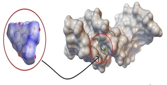
In order to obtain suitable crystals for XRD measurements, 100 mg of commercially available 2,5-dimethoxybenzaldehyde (C9H10O3, Aldrich, St. Louis, MO, USA, 99.0% pure) (100 mg) was dissolved in 10-mL MeOH at room temperature. After 2 days of evaporation at this temperature, colorless crystals were slowly formed.

2.4. BNA Docking

Docking studies were performed using the Autodock4.2 running on an Intel(R) Core(TM) i5 CPU (3 GHz) processor with a Windows 2007 operating system, Palo Alto, California, USA. The isomer structures were prepared using ChemDraw. The docking was performed using the Gasteiger charges, the water molecules were erased, and the nonpolar hydrogen atoms were merged using AutoDock4 [24]. The X-ray crystal of PDB ID: 1BNA DNA was freely obtained from the Protein Data Bank [25].

3. Results and Discussion

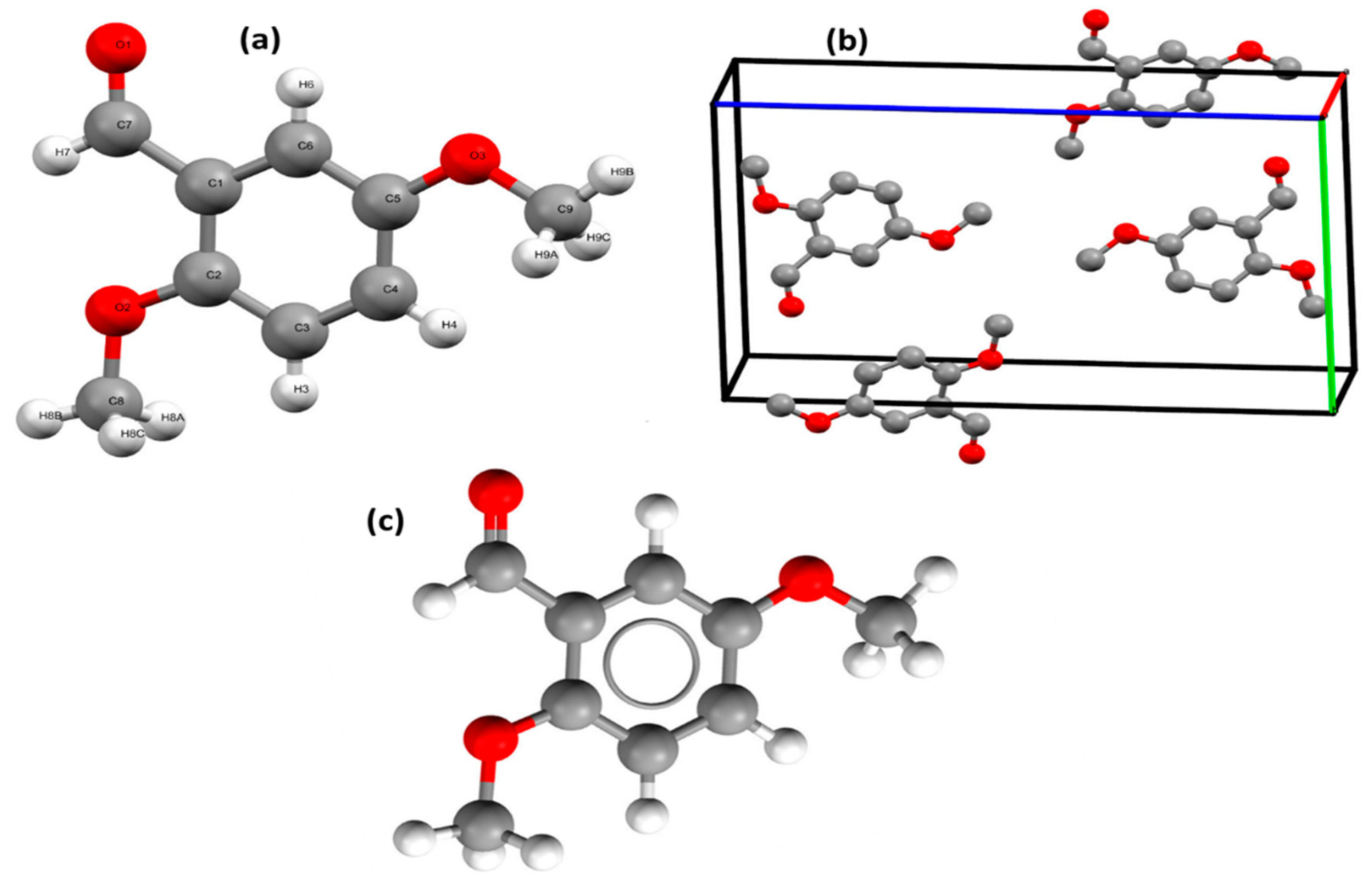
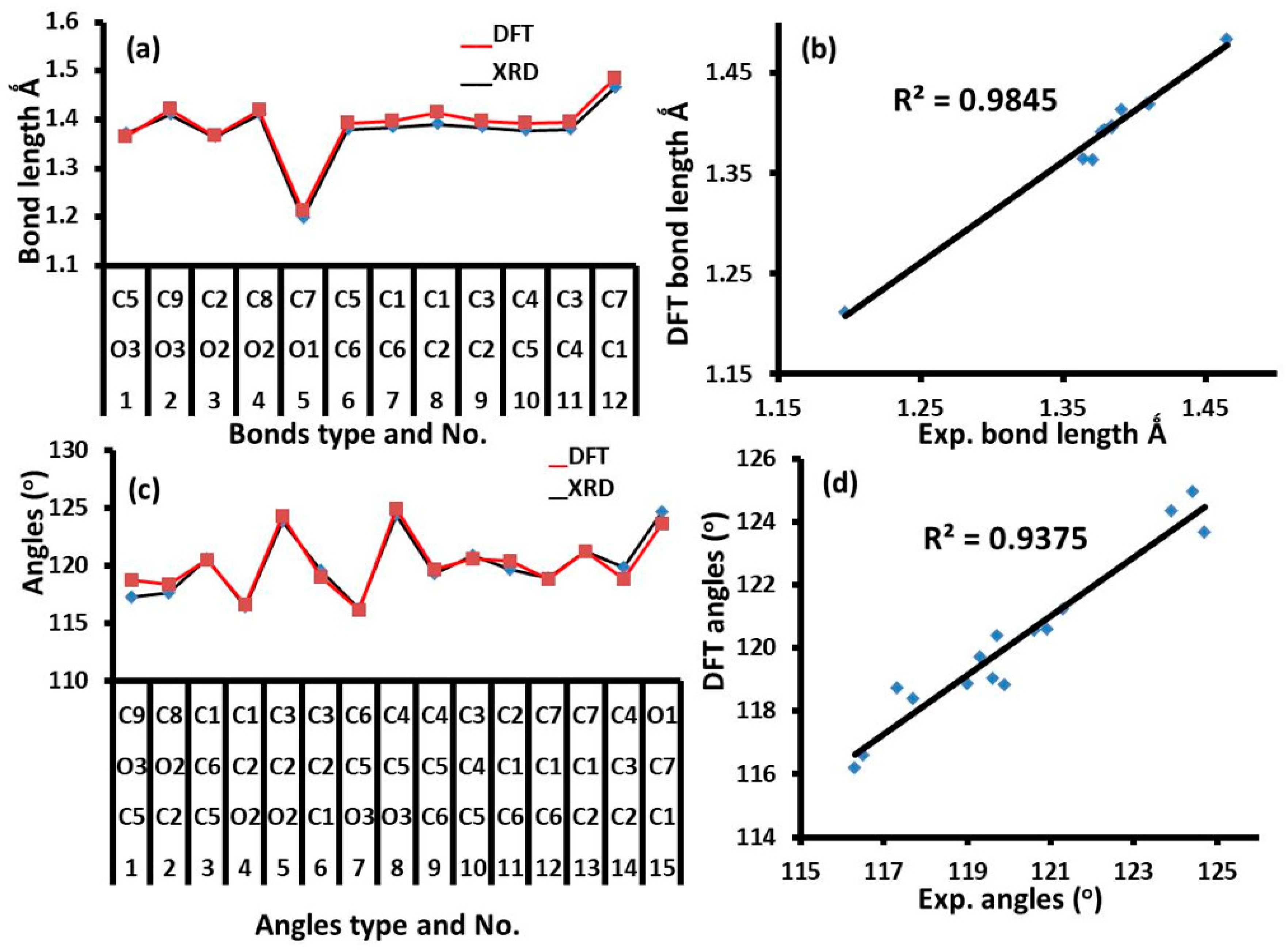
3.1. XRD and DFT Structure Analysis

XRD analysis indicated that exo-2,5-dimethoxybenzaldehyde (A) was the kinetically favored isomer with a dihedral angle (τO1–C2–C3–C4) of 179.95°. In contrast, the thermodynamically favored endo-isomer (B) with τO1C2C3C4 = 0° was not detected by XRD (Scheme 1).
2,5-Dimethoxybenzaldehyde crystallized in the kinetically favored exo-isomer form (Figure 1a) was monoclinic, with a p21/n space group, and four molecules were crystallized in a packing unit cell (Figure 1b). In the gaseous phase, the B3LYP/6-311G(d,p)-optimized exo-isomer structure was consistent with the XRD experimental result of the solid state, as shown in Figure 1c and Table 2.

3.2. B3LYP/6-311G(d,p) Structures

The bond distances and angles determined by DFT and XRD were almost identical, as shown in Table 2 and Figure 2. Specifically, the correlation (R2) between the calculated/experimental bond lengths was found to be 0.9845 (Figure 2a,b) and that between the calculated and experimental angles was 0.9357 (Figure 2c,d). Slight differences were only observed, because the DFT was performed in the gaseous phase, while XRD in the solid state.

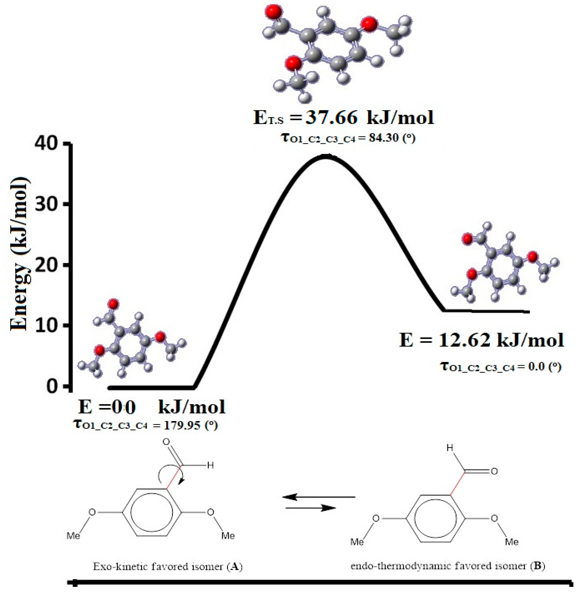
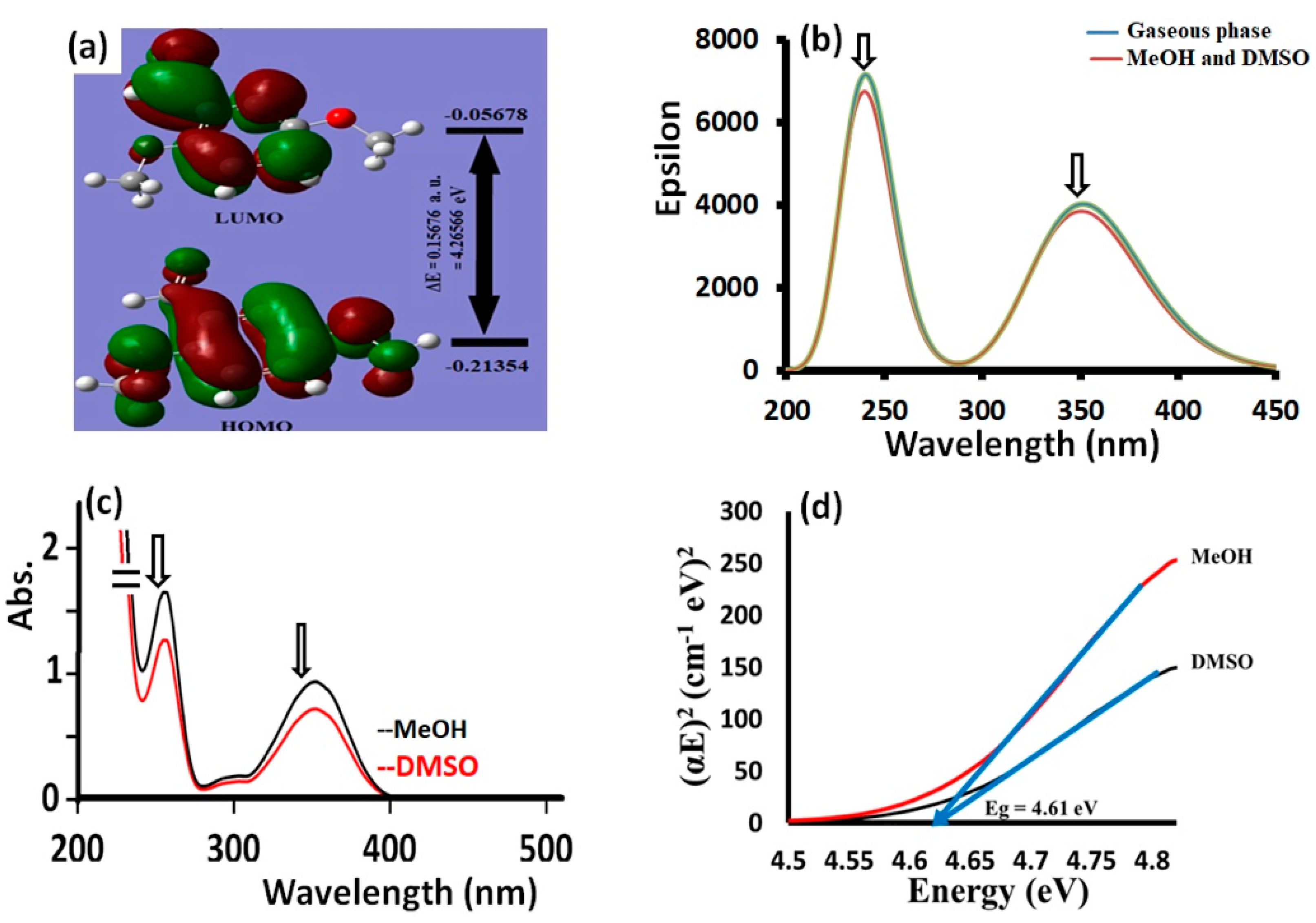
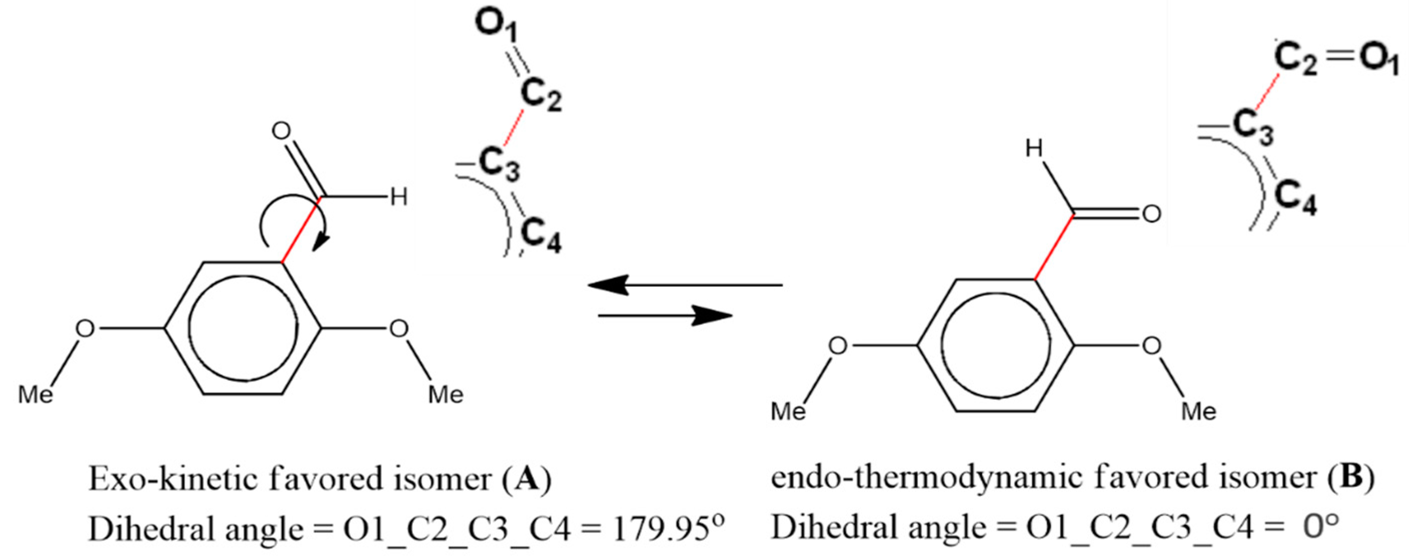
3.3. Exo⇔Endo Computational Isomerism

The exo-kinetic isomer did not favor the coordination of a metal ion, as the carbonyl oxygen and 2-OCH3 oxygen atoms were in the opposite direction. Although its structure was sterically favored, it did not serve as a good OՈO bidentate ligand. In contrast, the endo-isomer was expected to be a good OՈO bidentate chelate ligand, as the two oxygen atoms were in the same direction. Although the geometry of the exo-isomer was not appropriate for ligation, the formation of a stable S6-fused metal–heterocyclic ring after isomerization was confirmed by XRD crystal analysis (Figure 3).
Therefore, here, we investigated the exo⇔endo isomerization based on theoretical measurements to identify the energy required to switch between the two isomers. As shown in Scheme 1, the stereochemical difference between the two isomers is controlled by a 90° single rotation around the cited Csp2-Csp2 single bond, which leads to a significant change in the τO1_C2_C3_C4 dihedral angle from 180° (exo) to 0° (endo). Based on this change, and ignoring all the intermolecular forces in both isomers, high-level DFT/B3LYP/6-311G(d) optimization calculations were performed in the gaseous state for both isomers. Moreover, the QTS2 computation method was applied to detect the transition state (TS) of the exoendo isomerization reaction (Figure 4).
Based on the energy profile of the exoendo isomerization of 2,5-dimethoxybenzaldehyde (Figure 4), the exo-isomer energy was −574.76611449 a.u., Eexo = 0.0 kJ, whereas that of the endo-isomer was found at −574.76130900 a.u., Eendo = 12.62 kJ. Moreover, the TS energy was higher than that of both isomers (−574.75176736 a.u., ETS = 37.66 kJ), and its structure was between the structure of the endo- and exo-isomers with a dihedral angle of 84.3°. Therefore, we demonstrated that, energetically, the stable exo-isomer could give the unfavorable endo-isomer, since the energy required for isomerization was not too high and could be easily provided by the surrounding environment or solvents.

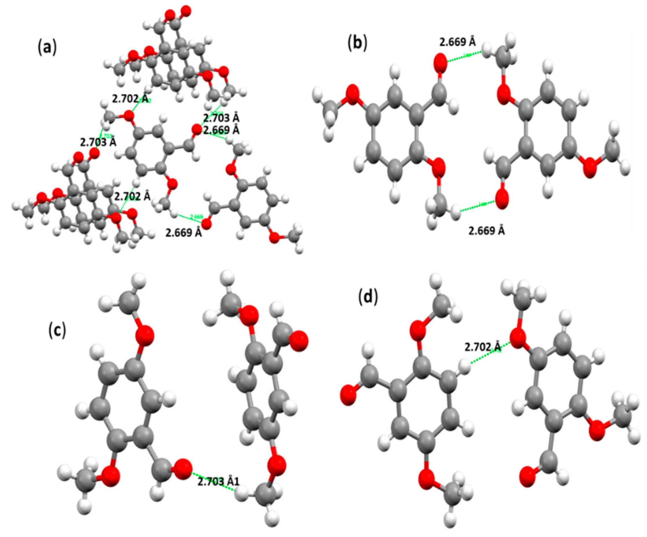
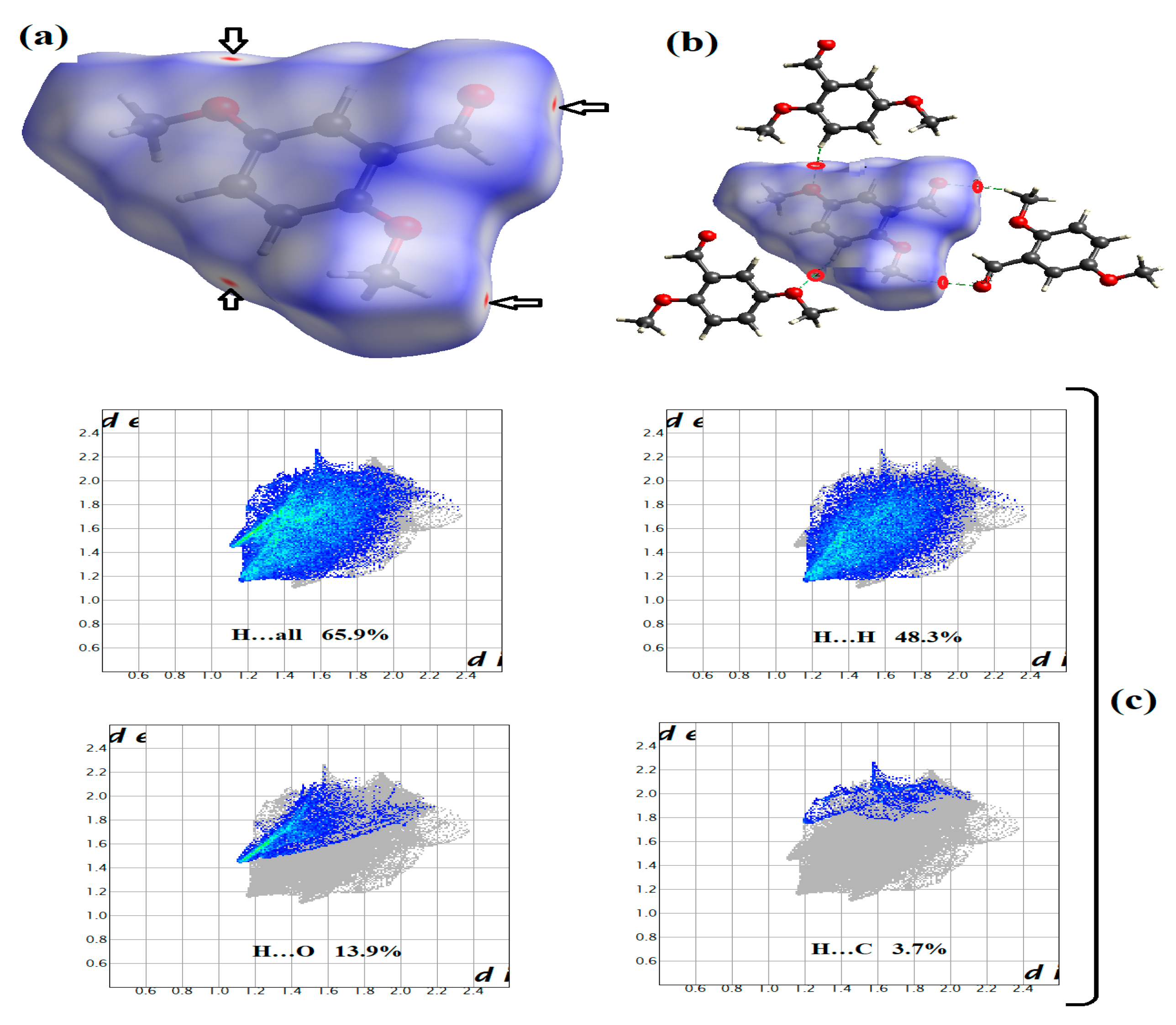
3.4. Crystal Interactions and HSA Investigation

Three main H∙∙∙O hydrogen bond interactions were detected in the crystal lattice of exo-2,5-dimethoxybenzaldehyde molecules, while each molecule was bound to its surrounding molecules through six hydrogen bonding interactions (Figure 5a), and no other types of interactions were identified. The two shortest interactions were assigned to the C=O∙∙∙HMe hydrogen-bonding interactions with a distance of 2.669 Å, forming a semi-dimer S14 supramolecular system (Figure 5b), while two C=O∙∙∙HMe hydrogen bonds with 2.703 Å (Figure 5c) and two MeO∙∙∙Hph hydrogen bonds with 2.702 Å (Figure 5d) could also be detected.
During HSA, four red spots were detected on the dnorm surface of the computed molecule [26,27,28,29,30], which were all attributed to the formation of H∙∙∙O hydrogen bonds (Figure 6a). It should be noted that the type and the number of the hydrogen bonds identified by HSA were consistent with those detected by an XRD packing analysis (Figure 6b). In addition, the HSA 2D fingerprint plots over the computed surface molecule indicated the presence of 65.9% total hydrogen interactions, which corresponded to three types of hydrogen-bonding interactions, namely H∙∙∙H (48.3%) > H∙∙∙O (13.9%) > H∙∙∙C (3.7%) (Figure 6c).

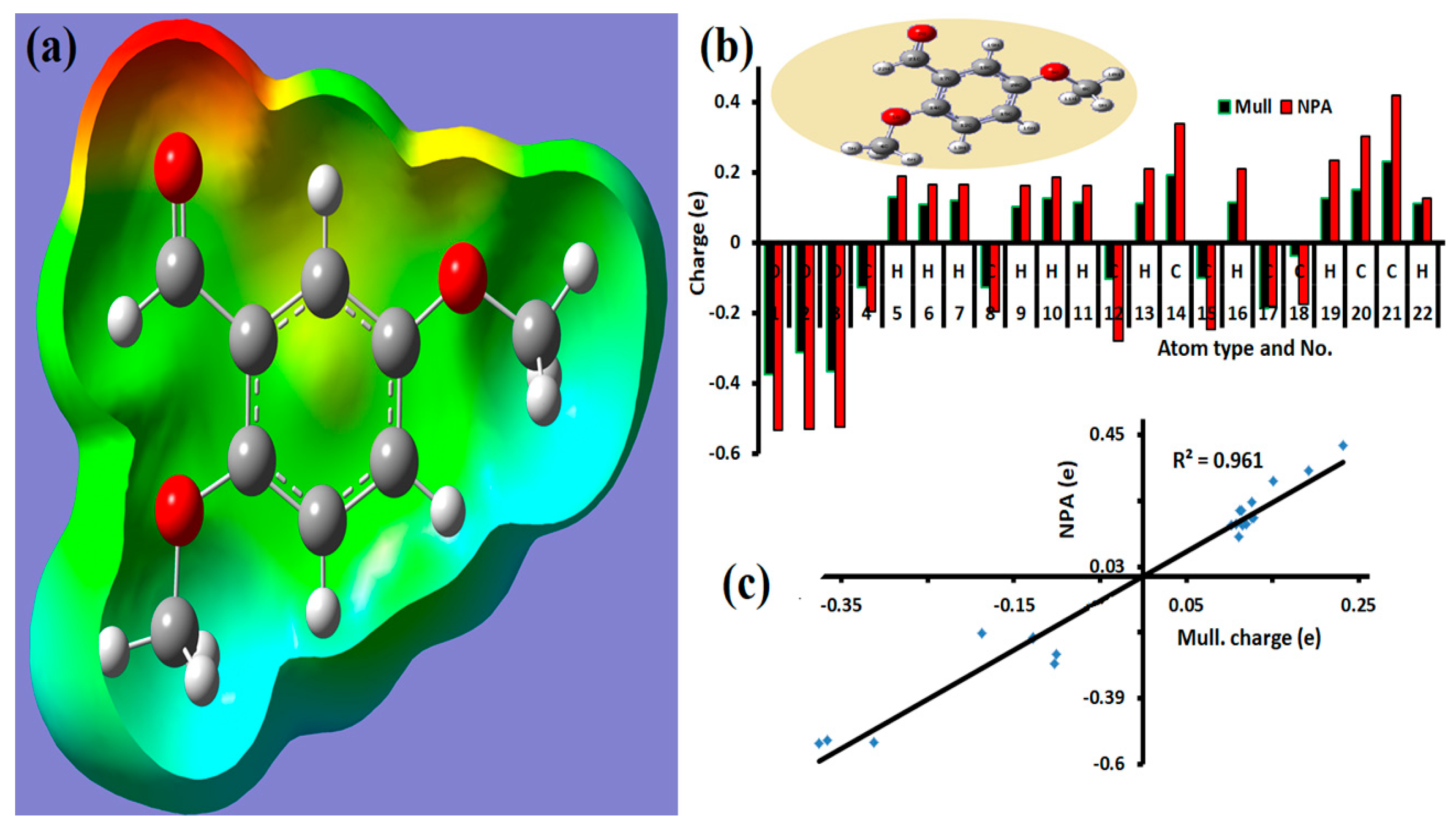
3.5. MEP Analysis and Atomic Charge Populations

The MEP analysis suggested the presence of both electrophilic and nucleophilic sites on the molecule surface (Figure 7a). The carbonyl oxygen atom was indicated as the strongest nucleophile site (red), while the other oxygen atoms were less nucleophilic (yellow). Moreover, the phenyl and methyl hydrogen atoms had strong electrophilic positions (blue). These findings strongly supported the formation of H∙∙∙O hydrogen bonds [31], as already confirmed by the XRD experimental results and HSA computations.
The determination of the Mulliken and NPA atomic charges revealed the presence of digital electron-poor and electron-rich atoms (Figure 7b). In general, the NPA atomic charges were higher than the Mulliken atomic charges, while the Mulliken and NPA values confirmed that all the oxygen atoms, as well as the C4, C8, C12, C15, C17, and C18 carbon atoms, acted as nucleophilic sites (Table 3). Accordingly, the electrophilic sites corresponded to the C13, C14, C20, and C21 carbon atoms. Moreover, all hydrogen atoms showed positive charge values, with H5, H10, and H19 being the most electrophilic (Table 3). A high correlation between Mulliken and NPA charge with R = 0.9614 was also observed based on the plot of Figure 7c. It is worth noting that the Mulliken and NPA data were in good agreement with the MEP, XRD packing, and HSA results.
The shape and energy diagram of HOMO and LUMO indicated that the electron donation capacity in the UV region was ΔEHOMO/LUMO = 4.266 eV (Figure 8a). Moreover, by measuring the B3LYP/6-311G(d,p) electron transfer in the gaseous state and in MeOH and DMSO (Figure 8b), two broad maxima bands at λmax = 245 and 355 nm were observed, corresponding mainly to HOMO-2-to-LUMO (76%) and HOMO-to-LUMO (96%) transitions, respectively. Similar electron transition results were obtained by experimental UV spectra, as shown in Figure 8c. Specifically, two peaks with λmax at 250 and 350 nm were detected in MeOH and DMSO, which were assigned to π→π* and n→π* electron transitions, respectively. Moreover, no solvatochromism effect was observed by changing the solvents in both the experimental and theoretical studies. The small Δλ shift (~5 nm) between the experimental and DFT data could be attributed to the solute–solvent interaction [32]. In addition, the experimental optical energy band gap (Eg) in MeOH and DMSO was determined using the Tauc equation [33]. Based also on Figure 8d, the direct Eg value was found to be ~4.51 eV in both solvents, which was close to the ΔEHOMO/LUMO value (~4.3 eV).
The GRD quantum parameters of the ligand, including softness (σ), hardness (η), chemical potential (μ), electrophilicity (ω), and electronegativity (χ), were also calculated by the following equations (Table 4):
I: Ionization potential = −EHOMO
A: Electron affinity = −ELUMO
ΔΕgap: Energy gap = EHOMO − ELUMO
χ: Absolute electronegativity = (I + A)/2
η: Global hardness = (I − A)/2
σ: Global softness = l/η
μ: Chemical potential = − χ
ω: Electrophilicity = μ2/2η

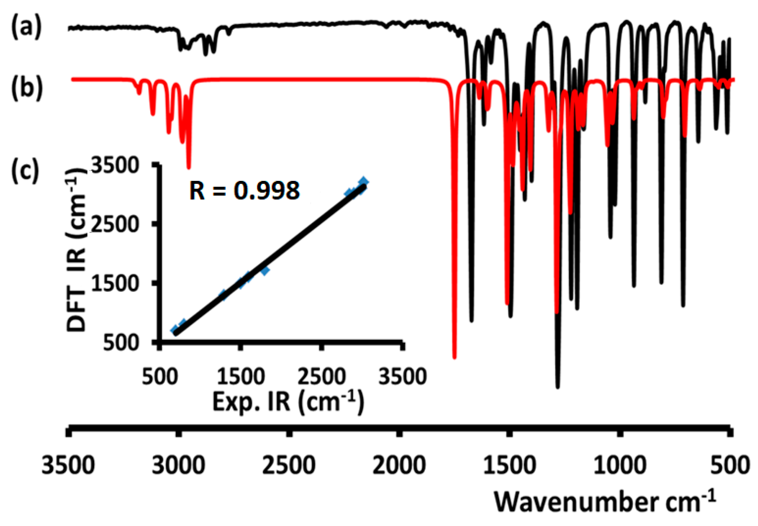
3.6. FTIR Investigations

The experimental FTIR spectrum of 2,5-dimethoxybenzaldehyde in a solid state indicated the presence of several functional groups, which were consistent with its chemical formula. In particular, the peaks at ~3050, 2950-2840, and 1620 cm−1 were attributed to the C–Hph, C–HCH3, and C=O stretching vibrations, respectively (Figure 9a). Moreover, it is clear from Figure 9b that the experimental and DFT-calculated spectra were very similar, while their high compatibility was further confirmed by their excellent correlation with R2 = 0.998 (Figure 9c).

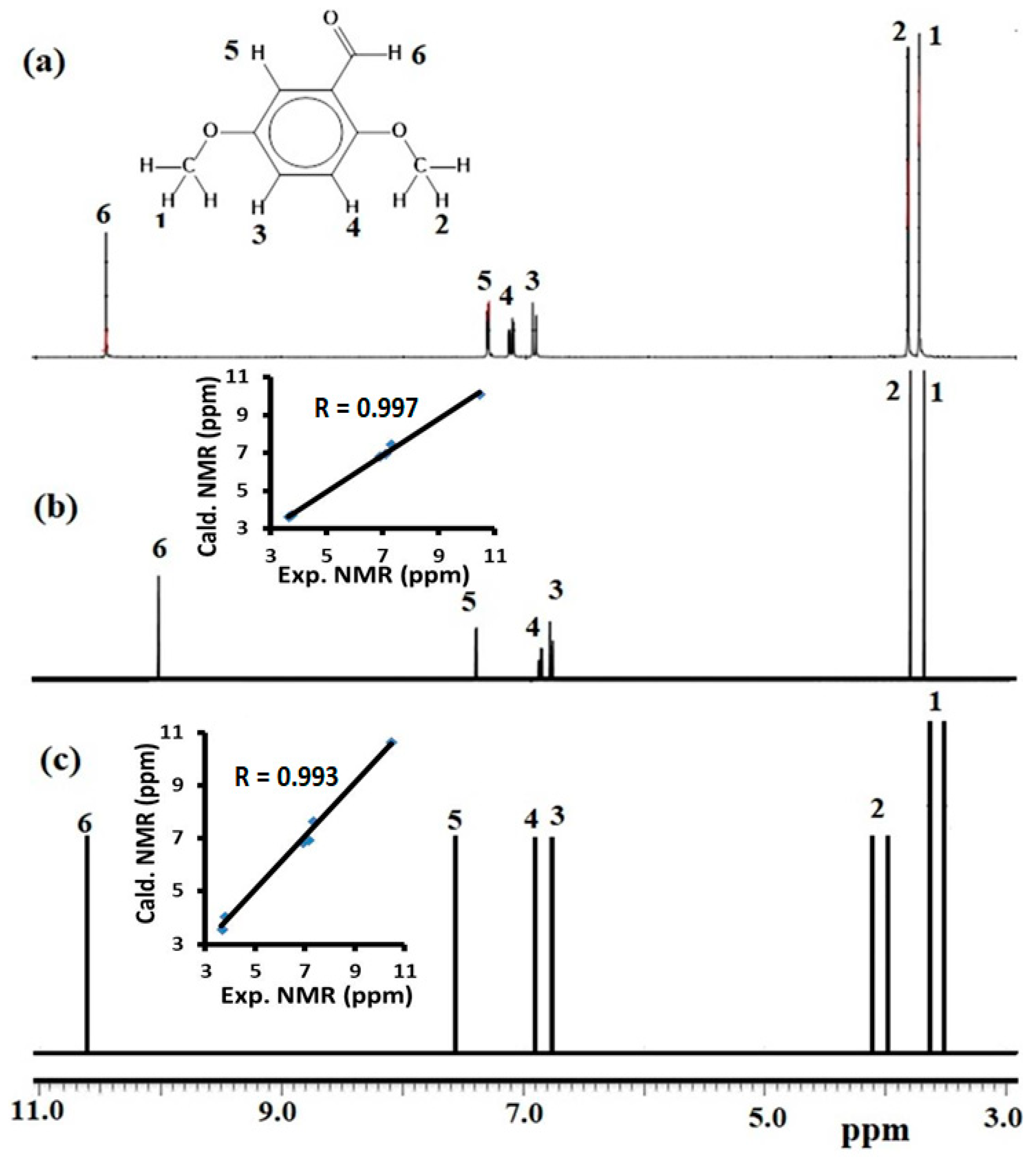
3.7. Computed and Experimental 1H NMR

The experimental 1H-NMR spectrum of 2,5-dimethoxybenzaldehyde was recorded in CDCl3 (Figure 10a). In the aliphatic region, two broad peaks were detected at 3.65 and 3.76 ppm corresponding to OCH3, while the peaks at 6.91 (d), 7.11 (d), and 7.31 (s) ppm were assigned to the three aromatic protons. The aldehyde proton was detected at 10.45 ppm as a singlet. The theoretical NMR-DB [16] (Figure 10b) and GIAO-NMR (Figure 10c) in CDCl3 were similar to the experimental spectrum, while the calculated and experimental proton chemical shifts showed a very good correlation, with R2 values of 0.9975 and 0.9929, respectively.

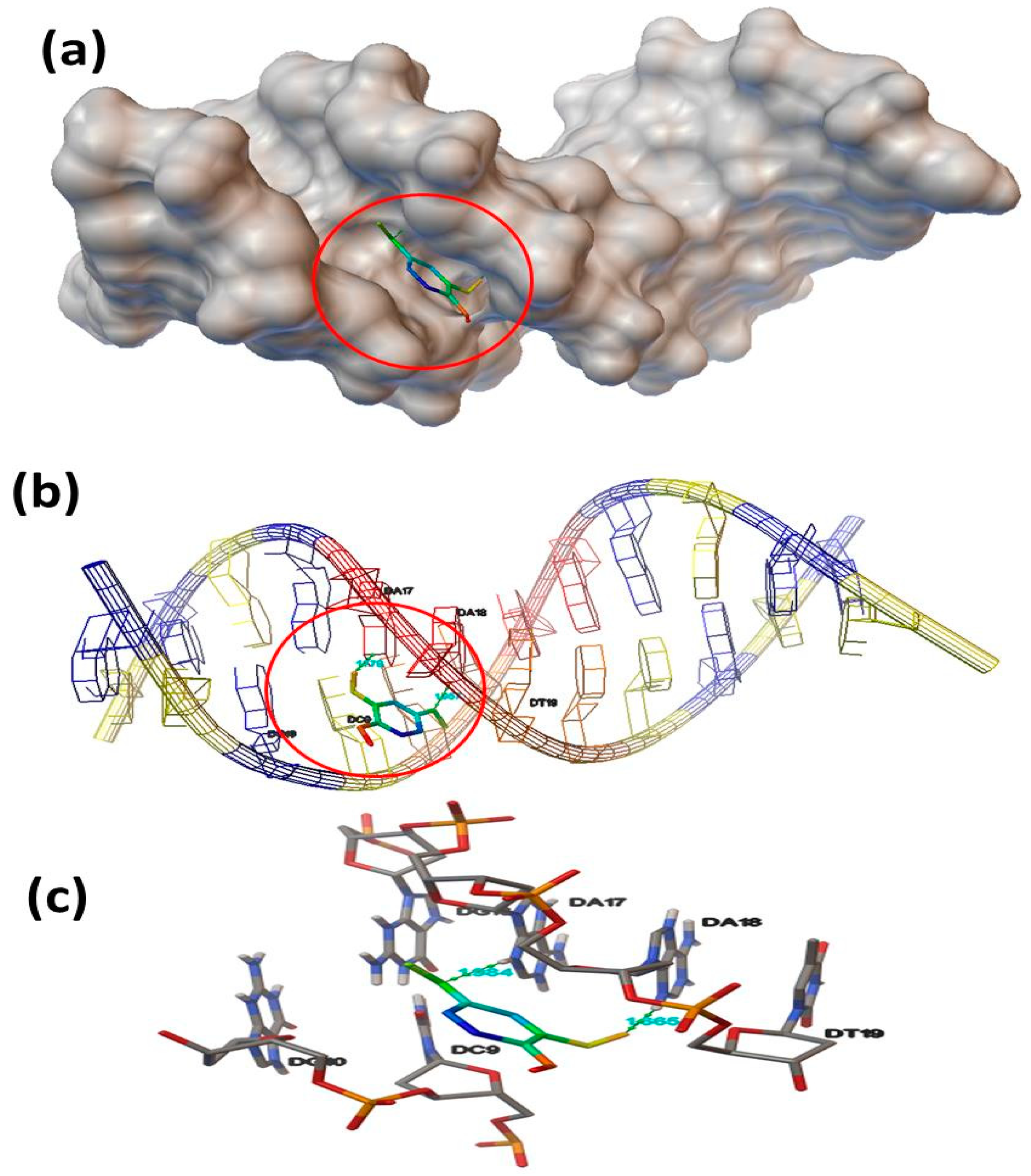
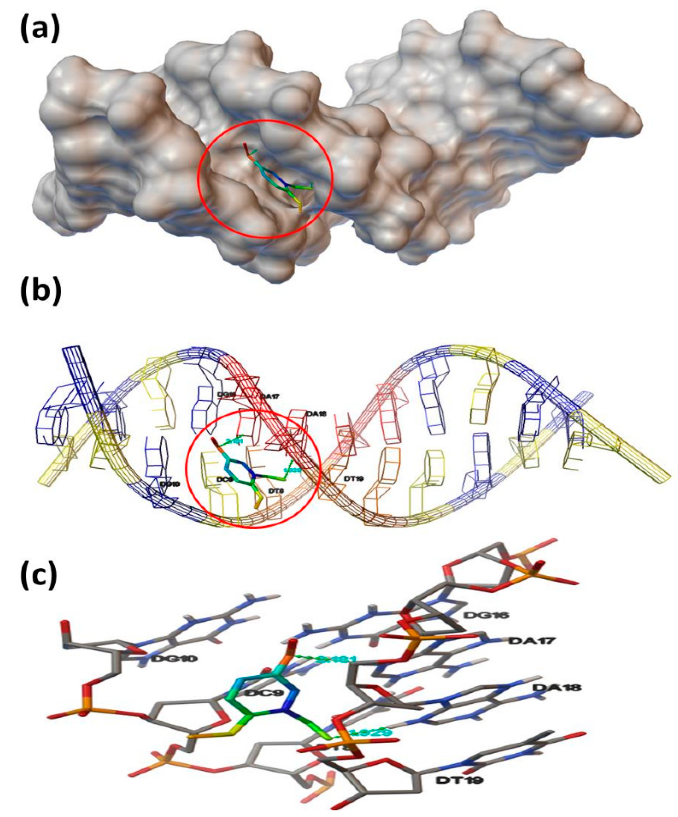
3.8. Molecular Docking

Both exo- and endo-isomers of 2,5-dimethoxybenzaldehyde were docked to DNA (PDB ID: 1BNA) under the same level of theory based on the existing data [25]. Interestingly, both isomers showed good and similar docking behaviors and were cross-linked to one DNA helix via two hydrogen bonds to form a (DNA:isomer) complex. No π–π stacking interactions were observed (Figure 11), while the polar 3-OCH3 functional group of both isomers did not develop hydrogen-bonding interactions with DNA, as it did not interfere with the DNA helix in the crystal lattice, which supported the minor groove DNA intercalation.
In addition, the binding affinity of the exo-isomer indicated its close contact with the DNA surface through a minor groove intercalation mode (Figure 11a) and two short hydrogen bonds to the adenosines of the one DNA helix (Figure 11b). The hydrogen-bonding interactions were assigned to DNA DA17: H∙∙∙OMe (ligand) with 1.984 Å and DNA DA18: H∙∙∙O=C (ligand) with 1.665 Å (Figure 11c). The docking results were consistent with the hydrogen bonds detected in the crystal lattice of the solved structure. In general, the docking effect can be considered a good result when the root mean square deviation (RMSD) value is below 2 Å [32]. The theoretical binding constant (Kb) and free energy change for the exo-isomer were found to be 1.63 × 10⁴ and −5.72 kcal/mol, respectively.
Similar to the exo-isomer, the endo-isomer was also in contact with DNA via a minor groove intercalation mode (Figure 12a), and one helix binding was observed (Figure 12b). However, this isomer developed two longer hydrogen-bonding interactions with the DNA adenosines, i.e., DA17:H∙∙∙OMe (ligand) with 2.481 Å and DNA DA18:H∙∙∙O=C (ligand) with 1.927 Å (Figure 12c). According to the RMSD, one bond was considered to be a good interaction. The theoretical Kb and free energy change were determined at 1.10 × 10⁴ and −5.49 kcal/mol, respectively.
The study showed significant convergence in the docking behavior of both isomers. However, the exo-isomer seemed to be slightly more active than the endo-isomer, since its hydrogen bonds were stronger, and its binding energy and Kb values were higher. These results could be expected, as the exo-isomer is more stable, and its structural shape selectivity is more suitable for structure-based drug discovery [16].

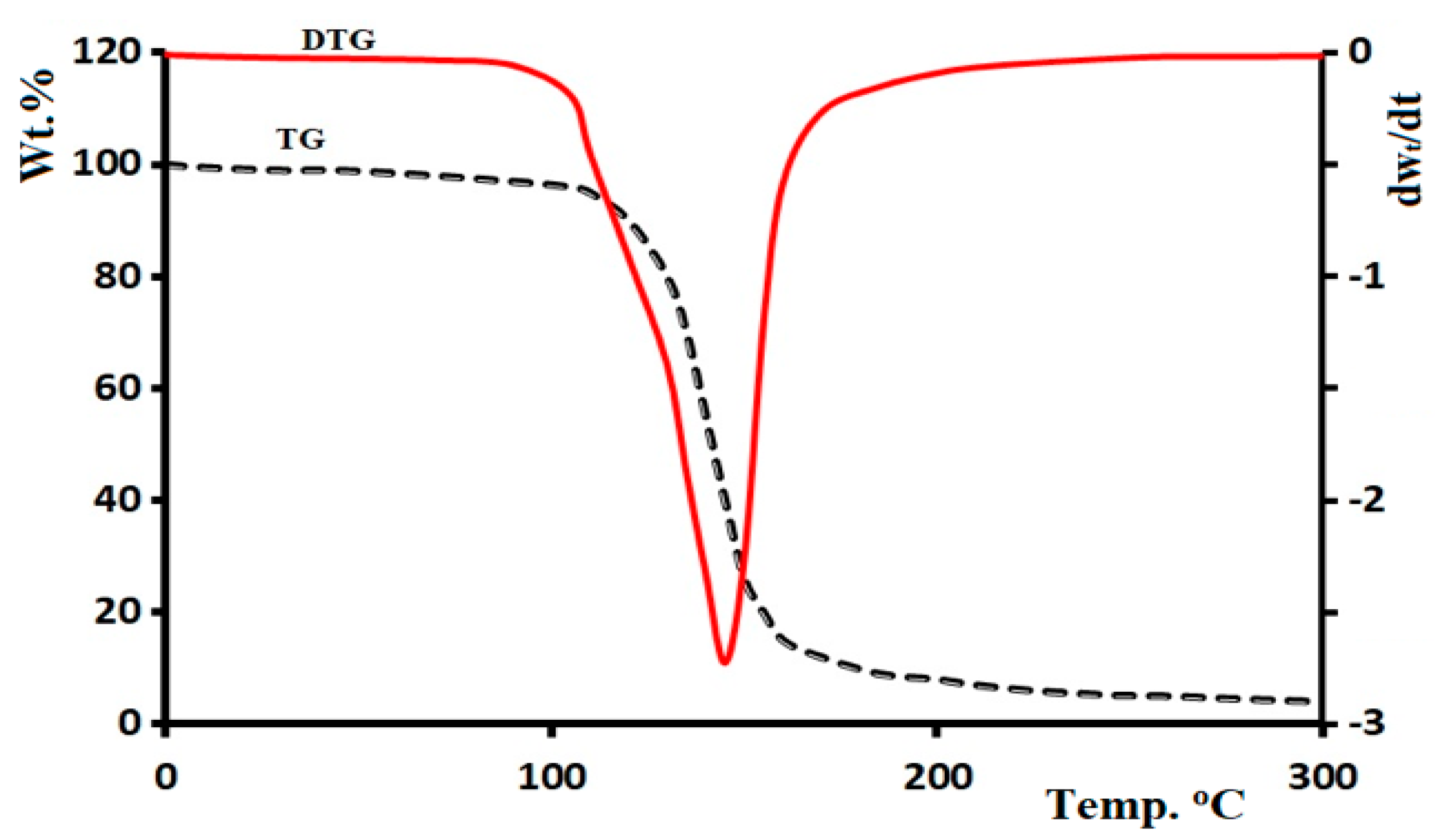
3.9. Thermal Analysis

The thermal properties of 2,5-dimethoxybenzaldehyde were also evaluated by thermogravimetric–derivative thermogravimetric (TG/DTG) analysis. The TG/DTG curves were obtained at a temperature range of 0–300 °C at a heating rate of 5 °C/min in an open atmosphere (Figure 13). The ligand exhibited acceptable stability up to = 95 °C, while, at temperatures above 95 °C, the ligand was gradually decomposed, and its weight decreased from 100 wt% to 0 wt% via a single broad-step reaction mechanism with Toff ≈ 200 °C and complete thermal decomposition.

4. Conclusions

In this study, we explored the exoendo isomerization of 2,5-dimethoxybenzaldehyde based on DFT studies, and the results were compared and confirmed by experimental studies. The formation of the exo-isomer was confirmed by the XRD crystallographic analysis structure, while the DFT/XRD structure parameters reflected semi-unity graphical correlations. The hydrogen bonds computed by HSA and MEP analyses were in excellent agreement with the experimental XRD packing results, while the Mulliken and NPA population charge analyses indicated the presence of both nucleophilic and electrophilic sites on the ligand surface. Moreover, the DFT/B3LYP/6-311G(d,p) computational study of the exoendo isomerization process allowed the identification of the QST2 TS. Furthermore, the TD-SCF/DFT, B3LYP-IR, NMR-DB, and GIAO-NMR calculations were similar to the experimental UV–Vis, direct Eg, FTIR, and 1H NMR spectra, respectively. The calculated Mulliken and NPA population charges, along with the HOMO/LUMO and GRD quantum parameters, further supported the exo-isomer formation. The ligand also exhibited good thermal stability, with a one-step decomposition mechanism in the range of 100–200 °C. In addition, both isomers showed a very good DNA docking effect, where one helix minor grove with two hydrogen bonds was observed for both isomers. Such compounds can be used in future works as DNA-binding promising drugs.

Author Contributions

Data curation: N.A.-Z. and I.W.; formal analysis: M.S.; funding acquisition: N.A.-Z. and A.A. (Amjad Alsyahi); investigation: A.A.-A., K.A., and N.K.L.; methodology: A.Z., F.A.A., and A.A.-T.; project administration: N.A.-Z. and A.A. (Ali Alsalme), software: I.W.; validation: N.A.-Z.; writing—original draft: I.W. and A.A.-A.; writing—review and editing: K.K., N.K.L., A.Z., and M.S. All authors have read and agreed to the published version of the manuscript.

Funding

The authors extend their appreciation to the Deputyship for Research & Innovation, “Ministry of Education” in Saudi Arabia for funding this research work through the project number IFKSURG-1440-141.

Conflicts of Interest

The authors declare no conflict of interest.

References

  1. Avci, D.; Tamer, Ö.; Başoğlu, A.; Atalay, Y. 5-Methyl-2-thiophenecarboxaldehyde: Experimental and TD/DFT study. J. Mol. Struct. 2018, 1174, 52–59. [Google Scholar] [CrossRef]
  2. Da Silva, M.A.R.; Santos, A.F.L. Experimental thermochemical study of 3-acetyl-2-methyl-5-phenylthiophene. J. Chem. Thermodyn. 2010, 42, 128–133. [Google Scholar] [CrossRef]
  3. Brugman, S.J.T.; Engwerda, A.H.J.; Kalkman, E.; De Ronde, E.; Tinnemans, P.; Vlieg, E. The crystal structures of four dimethoxybenzaldehyde isomers. Acta Crystallogr. Sect. E Crystallogr. Commun. 2019, 75, 38–42. [Google Scholar] [CrossRef]
  4. Ma, C.; Liu, S.; Zhang, S.; Xu, T.; Yu, X.; Gao, Y.; Zhai, C.; Li, C.; Lei, C.; Fan, S.; et al. Evidence and perspective for the role of the NLRP3 inflammasome signaling pathway in ischemic stroke and its therapeutic potential (Review). Int. J. Mol. Med. 2018, 42, 2979–2990. [Google Scholar] [CrossRef] [Green Version]
  5. Atta, A.K.; Kim, S.-B.; Cho, D.-G. Catalytic Oxidative Conversion of Aldehydes to Carboxylic Esters and Acids under Mild Conditions. Bull. Korean Chem. Soc. 2011, 32, 2070–2072. [Google Scholar] [CrossRef] [Green Version]
  6. Hodgson, D.M.; Charlton, A. Methods for direct generation of a-alkyl-substituted aldehydes. Tetrahedron 2014, 70, 2207. [Google Scholar] [CrossRef]
  7. Ding, Y.-Q.; Cui, Y.; Li, T.-D. New Views on the Reaction of Primary Amine and Aldehyde from DFT Study. J. Phys. Chem. A 2015, 119, 4252–4260. [Google Scholar] [CrossRef] [PubMed]
  8. Arjunan, V.; Santhanam, R.; Rani, T.; Rosi, H.; Mohan, S. Conformational, vibrational, NMR and DFT studies of N-methylacetanilide. Spectrochim. Acta Part A Mol. Biomol. Spectrosc. 2013, 104, 182–196. [Google Scholar] [CrossRef] [PubMed]
  9. Sevvanthi, S.; Muthu, S.; Raja, M. Molecular docking, vibrational spectroscopy studies of (RS)-2-(tert-butylamino)-1-(3-chlorophenyl)propan-1-one: A potential adrenaline uptake inhibitor. J. Mol. Struct. 2018, 1173, 251–260. [Google Scholar] [CrossRef]
  10. Barakat, A.; Islam, M.S.; Al-Majid, A.M.; Ghabbour, H.A.; Atef, S.; Zarrouk, A.; Warad, I.; Gu, Y.; Liu, Y. Quantum chemical insight into the molecular structure of L-chemosensor 1,3-dimethyl-5-(thien-2-ylmethylene)-pyrimidine-2,4,6-(1H,3H,5H)-trione: Naked-eye colorimetric detection of copper(II) anions. J. Theor. Comput. Chem. 2018, 17, 1850005. [Google Scholar] [CrossRef]
  11. Barakat, A.; Soliman, S.M.; Ghabbour, H.A.; Ali, M.; Al-Majid, A.M.; Zarrouk, A.; Warad, I. Intermolecular interactions in crystal structure, Hirshfeld surface, characterization, DFT and thermal analysis of 5-((5-bromo-1 H -indol-3-yl)methylene)-1,3-dimethylpyrimidine-2,4,6(1 H, 3 H, 5 H )-trione indole. J. Mol. Struct. 2017, 1137, 354–361. [Google Scholar] [CrossRef]
  12. Zi, Y.; Zhu, M.; Li, X.; Xu, Y.; Wei, H.; Li, D.; Mu, C. Effects of carboxyl and aldehyde groups on the antibacterial activity of oxidized amylose. Carbohydr. Polym. 2018, 192, 118–125. [Google Scholar] [CrossRef] [PubMed]
  13. Barnsley, J.E.; Wagner, P.; Officer, D.L.; Gordon, K.C. Aldehyde isomers of porphyrin: A spectroscopic and computational study. J. Mol. Struct. 2018, 1173, 665–670. [Google Scholar] [CrossRef]
  14. Meng, X.-Y.; Zhang, H.-X.; Mezei, M.; Cui, M. Molecular Docking: A Powerful Approach for Structure-Based Drug Discovery. Curr. Comput. Drug Des. 2011, 7, 146–157. [Google Scholar] [CrossRef]
  15. Guedes, I.A.; Camila, S.D.M.; Dardenne, L.E. Receptor–ligand molecular docking. Biophys. Rev. 2014, 6, 75. [Google Scholar] [CrossRef] [Green Version]
  16. Hamilton, P.L.; Arya, D.P. Natural product DNA major groove binders. Nat. Prod. Rep. 2011, 29, 134–143. [Google Scholar] [CrossRef]
  17. Rehman, S.U.; Sarwar, T.; Husain, M.A.; Ishqi, H.M.; Tabish, M. Studying non-covalent drug–DNA interactions. Arch. Biochem. Biophys. 2015, 576, 49. [Google Scholar] [CrossRef]
  18. Arjmand, F.; Parveen, S.; Afzal, M.; Shahid, M. Synthesis, characterization, biological studies (DNA binding, cleavage, antibacterial and topoisomerase I) and molecular docking of copper(II) benzimidazole complexes. J. Photochem. Photobiol. B Biol. 2012, 114, 15–26. [Google Scholar] [CrossRef]
  19. Barnes, E.C.; Petersson, G.A.; Montgomery, J.A.; Frisch, M.J.; Martin, J.M.L.; Martin, J.M.L. Unrestricted Coupled Cluster and Brueckner Doubles Variations of W1 Theory. J. Chem. Theory Comput. 2009, 5, 2687–2693. [Google Scholar] [CrossRef]
  20. Becke, A.D. Density-functional exchange-energy approximation with correct asymptotic behavior. Phys. Rev. A 1988, 38, 3098–3100. [Google Scholar] [CrossRef]
  21. Perdew, J.P.; Burke, K.; Ernzerhof, M. Generalized Gradient Approximation Made Simple. Phys. Rev. Lett. 1996, 77, 3865–3868. [Google Scholar] [CrossRef] [PubMed] [Green Version]
  22. Wolff, S.K.; Grimwood, D.J.; McKinnon, J.J.; Jayatilaka, D.; Spackman, M.A. CrystalExplorer 2.1; University of Western Australia: Perth, Australia, 2007. [Google Scholar]
  23. Sheldrick, G. A short history ofSHELX. Acta Crystallogr. Sect. A Found. Crystallogr. 2008, 64, 112–122. [Google Scholar] [CrossRef] [PubMed] [Green Version]
  24. Trott, O.; Olson, A.J. AutoDock Vina: Improving the speed and accuracy of docking with a new scoring function, efficient optimization, and multithreading. J. Comput. Chem. 2009, 31, 455–461. [Google Scholar] [CrossRef] [PubMed] [Green Version]
  25. Drew, H.R.; Wing, R.M.; Takano, T.; Broka, C.A.; Tanaka, S.; Itakura, K.; Dickerson, R.E. Structure of a B-DNA dodecamer: Conformation and dynamics. Proc. Natl. Acad. Sci. USA 1981, 78, 2179–2183. [Google Scholar] [CrossRef] [Green Version]
  26. Titi, A.; Warad, I.; Almutairi, S.M.; Fettouhi, M.; Messali, M.; Aljuhani, A.; Touzani, R.; Zarrouk, A. One-pot liquid microwave-assisted green synthesis of neutral trans-Cl2Cu(NNOH)2: XRD/HSA-interactions, antifungal and antibacterial evaluations. Inorg. Chem. Commun. 2020, 122, 108292. [Google Scholar] [CrossRef]
  27. Warad, I.; Awwadi, F.F.; Abd Al-Ghani, B.; Sawafta, A.; Shivalingegowda, N.; Lokanath, N.K.; Mubarak, M.S.; Ben Hadda, T.; Zarrouk, A.; Al-Rimawi, F.; et al. Ultrasound-assisted synthesis of two novel [CuBr(diamine)2. H2O]Br complexes: Solvatochromism, crystal structure, physicochemical, Hirshfeld surface thermal, DNA/binding, antitumor and antibacterial activities. Ultrason. Sonochem. 2018, 48, 1. [Google Scholar] [CrossRef]
  28. Warad, I.; Musameh, S.; Sawafta, A.; Brandão, P.; Tavares, C.J.; Zarrouk, A.; Amereih, S.; Al Ali, A.; Shariah, R. Ultrasonic synthesis of Oct. trans-Br2Cu(N ∩ N)2 Jahn-Teller distortion complex: XRD-properties, solvatochromism, thermal, kinetic and DNA-binding evaluations. Ultrason. Sonochemistry 2019, 52, 428–436. [Google Scholar] [CrossRef]
  29. Abu Saleemh, F.; Musameh, S.; Sawafta, A.; Brandao, P.; Tavares, C.J.; Ferdov, S.; Barakat, A.; Al Ali, A.; Hamani, H.; Warad, I. Diethylenetriamine/diamines/copper (II) complexes [Cu(dien)(NN)]Br 2: Synthesis, solvatochromism, thermal, electrochemistry, single crystal, Hirshfeld surface analysis and antibacterial activity. Arab. J. Chem. 2017, 10, 845–854. [Google Scholar] [CrossRef] [Green Version]
  30. Aouad, M.R.; Messali, M.; Rezki, N.; Al-Zaqri, N.; Warad, I. Single proton intramigration in novel 4-phenyl-3-((4-phenyl-1H-1,2,3-triazol-1-yl)methyl)-1H-1,2,4-triazole-5(4H)-thione: XRD-crystal interactions, physicochemical, thermal, Hirshfeld surface, DFT realization of thiol/thione tautomerism. J. Mol. Liq. 2018, 264, 621–630. [Google Scholar] [CrossRef]
  31. Tauc, J.; Menth, A. States in the gap. J. Non-Cryst. Solids 1972, 8, 569–585. [Google Scholar] [CrossRef]
  32. Banfi, D.; Patiny, L. www.nmrdb.org: Resurrecting and Processing NMR Spectra On-line. Chim. Int. J. Chem. 2008, 62, 280–281. [Google Scholar] [CrossRef]
  33. Shi, J.-H.; Lou, Y.-Y.; Zhou, K.-L.; Pan, D.-Q. Exploration of intermolecular interaction of calf thymus DNA with sulfosulfuron using multi-spectroscopic and molecular docking techniques. Spectrochim. Acta Part A Mol. Biomol. Spectrosc. 2018, 204, 209–216. [Google Scholar] [CrossRef] [PubMed]
Scheme 1. Exoendo isomerization of 2,5-dimethoxybenzaldehyde and the O1–C2–C3–C4 dihedral angles of the corresponding isomers.
Molecules 25 05970 sch001
Figure 1. (a) ORTEP drawing of exo-2,5-dimethoxybenzaldehyde. (b) Packing unit cell with four crystallized exo-2,5-dimethoxybenzaldehyde molecules. (c) B3LYP/6-311G(d,p)-optimized structure of exo-2,5-dimethoxybenzaldehyde.
Molecules 25 05970 g001
Figure 2. Histograms of (a) bond lengths and (c) angles determined by X-ray diffraction (XRD) and density functional theory (DFT). (b) and (d) Graphical correlations of the bond lengths and angles determined by XRD and DFT, respectively.
Molecules 25 05970 g002
Figure 3. Formation of a S6-fused metal–heterocyclic ring after the exoendo isomerization of 2,5-dimethoxybenzaldehyde.
Molecules 25 05970 g003
Figure 4. Energy profile and global minimum structures of the exo- and endo-2,5-dimethoxybenzaldehyde isomers and the transition state (TS) of the exoendo isomerization.
Molecules 25 05970 g004
Figure 5. (a) Six hydrogen bond interactions determined in the crystal lattice of exo-2,5-dimethoxybenzaldehyde molecules. (b) C=O∙∙∙HMe: 2.669 Å, (c) C=O∙∙∙HMe: 2.703 Å, and (d) MeO∙∙∙Hph: 2.702 Å.
Molecules 25 05970 g005
Figure 6. (a) Map of the dnorm surface of the computed molecule. (b) Hydrogen bonding interactions of the computed molecule with its neighboring molecules. (c) Hirshfeld surface analysis (HSA) 2D fingerprint plots indicating the H∙∙∙H, H∙∙∙O, and H∙∙∙C hydrogen-bonding interactions.
Molecules 25 05970 g006
Figure 7. (a) B3LYP/6-311G(d,p) molecular electrostatic potential (MEP) map of exo-2,5-dimethoxybenzaldehyde. (b) Mulliken (Mull) and natural population analysis (NPA) atomic charges and (c) their graphical correlation. R2: correlation.
Molecules 25 05970 g007
Figure 8. (a) HOMO/LUMO shapes and energy diagram. (b) TD-DFT. (c) Experimental UV spectra, and (d) optical energy band gap (Eg) of the 2.2 × 10⁻⁶ M of 2,5-dimethoxybenzaldehyde in MeOH and DMSO.
Molecules 25 05970 g008
Figure 9. (a) Experimental and (b) DFT-calculated FTIR spectra of 2,5-dimethoxybenzaldehyde, and (c) their graphical correlation.
Molecules 25 05970 g009
Figure 10. (a) Experimental 1H NMR spectrum of 2,5-dimethoxybenzaldehyde in CDCl3. Theoretical (b) NMR-DB and (c) GIAO-NMR spectrum of 2,5-dimethoxybenzaldehyde in CDCl3.
Molecules 25 05970 g010
Figure 11. (a) Schematic representation of the binding position of the exo-isomer to DNA. (b) Interactions of the exo-isomer with the nuclides of the one DNA helix. (c) Hydrogen-bonding interactions of the exo-isomer with DNA adenosines (DA17: H∙∙∙OMe and DA18: H∙∙∙O=C).
Molecules 25 05970 g011
Figure 12. (a) Schematic representation of the binding position of the endo-isomer to DNA. (b) Interactions of the endo-isomer with the nuclides of the one DNA helix. (c) Nuclides-Z-isomer positional binding and hydrogen-bonding interactions of the exo-isomer with DNA adenosines (DA17: H∙∙∙OMe and DA18: H∙∙∙O=C).
Molecules 25 05970 g012
Figure 13. Thermogravimetric–derivative thermogravimetric (TG/DTG) curves of 2,5-dimethoxybenzaldehyde.
Molecules 25 05970 g013
Table 1. Crystallographic refined parameters of the desired molecular structure.
Empirical FormulaC9H10O3
CCDC1860213
Temperature293(2) K
Formula weight166.17
Wavelength0.71073 Å
Crystal system, space groupMonoclinic, p21/n
Volume639.55(5) Å3
Unit cell dimensionsa = 3.9469 (9), b = 11.580 (3), c = 17.886 (4) Å
β91.442 (17)°
V817.2 (3) (Å)3
Crystal size0.29 × 0.26 × 0.23 mm
Z4
Absorption coefficient0.1 mm−1
No. of reflections1837
Refinement methodFull-matrix least-squares on F2
R(int), (sin θ/λ)max0.115, 0.650 (Å−1)
S 1.05
R[F2 > 2σ(F2)], wR(F2) 0.062, 0.190
Largest diff. peak and hole0.14, −0.20 eÅ−3
Table 2. Density functional theory (DFT)-calculated angles (o) and bond lengths (Å) compared to the corresponding X-ray diffraction (XRD) experimental results (exp. XRD).
Bond No.BondsExp. XRDDFTAngle No.Angles (°)Exp. XRDDFT
1O3C51.371(2)1.36391C5O3C9117.3(2)118.73
2O3C91.409(3)1.42072C2O2C8117.7(2)118.38
3O2C21.364(2)1.36553C5C6C1120.6(2)120.56
4O2C81.410(3)1.41894O2C2C1116.5(2)116.62
5O1C71.197(3)1.21245O2C2C3123.9(2)124.35
6C6C51.379(3)1.3936C1C2C3119.6(2)119.03
7C6C11.384(3)1.39787O3C5C6116.3(2)116.2
8C2C11.391(3)1.41388O3C5C4124.4(2)124.95
9C2C31.384(3)1.39649C6C5C4119.3(2)119.73
10C5C41.377(3)1.391910C5C4C3120.9(2)120.58
11C4C31.379(3)1.393911C6C1C2119.7(2)120.39
12C1C71.465(3)1.483812C6C1C7119.0(2)118.88
13C2C1C7121.3(2)121.25
14C2C3C4119.9(2)118.84
15C1C7O1124.7(2)123.68
Table 3. Mulliken (Mull) and natural population analysis (NPA) atomic charges.
Atom No.AtomMullNPAAtom No.AtomMullNPA
1O−0.37563−0.5323812C−0.10245−0.27906
2O−0.31227−0.5311313H0.1119930.2103
3O−0.36585−0.523414C0.1918260.33756
4C−0.12813−0.1954415C−0.10028−0.24795
5H0.1280880.187616H0.1139170.20922
6H0.1078740.1650517C−0.1867−0.18217
7H0.119550.1650518C−0.03829−0.17568
8C−0.12758−0.1974919H0.1265190.23468
9H0.1026420.1617920C0.1510510.30193
10H0.1262940.1872421C0.231680.41763
11H0.1152550.1617822H0.1104880.12487
HOMO-LUMO, TD-SCF-B3LYP, absorbance, optical energy gap (Eg), and global reactivity descriptors (GRD).
Table 4. Global reactivity descriptor (GRD) quantum parameters calculated for exo-2,5-dimethoxybenzaldehyde.
GRDValue
Global total energy ET−574.76611449 a.u
Low unoccupied molecular orbitalLUMO−0.05678 a.u
High occupied molecular orbitalHOMO−0.21354 a.u
Energy difference ΔEgap0.15676 a.u
4.26565 eV
Electron affinityA1.545063 eV
Ionization potentialI5.810721 eV
Global hardnessƞ2.13505 eV
Global softnessσ0.468372 eV
Chemical potentialμ−3.67789 eV
Electronegativityχ3.67789 eV
Electrophilicityω3.16781 eV
Dipole momentu6.4226 D
Sample Availability: Samples of the compounds are available from the authors.
Publisher’s Note: MDPI stays neutral with regard to jurisdictional claims in published maps and institutional affiliations.

