
Ruthenium Complexes as Promising Candidates against Lung Cancer

1
International Cancer Center, Guangdong Key Laboratory for Genome Stability & Disease Prevention, Department of Pharmacology, Shenzhen University Health Science Center, Shenzhen 518060, China
2
College of Chemistry and Environmental Engineering, Shenzhen University, Shenzhen 518060, China
3
Key Laboratory for Advanced Materials of MOE, School of Chemistry & Molecular Engineering, East China University of Science and Technology, Shanghai 200237, China
4
Department of Surgery, The University of Hong Kong-Shenzhen Hospital, Haiyuan 1st Road, Futian District, Shenzhen 518053, China
*
Authors to whom correspondence should be addressed.
†
Contributed equally to this work.
Molecules 2021, 26(15), 4389; https://0-doi-org.brum.beds.ac.uk/10.3390/molecules26154389
Submission received: 10 June 2021
/
Revised: 13 July 2021
/
Accepted: 15 July 2021
/
Published: 21 July 2021
(This article belongs to the Section Inorganic Chemistry)
Abstract
:Lung cancer is one of the most common malignancies with the highest mortality rate and the second-highest incidence rate after breast cancer, posing a serious threat to human health. The accidental discovery of the antitumor properties of cisplatin in the early 1960s aroused a growing interest in metal-based compounds for cancer treatment. However, the clinical application of cisplatin is limited by serious side effects and drug resistance. Therefore, other transition metal complexes have been developed for the treatment of different malignant cancers. Among them, Ru(II/III)-based complexes have emerged as promising anticancer drug candidates due to their potential anticancer properties and selective cytotoxic activity. In this review, we summarized the latest developments of Ru(II/III) complexes against lung cancer, focusing mainly on the mechanisms of their biological activities, including induction of apoptosis, necroptosis, autophagy, cell cycle arrest, inhibition of cell proliferation, and invasion and metastasis of lung cancer cells.
1. Introduction
Primary bronchogenic carcinoma, also known as lung cancer, is a malignant tumor that originates from the bronchial mucosa or gland. With a high incidence and mortality rate, lung cancer poses a serious threat to human health, while its cases and deaths rise every year [1]. Although its incidence follows breast cancer, lung cancer remains the leading cause of cancer death, with approximately 1.8 million deaths (18%) worldwide [2]. Moreover, the global cancer burden is expected to increase by 47% in 2040 compared to 2020, reaching a total of 28.4 million cases [2]. However, current advances in novel chemotherapeutic agents, targeted therapies, standardized diagnosis, and staging and multidisciplinary treatment of lung cancer have improved patient survival rates [3]. Nevertheless, the prognosis of lung cancer patients is still poor due to insufficient early diagnosis.
Lung cancer can be classified into central and peripheral lung cancer depending on the anatomical part affected, as well as into two main pathological entities: non-small-cell lung cancer (NSCLC) and small-cell lung cancer (SCLC). NSCLC can be further subdivided into three histological subtypes: lung squamous cell carcinoma, lung adenocarcinoma, and large cell carcinoma. NSCLC, which accounts for about approximately 85% of lung cancer cases, has shown an increased mortality rate in recent years. Radical surgery is the most common treatment applied to early-stage NSCLC patients, while chemotherapy is mainly used for NSCLC patients in advanced or recurrent stages [3]. Moreover, NSCLC patients with unresectable tumors in the advanced stage are still treated with Pt-based doublet chemotherapies such as cisplatin–etoposide and carboplatin–paclitaxel [4]. Currently, chemotherapy combined with Pt-based antineoplastic agents, such as cisplatin, oxaliplatin, and carboplatin, has been efficiently used to treat various cancers, including NSCLC. However, lung cancer patients show different sensitivity to Pt-based chemotherapy and 20–40% of them tend to relapse within six months after treatment [5]. It is also known that the anticancer activity of cisplatin targeting nuclear DNA is based on the formation of cisplatin–DNA adducts, which stop DNA replication and transcription, while triggering cancer cell apoptosis [6,7]. However, tumor resistance to cisplatin reduces the accumulation of drugs in cancer cells, rapid DNA repair, and upregulation of transcription factors [8], thus significantly limiting its clinical application. Moreover, Pt drugs can lead to serious side effects, such as nephrotoxicity, ototoxicity, nausea, vomiting, hair loss, etc., further limiting their effective use [8,9]. Therefore, researchers have focused on the development of alternative anticancer drugs to overcome the drawbacks of Pt-based agents in NSCLC patients.
Considering the effectiveness of cisplatin and its derivatives, other transition-metal complexes, such as Ru-, Ir-, Rh-, Pd-, Au-, and Os-based complexes, have emerged as a new generation of promising anticancer agents due to their potential anticancer properties and selective cytotoxic activity [10,11,12,13]. Among them, Ru complexes have received particular attention owing to their good biodistribution and multimodal actions. Moreover, Ru compounds can effectively bind to the serum transferrin receptor, which is highly expressed in tumor cells, thus increasing the number of Ru–transferrin complexes that could preferably be delivered at the tumor site [14,15]. Ru can be found in two stable oxidation states (II and III) [16], which can coordinate with ancillary ligands of different geometries to prepare diverse Ru(II/III) complexes with different steric and electronic properties [17]. For example, arene has been widely used as a ligand, as it can stabilize the oxidation state of metal complexes. Hence, a series of hydrophilic and hydrophobic arene Ru(II/III) complexes have been designed and synthesized with great potential for the development of metal-based chemotherapeutic drugs [18,19]. There are few studies focused on Ru(IV) complexes that search for efficient anticancer candidates. Of note are the recent research reported by Lu, Y et al. who have proposed a novel dual-targeting Ru(IV) candidate with antitumor and antimetastatic properties in vitro and in vivo studies via the PARP/ATM pathway [20]. There are different signaling pathways that participate in the anticancer activity of various Ru complexes, including the mitochondria-mediated pathway, the DNA damage-mediated pathway, and the death receptor-mediated pathway [21,22].
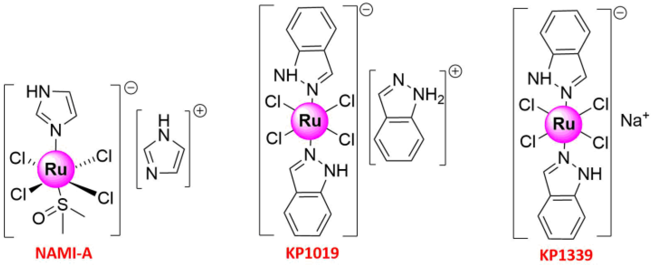
Ru complexes can also trigger phototoxicity in cells, which induces a series of photochemical and photobiologic processes, leading to irreversible photodamage in tumor tissues [16]. Thus, Ru compounds, such as Ru(II) polypyridyl complexes, which could interact with bovine serum albumin (BSA) and with DNA via minor grooves [23,24,25,26], are considered attractive photo-mediated activation prodrugs for photodynamic therapy (PDT) and photoactivated chemotherapy (PACT) [27]. Given also that photofrin, a hematoporphyrin derivative, is the only PDT drug approved by the Food and Drug Administration for cancer therapy, including early and advanced lung cancer [28,29], extensive studies have been performed to develop novel Ru(II/III) complexes for efficient cancer treatment [16,18,30]. Metal-based anticancer candidate imidazolium [trans-RuCl4(1H-imidazole) (DMSO-S) (NAMI-A; DMSO = dimethyl sulfoxide) (Figure 1) was the first Ru compound to be studied on human beings, which has reached the phase II stage [31,32,33,34]. The study was launched in 2008 when NAMI-A administered in combination with gemcitabine was given to patients with advanced NSCLC [34]. Previously, fundamental works have shown that its lower molar cytotoxicity over cisplatin results from its reduced reactivity against DNA in intact cells, and more studies in animal models have exhibited the excellent and selective activity against lung metastases of some solid metastasizing tumors at a concentration with relatively mild toxicity [32,33,35]. The other two important promising Ru(III) complexes that have entered clinical trials were indazolium [trans-RuCl4(1H-indazole)2] (KP1019) [36,37,38,39] and sodium [trans-RuCl4(1H-indazole)2] (KP1339) [40] (Figure 1). The pharmacokinetics of KP1019, which was characterized by a small volume of distribution, low clearance, and long half-life, has been researched in a phase I dose-escalation study, in which five out of six patients treated with KP1019 experienced disease stabilization with no severe side effects [36,39]. With these pioneering works, Ru complexes are attracting increasing attention from chemical researchers. The recent years have witnessed the development of Ruthenium complexes as second-generation metal-based anticancer agents, possessing high potency of targeting cancer cells due to their low toxicity, the ability to induce apoptosis, selective anti-invasion, and anti-metastasis activity [41,42,43]. Some of them have also shown anti-angiogenic properties and could therefore be used to inhibit angiogenesis, the basis of tumor growth and metastasis [44]. Moreover, with the purpose of improving their in vivo stability, solubility, cellular uptake, and effectiveness, some research groups with meticulous design have developed special drug delivery systems (nanoparticles, liposomes, etc.), which could encapsulate Ru-based compounds appropriately [30,45,46,47].
2. Ru(III) Complexes
Ru(III) complexes are rapidly evolving into next-generation anticancer drugs. Ru(III) hydrazone complexes owing uncoordinated phenolic oxygen were offered as better drug candidates than Ru(II) complexes with the same ligands because the phenolic oxygen in Ru(III) complexes could effectively interact with biomolecules by hydrogen bond, while phenolic oxygen of Ru(II) complexes was deprotonated for coordinating with Ru metal [50]. Moreover, several Ru(III) complexes that have entered clinical trials in different phases, such as KP1019 [36,37,38,39], KP-1339 [40], and NAMI-A [31,32,33,65], showed promising anticancer activity with limited side effects and have been used to prepare various derivatives.
2.1. Apoptosis
Recent studies have shown that Ru(III) complexes inhibit tumor cell proliferation by inducing apoptosis, which refers to genetically identified programmed cell death and plays a vital role in normal tissue homeostasis [66]. Apoptosis is not only involved in the occurrence and growth of tumors but can also trigger cancer cell death, making it a suitable pathway for the development of anticancer drugs [67]. There are two main types of apoptosis pathways: the intrinsic pathway, which depends on mitochondria, and the extrinsic pathway, which depends on death receptors [67,68].
The tryptamine-based mixed ligand Schiff base Ru(III) complexes Ru(Cl)2(SB) (phen = 1,10-Phenenthroline) (SB = Schiff base) (Ru1) and Ru(Cl)2(SB)(bipy) (bipy = 4,4′-bipyridine) (Ru2) (Figure 3) showed moderate antibacterial activity against Gram-positive and Gram-negative strains, but significantly high anticancer activity against NSCLC cells (H1299). In addition, they exhibited high cytotoxicity to NSCLC cells with IC50 = 10–12.5 ± 0.5 and 15–20 ± 0.5 μg/mL, respectively, but low toxicity to human erythrocytes compared to cisplatin [48]. Further cell studies indicated that the mechanism of action of Ru(III) complexes bearing tetradentate bis (aminophenolate) ligands against various cell types, including lung cancer, was based on the induction of programmed cell death [69]. In the case of mer- [RuCl3(PPh3)(dmpbt)] (Ru3) and fac- [RuCl3(PPh3)(dmpbt)] (Ru4) (PPh3 = triphenylphosphine, dmpbt = 2-(3,5-dimethylpyrazoll-yl) benzothiazole) (Figure 3), apoptosis was further promoted by their caspase 3/7 activity [49]. In another study, Ru(II) and Ru(III) hydrazone complexes {[RuIII(HL) Cl2(PPh3)2] (Ru5), [RuII(L)(CO)(PPh3)2] (Ru6), ([RuIII(HL) Cl2(AsPh3)2] (Ru7), and [RuII(L)(CO)(AsPh3)2] (Ru8)} (Figure 3) were prepared through a one-pot process and enhanced the release of lactate dehydrogenase, nitric oxide, and ROS [50]. The chromatin condensation, nuclear shrinkage, and plasma membrane blebbing were also observed using fluorescence microscopy, indicating that their in vitro cytotoxicity toward A549 cells resulted from apoptosis induction [50].
2.2. Anti-Metastasis
Cancer therapies generally fail when genetically unstable cancer cells adapt to the tissue microenvironment, leading to tumor metastasis [70,71]. Tumor metastasis involves several steps, including loss of cell adhesion, increased motility and invasiveness, entry and survival into the circulation, and eventual settlement into new tissues or distant organs [72]. Metastatic tumors are resistant to various cancer therapies, especially drugs and radiotherapy [73], indicating the need for the development of new compounds targeting the tumor metastasis pathway [74]. To date, various analogs have been designed and synthesized to prevent metastasis and/or inhibit the growth of metastatic tumors. Antineoplastic Ru(III) complexes are currently the most effective, such as NAMI-A, which can greatly reduce lung metastasis and the formation of solid metastasizing tumors in mice [75]. Therefore, a series of NAMI-A derivatives have been prepared that could modify important metastasis parameters such as tumor invasion, matrix metalloproteinases (MMP) activity, and cell cycle progression [42]. The new analogs also maintained the potent characteristics of NAMI-A and could selectively interact with solid metastatic tumors. Furthermore, the introduction of different ligands improved their stability in aqueous solutions [76]. Two NAMI-A derivatives bearing a pyridine ligand, Ru9/G26b and Ru10/G94a (Figure 4), displayed little direct cytotoxicity to human (A549) and mouse Lewis lung cancer cells but had a significant suppressive effect on the invasion and migration of cancer cells [51]. Like with NAMI-A, in vivo studies in 4T1 mammary carcinoma-bearing mice showed that the occurrence and development of lung metastasis were suppressed significantly and that no retinal toxicity or hepatotoxicity was found in mice after intraperitoneal injection of Ru9 and Ru10 at a dose of 17.5 mg/kg per day for consecutive 6 days, with three times in a 1-day interval. Ru9 in particular could suppress important molecules involved in metastasis, such as MMP-2 and MMP-9, and the vascular endothelial growth factor [51]. More interestingly, plasma atomic emission spectrometry showed that Ru9 possessed higher metabolic stability due to having a longer Ru-elimination time in the lungs, indicating its better anti-metastatic effect compared to NAMI-A and Ru10 [51]. In an earlier study, the hetero multinuclear complex [Na2] {[RuCl4(DMSO-S)(μ-pyz)]2PtCl2}, AH197 (Ru11/AH197) was also synthesized [77], and its effect on the motility and DNA electrophoretic mobility of NSCLC (A549) and breast cancer (MDA-MB-231) cells was compared to that of [K] [RuCl4(DMSO-S)(μ-pyz)Pt(DMSO-S)Cl2] (IT127), NAMI-A, and Na [trans-RuCl4(DMSO)(pyz)] (AH403) [78]. The Ru2Pt trinuclear species showed higher inhibitory activity in the order Ru11 > IT127 > NAMI-A > AH403 (Figure 4) [78], while it was shown that the inhibition of cell motility might contribute to the anti-metastatic properties of the complexes [79].
3. Ru(II) Complexes
Ru(II) complexes are also known for their low toxicity, different modes of action, and non-cross resistance to traditional Pt-based drugs, especially cisplatin. Therefore, recent studies have focused on understanding the anticancer mechanism of Ru(II) complexes to develop more effective Ru(II)-based drug candidates.
3.1. Apoptosis
With air stability, aqueous solubility, and structural diversity, the versatile half-sandwich Ru(II)-η6-p-cymene complexes have been reported as potential anticancer drugs, as they show distinct anti-proliferative activity and can effectively induce apoptosis [80]. For instance, [(Ru(η6-p-cymene)Cl)2(1,3,5-triaza-7-phosphaadamantane)] (RAPTA-C) (Figure 5) inhibited tumor metastasis and growth by inducing the apoptosis of Ehrlich ascites carcinoma cells through mitochondrial and p53-JNK pathways [81]. Similarly to RAPTA-C, a bimetallic Ru(II) cymene complex, [(Ru(η6-p-cymene) Cl)2(1,3-bis(2-methyl-6-(pyridin-2-yl) pyrimidin-4-yl) benzene)] (Ru12) (Figure 5), showed strong anticancer activity toward human NSCLC A549 and A427 cancer cells by inhibiting cell proliferation, migration, and invasion, which was stable in solution state in D2O/DMSO-d6 mixture as well as in solid state under air and light [82]. Mitochondria-mediated apoptosis of NSCLC was also observed upon treatment with Ru12, followed by an increase in the apoptosis regulator Bax and caspase-3/-9 activation. Thus, Ru12 induced DNA damage and cell death via caspase-dependent apoptosis by activating poly(ADP-ribose) polymerase(PARP) and triggering the p53-dependent pathway [82]. Moreover, Ru12 inhibited cancer cell migration and invasion, which in turn blocked the expression of the c-Myc(myelocytomatosis) oncogene that is important for cell cycle progression, apoptosis, and cellular transformation, and it has been related to cancer metastasis [82,83]. In a current study, another two sets of organometallic arene Ru(II) complexes against cancer cells (NSCLC A549, colon adenocarcinoma LoVo, and hepato cellular carcinoma HuH-7) have been reported by Balaji, S and coworkers. Structurally, the complexes with p-cymene moiety outperformed the anticancer activity of the complexes containing benzene moiety, in that the latter had less hydrophobic interaction with the cell membrane [84].
Recently, six new bimetallic Ru(II) arene complexes [Ru2(η6-p-cymene)2(1,3-bib)2Cl2]X2 (X = Cl− (Ru13), I− (Ru14), NO3− (Ru15), BF4− (Ru16), PF6− (Ru17), and CF3SO3− (Ru18); 1,3-bib = 1,3-di(1H-imidazol-1-yl) benzene) were designed and synthesized [85]. Ru13–18 (Figure 5) performed good stability in aqueous solution, and UV-Vis spectra also suggested that bidentate imidazole-based ligand strengthens the stability of the Ru-arene complex, comparing to mononuclear N-heterocyclic ligand. All showed moderate proliferative or anti-proliferative activity due to their interaction with glutathione on lung (A549) cancer cells, but strong intercalative binding ability to ctDNA. Moreover, Ru14 showed a relatively better anti-proliferative activity compared to the other complexes due to the increased polarization of I−. Further treatment of A549 cells with Ru14 led to concentration-dependent late apoptosis and cell cycle arrest in the G1/G0 phase. In a subsequent study, a hydrazone moiety was rationally incorporated into two tetranuclear arene Ru(II) complexes, Ru19 and Ru20 (Figure 5), to improve their pharmacological activity [86]. With high stability in DMEM containing 10%FBS and PBS with 10%DMSO, both complexes had high anticancer activity in vitro and could induce the apoptosis of various human cancer cell lines, including cisplatin-resistant lung (A549) cancer cells. To certify the systemic toxicity of Ru19, a series of animal experiments were conducted by the group. Impressively, no damage was observed in major organs, including the kidney, in the mice treatment with Ru19 (6 mg/kg), while a mass of vacuolization in the cell cytoplasm of renal tubules were found in the cisplatin-treated mice, suggesting that the compound Ru19 exhibited lower systemic toxicity and was potentially more tolerated by animals than cisplatin [86]. Apart from targeting cisplatin-resistant cancer cells to address chemoresistance issue, Teixeira, R. G et al. proposed a novel approach that organometallic Ru(II) compounds increased cisplatin cytotoxicity up to 1390-fold at nontoxic doses by inhibiting multidrug resistance-associated protein 1 (MRP1) and the P-glycoprotein 1 (Pgp) transporters [87]. It further promoted Ru(II) compounds as more valuable and prospective agents for lung cancer chemotherapy, in particular for those patients with cisplatin resistance.
A series of Ru(II) methylimidazole complexes (Ru21–24) (Figure 6) with strong anti-proliferative activity against various human cancer cells were also synthesized [88]. Although Ru12 induced NSCLC apoptosis via the mitochondria-dependent pathway [82], two pathways were involved in the mechanism of action of Ru21. Specifically, Ru21 activated the BID protein and depleted the mitochondrial membrane potential in A549 cells by regulating the expression of pro-survival and pro-apoptotic Bcl-2 family proteins [88]. Moreover, the Ru(II) polypyridyl complexes Ru25 and Ru28 (Figure 6) showed completely different mechanisms [52]. Ru25 induced marginal oxidative stress and preferably accumulated in lysosomes, triggering apoptosis via an intrinsic mitochondrial pathway, while enlarged mitochondria were detected in Ru25-treated A549 cells. In contrast, Ru28, which was mainly localized in mitochondria and endoplasmic reticulum, did not have the same effect and induced caspase-independent apoptosis [52].
ROS are generated under normal cell activity and are involved in cellular signaling. However, high ROS levels produced mainly by mitochondrial dysfunction may lead to oxidative damage of cellular structures such as DNA damage and apoptosis [89,90]. Therefore, agents modulating ROS generation have been designed for clinical cancer therapy. For instance, Ru30 [54], Ru31–32 [55], Ru33 [56], Ru36 [57], and Ru38 [58] (Figure 6 and Figure 7) altered the mitochondrial function and generated ROS in lung tumor cells, leading to various adverse effects. [cis-[Ru(η2-O2CC7H7O2)(dppm)2]PF6] (dppm = bis(diphenylphosphino)methane) (Ru30), with stability in DMSO monitored by 31P {1H} NMR experiments, showed high cytotoxic activity against Leishmania promastigotes [91,92] and selectively targeted lung target tumor cells [54], while no toxic effect was observed on normal bronchial epithelial BEAS-2B cells. In addition, the increased ROS levels generated by 3.8 µM Ru30 in A549 lung tumor cells caused oxidative stress, which led to proliferation inhibition, changes in the morphology and organization patterns of the actin cytoskeleton, G2/M phase arrest, apoptosis, changes in the mitochondrial membrane potential, and DNA damage [54]. Ru31 [Ru(dip)2(SA)] and Ru32 [Ru(dmp)2(SA)] (dip = 4,7-diphenyl-1,10-phenanthroline; dmp = 2,9-dimethyl-1,10-phenanthroline; SA = salicylate), two Ru(II) complexes bearing O,O-chelated ligands with low toxicity to BEAS-2B, could also induce apoptosis in A549 cells via caspase family proteins and PARP activation, ROS accumulation, DNA damage, MMP reduction, and Cytochrome c release from mitochondria [55]. Significant in vitro anticancer activity, combined with proven solution stability and hydrophobic property, prompted the group to further conduct in vivo experiments to test the toxicity [55]. Accordingly, developing zebrafish embryos were used, incubating with various concentrations (0, 12.5, 25, 50, 100, and 200 μM) of Ru31 in water. Although lower cumulative hatch rate and increased lethality rate, even unhealthy features such as pericardial cysts and spine curvature were notably observed with treatment concentrations up to 200 μM, but there were no apparent side effects found in zebrafish embryos after treatment with concentrations from 12.5 to 50 μM of Ru31. Combined with low toxicity towards both normal BEAS-2B cells in vitro and zebrafish embryos in vivo implied that Ru31 has great potential to develop as a promising therapeutic agent against lung cancer with safety profiles [55].
Caspase-related family proteins are key components of induction and transduction of apoptotic signaling in cells and are closely related to BCL-2 family proteins that either induce (pro-apoptotic) or inhibit (anti-apoptotic) apoptosis [93,94]. Ru31 downregulated the expression levels of the anti-apoptotic proteins BCL-2 and BCL-xL, whereas it upregulated the levels of the pro-apoptotic proteins BAX and BAD [55]. Since the high expression levels of BCL-2 and BCL-xL have been associated with cisplatin resistance and tumor recurrence in NSCLC patients [95], Ru31 may indirectly inhibit the resistance and relapse of lung cancer. [Ru(dmp)2(pddppn)](ClO4)2 (pddppn = phenantheno [1,2-b]-1,4-diazabenzo [i] dipyrido [3,2-a:2′,3′-c]phenazine) (Ru33) could also downregulate the expression of BCL-2, BCL-x, BAK, and BIM, while upregulating the expression of BAG-1 and BAD, thus inducing apoptosis of NSCLC A549 cells via an intrinsic ROS-mediated mitochondrial dysfunction pathway [56]. Moreover, Ru33 effectively inhibited the growth of BEL-7402 (human hepatocellular cell line), HeLa (human cervical cancer cell line), MG-63 (human osteosarcoma cell line), and A549 cancer cells with IC50 values of 1.6 ± 0.4, 9.0 ± 0.8, 1.5 ± 0.2, and 1.5 ± 0.3 μM, respectively [56].
Another study investigated the mechanism of action of Ru complexes, showing that Ru36 can lead to significant ROS production and induce apoptosis, while increasing the BAX/BCL-2 ratio and PERK levels without affecting the expression of caspase-3 [57]. Four additional Ru(II) complexes with different ancillary ligands { [Ru(bpy)2(dmbpy)](PF6)2 (Ru34), [Ru(phen)2(dmbpy)](PF6)2 (Ru35), [Ru(bphen)2(dmbpy)](PF6)2 (Ru36), [Ru(BPS)2dmbpy]Na2 (Ru37) (dmbpy = dimethyl-2,2′-bipyridine)} (Figure 7) were also prepared in the same study, and their effect on NSCLC (A549) and triple-negative breast (MDA-MB-231) cancer cells were investigated [57]. The increase in lipophilicity as a strategy to enhance the cellular uptake and the antiproliferative activity of the Ruthenium complexes on cancer cells is important [96,97]. Among them, Ru36 was the only lipophilic complex and exhibited the highest cellular uptake and significant phototoxicity [57]. [Ru(MeIm)4(p-cpip)]2+ (p-cpip = 2-(4-chlorophenyl)-1H-imidazo [4,5-f] [1,10]phenanthroline, MeIm = 1-methylimidazole) (Ru38) has also been recently synthesized and characterized, exhibiting relatively high cytotoxicity against lung cancer (A549) cells, as well as high selectivity to tumor vs. normal cells compared to cisplatin [58]. In addition, Ru38 induced apoptosis via the mitochondrial pathway, which involved ROS accumulation, mitochondrial dysfunction, and activation of BCL-2 and caspase family proteins [58].
3.2. Autophagy
Unlike apoptosis, autophagy is an evolutionarily conserved degradation pathway for the recycling of cytoplasmic components and plays a significant role in response to metabolic and therapeutic stresses [98,99]. Recent studies presented scientific evidence supporting the participation of autophagy in tumor progression, such as the development of multidrug resistance of cancer cells, but autophagy also can help in killing cancer cells that are resistant to anticancer agents as a potential target for cancer therapy [99,100].
To the best of our knowledge, there are only a few studies on the inhibition of cell growth caused by metal complexes through autophagy induction. For instance, a Pt-based complex, [Pt(O,O’-acac)(γ-acac)(DMS)], induced autophagy in Caki-1 renal cancer cells [101] and rat B50 neuroblastoma [102]. Apart from Pt-based agents, Piccolo M et al. [103] and Irace C et al. [104] have certified that the activation of autophagic pathways is a common feature with Ru(III) complexes against breast cancer cells. These findings could pave the way for the development of other autophagy-inducing metal complexes to overcome apoptosis-resistant cancer cells. In 2016, the mechanism of Ru(II) complex targeting lung cancer cells by autophagy induction was reported by Chen, L et al. for the first time [59]. Specifically, the formation of autophagosomes and acidic vesicular organelles along with LC3-II upregulation was observed in A549 and NCI-H460 cancer cells treated with a Ru(II) imidazole complex, [Ru(Im)4(dppz)]2+ (dppz = pyrido [3.2-a:2′,3′-c]phenazine) (Ru39) (Figure 7). Ru39 caused mitochondrial dysfunction and ROS generation in A549 cells, thus partially inducing caspase-3-dependent apoptosis, which causes cell death, as well as autophagy mediated by the extracellular signal-regulated kinase (ERK) signaling pathway. Eventually, the accumulation of Ru39 in mitochondria could induce autophagy and compete with mitochondria-mediated apoptosis [59]. Furthermore, the antitumor activity of Ru39 was further evaluated in vivo mice bearing A549 xenografts, which were treated with Ru39 at 10 or 20 mg/kg. After 28 days, it was observed that the weight and volume of tumors were significantly reduced, and the expressions of LC3-II, cleaved caspase-3, CD-31, and Ki-67 were up-regulated via immunohistochemical analysis. Accordingly, Ru39 could inhibit tumor growth very well in vitro and in vivo and develop as a promising anticancer candidate promoting cell death via both apoptosis and autophagy pathway [59]. Resembled Ru-based complexes were also highly active against glioblastoma cell lines, inducing cell death via apoptosis and autophagy in a p53-independent manner [105].
3.3. Necroptosis
Promoting apoptosis is one of the main methods to treat tumors. However, the anti-apoptotic properties may lead to resistance to cytotoxic chemotherapeutic drugs or radiotherapy [106]. Necroptosis is another type of regulated cell death closely related to the receptor-interacting protein kinases 1 and 3 (RIPK1 and RIPK3) and mixed-lineage kinase domain-like protein (MLKL) [107]. Necroptosis begins with the activation of RIPK and MLKL, which increase the levels of Ca2+, causing lysosomal membrane permeabilization, and release cathepsins into the cytosol [108]. Although relevant studies are scarce, a recent report has shown that inducing necroptosis in cancer cells can overcome chemotherapy failure due to apoptotic resistance [109], while metal complexes have been shown to induce cancer cell death through necroptosis [110,111].
A series of Ru(II) complexes bearing 1,1-(pyrazin-2-yl) pyreno [4,5-e] [1,2,4] triazine with different ancillary ligands have been recently prepared and used to induce necroptosis by Xiong K et al. (Ru40–46) (Figure 8) [60]. All analogs showed significant antitumor activity against drug-resistant cancer cells, including A549 lung cancer cells (IC50 (Ru46) = 3.0 ± 0.1 μM). More importantly, Ru40–46 prevented the DNA binding of topoisomerases (topo) I and II, which are crucial nuclear enzymes that regulate DNA replication, transcription, recombination, and chromosome segregation during mitosis [60,112]. The characteristic indicators of necroptosis, including ROS burst, plasma membrane permeabilization, and cytosolic ATP reduction, were also reported, while the cell signaling pathway from topo I and II inhibition, DNA damage, and PARP1 activation to necroptosis induced by the activation of RIPK1, RIPK3, and MLKL were elucidated [60]. In contrast, the group also reported a collection of Ru(II) complexes containing asymmetric tridentate ligands that could induce DNA damage and cancer cell death through apoptosis, which were also topo I and II inhibitors [113]. Moreover, caspase-8 has been proven to regulate the switch from apoptosis to necroptosis [114].
3.4. Anti-Metastasis
Ru(II) compounds can not only suppress the primary tumor but also effectively inhibit malignant tumor metastasis. A series of recently synthesized Ru(II)-containing polypyridyl ligand complexes (Ru25–29 (Figure 6) [53] and Ru47 (Figure 9) [61]) showed anti-metastatic properties, which was attributed to their targeting ability to MMPs [53]. Ru25–29 bearing 2,2′-bipyridine substituted with a semicarbazone-2-formylopyridine moiety as one of the ligands and 4,4′-di-tert-butyl-2,2′-dipyridyl or 4,7-diphenyl-1,10-phenanthroline as auxiliary ligands have been studied for their effect on the adhesion properties of human A549 and pancreatic cancer cells [53]. All complexes enhanced the cell adherent properties and could directly inhibit the activity of MMP2 and MMP9 enzymes in vitro. Among them, Ru28 led to the most significant enhancement of cell adhesion with increasing concentration [53].
Another Ru polypyridyl complex bearing an nitroimdazole unit {[Ru(dip)2(bpy-2-nitroIm)]Cl2 (dip = 4,7-diphenyl-1,10-phenanthroline, bpy-2-nitroIm = 4- [3-(2-nitro-1H-imidazol-1-yl) propyl]-2,2′-bipyridine)} (Ru47) could reduce the activity of MMP enzymes to inhibit tumor metastasis [115], while its effect on cancer and endothelial cells has also been reported [61]. Similar to Ru25–29, Ru47 changed the cell adhesion properties, thus reducing the number of adherent cells on different surfaces (fibronectin, collagen, and plastic) and the expression levels of several MMPs (MMP1a, MMP3, MMP9, LOX, TIMP1, THBS1, and ITB1) and protein-lysine 6-oxidase, while increasing the expression of the extracellular matrix inhibitor [61]. Ru47 was also studied in hypoxia, as hypoxia results from poor vascular organization of the tumor, promoting drug resistance and malignant progression [116,117]. The results suggested that Ru47 was more cytotoxic against human breast (4T1) and lung (A549) cancer cells than cisplatin, while it induced cell apoptosis and affected endothelial cell vasculature by activating oxidative stress and modulating the mRNA expression profile of ICAM-1 and VCAM-1 genes involved in metastasis and angiogenesis [61].
Two other complexes, Ru48/Ru-hq1 [η6-p-cymene) Ru(5-bromo-8-hydroxyquinolinato) Cl] and Ru49/Ru-hq2 [(η6-p-cymene) Ru (k2-O,N-5,7-dibromo-HyQ) Cl] (Figure 9), have also been screened as potential novel agents for bone, lung, and breast cancer chemotherapy [62]. Both complexes are stable in DMSO and DMEM solution within the time-frame of the biological experiments and attenuated cell viability with greater selectivity and specificity than cisplatin, while they inhibited cell proliferation, migration, and invasion on cell monolayers at lower concentrations (2.5–10 µM). The higher inhibitory effect of Ru48/Ru-hq1 and Ru49/Ru-hq2 on cell invasion compared to cisplatin was further confirm using 3D multicellular spheroid models [62]. Similar results were obtained for Ru19, which inhibited cancer cell migration and invasion and had higher safety margins for animals than cisplatin [86]. Therefore, Ru48–49 and Ru19 may be used as cisplatin alternatives to manage lung cancer and inhibit tumor metastasis.
3.5. Cell Cycle Arrest
The cell cycle, which involves four main phases (G1, S, G2, and M), contributes significantly to the regulation of cell self-renewal and differentiation, while cyclin-dependent kinases (CDKs) and cycle proteins play a key role in the process [118]. Therefore, clinical trials have been performed recently to explore the synergistic effect of CDK inhibitors and different anticancer drugs on various cancer types [119,120]. Additional studies have shown that metallodrugs can target CDKs and cycle proteins to block the cell cycle in different phases. For instance, Ru(II), Rh(III), Mn(II), and Zn(II) complexes blocked the cell cycle in the S-phase, thus inhibiting cell proliferation by reducing the levels of cyclins A2/B1/D1/E1, CDK-2/6, and PCNA and increasing the levels of p21, p27, p53, and CDC25A [121].
Given that the dysregulation of the cell cycle is associated with the development of cancer, novel Ru(II)-based drugs that inhibited tumor proliferation were synthesized and identified to disrupt the cell cycle in the G0/G1, G1/S, S, and G2/M phases (Figure 10). Ru12 [82], Ru14 [85], Ru33 [56], Ru28 [52], and Ru38 [58] are typical examples of compounds causing cell death via cell cycle arrest in the G0/G1 phase. Investigation of the mechanism of action revealed that Ru12 could induce cell cycle arrest in the G0/G1 phase; suppress the expression of cell-cycle-regulatory proteins, such as cyclins D1, A1, and B1, upregulate p53, p21, and p15; and cleave PARP [82]. Interestingly, the anti-proliferative complex Ru33 exhibited different mechanisms against different cancer cells: G2/M phase arrest in BEL-7402 and MG-63 cells and G0/G1 phase arrest in A549 cells [56]. In addition, Ru28 promoted cell accumulation in the G0/G1 phase, while the number of Ru25-treated A549 cells increased remarkably in the S-phase but decreased in the G2/M phase [52]. Furthermore, the Ru(II) methylimidazole complex Ru21 inhibited the growth of A549 lung cells by inducing apoptotic cell death, as confirmed by the generation of a significant apoptosis peak in the sub-G1 phase [88].
The cytotoxicity of {[Ru(pipe)(dppb)(bipy)]PF6} (pipe = piperonylic acid, dppb = 1,4-bis(diphenylphosphino) butane) (Ru50) on A549 cells has been associated with the induction of cell apoptosis via the intrinsic pathway [63]. The number of cells was significantly increased in the G0/G1 phase of the cell cycle, while the population of A549 cells in the S-phase was reduced when 9 μM of Ru50 was used. These results indicated that the G1/S transition depended on the antineoplastic ability of Ru50, which reduced the cyclin D1 expression levels and attenuated ERK phosphorylation [63]. Furthermore, Ru(II)-based compounds, such as the polypyridyl Ru complex Ru47 [61], arrested cell growth in the S-phase. Similar results were also obtained for the arene Ru(II) carbazole-based hydrazone complexes Ru51–53 [(η6-benzene) Ru(L) Cl] (L = carbazolone benzhydrazone ligands) [64] (Figure 9), whereas Ru30 [54] and Ru48–49 [62] induced apoptosis and cell cycle arrest of A549 lung cancer cells in the G2/M phase.
4. Conclusions/Discussions
Lung cancer remains a life-threatening malignancy due to poor prognosis and drug resistance, which is one of the most severe challenges that still need to be addressed to improve patients’ prognosis and survival rate. However, specific drugs preventing tumor metastasis and recurrence have not yet been efficiently developed. In order to design and synthesize effective anticancer agents, transition metal-based compounds have gradually evolved as promising drug candidates due to their cytotoxicity and ability to prevent drug resistance in tumor cells. Among them, Ru(II/III)-based compounds proved to be the most effective, as they show high cytotoxicity, which induces apoptosis, necroptosis, or autophagy and cell cycle arrest, thus inhibiting cell proliferation, invasion, and metastasis (Figure 2 and Figure 10). Most studies suggested that Ruthenium complexes were low in toxicity, easily absorbed, and excreted quickly. More importantly, Ruthenium complexes were easily absorbed by tumor tissues.
In this review, we summarized the recent developments of Ru(II) and Ru(III) complexes and discussed their biological activity and mechanism of action against lung cancer. The existing findings clearly support that Ru complexes can be used to develop effective chemotherapeutic agents for human lung cancer, while they may serve as a guide for the design of other metallodrugs with higher efficiency and better clinical application potential. Nevertheless, the anticancer mechanisms of Ru(II/III) complexes require further investigation, and their efficiency against other cancer types should also be explored in future studies.
Author Contributions
Conceptualization, Q.S. and Y.L.; methodology, Q.S. and Y.L.; software, Y.L.; validation, H.S., Y.W., J.Z. and Q.Z.; formal analysis, Q.S.; investigation, Q.S. and Y.L.; resources, J.Z. and Q.Z.; data curation, Q.S. and Y.L.; writing—original draft preparation, Q.S. and Y.L.; writing—review and editing, H.S., Y.W., J.Z. and Q.Z.; visualization, Y.L.; supervision, J.Z. and Q.Z.; project administration, Q.S. and Y.L.; funding acquisition, J.Z. and Q.Z. All authors have read and agreed to the published version of the manuscript.
Funding
This research was funded by the National Natural Science Foundation of China (21877103, 81602344, 21907069, 81802662), the Project of Natural Science Foundation of Guangdong Province (2019A1515011958), Shenzhen Science and Technology Innovation Commission (Grant No. KQJSCX20170331104516), SZU Medical Young Scientists Program (Grant No. 71201-000001). Medical Science and Technology Research Foundation of Guangdong Province (Grant No. A2019475).
Acknowledgments
The authors thank Collaborative Innovation Center of Suzhou Nano Science and Technology for support.
Conflicts of Interest
There are no conflict of interest.
Abbreviations
Ru: Ruthenium; NSCLC: non-small-cell lung cancer; SCLC: small cell lung cancer; LUSC: lung squamous cell carcinoma; LUAD: lung adenocarcinoma; BSA: bovine serum albumin; PDT: photodynamic therapy; PACT: photoactivated chemotherapy; MRP1: multidrug resistance-associated protein 1; Pgp: P-glycoprotein 1; RIPK: receptor-interacting protein kinase; MLKL: mixed lineage kinase domain-like protein; Topo: topoisomerase; MCS: multicellular spheroids; CDK: cyclin-dependent kinase; PARP: Poly (ADP-ribose) polymerase protein; ERK: extracellular signal-regulated kinase; EAC: Ehrlich ascites carcinoma; ROS: reactive oxygen species; LLC: lung cancer Lewis; VEGF: vascular endothelial growth factor; AVOs: acidic vesicular organelles; Pt: platinum; MMPs: matrix metalloproteinases.
References
- Bade, B.C.; Dela Cruz, C.S. Lung Cancer 2020: Epidemiology, Etiology, and Prevention. Clin. Chest Med. 2020, 41, 1–24. [Google Scholar] [CrossRef] [PubMed]
- Sung, H.; Ferlay, J.; Siegel, R.L.; Laversanne, M.; Soerjomataram, I.; Jemal, A.; Bray, F. Global Cancer Statistics 2020: GLOBOCAN Estimates of Incidence and Mortality Worldwide for 36 Cancers in 185 Countries. CA Cancer J. Clin. 2021, 71, 209–249. [Google Scholar] [CrossRef] [PubMed]
- Zappa, C.; Mousa, S.A. Non-small cell lung cancer: Current treatment and future advances. Transl. Lung. Cancer Res. 2016, 5, 288–300. [Google Scholar] [CrossRef] [PubMed] [Green Version]
- Rossi, A.; Di Maio, M. Platinum-based chemotherapy in advanced non-small-cell lung cancer: Optimal number of treatment cycles. Expert Rev. Anticancer Ther. 2016, 16, 653–660. [Google Scholar] [CrossRef]
- Giaccone, G.; Splinter, T.A.; Debruyne, C.; Kho, G.S.; Lianes, P.; van Zandwijk, N.; Pennucci, M.C.; Scagliotti, G.; van Meerbeeck, J.; van Hoesel, Q.; et al. Randomized study of paclitaxel-cisplatin versus cisplatin-teniposide in patients with advanced non-small-cell lung cancer. The European Organization for Research and Treatment of Cancer Lung Cancer Cooperative Group. J. Clin. Oncol. 1998, 16, 2133–2141. [Google Scholar] [CrossRef]
- Jamieson, E.R.; Lippard, S.J. Structure, Recognition, and Processing of Cisplatin-DNA Adducts. Chem. Rev. 1999, 99, 2467–2498. [Google Scholar] [CrossRef]
- Ghosh, S. Cisplatin: The first metal based anticancer drug. Bioorg. Chem. 2019, 88, 102925. [Google Scholar] [CrossRef] [PubMed]
- Boulikas, T.; Vougiouka, M. Cisplatin and platinum drugs at the molecular level (Review). Oncol. Rep. 2003, 10, 1663–1682. [Google Scholar] [CrossRef] [PubMed]
- Oun, R.; Moussa, Y.E.; Wheate, N.J. The side effects of platinum-based chemotherapy drugs: A review for chemists. Dalton Trans. 2018, 47, 6645–6653. [Google Scholar] [CrossRef]
- Ma, D.L.; He, H.Z.; Leung, K.H.; Chan, D.S.; Leung, C.H. Bioactive luminescent transition-metal complexes for biomedical applications. Angew. Chem. Int. Ed. Engl. 2013, 52, 7666–7682. [Google Scholar] [CrossRef]
- Ma, D.L.; Wu, C.; Wu, K.J.; Leung, C.H. Iridium(III) Complexes Targeting Apoptotic Cell Death in Cancer Cells. Molecules 2019, 24, 2739. [Google Scholar] [CrossRef] [PubMed] [Green Version]
- Ohata, J.; Ball, Z.T. Rhodium at the chemistry-biology interface. Dalton Trans. 2018, 47, 14855–14860. [Google Scholar] [CrossRef] [PubMed]
- Omondi, R.O.; Ojwach, S.O.; Jaganyi, D. Review of comparative studies of cytotoxic activities of Pt(II), Pd(II), Ru(II)/(III) and Au(III) complexes, their kinetics of ligand substitution reactions and DNA/BSA interactions. Inorg. Chim. Acta 2020, 512, 119883. [Google Scholar] [CrossRef]
- Koiri, R.K.; Mehrotra, A.; Trigun, S.K. Targetting cancer with Ru(III/II)-phosphodiesterase inhibitor adducts: A novel approach in the treatment of cancer. Med. Hypotheses 2013, 80, 841–846. [Google Scholar] [CrossRef] [PubMed]
- Guo, W.; Zheng, W.; Luo, Q.; Li, X.; Zhao, Y.; Xiong, S.; Wang, F. Transferrin serves as a mediator to deliver organometallic ruthenium(II) anticancer complexes into cells. Inorg. Chem. 2013, 52, 5328–5338. [Google Scholar] [CrossRef]
- Lee, S.Y.; Kim, C.Y.; Nam, T.G. Ruthenium Complexes as Anticancer Agents: A Brief History and Perspectives. Drug Des. Dev. Ther. 2020, 14, 5375–5392. [Google Scholar] [CrossRef]
- Jablonska-Wawrzycka, A.; Rogala, P.; Michalkiewicz, S.; Hodorowicz, M.; Barszcz, B. Ruthenium complexes in different oxidation states: Synthesis, crystal structure, spectra and redox properties. Dalton Trans. 2013, 42, 6092–6101. [Google Scholar] [CrossRef]
- Suss-Fink, G. Arene ruthenium complexes as anticancer agents. Dalton Trans. 2010, 39, 1673–1688. [Google Scholar] [CrossRef]
- Hairat, S.; Zaki, M. Half sandwiched Ruthenium(II) complexes: En Route towards the targeted delivery by Human Serum Albumin (HSA). J. Organomet. Chem. 2021, 937, 121732, Corrigendum in 2021, 943, 121734. [Google Scholar] [CrossRef]
- Lu, Y.; Zhu, D.; Gui, L.; Li, Y.; Wang, W.; Liu, J.; Wang, Y. A dual-targeting ruthenium nanodrug that inhibits primary tumor growth and lung metastasis via the PARP/ATM pathway. J. Nanobiotechnol. 2021, 19, 115. [Google Scholar] [CrossRef]
- Lin, K.; Zhao, Z.Z.; Bo, H.B.; Hao, X.J.; Wang, J.Q. Applications of Ruthenium Complex in Tumor Diagnosis and Therapy. Front. Pharmacol. 2018, 9, 1323. [Google Scholar] [CrossRef] [Green Version]
- Liu, J.; Lai, H.; Xiong, Z.; Chen, B.; Chen, T. Functionalization and cancer-targeting design of ruthenium complexes for precise cancer therapy. Chem. Commun. 2019, 55, 9904–9914. [Google Scholar] [CrossRef]
- Jakubaszek, M.; Goud, B.; Ferrari, S.; Gasser, G. Mechanisms of action of Ru(ii) polypyridyl complexes in living cells upon light irradiation. Chem. Commun. 2018, 54, 13040–13059. [Google Scholar] [CrossRef] [Green Version]
- Mede, T.; Jager, M.; Schubert, U.S. “Chemistry-on-the-complex”: Functional Ru(II) polypyridyl-type sensitizers as divergent building blocks. Chem. Soc. Rev. 2018, 47, 7577–7627. [Google Scholar] [CrossRef]
- Oliveira, K.M.; Honorato, J.; Goncalves, G.R.; Cominetti, M.R.; Batista, A.A.; Correa, R.S. Ru(II)/diclofenac-based complexes: DNA, BSA interaction and their anticancer evaluation against lung and breast tumor cells. Dalton Trans. 2020, 49, 12643–12652. [Google Scholar] [CrossRef]
- Rilak Simović, A.; Masnikosa, R.; Bratsos, I.; Alessio, E. Chemistry and reactivity of ruthenium(II) complexes: DNA/protein binding mode and anticancer activity are related to the complex structure. Coord. Chem. Rev. 2019, 398, 113011. [Google Scholar] [CrossRef]
- Mari, C.; Gasser, G. Lightening up Ruthenium Complexes to Fight Cancer? Chimia 2015, 69, 176–181. [Google Scholar] [CrossRef] [PubMed] [Green Version]
- Dougherty, T.J.; Gomer, C.J.; Henderson, B.W.; Jori, G.; Kessel, D.; Korbelik, M.; Moan, J.; Peng, Q. Photodynamic therapy. J. Nat. Inst. 1998, 90, 889–905. [Google Scholar] [CrossRef] [PubMed] [Green Version]
- Carruth, J.A. Clinical applications of photodynamic therapy. Int. J. Clin. Pract. 1998, 52, 39–42. [Google Scholar] [CrossRef]
- Thota, S.; Rodrigues, D.A.; Crans, D.C.; Barreiro, E.J. Ru(II) Compounds: Next-Generation Anticancer Metallotherapeutics? J. Med. Chem. 2018, 61, 5805–5821. [Google Scholar] [CrossRef]
- Alessio, E.; Messori, L. NAMI-A and KP1019/1339, Two Iconic Ruthenium Anticancer Drug Candidates Face-to-Face: A Case Story in Medicinal Inorganic Chemistry. Molecules 2019, 24, 1995. [Google Scholar] [CrossRef] [Green Version]
- Alessio, E. Thirty Years of the Drug Candidate NAMI-A and the Myths in the Field of Ruthenium Anticancer Compounds: A Personal Perspective. Eur. J. Inorg. Chem. 2017, 1549–1560. [Google Scholar] [CrossRef]
- Coverdale, J.; Laroiya-McCarron, T.; Romero-Canelón, I. Designing Ruthenium Anticancer Drugs: What Have We Learnt from the Key Drug Candidates? Inorganics 2019, 7, 31. [Google Scholar] [CrossRef] [Green Version]
- Leijen, S.; Burgers, S.A.; Baas, P.; Pluim, D.; Tibben, M.; van Werkhoven, E.; Alessio, E.; Sava, G.; Beijnen, J.H.; Schellens, J.H. Phase I/II study with ruthenium compound NAMI-A and gemcitabine in patients with non-small cell lung cancer after first line therapy. Investig. New Drugs 2015, 33, 201–214. [Google Scholar] [CrossRef] [Green Version]
- Pluim, D.; van Waardenburg, R.C.; Beijnen, J.H.; Schellens, J.H. Cytotoxicity of the organic ruthenium anticancer drug Nami-A is correlated with DNA binding in four different human tumor cell lines. Cancer Chemother. Pharmacol. 2004, 54, 71–78. [Google Scholar] [CrossRef]
- Hartinger, C.G.; Jakupec, M.A.; Zorbas-Seifried, S.; Groessl, M.; Egger, A.; Berger, W.; Zorbas, H.; Dyson, P.J.; Keppler, B.K. KP1019, a new redox-active anticancer agent--preclinical development and results of a clinical phase I study in tumor patients. Chem. Biodivers. 2008, 5, 2140–2155. [Google Scholar] [CrossRef] [PubMed]
- Hartinger, C.G.; Zorbas-Seifried, S.; Jakupec, M.A.; Kynast, B.; Zorbas, H.; Keppler, B.K. From bench to bedside--preclinical and early clinical development of the anticancer agent indazolium trans-[tetrachlorobis(1H-indazole)ruthenate(III)] (KP1019 or FFC14A). J. Inorg. Biochem. 2006, 100, 891–904. [Google Scholar] [CrossRef]
- Jakupec, M.A.; Arion, V.B.; Kapitza, S.; Reisner, E.; Eichinger, A.; Pongratz, M.; Marian, B.; Graf von Keyserlingk, N.; Keppler, B.K. KP1019 (FFC14A) from bench to bedside: Preclinical and early clinical development—An overview. Int. J. Clin. Pharmacol. Ther. 2005, 43, 595–596. [Google Scholar] [CrossRef] [PubMed]
- Lentz, F.; Drescher, A.; Lindauer, A.; Henke, M.; Hilger, R.A.; Hartinger, C.G.; Scheulen, M.E.; Dittrich, C.; Keppler, B.K.; Jaehde, U.; et al. Pharmacokinetics of a novel anticancer ruthenium complex (KP1019, FFC14A) in a phase I dose-escalation study. Anticancer Drugs 2009, 20, 97–103. [Google Scholar] [CrossRef] [PubMed]
- Kapitza, S.; Pongratz, M.; Jakupec, M.A.; Heffeter, P.; Berger, W.; Lackinger, L.; Keppler, B.K.; Marian, B. Heterocyclic complexes of ruthenium(III) induce apoptosis in colorectal carcinoma cells. J. Cancer Res. Clin. Oncol. 2005, 131, 101–110. [Google Scholar] [CrossRef]
- Biersack, B.; Schobert, R. Current State of Metal-Based Drugs for the Efficient Therapy of Lung Cancers and Lung Metastases. Adv. Exp. Med. Biol. 2016, 893, 211–224. [Google Scholar]
- Alessio, E.; Mestroni, G.; Bergamo, A.; Sava, G. Ruthenium antimetastatic agents. Curr. Top. Med. Chem. 2004, 4, 1525–1535. [Google Scholar] [CrossRef]
- Zheng, K.; Wu, Q.; Wang, C.; Tan, W.; Mei, W. Ruthenium(II) Complexes as Potential Apoptosis Inducers in Chemotherapy. Anticancer Agents Med. Chem. 2017, 17, 29–39. [Google Scholar]
- Sun, R.W.; Ng, M.F.; Wong, E.L.; Zhang, J.; Chui, S.S.; Shek, L.; Lau, T.C.; Che, C.M. Dual anti-angiogenic and cytotoxic properties of ruthenium(III) complexes containing pyrazolato and/or pyrazole ligands. Dalton Trans. 2009, 48, 10712–10716. [Google Scholar] [CrossRef]
- Riccardi, C.; Musumeci, D.; Irace, C.; Paduano, L.; Montesarchio, D. RuIIIComplexes for Anticancer Therapy: The Importance of Being Nucleolipidic. Eur. J. Org. Chem. 2017, 2017, 1100–1119. [Google Scholar] [CrossRef]
- Riccardi, C.; Musumeci, D.; Trifuoggi, M.; Irace, C.; Paduano, L.; Montesarchio, D. Anticancer Ruthenium(III) Complexes and Ru(III)-Containing Nanoformulations: An Update on the Mechanism of Action and Biological Activity. Pharmaceuticals 2019, 12, 146. [Google Scholar] [CrossRef] [PubMed] [Green Version]
- Zeng, L.; Gupta, P.; Chen, Y.; Wang, E.; Ji, L.; Chao, H.; Chen, Z.S. The development of anticancer ruthenium(ii) complexes: From single molecule compounds to nanomaterials. Chem. Soc. Rev. 2017, 46, 5771–5804. [Google Scholar] [CrossRef] [PubMed]
- Malik, M.A.; Raza, M.K.; Dar, O.A.; Amadudin, A.; Abid, M.; Wani, M.Y.; Al-Bogami, A.S.; Hashmi, A.A. Probing the antibacterial and anticancer potential of tryptamine based mixed ligand Schiff base Ruthenium(III) complexes. Bioorg. Chem. 2019, 87, 773–782. [Google Scholar] [CrossRef] [PubMed]
- Richert, M.; Walczyk, M.; Cieslak, M.J.; Kazmierczak-Baranska, J.; Krolewska-Golinska, K.; Wrzeszcz, G.; Muziol, T.; Biniak, S. Synthesis, X-ray structure, physicochemical properties and anticancer activity of mer and fac Ru(iii) triphenylphosphine complexes with a benzothiazole derivative as a co-ligand. Dalton Trans. 2019, 48, 10689–10702. [Google Scholar] [CrossRef] [PubMed]
- Jayanthi, E.; Kalaiselvi, S.; Padma, V.V.; Bhuvanesh, N.S.; Dharmaraj, N. Solvent assisted formation of ruthenium(III) and ruthenium(II) hydrazone complexes in one-pot with potential in vitro cytotoxicity and enhanced LDH, NO and ROS release. Dalton Trans. 2016, 45, 1693–1707. [Google Scholar] [CrossRef]
- Gu, L.; Li, X.; Ran, Q.; Kang, C.; Lee, C.; Shen, J. Antimetastatic activity of novel ruthenium (III) pyridine complexes. Cancer Med. 2016, 5, 2850–2860. [Google Scholar] [CrossRef] [PubMed]
- Lomzik, M.; Mazuryk, O.; Rutkowska-Zbik, D.; Stochel, G.; Gros, P.C.; Brindell, M. New ruthenium compounds bearing semicarbazone 2-formylopyridine moiety: Playing with auxiliary ligands for tuning the mechanism of biological activity. J. Inorg. Biochem. 2017, 175, 80–91. [Google Scholar] [CrossRef] [PubMed] [Green Version]
- Gurgul, I.; Mazuryk, O.; Lomzik, M.; Gros, P.C.; Rutkowska-Zbik, D.; Brindell, M. Unexplored features of Ru(ii) polypyridyl complexes towards combined cytotoxic and antimetastatic activity. Metallomics 2020, 12, 784–793. [Google Scholar] [CrossRef] [PubMed]
- Costa, M.S.; Goncalves, Y.G.; Borges, B.C.; Silva, M.J.B.; Amstalden, M.K.; Costa, T.R.; Antunes, L.M.G.; Rodrigues, R.S.; Rodrigues, V.M.; de Faria Franca, E.; et al. Ruthenium (II) complex cis-[Ru(II)(ŋ(2)-O2CC7H7O2)(dppm)2]PF6-hmxbato induces ROS-mediated apoptosis in lung tumor cells producing selective cytotoxicity. Sci. Rep. 2020, 10, 15410. [Google Scholar] [CrossRef] [PubMed]
- Chen, J.; Wang, J.; Deng, Y.; Wang, T.; Miao, T.; Li, C.; Cai, X.; Liu, Y.; Henri, J.; Chen, L. Ru(II) Complexes Bearing O, O-Chelated Ligands Induced Apoptosis in A549 Cells through the Mitochondrial Apoptotic Pathway. Bioinorg. Chem. Appl. 2020, 2020, 8890950. [Google Scholar] [CrossRef] [PubMed]
- Lai, S.H.; Jiang, G.B.; Yao, J.H.; Li, W.; Han, B.J.; Zhang, C.; Zeng, C.C.; Liu, Y.J. Cytotoxic activity, DNA damage, cellular uptake, apoptosis and western blot analysis of ruthenium(II) polypyridyl complex against human lung decarcinoma A549 cell. J. Inorg. Biochem. 2015, 152, 1–9. [Google Scholar] [CrossRef]
- Fayad, C.; Audi, H.; Khnayzer, R.S.; Daher, C.F. The anti-cancer effect of series of strained photoactivatable Ru(II) polypyridyl complexes on non-small-cell lung cancer and triple negative breast cancer cells. J. Biol. Inorg. Chem. 2021, 26, 43–55. [Google Scholar] [CrossRef] [PubMed]
- Chen, L.M.; Peng, F.; Li, G.D.; Jie, X.M.; Cai, K.R.; Cai, C.; Zhong, Y.; Zeng, H.; Li, W.; Zhang, Z.; et al. The studies on the cytotoxicity in vitro, cellular uptake, cell cycle arrest and apoptosis-inducing properties of ruthenium methylimidazole complex [Ru(MeIm)4(p-cpip)](2.). J. Inorg. Biochem. 2016, 156, 64–74. [Google Scholar] [CrossRef]
- Chen, L.; Li, G.; Peng, F.; Jie, X.; Dongye, G.; Cai, K.; Feng, R.; Li, B.; Zeng, Q.; Lun, K.; et al. The induction of autophagy against mitochondria-mediated apoptosis in lung cancer cells by a ruthenium (II) imidazole complex. Oncotarget 2016, 7, 80716–80734. [Google Scholar] [CrossRef] [Green Version]
- Xiong, K.; Qian, C.; Yuan, Y.; Wei, L.; Liao, X.; He, L.; Rees, T.W.; Chen, Y.; Wan, J.; Ji, L.; et al. Necroptosis Induced by Ruthenium(II) Complexes as Dual Catalytic Inhibitors of Topoisomerase I/II. Angew. Chem. Int. Ed. Engl. 2020, 59, 16631–16637. [Google Scholar] [CrossRef]
- Mazuryk, O.; Suzenet, F.; Kieda, C.; Brindell, M. The biological effect of the nitroimidazole derivative of a polypyridyl ruthenium complex on cancer and endothelial cells. Metallomics 2015, 7, 553–566. [Google Scholar] [CrossRef] [PubMed]
- Ruiz, M.C.; Kljun, J.; Turel, I.; Di Virgilio, A.L.; Leon, I.E. Comparative antitumor studies of organoruthenium complexes with 8-hydroxyquinolines on 2D and 3D cell models of bone, lung and breast cancer. Metallomics 2019, 11, 666–675. [Google Scholar] [CrossRef] [PubMed]
- Ferreira-Silva, G.A.; Ortega, M.M.; Banionis, M.A.; Garavelli, G.Y.; Martins, F.T.; Dias, J.S.M.; Viegas, C., Jr.; Oliveira, J.C.; Nascimento, F.B.D.; Doriguetto, A.C.; et al. [Ru(pipe)(dppb)(bipy)]PF6: A novel ruthenium complex that effectively inhibits ERK activation and cyclin D1 expression in A549 cells. Toxicol. In Vitro 2017, 44, 382–391. [Google Scholar] [CrossRef] [PubMed]
- Sathiya Kamatchi, T.; Mohamed Subarkhan, M.K.; Ramesh, R.; Wang, H.; Malecki, J.G. Investigation into antiproliferative activity and apoptosis mechanism of new arene Ru(ii) carbazole-based hydrazone complexes. Dalton Trans. 2020, 49, 11385–11395. [Google Scholar] [CrossRef]
- Rademaker-Lakhai, J.M.; van den Bongard, D.; Pluim, D.; Beijnen, J.H.; Schellens, J.H. A Phase I and pharmacological study with imidazolium-trans-DMSO-imidazole-tetrachlororuthenate, a novel ruthenium anticancer agent. Clin. Cancer Res. 2004, 10, 3717–3727. [Google Scholar] [CrossRef] [Green Version]
- Fleisher, T.A. Apoptosis. Ann. Allergy Asthma Immunol. 1997, 78, 245–249; quiz 249–250. [Google Scholar] [CrossRef]
- Pistritto, G.; Trisciuoglio, D.; Ceci, C.; Garufi, A.; D’Orazi, G. Apoptosis as anticancer mechanism: Function and dysfunction of its modulators and targeted therapeutic strategies. Aging 2016, 8, 603–619. [Google Scholar] [CrossRef] [Green Version]
- Tait, S.W.; Green, D.R. Caspase-independent cell death: Leaving the set without the final cut. Oncogene 2008, 27, 6452–6461. [Google Scholar] [CrossRef] [Green Version]
- Domotor, O.; de Almeida, R.F.; Corte-Real, L.; Matos, C.P.; Marques, F.; Matos, A.; Real, C.; Kiss, T.; Enyedy, E.A.; Helena Garcia, M.; et al. Studies on the mechanism of action of antitumor bis(aminophenolate) ruthenium(III) complexes. J. Inorg. Biochem. 2017, 168, 27–37. [Google Scholar] [CrossRef] [Green Version]
- Gupta, G.P.; Massague, J. Cancer metastasis: Building a framework. Cell 2006, 127, 679–695. [Google Scholar] [CrossRef] [Green Version]
- Wang, J.; Wu, Y.; Guo, J.; Fei, X.; Yu, L.; Ma, S. Adipocyte-derived exosomes promote lung cancer metastasis by increasing MMP9 activity via transferring MMP3 to lung cancer cells. Oncotarget 2017, 8, 81880–81891. [Google Scholar] [CrossRef]
- Chambers, A.F.; Groom, A.C.; MacDonald, I.C. Dissemination and growth of cancer cells in metastatic sites. Nat. Rev. Cancer 2002, 2, 563–572. [Google Scholar] [CrossRef] [PubMed]
- Sambi, M.; Qorri, B.; Harless, W.; Szewczuk, M.R. Therapeutic Options for Metastatic Breast Cancer. Adv. Exp. Med. Biol. 2019, 1152, 131–172. [Google Scholar]
- Sava, G.; Bergamo, A. Drug control of solid tumour metastases: A critical view. Anticancer Res. 1999, 19, 1117–1124. [Google Scholar] [PubMed]
- Sava, G.; Clerici, K.; Capozzi, I.; Cocchietto, M.; Gagliardi, R.; Alessio, E.; Mestroni, G.; Perbellini, A. Reduction of lung metastasis by ImH[trans-RuCl4(DMSO)Im]: Mechanism of the selective action investigated on mouse tumors. Anticancer Drugs 1999, 10, 129–138. [Google Scholar] [CrossRef]
- Bergamo, A.; Gava, B.; Alessio, E.; Mestroni, G.; Serli, B.; Cocchietto, M.; Zorzet, S.; Sava, G. Ruthenium-based NAMI-A type complexes with in vivo selective metastasis reduction and in vitro invasion inhibition unrelated to cell cytotoxicity. Int. J. Oncol. 2002, 21, 1331–1338. [Google Scholar] [CrossRef] [PubMed]
- Herman, A.; Tanski, J.M.; Tibbetts, M.F.; Anderson, C.M. Synthesis, characterization, and in vitro evaluation of a potentially selective anticancer, mixed-metal [ruthenium(III)-platinum(II)] trinuclear complex. Inorg. Chem. 2008, 47, 274–280. [Google Scholar] [CrossRef]
- Anderson, C.M.; Taylor, I.R.; Tibbetts, M.F.; Philpott, J.; Hu, Y.; Tanski, J.M. Hetero-multinuclear ruthenium(III)/platinum(II) complexes that potentially exhibit both antimetastatic and antineoplastic properties. Inorg. Chem. 2012, 51, 12917–12924. [Google Scholar] [CrossRef] [PubMed]
- Liu, M.; Lim, Z.J.; Gwee, Y.Y.; Levina, A.; Lay, P.A. Characterization of a ruthenium(III)/NAMI-A adduct with bovine serum albumin that exhibits a high anti-metastatic activity. Angew. Chem. Int. Ed. Engl. 2010, 49, 1661–1664. [Google Scholar] [CrossRef]
- Biancalana, L.; Pampaloni, G.; Marchetti, F. Arene Ruthenium(II) Complexes with Phosphorous Ligands as Possible Anticancer Agents. Chimia 2017, 71, 573–579. [Google Scholar] [CrossRef]
- Chatterjee, S.; Kundu, S.; Bhattacharyya, A.; Hartinger, C.G.; Dyson, P.J. The ruthenium(II)-arene compound RAPTA-C induces apoptosis in EAC cells through mitochondrial and p53-JNK pathways. J. Biol. Inorg. Chem. 2008, 13, 1149–1155. [Google Scholar] [CrossRef]
- Bhatti, M.Z.; Ali, A.; Duong, H.Q.; Chen, J.; Rahman, F.U. Anticancer activity and mechanism of bis-pyrimidine based dimetallic Ru(II)(eta(6)-p-cymene) complex in human non-small cell lung cancer via p53-dependent pathway. J. Inorg. Biochem. 2019, 194, 52–64. [Google Scholar] [CrossRef]
- Sachdeva, M.; Zhu, S.; Wu, F.; Wu, H.; Walia, V.; Kumar, S.; Elble, R.; Watabe, K.; Mo, Y.Y. p53 represses c-Myc through induction of the tumor suppressor miR-145. Proc. Natl. Acad. Sci. USA 2009, 106, 3207–3212. [Google Scholar] [CrossRef] [Green Version]
- Balaji, S.; Mohamed Subarkhan, M.K.; Ramesh, R.; Wang, H.; Semeril, D. Synthesis and Structure of Arene Ru(II) N∧O-Chelating Complexes: In Vitro Cytotoxicity and Cancer Cell Death Mechanism. Organometallics 2020, 39, 1366–1375. [Google Scholar] [CrossRef]
- Wu, Q.; Liu, L.Y.; Li, S.; Wang, F.X.; Li, J.; Qian, Y.; Su, Z.; Mao, Z.W.; Sadler, P.J.; Liu, H.K. Rigid dinuclear ruthenium-arene complexes showing strong DNA interactions. J. Inorg. Biochem. 2018, 189, 30–39. [Google Scholar] [CrossRef] [PubMed]
- Mohamed Subarkhan, M.K.; Ren, L.; Xie, B.; Chen, C.; Wang, Y.; Wang, H. Novel tetranuclear ruthenium(II) arene complexes showing potent cytotoxic and antimetastatic activity as well as low toxicity in vivo. Eur. J. Med. Chem. 2019, 179, 246–256. [Google Scholar] [CrossRef] [PubMed]
- Teixeira, R.G.; Belisario, D.C.; Fontrodona, X.; Romero, I.; Tomaz, A.I.; Garcia, M.H.; Riganti, C.; Valente, A. Unprecedented collateral sensitivity for cisplatin-resistant lung cancer cells presented by new ruthenium organometallic compounds. Inorg. Chem. Front. 2021, 8, 1983–1996. [Google Scholar] [CrossRef]
- Yang, X.; Chen, L.; Liu, Y.; Yang, Y.; Chen, T.; Zheng, W.; Liu, J.; He, Q.Y. Ruthenium methylimidazole complexes induced apoptosis in lung cancer A549 cells through intrinsic mitochondrial pathway. Biochimie 2012, 94, 345–353. [Google Scholar] [CrossRef] [PubMed]
- Yang, J.; Zhao, X.; Tang, M.; Li, L.; Lei, Y.; Cheng, P.; Guo, W.; Zheng, Y.; Wang, W.; Luo, N.; et al. The role of ROS and subsequent DNA-damage response in PUMA-induced apoptosis of ovarian cancer cells. Oncotarget 2017, 8, 23492–23506. [Google Scholar] [CrossRef] [PubMed] [Green Version]
- Srinivas, U.S.; Tan, B.W.Q.; Vellayappan, B.A.; Jeyasekharan, A.D. ROS and the DNA damage response in cancer. Redox Biol. 2019, 25, 101084. [Google Scholar] [CrossRef]
- Costa, M.S.; Goncalves, Y.G.; Teixeira, S.C.; Nunes, D.C.O.; Lopes, D.S.; da Silva, C.V.; da Silva, M.S.; Borges, B.C.; Silva, M.J.B.; Rodrigues, R.S.; et al. Increased ROS generation causes apoptosis-like death: Mechanistic insights into the anti-Leishmania activity of a potent ruthenium(II) complex. J. Inorg. Biochem. 2019, 195, 1–12. [Google Scholar] [CrossRef] [PubMed]
- Costa, M.S.; Goncalves, Y.G.; Nunes, D.C.O.; Napolitano, D.R.; Maia, P.I.S.; Rodrigues, R.S.; Rodrigues, V.M.; Von Poelhsitz, G.; Yoneyama, K.A.G. Anti-Leishmania activity of new ruthenium(II) complexes: Effect on parasite-host interaction. J. Inorg. Biochem. 2017, 175, 225–231. [Google Scholar] [CrossRef] [PubMed]
- Fan, T.J.; Han, L.H.; Cong, R.S.; Liang, J. Caspase family proteases and apoptosis. Acta Biochim. Biophys. Sin. 2005, 37, 719–727. [Google Scholar] [CrossRef] [PubMed] [Green Version]
- Shi, Y. Mechanisms of caspase activation and inhibition during apoptosis. Mol. Cell 2002, 9, 459–470. [Google Scholar] [CrossRef]
- Han, J.Y.; Hong, E.K.; Choi, B.G.; Park, J.N.; Kim, K.W.; Kang, J.H.; Jin, J.Y.; Park, S.Y.; Hong, Y.S.; Lee, K.S. Death receptor 5 and Bcl-2 protein expression as predictors of tumor response to gemcitabine and cisplatin in patients with advanced non-small-cell lung cancer. Med. Oncol. 2003, 20, 355–362. [Google Scholar] [CrossRef]
- Riccardi, C.; Musumeci, D.; Capuozzo, A.; Irace, C.; King, S.; Russo Krauss, I.; Paduano, L.; Montesarchio, D. “Dressing up” an Old Drug: An Aminoacyl Lipid for the Functionalization of Ru(III)-Based Anticancer Agents. ACS Biomater. Sci. Eng. 2018, 4, 163–174. [Google Scholar] [CrossRef]
- Webb, M.I.; Chard, R.A.; Al-Jobory, Y.M.; Jones, M.R.; Wong, E.W.; Walsby, C.J. Pyridine analogues of the antimetastatic Ru(III) complex NAMI-A targeting non-covalent interactions with albumin. Inorg. Chem. 2012, 51, 954–966. [Google Scholar] [CrossRef] [PubMed]
- Onorati, A.V.; Dyczynski, M.; Ojha, R.; Amaravadi, R.K. Targeting autophagy in cancer. Cancer 2018, 124, 3307–3318. [Google Scholar] [CrossRef] [Green Version]
- Kocaturk, N.M.; Akkoc, Y.; Kig, C.; Bayraktar, O.; Gozuacik, D.; Kutlu, O. Autophagy as a molecular target for cancer treatment. Eur. J. Pharm. Sci. 2019, 134, 116–137. [Google Scholar] [CrossRef]
- Li, Y.J.; Lei, Y.H.; Yao, N.; Wang, C.R.; Hu, N.; Ye, W.C.; Zhang, D.M.; Chen, Z.S. Autophagy and multidrug resistance in cancer. Chin. J. Cancer 2017, 36, 52. [Google Scholar] [CrossRef]
- Antonaci, G.; Cossa, L.G.; Muscella, A.; Vetrugno, C.; De Pascali, S.A.; Fanizzi, F.P.; Marsigliante, S. [Pt(O,O’-acac)(gamma-acac)(DMS)] Induces Autophagy in Caki-1 Renal Cancer Cells. Biomolecules 2019, 9, 92. [Google Scholar] [CrossRef] [PubMed] [Green Version]
- Grimaldi, M.; Bo, V.D.; Ferrari, B.; Roda, E.; De Luca, F.; Veneroni, P.; Barni, S.; Verri, M.; De Pascali, S.A.; Fanizzi, F.P.; et al. Long-term effects after treatment with platinum compounds, cisplatin and [Pt(O,O’-acac)(gamma-acac)(DMS)]: Autophagy activation in rat B50 neuroblastoma cells. Toxicol. Appl. Pharmacol. 2019, 364, 1–11. [Google Scholar] [CrossRef] [PubMed]
- Piccolo, M.; Misso, G.; Ferraro, M.G.; Riccardi, C.; Capuozzo, A.; Zarone, M.R.; Maione, F.; Trifuoggi, M.; Stiuso, P.; D’Errico, G.; et al. Exploring cellular uptake, accumulation and mechanism of action of a cationic Ru-based nanosystem in human preclinical models of breast cancer. Sci. Rep. 2019, 9, 7006. [Google Scholar] [CrossRef] [Green Version]
- Irace, C.; Misso, G.; Capuozzo, A.; Piccolo, M.; Riccardi, C.; Luchini, A.; Caraglia, M.; Paduano, L.; Montesarchio, D.; Santamaria, R. Antiproliferative effects of ruthenium-based nucleolipidic nanoaggregates in human models of breast cancer in vitro: Insights into their mode of action. Sci. Rep. 2017, 7, 45236. [Google Scholar] [CrossRef] [Green Version]
- Montel, A.M.; Dos Santos, R.G.; da Costa, P.R.; Silveira-Lacerda, E.P.; Batista, A.A.; Dos Santos, W.G. Neutron activation increases activity of ruthenium-based complexes and induces cell death in glioma cells independent of p53 tumor suppressor gene. Biometals 2017, 30, 295–305. [Google Scholar] [CrossRef] [PubMed] [Green Version]
- Manda, S.; Lee, N.K.; Oh, D.C.; Lee, J. Design, Synthesis, and Biological Evaluation of Proteolysis Targeting Chimeras (PROTACs) for the Dual Degradation of IGF-1R and Src. Molecules 2020, 25, 1948. [Google Scholar] [CrossRef] [PubMed] [Green Version]
- Galluzzi, L.; Kepp, O.; Chan, F.K.; Kroemer, G. Necroptosis: Mechanisms and Relevance to Disease. Annu. Rev. Pathol. 2017, 12, 103–130. [Google Scholar] [CrossRef] [PubMed]
- Sharapova, T.N.; Romanova, E.A.; Sashchenko, L.P.; Yashin, D.V. FasL on the surface of Tag7 (PGRP-S)-activated lymphocytes induces necroptosis in HLA-negative tumor cells with the involvement of lysosomes and mitochondria. Biochimie 2018, 152, 174–180. [Google Scholar] [CrossRef] [PubMed]
- Gong, Y.; Fan, Z.; Luo, G.; Yang, C.; Huang, Q.; Fan, K.; Cheng, H.; Jin, K.; Ni, Q.; Yu, X.; et al. The role of necroptosis in cancer biology and therapy. Mol. Cancer 2019, 18, 100. [Google Scholar] [CrossRef] [Green Version]
- Suntharalingam, K.; Awuah, S.G.; Bruno, P.M.; Johnstone, T.C.; Wang, F.; Lin, W.; Zheng, Y.R.; Page, J.E.; Hemann, M.T.; Lippard, S.J. Necroptosis-inducing rhenium(V) oxo complexes. J. Am. Chem. Soc. 2015, 137, 2967–2974. [Google Scholar] [CrossRef] [Green Version]
- Flamme, M.; Cressey, P.B.; Lu, C.; Bruno, P.M.; Eskandari, A.; Hemann, M.T.; Hogarth, G.; Suntharalingam, K. Induction of Necroptosis in Cancer Stem Cells using a Nickel(II)-Dithiocarbamate Phenanthroline Complex. Chemistry 2017, 23, 9674–9682. [Google Scholar] [CrossRef] [Green Version]
- Champoux, J.J. DNA topoisomerases: Structure, function, and mechanism. Annu. Rev. Biochem. 2001, 70, 369–413. [Google Scholar] [CrossRef] [PubMed] [Green Version]
- Du, K.; Liang, J.; Wang, Y.; Kou, J.; Qian, C.; Ji, L.; Chao, H. Dual inhibition of topoisomerases I and IIalpha by ruthenium(II) complexes containing asymmetric tridentate ligands. Dalton Trans. 2014, 43, 17303–17316. [Google Scholar] [CrossRef] [PubMed]
- Fritsch, M.; Gunther, S.D.; Schwarzer, R.; Albert, M.C.; Schorn, F.; Werthenbach, J.P.; Schiffmann, L.M.; Stair, N.; Stocks, H.; Seeger, J.M.; et al. Caspase-8 is the molecular switch for apoptosis, necroptosis and pyroptosis. Nature 2019, 575, 683–687. [Google Scholar] [CrossRef] [PubMed]
- Mazuryk, O.; Maciuszek, M.; Stochel, G.; Suzenet, F.; Brindell, M. 2-Nitroimidazole-ruthenium polypyridyl complex as a new conjugate for cancer treatment and visualization. J. Inorg. Biochem. 2014, 134, 83–91. [Google Scholar] [CrossRef]
- Brown, J.M.; Wilson, W.R. Exploiting tumour hypoxia in cancer treatment. Nat. Rev. Cancer 2004, 4, 437–447. [Google Scholar] [CrossRef]
- Peerlings, J.; Van De Voorde, L.; Mitea, C.; Larue, R.; Yaromina, A.; Sandeleanu, S.; Spiegelberg, L.; Dubois, L.; Lambin, P.; Mottaghy, F.M. Hypoxia and hypoxia response-associated molecular markers in esophageal cancer: A systematic review. Methods 2017, 130, 51–62. [Google Scholar] [CrossRef]
- Gao, S.W.; Liu, F. Novel insights into cell cycle regulation of cell fate determination. J. Zhejiang Univ. Sci. B 2019, 20, 467–475. [Google Scholar] [CrossRef] [PubMed]
- Bonelli, M.; La Monica, S.; Fumarola, C.; Alfieri, R. Multiple effects of CDK4/6 inhibition in cancer: From cell cycle arrest to immunomodulation. Biochem. Pharmacol. 2019, 170, 113676. [Google Scholar] [CrossRef] [PubMed]
- Mills, C.C.; Kolb, E.A.; Sampson, V.B. Development of Chemotherapy with Cell-Cycle Inhibitors for Adult and Pediatric Cancer Therapy. Cancer Res. 2018, 78, 320–325. [Google Scholar] [CrossRef] [Green Version]
- Qin, J.L.; Meng, T.; Chen, Z.F.; Xie, X.L.; Qin, Q.P.; He, X.J.; Huang, K.B.; Liang, H. Facile total synthesis of lysicamine and the anticancer activities of the Ru(II), Rh(III), Mn(II) and Zn(II) complexes of lysicamine. Oncotarget 2017, 8, 59359–59375. [Google Scholar] [CrossRef] [PubMed] [Green Version]
Figure 1.
Structures of three important Ruthenium complexes entering clinical trials.

Figure 2.
The mechanism of Ruthenium complexes against lung cancer.

Figure 3.
Structures of promising Ru(III) complexes inducing cell apoptosis.

Figure 4.
Structures of promising Ru(III) complexes inhibiting tumor metastasis.

Figure 5.
Structures of promising Ru(II) arene complexes inducing cell apoptosis.

Figure 6.
Structures of promising Ru(II) complexes inducing cell apoptosis.

Figure 7.
Structures of promising Ru(II) complexes inducing cell apoptosis and autophagy.

Figure 8.
Structures of promising Ru(II) complexes inducing cell necroptosis.

Figure 9.
Structures of promising Ru(II) complexes inhibiting tumor metastasis.

Figure 10.
Ru complexes induce cell cycle arrest.


Table 1.
Ruthenium complexes as promising candidates against lung cancer.
| No. | IC50 (μ/M) | Cell Lines | Biology and Mechanism | Ref. |
|---|---|---|---|---|
| Ru1 | 10–12.5 ± 0.5 | H1299 | Cytotoxicity | [48] |
| Ru2 | 15–20 ± 0.5 | H1299 | ||
| Ru3 | 30 ± 5 | A549 | (1) Anti-proliferation (2) Pro-apoptosis (3) Caspase 3/7-dependent apoptosis | [49] |
| Ru4 | 5 ± 2.6 | A549 | ||
| Ru5 | 18 ± 0.67 | A549 | (1) Anti-proliferation (2) Pro-apoptosis (3) Enhanced the LDH, NO, and ROS release | [50] |
| Ru6 | 24 ± 1.0 | A549 | ||
| Ru7 | 22 ± 1.17 | A549 | ||
| Ru8 | 24 ± 0.93 | A549 | ||
| Ru9/G26b Ru10/G94a | >200 | A549 | (1) Anti-proliferation (2) Anti-metastasis (3) Suppressed MMP-2, MMP-9 and VEGF | [51] |
| Ru11/AH197 | 5.0 | HOP-62 | Anti-metastasis and inhibited cell motility | [51] |
| Ru12 | 0–20 0–20 | A549 A427 | (1) Anti-proliferation (2) Pro-apoptosis (3) G0/G1 phase arrest (4) DNA damage, caspase-dependent apoptosis involving PARP activation and induction of p53-dependent pathway | [51] |
| Ru13 | - | A549 | (1) Anti-proliferation | [51] |
| Ru14 | 30.9 | A549 | (1) Anti-proliferation (2) G0/G1 phase arrest (3) induced late apoptosis | [51] |
| Ru15–18 | - | A549 | (1) Anti-proliferation | [51] |
| Ru19 | 1.39 ± 0.15 3.39 ± 0.47 | A549 A549cisR | (1) Anti-proliferation (2) Induced apoptosis (3) Anti-migration and anti-invasion | [51] |
| Ru20 | 1.41 ± 0.23 5.70 ± 0.33 | A549 A549cisR | (1) Anti-proliferation (2) Induced apoptosis | [51] |
| Ru21 | 17.34 ± 0.42 51.59 ± 2.57 | A549 HBE | (1) Anti-proliferation (2) Pro-apoptosis (3) Generated a peak of apoptosis in sub-G1 phase, via both mitochondrial and death receptor apoptotic pathways | [51] |
| Ru22 | 21.97 ± 2.31 | A549 | Anti-proliferation | [51] |
| Ru23 | 444.38 ± 3.19 | A549 | ||
| Ru24 | 37.62 ± 2.83 | A549 | ||
| Ru25 | >240 | A549 | (1) Anti-proliferation (2) Apoptosis induction (3) S-phase arrest, G2/M phase arrest (4) Anti-metastasis (5) Inhibited MMP2 and MMP9 enzyme activities, intrinsic mitochondrial pathway-triggered apoptosis | [52] |
| Ru26 | 158 ± 15 | A549 | (1) Anti-proliferation (2) Anti-metastasis (3) Inhibited MMP2 and MMP9 enzyme activities | [53] |
| Ru27 | 14.1 ± 0.3 | A549 | ||
| Ru28 | 10.7 ± 0.7 | A549 | (1) Anti-proliferation (2) Anti-metastasis (3) G0/G1 phase arrest (4) Pro-apoptosis (5) Inhibited MMP2 and MMP9 enzyme activities, caspase-independent apoptosis | [52] |
| Ru29 | >240 | A549 | (1) Anti-proliferation (2) Anti-metastasis (3) Inhibited MMP2 and MMP9 enzyme activities | [53] |
| Ru30 | 3.8 (2.3–6.2) 40.3 (22.6–71.7) | A549 BEAS-2B | (1) Anti-proliferation (2) G2/M phase arrest (3) The changes in morphology and organization patterns of the actin cytoskeleton, apoptosis, mitochondrial membrane potential changes and DNA damage result from increased ROS | [54] |
| Ru31 | 11.3 ± 1.1 54.3 ± 3.4 | A549 BEAS-2B | (1) Anti-proliferation (2) Induced apoptosis (3) Through Mitochondrial Apoptotic Pathway, and ROS accumulation, DNA damage | [55] |
| Ru32 | 30.1 ± 1.2 48.1 ± 3.7 | A549 BEAS-2B | ||
| Ru33 | 1.5 ± 0.3 | A549 | (1) Anti-proliferation (2) Induced apoptosis (3) G0/G1 phase arrest (4) Through an intrinsic ROS-mediated mitochondrial dysfunction pathway | [56] |
| Ru34 | 12.94 ± 0.43 (Dark) 13.84 ± 3.57 (Light) | A549 | (1) Anti-proliferation | [57] |
| Ru35 | 17.42 ± 2.39 (Dark) 2.90 ± 0.83 (Light) | A549 | ||
| Ru36 | 6.03 ± 0.89 (Dark) 1.25 ± 0.17 (Light) | A549 | (1) Anti-proliferation (2) Phototoxicity (3) Induced apoptosis (4) ROS production and increased Bax/Bcl2 ratio and PERK levels | [57] |
| Ru37 | 89.30 ± 3.95 (Dark) 21.89 ± 4.53 (Light) | A549 | (1) Anti-proliferation | [57] |
| Ru38 | 18.3 ± 2.7 | A549 | (1) Anti-proliferation (2) Induced apoptosis (3) G0/G1 phase arrest (4) Via the mitochondrial pathway, ROS accumulation, the mitochondrial dysfunction and Bcl-2 and caspase correlative family member activation | [58] |
| Ru39 | 21.24 ± 1.24 23.10 ± 3.2 176.47 ± 13.4 | A549 NCI-H460 HBE | (1) Anti-proliferation (2) Apoptosis and autophagy induction (3) Mitochondrial dysfunction, ROS generation, caspase 3-dependent apoptosis, ERK mediated-autophagy | [59] |
| Ru46 | 3.0 ± 0.1 | A549 | (1) topo I and II inhibitors (2) induced necroptosis, via ROS burst, plasma membrane permeabilization, and cytosolic ATP reduction (3) induced DNA damage, activated PARP1, RIPK1, RIPK3, and MLKL | [60] |
| Ru47 | Normoxia 17.5 ± 5.7 (24 h) 3.4 ± 0.5 (48 h) Hypoxia 10.9 ± 2.8 (24 h) 4.9 ± 1.6 (48 h) | A549 | (1) Anti-proliferation (2) Anti-metastasis, Anti-invasion (3) Pro-apoptosis (4) S-phase arrest (5) Decreased the number of adherent cells to different surfaces (fibronectin, collagen, plastic) and the expression of several MMPs and protein-lysine 6-oxidase, increased the expression of the extracellular matrix inhibitor | [61] |
| Ru48/Ru-hq1 | 50.9 ± 5.3 (2D) 103.9 ± 10.8 (3D) | A549 | (1) Anti-proliferation (2) Anti-migration, anti-invasion (3) Pro-apoptosis (4) G2/M phase arrest | [62] |
| Ru49/Ru-hq2 | 24.9 ± 6.5 (2D) 213.6.9 ± 8.6 (2D) | A549 | ||
| Ru50/PIPE | 17.99 ± 0.39 (24 h) 4.11 ± 0.27 (48 h) | A549 | (1) Anti-proliferation (2) Induced apoptosis (3) G1/S phase arrest (4) Reduced cyclin D1 expression and ERK phosphorylation levels, and induced apoptosis by intrinsic pathway | [63] |
| Ru51 | 10.0 ± 1.0 | A549 | (1) Anti-proliferation (2) S-phase arrest | [64] |
| Ru52 | 7.0 ± 0.5 | A549 | ||
| Ru53 | 3.0 ± 0.5 | A549 |
Publisher’s Note: MDPI stays neutral with regard to jurisdictional claims in published maps and institutional affiliations. |
© 2021 by the authors. Licensee MDPI, Basel, Switzerland. This article is an open access article distributed under the terms and conditions of the Creative Commons Attribution (CC BY) license (https://creativecommons.org/licenses/by/4.0/).
Share and Cite
MDPI and ACS Style
Sun, Q.; Li, Y.; Shi, H.; Wang, Y.; Zhang, J.; Zhang, Q. Ruthenium Complexes as Promising Candidates against Lung Cancer. Molecules 2021, 26, 4389. https://0-doi-org.brum.beds.ac.uk/10.3390/molecules26154389
AMA Style
Sun Q, Li Y, Shi H, Wang Y, Zhang J, Zhang Q. Ruthenium Complexes as Promising Candidates against Lung Cancer. Molecules. 2021; 26(15):4389. https://0-doi-org.brum.beds.ac.uk/10.3390/molecules26154389
Chicago/Turabian StyleSun, Qi, Yingsi Li, Hongdong Shi, Yi Wang, Jitian Zhang, and Qianling Zhang. 2021. "Ruthenium Complexes as Promising Candidates against Lung Cancer" Molecules 26, no. 15: 4389. https://0-doi-org.brum.beds.ac.uk/10.3390/molecules26154389