Article

Comparative Studies of 5S rDNA Profiles and Cyt b Sequences in two Onychostoma Species (Cyprinidae)

1
National Museum of Marine Biology and Aquarium, Pingtung 944, Taiwan
2
Graduate Institute of Marine Biology, National Dong Hwa University, Hualien 974, Taiwan
3
Department of Tropical Agriculture and International Cooperation, National Pingtung University of Science and Technology, Pingtung 912, Taiwan
4
Institute of Aquaculture, National Pingtung University of Science and Technology, Pingtung 912, Taiwan
*
Author to whom correspondence should be addressed.
Int. J. Mol. Sci. 2015, 16(12), 29663-29672; https://0-doi-org.brum.beds.ac.uk/10.3390/ijms161226193
Submission received: 24 September 2015 / Revised: 27 November 2015 / Accepted: 1 December 2015 / Published: 11 December 2015
(This article belongs to the Special Issue Fish Molecular Biology)

Abstract

:
Onychostoma barbatulum and O. alticorpus, two primarily freshwater cyprinid fish, have similar morphological characters and partially overlapping ecological habitats. In order to explore the genetic differences between these two species, chromosomal characteristics and genetic variations were examined by fluorescence in situ hybridization (FISH) of 5S rDNA and cytochrome (Cyt) b gene analysis. Ten specimens of O. barbatulum and O. alticorpus were collected from the Nanzihsian Stream in southern Taiwan. FISH revealed that the 5S rDNA loci of O. barbatulum and O. alticorpus were found at a pericentromeric and subtelomeric position, respectively, in a pair of submetacentric chromosomes. Cyt b genes were amplified and sequenced from five individuals of each species. Intraspecific genetic distances ranged from 0.001–0.004 in O. barbatulum and from 0.001–0.006 in O. alticorpus. Genetic distances between these two species ranged from 0.132–0.142. The phylogenetic tree showed these two species are not sister species. In conclusion, FISH cytogenetic information and Cyt b gene analyses indicated that these two species have significantly different genetic characteristics; nevertheless, their morphological similarities may be due to environmental adaptation.

1. Introduction

The genus Onychostoma contains about 20 species distributed widely in East Asia, and some of them are economically important. Both O. barbatulum (Pellegrin, 1908) and O. alticorpus (Oshima, 1920) reside on Taiwan Island and are cyprinid fish that have similar external morphological traits and almost identical body shapes before reaching a length of about 7 cm. Consequently, it is difficult to distinguish young individuals of these two species based on external traits. Upon reaching adulthood, their body shapes gradually become distinctive due to O. alticorpus expressing a greater body depth and a more obvious fusiform shape than O. barbatulum. In addition, they share partially sympatric habitats. Most individuals of O. barbatulum and O. alticorpus dwell along the stream bottom, feeding on algae and aquatic insects, but they prefer different micro-environments and have different distributions. O. barbatulum resides in upper stream but migrates to midstream or downstream for winter feeding [1]. It is found in all of Taiwan’s rivers except the smaller streams of the southernmost Hengchun [2]. The Taiwan-endemic O. alticorpus resides in the midstream and downstream year-round, and has a narrower south-to-east distribution range that includes the Kaoping, the Hsiukuluan, the Beinan, and the Taimali Rivers (Figure 1) [3,4]. However, previous studies considered the two species to have seasonal sympatric habitats.
Figure 1. Distribution of Onychostoma barbatulum (gray area) and O. alticorpus (black triangles). Asterisk indicates the sampling location.
Ijms 16 26193 g001
The development of cytogenetics with modern molecular techniques that has resulted in a better understanding in genome structure and organization provides a useful tool to study species evolution and efficiently characterize species and populations [5,6]. The cytogenetics of Onychostoma is currently not fully understood and only a few basic studies have been published simply comprising chromosome number and fundamental arm number [7,8,9]. The highly limited cytogenetic techniques applied to fish chromosomes have been greatly thrust by the convenience of fluorescence in situ hybridization (FISH) and FISH-derived procedures [10]. Repetitive DNAs represent a large fraction of vertebrate genomes and are the most frequently used probes to study the organization of fish genomes [11], particularly the multigene family of ribosomal DNAs (rDNAs) [12,13]. It is usually found at one or several different chromosomal loci with a tandem repeated arrangement in higher eukaryotes [14]. Molecular tools, especially the cytochrome b (Cyt b) gene, are routinely used in studies of species identification and systematics [15,16].
The aim of the present study was to explore the genetic differences and evolution of O. barbatulum and O. alticorpus by examining their 5S rDNA profiles using FISH and Cyt b gene analysis.

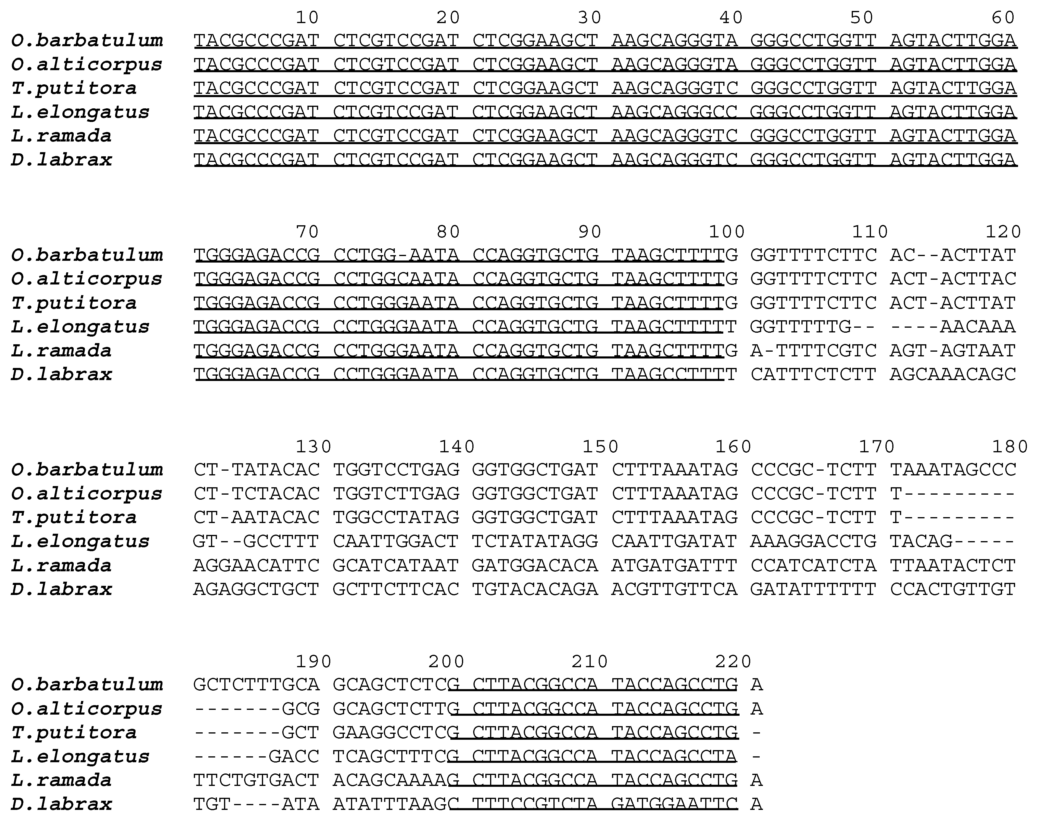
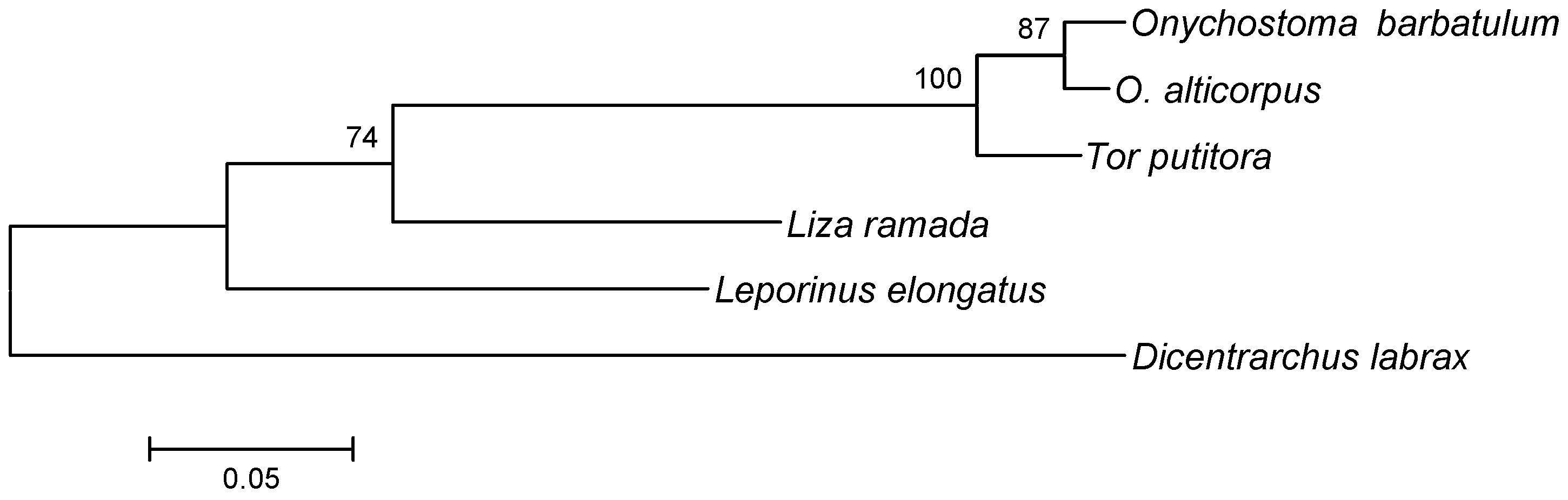
2. Results and Discussion

Cytogenetic studies by Giemsa staining of Onychostoma barbatulum and O. alticorpus indicated that the diploid number for both species was 2n = 50 (Figure 2). The Cyprinidae, which contains more than 2400 species, is the richest family of fish with very divergent chromosome patterns [9,17,18]. The diploid number of 2n = 50 should be a synapomorphic character of cyprinids. In this study, O. barbatulum and O. alticorpus also have the ancestral and presumably primitive characteristic of 2n = 50. O. barbatulum and O. alticorpus had previously been classified Varicorhinus [1] based on external traits of the mouth. However, V. nelspruitensis Gilchrist and Thompson, 1911, V. beso Rüppell, 1836 and V. capoeta sevangi (De Filippi, 1865) are known for a much larger chromosome number (2n = 150) [19,20,21,22] than O. barbatulum and O. alticorpus.
Figure 2. Karyotypes of O. alticorpus (A) and O. barbatulum (B) females after Giemsa staining. Both species show 2n = 50; m = metacentric, sm = submetacentric, st = subtelocentric, t = telocentric.
Ijms 16 26193 g002
In our study, the karyotypic formulae of female O. barbatulum and O. alticorpus were 10 m + 22 sm + 10 st + 8 t with fundamental arm numbers (FN) = 82 and 14 m + 18 sm + 8 st + 10 t with FN = 82, respectively. The previous studies also reported that the karyotypic formulae of O. simum (Sauvage and Dabry de Thiersant, 1874) and O. elongatum (Pellegrin & Chevey, 1934) were 10 m + 16 sm + 16 st + 8 t and 12 m + 12 sm + 14 st + 12 t with FN = 76 and 74, respectively [8,9]. Most of their chromosomes were metacentric (m) and submetacentric (sm), which are the conservative characteristics of Onychostoma.
The 5S rDNA sequences were subcloned from O. barbatulum and O. alticorpus, sequencing a total of 216 (G + C ratio = 51.9%) and 202 (G + C ratio = 52.9%) nucleotides, respectively. Their sequences were aligned to Tor putitora (Hamilton, 1822), Leporinus elongatus Valenciennes, 1850, Liza ramada (Risso, 1827), and Dicentrarchus labrax (Linnaeus, 1758), which were selected randomly from the NCBI GenBank (Available online: http://0-www-ncbi-nlm-nih-gov.brum.beds.ac.uk/genbank/) and have 5S rDNA sequences of high similarity (75%) across orders. In brief, it is known that the 120 bp corresponding to the transcribed region is highly conserved among distant species, while the non-transcribed regions (NTRs) are highly variable (Figure 3).
All these sequences were used to reconstruct the genealogic tree of 5S rDNA. The interspecific Kimura-2-parameter (K-2-P) genetic distance of 5S rDNA sequences between O. barbatulum and O. alticorpus (0.026) was significantly smaller than those (0.052–0.572) of Onychostoma to other referenced sequences in this study. A neighbor-joining analysis with an 87% bootstrapping value showed that O. barbatulum and O. alticorpus had a close relationship (Figure 4).
Two different 5S rDNA probes were prepared separately and individually hybridized with the chromosomes of these two species by FISH, both representing a single pair of signals in each species. It is noteworthy that the 5S rDNA locus of O. barbatulum was found at a pericentromeric region and that of O. alticorpus was found on the subtelomeric position of a pair of submetacentric chromosomes (Figure 5). One pair of 5S rDNA-bearing chromosomes has been presented as the plesiomorphic character state in fishes [23]. Nevertheless, the occurrence of multiple 5S rDNA sites had also been found in a few species, such as Acheilognathus tabira and Cyprinus carpio [24,25].
Figure 3. Aligned 5S rDNA sequences subcloned from Onychostoma barbatulum and O. alticorpus and four reference sequences obtained from NCBI GenBank (with accession numbers): Tor putitora (EU621853), Leporinus elongatus (AF284729), Liza ramada (AM706452), and Dicentrarchus labrax (HM014367). The first 99 bp and the last 21 bp, corresponding to the 5S rDNA transcribed sequences, are underlined.
Ijms 16 26193 g003
Figure 4. Neighbor-joining tree reconstructed based on 5S rDNA sequences from two Onychostoma species and four reference sequences from NCBI GenBank: Tor putitora, Leporinus elongatus, Liza ramada, and Dicentrarchus labrax. The scale bar indicates the nucleotide diversity between sequences. The bootstrapping value of each branch is shown in front of the node.
Ijms 16 26193 g004
Figure 5. Mitotic metaphase plates of (A) Onychostoma barbatulum and (B) O. alticorpus chromosomes after fluorescence in situ hybridization (FISH) with 5S rDNA probes. Arrows indicate the locations of 5S rDNA loci (light blue). Bars equal 5 μm.
Ijms 16 26193 g005
The total length of the Cyt b gene was 1141 bp among 10 cloned sequences of each of the five specimens of O. barbatulum and O. alticorpus as well as in all the reference sequences of the cyprinidae species O. ovale Pellegrin and Chevey, 1936, O. lini (Wu, 1939), O. simum, Acrossocheilus cinctus (Lin, 1931), A. paradoxus (Günther, 1868), Varicorhinus maroccanus (Günther, 1902), V. beso, V. steindachneri Boulenger, 1910, Barbus barbus (Linnaeus, 1758), B. rebeli Koller, 1926, Spinibarbus hollandi Oshima, 1919, and S. denticulatus (Oshima, 1926). The percentages of nucleotide composition did not differ significantly among the five Onychostoma species. Altogether, 234 polymorphic sites encompassing 82 singleton variable sites and 152 parsimony informative sites occurred within 11 different sequences of five Onychostoma species. Four different haplotypes were found in both O. barbatulum and O. alticorpus and their intraspecific nucleotide diversity (Pi) values were 0.0019 and 0.0035, respectively. Intraspecific Kimura-2-parameter genetic distances among different haplotypes ranged from 0.001 to 0.004 for O. barbatulum and from 0.001 to 0.006 for O. alticorpus. The interspecific distances of Onychostoma samples ranged from 0.064 (O. alticorpus vs. O. simum) to 0.142 (O. alticorpus vs. O. barbatulum). Cyt b genes of O. barbatulum and O. alticorpus have very different nucleotide compositions. Numbers of different nucleotides between these two species ranged from 135 (A2/A3 vs. B5) to 144 (A4 vs. B2) (Table 1). A neighbor-joining analysis with 100% bootstrap support showed that specimens of O. barbatulum and O. alticorpus were on two different monophyletic branches of the phylogenetic tree (Figure 6), suggesting that they are genetically different. O. barbatulum and O. alticorpus had both been classified under Varicorhinus [1,3] based on their mouth shape. Therefore, their systematics still remains open to question. In this study, the intergeneric genetic distances between Onychostoma and Varicorhinus spp. ranged from 0.159 (O. alticorpus vs. V. maroccanus) to 0.192 (O. barbatulum vs. V. maroccanus) which is higher than the intrageneric distance of Onychostoma or Varicorhinus (Table 1).
O. barbatulum and O. alticorpus are the only two Onychostoma species presently residing in Taiwan streams. However, these two Onychostoma species are not sister species. In this study, the phylogeny reconstructed using the Cyt b gene sequence assigned O. barbatulum and O. alticorpus to different clades, not sharing a recent common ancestor. O. barbatulum and O. lini have a closer evolutionary relationship with Acrossocheilus sp. than with the other Onychostoma sp., suggesting that the Onychostoma genus is not monophyletic; nevertheless, O. barbatulum and O. alticorpus have similar juvenile body shapes and external morphological characteristics, which are the result of environmental adaptation.
Figure 6. The neighbor-joining evolutionary tree reconstructed based on Cyt b gene sequences from five Onychostoma barbatulum (B1–B5) and five O. alticorpus (A1–A5) from Taiwan and 12 reference sequences from NCBI GenBank (with accession numbers): Acrossocheilus cinctus (JX066772), A. paradoxus (HQ443699), Onychostoma lini (JQ343982), O. ovale (JX074246), O. simum (HM536801), Barbaus barbus (KC465927), B. rebeli (AF090791), Spinibarbus hollandi (NC026129), S. denticulatus (NC021616), Varicorhinus maroccanus (AF287457), V. beso (JQ716388), and V. steindachneri (AF180865). The scale bar indicates the nucleotide diversity between sequences. The bootstrapping value of each branch is shown in front of the node.
Ijms 16 26193 g006
Table 1. Nucleotide numbers, interspecific and intraspecific Kimura-2-parameter genetic distances (gray area) estimated from 10 cytochrome b gene sequences and four reference sequences. Values of genetic distances are decimals, while different nucleotide numbers are integers. Specimens A1–A5 are Onychostoma alticorpus and B1–B5 are O. barbatulum. Reference sequences from NCBI GenBank (with accession numbers) included Osi, O. simum (HM536801); Oov, O. ovale (JX074246); Vma, Varicorhinus maroccanus (AF287457); Vbe, V. beso (JQ716388).
A1A2 and A3A4A5B2B1 and B3B4B5OsiOovVmaVbe
A10.0010.0060.0020.1360.1340.1340.1330.0650.0970.1610.168
A2 and A310.0050.0010.1350.1330.1330.1320.0640.0960.1600.167
A4760.0060.1420.1400.1400.1380.0700.1020.1670.174
A52170.1360.1340.1340.1330.0650.0970.1590.168
B21391381441390.0020.0040.0030.1290.1300.1920.178
B1 and B313713614213720.0020.0010.1270.1280.1910.178
B4137136142137420.0010.1270.1280.1910.179
B51361351411363110.1250.1270.1900.178
Osi706975701321301301290.0950.1660.169
Oov1021011071021331311311301000.1640.172
Vma1621611671601881871871861661640.105
Vbe168167173168177177178177169171109

3. Experimental Section

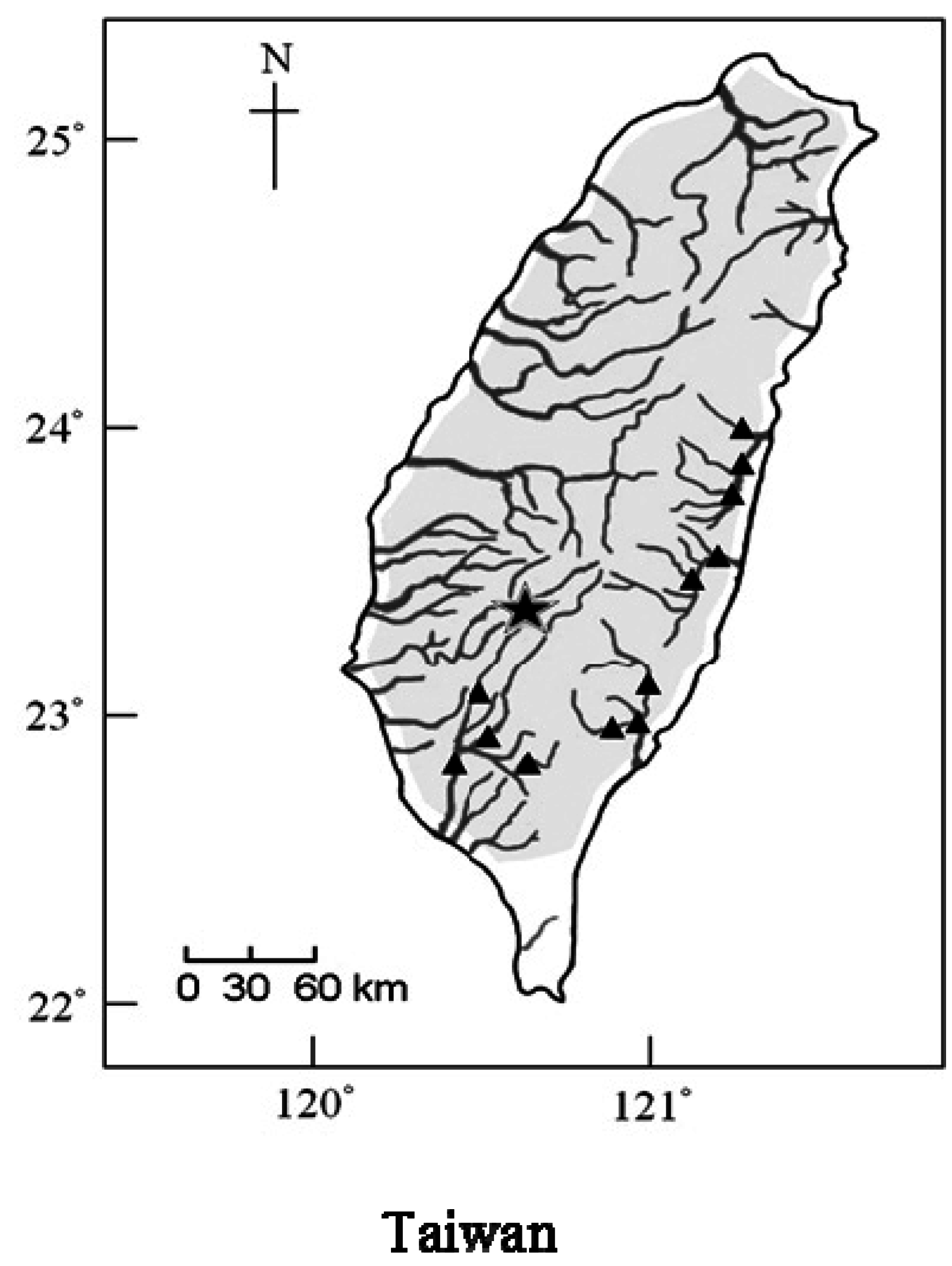
3.1. Fish Collection

Each of the five specimens of O. barbatulum and O. alticorpus were obtained from Nanzihsian Stream, a tributary of the Kaoping River in southwestern Taiwan (Figure 1). Live specimens were transported to the laboratory and maintained in fresh water in 500 L aquaria according to the Guide for the care and use of laboratory animals [26] until being sacrificed.

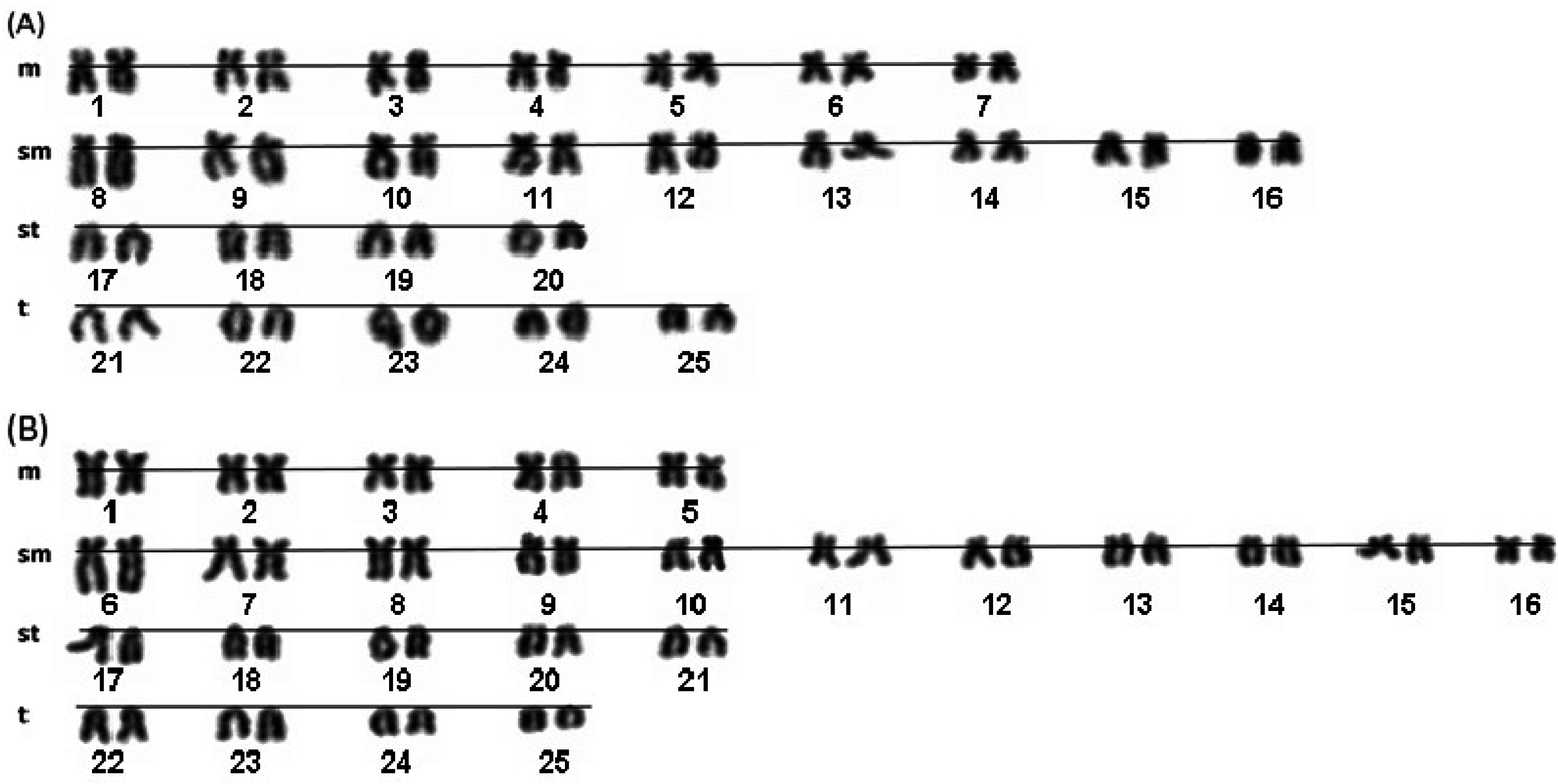
3.2. Chromosome Preparations and Staining

Before the experiment, fish were anesthetized using 140 μg/mL tricaine methanesulphonate (MS222, Sigma-Aldrich, St. Louis, MO, USA). Mitotic chromosomes were prepared from the cephalic kidney of each specimen. Briefly, cells were cultured in Minimum Essential Medium (MEM) with 15% fetal bovine serum (GIBCO, New York, NY, USA) and mitotic inhibitor (0.0001% Colchicine) using a Rota-mixer (Firstek Scientific Co., Taipei, Taiwan) at 100 rpm and room temperature for 2 h. A hypotonic solution (0.075 M KCl) was added to the cells for 30 min at room temperature and then centrifuged (3000 rpm, 5 min). Supernatant was decanted. Cells were fixed in a methanol-acetic acid solution at room temperature for 15 min followed by centrifugation (3000 rpm, 5 min), and supernatant was decanted. The fixation process was repeated thrice. One to two drops of cell suspension were dripped onto hot slides from a height of 10–30 cm and stained in 5% Giemsa solution for 10 min. Ten metaphase plates for each species were observed and photographed under a Leica DM2500 light microscope (Leica Biosystems, Wetzlar, Germany) with a BASLER charge-coupled device (CCD) scA1400-17gmASI camera (Basler AG, Ahrensburg, Germany). Metaphase displaying distinct chromosome morphology was used for preparing karyograms. Chromosome morphology was defined according to Levan et al. [27]. Chromosome numbers of species were counted and manually classified with BandView karyotyping software (ASI, Migdal Haemek, Israel).

3.3. 5S rDNA Purification, Subcloning and Analysis

DNA was extracted from 5 to 10 mg of muscle tissue of one specimen of both O. barbatulum and O. alticorpus using a Puregene Core kit A (Qiagen Sciences, Germantown, MD, USA). The 5S rDNA of the two species was amplified by a polymerase chain reaction (PCR) using 5S forward 5′-TACGCCCGATCTCGTCCGATC-3′ and reverse 5′-CAGGCTGGTATGGCCGTAAGC-3′ primers [23]. Amplification was performed in a Px2 Thermal Cycler (Thermo Fisher Scientific, Waltham, MA, USA). The reaction solution consisted of approximately 5 ng genomic DNA, 50 pmol each of the forward and reverse primers, 25 mM dNTP, 0.05–0.1 mM MgCl2, 10× buffer, and 5 U Taq polymerase (Takara Shuzo, Shiga, Japan) brought up to 125 μL with sterile water. The PCR program included one cycle of 4 min at 94 °C, 35 cycles of 30 s at 94 °C, 30 s at 62 °C, and 1 min at 72 °C, followed by a single further extension of 5 min at 72 °C. We evaluated 10 μL of this product on a 1.0% agarose gel to check the PCR success and confirm product sizes. The remaining PCR products were run on 1.0% agarose gels and purified using a DNA Clean/Extraction kit (GeneMark, Taichung, Taiwan). Purified DNA was subcloned into pGEM-T easy vector (Promega, Madison, WI, USA) and transformed into the Escherichia coli JM109 strain. Plasmid DNA was isolated using a mini plasmid kit (Geneaid, Taichung, Taiwan). Two clones from O. barbatulum and O. alticorpus were sequenced on an Applied Biosystems (ABI, Foster City, CA, USA) automated DNA sequencer ABI3730x1 using a Bigdye sequencing kit (Perkin-Elmer, Wellesley, MA, USA). T7 and SP6 primers were used in the sequencing reaction and the PCR cycle parameters for sequencing were 35 cycles of 30 s at 95 °C, 30 s at 50 °C, and 1 min at 72 °C. The genealogic analysis of 5S rDNA sequences depended on the neighbour-joining (NJ) method [28] and the significance of the clusters was assessed using bootstrap analysis with 1000 replications. The phylogeny analyses were carried out using MEGA software version 4.0 [29]. Several reference sequences, Tor putitora (EU621853), Leporinus elongatus (AF284729), Liza ramada (AM706452), and Dicentrarchus labrax (HM014367), from NCBI were added to this analysis.

3.4. Fluorescence in Situ Hybridization of 5S rDNA Probe

Labeled 5S rDNA probes of the two species were generated using a PCR fluorescein labeling mix kit (Roche, Mannheim, Germany). The reaction solution contained 10× buffer, 4 mM MgCl2, 200 μM dNTP, 1 μM 5S rDNA primers [23], 50 ng plasmid DNA, and 2 U Taq polymerase, and was brought up to 100 μL with sterile water. Fluorescein-labeled probes were purified by ethanol precipitation. Chromosomes were prepared with 100 μg/mL RNase A in 2× SSC buffer at 37 °C for 1 h, and washed thrice in 2× SSC for 5 min. Slides were then quickly immersed in a cold series of ethanol solutions (70%, 95% and 100%) for dehydrating the chromatin. After air-drying, chromosome spreads were done by exposing chromosomes to 0.005% pepsin (Roche, Mannheim, Germany) in 10 mM HCl at 37 °C for 10 min to remove residual proteins, and then washed in phosphate-buffered saline (PBS). Slides were then quickly dehydrated through a cold ethanol series and air-dried. Chromosomes were denatured at 80 °C for 5 min in hybridization buffer (2× SSC, 10% dextran sulfate, and 50% deionized formamide). All slides were placed on ice for 3–5 min prior to the addition of 35 μL hybridization buffer containing 50 ng labeling probe. Hybridization occurred at 37 °C for 12–16 h. Post-hybridization washes were carried out at 42 °C for 15 min in 1× SSC with 50% deionized formamide, followed by 0.1× SSC at 60 °C for 5 min thrice, and then rinsed thrice in PBS buffer with 0.2% Tween 20 at 37 °C for 5 min. Fluorescent signals were amplified using a rabbit anti-FITC antibody (Invitrogen, Carlsbad, CA, USA) which was diluted to 1:200 using the TNB buffer (100 mM Tris-HCl (pH 7.5), 150 mM NaCl, 0.5% Blocking reagent). The antibody solution (100 μL) was added to the slide at 37 °C for 30 min and then washed for 5 min thrice in TNT buffer (100 mM Tris-HCl (pH 7.5), 150 mM NaCl, 0.05% Tween 20) before being transferred to room temperature. A FITC-labeled goat anti-rabbit antibody (Jackson ImmunoResearch Laboratories, West Grove, PA, USA) was diluted to 1:250 using the TNB buffer. Then 100 uL of the antibody solution was added to the slide and incubated at 37 °C for 30 min, and was washed thrice in TNT buffer at room temperature for 5 min. The slide was immersed in 70%–100% ethanol series for dehydrating chromatin. Chromosomes were counterstained with 0.05 μg/mL of 4′,6-diamidino-2-phenylindole (DAPI) and mounted in 1,4-diazabicyclo[2.2.2]octane (DABCO) anti-fading solution (50% glycerol and 2% DABCO in PBS). All slides were observed on a Leica DM 2500 microscope (Leica Microsystems, Wetzlar, Germany) equipped with a CCD camera. FISHView EXPO 4.0 software (Applied Spectral Imaging, Migdal HaEmek, Israel) was used for image analysis.

3.5. Cytochrome b Gene Cloning and Analysis

DNA was extracted from 5 to 10 mg of muscle tissue of each of the five specimens of O. barbatulum and O. alticorpus. The complete Cyt b gene was amplified using the FishcytB-F primer 5′-ACCACCGTTGTTATTCAACTACAAGAAC-3′ and TruccytB-R primer 5′-CCGACTTCCGGATTACAAGACCG-3′ [30]. The reaction solution consisted of approximately 5 ng genomic DNA, 50 pmol each of the forward and reverse primers, 25 mM dNTP, 0.05–0.1 mM MgCl2, 10× buffer, and 5 U Taq polymerase (Takara Shuzo, Shiga, Japan), and brought up to 125 μL with Milli-Q water (Millipore, Billerica, MA, USA). The PCR program included one cycle of 4 min at 95 °C, 38 cycles of 1 min at 94 °C, 50 s at 54 °C, and 1 min at 72 °C, followed by a single further extension of 10 min at 72 °C. We evaluated 10 μL of each product on a 0.8% agarose gel to check PCR success and confirm product sizes. The remaining PCR products were run on 0.8% agarose gels and purified using a DNA Clean/Extraction kit (GeneMark, Taichung, Taiwan). Purified DNA was subcloned into the pGEM-T easy vector (Promega, Madison, WI, USA) and transformed into Escherichia coli JM109 strain. Plasmid DNA was isolated using a mini plasmid kit (Geneaid, Taichung, Taiwan). Different clones from each of five individuals of O. barbatulum and O. alticorpus were sequenced on an Applied Biosystems (ABI, Foster City, CA, USA) automated DNA sequencer ABI3730x1 using a Bigdye sequencing kit (Perkin-Elmer, Wellesley, MA, USA) as mentioned above.
In total, 10 Cyt b sequences were subcloned and deposited in GenBank. Homologous sequences of O. ovale, O. lini, O. simum, Acrossocheilus cinctus, A. paradoxus, Varicorhinus maroccanus, V. beso, V. steindachneri, Barbaus barbus, B. rebeli, Spinibarbus hollandi, and S. denticulatus derived from NCBI (Accession nos. JX074246, JQ343982, HM536801, JX066772, HQ443699, AF287457, JQ716388, AF180865, KC465927, AF090791, NC026129, and NC021616) were selected as reference sequences. All sequences were aligned using ClustalW [31] and then manually checked. Intraspecific and interspecific genetic distances and number of different nucleotides were calculated using MEGA software [29]. Variable site numbers, parsimony informative site numbers among the samples of the two Onychostoma species, and the intraspecies nucleotide diversity of O. alticorpus and O. barbatulum specimens were computed by DnaSP v5 [32]. The phylogenetic trees of Cyt b sequences were constructed using NJ methods [28]. Cluster confidence level was assessed using a bootstrap analysis with 1000 replications.

4. Conclusions

Onychostoma barbatulum and O. alticorpus have identical chromosome numbers 2n = 50, but variable karyotypes. The 5S rDNAs of these two Onychostoma species were cloned, sequenced, and mapped by FISH on metaphase plates. The 5S rDNA sequences showed differences in nucleotide lengths of NTRs with the chromosome location being pericentromeric in O. barbatulum and subtelomeric in O. alticorpus. The Cyt b gene sequences of O. barbatulum and O. alticorpus clearly revealed that these two species are not sister species and the Onychostoma genus is not monophyletic; nevertheless, their morphological similarities may be due to environmental adaptation.

Acknowledgments

The authors express their gratitude to Hung-Du Lin for collecting specimens. We also thank Jyun-Cheng Shih and Yin-Huei Hung for assistance with laboratory work. Research funding was provided to Mei-Chen Tseng by the National Science Council, Taiwan (NCS98-2313-B-020-007-MY3).

Author Contributions

Chiao-Chuan Han assisted sample collection, conducted experimental design and data analyses; Tsair-Bor Yen provided assistance on fluorescence microscopy, chromosome image analysis and quantitative analysis; Nian-Cih Chen performed chromosome preparations and molecular cloning, and Mei-Chen Tseng was responsible for data analyses and execution of this research project.

Conflicts of Interest

The authors declare no conflict of interest.

References

  1. Wang, J.P.; Lin, H.D.; Huang, S.; Pan, C.H.; Chen, X.L.; Chiang, T.Y. Phylogeography of Varicorhinus barbatulus (Cyprinidae) in Taiwan based on nucleotide variation of mtDNA and allozymes. Mol. Phylogenet. Evol. 2004, 31, 1143–1156. [Google Scholar] [CrossRef] [PubMed]
  2. Tzeng, C.S. Distribution of the freshwater fishes of Taiwan. J. Taiwan Mus. 1986, 39, 127–146. [Google Scholar]
  3. Shen, S.C. Fishes of Taiwan; Department of Zoology, National Taiwan University: Taipei, Taiwan, 1993. [Google Scholar]
  4. Han, C.C.; Tew, K.S.; Chen, I.S.; Su, L.Y.; Fang, L.S. Environmental biology of an endemic cyprinid, Varicorhinus alticorpus, in a subtropical mountain stream of Taiwan. Environ. Biol. Fishes 2000, 59, 153–161. [Google Scholar] [CrossRef]
  5. Eichler, E.E.; Sankoff, D. Structural dynamics of eukaryotic chromosome evolution. Science 2003, 301, 793–797. [Google Scholar] [CrossRef] [PubMed]
  6. Foresti de Almeida-Toledo, L.; Daniel-Silva, M.Z.; Moysés, C.B.; Foresti, F. Chromosome variability in Gymnotiformes (Teleostei: Ostariophysi). In Fish Cytogenetics; Pisano, E., Ozouf-Costaz, C., Foresti, F., Kapoor, B.G., Eds.; CRC Press: Boca Raton, FL, USA, 2007; pp. 17–40. [Google Scholar]
  7. Dai, Y. Karyotype and evolution analysis of vulnerable fish Onychostoma lini from China. In Proceedings of the 7th International Conference on Systems Biology, Huangshan, China, 23–25 August 2013.
  8. Li, Y.; Li, K.; Jiang, J.; Sun, Q.; Zhou, T. Studies on the karyotypes of Chinese cyprinid fishes X. Karyotypes of five species of barbinae and four species of gobioninae. Zool. Res. 1986, 7, 183–189. [Google Scholar]
  9. Gui, J.; Li, Y.; Li, K.; Zhou, T. Studies on karyotypes of Chinese cyprinid fishes. VIII. Karyotype analyses of fifteen species of barbines with considerations for their phyletic evolution. Trans. Chin. Ichthyol. Soc. 1986, 5, 119–127. [Google Scholar]
  10. Phillips, R.B. Application of fluorescence in situ hybridization (FISH) to genome mapping in fishes. In Fish Cytogenetics; Pisano, E., Ozouf-Costaz, C., Foresti, F., Kapoor, B.G., Eds.; CRC Press: Boca Raton, FL, USA, 2007; pp. 455–471. [Google Scholar]
  11. Martins, C. Chromosomes and Repetitive DNAs: A contribution to the knowledge of the fish genome. In Fish Cytogenetics; Pisano, E., Ozouf-Costaz, C., Foresti, F., Kapoor, B.G., Eds.; CRC Press: Boca Raton, FL, USA, 2007; pp. 421–432. [Google Scholar]
  12. Wasko, A.P.; Martins, C.; Wright, J.M.; Galetti, P.M., Jr. Molecular organization of 5S rDNA in fishes of the genus Brycon. Genome 2001, 44, 893–902. [Google Scholar] [CrossRef] [PubMed]
  13. Affonso, P.R.A.M.; Galetti, P.M., Jr. Chromosomal diversification of reef fishes from genus Centropyge (Perciformes, Pomacanthidae). Genetica 2005, 123, 227–233. [Google Scholar] [CrossRef] [PubMed]
  14. Van Campenhout, S.; Aert, R.; Volckaert, G. Orthologous DNA sequence variation among 5S ribosomal RNA gene spacer sequences on homologous chromosomes 1B, 1D, and 1R of wheat and rye. Genome 1998, 41, 244–255. [Google Scholar] [CrossRef] [PubMed]
  15. Podlesnykh, A.V.; Apalikova, O.V.; Brykov, V.A. Phylogenetic relationships of silver crucian carp in Carassius auratus complex based on mtDNA analysis. Russian J. Genet. 2012, 48, 1207–1217. [Google Scholar] [CrossRef]
  16. Wali, A.; Ahmad, S.M.; Balkhi, M.H.; Bhat, F.A.; Bhat, B.A.; Darzi, M.M. Genetic diversity of Cyprinus carpio var. communis, Cyprinus carpio var. specularis and Carassius carassius by DNA based markers. Int. J. Aquacul. 2013, 24, 138–146. [Google Scholar]
  17. Shen, J.B.; Fan, Z.T.; Wang, G.R. Karyotype studies of maletriploid Crucian carp (Fangzheng Crucian Carp) in Heilongjiang. J. Genet. Genom. 1983, 10, 133–136. [Google Scholar]
  18. Arai, R. Fish Karyotypes: A Check List; Springer: Tokyo, Japan, 2011; p. 340. [Google Scholar]
  19. Golubtsov, A.S.; Krysanov, E.Y. Karyological study of some cyprinid species from Ethiopia. The ploidy differences between large and small Barbus of Africa. J. Fish Biol. 1993, 42, 445–455. [Google Scholar] [CrossRef]
  20. Krysanov, E.Y.; Golubtsov, A.S. Karyotypes of some Ethiopian Barbus and Varicorhinus from the Nile basin including Lake Tana morphotypes. Folia Zool. 1996, 45, 67–75. [Google Scholar]
  21. Krysanov, E.Y. Karyotypes of Varicorhinus capoeta and Barbus goktschaicus (Cypriniformes) from lake Sevan, Armenia. J. Ichthy. 1999, 39, 262–264. [Google Scholar]
  22. Oellermann, L.K.; Skelton, P.H. Hexaploidy in yellowfish species (Barbus, Pisces, Cyprinidae) from southern Africa. J. Fish Biol. 1990, 37, 105–115. [Google Scholar] [CrossRef]
  23. Martins, C.; Galetti, P.M. Chromosomal localization of 5S rDNA gene in Leporinus fish (Anostomidae, Characiformes). Chrom. Res. 1999, 7, 363–367. [Google Scholar] [CrossRef] [PubMed]
  24. Inafuku, J.; Nabeyama, M.; Kikuma, Y.; Saitoh, J.; Kubota, S.; Kohno, S. Chromosomal location and nucleotide sequences of 5S ribosomal DNA of two cyprinid species (Osteichthyes, Pisces). Chrom. Res. 2000, 8, 193–199. [Google Scholar] [CrossRef] [PubMed]
  25. Martins, C.; Wasko, A.P. Organization and evolution of 5S ribosomal DNA in the fish genome. In Focus on Genome Research; Williams, C.R., Ed.; Nova Science Publishers: Hauppauge, NY, USA, 2004; pp. 289–318. [Google Scholar]
  26. Committee for the Update of the Guide for the Care and Use of Laboratory Animals. Guide Laboratory Animals for the Care and Use of Laboratory Animals, 8th ed.; The National Academies Press: Washington, DC, USA, 2011; p. 248. [Google Scholar]
  27. Levan, A.; Fredgra, K.; Sandberg, A.A. Nomenclature for centromeric position on chromosomes. Hereditas 1964, 52, 201–220. [Google Scholar] [CrossRef]
  28. Nei, M.; Kumar, S. Molecular Evolution and Phylogenetics; Oxford University Press: New York, NY, USA, 2000. [Google Scholar]
  29. Tamura, K.; Dudley, J.; Nei, M.; Kumar, S. MEGA4: Molecular evolutionary genetics analysis (MEGA) software version 4.0. Mol. Biol. Evol. 2007, 24, 1596–1599. [Google Scholar] [CrossRef] [PubMed]
  30. Sevilla, R.G.; Diez, A.; Noren, M.; Mouchel, O.; Jerome, M.; Verrez-Bagnis, V.; van Pelt, H.; Favre-Krey, L.; Krey, G.; Bautista, J.M. Primers and polymerase chain reaction conditions for DNA barcoding teleost fish based on the mitochondrial cytochrome b and nuclear rhodopsin genes. Mol. Ecol. Notes 2007, 7, 730–734. [Google Scholar] [CrossRef]
  31. Thompson, J.D.; Higgins, D.G.; Gibson, T.J. ClustalW: Improving the sensitivity of progressive multiple sequence alignment through sequence weighting, position-specific gap penalties and weight matrix choice. Nucleic Acids Res. 1994, 22, 4673–4680. [Google Scholar] [CrossRef] [PubMed]
  32. Librado, P.; Rozas, J. DnaSP v5: A software for comprehensive analysis of DNA polymorphism data. Bioinformatics 2009, 25, 1451–1452. [Google Scholar] [CrossRef] [PubMed]

