Article

Signaling Modulations of miR-206-3p in Tooth Morphogenesis

1
Department of Biochemistry, School of Dentistry, Kyungpook National University, Daegu 41940, Korea
2
Department of Biochemistry and Cell Biology, Stony Brook University, Stony Brook, NY 11794-5215, USA
3
Department of Oral and Maxillofacial Radiology, School of Dentistry, Kyungpook National University, Daegu 41940, Korea
4
Department of Dental Hygiene, College of Health Science, Gachon University, Incheon 21936, Korea
5
Department of Histology and Developmental Biology, Tokyo Dental College, Tokyo 101-0061, Japan
6
Pre-Major of Cosmetics and Pharmaceutics, Daegu Haany University, Gyeongsan 38610, Korea
*
Authors to whom correspondence should be addressed.
Int. J. Mol. Sci. 2020, 21(15), 5251; https://0-doi-org.brum.beds.ac.uk/10.3390/ijms21155251
Submission received: 24 June 2020 / Revised: 22 July 2020 / Accepted: 22 July 2020 / Published: 24 July 2020
(This article belongs to the Section Molecular Genetics and Genomics)

Abstract

:
MicroRNAs (miRNAs) are a class of naturally occurring small non-coding RNAs that post-transcriptionally regulate gene expression in organisms. Most mammalian miRNAs influence biological processes, including developmental changes, tissue morphogenesis and the maintenance of tissue identity, cell growth, differentiation, apoptosis, and metabolism. The miR-206-3p has been correlated with cancer; however, developmental roles of this miRNA are unclear. In this study, we examined the expression pattern and evaluated the developmental regulation of miR-206-3p during tooth morphogenesis using ex-vivo culture method. The expression pattern of miR-206-3p was examined in the epithelium and mesenchyme of developing tooth germ with stage-specific manners. Perturbation of the expression of miR-206-3p clearly altered expression patterns of dental-development–related signaling molecules, including Axin2, Bmp2, Fgf4, Lef1 and Shh. The gene expression complemented with change in cellular events including, apoptosis and proliferation which caused altered crown and pulp morphogenesis in renal-capsule–calcified teeth. Especially, mislocalization of β-Catenin and SMAD1/5/8 were observed alongside dramatic alterations in the expression patterns of Fgf4 and Shh. Overall, our data suggest that the miR-206-3p regulate the cellular physiology during tooth morphogenesis through modulation of the Wnt, Bmp, Fgf, and Shh signaling pathways to form proper tooth pulp and crown.

1. Introduction

Tooth development is a molecularly and morphologically well-defined process. For decades, gene expression patterns and functions of a range of signaling factors in tooth development have been examined using cells, tissues, and genetically manipulated animals [1]. However, we still do not understand the details of the modulations of the signaling pathways that underlie tooth morphogenesis. The missing link manages the cross-talk among the signaling pathways by modulating the levels of their dental-development–related components, and is known as the key factors for tooth development. It includes paracrine and transcription factors, and needs to be elucidated to understand the detailed molecular mechanisms underlying tooth development and regeneration.
Tooth development progresses through the interactions of dental epithelium and neural-crest–derived mesenchyme, and is mediated by multiple members of the Bmp, Fgf, Shh, and Wnt signaling pathways [1]. These pathways are known to be regulated by activators, inhibitors and mediators for proper tooth morphogenesis [1,2]. Particularly, Wnt signaling components, including Wnt ligands, receptors, transducers, transcription factors, and antagonists are expressed in the dental epithelium and mesenchyme during tooth development in humans and mice [3,4]. In dental epithelium, Wnt3, Wnt4, Wnt6, Wnt7b, and Wnt10b are expressed, whereas Wnt5a is expressed in the mesenchyme [4] in both mice and humans [3]. During tooth development, the canonical Wnt signaling pathway is activated at multiple stages of tooth morphogenesis, [5]. Modulation of the Wnt signaling results in various tooth phenotypes including, multiple tooth formation, shape changes, developmental arrest, and thin incisors [5]. The activation of the Wnt signaling component β-catenin in the epithelium and mesenchyme results in continuous tooth generation [5], and inhibits the formation of posterior molars [6], respectively. Similarly, other paracrine and transcription factors, including FGF4, LEF1, and BMP4 are involved in the critical stages of tooth development, and perturbation of these factors results in altered proliferation, apoptosis, differentiation or migration of cells, leading to developmental defects, such as tooth agenesis, macrodontia, microdontia, oligodontia, and enamel and dentine defects [7]. However, tooth development has not been sufficiently described and understood only with these signaling molecules, including paracrine and transcription factors, to understand the detailed molecular mechanisms underlying tooth morphogenesis. It is necessary to reveal the fine-tuned signaling regulations with the specific modulators, which would mediate the proper tooth morphogenesis for understanding the precise mechanisms of tooth morphogenesis, especially crown morphogenesis.
MicroRNAs (miRNAs) are 19–25-nt non-coding single-stranded RNAs that modulate gene expression at the post-transcriptional level, and this regulatory mechanism is believed to be important for fine-tuning the cross talk among signaling pathways during development. The developmental roles of miRNA has been demonstrated in various ectodermal organs like skin, hair and teeth [8,9,10]. Deletion of Dicer1 in the mesenchyme results in enamel-free incisors and cuspless molars [11,12,13]. Similarly, miRNA 135a, miR-224, miR-145 and miR-143 have been reported to regulate the Bmp signaling [14], enamel mineralization [13,15], and odontoblast differentiation [16], respectively. In swine, ssc-mir-133b regulates apoptosis [17] during tooth development. These previous observations suggest that miRNAs would be fine modulators for playing important roles in signaling regulations during tooth morphogenesis in mammals.
In this study, we selected one of miRNAs, miR-206-3p, which shows specific expression pattern in developing tooth germ and skin, and also have role to inhibit adipogenesis [18], but not yet evaluated its developmental function in tooth morphogenesis. In in vitro cell cultivation, miR-206-3p inhibits adipogenesis of adipocyte through silencing c-Met and inactivating the PI3K/Akt signaling pathway [18]. Moreover, PI3K/Akt signaling pathway converge with GSK3β and β-catenin of Wnt signaling [19] suggesting that miR-206-3p can modulate Wnt signaling in other model systems including, tooth development. Here, we investigated the precise developmental roles of miR-206-3p in modulation of the Wnt signaling, which alters the cross-talks among other signaling pathways during tooth crown and pulp morphogenesis. Therefore, this study aimed to illuminate the modulating roles of miR-206-3p in tooth development and broaden our understanding of how miRNAs coordinate developmental signaling pathways during organogenesis.

2. Results

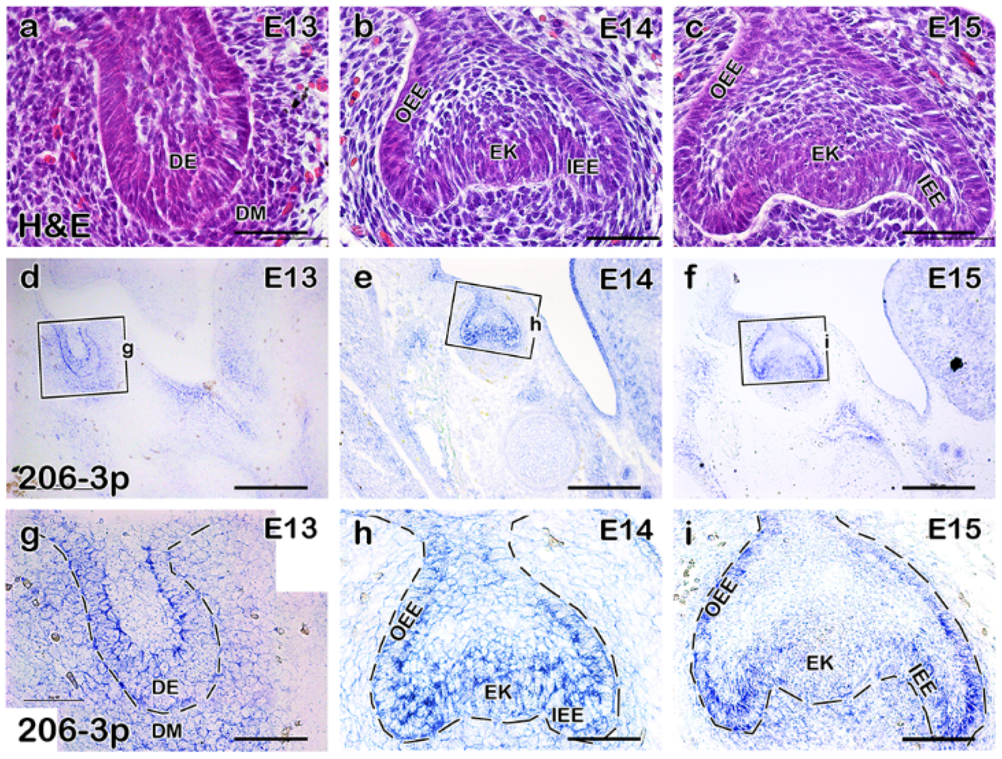
2.1. miR-206-3p Is Moderately Expressed in the Developing Molar

Although specific molecular signaling pathways for tooth development have been described, role of miRNA in tooth development is limited. Therefore, we aimed to identify and determine the developmental roles of miRNA in developing mouse molar. Our pilot screening for miRNA from developing mouse molar at E14 suggested that miR-206-3p is highly expressed in developing tooth which led us to select the miR-206-3p for further evaluation. The bud-stage (Figure 1a) and cap-stage (Figure 1b,c) developing teeth of E13, and E14/E15 mice, respectively, were used to understand the detailed expression of miR-206-3p. Section in situ hybridization using miRCURY LNATM miRNA Custom Detection Probe for miR-206-3p was used to detect the expression of miR-206-3p in the developing tooth at the bud and cap stages (Figure 1d–i).
The expression of miR-206-3p was specifically detected in the dental epithelium and condensed mesenchyme at E13 (Figure 1d,g). The expression was more restricted and punctate in the epithelium, including IEE (inner enamel epithelium), OEE (outer enamel epithelium), and EK (enamel knot) at E14 (Figure 1e,h). At the late cap stage (E15), the expression was observed in the IEE and OEE (Figure 1f–i). MicroRNA miR-206-3p was also broadly expressed in the dental mesenchyme at E14 and E15 (Figure 1e,f,i,j). IEE and dental mesenchyme are important for hard tissue morphogenesis, including, enamel, and dentine, respectively, as well as dental pulp morphogenesis [20], suggesting that the expression patterns of miR-206-3p in developing tooth may provide insight into crown and pulp morphogenesis.

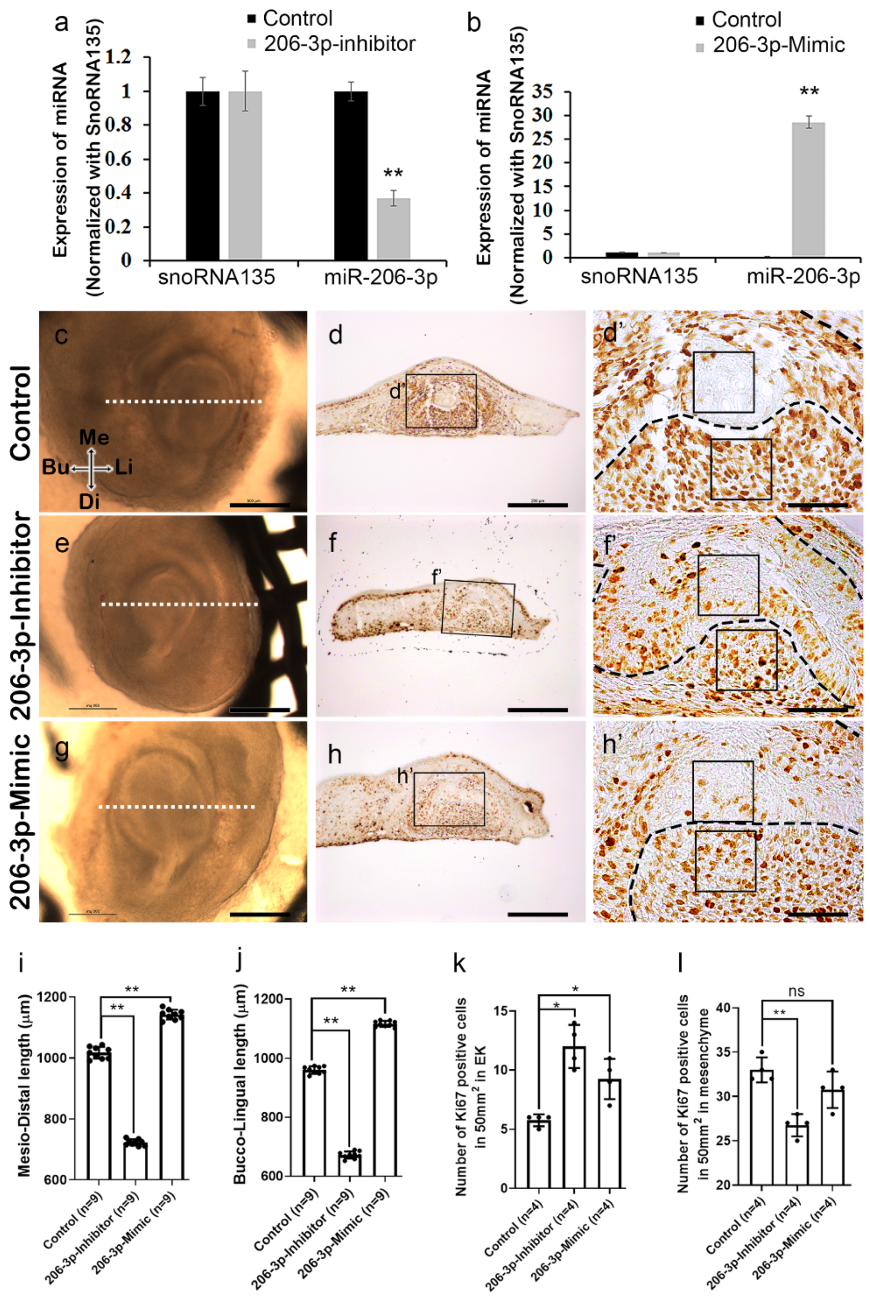
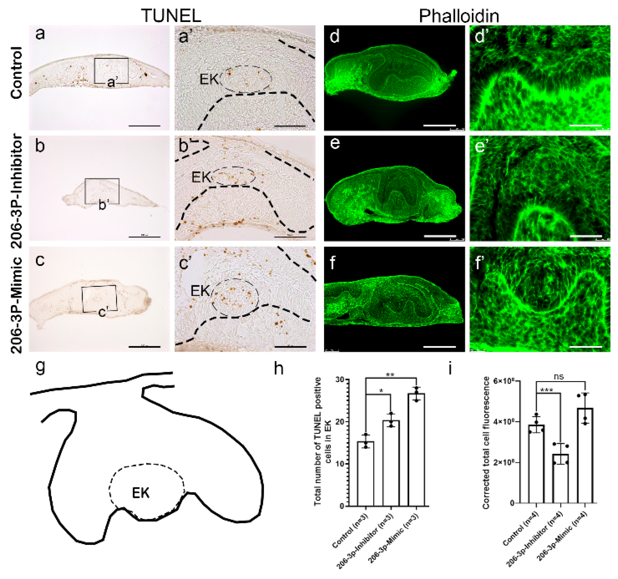
2.2. miR-206-3p Regulates Cell Dynamics Ex Vivo

The inhibitor against miR-206-3p, and mimic for miR-206-3p were used, respectively to knockdown and increase the function of miR-206-3p during in vitro organ cultivation prior to cap stage morphogenesis at E13 (Figure 2a,b). A 36 h cultivation at E13 with the miR-206-3p inhibitor and mimic, the expression level of the miR-206-3p was evaluated using qPCR (Figure 2a,b), which showed significant decrease, and increase of miR-206-3p expression, respectively. When developing teeth at E13 were cultivated for 2 days, the control specimen (n = 9) showed proper tooth size and structure after the cultivation (Figure 2c). The inhibitor-and mimic treated specimen (n = 9) showed decreased and increased tooth size respectively in both mesio-distally and bucco-lingually (Figure 2e,g,i–j), suggesting that there is alteration in, either cell proliferation or apoptosis. The inhibitor and mimic were fluorescently labelled to monitor the transfections (Figure S1). Epithelial morphogenesis in mouse molar relies on asymmetrical growth, differential cell proliferation, and dynamic cell rearrangement [21,22,23,24] that ensure proper crown and pulp development [25]. In control (n = 4), cellular proliferation was observed mostly in the epithelium and mesenchyme, except the EK (Figure 2d–d’,k–l). In contrast, the inhibitor and mimic treated specimen (n = 4) showed increased cell proliferation in the EK (Figure 2f–f’,h–h’, k). In addition, inhibitor treated specimen (n = 4) showed reduced cell proliferation in the mesenchyme (Figure 2f–f’,l). On the other hand, mimic treated specimen showed no significant proliferation in the mesenchyme (Figure 2h–h’,l).
These observations of cell proliferation suggested that decrease in tooth germ size after treatment with inhibitor is directly related to the decreased mesenchymal cell proliferation. The increased cell proliferation in EK of both the inhibitor and mimic treated specimen suggest that EK morphogenesis is altered which affects the EK signaling, ultimately affecting crown morphogenesis. However, increase in tooth size even after not significant increase in tooth size with mimic treatment implies that cell adhesion and actin remodelling may have impacted.
Meanwhile, epithelial and mesenchymal apoptosis increased after inhibition and or mimicking the miR-206-3p (n = 3) during in vitro organ cultivation (Figure 3b–b’,c–c’), compared to the control (Figure 3a–a’) (n = 3). In particular, apoptosis increased in the EK after treatment with inhibitor and mimic (Figure 3b’,c’,h), which further suggests that miR-206-3p have important role in EK morphogenesis that ultimately leads to crown morphogenesis. The final crown shape is determined by epithelial invagination and requires the changes in the cell motility and cell adhesion regulated by RAC1 and RHO-A [26], and can be evaluated by actin staining. The actin staining in the IEE (determined by phalloidin staining) was decreased after inhibiting miR-206-3p (Figure 3e–e’,i) compared with that in the control (Figure 3d–d’,i) (n = 4). Conversely, the actin staining intensity was not altered significantly in the IEE of mimic-treated specimens (Figure 3f–f’,i) (n = 4). At the same time, E-cadherin, an epithelial cell adhesion marker was reduced in the tooth epithelium after mimic treatment (Figure S2b) suggesting that miR-206-3p also regulate cell adhesion.

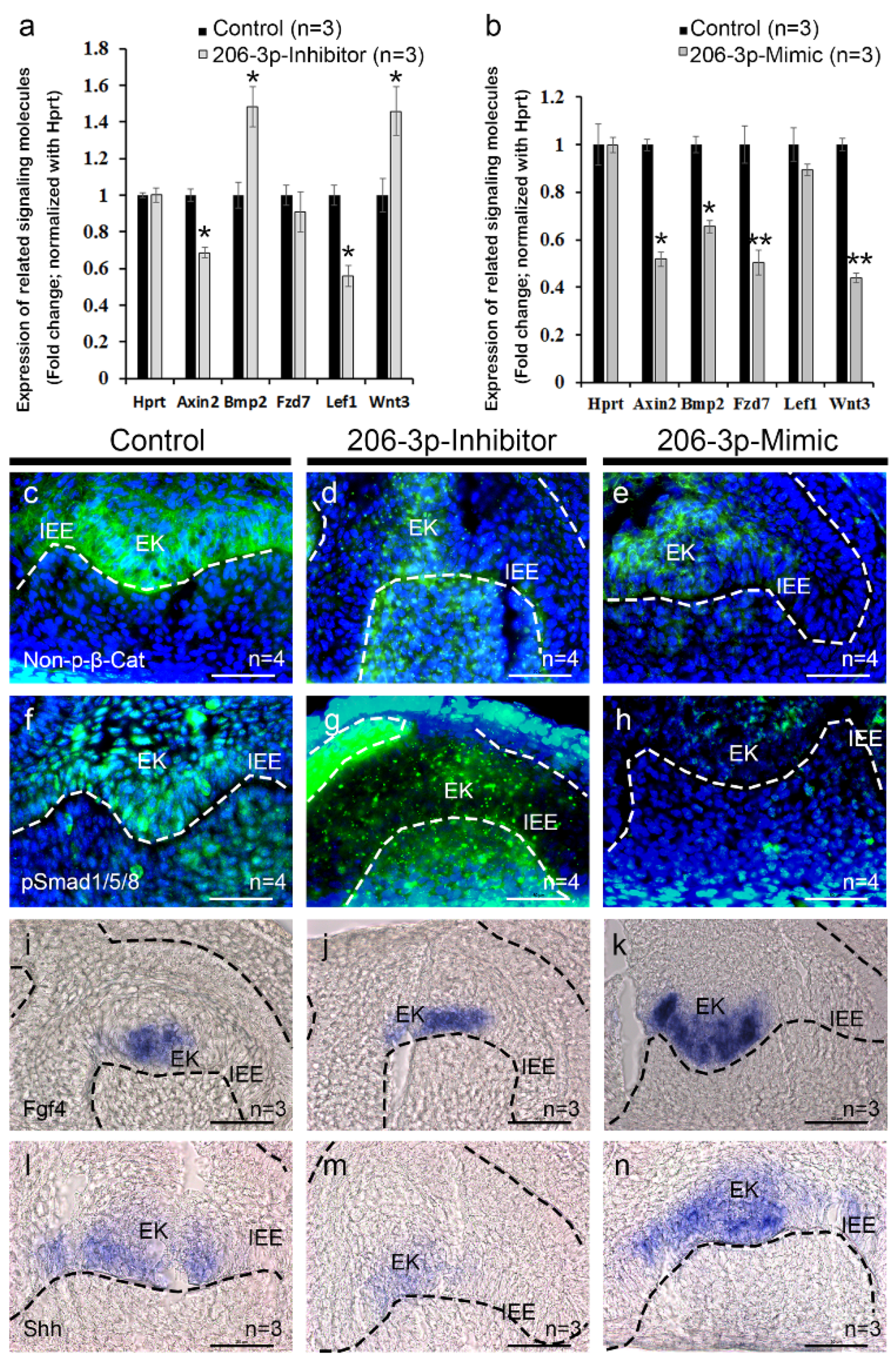
2.3. miR-206-3p Modulates Tooth Specific Signaling

According to the target database TargetScan [27], miR-206-3p is broadly conserved among vertebrates and predicted to target Wnt3 and Fzd7 (Figure S3), important components of the canonical Wnt signaling and are also expressed during tooth development [4]. After successful transfection, expression levels of the genes related to tooth development were examined by qPCR (Figure 4a,b). The qPCR results showed that the Wnt3 level was increased upon inhibition of miR-206-3p (Figure 4a). Meanwhile, the Lef1 and Axin2 levels were downregulated, and the Bmp2 level was upregulated, whereas the Fzd7 level was not significantly altered (Figure 4a). However, after treatment with miR-206-3p mimic, the Wnt3, Fzd7, Bmp2, and Axin2 levels were significantly down-regulated (Figure 4b). To understand the altered expression patterns of signaling molecules in detail, we examined the localization of the proteins involved in the Wnt and Bmp signaling pathways, including active β-catenin and pSMAD1/5/8 in the cultivated teeth (Figure 4c–h).
The β-catenin is reported to have role in tooth development from early placode to late bell stage [5] and tooth root formation [28] and nuclear β-catenin is used as an alternate readout for Wnt signaling activity [29]. In control (n = 4), the β-catenin was localized both in the nuclear and cytoplasmic regions of EK and adjacent cells in the mesenchyme (Figure 4c). In contrast, the protein level decreased, and its localization patterns were impaired in the IEE and EK after inhibiting miR-206-3p (Figure 4d) (n = 4), compared with those in the control (Figure 4c). However, a higher nuclear and cytoplasmic mesenchymal β-catenin level was observed (Figure 4d). Meanwhile, the mimic-treated specimen (n = 4) showed lower β-catenin levels in the IEE and EK when compared with the control (Figure 4e). The altered β-catenin localization in tooth suggests that Wnt signaling is affected, which can have further effect on other signaling regulation.
During tooth development, Wnt and BMP signaling crosstalk in regulating the enamel knot morphogenesis and subsequent induction of BMP signaling in the mesenchyme [30]. Moreover, BMP signaling is transmitted through SMAD1 and 5 which are localized within the IEE and cranial neural crest derived dental mesenchyme at the cap and bell stage and regulate epithelial mesenchymal interaction during tooth morphogenesis [31]. To understand whether BMP signaling is altered after inhibition and mimicking the miR-206-3p, we used pSMAD1/5/8 immunohistochemistry. The epithelial and mesenchymal pSMAD1/5/8 levels were reduced, and the localization patterns were impaired after inhibiting miR-206-3p (n = 4) during the in vitro organ cultivation (Figure 4g), compared with those in the control (Figure 4f) (n = 4). The mimic-treated specimen showed reduced pSMAD1/5/8 levels in both the epithelium and mesenchyme (Figure 4h) (n = 4).
To evaluate the role of miR-206-3p further in the morphogenesis of the enamel knot, we examined the expression of Fgf4 and Shh by whole-mount or cryo-section in situ hybridization (Figure S4, Figure 4i–n). Fgf4 was expressed within a restricted area in the EK of the control specimen (Figure 3i, Figure S4a) (n = 3). However, the expression expanded in the IEE after inhibiting miR-206-3p (Figure 4j, Figure S4b) (n = 3). The mimic-treated specimen (n = 3) increased and expanded the expression of Fgf4 in the EK (Figure 4k, Figure S4c). Similarly, the Shh level was downregulated after the inhibition of miR-206-3p (Figure 4m, Figure S4e) relative to that in the control (Figure 4l, Figure S4d) but was upregulated in the mimic-treated specimens (Figure 4n, Figure S4f) (n = 3).

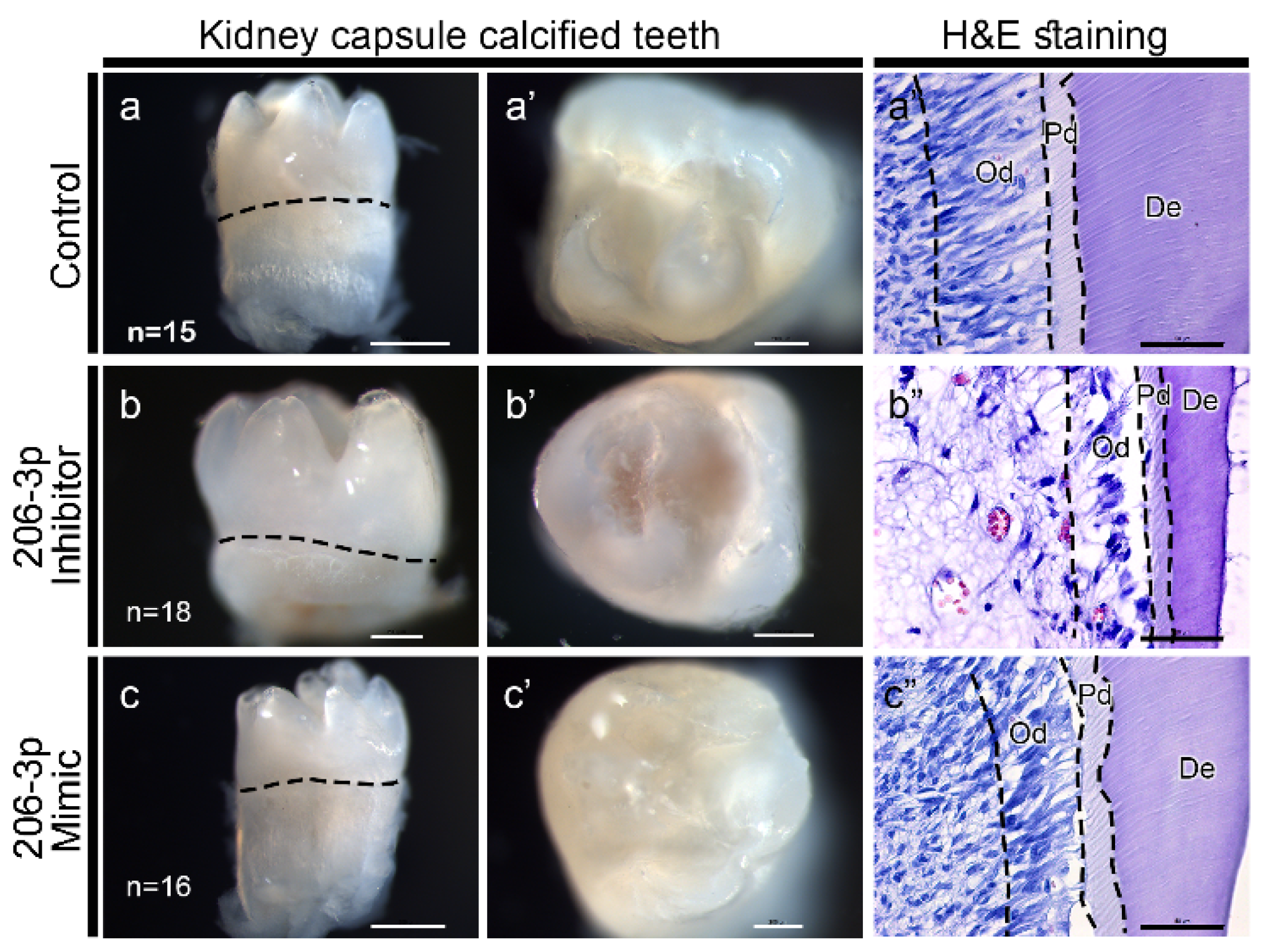
2.4. miR-206-3p Regulates Tooth Pulp Morphogenesis

To identify the role of miR-206-3p in tooth development, we examined renal-capsule–calcified teeth (Figure 5a–c). The inhibitor-treated teeth (n = 18) were very small after 21 days of calcification in the renal capsule (Figure 5b–b’, Figure S5b) relative to the control (n = 15) or mimic-treated teeth (n = 16) (Figure 5a–a’,c–c’, Figure S5a,c). The histological examination of the calcified teeth showed a properly arranged odontoblast layer, predentin, and dentin in the control specimen (Figure 5a”, Figure S5a). Meanwhile, the inhibitor-treated specimen decreased in cell mass in the pulp region, and had a poorly differentiated and developed odontoblast layer with thinner predentin and dentin layers, relative to those in the control (Figure 5b”, Figure S5b). However, the mimic-treated specimen showed a proper amount of cell mass in the dental pulp, but the arrangement of the odontoblast layer was irregular and also showed irregular predentin and dentin layers (Figure 5c”, Figure S5c). These observations suggest that signaling modulation, mediated by miR-206-3p, is crucial for tooth crown and pulp morphogenesis.

3. Discussion

Signaling regulation mediated by epithelial-mesenchymal interactions during tooth development is crucial to form optimal tooth size with proper crown and pulp development [30]. The Wnt is one of the important signaling in tooth development and has been extensively studied [5]. Here, we report that miR-206-3p in tooth development primarily regulates Wnt signaling that mediate other signaling such as BMP, SHH and FGF for proper morphogenesis of tooth crown and pulp.
The stage-specific expression pattern of miR-206-3p in the dental epithelium and mesenchyme at the bud stage, and restricted expression in the epithelium at the later stages of tooth development coincided with the expression patterns of other signaling factors, including β-catenin [5], Lef1 [7], Pitx2 [32] and Wnt3 [4]. This coincided expression pattern suggested that miR-206-3p would have major roles in modulating signaling factors related with tooth development. Accordingly, we examined the precise role of miR-206-3p in tooth development by employing in vitro organ cultivation of embryonic tooth using an inhibitor or mimic of miR-206-3p.
In this research we found that miR-206-3p controls tooth size by regulating the cell dynamics including cell adhesion, proliferation, apoptosis and cytoskeletal remodelling. Our results showed that cell proliferation still remained in the EK after inhibiting and mimicking the miR-206-3p, suggesting that there is decrease in cell adhesion in the epithelium, which later affects the signaling between IEE and dental papilla. At the same time, apoptosis increased in the EK after inhibiting and mimicking the miR-206-3p, which seems unusual because proliferation is also persistent in the EK. These data suggest that tooth EK tends to acquire the proper tooth morphology by limiting the numbers of proliferative cells in the EK. In zebrafish study, both the overexpression and knockdown of miR206 impaired the actin filament formation during gastrulation [33]. Moreover, developing tooth maintains its morphology through actin fibers, regulated by Rho-associated protein kinase (ROCK) and important for proper ameloblast and odontoblast formation [34]. The inhibition of miR-206-3p reduced the actin reorganization suggesting that a more pronounced changes results in the tooth crown and pulp formation. As reported in a previous study, the mesenchymal cell number is an important factor for tooth size determination [22], which suggest that, in our study, a reduced number of proliferative cells in the mesenchyme after treatment with inhibitor, also contributed to significant reduction of tooth size (Figure 2e). Meanwhile, reduced localization of E-cadherin in the tooth organ (Figure S2) might be responsible for increased size of tooth germ in the mimic treated specimen, due to reduced adhesion as the reduction in E-cadherin is associated with the loss of contact inhibition and increased cell motility and proliferation [35]. Our observations provide strong evidence for the involvement of miR-206-3p in EK morphogenesis and regulation of gene expression, as previously reported [36].
Epithelial-mesenchymal interactions are important throughout tooth development [37]. Moreover, previous reports have implicated that EK serves as the primary signaling center during tooth development [38], and altered EK affects the functional structure of the tooth, particularly the cusp pattern [39]. Shh expressed in the epithelium regulates Bmp s in the mesenchyme, which regulates Dspp during terminal differentiation of odontoblasts [40,41]. Loss of Lef1 causes arrested tooth development at bud stage, loss of expression of LEF1/β- catenin target gene Fgf4 and failure of survival of dental epithelial cells [7,42]. Bmp2 is required for odontoblast differentiation and pulp vasculogenesis [43]. Beta catenin enhances odontoblastic differentiation in pulp cells through regulation of Runx2 [44]. Inhibition of miR-206-3p results in reduced expression of Lef1 and Axin2, and increased the expression of Bmp2 and Wnt3 suggesting the crosstalk of Bmp and Wnt signalings. miR-206-3p regulates Wnt signaling, as previously predicted by the TargetScan database [27], further suggesting that loss of miR-206-3p regulate Bmp signaling indirectly via Wnt signaling. Meanwhile, expression pattern of β-catenin, the mediator of the Wnt signalling, was upregulated after the inhibition of miR-206-3p and would alter the fate of tooth [45].
Conversely, we found that, Axin2, Bmp2, Fzd7 and Wnt3 were downregulated in mimic-treated specimens and showed disrupted localization patterns of β-catenin and pSMAD1/5/8 further suggests that miR-206-3p also regulates the Bmp signaling through the regulation of the Wnt signaling. Down-regulation of Axin2 both in the inhibitor and mimic treated specimen suggested that Axin2 functions autonomously in treated tissues not working as a negative feedback regulator of Wnt signaling. Further, the levels of Fgf4 and Shh, two important signaling factors for tooth development [46,47], were altered in the EK and IEE, presumably contributing to the altered morphogenesis of EK and overall tooth structure. These results suggested that miR-206-3p regulated tooth development through the Wnt, Bmp, and Shh signaling pathways. However, further analyses using Wnt antagonists and activators are needed for confirmation.
Calcified teeth by renal capsule transplantation revealed that miR-206-3p was indispensable during tooth development. It has previously been reported that the Shh signaling in the epithelium signals the mesenchyme to induce the Bmp signaling during the differentiation stages [43,48]. This observation suggests that differentiation during tooth development was altered after treatment with the miR-206-3p inhibitor or mimic in the current study. Moreover, extra-folding of IEE, altered ameloblast differentiation, and defective enamel formation in the Dicer-1 conditional knockout mouse suggests that fine-tuning of gene expression is necessary for proper hard-tissue formation [49]. In addition, other recent studies have suggested that miRNA 200c/141 knockout mice are defective in enamel formation through changes in the Bmp signaling [50]. In this study, altered epithelial and mesenchymal Wnt signaling upon modulation of the miR-206-3p levels resulted in changes in the downstream signaling during tooth development, leading to altered tooth formation. Thus, miR-206-3p appears to be a pivotal factor regulating tooth formation by targeting the Wnt signaling.
Based on our results, we conclude that miRNA–mediated modulation of the Wnt signaling is necessary for proper tooth morphogenesis through tight regulation of tooth developmental processes. Disruption of miRNA balance would lead to significant alterations in tooth morphology including tooth size and hard tissue matrices formation. A better understanding of the fine-tuning of tooth developmental signaling pathways, through single miRNA modulation during tooth development is needed to understand the etiopathology of dental defects in humans.

4. Materials and Methods

All experimental protocols were approved by the Kyungpook National University, School of Dentistry, Animal Care and Use Committee (KNU-2012-42, 3 April 2012) and performed according to the ARRIVE guidelines for the care and use of laboratory.

4.1. Animals

Adult ICR (Institute for Cancer Research) mice were housed in a temperature-controlled room (22 °C) under artificial illumination (lights on from 05:00 to 17:00) and 55% relative humidity with access to food and water ad libitum. The mouse embryos were obtained from time-mated pregnant mice. The day on which a vaginal plug was confirmed were designated as embryonic day 0 (E0). The embryos at E13, E14, and E15 were used.
All experiments described in this study were performed for 3 or more times, independently.

4.2. In Situ Hybridization

For miRNA in situ hybridizations, a 5’- and 3’- DIG-labeled miRCURY LNATM miRNA custom detection probe at 80 nM final concentration was hybridized at 54 °C using the miRNA ISH buffer set (Exiqon, Skelstedet, Denmark, cat no. 90000) and processed according to the manufacturer’s instructions. The miRCURY LNATM miRNA custom detection probe for miR-206-3p (/5’DigN/CCACACACTTCCTTACATTCCA/3’Dig_N/) was obtained from Qiagen (Qiagen, Hilden, Germany, cat. no. 339115 YCD0072334-BCG). Whole-mount or section in situ hybridizations were performed, as previously described [51], using standard protocols in RNAse free conditions. Briefly, for section in situ hybridization, 7 µm thick tissue sections were deparaffinised and rehydrated in PBS and treated with proteinase K for 15 min. The sections were postfixed in 4% PFA and processed for acetylation using mixture of acetic acid anhydride and triethanolamine. For whole mount in situ hybridization, the tissues were rehydrated using methanol and treated with proteinase K. The tissue were then postfixed with mixture of glutraldehyde and paraformaldehyde and prehybridized in hybridization solutions. For both hybridizations, digoxigenin (DIG)-labeled antisense RNA probes were pre-warmed to 80 °C and hybridized overnight at 62 °C. After whole-mount in situ hybridization, 20-μm thick frontal frozen sections were prepared to examine the detailed expression patterns.

4.3. In Vitro Organ Cultivation and Renal Capsule Transplantations

The embryonic mice molar tooth buds were micro-dissected out from the lower jaws of E13 mice in PBS under a stereo-microscope. The tooth buds were cultured in DMEM (HyClone, Logan, UT, USA; cat. no.-SH30243.01) with 10% fetal bovine serum (Hyclone, Logan, UT, USA) and antibiotics using a modified Trowell’s culture method for a two days, as previously described [52]. During the cultivation, the tooth germs were transfected with 400 nM of the inhibitor or mimic of the miR-206-3p as previously described [53]. The inhibitor and mimic were purchased from Qiagen (Qiagen, Hilden, Germany, miCURY LNATM miRNA Power Inhibitor, cat no. 339131 YI04100975-DDB; miCURY LNATM miRNA Mimic, cat no. 339173 YM00472240-ADB). The cultivated tooth germs were transplanted into the renal subcapsular layer of the adult male mice as previously described [22]. After 3 weeks, the host mice were sacrificed, and kidneys were dissected out to obtain the calcified teeth.

4.4. Histology and Immunohistochemistry

Immunostaining and routine histological analyses by H and E staining were carried out, as previously described [51]. Calcified teeth were sectioned following decalcification using 0.5 M EDTA for three weeks. For immunostaining, sections were first rehydrated and then processed for antigen retrieval. Blocking was performed by incubating the sections in the 1X western blocking solution (Roche, Mannheim, Germany; Ref. 11921673001) for one hour at room temperature. The primary antibodies used in this study were directed against Ki67 (Thermo Scientific, Waltham, MA, USA, cat. no. RM-9106-s), non-phospho β-catenin (Cell Signaling Technology, Danvers, MA, USA, cat. no. 8814S), and pSMAD (Cell Signaling Technology, Danvers, MA, USA; cat. no. 9511S). The secondary antibodies used in this study were biotinylated goat anti-rabbit IgG (Invitrogen, Waltham, MA, USA) and goat anti-rabbit IgG Flamma 488 (BioActs, Incheon, Korea, cat. no. RSA1241).

4.5. TUNEL Assay

TUNEL assay was performed as previously described [54] using an in situ cell apoptosis detection kit (Trevigen, Gaithersburg, MD, USA, cat. no. 4810-30-K) according to the manufacturer’s instructions.

4.6. Phalloidin Staining

Phalloidin staining was performed as previously described [54]. Frozen sections were incubated with phalloidin-fluorescein isothiocyanate (Sigma-Aldrich, St. Louis, MO, USA, Cat no. p5282; Sigma) at room temperature for 1 h, and then visualized under a fluorescence microscope DM2500 (Leica, Wetzlar, Germany).

4.7. Quantitative PCR (qPCR)

RNA was extracted from cultivated tooth germs using the RNeasy micro Kit (Qiagen, Hilden, Germany, cat. no. 74004), and qPCR was carried out, as previously described [51]. For miRNA qPCR, RNA was extracted from the cultivated teeth using the miRNeasy Micro Kit (Qiagen, Hilden, Germany, cat. no. 217084), and cDNA was prepared using the miScript II RT Kit (Qiagen, Hilden, Germany, cat. no. 218161). The primers for miRNA qPCR were designed using miRprimer [55]. The mRNA and the miRNA qPCR results for each sample were normalized to those of Hprt and snoRNA135, respectively. The data are expressed as means ± standard deviations (SDs). The mean expression levels were compared between the experimental and control groups using Student’s t-test. p-values < 0.05 were considered significant. The primers used in this study are presented in Table 1. This experiment is repeated for 3 independent biological samples.

Supplementary Materials

The following are available online at https://0-www-mdpi-com.brum.beds.ac.uk/1422-0067/21/15/5251/s1. Figure S1. Evaluation of the efficiency of inhibitor/mimic transfection during in vitro organ cultivation. Figure S2. Localization of E-Cadherin in E13+2 day teeth. Figure S3. The putative binding sites of mmu-miR-206-3P are broadly conserved among the Wnt3 and Fzd7 3’ UTRs of vertebrates. Figure S4. Whole-mount in situ hybridization. Figure S5. Lower magnification of H&E-stained histological sections of the renal-capsule–calcified teeth.

Author Contributions

S.N.: Conceptualization, methodology, investigation, visualization, formal analysis, writing-original draft, funding acquisition; Y.P.A.: Resources, investigation, visualization, writing review and editing; T.-Y.K.: Resources, investigation, visualization; C.-Y.Y.: Resources, investigation, visualization; C.-H.A.: Validation, resources, supervision, writing review and editing; J.-Y.K. (Ji-Youn Kim): Validation, resources, formal analysis, writing review and editing; H.Y.: Validation, resources, formal analysis, writing review and editing Y.L.: Validation, resources, supervision, writing review and editing; W.-J.S.: Validation, resources, supervision, writing review and editing; J.-Y.K. (Jae-Young Kim): Conceptualization, methodology, investigation, visualization, formal analysis, writing-original draft, supervision, project administration, funding acquisition. All authors have read and agreed to the published version of the manuscript

Funding

This study was supported by the National Research Foundation of Korea (NRF) grant funded by the Korean government (MEST) (NRF-2017R1D1A1B03034378, NRF-2017R1A5A2015391, NRF-2018R1A2A3075600 and NRF-2018R1D1A1A02086098).

Conflicts of Interest

The authors declare no conflict of interest. The funders had no role in the design of the study; in the collection, analyses, or interpretation of data; in the writing of the manuscript, or in the decision to publish the results.

References

  1. Balic, A.; Thesleff, I. Tissue Interactions Regulating Tooth Development and Renewal. Curr. Top. Dev. Biol. 2015, 115, 157–186. [Google Scholar] [CrossRef] [PubMed]
  2. Aurrekoetxea, M.; Irastorza, I.; García-Gallastegui, P.; Jiménez-Rojo, L.; Nakamura, T.; Yamada, Y.; Ibarretxe, G.; Unda, F.J. Wnt/β-Catenin Regulates the Activity of Epiprofin/Sp6, SHH, FGF, and BMP to Coordinate the Stages of Odontogenesis. Front. Cell Dev. Biol. 2016, 4, 1–14. [Google Scholar] [CrossRef] [PubMed] [Green Version]
  3. Wang, B.; Li, H.; Liu, Y.; Lin, X.; Lin, Y.; Wang, Y.; Hu, X.; Zhang, Y. Expression patterns of WNT/β -CATENIN signaling molecules during human tooth development. J. Mol. Histol. 2014, 45, 487–496. [Google Scholar] [CrossRef] [PubMed]
  4. Sarkar, L.; Sharpe, P.T. Expression of Wnt signalling pathway genes during tooth development. Mech. Dev. 1999, 85, 197–200. [Google Scholar] [CrossRef]
  5. Liu, F.; Chu, E.Y.; Watt, B.; Zhang, Y.; Gallant, N.M.; Andl, T.; Yang, S.H.; Lu, M.M.; Piccolo, S.; Schmidt-Ullrich, R.; et al. Wnt/β-catenin signaling directs multiple stages of tooth morphogenesis. Dev. Biol. 2008, 313, 210–224. [Google Scholar] [CrossRef] [Green Version]
  6. Järvinen, E.; Shimomura-Kuroki, J.; Balic, A.; Jussila, M.; Thesleff, I. Mesenchymal Wnt/β-catenin signaling limits tooth number. Development 2018, 145. [Google Scholar] [CrossRef] [Green Version]
  7. Kratochwil, K.; Galceran, J.; Tontsch, S.; Roth, W.; Grosschedl, R. FGF4, a direct target of LEF1 and Wnt signaling, can rescue the arrest of tooth organogenesis in Lef1−/− mice. Genes Dev. 2002, 16, 3173–3185. [Google Scholar] [CrossRef] [Green Version]
  8. Yi, R.; O’Carroll, D.; Pasolli, H.A.; Zhang, Z.; Dietrich, F.S.; Tarakhovsky, A.; Fuchs, E. Morphogenesis in skin is governed by discrete sets of differentially expressed microRNAs. Nat. Genet. 2006, 38, 356–362. [Google Scholar] [CrossRef]
  9. Andl, T.; Murchison, E.P.; Liu, F.; Zhang, Y.; Yunta-Gonzalez, M.; Tobias, J.W.; Andl, C.D.; Seykora, J.T.; Hannon, G.J.; Millar, S.E. The miRNA-Processing Enzyme Dicer Is Essential for the Morphogenesis and Maintenance of Hair Follicles. Curr. Biol. 2006, 16, 1041–1049. [Google Scholar] [CrossRef] [Green Version]
  10. Cao, H.; Huang, Z.; Amendt, B.A.; Venugopalan, S.R.; Martin, J.F.; Elangovan, S.; Skobe, Z.; Florez, S.; Margolis, H.C.; Wang, J.; et al. MicroRNAs Play a Critical Role in Tooth Development. J. Dent. Res. 2010, 89, 779–784. [Google Scholar] [CrossRef]
  11. Oommen, S.; Otsuka-Tanaka, Y.; Imam, N.; Kawasaki, M.; Kawasaki, K.; Jalani-Ghazani, F.; Anderegg, A.; Awatramani, R.; Hindges, R.; Sharpe, P.T.; et al. Distinct roles of MicroRNAs in epithelium and mesenchyme during tooth development. Dev. Dyn. 2012, 241, 1465–1472. [Google Scholar] [CrossRef] [PubMed]
  12. Sehic, A.; Tulek, A.; Khuu, C.; Nirvani, M.; Sand, L.P.; Utheim, T.P. Regulatory roles of microRNAs in human dental tissues. Gene 2017, 596, 9–18. [Google Scholar] [CrossRef] [PubMed]
  13. Khuu, C.; Nirvani, M.; Utheim, T.P.; Sehic, A. MicroRNAs: Modulators of Tooth Development. MicroRNA 2016, 5, 132–139. [Google Scholar] [CrossRef] [PubMed]
  14. Kim, E.J.; Lee, M.J.; Li, L.; Yoon, K.S.; Kim, K.S.; Jung, H.S. Failure of tooth formation mediated by miR-135a overexpression via BMP signaling. J. Dent. Res. 2014, 93, 571–575. [Google Scholar] [CrossRef] [PubMed]
  15. Jheon, A.H.; Li, C.Y.; Wen, T.; Michon, F.; Klein, O.D. Expression of micrornas in the stem cell niche of the adult mouse incisor. PLoS ONE 2011, 6, e24536. [Google Scholar] [CrossRef] [PubMed] [Green Version]
  16. Liu, H.; Lin, H.; Zhang, L.; Sun, Q.; Yuan, G.; Zhang, L.; Chen, S.; Chen, Z. miR-145 and miR-143 Regulate Odontoblast Differentiation through Targeting Klf4 and Osx Genes in a Feedback Loop. J. Biol. Chem. 2013, 288, 9261–9271. [Google Scholar] [CrossRef] [PubMed] [Green Version]
  17. Li, Y.; Wang, X.; Ren, J.; Wu, X.; Li, G.; Fan, Z.; Zhang, C.; Li, A.; Wang, S. Mandible exosomal ssc-mir-133b regulates tooth development in miniature swine via endogenous apoptosis. Bone Res. 2018, 6. [Google Scholar] [CrossRef]
  18. Tang, R.; Ma, F.; Li, W.; Ouyang, S.; Liu, Z.; Wu, J. mir-206-3p inhibits 3T3-L1 cell adipogenesis via the c-Met/Pi3k/Akt pathway. Int. J. Mol. Sci. 2017, 18, 1510. [Google Scholar] [CrossRef] [Green Version]
  19. Mcconechy, M.K.; Ding, J.; Senz, J.; Yang, W.; Melnyk, N.; Tone, A.A.; Prentice, L.M.; Wiegand, K.; Mcalpine, J.N.; Sohrab, P.; et al. Ovarian and endometrial endometrioid carcinomas have distinct CTNNB1 and PTEN mutation profiles. Mod. Pathol. 2014, 27, 128–134. [Google Scholar] [CrossRef] [Green Version]
  20. Jukka Jernvall, I.T. Reiterative Signaling and Patterning during Mammalian/Rtooth Morphogenesis; Elsevier: Amsterdam, The Netherlands, 2000; pp. 19–29. [Google Scholar]
  21. Cho, S.W.; Lee, H.A.; Cai, J.; Lee, M.J.; Kim, J.Y.; Ohshima, H.; Jung, H.S. The primary enamel knot determines the position of the first buccal cusp in developing mice molars. Differentiation 2007, 75, 441–451. [Google Scholar] [CrossRef]
  22. Cai, J.; Cho, S.W.; Kim, J.Y.; Lee, M.J.; Cha, Y.G.; Jung, H.S. Patterning the size and number of tooth and its cusps. Dev. Biol. 2007, 304, 499–507. [Google Scholar] [CrossRef] [PubMed] [Green Version]
  23. Cao, Z.; Zhang, H.; Zhou, X.; Han, X.; Ren, Y.; Gao, T.; Xiao, Y.; De Crombrugghe, B.; Somerman, M.J.; Feng, J.Q. Genetic evidence for the vital function of osterix in cementogenesis. J. Bone Miner. Res. 2012, 27, 1080–1092. [Google Scholar] [CrossRef] [PubMed] [Green Version]
  24. Obara, N.; Lesot, H. Asymmetrical growth, differential cell proliferation, and dynamic cell rearrangement underlie epithelial morphogenesis in mouse molar development. Cell Tissue Res. 2007, 330, 461–473. [Google Scholar] [CrossRef] [PubMed]
  25. Seo, H.; Kim, J.; Hwang, J.J.; Jeong, H.G.; Han, S.S.; Park, W.; Ryu, K.; Seomun, H.; Kim, J.Y.; Cho, E.S.; et al. Regulation of root patterns in mammalian teeth. Sci. Rep. 2017, 7, 12714. [Google Scholar] [CrossRef] [Green Version]
  26. Hoon, J.; Tan, M.; Koh, C.-G. The Regulation of Cellular Responses to Mechanical Cues by Rho GTPases. Cells 2016, 5, 17. [Google Scholar] [CrossRef] [Green Version]
  27. Agarwal, V.; Bell, G.W.; Nam, J.-W.; Bartel, D.P. Predicting effective microRNA target sites in mammalian mRNAs. Elife 2015, 4, e05005. [Google Scholar] [CrossRef]
  28. Kim, T.H.; Bae, C.H.; Lee, J.C.; Ko, S.O.; Yang, X.; Jiang, R.; Cho, E.S. β-catenin is Required in Odontoblasts for Tooth Root Formation. J. Dent. Res. 2013, 92, 215–221. [Google Scholar] [CrossRef]
  29. Andl, T.; Reddy, S.T.; Gaddapara, T.; Millar, S.E. WNT signals are required for the initiation of hair follicle development. Dev. Cell 2002, 2, 643–653. [Google Scholar] [CrossRef]
  30. Lan, Y.; Jia, S.; Jiang, R. Molecular patterning of the mammalian dentition. Semin. Cell Dev. Biol. 2014, 25–26, 61–70. [Google Scholar] [CrossRef] [Green Version]
  31. Xu, X.; Jeong, L.; Han, J.; Ito, Y.; Bringas, P.; Chai, Y. Developmental expression of Smad1-7 suggests critical function of TGF-β/BMP signaling in regulating epithelial-mesenchymal interaction during tooth morphogenesis. Int. J. Dev. Biol. 2003, 47, 31–39. [Google Scholar] [CrossRef]
  32. Liu, W.; Selever, J.; Lu, M.-F.; Martin, J.F. Genetic dissection of Pitx2 in craniofacial development uncovers new functions in branchial arch morphogenesis, late aspects of tooth morphogenesis and cell migration. Development 2003, 130, 6375–6385. [Google Scholar] [CrossRef] [PubMed] [Green Version]
  33. Liu, X.; Ning, G.; Meng, A.; Wang, Q. MicroRNA-206 Regulates Cell Movements during Zebrafish Gastrulation by Targeting prickle1a and Regulating c-Jun N-Terminal Kinase 2 Phosphorylation. Mol. Cell. Biol. 2012, 32, 2934–2942. [Google Scholar] [CrossRef] [PubMed] [Green Version]
  34. Otsu, K.; Kishigami, R.; Fujiwara, N.; Ishizeki, K.; Harada, H. Functional role of rho-kinase in ameloblast differentiation. J. Cell. Physiol. 2011, 226, 2527–2534. [Google Scholar] [CrossRef] [PubMed]
  35. Mendonsa, A.M.; Na, T.Y.; Gumbiner, B.M. E-cadherin in contact inhibition and cancer. Oncogene 2018, 37, 4769–4780. [Google Scholar] [CrossRef] [PubMed]
  36. Catalanotto, C.; Cogoni, C.; Zardo, G. MicroRNA in Control of Gene Expression: An Overview of Nuclear Functions. Int. J. Mol. Sci. 2016, 17, 1712. [Google Scholar] [CrossRef] [Green Version]
  37. Thesleff, I. Signalling regulating tooth morphogenesis Signalling In Tooth Development. J. Cell Sci. 2003, 116, 1647–1648. [Google Scholar] [CrossRef] [Green Version]
  38. Du, W.; Kuang-Hsien Hu, J.; Du, W.; Klein, O.D. Lineage tracing of epithelial cells in developing teeth reveals two strategies for building signaling centers. J. Biol. Chem. 2017, 292, 15062–15069. [Google Scholar] [CrossRef] [Green Version]
  39. Kwon, H.-J.E.; Li, L.; Jung, H.-S. Hippo pathway/Yap regulates primary enamel knot and dental cusp patterning in tooth morphogenesis. Cell Tissue Res. 2015, 362, 447–451. [Google Scholar] [CrossRef]
  40. Chen, J.; Lan, Y.; Baek, J.A.; Gao, Y.; Jiang, R. Wnt/beta-catenin signaling plays an essential role in activation of odontogenic mesenchyme during early tooth development. Dev. Biol. 2009, 334, 174–185. [Google Scholar] [CrossRef] [Green Version]
  41. Gluhak-Heinrich, J.; Guo, D.; Yang, W.; Harris, M.A.; Lichtler, A.; Kream, B.; Zhang, J.; Feng, J.Q.; Smith, L.C.; Dechow, P.; et al. New roles and mechanism of action of BMP4 in postnatal tooth cytodifferentiation. Bone 2010, 46, 1533–1545. [Google Scholar] [CrossRef] [Green Version]
  42. Sasaki, T.; Ito, Y.; Xu, X.; Han, J.; Bringas, P.; Maeda, T.; Slavkin, H.C.; Grosschedl, R.; Chai, Y. LEF1 is a critical epithelial survival factor during tooth morphogenesis. Dev. Biol. 2005, 278, 130–143. [Google Scholar] [CrossRef] [PubMed] [Green Version]
  43. Yang, W.; Harris, M.A.; Cui, Y.; Mishina, Y.; Harris, S.E.; Gluhak-Heinrich, J. Bmp2 is required for odontoblast differentiation and pulp vasculogenesis. J. Dent. Res. 2012, 91, 58–64. [Google Scholar] [CrossRef] [PubMed]
  44. Han, N.; Zheng, Y.; Li, R.; Li, X.; Zhou, M.; Niu, Y.; Zhang, Q. β-catenin enhances odontoblastic differentiation of dental pulp cells through activation of Runx2. PLoS ONE 2014, 9, e88890. [Google Scholar] [CrossRef] [Green Version]
  45. MacDonald, B.T.; Tamai, K.; He, X. Wnt/beta-catenin signaling: Components, mechanisms, and diseases. Dev. Cell 2009, 17, 9–26. [Google Scholar] [CrossRef] [PubMed] [Green Version]
  46. Seppala, M.; Fraser, G.J.; Birjandi, A.A.; Xavier, G.M.; Cobourne, M.T.; Roelink, H.; Conway, S.J. Sonic Hedgehog Signaling and Development of the Dentition. J. Dev. Biol. 2017, 5, 6. [Google Scholar] [CrossRef] [Green Version]
  47. Kim, J.; Ahn, Y.; Adasooriya, D.; Woo, E.J.; Kim, H.J.; Hu, K.S.; Krumlauf, R.; Cho, S.W. Shh Plays an Inhibitory Role in Cusp Patterning by Regulation of Sostdc1. J. Dent. Res. 2019, 98, 98–106. [Google Scholar] [CrossRef]
  48. Lu, C.P.; Polak, L.; Keyes, B.E.; Fuchs, E. Spatiotemporal antagonism in mesenchymal-epithelial signaling in sweat versus hair fate decision. Science 2016, 354, aah6102. [Google Scholar] [CrossRef] [Green Version]
  49. Michon, F.; Tummers, M.; Kyyrönen, M.; Frilander, M.J.; Thesleff, I. Tooth morphogenesis and ameloblast differentiation are regulated by micro-RNAs. Dev. Biol. 2010, 340, 355–368. [Google Scholar] [CrossRef] [Green Version]
  50. Cao, H.; Jheon, A.; Li, X.; Sun, Z.; Wang, J.; Florez, S.; Zhang, Z.; McManus, M.T.; Klein, O.D.; Amendt, B.A. The Pitx2:miR-200c/141:noggin pathway regulates Bmp signaling and ameloblast differentiation. Development 2013, 140, 3348–3359. [Google Scholar] [CrossRef] [Green Version]
  51. Neupane, S.; Sohn, W.J.; Rijal, G.; Lee, Y.J.; Lee, S.; Yamamoto, H.; An, C.H.; Cho, S.W.; Lee, Y.; Shin, H.I.; et al. Developmental regulations of Perp in mice molar morphogenesis. Cell Tissue Res. 2014, 358, 109–121. [Google Scholar] [CrossRef]
  52. Kim, J.Y.; Lee, M.J.; Cho, K.W.; Lee, J.M.; Kim, Y.J.; Kim, J.Y.; Jung, H.I.; Cho, J.Y.; Cho, S.W.; Jung, H.S. Shh and ROCK1 modulate the dynamic epithelial morphogenesis in circumvallate papilla development. Dev. Biol. 2009, 325, 273–280. [Google Scholar] [CrossRef] [PubMed] [Green Version]
  53. Hayashi, T.; Lombaert, I.M.A.; Hauser, B.R.; Patel, V.N.; Hoffman, M.P. Exosomal MicroRNA Transport from Salivary Mesenchyme Regulates Epithelial Progenitor Expansion during Organogenesis. Dev. Cell 2017, 40, 95–103. [Google Scholar] [CrossRef] [Green Version]
  54. Adhikari, N.; Neupane, S.; Gwon, G.J.; Kim, J.Y.; An, C.H.; Lee, S.; Sohn, W.J.; Lee, Y.; Kim, J.Y. Grhl3 modulates epithelial structure formation of the circumvallate papilla during mouse development. Histochem. Cell Biol. 2017, 147, 5–16. [Google Scholar] [CrossRef]
  55. Busk, P.K. A tool for design of primers for microRNA-specific quantitative RT-qPCR. BMC Bioinform. 2014, 15, 29. [Google Scholar] [CrossRef] [Green Version]
  56. Neupane, S.; Sohn, W.J.; Gwon, G.J.; Kim, K.R.; Lee, S.; An, C.H.; Suh, J.Y.; Shin, H.I.; Yamamoto, H.; Cho, S.W.; et al. The role of APCDD1 in epithelial rearrangement in tooth morphogenesis. Histochem. Cell Biol. 2015, 144, 377–387. [Google Scholar] [CrossRef]
Figure 1. Expression of miR-206-3p in the developing tooth. Expression of miR-206-3p in the developing tooth. Developing teeth are at the bud and cap stages at E13-E15 (ac). Section in situ hybridization using miRCURY LNATM miRNA Custom Detection Probe for miR-206-3p was performed to detect the expression of miR-206-3p in the developing tooth at the bud and cap stages (di). Specific expression of miR-206-3p was detected in the dental epithelium and condensed mesenchyme at E13 (d,g). The expression was more restricted to the epithelium, including IEE, OEE, and EK at E14 (e,h). At E15, the expression was detected in IEE and OEE (f,i). Schematic diagram showing the expression of miR-206-3p (gi). DE; dental epithelium, DM; dental mesenchyme, EK; enamel knot, IEE; inner enamel epithelium, OEE; outer enamel epithelium. Rectangular boxes indicate the magnified regions of the developing tooth (df), and the dotted lines indicate the boundary of the epithelium (gi). Scale bars 200 µm (df), 50 µm (ac,gi).
Ijms 21 05251 g001
Figure 2. Loss- or gain-of-function of miR-206-3p using organ culture. Inhibitor and mimic significantly altered the expression of miR-206-3p at E13+36 h cultivated teeth (a,b). Pre–cap-stage developing teeth at E13 were cultivated for 2 days (c,e,g). The control specimen showed a proper tooth size and structure after 2 days of cultivation (c,i,j). The specimen treated with the inhibitor showed decreased tooth size both bucco-lingually and mesio-distally (e,i,j). The mimic-treated specimen showed a larger tooth structure (g,i,j) after 2 days of cultivation. In comparison with the control (dd’,k), cellular proliferation increased in both the inhibitor and mimic treated specimen in EK (50 µm2) (ff’,hh’,k) and decreased in the mesenchyme (l). Bu; buccal, Li; lingual, Me; Mesial, Di; distal, EK; enamel knot. Dotted lines indicate the section levels (c,e,g) and epithelial boundary (d’,f’,h’), and boxes denote the magnified regions (d,f,h) and 50 µm2 regions in EK and mesenchyme (d’,f’,h’). ns, * and ** indicate not signigicant, p < 0.05 and p < 0.01, respectively. Scale bars 500 µm (c,e,g), 200 µm (d,f,h), 50 µm (d’,f’,h’).
Ijms 21 05251 g002
Figure 3. Cellular death and rearrangement after loss-and gain of function of miR-206-3p. Compared with the control (aa’), TUNEL assay showed that epithelial and mesenchymal apoptosis were increased after inhibiting miR-206-3p (bb’). Apoptosis especially increased in the EK after inhibiting miR-206-3p (b’,h). The mimic-treated specimen showed increased apoptosis in the epithelium, including EK, (cc’) but not in the IEE. Cell rearrangement patterns were examined using phalloidin staining (df). Phalloidin staining intensity is decreased in the tooth organ after inhibiting miR-206-3p (ee’,i) compared with that in the control (dd’,i). However, the staining intensity was not alteredin the mimic-treated specimens (ff’,i). Schematic of tooth organ with EK (g). EK; enamel knot. Boxes denote the magnified regions (ac). Dotted lines indicate the epithelial boundary (a’c’) and dotted ovals and or circles indicate the EK region (a’c’,g). ns, *,** and *** indicate not significant, p < 0.05, p < 0.01 and p < 0.001, respectively. Scale bars 200 µm (af), 50 µm (a’f’).
Ijms 21 05251 g003
Figure 4. Altered signaling after loss or gain of function of miR-206-3p. Signaling factors related to tooth development were examined by qPCR (ab). The expression of active β-catenin was reduced, and the localization patterns in the IEE and EK were destroyed after inhibiting miR-206-3p (d) compared with those in the control (c). However, a stronger mesenchymal expression of β-Catenin was observed (d). The specimen treated with the miR-206-3p mimic showed weaker expressions of β-catenin in the IEE and EK (e). The epithelial and mesenchymal expressions of pSMAD1/5/8 were reduced, and the localization patterns were impaired upon inhibition of miR-206-3p during the in vitro organ cultivation (g) compared with those in the control (f). The specimen treated with the mimic showed reduced expression of pSMAD1/5/8 in both, the epithelium and mesenchyme (h). FGF4 expression was detected within a restricted area in the EK of the control specimen (i). However, the expression was expanded in the IEE after inhibiting miR-206-3p (j). The specimen treated with the mimic showed an increased and expanded expression of Fgf4 in the EK (k). Similarly, the Shh expression was decreased after the inhibition of miR-206-3p (m) compared with that in the control (l) but was increased in the specimens treated with the mimic (n). Dotted lines indicate the epithelial boundary (cn). EK; enamel knot, IEE; inner enamel epithelium. Scale bars 50 µm (cn). * and ** indicate not signigicant, p < 0.05 and p < 0.01, respectively. Scale bars 50 µm (cn).
Ijms 21 05251 g004
Figure 5. Renal-capsule–calcified teeth for 21 days (ac). The buccal view of the teeth treated with the control (a), miR-206-3p inhibitor (b), or miR-206-3p mimic (c). The inhibitor-treated teeth are very small (b) relative to the control (a). Occlusal views showing the crown morphology (a’c’). The histological sections of the calcified teeth show properly arranged odontoblast layer, predentin, and dentin in the control specimen (a’’). The inhibitor-treated specimen showed decreased cell mass in the pulp region with a poorly differentiated and developed odontoblast layer [(b’’) with thinner predentin and dentin layers (b’’)]. The mimic-treated specimen showed the proper amount of cellular mass in the dental pulp but irregularly arranged odontoblast, predentin, and dentin layers (c’’). Scale bars 500 µm (a,e), 200 µm (a’,bb’,c’), 50 µm (a’’c’’).
Ijms 21 05251 g005
Table 1. Primer sequences for qPCR.
GeneAccessionPrimer SequenceReferencesProduct Size (bp)Remark
Axin2BC057338.1ForwardTGAAGAAGAGGAGTGGACGT[56]115Wnt signaling
ReverseAGCTGTTTCCGTGGATCTCA
Bmp2NM_007553.3ForwardAAGTGGCCCATTTAGAGGAG[56]104Bmp signaling
ReverseCAATGGCCTTATCTGTGACC
Fzd7NM_008057.3ForwardAAGGGGGAAACTGCGGTATG 203Wnt Signaling
ReverseTCAAAACCATCTCTCGCCCC
Lef1NM_010703.4ForwardACAGCGACGAGCACTTTTCT[56]82EK signaling
ReverseTGTCTGGACATGCCTTGCTT
Wnt3NM_009521.2ForwardTCGAGGCTTACCTTTGCACAT 114Wnt signaling
ReverseTGGTCTTGTCCTTCCCACCA
HprtNM_013556.1ForwardCCTAAGATGATCGCAAGTTG[56]86Internal standard
ReverseCCACAGGGACTAGAACACCTGCTAA
Primer Sequences for miRNA qPCR
GeneAccessionPrimer Sequence
miR-206-3pMI0000249
(miRBase)
ForwardGCAGTGGAATGTAAGGAAGT
ReverseCCAGTTTTTTTTTTTTTTTCCACACA
snoRNA135AF357323
(NCBI)
ForwardTGGAATTACCGGCAGATTGGTAGTGGTGAGCCTATGGT
ReverseTCCAGTTTTTTTTTTTTTTTCTTCAGA

