Article

Discriminating miRNA Profiles between Endometrioid Well- and Poorly-Differentiated Tumours and Endometrioid and Serous Subtypes of Endometrial Cancers

1
Department of Genetics, Cancer Research Institute, Biomedical Research Center of Slovak Academy of Sciences, 845 05 Bratislava, Slovakia
2
Department of Pathology, St. Elisabeth Cancer Institute, 812 50 Bratislava, Slovakia
3
Laboratory of Bioinformatics, Biomedical Research Center of Slovak Academy of Sciences, 845 05 Bratislava, Slovakia
*
Author to whom correspondence should be addressed.
Int. J. Mol. Sci. 2020, 21(17), 6071; https://0-doi-org.brum.beds.ac.uk/10.3390/ijms21176071
Submission received: 27 July 2020 / Revised: 17 August 2020 / Accepted: 19 August 2020 / Published: 23 August 2020

Abstract

:
The discrimination of different subtypes of endometrial carcinoma (EC) is frequently problematic when using the current histomorphological classification; therefore, new markers for this differentiation are needed. Here, we examined differences in miRNA expression between well- and poorly-differentiated (grades 1 and 3) endometrioid endometrial carcinoma (EEC) and between EEC and serous endometrial carcinoma (SEC). The expression of 84 tumour-suppressor miRNAs was analysed by real-time polymerase chain reactions in 62 EC and 20 non-neoplastic endometrial specimens. The potential functions of the differentially expressed miRNAs were determined by bioinformatics analyses. The expression of let-7c-5p, miR-125b-5p, miR-23b-3p, and miR-99a-5p in grade 3 EEC was decreased compared to grade 1 EEC. To discriminate between EEC and SEC, let-7g-5p, miR-195-5p, miR-34a-5p, and miR-497-5p expression was significantly downregulated in SEC. In bioinformatic analyses, miRNAs that could discriminate grade 1 from grade 3 mainly targeted genes involved in PI3K-AKT signaling, whereas miRNAs that could discriminate EEC from SEC targeted genes involved in several signaling pathways, but mainly MAPK signaling. Taken collectively, our results indicate that the activation of certain signaling pathways can be useful in the molecular characterization of EEC and SEC.

1. Introduction

Endometrial carcinoma (EC) is one of the most common gynaecological malignancies and it associates with increasing mortality. According to the Global Cancer Statistics Study from 2018, uterine cancer represents 37% of all reproductive malignancies and accounts for 21.2% of all cancer deaths [1]. Approximately 75% of individuals are diagnosed with EC at early stages (FIGO (International Federation of Gynecology and Obstetrics) I and II) and the 5-year overall survival rate of these individuals ranges from 74% to 91% compared to 66% and 26% for those diagnosed at late stages (FIGO III and IV) [2,3].
Bokhman’s classification is based on the clinical, metabolic, and endocrinological characteristics of EC [4]. Type I (70–80% of all ECs) represents endometrioid endometrial carcinoma (EEC) with good or moderate differentiation, good prognosis, and hormone receptor overexpression. This type of EC develops from atypical hyperplasia, and the risk factors are endogenous oestrogens, nulliparity, and obesity. Type II (10–20% of all ECs) represents serous endometrial carcinoma (SEC) with poor differentiation, as well as other types, namely clear cell EC, high-grade EEC, and other undifferentiated/de-differentiated carcinomas with mesenchymal phenotypes. This type of EC is common in older women without hormonal or metabolic changes, and it develops from an atrophic endometrium and premalignant lesions. The prognosis of type II EC is poor [5]. On the other hand, the current World Health Organization classification is based on the cytomorphological characteristics of EC [6], although these characteristics do not provide sufficient information on tumour behaviour. Moreover, the classification of ECs with ambiguous morphologies is problematic.
For these reasons, it is important to identify other biological characteristics to predict tumour behaviour and prognosis. This is also important for treatment because patients are diagnosed according to type, grade, and stage of the disease. Generally, low-grade EEC is diagnosed at early stages and treated with radiotherapy, whereas high-grade EEC, SEC, clear cell EC, and carcinosarcomas are often diagnosed at late stages and treated additionally with adjuvant chemotherapy [7]. Although the phenotypic features of tumours reflect the molecular changes occurring during disease progression, the most promising approach for cancer management is the identification of biomarkers that could further sub-classify the tumours.
In 2012, the Cancer Genome Atlas reported frequent mutations in PTEN, CTNNB1, PIK3CA, ARID1A, KRAS, and ARID5B, as well as rare TP53 mutations and copy number changes in most types of EECs. A subgroup of EECs manifested hot-spot POLE mutations and microsatellite instability (MSI), whereas, 25% of high-grade EECs and SECs showed frequent mutations in TP53, extensive changes in copy number, DNA methylation, and low hormone receptor expression. These findings resulted in the establishment of a new EC classification comprised of four categories, namely POLE ultra-mutated, MSI hyper-mutated, low copy-number, and high copy-number [6,7,8,9]. Although mutations have been identified in different genes, these mutated genes are not restricted to a particular subtype [3,8,10]. Therefore, the criteria for the precise discrimination of different EC subtypes have not been fulfilled.
Aberrant DNA hypermethylation is an important epigenetic event in the development and progression of EC. Hypermethylation of MLH1, RASSF1A/2A, PTEN, and APC has been reported in EEC and less frequently in SEC [11,12,13,14], which may be a consequence of the increased expression of DNA methyltransferases DNMT1 and DNMT3B in EEC [15]. Furthermore, hypermethylation of HOXA10 in EC and ADCYAP1 and HAND2 in benign lesions may increase the risk of EC [16,17]. In comparative whole-genome analyses of the DNA methylome, 27,009 and 15,676 differentially methylated regions were identified in EEC and SEC, respectively, indicating the complex and distinct regulation of promoters and enhancers in these two subtypes of EC [18].
Two decades ago, a new mechanism of epigenetic regulation involving protein non-coding RNA molecules, microRNAs (miRNAs), was described. Aberrant expression of miRNAs can contribute to the development and progression of many types of cancer. Furthermore, miRNAs can function as oncogenes, tumour suppressors, modulators of metastatic dissemination, and regulators of cancer stem cells. Therefore, they may represent a new class of biomarkers in the diagnosis and treatment of EC [19]. In a database that summarized miRNA expression profiles in 184 different types of cancer (http://mircancer.ecu.edu, updated on 31 October 2019), only 71 miRNAs associated with EC compared with lung, colorectal, and breast cancers with 815, 636, and 572 miRNAs, respectively [20]. A recent meta-analysis of 74 original articles and eight literature reviews identified 261 miRNAs involved in EC, including 133 oncogenic miRNAs, 110 miRNAs with tumour-suppressor activities, and 18 miRNAs with ambiguous functions [21].
Several studies have reported correlations between abnormal miRNA expression and pathological characteristics. In a study of 141 EC patients, significant differences in miRNA expression were observed between EC and the normal endometrium, as well as in early EC (FIGO stage 1A and grade 1). In advanced EC, miR-199c overexpression was a predictor of better prognosis [22]. In another study, decreased expression of miR-101, miR-10b*, miR-139-5p, miR-152, miR-29b, and miR-455-5p associated with a worse prognosis of SEC [23].
To identify new markers in order to improve the early detection of EC, the roles of 37 miRNAs in EC were evaluated by bioinformatics analyses. Using in silico analyses, 12 miRNAs were identified, which targeted genes belonging to three groups. Group 1 genes regulated cell survival by activating EGFR, PTEN, PIK3CA, ILK, AKT3, and FOXO3. Group 2 genes regulated cell proliferation and growth by activating SOS1, KRAS, RAF1, MAP2K3, GSK3b, AXIN2, LEF1, and CCND1. Finally, group 3 genes regulated the cell cycle, DNA repair, and apoptosis by activating TP53 [24].
In this retrospective study, we identified four miRNAs that were downregulated in poorly differentiated (grade 3) EEC. We also identified another four miRNAs for discriminating EEC from SEC, which were significantly downregulated in SEC. Using bioinformatic analyses, we identified the potential functions of these miRNAs. We found that miRNAs discriminating grade 1 EEC from grade 3 EEC targeted several genes in the PI3K-AKT pathway, whereas miRNAs discriminating EEC from SEC targeted genes involved in several signaling pathways, but mainly the MAPK pathway.

2. Results

A set of 84 miRNAs showing tumour suppressor functions in different types of cancer was used for miRNA profiling in well- and poorly-differentiated EEC and SEC. The analyses were performed in 62 EC patients and 20 control individuals and there was no statistically significant difference in age between populations (t-test, p = 0.91).

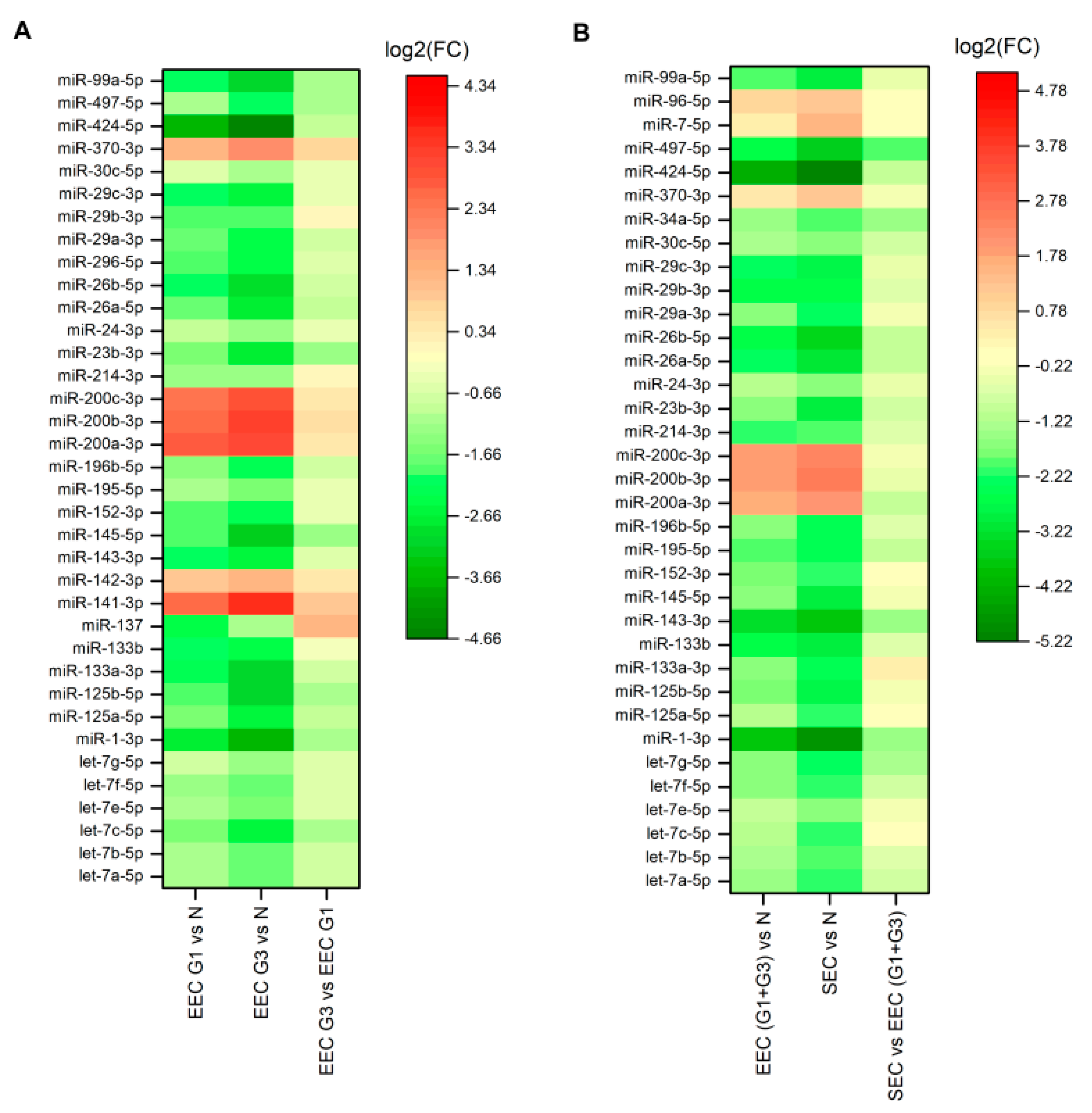
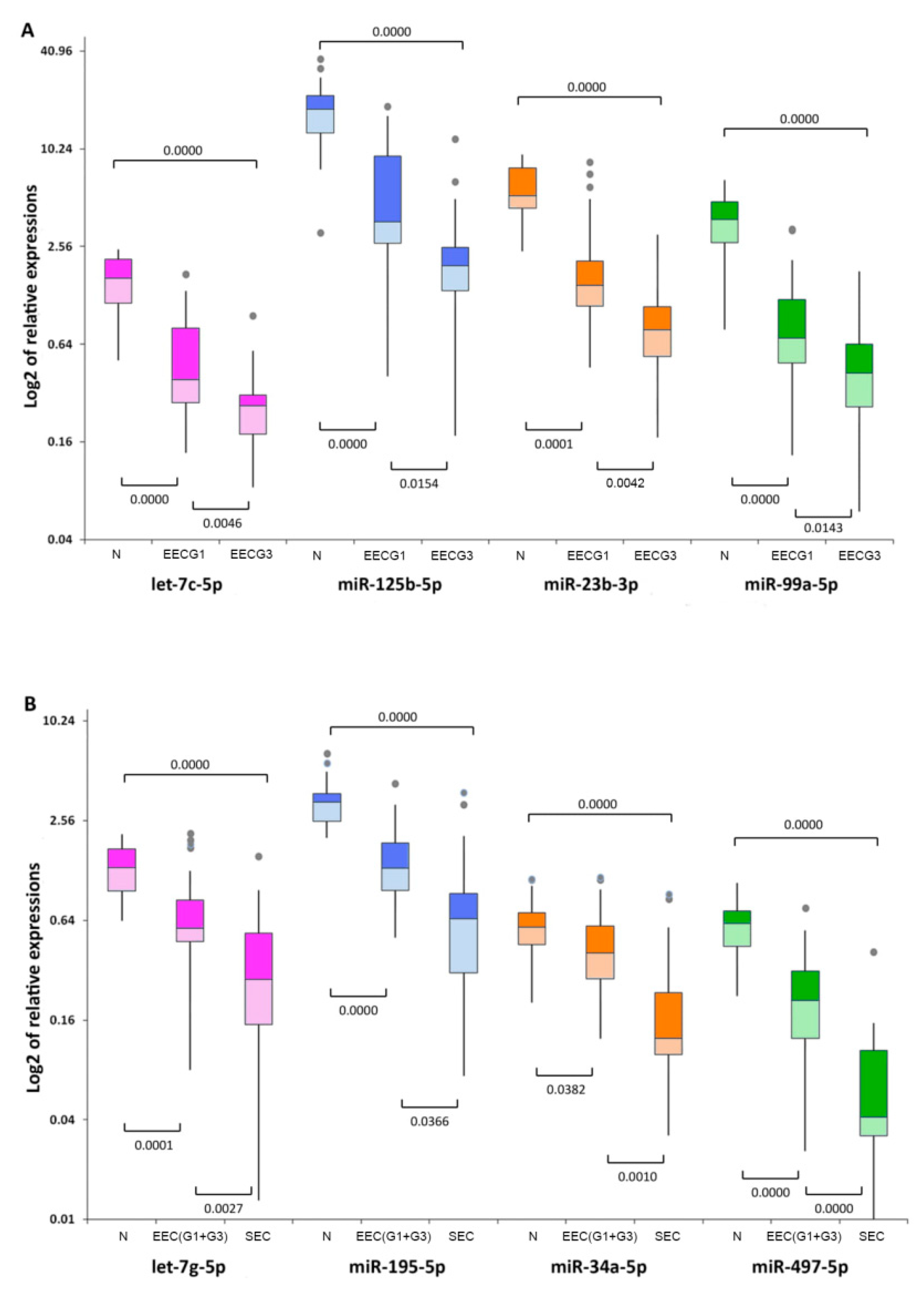
2.1. Differences in miRNA Expression between Well- and Poorly-Differentiated EEC

There were significant differences in 36 miRNAs between grade 1 EEC and grade 3 EEC and controls by miRNA profiling (Table 1), in which five genes were upregulated and 27 genes were downregulated in grade 1 and six genes were upregulated and 30 genes were downregulated in grade 3 compared to controls (Figure 1A). Of these, let-7c-5p (fold change 0.4866, p = 0.002877), miR-125b-5p (fold change 0.4694, p = 0.012155), miR-23b-3p (fold change 0.4037, p = 0.002478), and miR-99a-5p (fold change 0.4804, p = 0.011175) were significantly downregulated in grade 3 EEC compared to grade 1 EEC. The normalized miRNA expression levels represented by 2−ΔCt for let-7c-5p, miR-125b-5p, miR-23b-3p, and miR-99a-5p were 0.514 and 0.250, 4.428 and 2.078, 1.863 and 0.752, and 0.827 and 0.397 for grades 1 and 3, respectively, compared to 1.455, 16.846, 5.279, and 3.301 for controls, respectively. Differentially expressed miRNAs in grades 1 and 3 and controls are summarized in Figure 2A.

2.2. Differences in miRNA Expression between Endometrioid and Serous EC

There were differences in 35 miRNAs between EEC and SEC by miRNA profiling (Table 1), in which six genes were upregulated and 26 genes were downregulated in EEC and six genes were upregulated and 29 genes were downregulated in SEC compared to controls (Figure 1B). Of these, miRNAs let-7g-5p (fold change 0.4133, p = 0.004899), miR-195-5p (fold change 0.4829, p = 0.022248), miR-34a-5p (fold change 0.367, p = 0.000961), and miR-497-5p (fold change 0.2508, p = 0.00005) were significantly downregulated in SEC compared to EEC. The normalized miRNA expression levels were 0.643 and 0.266 for let-7g-5p, 1.350 and 0.652 for miR-195-5p, 0.428 and 0.157 for miR-34a-5p, and 0.187 and 0.047 for miR-497-5p in EEC and SEC, respectively, compared to 1.260, 3.421, 0.593, and 0.552 for controls (Figure 2B).
Furthermore, different EC-specific miRNA expression profiles were evidenced for miR-145-5b in the comparison of the two EEC grades, for miR-143-5p and miR-424-5p in the comparison of EEC and SEC, and for miR-1-3p in both comparisons. Compared to controls, the levels of all miRNAs were downregulated, although the differences were not significant. Furthermore, miR-137 was downregulated in grade 1 but upregulated in grade 3, whereas miR-1-3p was downregulated in EC regardless of the de-differentiation level and histological subtype.

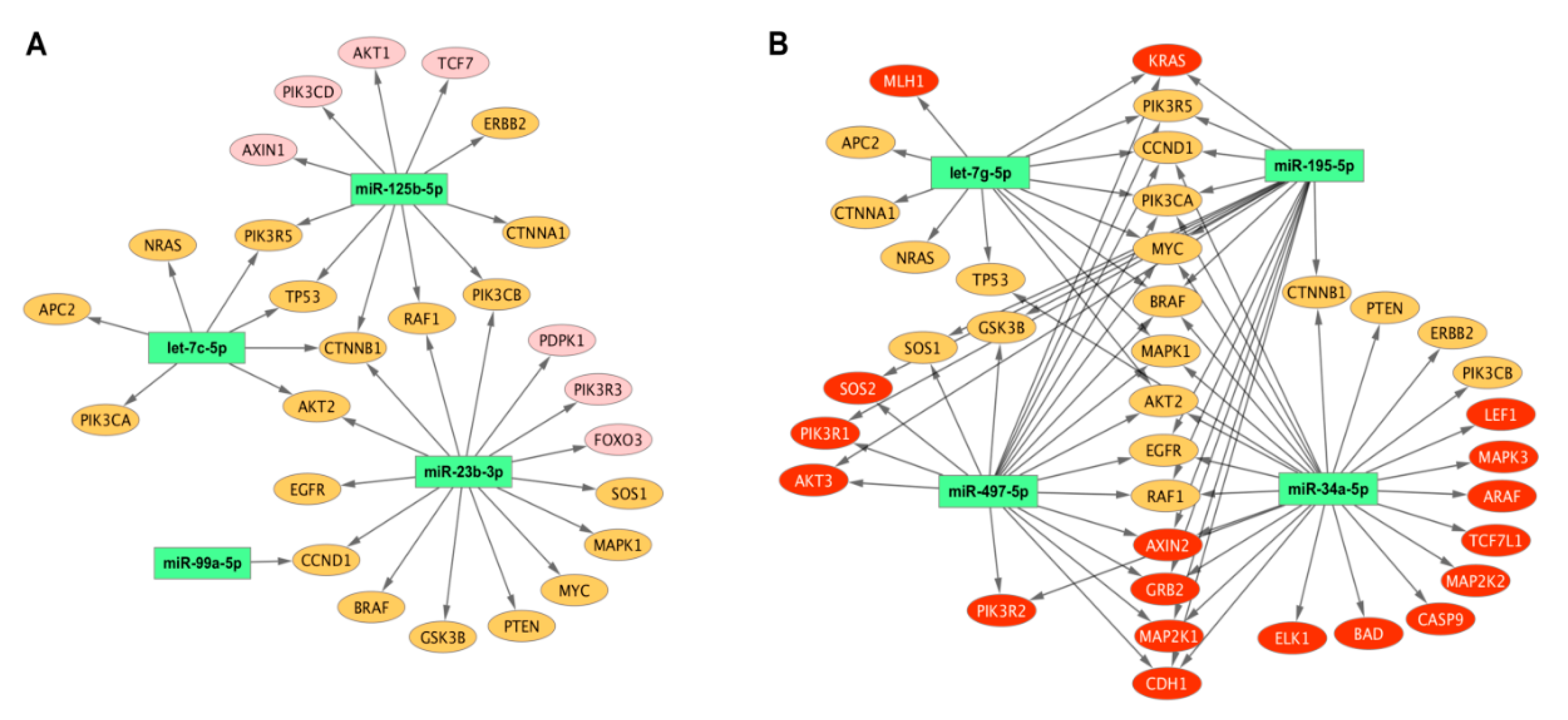
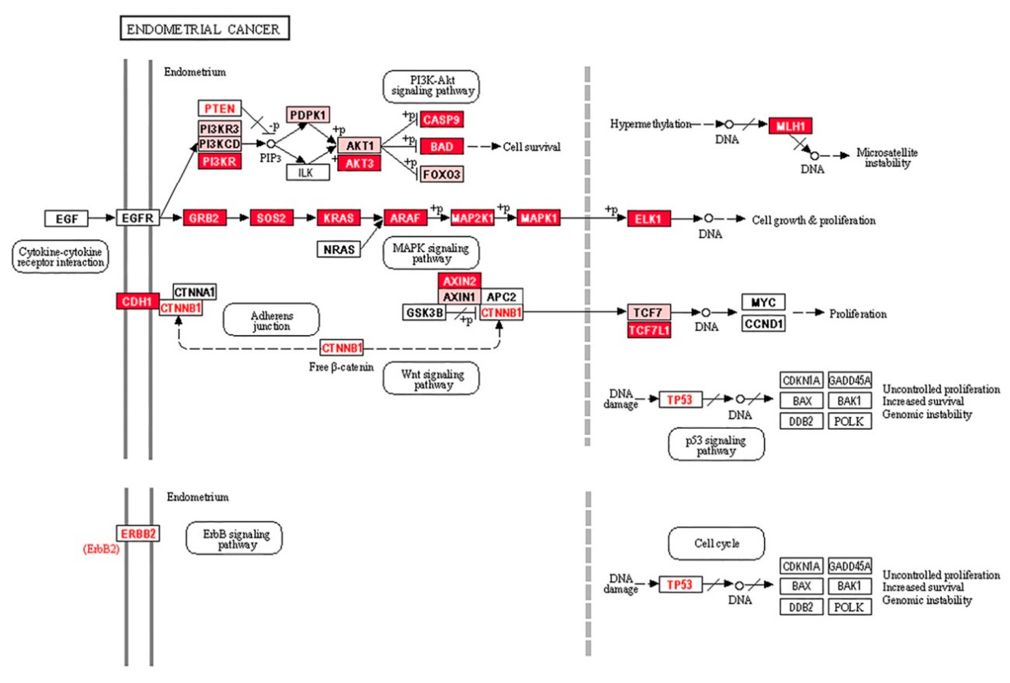
2.3. Aberrant miRNA Expression in EC Tumourigenesis

To investigate the roles of the differentially expressed miRNAs in endometrial tumourigenesis, we identified the target genes of all significantly downregulated miRNAs discriminating grade 1 from grade 3 EEC and EEC from SEC using the reverse search module in the miRPath software. Target genes, as well as their p-values, are summarized in Table 2. For several miRNA–target interactions, the strong evidence was experimentally confirmed by reporter assays, Western blotting, or qPCR (Table 2, Table S1).
Furthermore, we analyzed 44 target genes with respect to the eight discriminating miRNAs. We found that these genes could be divided into three distinct groups according to the criteria for discriminating between grades 1 and 3 or EEC and SEC or both (Table 3). For the first group, miR-125b-5p and miR-23b-3p potentially regulated seven genes. For the second group, different miRNAs identified in both comparisons potentially regulated 19 target genes. For the third group, four miRNAs potentially regulated 18 genes (Table 3). miRNA–target gene interactions are graphically depicted in Figure 3.
To confirm the involvement of the 44 target genes in EC, we used another bioinformatics tool, David 6.7. In this analysis, the 44 target genes were found to be involved in EC, but also in prostate, colorectal, non-small lung, or acute myeloid cancers (Table S2). The Pathview software was used to identify the genes involved in different signaling pathways (Figure 4).
Five out of the top seven genes in the first group, which were regulated by miRNAs discriminating grade 1 EEC from grade 3 EEC (Table 3), were involved in PI3K-AKT signaling. However, miRNAs discriminating EEC from SEC were involved in several signaling pathways, but mainly in MAPK signaling (Figure 4).

3. Discussion

Accurate therapeutic management of patients with EC depends on comprehensive clinical and pathological evaluations. Histomorphological examinations are critical in the diagnosis of EC, but the classification of ECs by histomorphological criteria has limited reproducibility [25]. More precise tools for distinguishing of these tumours are needed, which should facilitate subtype-specific research and treatment of EC [26].
Cell differentiation is the meaningful process resulting in well-defined tissues and organs, which is in cancer cells usually disrupted. The pathological grading of malignant tumours represents the extent of reversion of the differentiated phenotype; therefore, the score inversely correlates with the extent of cell differentiation. The multifunctional tumour suppressor p53 has roles in the cell cycle, apoptosis, senescence, DNA repair, and differentiation [27,28], although its role in de-differentiation is unknown. In EC, TP53 is frequently mutated; however, the high p53 immunohistochemical staining simultaneously with TP53 point mutations were found [29]. Regarding the molecular variability, it is difficult to interpret the p53 immunohistochemical results [30]. A previous study reported that miR-125b-5p targets TP53 [31]. In this study, a downregulation in the miR-125b-5p level in grade 3 EEC suggested that TP53 expression may increase, thereby affecting the process of cell differentiation. A downregulation in the miR-34a-5p level in SEC affected TP53 expression and could induce frequent de-differentiation of serous cancer cells [32].
Members of let-7 family are aberrantly expressed in many cancers and a correlation between weak let-7 expression and poor differentiation of aggressive tumours was observed [33]. Consistent with our results, a previous study has reported decreased let-7c-5p and miR-99a-3p expression in early stage EEC [34], as well as decreased let-7c-5p expression in grade 3 [35]. The PI3K/AKT/mTOR signaling pathway regulates cancer cell proliferation, migration, survival, and angiogenesis, and AKT1 and mTOR genes have been identified as targets of miR-99a-3p in other types of cancers [36,37]. Compared to controls, miR-99a-3p expression was significantly decreased and negatively correlated with EEC tumour grade [38,39]. In another study, decreased miR-125b expression increased the expression of ERBB2, its target gene [40]. ERBB2/HER2 overexpression was identified in very aggressive primary tumours and metastatic lesions of EC, mostly in the serous subtype, suggesting that these changes induce AKT phosphorylation [40,41]. Histologically, among the poorly differentiated ECs, SEC predominantly shows nuclear atypia and papillary growth compared to EEC. Grade 3 EEC is characterized by solid glandular growth and less frequent papillary formation with transition from one to another component. These features allow clinicians to discriminate between endometrioid and serous EC, but in many patients, reproducible classification has not been possible [42].
Of the four discriminating miRNAs between EEC and SEC identified in this study, only miR-34a-5p has been studied extensively. In non-selective EC, a decreased miR-34a-5p level was reported to associate with higher stage and metastasis [43,44], whereas another study revealed that miR-34a-5p targets SNAIL and other genes such as those involved in epithelial-mesenchymal transition (EMT) [45]. In another study, decreased miR-34a-5p expression upregulated the L1CAM level in EC cell lines and tumours [46]. In stage I EC patients, L1CAM expression was found to be a strong independent predictor of disease recurrence and poor prognosis [47]. On the other hand, increased miR-34a-5p expression was found to decrease the expression of its target genes NOTCH1 and DLL1 in EC. NOTCH1 and DLL1 are regulators of cell proliferation, differentiation, and apoptosis; therefore, these expression changes contribute to EC [48]. For other miRNAs, such as miR-195 and miR-497, changes in expression have been reported in SEC [23], but further studies are needed.
There is only one published comparative study of miRNA expression profiles in 14 and nine samples of EEC and SEC, respectively. Of 667 miRNAs, only 17 were significantly discriminating between these two EC subtypes. Of these, let-7c, miR-9, miR-146a, miR-450a, miR-504, and miR-542-3p showed at least a 2-fold difference in expression between EEC and SEC [49]. In this study, we identified another four miRNAs downregulated in EEC, whose levels decreased even further in SEC. The higher expression of their target genes in a more aggressive subtype of SEC indicate the important roles of these miRNAs in EC progression.
In endometrial cancer cells, molecular changes in PI3K/AKT/mTOR, MAPK/ERK, WNT/β-catenin, VEGF/VEGFR, ERBB, P53/P21, and P16INK4a/pRB signaling pathways are responsible for evading apoptosis, increasing cell proliferation, inhibiting differentiation, and stimulating angiogenesis [50]. For instance, PI3K/AKT signaling is critical for EMT and the stemness phenotype in endometrioid and non-endometrioid EC, and these changes have been reported to be mediated by several miRNAs that target PTEN, TWIST1, ZEB1, and BMI1 [51].
There is little knowledge on the roles of specific miRNAs in EC regarding their aberrant miRNA expression, target genes, and functions in EC tumourigenesis; therefore, to understand the clinical significance of our results, we performed bioinformatic analyses. To differentiate between grades 1 and 3, we identified let-7c-5p, miR-125b-5p, miR-23b-3p, and miR-99a-5p, as well as seven in silico generated target genes involved in EC progression. These target genes were regulated by miR-125b-5p or miR-23b-3p, and five are members of the PI3K/AKT signaling pathway, including phosphatidylinositol 3-kinases (PIK3CD, PIK3R3), serine/threonine kinases (PDPK1, AKT1), and the transcription factor FOXO3. Similarly, let-7g-5p, miR-195-5p, miR-34a-5p, and miR-497-5p, which differentiated EEC from SEC, regulated target genes participating in PI3K/AKT signaling. These target genes are also members of the WNT signaling pathway. They can also contribute to microsatellite instability by targeting MLH1 and exclusively influence MAPK signaling by regulating cell proliferation and growth [50]. Furthermore, overexpression of growth factors in cancers can constitutively and autonomously activate MAPK signaling [52]. It is possible that these miRNAs can affect the expression of GRB2, SOS2, KRAS, ARAF, MAPK2K1, MAPK1, and ELK1. For instance, KRAS is a small GTPase that transduces signals between cell surface receptors and the nucleus. Mutations in KRAS were identified in 20–30% of EEC cases compared to 3% of SECs [9], suggesting that the regulation of KRAS by let-7g-5p, miR-195-5p, or miR-497-5p could be another mechanism contributing to SEC.
Cancer development and progression involves the deregulation of essential cellular processes. miRNAs regulate the expression of multiple genes; however, it is challenging to characterize all the consequences of these changes. Here, we identified two sets of downregulated miRNAs and two key signaling pathways that could be used to differentiate between grade 1 and grade 3 EEC and between EEC and SEC. The associations between activated proteins of these pathways and EC cell phenotypes or behaviours need to be widely investigated; however, our findings may be helpful in the differential diagnosis, especially in the classification of morphologically mixed or problematic EC.

4. Materials and Methods

4.1. Patients

In this retrospective study, we analyzed endometrial tumour tissues isolated from FFPE specimens from histomorphologically-characterized EC. In brief, 20 and 21 specimens were from patients with grade 1 EEC and grade 3 EEC, respectively; and 21 specimens were from patients with SEC. The age of all 62 EC patients ranged from 45 to 86 years, with a mean 65.18 ± 8.85 years (63.75, 63.76, and 67.95 years for EEC grade 1, EEC grade 3, and SEC groups, respectively). We also included 20 non-neoplastic endometrial specimens from women in matched age from 53 to 79 years (mean, 65.4 ± 6.75 years). All specimens were obtained from the tissue archives of the St. Elizabeth Cancer Institute, Bratislava, Slovakia, and prepared before 2018. The St. Elizabeth Cancer Institute Review Board approved this retrospective study and waived the consent of patients and control persons. No patient underwent preoperative radiotherapy or chemotherapy before specimen collection. Controls had no signs or symptoms of cancer or other serious diseases.

4.2. miRNA Extraction and Real-Time PCR

miRNAs from FFPE endometrial tumour tissues were isolated using the miRNeasy FFPE Kit (Qiagen, Hilden, Germany) according to the manufacturer’s instructions. miRNA samples of suitable purity were reversely transcribed into cDNA using the miScript II RT Kit (Qiagen) according to the manufacturer’s instructions. For real-time polymerase chain reactions, we used the miScript miRNA PCR Array Human Tumor Suppressor miRNAs Kit containing 84 known tumour suppressor miRNAs in different cancer types (MIHS-119Z, Qiagen) using the miScript SYBR Green PCR Kit (Qiagen). RT-PCR reactions were carried out in an AriaMx Real-Time PCR System (Agilent, Santa Clara, CA, USA) using conditions as follows: Predenaturation at 95 °C for 15 min, followed by 40 cycles at 94 °C for 15 s, 55 °C at 30 s, and 70 °C for 30 s.

4.3. Statistical Analysis

Real-time PCR data was statistically analyzed using the miScript miRNA PCR Array Data Analysis Software (Qiagen). Global normalization was performed using six endogenous small RNAs, namely SNORD61, SNORD68, SNORD72, SNORD95, SNORS96A, and RNU6-6P. For the quantification of relative gene expression, the ΔΔCT method was used [53]. Fold-change 2−ΔΔCt was the normalized gene expression 2−ΔCt in the test sample divided by the normalized gene expression 2−ΔCt in the control sample. The upregulation of miRNA expression was indicated by fold-change values greater than 2, and the fold-regulation was equal to the fold-change. By contrast, the downregulation of miRNA expression was indicated by fold-change values less than 0.5, and the fold-regulation was the negative inverse of the fold-change (–1/x). p-Values were calculated based on the Student’s t-test of replicate 2−ΔCt values for each gene in both groups, and statistical significance was considered at p < 0.05. In bioinformatic analyses, the reverse-search module of the DIANA-miRPath v3.0 online software suite was used to identify the most relevant target genes involved in EC for eight discriminating miRNAs [54]. A reverse-search was performed separately against every available database included in miRPath such as Tarbase v.7.0, TargetScan v.6, and microT-CDS v.5.0. For the independent confirmation of the identified target genes with respect to pathways involved in EC and other cancer types, the online software tool David 6.7 was used [55]. For network visualization of miRNA–gene interactions, Cytoscape v.3.8.0 was used [56]. Experimentally validated miRNA–target interactions were adopted from miRTarBase [57]. For pathway analysis, the KEGG Database and Pathview Tool were used [58]. Heat maps were generated using the OriginPro 2015 software (OriginLab Corp., Northampton, MA, USA).

Supplementary Materials

Author Contributions

I.F. and K.K., conception and research design; K.K. and P.S., clinical data collection, selection of patients, and sample preparation; L.K. and I.Z., establishment of laboratory methodologies; L.K. and L.W., expression analysis; L.K., evaluation of expression analyses; M.K., bioinformatic analyses; I.F., writing—original draft preparation, L.K. and K.K., writing—review and editing; I.F., supervision, project administrator, and funding. All authors have read and agreed to the published version of the manuscript.

Funding

This work was supported by the Ministry of Health of the Slovak Republic under the project registration number 2018/45-SAV-4.

Acknowledgments

The authors would like to thank Viola Stevurkova for excellent technical assistance.

Conflicts of Interest

The authors declare no conflict of interest.

Abbreviations

ECEndometrial Carcinoma
EECEndometrioid Endometrial Carcinoma
SECSerous Endometrial Carcinoma
TCGAThe Cancer Genome Atlas
MSIMicrosatellite Instability
miRNAMicroRNA
G3Grade 3, Poorly-differentiated tumour
G1Grade 1, Well-differentiated tumour
FFPEFormalin-Fixed Paraffin Embedded
EMTEpithelial-Mesenchymal Transition

References

  1. Bray, F.; Ferlay, J.; Soerjomataram, I.; Siegel, R.L.; Torre, L.A.; Jemal, A. Global cancer statistics 2018: GLOBOCAN estimates of incidence and mortality worldwide for 36 cancers in 185 countries. CA Cancer J. Clin. 2018, 68, 394–424. [Google Scholar] [CrossRef] [PubMed] [Green Version]
  2. Siegel, R.L.; Miller, K.D.; Jemal, A. Cancer statistics, 2016. CA Cancer J. Clin. 2016, 66, 7–30. [Google Scholar] [CrossRef] [Green Version]
  3. Murali, R.; Soslow, R.A.; Weigelt, B. Classification of endometrial carcinoma: More than two types. Lancet Oncol. 2014, 7, e268–e278. [Google Scholar] [CrossRef]
  4. Bokhman, J.V. Two pathogenetic types of endometrial carcinoma. Gynecol. Oncol. 1983, 15, 10–17. [Google Scholar] [CrossRef]
  5. Matias-Guiu, X.; Prat, J. Molecular pathology of endometrial carcinoma. Histopathology 2013, 62, 111–123. [Google Scholar] [CrossRef] [PubMed]
  6. Zaino, R.; Carinelli, S.G.; Ellenson, L.H.; End, C.; Katabuchi, H.; Konishi, I.; Lax, S.; Matias-Guiu, X.; Mutter, G.L.; Peters, W.A., III; et al. Epithelial tumours and precursors. In WHO Classification of Tumours of Female Reproductive Organs, 4th ed.; Kurman, R., Carcangiu, M.L., Herrington, S., Young, R.H., Eds.; IARC: Lyon, France, 2014; Volume 6, pp. 125–134. [Google Scholar]
  7. Colombo, N.; Preti, E.; Landoni, F.; Carinelli, S.; Colombo, A.; Marini, C.; Sessa, C. ESMO Guidelines Working Group Endometrial Cancer. ESMO Clinical Practice Guidelines for Diagnosis, Treatment and Follow-up. Ann. Anncol. 2013, 24, vi33–vi38. [Google Scholar] [CrossRef]
  8. Cancer Genome Atlas Research Network; Kandoth, C.; Schultz, N.; Cherniack, A.D.; Akbani, R.; Liu, Y.; Shen, H.; Robertson, A.G.; Pashtan, I.; Shen, R.; et al. Integrated genomic characterization of endometrial carcinoma. Nature 2013, 497, 67–73. [Google Scholar]
  9. Morice, P.; Leary, A.; Creutzberg, C.; Abu-Rustum, N.; Darai, E. Endometrial cancer. Lancet 2016, 387, 1094–1108. [Google Scholar] [CrossRef]
  10. Bell, D.W. Novel genetic targets in endometrial cancer. Expert Opin. Ther. Targets 2014, 18, 725–730. [Google Scholar] [CrossRef]
  11. Esteller, M.; Catasus, L.; Matias-Guiu, X.; Mutter, G.L.; Prat, J.; Baylin, S.B.; Herman, J.G. hMLH1 promoter hypermethylation is an early event in human endometrial tumorigenesis. Am. J. Pathol. 1999, 155, 1767–1772. [Google Scholar] [CrossRef] [Green Version]
  12. Salvesen, H.B.; MacDonald, N.; Ryan, A.; Jacobs, I.J.; Lynch, E.D.; Akslen, L.A.; Das, S. PTEN methylation is associated with advanced stage and microsatellite instability in endometrial carcinoma. Int. J. Cancer 2001, 91, 22–26. [Google Scholar] [CrossRef]
  13. Liao, X.; Siu, M.K.; Chan, K.Y.; Wong, E.S.; Ngan, H.Y.; Chan, Q.K.; Li, A.S.; Khoo, U.S.; Cheung, A.N. Hypermethylation of RAS effector related genes and DNA methyltransferase 1 expression in endometrial carcinogenesis. Int. J. Cancer 2008, 123, 296–302. [Google Scholar] [CrossRef] [PubMed]
  14. Moreno-Bueno, G.; Hardisson, D.; Sanchez, C.; Sarrio, D.; Cassia, R.; Garcia-Rostan, G.; Prat, J.; Guo, M.; Herman, J.G.; Matias-Guiu, X.; et al. Abnormalities of the APC/beta-catenin pathway in endometrial cancer. Oncogene 2002, 21, 7981–7990. [Google Scholar] [CrossRef] [PubMed] [Green Version]
  15. Xiong, Y.; Dowdy, S.C.; Xue, A.; Shujuan, J.; Eberhardt, N.L.; Podratz, K.C.; Jiang, S.W. Opposite alterations of DNA methyltransferase gene expression in endometrioid and serous endometrial cancers. Gynecol. Oncol. 2005, 96, 601–609. [Google Scholar] [CrossRef] [PubMed]
  16. Fambrini, M.; Bussani, C.; Sorbi, F.; Pieralli, A.; Cioni, R. Methylation of the HOXA10 homeobox gene promoter is associated with endometrial cancer: A pilot study. J. Obstet. Gynaecol. 2013, 33, 519–520. [Google Scholar] [CrossRef] [PubMed]
  17. Multinu, F.; Chen, J.; Madison, J.D.; Torres, M.; Casarin, J.; Visscher, D.; Shridhar, V.; Bakkum-Gamez, J.; Sherman, M.; Wentzensen, N.; et al. Analysis of DNA methylation in endometrial biopsies to predict risk of endometrial cancer. Gynecol. Oncol. 2020, 156, 682–688. [Google Scholar] [CrossRef] [PubMed] [Green Version]
  18. Zhang, B.; Xing, X.; Li, J.; Lowdon, R.F.; Zhou, Y.; Lin, N.; Zhang, B.; Sundaram, V.; Chiappinelli, K.B.; Hagemann, I.S.; et al. Comparative DNA methylome analysis of endometrial carcinoma reveals complex and distinct deregulation of cancer promoters and enhancers. BMC Genom. 2014, 15, 868. [Google Scholar] [CrossRef] [Green Version]
  19. van Schooneveld, E.; Wildiers, H.; Vergote, I.; Vermeulen, P.B.; Dirix, L.Y.; Van Laere, S.J. Dysregulation of microRNAs in breast cancer and their potential role as prognostic and predictive biomarkers in patient management. Breast Cancer Res. 2015, 18, 17–21. [Google Scholar] [CrossRef] [Green Version]
  20. Xie, B.; Ding, Q.; Han, H.; Wu, D. miRCancer: A microRNA-cancer association database constructed by text mining on literature. Bioinformatics 2013, 29, 638–644. [Google Scholar] [CrossRef]
  21. Delangle, R.; De Foucher, T.; Larsen, A.K.; Sabbah, M.; Azaïs, H.; Bendifallah, S.; Daraï, E.; Ballester, M.; Mehats, C.; Uzan, C.; et al. The Use of microRNAs in the Management of Endometrial Cancer: A Meta-Analysis. Cancers 2019, 11, 832. [Google Scholar] [CrossRef] [Green Version]
  22. Cohn, D.E.; Fabbri, M.; Valeri, N.; Alder, H.; Ivanov, I.; Liu, C.G.; Croce, C.M.; Resnick, K.E. Comprehensive miRNA profiling of surgically staged endometrial cancer. Am. J. Obstet. Gynecol. 2010, 202, e1–e8. [Google Scholar] [CrossRef] [PubMed] [Green Version]
  23. Hiroki, E.; Akahira, J.; Suzuki, F.; Nagase, S.; Ito, K.; Suzuki, T.; Sasano, H.; Yaegashi, N. Changes in microRNA expression levels correlate with clinicopathological features and prognoses in endometrial serous adenocarcinomas. Cancer Sci. 2010, 101, 241–249. [Google Scholar] [CrossRef] [PubMed]
  24. Widodo; Djati, M.S.; Rifa’I, M. Role of MicroRNAs in carcinogenesis that potential for biomarker of endometrial cancer. Ann. Med. Surg. 2016, 7, 9–13. [Google Scholar] [CrossRef] [PubMed]
  25. Gilks, C.B.; Oliva, E.; Soslow, R.A. Poor interobserver reproducibility in diagnosis of high-grade endometrial carcinoma. Am. J. Surg. Pathol. 2013, 37, 874–881. [Google Scholar] [CrossRef]
  26. Talhouk, A.; McAlpine, J.N. New classification of endometrial cancers: The development and potential applications of genomic-based classification in research and clinical care. Gynecol. Oncol. Res. Pract. 2016, 3, 14. [Google Scholar] [CrossRef] [Green Version]
  27. Molchadsky, A.; Rivlin, N.; Brosh, R.; Rotter, V.; Sarig, R. p53 is balancing development, differentiation and de-differentiation to assure cancer prevention. Carcinogenesis 2010, 31, 1501–1508. [Google Scholar] [CrossRef] [Green Version]
  28. Rivlin, N.; Koifman, G.; Rotter, V. p53 orchestrates between normal differentiation and cancer. Semin. Cancer Biol. 2015, 32, 10–17. [Google Scholar] [CrossRef]
  29. Kohler, M.F.; Berchuck, A.; Davidoff, A.M.; Humphrey, P.A.; Dodge, R.K.; Iglehart, J.D.; Soper, J.T.; Clarke-Pearson, D.L.; Bast, R.C., Jr.; Marks, J.R. Overexpression and mutation of p53 in endometrial carcinoma. Cancer Res. 1992, 52, 1622–1627. [Google Scholar]
  30. Köbel, M.; Ronnett, B.M.; Singh, N.; Soslow, R.A.; Gilks, C.B.; McCluggage, W.G. Interpretation of P53 Immunohistochemistry in Endometrial Carcinomas: Toward Increased Reproducibility. Int. J. Gynecol. Pathol. 2019, 38, S123–S131. [Google Scholar] [CrossRef]
  31. Le, M.T.; Teh, C.; Shyh-Chang, N.; Xie, H.; Zhou, B.; Korzh, V.; Lodish, H.F.; Lim, B. MicroRNA-125b is a novel negative regulator of p53. Genes Dev. 2009, 23, 862–876. [Google Scholar] [CrossRef] [Green Version]
  32. Navarro, F.; Lieberman, J. miR-34 and p53: New Insights into a Complex Functional Relationship. PLoS ONE 2015, 10, e0132767. [Google Scholar] [CrossRef] [PubMed]
  33. Boyerinas, B.; Park, S.M.; Hau, A.; Murmann, A.E.; Peter, M.E. The role of let-7 in cell differentiation and cancer. Endocr.-Relat. Cancer 2010, 17, F19–F36. [Google Scholar] [CrossRef] [PubMed]
  34. Xiong, H.; Li, Q.; Liu, S.; Wang, F.; Xiong, Z.; Chen, J.; Chen, H.; Yang, Y.; Tan, X.; Luo, Q.; et al. Integrated microRNA and mRNA transcriptome sequencing reveals the potential roles of miRNAs in stage I endometrioid endometrial carcinoma. PLoS ONE 2014, 9, e110163. [Google Scholar] [CrossRef] [PubMed] [Green Version]
  35. Ye, F.; Tang, Q.L.; Ma, F.; Cai, L.; Chen, M.; Ran, X.X.; Wang, X.Y.; Jiang, X.F. Analysis of the circular RNA transcriptome in the grade 3 endometrial cancer. Cancer Manag. Res. 2019, 11, 6215–6227. [Google Scholar] [CrossRef] [PubMed] [Green Version]
  36. Wang, L.; Chang, L.; Li, Z.; Gao, Q.; Cai, D.; Tian, Y.; Zeng, L.; Li, M. miR-99a and -99b inhibit cervical cancer cell proliferation and invasion by targeting mTOR signaling pathway. Med. Oncol. 2014, 31, 934. [Google Scholar] [CrossRef] [PubMed]
  37. Yu, S.; Zhang, C.; Deng, F.; Zhang, Y. miR-99a suppresses the metastasis of human nonsmall cell lung cancer cells by targeting AKT1 signaling pathway. J. Cell. Biochem. 2015, 116, 268–276. [Google Scholar] [CrossRef] [PubMed]
  38. Torres, A.; Torres, K.; Pesci, A.; Ceccaroni, M.; Paszkowski, T.; Cassandrini, P.; Zamboni, G.; Maciejewski, R. Deregulation of miR-100, miR-99a and miR-199b in tissues and plasma coexists with increased expression of mTOR kinase in endometrioid endometrial carcinoma. BMC Cancer 2012, 12, 369. [Google Scholar] [CrossRef] [Green Version]
  39. Li, Y.; Zhang, Z.; Zhang, X.; Lin, Y.; Luo, T.; Xiao, Z.; Zhou, Q. A dual PI3K/AKT/mTOR signaling inhibitor miR-99a suppresses endometrial carcinoma. Am. J. Transl. Res. 2016, 8, 719–731. [Google Scholar]
  40. Shang, C.; Lu, Y.M.; Meng, L.R. MicroRNA-125b down-regulation mediates endometrial cancer invasion by targeting ERBB2. Med. Sci. Monit. 2012, 18, BR149–BR155. [Google Scholar] [CrossRef]
  41. Halle, M.K.; Tangen, I.L.; Berg, H.F.; Hoivik, E.A.; Mauland, K.K.; Kusonmano, K.; Berg, A.; Hurtado, A.; Kalland, K.H.; Øyan, A.M.; et al. HER2 expression patterns in paired primary and metastatic endometrial cancer lesions. Br. J. Cancer 2018, 118, 378–387. [Google Scholar] [CrossRef] [Green Version]
  42. Murali, R.; Davidson, B.; Fadare, O.; Carlson, J.A.; Crum, C.P.; Gilks, C.B.; Irving, J.A.; Malpica, A.; Matias-Guiu, X.; McCluggage, W.G.; et al. High-grade Endometrial Carcinomas: Morphologic and Immunohistochemical Features, Diagnostic Challenges and Recommendations. Int. J. Gynecol. Pathol. 2019, 38, S40–S63. [Google Scholar] [CrossRef]
  43. Choi, Y.S.; Lee, K.E. The Significance of miR-34a Expression in Endometrial Carcinogenesis: Correlation with Expression of p16 and Ki-67 Proteins in Endometrial Cancers. J. Cancer Prev. 2015, 20, 268–274. [Google Scholar] [CrossRef] [PubMed] [Green Version]
  44. Wang, Z.; Wang, W.; Huang, K.; Wang, Y.; Li, J.; Yang, X. MicroRNA-34a inhibits cells proliferation and invasion by downregulating Notch1 in endometrial cancer. Oncotarget 2017, 8, 111258–111270. [Google Scholar] [CrossRef] [PubMed]
  45. Siemens, H.; Jackstadt, R.; Hünten, S.; Kaller, M.; Menssen, A.; Götz, U.; Hermeking, H. miR-34 and SNAIL form a double-negative feedback loop to regulate epithelial-mesenchymal transitions. Cell Cycle 2011, 10, 4256–4271. [Google Scholar] [CrossRef] [PubMed] [Green Version]
  46. Schirmer, U.; Doberstein, K.; Rupp, A.K.; Bretz, N.P.; Wuttig, D.; Kiefel, H.; Breunig, C.; Fiegl, H.; Müller-Holzner, E.; Zeillinger, R.; et al. Role of miR-34a as a suppressor of L1CAM in endometrial carcinoma. Oncotarget 2014, 5, 462–472. [Google Scholar] [CrossRef] [PubMed] [Green Version]
  47. Bosse, T.; Nout, R.A.; Stelloo, E.; Dreef, E.; Nijman, H.W.; Jürgenliemk-Schulz, I.M.; Jobsen, J.J.; Creutzberg, C.L.; Smit, V.T. L1 cell adhesion molecule is a strong predictor for distant recurrence and overall survival in early stage endometrial cancer: Pooled PORTEC trial results. Eur. J. Cancer 2014, 50, 2602–2610. [Google Scholar] [CrossRef] [PubMed]
  48. Jurcevic, S.; Klinga-Levan, K.; Olsson, B.; Ejeskär, K. Verification of microRNA expression in human endometrial adenocarcinoma. BMC Cancer 2016, 16, 261. [Google Scholar] [CrossRef] [Green Version]
  49. Devor, E.J.; Hovey, A.M.; Goodheart, M.J.; Ramachandran, S.; Leslie, K.K. microRNA expression profiling of endometrial endometrioid adenocarcinomas and serous adenocarcinomas reveals profiles containing shared, unique and differentiating groups of microRNAs. Oncol. Rep. 2011, 26, 995–1002. [Google Scholar] [CrossRef] [Green Version]
  50. Ma, X.; Ma, C.X.; Wang, J. Endometrial Carcinogenesis and Molecular Signaling Pathways. Am. J. Mol. Biol. 2014, 4, 134–149. [Google Scholar] [CrossRef] [Green Version]
  51. Dong, P.; Konno, Y.; Watari, H.; Hosaka, M.; Noguchi, M.; Sakuragi, N. The impact of microRNA-mediated PI3K/AKT signaling on epithelial-mesenchymal transition and cancer stemness in endometrial cancer. J. Transl. Med. 2014, 12, 231. [Google Scholar] [CrossRef]
  52. Hoshino, R.; Chatani, Y.; Yamori, T.; Tsuruo, T.; Oka, H.; Yoshida, O.; Shimada, Y.; Ari-i, S.; Wada, H.; Fujimoto, J.; et al. Constitutive Activation of the 41-/43-kDa Mitogen-Activated Protein Kinase Signaling Pathway in Human Tumors. Oncogene 1999, 18, 813–822. [Google Scholar] [CrossRef] [PubMed] [Green Version]
  53. Livak, K.J.; Schmittgen, T.D. Analysis of relative gene expression data using real-time quantitative PCR and the 2[-Delta Delta C[T]] Method. Methods 2001, 25, 402–408. [Google Scholar] [CrossRef]
  54. Vlachos, I.S.; Zagganas, K.; Paraskevopoulou, M.D.; Georgakilas, G.; Karagkouni, D.; Vergoulis, T.; Dalamagas, T.; Hatzigeorgiou, A.G. DIANA-miRPath v3.0: Deciphering microRNA function with experimental support. Nucleic Acids Res. 2015, 43, W460–W466. [Google Scholar] [CrossRef] [PubMed]
  55. Jiao, X.; Sherman, B.T.; Huang, D.; Stephens, R.; Baseler, M.W.; Lane, H.C.; Lempicki, R.A. DAVID-WS: A stateful web service to facilitate gene/protein list analysis. Bioinformatics 2012, 28, 1805–1806. [Google Scholar] [CrossRef] [PubMed] [Green Version]
  56. Shannon, P.; Markiel, A.; Ozier, O.; Baliga, N.S.; Wang, J.T.; Ramage, D.; Amin, N.; Schwikowski, B.; Ideker, T. Cytoscape: A software environment for integrated models of biomolecular interaction networks. Genome Res. 2003, 13, 2498–2504. [Google Scholar] [CrossRef]
  57. Huang, H.Y.; Lin, Y.C.; Li, J.; Huang, K.Y.; Shrestha, S.; Hong, H.C.; Tang, Y.; Chen, Y.G.; Jin, C.N.; Yu, Y.; et al. miRTarBase 2020: Updates to the experimentally validated microRNA-target interaction database. Nucleic Acids Res. 2020, 48, D148–D154. [Google Scholar] [CrossRef] [Green Version]
  58. Luo, W.; Pant, G.; Bhavnasi, Y.K.; Blanchard, S.G., Jr.; Brouwer, C. Pathview Web: User friendly pathway visualization and data integration. Nucleic Acids Res. 2017, 45, W501–W508. [Google Scholar] [CrossRef]
Figure 1. Heat maps of miRNA expression in endometrial cancer. Differences in miRNA expression between grade 1 (EEC G1) and grade 3 (EEC G3) endometrioid endometrial carcinoma compared with non-neoplastic endometrium (N), (A) and between endometrioid endometrial carcinoma (EEC (G1 + G3)) and serous endometrial carcinoma (SEC) compared with the non-neoplastic endometrium (N) (B). On the scale of fold regulation (log2(FC)), red corresponds to upregulated miRNAs and green corresponds to downregulated miRNAs.
Ijms 21 06071 g001
Figure 2. Relative expression of differentially expressed miRNAs. Differential expression of let-7c-5p, miR-125b-5p, miR-23b-3p, and miR-99a-5p between grade 1 (EEC G1) and grade 3 (EEC G3) endometrioid endometrial carcinoma compared with the non-neoplastic endometrium (N) (A), and differential expression of let-7g-5p, miR-195-5p, miR-34a-5p, and miR-497-5p between endometrioid endometrial carcinoma (EEC(G1 + G3)) and serous endometrial carcinoma (SEC) compared with the non-neoplastic endometrium (N) (B). Box lengths correspond to the interquartile range (IQR) and represent values between 75th and 25th percentiles. Values greater than 1.5 IQRs from the end of the box are labeled as outliers (grey points). Horizontal lines depict the medians. Statistical significance was considered at p < 0.05.
Ijms 21 06071 g002
Figure 3. MicroRNA–target gene network for discriminating miRNAs. MicroRNA–target gene network for miRNAs discriminating grade 1 from grade 3 (A) and endometrioid from serous endometrial carcinoma (B). Target genes regulated by miRNAs discriminating between different grades in endometrioid and endometrioid and serous carcinoma are depicted in pink and red, respectively. Target genes regulated by miRNAs from both sets are depicted in orange. The associations between microRNAs and target genes are represented by black arrows. The scheme was generated by CytoScape 3.8.0.
Ijms 21 06071 g003
Figure 4. Signaling pathways involved in EC. Pink boxes represent the first group of target genes, which are targeted by two miRNAs, miR-125b-5p and miR-23b-3p that can discriminate grade 1 from grade 3. Red boxes represent the third group of target genes for miRNAs discriminating serous from endometrioid endometrial samples. KRAS, CTNNB1, ERBB2, PTEN, and TP53 (letters in red, except KRAS for visibility) are frequently mutated, whereas CTNNB1, ERBB2, PTEN, and TP53 are regulated by both sets of discriminating miRNAs (second group of target genes).
Ijms 21 06071 g004
Table 1. Differentially expressed miRNAs in EC subtypes.
EC SubtypesDifferently Expressed miRNAs in Each of Two Evaluated Groups Compared to Controls
Endometrioid grade 1 vs. grade 3let-7a-5p, let-7b-5p, let-7c-5p, let-7e-5p, let-7f-5p, let-7g-5p, miR-1-3p, miR-125a-5p, miR-125b-5p, miR-133a-3p, miR-133b, miR-137, miR-141-3p, miR-142-3p, miR-143-3p, miR-145-5p, miR-152-3p, miR-195-5p, miR-196b-5p, miR-200a-3p, miR-200b-3p, miR-200c-3p, miR-214-3p, miR-23b-3p, miR-24-3p, miR-26a-5p, miR-26b-5p, miR-296-5p, miR-29a-3p, miR-29b-3p, miR-29c-3p, miR-30c-5p, miR-370-3p, miR-424-5p, miR-497-5p, miR-99a-5p
Endometrioid vs. Serouslet-7a-5p, let-7b-5p, let-7c-5p, let-7e-5p, let-7f-5p, let-7g-5p, miR-1-3p, miR-125a-5p, miR-125b-5p, miR-133a-3p, miR-133b, miR-143-3p, miR-145-5p, miR-152-3p, miR-195-5p, miR-196b-5p, miR-200a-3p, miR-200b-3p, miR-200c-3p, miR-214-3p, miR-23b-3p, miR-24-3p, miR-26a-5p, miR-26b-5p, miR-29a-3p, miR-29b-3p, miR-29c-3p, miR-30c-5p, miR-34a-5p, miR-370-3p, miR-424-5p, miR-497-5p, miR-7-5p, miR-96-5p, miR-99a-5p
Underlined miRNAs were significantly differentially expressed between grade 1 and 3 endometrioid endometrial carcinoma (EC) and between endometrioid and serous EC.
Table 2. Target genes of significantly downregulated miRNAs in more advanced subtypes of endometrial carcinoma identified by bioinformatic analysis.
EC SubtypesDownregulated miRNAs *DatabaseEnrichment p-valueTarget Genes
Endometrioid grade 1 vs. grade 3let-7c-5pTargetScan1.80 × 1020NRAS, PIK3R5, TP53, AKT2, CCND1, APC2, PIK3CA
microT-CDS5.74 × 103NRAS
miR-125b-5pTarbase1.80 × 1020ERBB2,RAF1, AXIN1,TP53, CTNNB1, CTNNA1,AKT1
TargetScan3.60 × 1011PIK3CB, PIK3R5,PIK3CD,TCF7
microT-CDS5.74 × 1003PIK3CB
miR-23b-3pTarbase6.13 × 1034RAF1, EGFR, AKT2, CCND1, CTNNB1, MYC, SOS1, FOXO3, PDPK1,PTEN, MAPK1
TargetScan2.79 × 1017GSK3B, PIK3R3, CCND1, SOS1, PDPK1, PTEN
microT-CDS3.54 × 1014BRAF, PIK3CB, PIK3R3, PDPK1, PTEN
miR-99a-5pTarbase5.74 × 103CCND1
Endometrioid vs. Serouslet-7g-5pTarbase4.49 × 1027BRAF, NRAS,KRAS, MLH1, TP53, CCND1, CTNNA1,MYC, MAPK1
TargetScan1.80 × 1020NRAS, PIK3R5, TP53,AKT2, CCND1, APC2, PIK3CA
microT-CDS5.74 × 103NRAS
miR-195-5pTarbase1.80 × 1020CDH1,CCND1, CTNNB1, MYC, AKT3, PIK3CA, GRB2
TargetScan4.89 × 1041GSK3B, SOS2, PIK3R5,RAF1, EGFR, KRAS, CDH1,CCND1, AXIN2, PIK3R1, SOS1, AKT3, MAP2K1
microT-CDS9.75 × 1024BRAF, SOS2,RAF1,CCND1, AXIN2, PIK3R1, AKT3, MAP2K1
miR-34a-5pTarbase1.11 × 1083BRAF, MAP2K2, PIK3R2, TCF7L1, RAF1, EGFR, ARAF,TP53, AKT2, CDH1,CCND1, CTNNB1,AXIN2,MYC, MAPK3, CASP9, PIK3CA,LEF1,MAP2K1, PTEN, MAPK1, GRB2, BAD, ELK1
TargetScan2.82 × 108CCND1,LEF1,MAP2K1
microT-CDS3.54 × 1014ERBB2, PIK3CB,AXIN2,LEF1,MAP2K1
miR-497-5pTarbase1.78 × 1030SOS2, PIK3R2, AKT2, CCND1, MYC, AKT3, PIK3CA,MAP2K1, MAPK1, GRB2
TargetScan4.89 × 1041GSK3B, SOS2, PIK3R5,RAF1, EGFR, KRAS, CDH1, CCND1, AXIN2, PIK3R1, SOS1, AKT3, MAP2K1
microT-CDS9.75 × 1024BRAF, GSK3B, SOS2,RAF1, AXIN2, PIK3R1, AKT3,MAP2K1
* miRNAs were significantly downregulated in both subtypes compared to controls. For underlined target genes, the strong evidence for miRNA targeting was experimentally confirmed by reporter assays, Western blotting, or qPCR (data from miRTarBase).
Table 3. List of 44 target genes identified by the miRPath software and divided into three distinct groups with respect to discriminating miRNAs for both comparisons of endometrial carcinomas.
Endometrioid Grade 1 vs. Grade 3Endometrioid vs. Serous
Target Geneslet-7c-5pmiR-125b-5pmiR-23b-3pmiR-99a-5plet-7g-5pmiR-195-5pmiR-34a-5pmiR-497-5pNumber of Targeting miRNAs
FOXO3 Yes 1
PIK3R3 Yes 1
AXIN1 Yes 1
PIK3CD Yes 1
TCF7 Yes 1
AKT1 Yes 1
PDPK1 Yes 1
APC2Yes Yes 2
NRASYes Yes 2
CTNNA1 Yes Yes 2
ERBB2 Yes Yes 2
PTEN Yes Yes 2
GSK3B Yes Yes Yes3
PIK3CB YesYes Yes 3
SOS1 Yes Yes Yes3
EGFR Yes YesYesYes4
MAPK1 Yes Yes YesYes4
TP53YesYes Yes Yes 4
AKT2Yes Yes Yes YesYes5
CTNNB1YesYesYes YesYes 5
MYC Yes YesYesYesYes5
RAF1 YesYes YesYesYes5
BRAF Yes YesYesYesYes5
PIK3R5YesYes YesYes Yes5
PIK3CAYes YesYesYesYes5
CCND1 YesYesYesYesYesYes6
CDH1 YesYesYes3
MAP2K1 YesYesYes3
GRB2 YesYesYes3
KRAS YesYes Yes3
AXIN2 YesYesYes3
SOS2 Yes Yes2
PIK3R2 YesYes2
AKT3 Yes Yes2
PIK3R1 Yes Yes2
MAP2K2 Yes 1
TCF7L1 Yes 1
ARAF Yes 1
MAPK3 Yes 1
CASP9 Yes 1
LEF1 Yes 1
BAD Yes 1
ELK1 Yes 1
MLH1 Yes 1
Genes targeted by the eight discriminating miRNAs that associate with EC are included (DIANA-miRPath v3.0 reverse-search module).

