MGMT Promoter Methylation and IDH1 Mutations Do Not Affect [18F]FDOPA Uptake in Primary Brain Tumors

 ,
,  , ,
, ,
Abstract
:1. Introduction
2. Materials and Methods
2.1. Patients
2.2. [18F]FDOPA PET/CT, SUVmax, SUVratio, and Image Evaluation
2.3. Statistical Analysis
2.4. Ethics Statement
3. Results
4. Discussion
5. Conclusions
Author Contributions
Funding
Acknowledgments
Conflicts of Interest
References
- Lapointe, S.; Perry, A.; Butowski, N.A. Primary brain tumours in adults. Lancet 2018, 392, 432–446. [Google Scholar] [CrossRef]
- Cheng, W.; Ren, X.; Zhang, C.; Cai, J.; Han, S.; Wu, A. Gene expression profiling stratifies IDH1-mutant glioma with distinct prognoses. Mol. Neurobiol. 2016, 54, 5996–6005. [Google Scholar] [CrossRef] [PubMed]
- Wick, W.; Hartmann, C.; Engel, C.; Stoffels, M.; Felsberg, J.; Stockhammer, F.; Sabel, M.C.; Koeppen, S.; Ketter, R.; Meyermann, R.; et al. NOA-04 randomized phase III trial of tequential radiochemotherapy of anaplastic glioma with procarbazine, lomustine, and vincristine or temozolomide. J. Clin. Oncol. 2009, 27, 5874–5880. [Google Scholar] [CrossRef] [PubMed]
- Weller, M.; Stupp, R.; Hegi, M.E.; van den Bent, M.; Tonn, J.C.; Sanson, M.; Wick, W.; Reifenberger, G. Personalized care in neuro-oncology coming of age: Why we need MGMT and 1p/19q testing for malignant glioma patients in clinical practice. Neuro Oncol. 2012, 14, 100–108. [Google Scholar] [CrossRef] [PubMed] [Green Version]
- Albert, N.L.; Weller, M.; Suchorska, B.; Galldiks, N.; Soffietti, R.; Kim, M.M.; la Fourgère, C.; Pope, W.; Law, I.; Arbizu, J.; et al. Response assessment in neuro-oncology working group and european association for neuro-oncology recommendations for the clinical use of PET imaging in gliomas. Neuro Oncol. 2016, 18, 1199–1208. [Google Scholar] [CrossRef] [PubMed]
- Lopci, E.; Riva, M.; Olivari, L.; Raneri, F.; Soffietti, R.; Piccardo, A.; Bizzi, A.; Navarria, P.; Ascolese, A.M.; Rudà, R.; et al. Prognostic value of molecular and imaging biomarkers in patients with supratentorial glioma. Eur. J. Nucl. Med. Mol. Imaging 2017, 44, 1155–1164. [Google Scholar] [CrossRef] [PubMed] [Green Version]
- Verger, A.; Stoffels, G.; Bauer, E.K.; Lohmann, P.; Blau, T.; Fink, G.R.; Neumaier, B.; Shah, N.J.; Langen, K.J.; Galldiks, N. Static and dynamic (18)F-FET PET for the characterization of gliomas defined by IDH and 1p/19q status. Eur. J. Nucl. Med. Mol. Imaging 2018, 45, 443–451. [Google Scholar] [CrossRef] [PubMed]
- Blanc-Durand, P.; Van Der Gucht, A.; Verger, A.; Langen, K.J.; Dunet, V.; Bloch, J.; Brouland, J.P.; Nicod-Lalonde, M.; Schaefer, N.O.; Prior, J. Voxel-based 18F-FET PET segmentation and automatic clustering of tumor voxels: A significant association with IDH1 mutation status and survival in patients with gliomas. PLoS ONE 2018, 13, e0199379. [Google Scholar] [CrossRef] [PubMed]
- Verger, A.; Metellus, P.; Sala, Q.; Colin, C.; Bialecki, E.; Taieb, D.; Chinot, O.; Figarella-Branger, D.; Guedj, E. IDH mutation is paradoxically associated with higher 18F-FDOPA PET uptake in diffuse grade II and grade III gliomas. Eur. J. Nucl. Med. Mol. Imaging 2017, 44, 1306–1311. [Google Scholar] [CrossRef] [PubMed]
- Cicone, F.; Carideo, L.; Scaringi, C.; Arcella, A.; Giangaspero, F.; Scopinaro, F.; Minniti, G. 18 F-DOPA uptake does not correlate with IDH mutation status and 1p/19q co-deletion in glioma. Ann. Nucl. Med. 2019, 33, 295–302. [Google Scholar] [CrossRef] [PubMed]
- Okita, Y.; Nonaka, M.; Shofuda, T.; Kanematsu, D.; Yoshioka, E.; Kodama, Y.; Mano, M.; Kinoshita, M.; Nonaka, M.; Nakajima., S.; et al. 11C-methionine uptake correlates with MGMT promoter methylation in nonenhancing gliomas. Clin. Neurol. Neurosurg. 2014, 125, 212–216. [Google Scholar] [CrossRef] [PubMed]
- Ribom, D.; Schoenmaekers, M.; Engler, H.; Smits, A. Evaluation of 11C-methionine PET as a surrogate endpoint after treatment of grade 2 gliomas. J. Neurooncol. 2005, 71, 325–332. [Google Scholar] [CrossRef] [PubMed]
- Parkinson, J.F.; Wheeler, H.R.; Clarkson, A.; McKenzie, C.A.; Biggs, M.T.; Little, N.S.; Cook, R.J.; Messina, M.; Robinson, B.G.; McDonald, K.L. Variation of O 6 -methylguanine-DNA methyltransferase (MGMT) promoter methylation in serial samples in glioblastoma. J. Neurooncol. 2008, 87, 71–78. [Google Scholar] [CrossRef] [PubMed]
- Brandes, A.A.; Franceschi, E.; Tosoni, A.; Bartolini, S.; Bacci, A.; Agati, R.; Ghimenton, C.; Turazzi, S.; Talacchi, A.; Skrap, M.; et al. O(6)-methylguanine DNA-methyltransferase methylation status can change between first surgery for newly diagnosed glioblastoma and second surgery for recurrence: Clinical implications. Neuro Oncol. 2010, 12, 283–288. [Google Scholar] [CrossRef] [PubMed]
- World Medical Association. World Medical Association Declaration of Helsinki: Ethical principles for medical research involving human subjects. JAMA 2013, 310, 2191–2194. [Google Scholar] [CrossRef] [PubMed] [Green Version]
- Chiaravalloti, A.; Esposito, V.; Ursini, F.; Di Giorgio, E.; Zinzi, M.; Calabria, F.; Cimini, A.; Schillaci, O. Overall survival and progression-free survival in patients with primary brain tumors after treatment: Is the outcome of [18 F] FDOPA PET a prognostic factor in these patients? Ann. Nucl. Med. 2019, 33, 471–480. [Google Scholar] [CrossRef] [PubMed] [Green Version]
- Chiaravalloti, A.; Fiorentini, A.; Villani, V.; Carapella, C.; Pace, A.; Di Pietro, B.; Di Russo, C.; Palumbo, B.; Floris, R.; Schillaci, O. Factors affecting (1)(8)F FDOPA standardized uptake value in patients with primary brain tumors after treatment. Nucl. Med. Biol. 2015, 42, 355–359. [Google Scholar] [CrossRef] [PubMed]
- Suchorska, B.; Albert, N.L.; Bauer, E.K.; Tonn, J.C.; Galldiks, N. The Role of Amino-Acid PET in the Light of the New WHO Classification 2016 for Brain Tumors. Q. J. Nucl. Med. Mol. Imaging 2018, 62, 267–271. [Google Scholar] [CrossRef] [PubMed]
- Adams, M.C.; Turkington, T.G.; Wilson, J.M.; Wong, T.Z. A systematic review of the factors affecting accuracy of SUV measurements. Am. J. Roentgenol. 2010, 195, 310–320. [Google Scholar] [CrossRef] [PubMed]
- Murphy, E.S.; Leyrer, C.M.; Parsons, M.; Suh, J.H.; Chao, S.T.; Yu, J.S.; Kotecha, R.; Jia, X.; Peereboom, D.M.; Prayson, R.A.; et al. Risk factors for malignant transformation of low-grade glioma. Int. J. Radiat. Oncol. Biol. Phys. 2018, 100, 965–971. [Google Scholar] [CrossRef] [PubMed]
Publisher’s Note: MDPI stays neutral with regard to jurisdictional claims in published maps and institutional affiliations. |


| Patient | Tumor Type | Tumor Grade | O⁶-methylguanine-DNA methyltransferase promoter (MGMTp) Methylation (Y/N) | Isocitrate Dehydrogenase 1 (IDH1) Mutation (Y/N) | SUVma × Lesion | SUVma × occ | SUVr |
|---|---|---|---|---|---|---|---|
| #1 | Astrocytoma | 2 | Y | 0.9 | 0.9 | 1 | |
| #2 | Astrocytoma | 3 | Y | 1.6 | 1.6 | 1 | |
| #3 | Oligodendroglioma | 3 | Y | Y | 3.2 | 1.3 | 2.46 |
| #4 | Oligodendroglioma | 3 | Y | Y | 3.4 | 2.2 | 1.54 |
| #5 | Oligodendroglioma | 3 | Y | 3.4 | 1.9 | 1.79 | |
| #6 | Anaplastic Astrocytoma | 3 | Y | 0.9 | 0.8 | 1.12 | |
| #7 | Astrocytoma | 2 | Y | Y | 0.9 | 0.7 | 1.28 |
| #8 | Anaplastic Astrocytoma | 3 | Y | Y | 1.2 | 0.9 | 1.33 |
| #9 | Astrocytoma | 2 | N | Y | 2.1 | 1.2 | 1.75 |
| #10 | Brain Stem Glioma | 2 | Y | 1.7 | 1.2 | 1.42 | |
| #11 | Astrocytoma | 2 | N | Y | 2.1 | 1.2 | 1.75 |
| #12 | Brain Stem Glioma | 2 | Y | 1.7 | 1.2 | 1.42 | |
| #13 | Astrocytoma | 3 | Y | 1.9 | 1.5 | 1.26 | |
| #14 | Oligodendroglioma | 2 | Y | 0.8 | 0.9 | 0.88 | |
| #15 | Astrocytoma | 3 | N | 2.6 | 1.6 | 1.62 | |
| #16 | Astrocytoma | 3 | N | 2.6 | 1.4 | 1.86 | |
| #17 | Oligodendroglioma | 2 | Y | Y | 1.7 | 1.4 | 1.21 |
| #18 | Oligodendroglioma | 2 | Y | Y | 1.5 | 1.3 | 1.15 |
| #19 | Oligodendroglioma | 2 | Y | 1.5 | 1 | 1.5 | |
| #20 | Astrocytoma | 2 | Y | N | 1.1 | 0.9 | 1.22 |
| #21 | Oligoastrocytoma | 2 | Y | 1.4 | 1.5 | 0.93 | |
| #22 | Astrocytoma | 2 | Y | 1.4 | 1.0 | 1.4 | |
| #23 | Astrocytoma | 3 | Y | 1.7 | 1.1 | 1.54 | |
| #24 | Oligoastrocytoma | 2 | Y | Y | 2.2 | 1.4 | 1.57 |
| #25 | Oligodendroglioma | 3 | Y | Y | 0.8 | 0.9 | 0.89 |
| #26 | Oligodendroglioma | 3 | Y | 1.5 | 1.5 | 1 | |
| #27 | Astrocytoma | 2 | Y | N | 2.9 | 1.5 | 1.93 |
| #28 | Oligodendroglioma | 3 | Y | Y | 0.94 | 0.72 | 1.3 |
| #29 | Astrocytoma | 3 | Y | 2.09 | 1.05 | 1.99 | |
| #30 | Oligodendroglioma | 2 | Y | Y | 1.2 | 0.9 | 1.33 |
| #31 | Oligodendroglioma | 2 | Y | 2 | 1.6 | 1.25 | |
| #32 | Astrocytoma | 3 | Y | N | 2.3 | 1 | 2.3 |
| #33 | Astrocytoma | 3 | Y | 2 | 1 | 2 | |
| #34 | Oligodendroglioma | 2 | Y | 1.3 | 0.8 | 1.62 | |
| #35 | Astrocytoma | 3 | N | Y | 2 | 1.4 | 1.43 |
| #36 | Astrocytoma | 3 | Y | Y | 1.2 | 0.9 | 1.33 |
| #37 | Astrocytoma | 3 | Y | 2.4 | 1.3 | 1.85 | |
| #38 | Astrocytoma | 2 | Y | 1.1 | 1 | 1.1 | |
| #39 | Astrocytoma | 2 | Y | 1.6 | 1.5 | 1.07 | |
| #40 | Oligodendroglioma | 2 | Y | Y | 5.2 | 1.8 | 2.89 |
| #41 | Astrocytoma | 3 | Y | Y | 0.9 | 1 | 0.9 |
| #42 | Astrocytoma | 3 | Y | Y | 1.5 | 1.1 | 1.36 |
| #43 | Oligodendroglioma | 3 | Y | Y | 1.2 | 1.1 | 1.09 |
| #44 | Astrocytoma | 2 | Y | Y | 1.2 | 1.4 | 0.86 |
| #45 | Anaplastic Astrocytoma | 3 | Y | Y | 1.7 | 1.3 | 1.31 |
| #46 | Oligodendroglioma | 3 | Y | Y | 1.5 | 0.8 | 1.87 |
| #47 | Oligodendroglioma | 3 | Y | 1.6 | 1.1 | 1.45 | |
| #48 | Oligodendroglioma | 2 | Y | Y | 1.6 | 1.4 | 1.14 |
| #49 | Oligodendroglioma | 3 | Y | Y | 3.04 | 2.17 | 1.4 |
| #50 | Oligodendroglioma | 3 | Y | 2.1 | 2.04 | 1.03 | |
| #51 | Oligodendroglioma | 3 | Y | 2.1 | 2.07 | 1.01 | |
| #52 | Astrocytoma | 3 | Y | Y | 8.2 | 1.95 | 4.2 |
| #53 | Astrocytoma | 3 | Y | 1.57 | 1.54 | 1.02 | |
| #54 | Astrocytoma | 3 | Y | 2.51 | 1.41 | 1.78 | |
| #55 | Astrocytoma | 3 | Y | 3.58 | 1.96 | 1.83 | |
| #56 | Astrocytoma | 3 | Y | 3.43 | 2.02 | 1.7 | |
| #57 | Astrocytoma | 3 | Y | Y | 2.88 | 2.21 | 1.3 |
| #58 | Astrocytoma | 3 | N | N | 5.13 | 4.6 | 1.11 |
| #59 | Astrocytoma | 3 | N | N | 2.34 | 2.3 | 1.02 |
| #60 | Astrocytoma | 3 | Y | N | 7.07 | 2.65 | 2.67 |
| #61 | Astrocytoma | 3 | Y | N | 2.85 | 1.56 | 1.83 |
| #62 | Astrocytoma | 3 | Y | N | 3.58 | 2.04 | 1.75 |
| #63 | Astrocytoma | 3 | Y | N | 2.72 | 1.73 | 1.57 |
| #64 | Oligodendroglioma | 3 | N | Y | 2.41 | 2.43 | 0.99 |
| #65 | Oligodendroglioma | 3 | N | Y | 3 | 3.03 | 0.99 |
| #66 | Oligodendroglioma | 2 | Y | N | 2.88 | 2.97 | 0.96 |
| #67 | Oligodendroglioma | 2 | Y | N | 1.94 | 2.22 | 0.87 |
| #68 | Oligodendroglioma | 2 | Y | Y | 3.77 | 2.68 | 1.4 |
| #69 | Astrocytoma | 3 | N | N | 2.18 | 2.24 | 0.89 |
| #70 | Oligodendroglioma | 2 | Y | Y | 2.81 | 2.36 | 1.19 |
| #71 | Astrocytoma | 2 | N | Y | 2.51 | 2.67 | 0.94 |
| #72 | Oligodendroglioma | 2 | Y | Y | 1.8 | 1.7 | 1.05 |
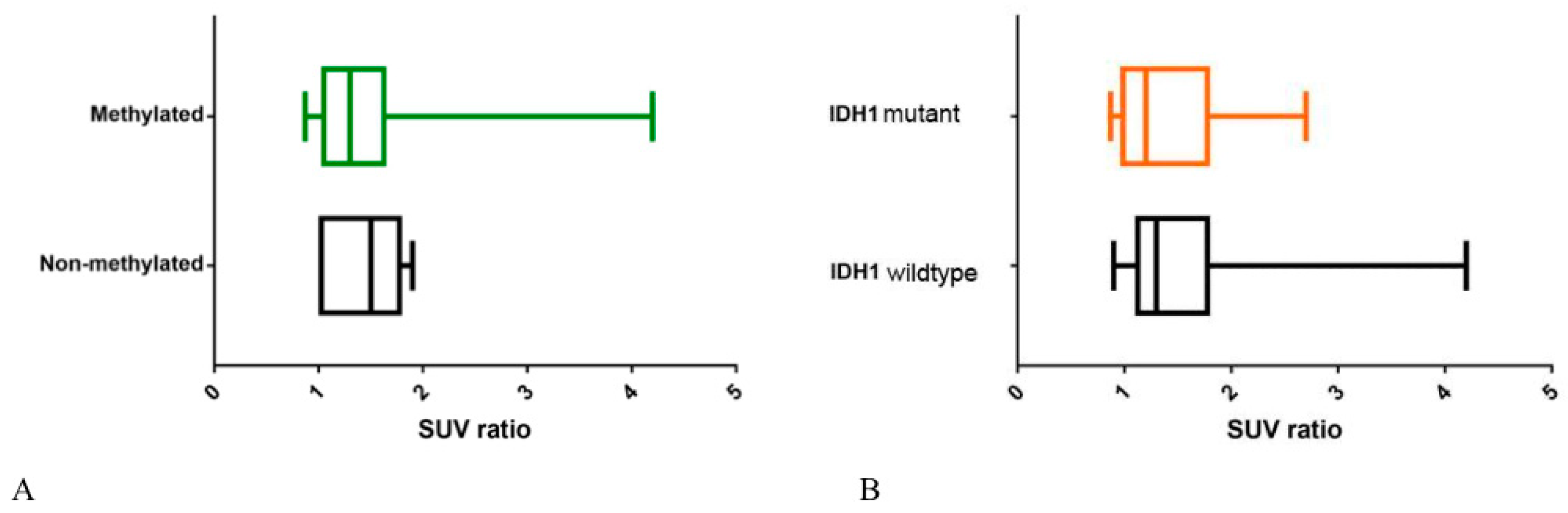
| Patients | Mean SUVr ± Standard Device | p (Mann Whitney U Test) |
|---|---|---|
| O⁶-methylguanine-DNA methyltransferase promoter (MGMTp) methylation vs. no MGMTp methylation | 1.44 ± 0.38 vs. 1.35 ± 0.48 | 0.15 |
| Isocitrate dehydrogenase 1 (IDH1) mutant vs. IDH1 wild-type | 1.438 ± 0.51 vs. 1.46 ± 0.56 | 0.79 |
© 2020 by the authors. Licensee MDPI, Basel, Switzerland. This article is an open access article distributed under the terms and conditions of the Creative Commons Attribution (CC BY) license (http://creativecommons.org/licenses/by/4.0/).
Share and Cite
Cimini, A.; Chiaravalloti, A.; Ricci, M.; Villani, V.; Vanni, G.; Schillaci, O. MGMT Promoter Methylation and IDH1 Mutations Do Not Affect [18F]FDOPA Uptake in Primary Brain Tumors. Int. J. Mol. Sci. 2020, 21, 7598. https://0-doi-org.brum.beds.ac.uk/10.3390/ijms21207598
Cimini A, Chiaravalloti A, Ricci M, Villani V, Vanni G, Schillaci O. MGMT Promoter Methylation and IDH1 Mutations Do Not Affect [18F]FDOPA Uptake in Primary Brain Tumors. International Journal of Molecular Sciences. 2020; 21(20):7598. https://0-doi-org.brum.beds.ac.uk/10.3390/ijms21207598
Chicago/Turabian StyleCimini, Andrea, Agostino Chiaravalloti, Maria Ricci, Veronica Villani, Gianluca Vanni, and Orazio Schillaci. 2020. "MGMT Promoter Methylation and IDH1 Mutations Do Not Affect [18F]FDOPA Uptake in Primary Brain Tumors" International Journal of Molecular Sciences 21, no. 20: 7598. https://0-doi-org.brum.beds.ac.uk/10.3390/ijms21207598