PD-1 and PD-L1 Expression on Circulating Lymphocytes as a Marker of Epstein-Barr Virus Reactivation-Associated Proliferative Glomerulonephritis

 , , , , and
Abstract
:1. Introduction
2. Results
2.1. Characteristics of Basic Parameters in Patients and Healthy Controls
2.2. PGN Patients Have a More “Exhausted” Lymphocyte Profile than NPGN Patients and Healthy Controls
2.3. Understanding the Phenomenon of Lymphocyte Exhaustion in PGN Patients and NPGN Patients
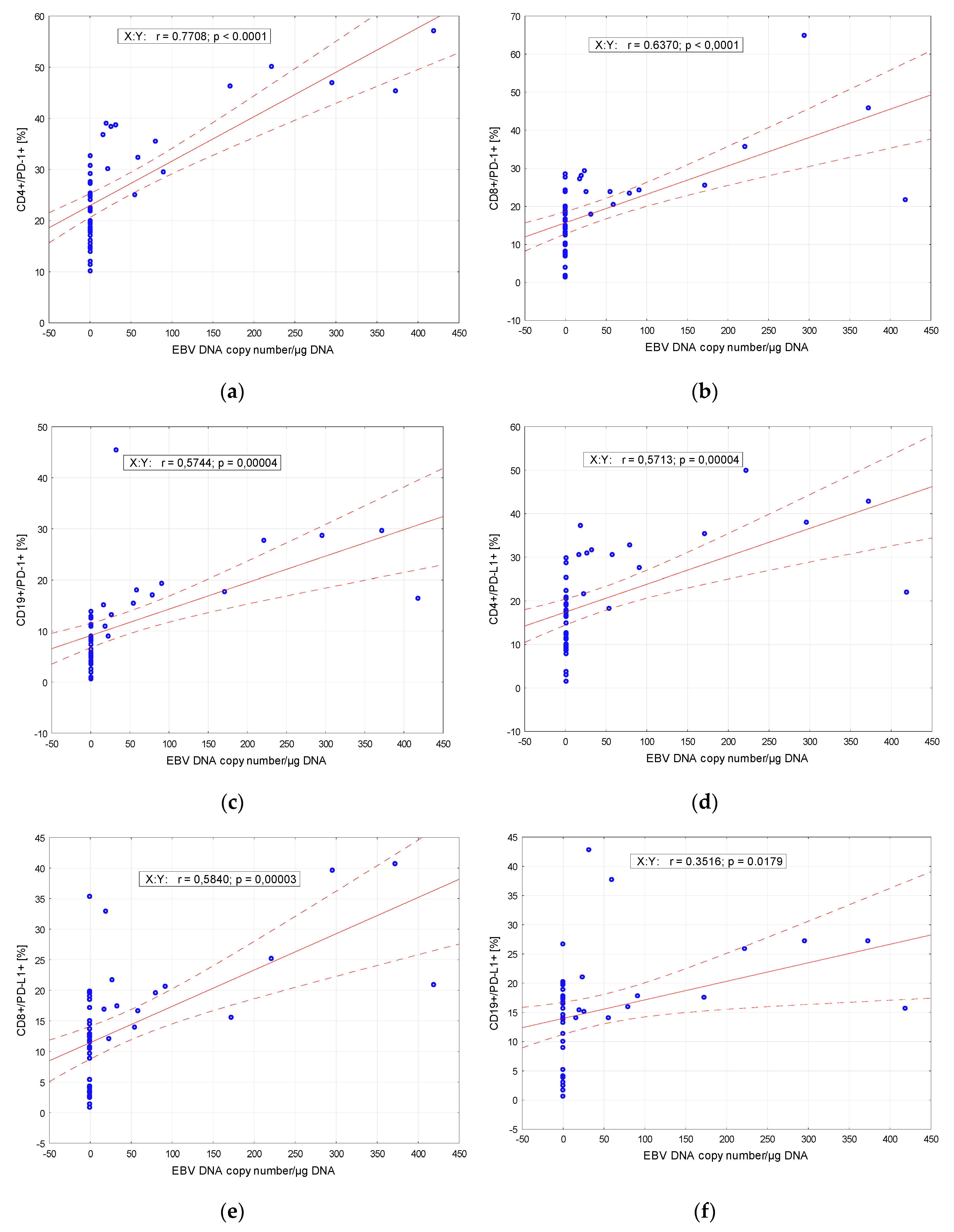
2.4. Correlation of PD-1 and PD-L1—Positive Cells and EBV DNA Copy Numbers
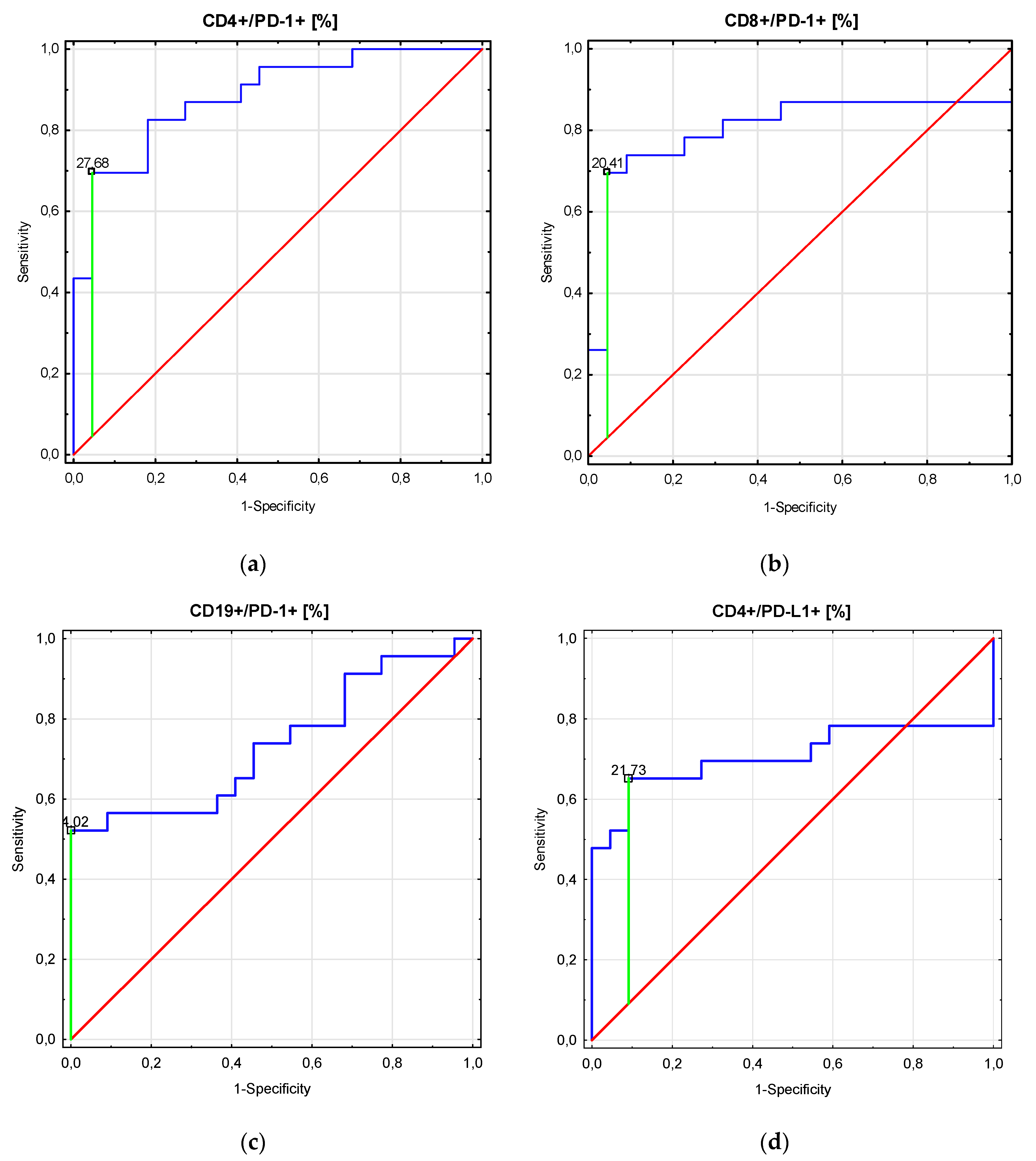
2.5. ROC Analyses of PD-1 and PD-L1—Positive Cells as Possible Non-Invasive Markers to Distinguish Type of Primary Glomerulonephritis
3. Discussion
Limitations of the Study
4. Materials and Methods
4.1. Patients and Controls
4.2. Preparation of Material
4.3. Immunophenotyping
4.4. Patients’ Infection Status Assessment
4.5. DNA Isolation and Calculation of EBV Load and Assessment of Anti-EBV Antibody Status
4.6. Statistical Analysis
5. Conclusions
Author Contributions
Funding
Acknowledgments
Conflicts of Interest
Abbreviations
| EBV | Epstein-Barr virus |
| GN | Glomerulonephritis |
| Ig | Immunoglobulin |
| NPGN | nonproliferative glomerulonephritis |
| PD-1 | programmed cell death protein 1 |
| PD-L1 | programmed death-ligand 1 |
| PGN | proliferative glomerulonephritis |
| ROC | receiver operating characteristic |
References
- Odorizzi, P.M.; Pauken, K.E.; Paley, M.A.; Sharpe, A.; Wherry, E.J. Genetic absence of PD-1 promotes accumulation of terminally differentiated exhausted CD8+ T cells. J. Exp. Med. 2015, 212, 1125–1137. [Google Scholar] [CrossRef] [PubMed]
- Huang, X.; Venet, F.; Wang, Y.L.; Lepape, A.; Yuan, Z.; Chen, Y.; Swan, R.; Kherouf, H.; Monneret, G.; Chung, C.-S.; et al. PD-1 expression by macrophages plays a pathologic role in altering microbial clearance and the innate inflammatory response to sepsis. Proc. Natl. Acad. Sci. USA 2009, 106, 6303–6308. [Google Scholar] [CrossRef] [PubMed] [Green Version]
- Kleffel, S.; Posch, C.; Barthel, S.R.; Mueller, H.; Schlapbach, C.; Guenova, E.; Elco, C.P.; Lee, N.; Juneja, V.R.; Zhan, Q.; et al. Melanoma cell-intrinsic PD-1 receptor functions promote tumor growth. Cell 2015, 162, 1242–1256. [Google Scholar] [CrossRef] [PubMed] [Green Version]
- Li, H.; Li, X.; Liu, S.; Guo, L.; Zhang, B.; Zhang, J.; Ye, Q. Programmed cell death-1 (PD-1) checkpoint blockade in combination with a mammalian target of rapamycin inhibitor restrains hepatocellular carcinoma growth induced by hepatoma cell–intrinsic PD-1. Hepatology 2017, 66, 6. [Google Scholar] [CrossRef] [Green Version]
- Chen, X.; Fosco, D.; Kline, D.E.; Meng, L.; Nishi, S.; Savage, P.A.; Kline, J. PD-1 regulates extrathymic regulatory T-cell differentiation. Eur. J. Immunol. 2014, 44, 2603–2616. [Google Scholar] [CrossRef] [PubMed] [Green Version]
- Zuazo, M.; Arasanz, H.; Bocanegra, A.; Chocarro, L.; Vera, R.; Escors, D.; Kagamu, H.; Kochan, G. Systemic CD4 immunity: A powerful clinical biomarker for PD-L1/PD-1 immunotherapy. EMBO Mol. Med. 2020. [Google Scholar] [CrossRef]
- Lim, T.S.; Chew, V.; Sieow, J.L.; Goh, S.; Yeong, J.P.-S.; Soon, A.L.; Ricciardi-Castagnoli, P. PD-1 expression on dendritic cells suppresses CD8+ T cell function and antitumor immunity. Oncoimmunology 2016, 5, e1085146. [Google Scholar] [CrossRef] [Green Version]
- McKay, J.T.; Egan, R.P.; Yammani, R.D.; Chen, L.; Shin, T.; Yagita, H.; Haas, K.M. PD-1 suppresses protective immunity to streptococcus pneumoniae through a B cell–intrinsic mechanism. J. Immunol. 2015, 194, 2289–2299. [Google Scholar] [CrossRef] [Green Version]
- Protein Atlas. Available online: https://www.proteinatlas.org/ENSG00000188389-PDCD1/cell (accessed on 18 September 2020).
- Chocarro de Erauso, L.; Zuazo, M.; Arasanz, H.; Bocanegra, A.; Hernandez, C.; Fernandez, G.; Garcia-Granda, M.J.; Blanco, E.; Vera, R.; Kochan, G.; et al. Resistance to PD-L1/PD-1 blockade immunotherapy. A tumor-intrinsic or tumor-extrinsic phenomenon? Front. Pharmacol. 2020, 11. [Google Scholar] [CrossRef]
- Ishida, Y. PD-1: Its discovery, involvement in cancer immunotherapy, and beyond. Cells 2020, 9, 1376. [Google Scholar] [CrossRef]
- Jiang, T.T.; Martinov, T.; Xin, L.; Kinder, J.M.; Spanier, J.A.; Fife, B.T.; Way, S.S. Programmed death-1 culls peripheral accumulation of high-affinity autoreactive CD4 T cells to protect against autoimmunity. Cell Rep. 2016, 17, 1783–1794. [Google Scholar] [CrossRef] [PubMed] [Green Version]
- Francisco, L.M.; Sage, P.T.; Sharpe, A.H. The PD-1 pathway in tolerance and autoimmunity. Immunol. Rev. 2010, 236, 219–242. [Google Scholar] [CrossRef] [PubMed]
- Hernandez, C.; Arasanz, H.; Chocarro, L.; Bocanegra, A.; Zuazo, M.; Fernandez-Hinojal, G.; Blanco, E.; Vera, R.; Escors, D.; Kochan, G. Systemic blood immune cell populations as biomarkers for the outcome of immune checkpoint inhibitor therapies. Int. J. Mol. Sci. 2020, 21, 2411. [Google Scholar] [CrossRef] [PubMed] [Green Version]
- Bocanegra, A.; Fernandez-Hinojal, G.; Zuazo-Ibarra, M.; Arasanz, H.; Garcia-Granda, M.; Hernandez, C.; Ibañez, M.; Hernandez-Marin, B.; Martinez-Aguillo, M.; Lecumberri, M.; et al. PD-L1 expression in systemic immune cell populations as a potential predictive biomarker of responses to PD-L1/PD-1 blockade therapy in lung cancer. Int. J. Mol. Sci. 2019, 20, 1631. [Google Scholar] [CrossRef] [Green Version]
- Tamura, H.; Ishibashi, M.; Sunakawa-Kii, M.; Inokuchi, K. PD-L1–PD-1 pathway in the pathophysiology of multiple myeloma. Cancers 2020, 12, 924. [Google Scholar] [CrossRef]
- Chadban, S.J.; Atkins, R.C. Glomerulonephritis. Lancet 2005, 365, 1797–1806. [Google Scholar] [CrossRef]
- Kim, A.H.; Markiewicz, M.A.; Shaw, A.S. New roles revealed for T cells and DCs in glomerulonephritis. J. Clin. Investig. 2009, 119, 1074–1076. [Google Scholar] [CrossRef] [Green Version]
- Sung, S.-S.; Bolton, W.K. T cells and dendritic cells in glomerular disease: The new glomerulotubular feedback loop. Kidney Int. 2010, 77, 393–399. [Google Scholar] [CrossRef] [Green Version]
- Maliakkal, J.G.; Hicks, M.J.; Michael, M.; Selewski, D.T.; Twombley, K.; Rheault, M.N.; Seamon, M.; Misurac, J.M.; Tran, C.L.; Reyes, L.; et al. Renal survival in children with glomerulonephritis with crescents: A pediatric nephrology research consortium cohort study. J. Clin. Med. 2020, 9, 2385. [Google Scholar] [CrossRef]
- Ferri, F. Ferri’s Clinical Advisor 2018, 1st ed.; Ferri, F., Ed.; Elsevier: Amsterdam, The Netherlands, 2017; ISBN 9780323529570. [Google Scholar]
- Mühlig, A.K.; Lee, J.Y.; Kemper, M.J.; Kronbichler, A.; Yang, J.W.; Lee, J.M.; Shin, J.I.; Oh, J. Levamisole in children with idiopathic nephrotic syndrome: Clinical efficacy and pathophysiological aspects. J. Clin. Med. 2019, 8, 860. [Google Scholar] [CrossRef] [Green Version]
- Hebert, L.A.; Parikh, S.; Prosek, J.; Nadasdy, T.; Rovin, B.H. Differential diagnosis of glomerular disease: A systematic and inclusive approach. Am. J. Nephrol. 2013, 38, 253–266. [Google Scholar] [CrossRef] [Green Version]
- Diwan, V.; Brown, L.; Gobe, G.C. Adenine-induced chronic kidney disease in rats. Nephrology 2018, 23, 5–11. [Google Scholar] [CrossRef] [Green Version]
- Mertowski, S.; Grywalska, E.; Gosik, K.; Smarz-Widelska, I.; Hymos, A.; Dworacki, G.; Niedźwiedzka-Rystwej, P.; Drop, B.; Roliński, J.; Załuska, W. TLR2 Expression on select lymphocyte subsets as a new marker in glomerulonephritis. J. Clin. Med. 2020, 9, 541. [Google Scholar] [CrossRef] [Green Version]
- Menè, P.; De Alexandris, L.; Moioli, A.; Raffa, S.; Stoppacciaro, A. Monoclonal gammopathies of renal significance: Renal biopsy and beyond. Cancers 2020, 12, 1741. [Google Scholar] [CrossRef]
- Kauffmann, M.; Bobot, M.; Daniel, L.; Torrents, J.; Knefati, Y.; Moranne, O.; Burtey, S.; Zandotti, C.; Jourde-Chiche, N. Parvovirus B19 infection and kidney injury: Report of 4 cases and analysis of immunization and viremia in an adult cohort of 100 patients undergoing a kidney biopsy. BMC Nephrol. 2020, 21, 260. [Google Scholar] [CrossRef]
- Marinaki, S.; Tsiakas, S.; Skalioti, C.; Lourida, P.; Argyraki, A.; Grigorakos, K.; Boletis, I. A Patient with cryoglobulinemic membranoproliferative GN (MPGN) who survived COVID-19 disease: Case presentation and current data of COVID-19 infection in dialysis and transplanted patients in Greece. Medicina 2020, 56, 355. [Google Scholar] [CrossRef]
- Brodsky, S.V.; Nadasdy, T.; Cassol, C.; Satoskar, A. IgA staining patterns differentiate between IgA nephropathy and IgA-dominant infection-associated glomerulonephritis. Kidney Int. Rep. 2020, 5, 909–911. [Google Scholar] [CrossRef]
- Grywalska, E.; Smarz-Widelska, I.; Krasowska-Zajac, E.; Korona-Glowniak, I.; Zaluska-Patel, K.; Mielnik, M.; Podgajna, M.; Malm, A.; Rolinski, J.; Zaluska, W. The PD-1/PD-L1 inhibitory pathway is altered in primary glomerulonephritides. Arch. Immunol. Ther. Exp. 2018, 66, 133–143. [Google Scholar] [CrossRef]
- De Paschale, M.; Clerici, P. Serological diagnosis of Epstein-Barr virus infection: Problems and solutions. World J. Virol. 2012, 1, 31–43. [Google Scholar] [CrossRef]
- KDIGO. KDIGO clinical practice guideline for glomerulonephritis. Kidney Int. 2012, 2 (Suppl. S2), 139–274. [Google Scholar]
- Floege, J.; Amann, K. Primary glomerulonephritides. Lancet 2016, 387, 2036–2048. [Google Scholar] [CrossRef]
- Kronbichler, A.; Gauckler, P.; Bruchfeld, A. Rituximab in minimal change disease and focal segmental glomerulosclerosis. Nephrol. Dial. Transpl. 2019, 23, 5–11. [Google Scholar] [CrossRef]
- Whittier, W.L.; Korbet, S.M. Timing of complications in percutaneous renal biopsy. J. Am. Soc. Nephrol. 2004, 15, 142–147. [Google Scholar] [CrossRef] [Green Version]
- Huang, Y.-T.; Lai, M.-Y.; Kan, W.-C.; Shiao, C.-C. Independent predictive ability of procalcitonin of acute kidney injury among critically ill patients. J. Clin. Med. 2020, 9, 1939. [Google Scholar] [CrossRef]
- Rodrigues, P.G.; Bringhenti, R.N.; do Nascimento, J.F.; Joelsons, G.; dos Santos, M.; Pereira, S.; Veronese, F.V. Expression patterns of podocyte-associated mRNAs in patients with proliferative or non-proliferative glomerulopathies. Int. J. Clin. Exp. Pathol. 2014, 7, 2185–2198. [Google Scholar]
- Chandra, P.; Kopp, J.B. Viruses and collapsing glomerulopathy: A brief critical review. Clin. Kidney J. 2013, 6, 1–5. [Google Scholar] [CrossRef] [Green Version]
- Karamadoukis, L.; Toth, T.; Tomson, C. Membranoproliferative GN associated with an Epstein-Barr virus infection. NDT Plus 2008, 1, 319–321. [Google Scholar]
- Grywalska, E.; Pasiarski, M.; Sosnowska-Pasiarska, B.; Macek, P.; Rolińska, A.; Samardakiewicz, M.; Ludian, J.; Góźdź, S.; Roliński, J. Programmed cell death 1 expression and Epstein-Barr virus infection in chronic lymphocytic leukaemia: A prospective cohort study. Cancer Manag. Res. 2019, 11, 7605–7618. [Google Scholar] [CrossRef] [Green Version]
- Korona-Glowniak, I.; Grywalska, E.; Grzegorczyk, A.; Roliński, J.; Glowniak, A.; Malm, A. Bacterial colonization in patients with chronic lymphocytic leukemia and factors associated with infections and colonization. J. Clin. Med. 2019, 8, 861. [Google Scholar] [CrossRef] [Green Version]
- Roliński, J.; Grywalska, E.; Pyzik, A.; Dzik, M.; Opoka-Winiarska, V.; Surdacka, A.; Maj, M.; Burdan, F.; Pirożyński, M.; Grabarczyk, P.; et al. Interferon alpha as antiviral therapy in chronic active Epstein-Barr virus disease with interstitial pneumonia-case report. BMC Infect. Dis. 2018, 18, 190. [Google Scholar] [CrossRef] [Green Version]
- Tomaszewski, M.; Grywalska, E.; Tomaszewski, A.; Błaszczak, P.; Kurzyna, M.; Roliński, J.; Kopeć, G. Overexpression of PD-1 on peripheral blood lymphocytes in patients with idiopathic pulmonary arterial hypertension and its association with high viral loads of Epstein-Barr virus and poor clinical parameters. J. Clin. Med. 2020, 9, 1966. [Google Scholar] [CrossRef]
- Morales-Sánchez, A.; Torres, J.; Cardenas-Mondragón, M.G.; Romo-González, C.; Camorlinga-Ponce, M.; Flores-Luna, L.; Fuentes-Pananá, E.M. Detection of Epstein-Barr Virus DNA in gastric biopsies of pediatric patients with dyspepsia. Pathogens 2020, 9, 623. [Google Scholar] [CrossRef]
- Krishna, G.; Soman Pillai, V.; Valiya Veettil, M. Upregulation of GLS1 isoforms KGA and GAC facilitates mitochondrial metabolism and cell proliferation in Epstein–Barr Virus infected cells. Viruses 2020, 12, 811. [Google Scholar] [CrossRef]
- Chau, S.-L.; Tong, J.-M.; Chow, C.; Kwan, J.-H.; Lung, R.-M.; Chung, L.-Y.; Tin, E.-Y.; Wong, S.-Y.; Cheung, A.-K.; Lau, R.-H.; et al. Distinct molecular landscape of Epstein–Barr virus associated pulmonary lymphoepithelioma-like carcinoma revealed by genomic sequencing. Cancers 2020, 12, 2065. [Google Scholar] [CrossRef]
- Lo, A.-F.; Dawson, C.W.; Lung, H.L.; Wong, K.-L.; Young, L.S. The therapeutic potential of targeting BARF1 in EBV-associated malignancies. Cancers 2020, 12, 1940. [Google Scholar] [CrossRef]
- Maple, P.A.C. Cytomegalovirus and Epstein–Barr virus associations with neurological diseases and the need for vaccine development. Vaccines 2020, 8, 35. [Google Scholar] [CrossRef] [Green Version]
- Tarlinton, R.E.; Khaibullin, T.; Granatov, E.; Martynova, E.; Rizvanov, A.; Khaiboullina, S. The interaction between viral and environmental risk factors in the pathogenesis of multiple sclerosis. Int. J. Mol. Sci. 2019, 20, 303. [Google Scholar] [CrossRef] [Green Version]
- Zhou, Y.; Shi, D.; Miao, J.; Wu, H.; Chen, J.; Zhou, X.; Hu, D.; Zhao, C.; Deng, W.; Xie, C. PD-L1 predicts poor prognosis for nasopharyngeal carcinoma irrespective of PD-1 and EBV-DNA load. Sci. Rep. 2017, 7, 43627. [Google Scholar] [CrossRef] [Green Version]
- Iwama, H.; Horikoshi, S.; Shirato, Y.; Tomino, Y. Epstein-Barr virus detection in kidney biopsy specimens correlates with glomerular mesangial injury. Am. J. Kidney Dis. 1998, 32, 785–793. [Google Scholar] [CrossRef]
- Lin, M.H.; Huang, J.J.; Chen, T.Y.; Chen, F.F.; Chang, K.C.; Liu, M.F.; Huang, W.T.; Su, W.C.; Tsao, C.J. EBER-1 positive diffuse large cell lymphoma presenting as lupus nephritis. Lupus 2003, 12, 486–489. [Google Scholar] [CrossRef]
- Mikhalkova, D.; Khanna, S.; Vaidya, R.; Sethi, S.; Hogan, M.C. Epstein–Barr virus-associated nephrotic syndrome. Epstein–Barr virus-associated nephrotic syndrome. Clin. Kidney J. 2012, 5, 50–52. [Google Scholar] [CrossRef] [Green Version]
- Igawa, T.; Nakashima, H.; Sadanaga, A.; Masutani, K.; Miyake, K.; Shimizu, S.; Takeda, A.; Hamano, S.; Yoshida, H. Deficiency in EBV-induced gene 3 (EBI3) in MRL/lpr mice results in pathological alteration of autoimmune glomerulonephritis and sialadenitis. Modern Rheumatol. 2009, 19, 33–41. [Google Scholar] [CrossRef]


| Parameters | Study Group (n = 45) | Control Goup (n = 20) | t/Z | p Value | ||
|---|---|---|---|---|---|---|
| Mean ± SD | Median (Range) | Mean ± SD | Median (Range) | |||
| WBC [103/mm3] | 6.8 ± 1.7 | 6.6 (4.3–9.9) | 6.8 ± 0.4 | 6.7 (6.3–7.6) | −0.55 | 0.58 |
| LYM [103/mm3] | 2.1 ± 0.7 | 1.96 (1.2–3.7) | 2.5 ± 0.6 | 2.5 (1.5–3.7) | −2.53 | 0.011 |
| RBC [106/mm3] | 4.7 ± 1.6 | 4.4 (3.3–11.8) | 5.2 ± 0.4 | 5.12 (4.5–5.8) | −4.7 | <0.0001 |
| HGB [g/dL] | 13.2 ± 1.6 | 13.2 (9.3–16.4) | 14.3 ± 1.2 | 14.35 (12.5–16.9) | −2.85 | 0.0058 |
| PLT [103/mm3] | 238.9 ± 61.3 | 222.0 (147.0–410.0) | 279.0 ± 57.0 | 281.5 (186.0–403.0) | −2.49 | 0.013 |
| Urea [mg/dL] | 51.9 ± 28.6 | 44.3 (13.0–115.7) | 31.4 ± 6.9 | 32.0 (18.0–42.0) | 2.59 | 0.0097 |
| BUN [mg/dL] | 24.3 ± 13.4 | 20.7 (6.1–54.0) | 14.7 ± 3.2 | 14.95 (8.4–19.6) | 2.59 | 0.0097 |
| Serum creatinine [mg/dL] | 1.2 ± 0.6 | 0.96 (0.37–2.3) | 0.9 ± 0.1 | 0.9 (0.7–1.1) | 1.07 | 0.29 |
| eGRF [ml/min/1.73 m2] | 86.1 ± 33.1 | 81.5 (26.7–146.8) | 125.6 ± 10.3 | 121.1 (115.0–148.2) | −5.2 | <0.0001 |
| Serum uric acid [mg/dL] | 6.9 ± 1.7 | 7.1 (3.8–11.9) | 6.2 ± 1.4 | 6.95 (3.7–7.9) | 1.73 | 0.083 |
| Total quantity of protein in a 24-h urine collection test [g/24 h] | 5.7 ± 5.6 | 4.8 (0.01–24.9) | 0.0 ± 0 | 0.0 | 6.39 | <0.0001 |
| Serum IgG [g/L] | 6.6 ± 2.8 | 6.3 (2.8–15.7) | 12.7 ± 1.4 | 12.79 (10.1–15.5) | −5.79 | <0.0001 |
| Serum IgM [g/L] | 1.7 ± 1.1 | 1.2 (0.2–3.98) | 1.7 ± 0.3 | 1.6 (1.2–2.2) | −1.02 | 0.31 |
| Serum IgA [g/L] | 2.4 ± 1.6 | 2.0 (0.3–6.1) | 2.4 ± 0.8 | 2.56 (0.9–3.9) | −0.76 | 0.45 |
| Serum total protein [g/dL] | 5.4 ± 1.0 | 5.4 (3.2–7.4) | 7.4 ± 0.6 | 7.35 (6.4–8.2) | −8.0 | <0.0001 |
| Serum albumin [g/L] | 2.6 ± 0.9 | 2.8 (0.6–4.1) | 4.2 ± 0.4 | 4.2 (3.5–4.75) | −7.75 | <0.0001 |
| Serum complement component C3 [g/L] | 1.2 ± 0.3 | 1.2 (0.4–2.0) | 1.3 ± 0.2 | 1.2 (0.95–1.8) | −0.85 | 0.40 |
| Serum complement component C4 [g/L] | 0.3 ± 0.08 | 0.28 (0.11–0.55) | 0.3 ± 0.08 | 0.3 (0.15–0.4) | 0.82 | 0.42 |
| Parameters | NPGN (n = 22) | PGN (n = 23) | t/Z | p Value | ||
|---|---|---|---|---|---|---|
| Mean ± SD | Median (Range) | Mean ± SD | Median (Range) | |||
| Male/Female, n (%) | 14 (63.6)/8 (36.4) | 14 (60.9)/9 (39.1) | 1.0 | |||
| Age [years] | 42.8 ± 13.2 | 37.4 (19.0–70.0) | 37.4 ± 14.3 | 35.0 (20.0–70.0) | 1.31 | 0.20 |
| Arterial hypertension, n (%) | 2 (9.1) | 13 (56.5) | 0.0012 | |||
| Urea [mg/dL] | 55.2 ± 31.5 | 45.1 (17.8–115.7) | 48.8 ± 25.9 | 44.3 (13.0–115.7) | 0.74 | 0.46 |
| BUN [mg/dL] | 25.8 ± 14.7 | 21.1 (8.3–54.0) | 22.8 ± 12.1 | 20.7 (6.1–54.0) | 0.74 | 0.46 |
| Serum creatinine [mg/dL] | 1.1 ± 0.6 | 0.9 (0.37–2.3) | 1.3 ± 0.6 | 1.2 (0.5–2.3) | −0.9 | 0.37 |
| eGRF [ml/min/1.73 m2] | 106. ± 28.2 | 115.7 (54.6–146.8) | 66.7 ± 25.1 | 69.9 (26.7–117.3) | 4.99 | <0.0001 |
| Serum uric acid [mg/dL] | 6.6 ± 1.9 | 6.4 (3.8–11.9) | 7.2 ± 1.5 | 7.6 (4.0–9.3) | −1.19 | 0.24 |
| Serum IgG [g/L] | 6.5 ± 2.1 | 6.7 (3.2–11.4) | 6.7 ± 3.4 | 6.1 (8.8–15.7) | −0.25 | 0.80 |
| Serum IgM [g/L] | 2.2 ± 1.1 | 2.7 (0.4–3.98) | 1.1 ± 0.7 | 1.1 (0.2–3.2) | 3.87 | 0.0004 |
| Serum IgA [g/L] | 2.0 ± 0.95 | 2.1 (0.6–3.6) | 2.6 ± 1.9 | 1.9 (0.3–6.1) | −1.34 | 0.19 |
| Serum total protein [g/dL] | 5.4 ± 1.0 | 5.2 (4.0–7.4) | 5.5 ± 1.0 | 5.7 (3.2–6.9) | −0.57 | 0.57 |
| Serum albumin [g/L] | 2.5 ± 0.8 | 2.6 (0.8–3.8) | 2.8 ± 0.9 | 2.8 (0.6–4.1) | −0.86 | 0.40 |
| Total quantity of protein in a 24-hour urine collection test [g/24 h] | 8.5 ± 6.1 | 6.3 (3.0–24.9) | 3.0 ± 3.6 | 0.9 (0.01–10.3) | 3.46 | 0.0005 |
| Serum complement component C3 [g/L] | 1.4 ± 0.3 | 1.3 (0.9–2.0) | 1.1 ± 0.3 | 1.2 (0.4–1.7) | 3.29 | 0.002 |
| Serum complement component C4 [g/L] | 0.32 ± 0.1 | 0.28 (0.2–0.55) | 0.3 ± 0.07 | 0.3 (0.1–0.5) | 1.32 | 0.19 |
| Variable (Unit) | Study Group (n = 45) | Control Goup (n = 20) | t/Z | p Value |
|---|---|---|---|---|
| Median (Range) | Median (Range) | |||
| Frequencies of T CD3+ lymphocytes (%) | 74.4 (5.2–87.98) | 72.4 (70.0–74.75) | 1.36 | 0.17 |
| Frequencies of B CD19+ lymphocytes (%) | 10.98 (1.7–70.3) | 10.8 (6.0–14.5) | 0.23 | 0.82 |
| Frequencies of NK cells (%) | 12.6 (1.9–36.8) | 14.5 (12.6–19.8) | −2.42 | 0.016 |
| Frequencies of T CD3+/CD4+ cells (%) | 43.1 (26.1–63.4) | 41.5 (40.5–44.2) | 0.68 | 0.50 |
| Frequencies of T CD3+/CD8+ cells (%) | 28.0 (18.1–48.7) | 29.97 (28.9–33.2) | −2.30 | 0.022 |
| T CD3+/CD4+: T CD3+/CD8+ ratio | 1.6 (0.7–3.2) | 1.4 (1.2–1.5) | 1.73 | 0.084 |
| CD4+/PD-1+ (%) | 24.7 (10.3–57.3) | 5.3 (2.65–7.7) | 6.39 | <0.0001 |
| CD8+/PD-1+ (%) | 18.0 (1.5–64.8) | 3.7 (1.4–6.2) | 5.55 | <0.0001 |
| CD19+/PD-1+ (%) | 10.8 (0.6–45.6) | 1.9 (0.4–3.4) | 5.31 | <0.0001 |
| CD4+/PD-L1+ (%) | 17.8 (1.4–49.8) | 1.7 (1.0–4.5) | 6.16 | <0.0001 |
| CD8+/PD-L1+ (%) | 12.8 (0.9–40.7) | 0.4 (0.3–0.7) | 6.39 | <0.0001 |
| CD19+/PD-L1+ (%) | 15.7 (0.7–43.0) | 0.2 (0.07–1.0) | 6.37 | <0.0001 |
| Variable (Unit) | NPGN (n = 22) | PGN (n = 23) | t/Z | p Value |
|---|---|---|---|---|
| Median (Range) | Median (Range) | |||
| Frequencies of T CD3+ lymphocytes (%) | 76.9 (5.2–87.98) | 73.3 (63.3–85.1) | −0.32 | 0.75 |
| Frequencies of B CD19+ lymphocytes (%) | 9.5 (1.7–70.3) | 11.3 (7.0–19.1) | 0.19 | 0.85 |
| Frequencies of NK cells (%) | 10.2 (1.9–36.8) | 12.9 (7.3–35.3) | −0.76 | 0.89 |
| Frequencies of T CD3+/CD4+ cells (%) | 44.7 (26.1–63.4) | 41.4 (34.5–53.3) | 1.06 | 0.45 |
| Frequencies of T CD3+/CD8+ cells (%) | 27.3 (18.4–48.7) | 28.9 (18.1–42.1) | −0.40 | 0.77 |
| T CD3+/CD4+: T CD3+/CD8+ ratio | 1.6 (0.7–3.2) | 1.4 (0.8–2.7) | 0.73 | 0.47 |
| CD4+/PD-1+ (%) | 18.6 (10.3–32.6) | 30.8 (16.3–57.3) | −5.38 | <0.0001 |
| CD8+/PD-1+ (%) | 13.2 (4.0–27.8) | 23.9 (1.5–64.28) | −3.22 | 0.0025 |
| CD19+/PD-1+ (%) | 7.9 (0.6–14.0) | 14.0 (0.9–45.6) | −2.99 | 0.0046 |
| CD4+/PD-L1+ (%) | 16.7 (9.0–28.7) | 27.7 (1.4–49.8) | −2.74 | 0.0088 |
| CD8+/PD-L1+ (%) | 12.1 (1.4–35.5) | 16.8 (0.9–40.7) | −1.39 | 0.17 |
| CD19+/PD-L1+ (%) | 14.4 (1.7–26.8) | 15.9 (0.7–43.0) | −1.56 | 0.13 |
| Parameter | Prognostic Value | AUC | 95% CI | p Value |
|---|---|---|---|---|
| CD4+/PD-1+ (%) | 27.68 | 0.89 | 0.79–0.98 | <0.0001 |
| CD8+/PD-1+ (%) | 20.4 | 0.80 | 0.66–0.95 | <0.0001 |
| CD19+/PD-1+ (%) | 14.2 | 0.74 | 0.59–0.88 | 0.002 |
| CD4+/PD-L1+ (%) | 21.73 | 0.70 | 0.54–0.88 | 0.017 |
| CD8+/PD-L1+ (%) | 15.51 | 0.62 | 0.45–0.79 | 0.15 |
| CD19+/PD-L1+ (%) | 20.27 | 0.62 | 0.45–0.78 | 0.18 |
| Parameter | Study Group (n = 45) | Control Group (n = 20) | t/Z | p Value | ||
|---|---|---|---|---|---|---|
| Mean ± SD | Median (Range) | Mean ± SD | Median (Range) | |||
| Age (years) | 40.0 ± 13.9 | 41.0 (19.0–70.0) | 41.4 ± 13.1 | 42.5 (20.0–60.0) | −0.38 | 0.71 |
| Diagnosis, n (%): | NA | NA | NA | |||
| Membranous glomerulonephritis | 11 (24.4) | |||||
| Minimal change disease | 11 (24.4) | |||||
| Membranoproliferative | 11 (24.4) | |||||
| glomerulonephritis | ||||||
| IgA nephropathy | 12 (26.7) | |||||
| Type of glomerulonephritis, n (%): | NA | NA | NA | |||
| Nonproliferative | 22 (48.9) | |||||
| Proliferative | 23 (51.1) | |||||
Publisher’s Note: MDPI stays neutral with regard to jurisdictional claims in published maps and institutional affiliations. |
© 2020 by the authors. Licensee MDPI, Basel, Switzerland. This article is an open access article distributed under the terms and conditions of the Creative Commons Attribution (CC BY) license (http://creativecommons.org/licenses/by/4.0/).
Share and Cite
Grywalska, E.; Smarz-Widelska, I.; Korona-Głowniak, I.; Mertowski, S.; Gosik, K.; Hymos, A.; Ludian, J.; Niedźwiedzka-Rystwej, P.; Roliński, J.; Załuska, W. PD-1 and PD-L1 Expression on Circulating Lymphocytes as a Marker of Epstein-Barr Virus Reactivation-Associated Proliferative Glomerulonephritis. Int. J. Mol. Sci. 2020, 21, 8001. https://0-doi-org.brum.beds.ac.uk/10.3390/ijms21218001
Grywalska E, Smarz-Widelska I, Korona-Głowniak I, Mertowski S, Gosik K, Hymos A, Ludian J, Niedźwiedzka-Rystwej P, Roliński J, Załuska W. PD-1 and PD-L1 Expression on Circulating Lymphocytes as a Marker of Epstein-Barr Virus Reactivation-Associated Proliferative Glomerulonephritis. International Journal of Molecular Sciences. 2020; 21(21):8001. https://0-doi-org.brum.beds.ac.uk/10.3390/ijms21218001
Chicago/Turabian StyleGrywalska, Ewelina, Iwona Smarz-Widelska, Izabela Korona-Głowniak, Sebastian Mertowski, Krzysztof Gosik, Anna Hymos, Jarosław Ludian, Paulina Niedźwiedzka-Rystwej, Jacek Roliński, and Wojciech Załuska. 2020. "PD-1 and PD-L1 Expression on Circulating Lymphocytes as a Marker of Epstein-Barr Virus Reactivation-Associated Proliferative Glomerulonephritis" International Journal of Molecular Sciences 21, no. 21: 8001. https://0-doi-org.brum.beds.ac.uk/10.3390/ijms21218001