Review

Methyl Jasmonate Induced Oxidative Stress and Accumulation of Secondary Metabolites in Plant Cell and Organ Cultures

by
Thanh-Tam Ho
1,
Hosakatte Niranjana Murthy
2 and
So-Young Park
3,*
1
Institute for Global Health Innovations, Duy Tan University, Danang 550000, Vietnam
2
Department of Botany, Karnatak University, Dharwad 580003, India
3
Department of Horticultural Science, Chungbuk National University, Cheongju 28644, Korea
*
Author to whom correspondence should be addressed.
Int. J. Mol. Sci. 2020, 21(3), 716; https://0-doi-org.brum.beds.ac.uk/10.3390/ijms21030716
Submission received: 16 December 2019 / Revised: 18 January 2020 / Accepted: 19 January 2020 / Published: 22 January 2020
(This article belongs to the Special Issue Jasmonic Acid Pathway in Plants)

Abstract

:
Recently, plant secondary metabolites are considered as important sources of pharmaceuticals, food additives, flavours, cosmetics, and other industrial products. The accumulation of secondary metabolites in plant cell and organ cultures often occurs when cultures are subjected to varied kinds of stresses including elicitors or signal molecules. Application of exogenous jasmonic acid (JA) and methyl jasmonate (MJ) is responsible for the induction of reactive oxygen species (ROS) and subsequent defence mechanisms in cultured cells and organs. It is also responsible for the induction of signal transduction, the expression of many defence genes followed by the accumulation of secondary metabolites. In this review, the application of exogenous MJ elicitation strategies on the induction of defence mechanism and secondary metabolite accumulation in cell and organ cultures is introduced and discussed. The information presented here is useful for efficient large-scale production of plant secondary metabolites by the plant cell and organ cultures.

1. Introduction

In plant defence systems, each cell has acquired the capability to respond to pathogens and environmental stresses and to build up a defence response. A plant defence mechanism is determined by several factors, mainly depending on their genetic characteristics and physiological state [1,2]. The supplementation of exogenous methyl jasmonate (MJ) to in vitro cultures is responsible for the induction of reactive oxygen species (ROS), regulation of defence response by an accumulation of antioxidant enzyme activity [3,4]. On the other hand, MJ also stimulates molecular signal transduction, regulation of gene expression which leads to accumulation of secondary metabolites [5,6].
Free radicals such as superoxide anion (O2•−), hydroxyl radical (OH), as well as non-radical molecules like hydrogen peroxide (H2O2), singlet oxygen (1O2), are accumulated in plant cells in response to stress mechanism [6,7]. Elicitation with MJ, salicylic acid (SA), environmental stresses, as well as pathogens attack lead to enhanced generation of ROS in plants due to disruption of cellular homeostasis [8,9]. When the level of ROS exceeds the defence mechanisms, a cell is said to be in a state of “oxidative stress” [10]. Although all ROS are extremely harmful to organisms at high concentrations, ROS are well-described second messengers in a variety of cellular processes including tolerance to environmental stresses at low concentration [11]. ROS when present in lower concentration, control many different processes in cells. However, higher concentration ROS in the cell is toxic and detrimental. Therefore, it is necessary for the cells to control the level of ROS tightly to avoid any oxidative injury and not to eliminate them completely [10]. The antioxidants enzymes include superoxide dismutase (SOD), catalase (CAT), guaiacol peroxidase (G-POD), enzymes of ascorbate-glutahione (AsA-GSH) cycle such as ascorbate peroxidase (APX), monodehydroascorbate reductase (MDHAR), dehydroascorbate reductase (DHAR), and glutathione reductase (GR). While ascorbate (AsA), glutathione (GSH), carotenoids, tocopherols, and phenolics serve as potent nonenzymic antioxidants within the cell, were achieved scavenging or detoxification of excess ROS [10].
Application of exogenous MJ to in vitro cultures has emerged as a novel technique for a hyperaccumulation of secondary metabolites. It has been demonstrated that application of MJ to in vitro cultures has induced the antioxidant enzyme activity, expression of defence-related genes, and over-production of secondary metabolites [3,4,12,13,14]. The process or methodology for enhancement of secondary metabolites in Polygonum multiflorum adventitious and hairy roots cultures by application of MJ has been presented in Figure 1. The first step is to establish an in vitro culture system, the second step is the optimization of culture conditions for biomass accumulation in the bioreactor culture system. Then, concentration and exposure time of MJ were determined in the third step, thereafter, scale-up of culture process could be achieved up to pilot-scale. HPLC and Fourier-transform infrared spectroscopy (FT-IR) analysis were applied to investigate the quality of bioactive compounds in the culture [15,16,17]. In this review, the role of MJ on the induction of oxidative stress, antioxidant enzyme activity, signal molecules transduction, and secondary metabolite accumulation in plant cell and organ culture system are introduced and discussed.

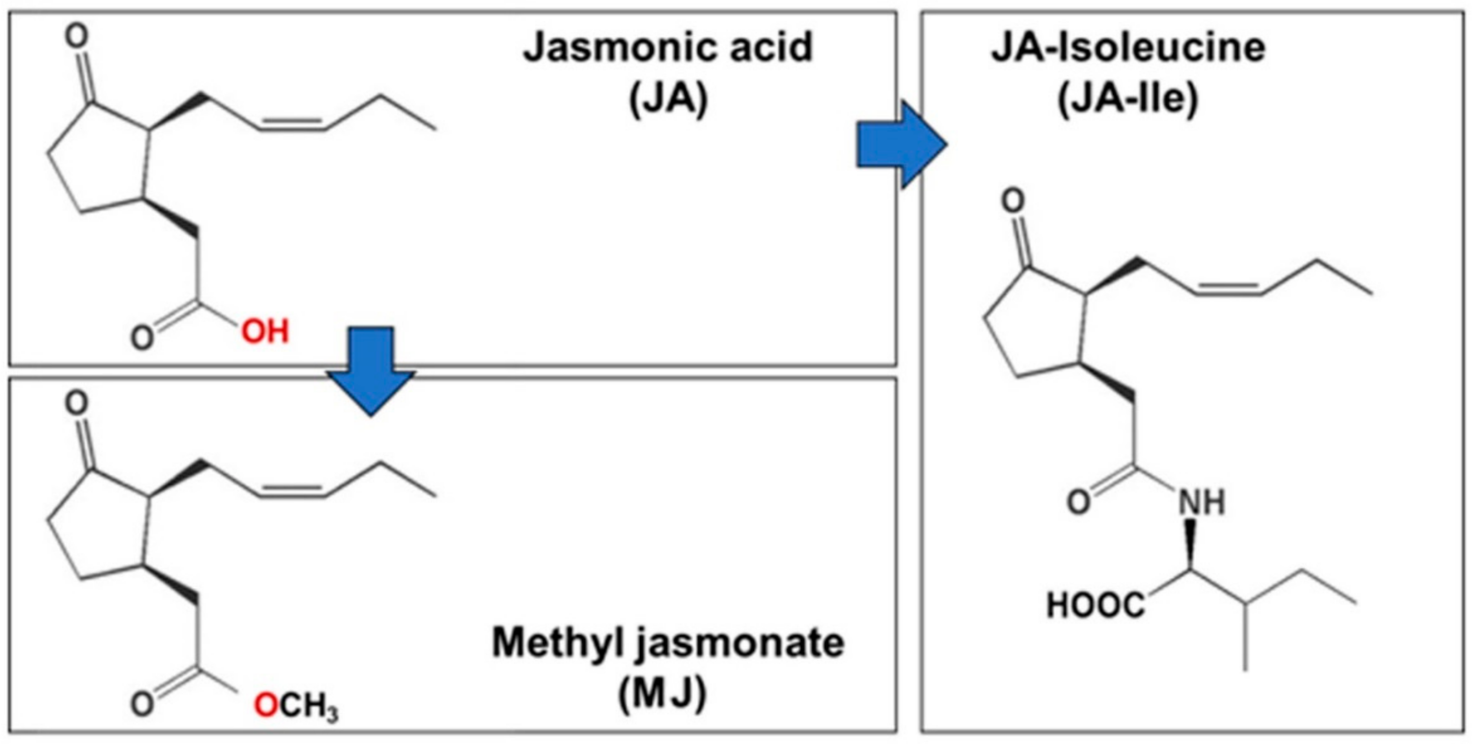
2. Jasmonic Acid (JA) and Methyl Jasmonate (MJ)

The Jasmonic acid (JA) and its derivatives were collectively called Jasmonates (JAs). JAs are cyclopentanone compounds or plant hormones derived from α-linolenic acid. It includes a group of oxygenated fatty acids, and JA is the main precursor of different compounds to this group like methyl jasmonate (MJ), which was first isolated from the essential oil of Jasminum grandiflorum [18], and the free acid was isolated subsequently from the culture filtrates of the fungus Lasiodiplodia theobromae [19]. JAs was synthesized from fatty acid including three steps. The first step occurs in the membrane chloroplast, where α-linolenic acid and hexadecatrienoic acid were released from membrane phospholipids [20,21]. Lipoxygenase (LOX), allene oxide synthase (AOS), allene oxide cyclase (AOC) are key enzyme of JA biosynthesis in chloroplast and they form (cis-(+)-12-oxophytodienoic acid (OPDA) and dn-OPDA (dinor-OPDA) [22,23]. The second step of JAs synthesis takes place in peroxisome by the process of β-oxidation to give finally jasmonic acid [21,23]. In the final step, JA is exported to the cytoplasm for further modification like MJ, JA-Ile (Figure 2) [21,24,25].
Jasmonic acid (JA) and its precursors and derivatives are known to possess many physiological processes in plant growth and development, and especially the mediation of plant responses to biotic and abiotic stresses [26,27,28]. They have been used extensively for elicitation studies within in vitro culture systems [29]. The elicitation process leads to crosstalk between jasmonates with their receptors which are present in the plasma membrane, which leads an array of defence responses with the cells, including the production of reactive oxygen species (ROS), reactive nitrogen species (RNS), and induction of enzymes of oxidative stress protection [29]. This also leads to synthesis and accumulation of signaling molecules such as JA, salicylic acid (SA), nitric oxide (NO), ethylene (ET) inside the cell and subsequent regulation of gene expression involved in secondary metabolite production [5,30,31]. Therefore, a large number of chemicals such as JA, MJ, SA, ET, are used for elicitation studies in vitro. However, MJ is the most commonly used elicitor which has shown a pronounced effect of accumulation secondary metabolites in plant cell and organ cultures [29,30]. Giri and Zaheer et al. [29] reported that MJ was most abundantly used chemical elicitor (60% of reports) in in vitro culture system, followed by SA and JA (approximately 15 and 10%, respectively). Cell suspension, adventitious roots and hairy roots are commonly used as culture system followed by multiple shoots and embryos for elicitation experiments [4,29,32].

3. Usage of Methyl Jasmonate (MJ) on Oxidative Stress and Antioxidant Response

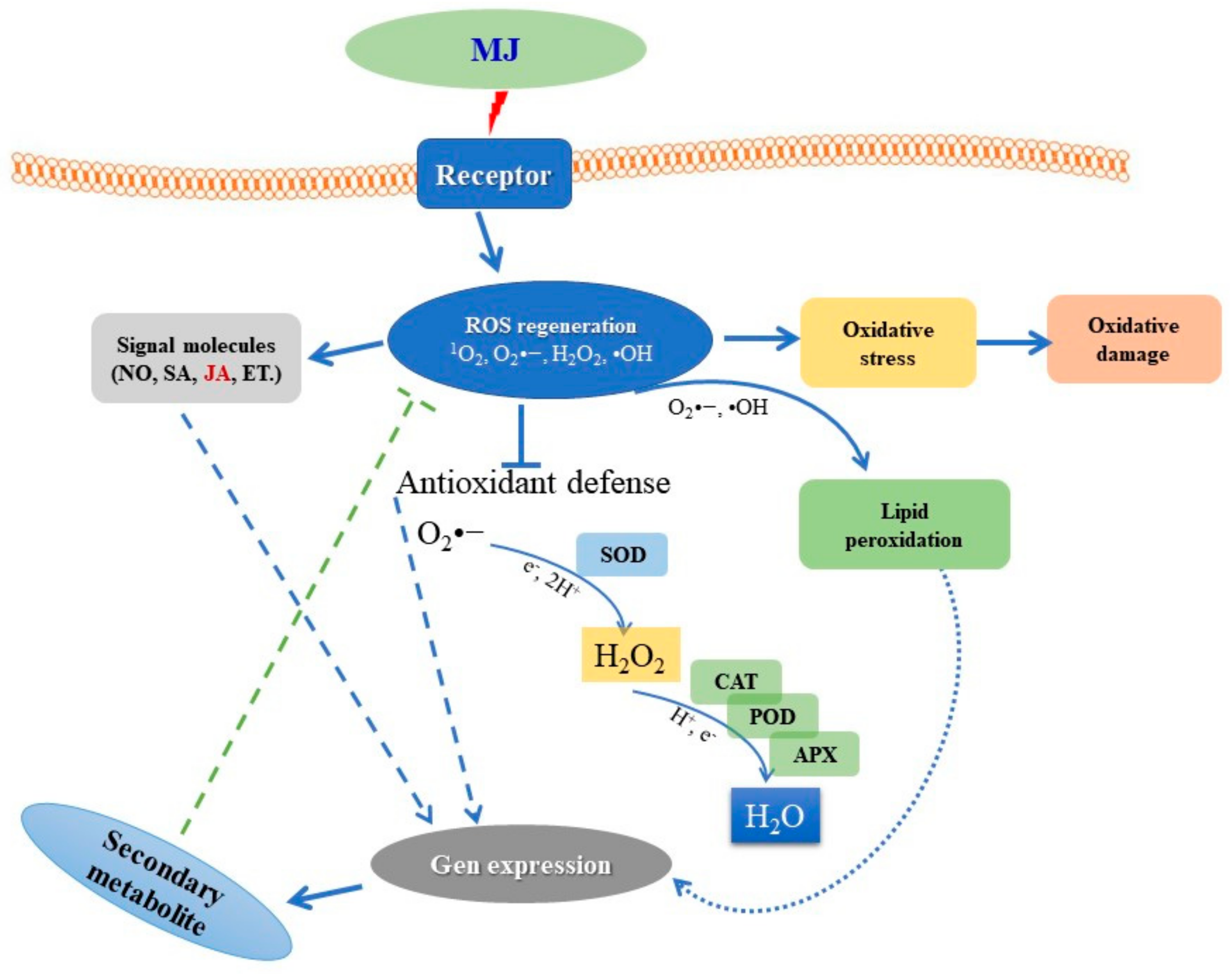
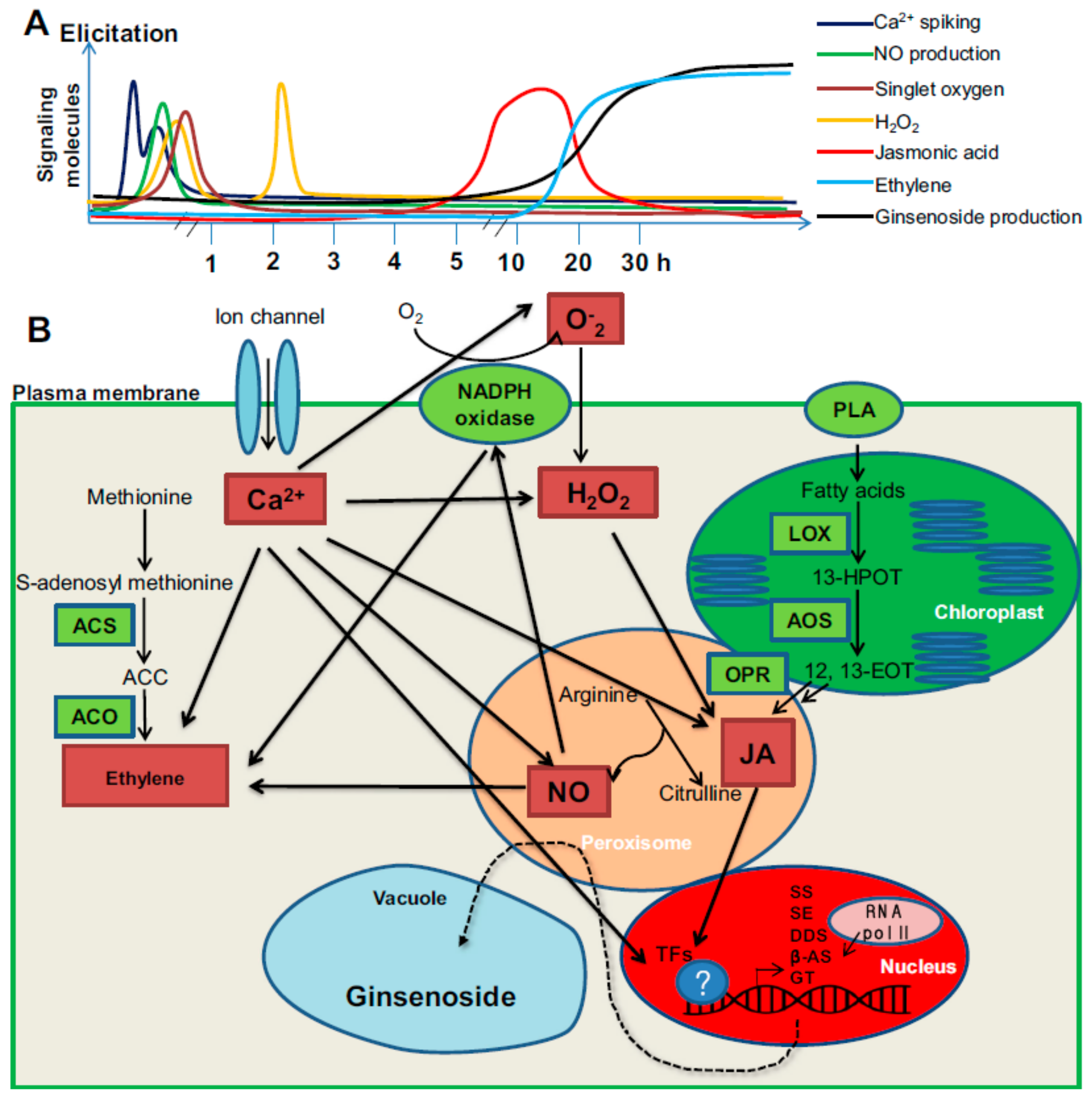
3.1. Summary of MJ Elicitation Mechanism

Summary of MJ-treated elicitation mechanism is presented in Figure 3. In the first step, the elicitors are perceived by the specific receptors localized in the plasma membrane, which initiates signalling processes that activate plant defence mechanism [31]. Majority of the studies have revealed that binding of elicitors to receptors leads to induction of pathogenesis-related proteins and the production of ROS and RNS, enzymes of oxidative stress protection, the activation of defence-related genes [30,31,32,33,34,35]. During the process of signal transduction several processes such as protein phosphorylation, lipid oxidation, enhanced antioxidant enzyme activity (SOD, G-POD, APX, CAT), and the activation and the de novo biosynthesis of transcription factors and subsequent expression secondary metabolite biosynthetic genes have been reported by several researchers [5,21].

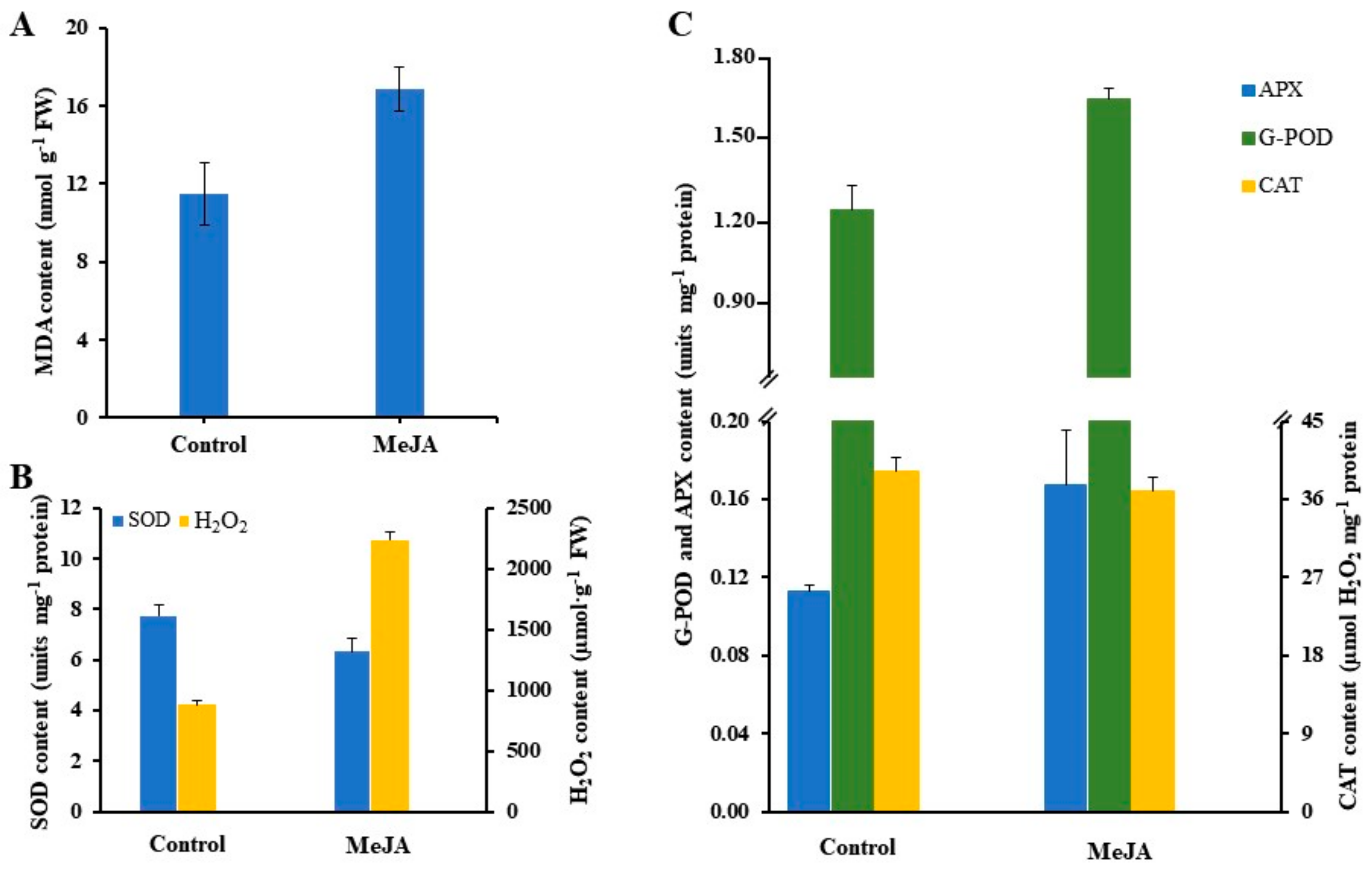
3.2. Oxidative Stress-Induced by Methyl Jasmonate

Recent studies suggest that environmental stresses can increase the oxygen-induced damage to cells due to increased generation of ROS. ROS brings about the peroxidation of membrane lipids, which leads to membrane damage [13,27]. Some studies report that induction of oxidative stress has been observed in plants exposed to MJ and SA [36,37,38]. In agreement with these early studies, exposure to MJ significantly increased the MDA content, an index of lipid peroxidation. O2 is known to be harmful to all membrane constituents (Figure 3 and Figure 4A). The accumulation of MDA content increased significantly in the MJ exposed roots of Cnidium officinale compared to the control (Figure 4A). This suggests that MJ lead to the production of O2, •OH resulting in increased lipid peroxidation. In other studies, the better protection in P. ginseng seems to result from the more efficient antioxidative system while a significant increase in MDA level in MJ-treated roots appeared to be derived from lower to increased activities of the enzymes [13,14].

3.3. Antioxidant Response

MJ is generally considered to modulate many physiological events in higher plants, such as defence responses, flowering and senescence [13,39]. Plants respond to a variety of environmental stresses (biotic and abiotic) through induction of antioxidant defence enzymes that provide protection against further damage [8,40]. Stress tolerance is closely related to the efficiency of antioxidant enzymes and these antioxidant enzymes and metabolites are reported to increase under various environmental stresses [8,12,41]. Numerous studies have demonstrated that MJ plays a key role in the enhancement of antioxidant enzymes activity such as APX, DHAR, MDHAR, AO, GR, GST, GPX, G-POD, CAT, SOD. Ali et al. [12] achieved that induced activity of SOD, G-POD and reduced ascorbate (ASC) contents indicated that antioxidants played an important role for protecting cells from MJ elicitation in Panax ginseng and Panax quinquefolium adventitious root cultures. In the apoplast and symplast of roots of sunflower (Helianthus annuus L.) seedlings, MJ-elicited roots showed a fast increase in ROS content, followed by a marked increase in the activity of H2O2-scavenging enzymes, G-POD, APX and CAT. The mechanisms responsible for MJ-induced H2O2 accumulation was investigated further by studying both the production and scavenging of H2O2 in the extracellular matrix [42].
The effects of MJ on the accumulation of antioxidant enzyme activity in adventitious root culture of Cnidium officinale has been presented in Figure 4B,C. The activity of SOD was significantly decreased in MJ-treated roots, while the accumulation of H2O2 was increased compared to control (un-treated roots). MJ treatments resulted in a significant increase in G-POD and APX activity which were approximately 1.33 and 1.48-fold higher than the control, respectively. However, the CAT activity was slightly decreased in C. officinale adventitious roots treated with MJ. Similar results were also reported by Ali et al. [13] in P. ginseng adventitious root cultures.
The main detoxifying enzymes in oxidative stress are SOD, APX, CAT and G-POD, monodehydroascorbate reductase (MDHAR), dehydroascorbate reductase (DHAR), and glutathione reductase (GR) also showed an important role in scavenging stress-induced ROS generated in plants [10,13,14]. These enzymes operate in different subcellular compartments and respond in concert when cells are exposed to oxidative stress [10]. SOD plays a central role in defence against oxidative stress in all aerobic organisms [37]. The enzyme SOD belongs to the group of metalloenzymes and catalyses the dismutation of O2 to O2 and H2O2, which in turn is detoxified by CAT or G-POD or APX reactions (Figure 3). Therefore, the control of the steady-state O2 levels by SOD is an important factor for protecting the cells against oxidative damage. Therefore, SOD is usually considered the first line of defence against oxidative stress [13,14,40,43]. CAT was the first enzyme to be discovered and characterized among antioxidant enzymes. It is a ubiquitous tetrameric heme-containing enzyme that catalyses the dismutation of two molecules of H2O2 into H2O and O2. G-POD is a heme-containing protein, preferably oxidizes aromatic electron donor such as guaiacol and pyrogallol at the expense of H2O2. APX is a central component of AsA-GSH cycle and plays an essential role in the control of intracellular ROS levels. APX uses two molecules of AsA to reduce H2O2 to water with a concomitant generation of two molecules of MDHA [10,11,44,45,46]. Farooq et al. [47] reported that the application of MJ minimized the oxidative stress, as revealed via a lower level of ROS synthesis in leaves of Brassica napus. This study also indicates that MJ plays an effective role in the regulation of multiple transcriptional pathways which were involved in oxidative stress responses, therefore enhanced enzymatic activities and gene expression of important antioxidants (SOD, APX, CAT, POD), secondary metabolites.

3.4. Signaling Pathway-Mediated Secondary Metabolite Accumulation in Plant Cell and Organ Cultures

Polyunsaturated fatty acids can produce signals, such as oxylipins, which include JA, JA methyl ester, JA amino acid conjugates, and further JA metabolites [48,49]. JA and its derivatives are known as the signalling molecules that can induce the biosynthetic enzymes involved in the formation of secondary metabolites in ginseng as shown in Figure 5 [5,50]. Zhao et al. [31] reported that exogenous application of MJ could induce endogenous JA biosynthesis, which stimulates expression of saponin biosynthetic genes in ginseng. Kim et al. [51] reported that the expression of farnesyl pyrophosphate synthase (FPS) was induced by MJ and produced the farnesyl diphosphate precursor for squalene biosynthesis. The transcription level of squalene synthase (SS) and squalene epoxidase (SE), key genes in ginseng biosynthesis was enhanced by MJ treatment in P. notoginseng and P. ginseng [5,51].
The treatment of in vitro cultures with MJ may induce cross-talks between different signal transduction (auxin, ET, ABA, SA, GA, Ca2+, O2, H2O2, NO) as in the case in vivo systems. JA signalling pathway triggers COL1 (coronatine insensitive1) gene in Arabidopsis, which encode F-box protein and involved in ubiquitination and removal of repressors of the JA signalling pathways [52]. Similarly, two other genes JAR1 (Jasmonate resistant1) and JIN1 (Jasmonate insentive1 and JIN1 are also known as MYC2) were identified in Arabidopsis, which are involved in conjugation of jasmonic acid to Ile [53] and transcription regulation of JA-responsive genes, respectively [54]. Another protein JAZ (Jasmonate zim domain) mediates the interaction between JAZ and COL1 or other transcriptional factors. COL1 protein, JAZ, and MYC2 constitute the core signal transduction mechanism of JA signalling and also responsible interaction with other transduction pathways [55]. For example, NtCOL1, NtJAZ, NtMYC2a/2b are responsible for nicotine biosynthesis in Nicotiana tabacum [55]. JA and auxin singling pathways participate in crosstalk and regulate various plant responses via COL1, MYC2 and JAZ components. When plants are activated with exogenous auxin, the auxin-TIR-AUX/IAA-ARF signalling is activated, which leads to JA synthesis. Contrarily, the endogenous JA induces the expression of auxin synthase gene (ASA1) and auxin levels, which leads the regulation of JAZ1 [55]. During JA and ET crosstalk ET3 (Ethylene insensitive3, which is involved in ET signalling pathway) and JAZ-MYC2 (which is involved in the JA signalling pathway) will interact and regulate the stress responses [55]. During JA and ET crosstalk JA and ET may antagonise or coordinately regulated the plant stress responses. At the time of ABA and JA signalling pathways, JAZ-MYC2 participates in the crosstalk between JA and ABA and they regulate the plant responses coordinately. ABA receptor PYL (Pyrabactin resistance1-like proteins) interacts with the JA signalling pathway in many plants [55]. The crosstalk between JA and SA signalling pathways involves many components such as MAPK (mitogen-activated protein kinase), GRX (redox regulators of glutathione) and TRX (thioredoxin). It was demonstrated that JA signalling inhibits SA biosynthesis and accumulation [55]. JA and GA singling pathways regulate plant responses either coordinately or antagonistically. The interaction between JA and SA was brought about by the C-terminus of JAZs with that of MYC2 or DELLA proteins. DELLA interacts with JAZ to release MYC2, leads to activation of MYC2 genes. Concurrently, DELLA interacts with JAZ to inhibit the expression of JA biosynthetic genes via MYB21 and MYB24. Within vitro cultures of ginseng, elicitation with MJ induces crosstalk between MJ and Ca2+, O2 (superoxide radical), H2O2, NO, ethylene and JA have been reported [5]. In the beginning, calcium signalling flux is required for further signalling, then both H2O2 and JA mediate early responses, whereas ethylene production is the late response in elicitor-induced for saponin biosynthesis (Figure 5) [5,56,57].

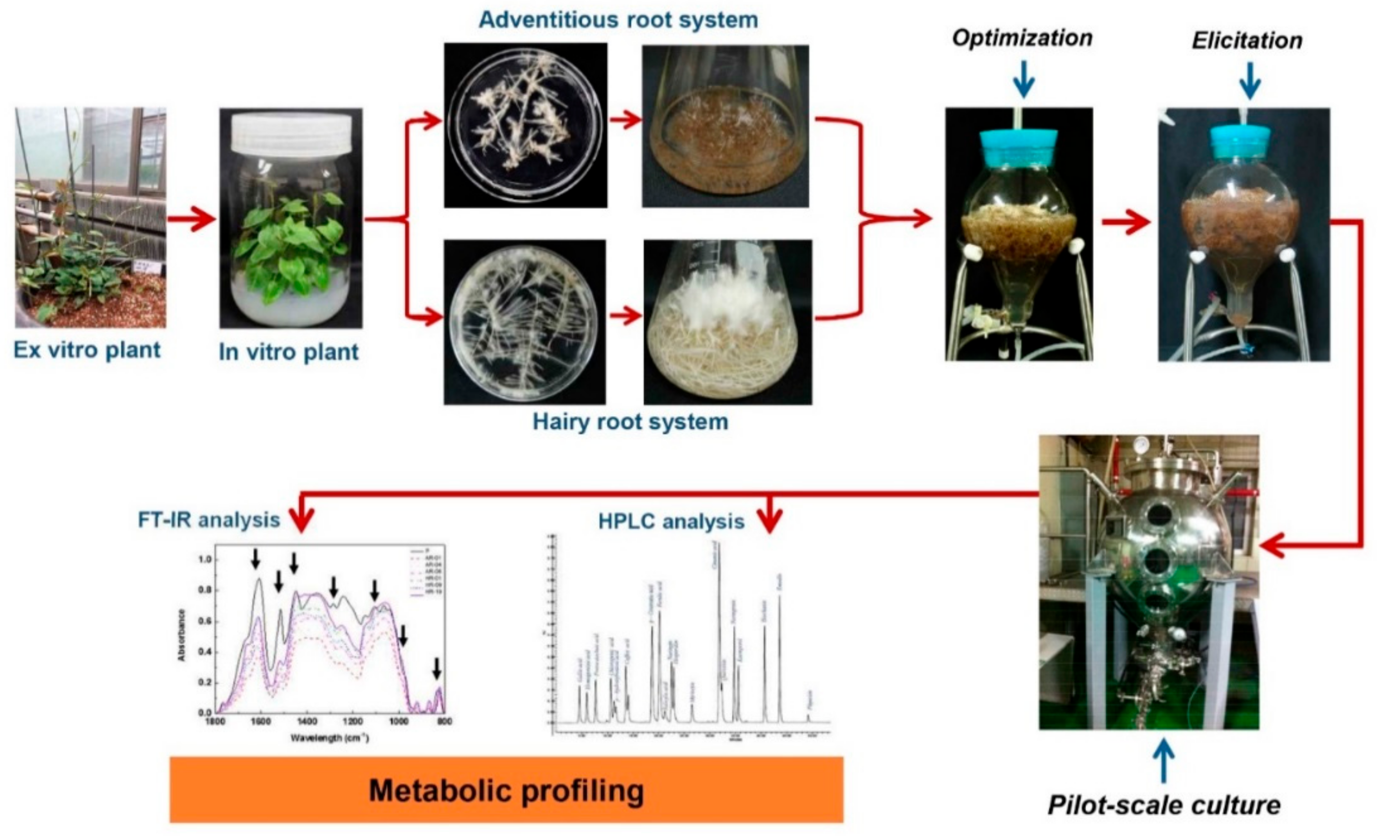
4. Usage of Methyl Jasmonate (MJ) in Plant Cell and Organ Cultures

4.1. Application of MJ for Enhancing Secondary Metabolite in In Vitro Culture System

The metabolic profiling of a plant is influenced by various micro-environmental and macro-environmental conditions. The plant responds to its surrounding environmental factors (as biotic and abiotic stress) through the stimulation of secondary metabolism to produce the desired compounds needed for its survival, the process is known as elicitation [58,59,60,61,62]. In recent days, MJ has been used extensively for elicitation studies involving in vitro culture systems such as cell suspension, adventitious root, hairy root and multiple shoot culture system (Table 1). Figure 6 shows the effect of MJ on phenolic compound production in adventitious root culture of Cnidium officinale. The adventitious roots treated with 100 µM MJ led to significantly higher yields (two-fold increment) of total phenolic compounds compared with the control treatments. These results corroborate the data for antioxidant enzyme activity (Figure 3 and Figure 4). Elicitor treatment might have resulted in the production of ROS due to stress. In order to mitigate the effects of ROS, plant tissues exhibit a stimulated antioxidant enzyme activity leading to an increased production of secondary metabolites [14,63,64]. Han and Yuan [65] indicated that activation of phenylalanine ammonia-lyase (PAL) activity and accumulation of phenolic compounds is regulated by the oxidative burst in suspension culture of Taxus cuspidata, that is believed to change the membrane permeability and lead to the induction of secondary metabolism.
MJ treatment stimulates the biosynthesis of secondary metabolites in plant cell cultures via a large number of control points and triggers the expression of key genes that increase cellular activities at biochemical and molecular levels through the involvement of signal compounds [29]. It also plays a role in the signal transduction, which speeds up enzyme catalysis, thereby leading to the formation of specific compounds such as polyphenol, terpenoids, flavonoids, and alkaloids [4,30]. Ho et al. [62] reported that phenolic levels increased approximately 2-fold in adventitious root samples treated with 50 μM MJ (22.08 mg·g−1 DW) versus the control (10.08 mg·g−1 DW) in Polygonum multiflorum. In addition, hairy root cultures of P. multiflorum, the highest total phenolic content (52.87 mg·g−1 DW) was 3.4-fold higher than the control, especially, MJ treatment led to significantly higher levels of almost all individual phenolics, such as 1.13-fold increase of physcion, 3.83-fold of quercetin, 1.58-fold of kaempferol, 5.48-fold of p-hydroxybenzoic acid, and 4.3-fold of salicylic acid [62]. Level of ginsenoside accumulation showed a seven-fold enhancement in the adventitious root cultures treated with 100 μM MJ when compared to the control [66]. MJ at 100 µM enhanced the maximum production of xanthotoxin and bergapten at 1.1-fold and 39.6-fold higher than control in Changium smyrnioides, respectively [67].
Optimization of MJ concentration, the growth stage and exposure time of cultures are important critical factors for improving secondary metabolite synthesis [29,66]. Elicitation with 100 µM MJ influenced silymarin pigment accumulation in cell culture of Silybum marimum after 3 days of treatment [68]. In another report, elicitation with 4 µM MJ for 2 weeks resulted in a 6.5-fold enhancement of solasodine content (9.33 mg g−1 DW) than un-elicited hairy root cultures of Solanum trilobatum [69]. In addition, increasing of withanolide derivatives accumulation (14-fold) was achieved in 40-day-old hairy roots elicited with 15 µM MJ for 4 h exposure time [70]. In the previous studies on P. multiflorum, the phenolic compound in adventitious root culture reached to 2-fold higher than the control after 7-days treatment with 50 µM MJ, whereas 3.4-fold higher was observed in hairy roots after 5-day of exposure time [34,62]. Besides, an increase in 4.5-fold of triterpenoid in hairy root cultures of Centella asiatica was reported [71]. Treatment of cultures with 100 µM MJ for 6 days has been reported to influence the accumulation of both phenolic and tanshinones in Salvia miltiorrhiza hairy root cultures [72].

4.2. Application of MJ for Enhancement of Secondary Metabolites in Bioreactor Cultures

Inhibition of root growth and decreased biomass accumulation are the major challenges with MJ treated cell and organ suspension cultures, this might be due to enhanced accumulation of ROS, which adversely affects root biomass. Airlift bioreactors appear to be ideal for plant cell and organ cultures by efficiently controlling the culture environment, foam generation, shear stress, and oxygen supply [78,83,84]. They are suitable for the cultivation of cell, hairy root, adventitious root and embryo suspension cultures of various medicinal plants [32,78,84,85], and are also applied in MJ treatment to reduce the inhibition of root biomass by two 2-stage cultures. The first step involves the optimization of root growth in bioreactors without elicitor and then concentration and time of elicitor application will have to be standardized in the second step. A numerous study has been reported in using balloon-type bubble bioreactor (BTBB) for both biomass production and enhancement of secondary metabolite treated with MJ. After 6 weeks of culture, the addition of 50 µM MJ for 1 week was found to be the optimal concentration for eleutheroside B and E, and chlorogenic acid production in adventitious root culture of Eleutherococcus koreanum [80]. Thanh et al. [80] used 200 µM MJ on day-15 during culture period in cell suspension culture of Panax ginseng showed the highest ginsenoside yield after 8 days of treatment. The ginsenoside, Rb group and Rg group content increased 2.9, 3.7, and 1.6-fold after treatment, respectively. In the study of the somatic embryo of Eleutherococcus senticosus, the total eleutheroside was increased 7.3-fold after treated with 200 µM MJ [83]. The maximum content of salidroside (4.74 mg·g−1 DW) was observed at 125 µM MJ, which was 5-fold higher than the control in Rhodiola sachalinensis callus culture [81]. Jiang et al. [80] reported that when added 200 µM MJ to culture medium after 30 days of culture, maximum productivity of bioactive compound was found 8 days after treatment in the adventitious culture of Olopanax elatus.
The effect of MJ on biomass and secondary metabolite production in adventitious root culture of Polygonum multiflorum and Echinacea purpurea is presented in Table 2 and Figure 7. The treatment with MJ inhibited root growth in both medicinal plants after 7 days of treatment. The biomass was decreased by 5.8% (in DW) of P. multiflorum and 22.97% (in DW) of E. purpurea. The roots also turn to brown with such treatments (Figure 7). However, MJ at 50 µM enhanced the phenolic content approximately 2-fold in P. multiflorum compared to non-treated, and also reached to higher the total phenolic compounds when compared to field-grown plants (Table 2). Whereas, total caffeic acid derivatives in E. purpurea was also increased 1.16-fold compared to the control with the treatment of 200 µM MJ. Moreover, total productivity was also increased in both plants with 1.92-fold and 1.12-fold compared with non-treated roots.
In addition, the treatment of MJ for improving bioactive compounds is also successfully applied in pilot-scale bioreactors up to 10,000 L [3,84,85,86]. A pilot-scale (500-L) bioreactor cultures were established in adventitious root culture of Echinacea angustifolia [87]. In this study, the authors achieved that although root biomass and growth ratio slightly decreased in the 500-L BTBB compared to the 5- and 20-L BTBBs, the highest concentrations of total phenolics (60.41 mg·g−1 DW), flavonoids (16.45 mg·g−1 DW), and total caffeic acid derivatives (33.44 mg·g−1 DW) were observed in the 500-L BTBB when added 100 µM MJ in the culture after 4 weeks. Especially, the accumulation of echinacoside (the major bioactive compound) in MJ-treated adventitious roots grown in the 500-L bioreactor was the highest (12.3 mg·g−1 DW), which is approximately three-fold higher than the non-MJ-treated roots cultured in 5- and 20-L bioreactors [80]. Baque et al. [88] achieved the highest accumulation of secondary metabolites in Morinda citriflora adventitious root cultures and cultures which were treated with 100 µM MJ after 4 weeks of culture. They reported higher accumulation of anthraquinones (205.75 mg·g−1 DW), phenolics (90.26 mg·g−1 DW) and flavonoids (93.34 mg·g−1 DW) in pilot-scale (500-L) bioreactor cultures when compared to small-scale bioreactor (3-L) cultures (approximately 1.8-fold, 1.3-fold, and 1.55-fold, respectively). In summary, two-stage culture systems should be addressed within in vitro culture system. First, a stage cultivation of cell suspension, adventitious roots and hairy roots for biomass production, followed by addition of MJ (at potential concentration and exposure time) in the second stage for enhancing metabolite production without decreasing root biomass. The application of scale-up bioreactor cultures is promising in the production of biomass and bioactive compounds and such materials/products will provide material for the pharmaceutical and cosmetic industry.

4.3. Effect of MJ on Gene Expression and Secondary Metabolite Accumulation

The variation of the expression of genes encoding key enzymes in a biosynthetic pathway directly influences the accumulation of the corresponding secondary metabolites [72,89,90]. The expression levels of the genes responsible for PAL, hydroxycinnamate coenzyme A ligase (4CL), cinnamic acid 4-hydroxylase (C4H), tyrosine aminotransferase (TAT), 4-Hydroxyphenylpyruvate reductase (HPPR), and rosmarinic acid synthase (RAS) were enhanced after 6-days of MJ treatment [72]. These enzymes were involved in the phenolic acid biosynthetic pathways which resulted in 197-fold increment phenolics in Salvia miltiorrhiza hairy roots [66]. Rahimi et al. [5] also demonstrated the enhanced regulation of genes involved in MEP (methylerythritol phosphate) and MVA (mevalonate) pathways with the treatment of MJ, in Panax ginseng [5]. Genes involved in andrographolide (diterpene) synthesis were influenced by MJ treatment in Andrographis paniculata [91]. The MEP pathway genes namely DXS (1-deoxy-d-xylulose 5-phosphate synthase), DXR (1-deoxy-D-xylulose 5-phosphate reductoisomerase), HDS (4-hydroxy-3-methylbut2-enyl-diphosphate synthase) and ISPH (1-Hydroxy-2-methyl-2-(E)-butenyl 4-diphosphate reductase) were up-regulated under MJ stimulation. Similarly, genes involved in the MVA pathway specially HMGS (3-hydroxy-3-methylglutaryl-Co-A synthase) and HMGR (3-hydroxy-3-methylglutaryl-CoA reductase) were also enhanced (Figure 8) by MJ treatment. A number of studies demonstrated that MJ treatment affected on MEP and MVA pathways and secondary metabolites biosynthesis [5,73,92,93].

Author Contributions

T.-T.H. contributed to writing of manuscript, H.N.M. revised for important intellectual content. S.-Y.P. made substantial contributions to interpreted data, conception and design this study. All authors have read and agree to the published version of the manuscript.

Funding

This work was funded by a grant from the Next Generation BioGreen21 Program (Project No. PJ013689), Rural Development Administration, Republic of Korea.

Acknowledgments

Authors were supported by Brain Korea (BK) 21 Plus Program through the National Research Foundation (NRF) of Korea.

Conflicts of Interest

The authors declare no conflict of interest.

Abbreviations

4CLHydroxycinnamate coenzyme A ligase
AOCAllene oxide cyclase
AOSAllene oxide synthase
APXAscorbate peroxidase
ASAAscorbate
AsA-GSHAscorbateglutahione
C4HCinnamic acid 4-hydroxylase
CATCatalase
DHARDehydroascorbate reductase
DXS1-Deoxy-d-xylulose 5-phosphate synthase
ETEthylene
FT-IRFourier-transform infrared spectroscopy
GGPPSGeranylgeranyl diphosphate synthase
G-PODGuaiacol peroxidase
GRGlutathione reductase
GSHGlutathione
H2O2Hydrogen peroxide
HDR1-Hydroxy-2-methyl-2-(E)-butenyl-4-diphosphate reductase
HPLCHigh-performance liquid chromatography
HPPL4-Hydroxyphenylpyruvate reductase
ISPH1-Hydroxy-2-methyl-2-(E)-butenyl 4-diphosphate reductase
JAJasmonic acid
JAsJasmonate
JA-Ile(+)-7-iso-jasmonoyl-L-isoleucine
LOXLipoxygenase
MJMethyl jasmonate
MEPMethylerythritol phosphate
MDHARMonodehydroascorbate reductase
MVAMevalonate
NONitric oxide
PALPhenylalanine ammonia-lyase
RASRosmarinic acid synthase
RNSReactive nitrogen species
ROSReactive oxygen species
SASalicylic acid
SODSuperoxide dismutase
TATTyrosine aminotransferase

References

  1. Ferrari, F.; Pasqua, G.; Monacelli, B. Xanthones from calli of Hypericum perforatum subsp. perforatum. Nat. Prod. Res. 2005, 19, 171–176. [Google Scholar] [CrossRef] [PubMed]
  2. Brugger, G.A.; Lamotte, O.; Vandelle, E.; Bourque, S.; Lecourieux, D.; Poinssot, B.; Wendehenne, D.; Pugin, A. Early signalling events induced by elicitors of plant defences. Mol. Plant Microbe Interact. 2006, 19, 711–724. [Google Scholar] [CrossRef] [PubMed] [Green Version]
  3. Paek, K.Y.; Murthy, H.N.; Hahn, E.J.; Zhong, J.J. Large scale culture of ginseng adventitious roots for production of ginsenosides. Adv. Biochem. Eng. Biotechnol. 2009, 113, 151–176. [Google Scholar]
  4. Murthy, H.N.; Lee, E.J.; Paek, K.Y. Production of secondary metabolites from cell and organ cultures: Strategies and approaches for biomass improvement and metabolite accumulation. Plant Cell Tissue Organ Cult. 2014, 118, 1–16. [Google Scholar] [CrossRef]
  5. Rahimi, S.; Kim, Y.J.; Yang, D.C. Production of ginseng saponins: Elicitation strategy and signal transductions. Appl. Microbiol. Biotechnol. 2015, 99, 6987–6996. [Google Scholar] [CrossRef] [PubMed]
  6. Foyer, C.H.; Harbinson, J. Oxygen metabolism and the regulation of photosynthetic electron transport. In Causes of Photooxidative Stresses and Amelioration of Defense Systems in Plants; Foyer, C.H., Mullineaux, P., Eds.; CRC Press: Boca Raton, FL, USA, 1994; pp. 1–42. [Google Scholar]
  7. Blokhina, O.; Fagerstedt, K.V. Reactive oxygen species and nitric oxide in plant mitochondria: Origin and redundant regulatory systems. Physiol. Plant. 2010, 138, 447–462. [Google Scholar] [CrossRef]
  8. Mittler, R. Oxidative stress, antioxidants and stress tolerance. Trends Plant Sci. 2002, 7, 405–410. [Google Scholar] [CrossRef]
  9. Hu, W.H.; Song, X.S.; Shi, K.; Xia, X.J.; Zhou, Y.H.; Yu, J.Q. Changes in electron transport, superoxide dismutase and ascorbate peroxidase isoenzymes in chloroplasts and mitochondria of cucumber leaves as influenced by chilling. Photosynthetica 2008, 46, 581–588. [Google Scholar] [CrossRef]
  10. Sharma, P.; Jha, A.B.; Dubey, R.S.; Pessarakli, M. Reactive oxygen species, oxidative damage, and antioxidative defense mechanism in plants under stressful conditions. J. Bot. 2012, 2012, 1–26. [Google Scholar] [CrossRef] [Green Version]
  11. Desikan, R.; Mackerness, S.A.H.; Hancock, S.J.T.; Neill, S.J. Regulation of the Arabidopsis transcriptome by oxidative stress. Plant Physiol. 2001, 127, 9–172. [Google Scholar] [CrossRef] [Green Version]
  12. Ali, M.B.; Yu, K.W.; Hahn, E.J.; Paek, K.Y. Differential responses of anti-oxidants enzymes, lipoxygenase activity, ascorbate content and the production of saponins in tissue cultured root of mountain Panax ginseng C.A. Mayer and Panax quinquefolium L. in bioreactor subjected to methyl jasmonate stress. Plant Sci. 2005, 169, 83–192. [Google Scholar]
  13. Ali, M.B.; Yu, K.W.; Hahn, E.J.; Paek, K.Y. Methyl jasmonate and salicylic acid elicitation induces ginsenosides accumulation, enzymatic and non-enzymatic antioxidant in suspension culture Panax ginseng roots in bioreactors. Plant Cell Rep. 2006, 25, 613–620. [Google Scholar] [CrossRef] [PubMed]
  14. Ali, M.B.; Hahn, E.J.; Paek, K.Y. Methyl jasmonate and salicylic acid induced oxidative stress and accumulation of phenolics in Panax ginseng bioreactor root suspension culture. Molecules 2007, 12, 607–621. [Google Scholar] [CrossRef] [PubMed] [Green Version]
  15. Ho, T.T. Strategies and Approaches for Improvement of Biomass and Bioactive Compounds in Adventitious Root and Hairy Root Cultures in Polygonum multiflorum. Ph.D. Thesis, Chungbuk National University, Cheongju, Korea, 2018. [Google Scholar]
  16. Ho, T.T.; Lee, K.J.; Lee, J.D.; Bhushan, S.; Paek, K.Y.; Park, S.Y. Adventitious root culture of Polygonum multiflorum for phenolic compounds and its pilot-scale production in 500 L-tank. Plant Cell Tissue Organ Cult. 2017, 130, 167–181. [Google Scholar] [CrossRef]
  17. Ho, T.T.; Murthy, H.N.; Dalawai, D.; Bhat, M.A.; Paek, K.Y.; Park, S.Y. Attributes of Polygonum multiflorum to transfigure red biotechnology. App. Microbiol. Biotechnol. 2019, 103, 3317–3326. [Google Scholar] [CrossRef] [PubMed]
  18. Demole, E.; Lederer, E.; Mercier, D. Isolement et détermination de la structure du jasmonate de méthyle, constituant odorant caractéristique de lessence de jasmin. Helv. Chim. Acta 1962, 45, 675–685. [Google Scholar] [CrossRef]
  19. Aldridge, D.C.; Galt, S.; Giles, D.; Turner, W.B. Metabolites of Lasiodiplodia theobromae. J. Chem. Soc. C 1971, 1623–1627. [Google Scholar] [CrossRef]
  20. Hyun, Y.; Choi, S.; Hwang, H.J.; Yu, J.; Nam, S.J.; Ko, J.; Park, J.Y.; Seo, Y.S.; Kim, E.Y.; Ryu, S.B.; et al. Cooperation and functional diversification of two closely related galactolipase genes for jasmonate biosynthesis. Dev. Cell 2008, 14, 183–192. [Google Scholar] [CrossRef] [Green Version]
  21. Santino, A.; Taurino, M.; De Domenico, S.; Bonsegna, S.; Pastor, V.; Flors, V. Jasmonate signalling in plant development and defence response to multiple abiotic stresses. Plant Cell Tissue Organ Cult. 2013, 32, 1085–1098. [Google Scholar]
  22. Ziegler, J.; Stenzel, I.; Hause, B.; Mauche, H.; Hamberg, M.; Grimm, R.; Ganal, M.; Wasternack, C. Molecular cloning of allene oxide cyclase. The enzyme establishing the stereochemistry of octadecanoids and jasmonates. J. Biol. Chem. 2000, 275, 19132–19138. [Google Scholar] [CrossRef] [Green Version]
  23. Wasternack, C.; Hause, B. Jasmonates and octadecanoids: Signal in plant stress responses and development. Prog. Nucl. Res. Mol. Biol. 2002, 72, 165–221. [Google Scholar]
  24. Yan, J.; Zhang, C.; Gu, M.; Bai, Z.; Zhang, W.; Qi, T.; Cheng, Z.; Peng, W.; Luo, H.; Nan, F.; et al. The Arabidopsis CORONATINE INSENSITIVE1 Protein is a jasmonate receptor. Plant Cell 2009, 21, 2220–2236. [Google Scholar] [CrossRef] [PubMed] [Green Version]
  25. Ruan, J.; Zhou, Y.; Zhou, M.; Yan, J.; Khurshid, M.; Weng, W.; Cheng, J.; Zhang, K. Jasmonic acid signalling pathways in plants. Int. J. Mol. Sci. 2019, 20, 2479. [Google Scholar] [CrossRef] [PubMed] [Green Version]
  26. Creelman, R.A.; Mullet, J.E. Jasmonic acid distribution and action in plants: Regulation during development and response to biotic and abiotic stress. Proc. Natl. Acad. Sci. USA 1995, 92, 4114–4119. [Google Scholar] [CrossRef] [Green Version]
  27. Bertini, L.; Palazzi, L.; Proietti, S.; Pollastri, S.; Arrigoni, G.; de Laureto, P.; Carusco, C. Proteomic analysis of MeJa-induced defense responses in rice against wounding. Int. J. Mol. Sci. 2019, 2020, 2525. [Google Scholar] [CrossRef] [Green Version]
  28. Bertini, L.; Proietti, S.; Focaracci, F.; Sabatin, B.; Carusco, C. Epigenetic control of dense genes following MeJA-induced priming in rice (O. sativa). J. Plant Physiol. 2018, 228, 166–177. [Google Scholar]
  29. Giri, C.C.; Zaheer, M. Chemical elicitors versus secondary metabolite production in vitro using plant cell, tissue and organ cultures: Recent trends and a sky eye view appraisal. Plant Cell Tissue Organ Cult. 2016, 126, 1–18. [Google Scholar] [CrossRef]
  30. Baenas, N.; Garcia-Viguera, C.; Moreno, D.A. Elicitation: A tool for enriching the bioactive composition of foods. Molecules 2014, 19, 13541–13563. [Google Scholar] [CrossRef] [Green Version]
  31. Zhao, J.; Davis, L.C.; Verpoorte, R. Elicitor signal transduction leading to production of plant secondary metabolites. Biotechnol. Adv. 2005, 23, 283–333. [Google Scholar] [CrossRef]
  32. Shohael, A.M.; Murthy, H.N.; Lee, H.L.; Hahn, E.J.; Paek, K.Y. Increased eleutheroside production in Eleutherococcus sessiliflorus embryogenic suspension cultures with methyl jasmonate treatment. Biochem. Eng. J. 2008, 38, 270–273. [Google Scholar] [CrossRef]
  33. Durango, D.; Pulgarin, N.; Echeverri, F.; Escobar, G.; Quiñones, W. Effect of salicylic acid and structurally related compounds in the accumulation of phytoalexins in cotyledons of common bean (Phaseolus vulgaris L.) cultivars. Molecules 2013, 18, 10609–10628. [Google Scholar] [CrossRef] [PubMed] [Green Version]
  34. Ho, T.T.; Lee, J.D.; Ahn, M.S.; Kim, S.W.; Park, S.Y. Enhanced production of phenolic compounds in hairy root cultures of Polygonum multiflorum and its metabolite discrimination using HPLC and FT-IR methods. Appl. Microbiol. Biotechnol. 2018, 102, 9563–9575. [Google Scholar] [CrossRef] [PubMed]
  35. Vasconsuelo, A.; Boland, R. Molecular aspects of the early stages of elicitation of secondary metabolites in plants. Plant Sci. 2007, 172, 861–875. [Google Scholar] [CrossRef]
  36. Mir, M.Y.; Hamid, S.; Kamili, A.N.; Hassan, Q.P. Sneak peek of Hypericum perforatum L.: Phytochemistry, phytochemical efficacy and biotechnological interventions. J. Plant Biochem. Biotechnol. 2019, 28, 357–375. [Google Scholar] [CrossRef]
  37. Scandalios, L.G. Oxygen stress and superoxide dismutase. Plant Physiol. 1993, 101, 7–12. [Google Scholar] [CrossRef] [PubMed] [Green Version]
  38. Yu, D.Q.; Cen, C.; Yang, M.L. Studies on the salicylic acid induced lipid peroxidation and defence gene expression in tabacco cell culture. Acta Bot. Sin. 1999, 41, 977–982. [Google Scholar]
  39. Chong, T.M.; Abdullah, M.A.; Fadzillah, N.M.; Lai, O.M.; Lajis, N.H. Jasmonic acid elicitation of anthraquinones with some associated. Enzyme Microb. Technol. 2005, 36, 469–477. [Google Scholar] [CrossRef]
  40. Creelman, R.A.; Mullet, J.E. Biosynthesis and action of jasmonates in plants. Annu. Rev. Plant. Physiol. Plant Mol. Biol. 1997, 48, 355–381. [Google Scholar] [CrossRef] [Green Version]
  41. Asada, K. The water-water cycle in chloroplasts: Scavenging of active oxygens and dissipation of excess photons. Annu. Rev. Plant Physiol. Plant Mol. Biol. 1999, 50, 601–639. [Google Scholar] [CrossRef]
  42. Seo, Y. Antioxidant activity of the chemical constituents from the flower buds of Magnolia denudata. Biotechnol. Bioprocess Eng. 2010, 15, 400–406. [Google Scholar] [CrossRef]
  43. Parra-Lobato, M.G.; Fernandez-Garcia, N.; Olmos, E.; Alvarez-Tinaut, M.G.; Gomez-Jiménez, M.C. Methyl jasmonate-induced antioxidant defence in root apoplast from sunflower seedlings. Environ. Exp. Bot. 2009, 66, 9–17. [Google Scholar] [CrossRef]
  44. Wasternack, C.; Parthier, B. Jasmonate signaled plant gene expression. Trends Plant Sci. 1997, 2, 302–307. [Google Scholar] [CrossRef]
  45. Yamauchi, Y.; Furutera, A.; Seki, K.; Toyoda, Y.; Tanaka, K.; Sugimoto, Y. Malondialdehyde generated from peroxidized linolenic acid causes protein modification in heat-stressed plants. Plant Physiol. Biochem. 2008, 8–9, 786–793. [Google Scholar] [CrossRef] [PubMed]
  46. Welinder, K.G. Superfamily of plant, fungal and bacterial peroxidases. Curr. Opin. Struct. Biol. 1992, 2, 388–393. [Google Scholar] [CrossRef]
  47. Farooq, M.A.; Gill, R.A.; Islam, F.; Ali, B.; Liu, H.; Xu, J.; He, S.; Zhou, W. Methyl jasmonate regulates antioxidant defence and suppresses arsenic uptake in Brassica napus L. Front. Plant Sci. 2016, 7, 468. [Google Scholar] [CrossRef] [Green Version]
  48. Wasternack, C. Jasmonates: An update on biosynthesis, signal transduction and action in plant stress response, growth and development. Ann. Bot. 2007, 100, 681–697. [Google Scholar] [CrossRef] [Green Version]
  49. Wasternack, C.; Hause, B. Jasmonates: Biosynthesis, perception, signal transduction and action in plant stress response, growth and development. An update to 2007 review in Annals of Botany. Ann. Bot. 2013, 111, 1021–1058. [Google Scholar] [CrossRef]
  50. Rahimi, S.; Devi, B.S.R.; Khorolragcha, A.; Kim, Y.J.; Kim, J.H.; Jung, S.K.; Yang, D.C. Effect of salicylic acid and yeast extract on the accumulation of jasmonic acid and sesquiterpenoids in Panax ginseng adventitious roots. Russ. J. Plant Physiol. 2014, 61, 811–817. [Google Scholar] [CrossRef]
  51. Kim, O.T.; Bang, K.H.; Kim, Y.C.; Hyun, D.Y.; Kim, M.Y.; Cha, S.W. Upregulation of ginsenoside and gene expression related to triterpene biosynthesis in ginseng hairy root cultures elicited by methyl jasmonate. Plant Cell Tissue Organ Cult. 2009, 98, 25–33. [Google Scholar] [CrossRef]
  52. Xie, D.X.; Feys, B.F.; James, S.; Nieto-Rostro, M.; Turner, J.G. COL1, an Arabidopsis gene required for jasmonae-regulated defense and fertility. Science 1998, 280, 1091–1094. [Google Scholar] [CrossRef]
  53. Staswick, P.E.; Tiryaki, I. The oxylipin signal jasmonic acid is activated by an enzyme that conjugates it it to isoleucine in Arabidopsis. Plan Cell 2004, 16, 2117–2127. [Google Scholar] [CrossRef] [PubMed] [Green Version]
  54. Lorenzo, O.; Chico, J.M.; Sanchez-Serrano, J.J.; Solano, R. JASMONATE INSENSTIVE1 encodes a MYC transcription factor essential to discriminate between different jasmonate-regulated defense responses in Arabidopsis. Plant Cell 2004, 16, 1938–1950. [Google Scholar] [CrossRef] [PubMed] [Green Version]
  55. Yang, J.; Duan, G.; Li, C.; Liu, L.; Han, G.; Zhang, Y.; Wang, C. The crosstalk between jasmonic acid and other plant hormone sginalling highlight the involvement of jasmonic acid as a core component in plant reponses to biotic and abiotic stresses. Front. Plant Sci. 2019, 10, 1349. [Google Scholar] [CrossRef] [PubMed] [Green Version]
  56. Hu, F.X.; Zhong, J.J. Jasmonic acid mediates gene transcription of ginsenoside biosynthesis in cell cultures of Panax notoginseng treated with chemically synthesized 2-hydroxyethyl jasmonate. Process Biochem. 2008, 43, 113–118. [Google Scholar] [CrossRef]
  57. Xu, X.; Hu, X.; Neill, S.J.; Fang, J.; Cai, W. Fungal elicitor induces singlet oxygen generation, ethylene release and saponin synthesis in cultured cells of Panax ginseng CA Meyer. Plant Cell Physiol. 2005, 46, 947–954. [Google Scholar] [CrossRef]
  58. Tewari, R.K.; Paek, K.Y. Salicylic acid-induced nitric oxide and ROS generation stimulate ginsenoside accumulation in Panax ginseng roots. J. Plant Growth Regul. 2011, 30, 396–404. [Google Scholar] [CrossRef]
  59. Jeong, G.T.; Park, D.H. Enhancement of growth and secondary metabolite biosynthesis: Effect of elicitor derived from plant and insects. Biotechnol. Bioprocess Eng. 2005, 10, 73–77. [Google Scholar] [CrossRef]
  60. Yin, Z.; Shangguan, X.; Chen, J.; Zhao, Q.; Li, D. Growth and triterpenic acid accumulation of Cyclocarya paliurus cell suspension cultures. Biotechnol. Bioprocess Eng. 2013, 18, 606–614. [Google Scholar] [CrossRef]
  61. Gorelick, J.; Bernstein, N. Elicitation: An underutilized tool in the development of medicinal plants as a source of therapeutic secondary metabolites. Adv. Agron. 2014, 124, 201–230. [Google Scholar]
  62. Ho, T.T.; Lee, J.D.; Jeong, C.S.; Paek, K.Y.; Park, S.Y. Improvement of biosynthesis and accumulation of bioactive compounds by elicitation in adventitious root cultures of Polygonum multiflorum. Appl. Microbiol. Biotechnol. 2018, 102, 199–209. [Google Scholar] [CrossRef]
  63. Belhadj, A.; Teleg, N.; Saigne, C.; Cluzet, S.; Barrieu, F.; Hamdi, S.; Merillon, J.M. Effect of methyl jasmonate in combination with carbohydrates on gene expression of PR proteins, stilbene and anthocyanin accumulation in grapevine cell cultures. Plant Physiol. Biochem. 2008, 46, 493–499. [Google Scholar] [CrossRef]
  64. Zhou, M.L.; Zhu, X.M.; Shao, J.R.; Tang, J.X.; Wu, Y.M. Production and metabolic engineering of bioactive substances in plant hairy root culture. Appl. Microbiol. Biotechnol. 2011, 90, 1229–1239. [Google Scholar] [CrossRef] [PubMed]
  65. Han, R.B.; Yuan, Y.J. Oxidative burst in suspension culture of Taxus cuspidata induced by a laminar shear stress in short-term. Biotechnol. Prog. 2004, 20, 507–513. [Google Scholar] [CrossRef]
  66. Kim, Y.S.; Hahn, E.J.; Murthy, H.N.; Paek, K.Y. Adventitious root growth and ginsenoside accumulation in Panax ginseng cultures as affected by methyl jasmonate. Biotechnol. Lett. 2004, 26, 1619–1622. [Google Scholar] [CrossRef]
  67. Cai, J.; Ma, Y.; Hu, P.; Zhang, Y.; Chen, J.; Li, X. Elicitation of furanocoumarins in Changium smyrnioides suspension cells. Plant Cell Tissue Organ Cult. 2017, 130, 1–12. [Google Scholar] [CrossRef]
  68. Sampedro, M.A.S.; Tárrago, J.F.; Corchete, P. Yeast extract and methyl jasmonate-induced silymarin production in cell cultures of Silybum marianum (L.) Gaertn. J. Biotechnol. 2005, 119, 60–69. [Google Scholar] [CrossRef] [PubMed]
  69. Baek, S.E.; Ho, T.T.; Lee, H.S.; Jung, G.Y.; Kim, Y.E.; Jeong, C.S.; Park, S.Y. Enhanced biosynthesis of triterpenoids in Centella asiatica hairy root culture by precursor feeding and elicitation. Plant Biotechnol. Rep. 2019. [Google Scholar] [CrossRef]
  70. Shilpha, J.; Satish, L.; Kavikkuil, M.; Largia, M.J.V.; Ramesh, M. Methyl jasmonate elicits the solasodine production and antioxidant activity in hairy root cultures of Solanum trilobatum L. Ind. Crop. Prod. 2015, 71, 54–64. [Google Scholar] [CrossRef]
  71. Sivanandhan, G.; Dev, K.G.; Jeyaraj, M.; Rajesh, M.; Arjunan, A.; Muthuselvam, M.; Manickavasagam, M.; Ganapathi, A. Increased production of withanolide A, withanone and withaferin A in hairy root cultures of Withania somnifera (L.) Dunal elicited with methyl jasmonate and salicylic acid. Plant Cell Tissue Organ Cult. 2013, 114, 121–129. [Google Scholar] [CrossRef]
  72. Xing, B.; Yang, D.; Liu, L.; Han, R.; Sun, Y.; Liang, Z. Phenolic acid production is more effectively enhanced than tanshinone production by methyl jasmonate in Salvia miltiorrhiza hairy roots. Plant Cell Tissue Organ Cult. 2018, 134, 119–129. [Google Scholar] [CrossRef]
  73. Wang, W.; Zhao, Z.J.; Xu, Y.; Qian, X.; Zhong, J.J. Efficient induction of ginsenoside biosynthesis and alteration of ginsenoside heterogeneity in cell cultures of Panax notoginseng by using chemically synthesized 2-hydroxyethyl jasmonate. Appl. Microbiol. Biotechnol. 2006, 70, 298–307. [Google Scholar] [CrossRef]
  74. Komaraiah, P.; Kavi-Kishor, P.B.; Carlsson, M.; Magnusson, K.E.; Mandenius, C.F. Enhancement of anthraquinone accumulation in Morinda citrifolia suspension cultures. Plant Sci. 2005, 168, 1337–1344. [Google Scholar] [CrossRef]
  75. Krzyzanowska, J.; Czubacka, A.; Pecio, L.; Przybys, M.; Doroszewska, T.; Stochmal, A.; Oleszek, W. The effects of jasmonic acid and methyl jasmonate on rosmarinic acid production in Mentha x piperita cell suspension cultures. Plant Cell Tissue Organ Cult. 2012, 108, 73–81. [Google Scholar] [CrossRef]
  76. Kang, S.M.; Jung, H.Y.; Kang, Y.M.; Yun, D.J.; Bahk, J.D.; Yang, J.K. Effects of methyl jasmonate and salicylic acid on the production of tropane alkaloids and the expression of PMT and H6H in adventitious root cultures of Scopolia praviflora. Plant Sci. 2004, 166, 745–751. [Google Scholar] [CrossRef]
  77. Zaker, A.; Sykora, C.; Gössnitzer, F.; Abrishamchi, P.; Asili, J.; Mousavi, S.H.; Wawrosch, C. Effects of some elicitors on tanshinone production in adventitious root cultures of Perovskia abrotanoides Karel. Ind. Crop. Prod. 2015, 67, 97–102. [Google Scholar] [CrossRef]
  78. Lee, E.J.; Park, S.Y.; Paek, K.Y. Enhancement strategies of bioactive compound production in adventitious root cultures of Eleutherococcus koreanum Nakai subjected to methyl jasmonate and salicylic acid elicitation through airlift bioreactors. Plant Cell Tissue Organ Cult. 2015, 120, 1–10. [Google Scholar] [CrossRef]
  79. Jiang, X.L.; Piao, X.C.; Gao, R.; Jin, M.Y.; Jiang, J.; Jin, X.H.; Lian, M.L. Improvement of bioactive compound accumulation in adventitious root cultures of an endangered plant species, Oplopanax elatus. Acta Physiol. Plant. 2017, 39, 226. [Google Scholar] [CrossRef]
  80. Thanh, N.T.; Murthy, H.N.; Yu, K.W.; Hahn, E.J.; Paek, K.Y. Methyl jasmonate elicitation enhanced synthesis of ginsenoside by cell suspension cultures of Panax ginseng in 5-l balloon type bubble bioreactor. Appl. Micorbiol. Biotechnol. 2005, 67, 197–201. [Google Scholar] [CrossRef] [PubMed]
  81. Li, Y.; Shao, C.H.; Park, S.Y.; Piao, X.C.; Lian, M.L. Production of salidroside and polysaccharides in Rhodiola sachalinensis using airlift bioreactor systems. Acta Physiol. Plant. 2014, 36, 2975–2983. [Google Scholar] [CrossRef]
  82. Shohael, A.M.; Murthy, H.N.; Lee, H.L.; Hahn, E.J.; Paek, K.Y. Methyl jasmonate induced overproduction of eleutherosides in somatic embryos of Eleutherococcus senticosus cultures in bioreactors. Electron. J. Biotechnol. 2007, 10, 633–637. [Google Scholar] [CrossRef] [Green Version]
  83. Jeong, J.A.; Wu, C.H.; Murthy, H.N.; Hahn, E.J.; Paek, K.Y. Application of an airlift bioreactor system for the production of adventitious root biomass and caffeic acid derivatives of Echinacea purpurea. Biotechnol. Bioprocess Eng. 2009, 14, 91–98. [Google Scholar] [CrossRef]
  84. Baque, M.A.; Moh, S.H.; Lee, E.J.; Zhong, J.J.; Paek, K.Y. Production of biomass and useful compounds from adventitious roots of high-value added medicinal plants using bioreactor. Biotechnol. Adv. 2012, 30, 1255–1267. [Google Scholar] [CrossRef] [PubMed]
  85. Murthy, H.N.; Dandin, V.S.; Paek, K.Y. Tools for biotechnological production of useful phytochemicals from adventitious root cultures. Phytochem. Rev. 2016, 15, 129–145. [Google Scholar] [CrossRef]
  86. Wang, G.R.; Qi, N.M. Influence of mist intervals and aeration rate on growth and second metabolite production of Pseudostellaria heterophylla adventitious roots in a siphon-mist bioreactor. Bioprocess Eng. 2010, 15, 1059–1098. [Google Scholar] [CrossRef]
  87. Cui, H.Y.; Baque, M.A.; Lee, E.J.; Paek, K.Y. Scale-up of adventitious root cultures of Echinacea angustifolia in a pilot-scale bioreactor for the production of biomass and caffeic acid derivatives. Plant. Biotechnol. Rep. 2013, 7, 297–308. [Google Scholar] [CrossRef]
  88. Baque, M.A.; Murthy, H.N.; Paek, K.Y. Adventitious root culture of Morinda citrifolia in bioreactors for production of bioactive compounds. In Production of Biomass and Bioactive Compounds Using Bioreactor Technology, 1st ed.; Paek, K.Y., Murthy, H.N., Zhong, J.J., Eds.; Springer: Dordrecht, The Netherlands, 2014; pp. 185–222. [Google Scholar]
  89. Yu, H.; Liu, X.; Gao, S.; Han, X.; Cheng, A.; Lou, H. Molecular cloning and functional characterization of a phenylalanine ammonialyase from liverwort Plagiochasma appendiculatum. Plant Cell Tissue Organ Cult. 2014, 117, 265–277. [Google Scholar] [CrossRef]
  90. Mueller, M.J.; Brodschelm, W.; Spannagl, E.; Zenk, M.H. Signaling in the elicitation process is mediated through the octadecanoid pathway leading to jasmonic acid. Proc. Natl. Acad. Sci. USA 1993, 90, 7490–7494. [Google Scholar] [CrossRef] [Green Version]
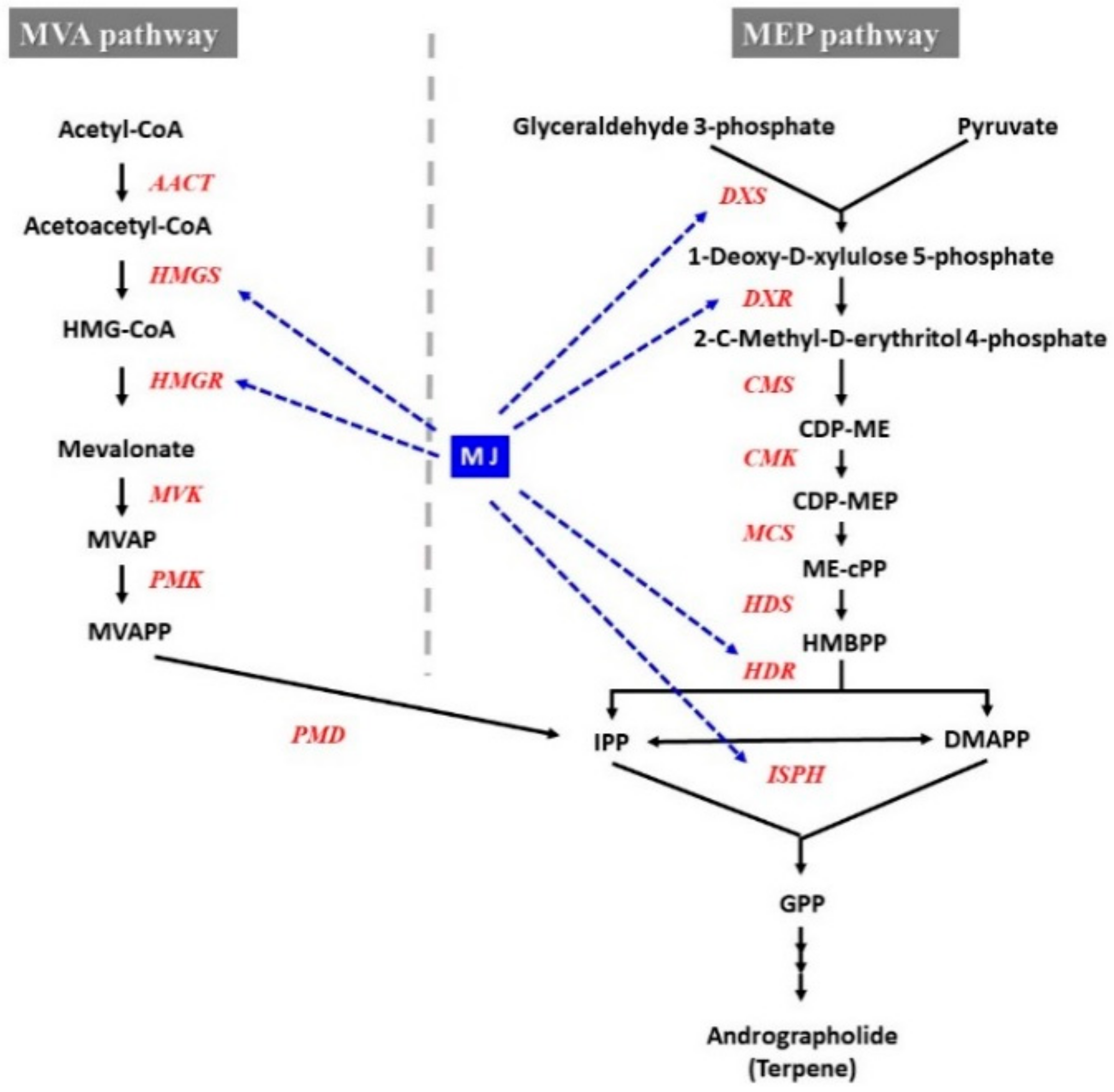
  91. Sinha, R.K.; Sharma, S.N.; Verma, S.S.; Zha, J. Effects of lovastin, fosmidomycin and methyl jasmonate on andrographolide biosynthesis in the Andrographis paniculata. Acta Physiol. Plant. 2008, 40, 165. [Google Scholar] [CrossRef]
  92. Yan, Y.; Stolz, S.; Chetelat, A.; Reymond, P.; Pagni, M.; Dubugnon, L.; Farmer, E.E. A downstream mediator in the growth repression limb of the jasmonate pathway. Plant Cell 2007, 19, 2470–2483. [Google Scholar] [CrossRef] [Green Version]
  93. Murthy, H.N.; Georgiev, M.I.; Kim, Y.S.; Jeong, C.S.; Kim, S.J.; Park, S.Y.; Paek, K.Y. Ginsenosides: Prospective for sustainable biotechnological production. Appl. Microbiol. Biotechnol. 2014, 98, 6243–6254. [Google Scholar] [CrossRef]
Figure 1. The experimental process of Polygonum multiflorum for enhanced production of secondary metabolites.
Ijms 21 00716 g001
Figure 2. Jasmonic acid and its derivatives.
Ijms 21 00716 g002
Figure 3. General mechanism after MJ perception. Abbreviations: ROS reactive oxygen species, SA salicylic acid, JA jasmonic acid, ET ethylene, O2 superoxide anion, •OH hydroxyl radical, H2O2 hydrogen peroxide, SOD superoxide dismutase, CAT catalase, G-POD Guaiacol peroxidase, APX ascorbate peroxidase.
Ijms 21 00716 g003
Figure 4. Effect of MJ on malondialdehyde (MDA) content (A), and antioxidant enzyme activity (B,C) in adventitious root cultures of Cnidium officinale. SOD superoxide dismutase, CAT catalase, G-POD guaiacol peroxidase, APX ascorbate peroxidase. Values represent mean ± SE (n = 3).
Ijms 21 00716 g004
Figure 5. Schematic illustration of the sequential signalling pathways activated in elicited ginseng (A). Model of cross-talk between different signal transductions (B). Cross talk between different signalling molecules is shown by bold arrows. ACC 1-aminocyclopropane-1-carboxylic acid, ACS 1-aminocyclopropane-1-carboxylic acid synthase, ACO 1-aminocyclopropane-1-carboxylic acid, AOS allene oxide synthase, β-AS beta-amyrin synthase, DDS dammarenediol synthase, 12,13-EOT 12,13(S)-epoxyoctadecatrienoic acid, H2O2 hydrogen peroxide, 13-HPOT (13S)-hydroperoxyoctadecatrienoic acid, JA jasmonic acid, LOX lipoxygenase, NO nitric oxide, NOS nitric oxide synthase, O2 superoxide radical, OPR oxophytodienoate reductase, PLA phospholipase, SS squalene synthase, SE squalene epoxidase, TFs transcription factors, UGRdGT UDPG/ginsenoside Rd glucosyltransferase (Adapted from Rahimi et al. [5]).
Ijms 21 00716 g005
Figure 6. Effect of MJ on phenolic compounds production in Cnidium officinale adventitious root culture.
Ijms 21 00716 g006
Figure 7. Effect of MJ on biomass production in Polygonum multiflorum and Echinacea purpurea adventitious root culture system. (A,B) control and 100 µM MJ in P. multiflorum, (C,D) control and 100 µM MJ in E. purpurea, respectively.
Ijms 21 00716 g007
Figure 8. Andrographolide biosynthetic gene activation by MJ elicitation in Andrographis paniculata [91]. Dashed blue arrows show the relationship between MJ signalling and andrographolide biosynthetic genes. HMG-CoA, 3-Hydroxy-3-methylglutaryl CoA. MVA, mevalonic acid. MVAP, mevalonic acid 5-phosphate. MVAPP, mevalonic acid 5-diphosphate. IPP, isopentenyl diphosphate. DMAPP, dimethylallyl diphosphate. MEP, 2-C-methyl-D-erythritol 4-phosphate. CDP-ME, 4-diphosphocytidyl-2-C-methyl-d-erythritol. CDP-MEP, 4-diphosphocytidyl2-C-methyl-D-erythritol 2-phosphate. ME-cPP, 2-C-methyl-D-erythritol 2,4-cyclodiphosphate. HMBPP, 1-hydroxy-2-methyl-2-(E)-butenyl 4-diphosphate. Enzymes of the MVA pathway: HMGS, HMG-CoA synthase. HMGR, HMG-CoA reductase. MVK, MVA kinase. PMK, MVAP kinase. PMD, MVAPP decarboxylase. Enzymes of the MEP pathway: DXS, DOXP synthase. DXR, DOXP reductoisomerase. CMS, CDP-ME synthase. CMK, CDP-ME kinase. MCS, ME-2,4cPP synthase. HDS, HMBPP synthase. HDR, HMBPP reductase, ISPH, 1-Hydroxy-2-methyl-2-(E)-butenyl 4-diphosphate reductase.
Ijms 21 00716 g008
Table 1. Usage of MJ for enhance secondary metabolite within in vitro culture system.
PlantMaterial zCulture SystemMJElicitor DurationBiomassTarget CompoundFold Increase CompoundReference
Panax notoginsengCSFlask200 µM7 dayNot significantRb, Rg ginsenosides3.38[73]
Silybum marimumCSFlask100 µM3 dayDecreasedSilymarin4[68]
Morinda citrifoliaCSFlask150 µM2 daySlightly decreasedAnthraquinone4[74]
Mentha x piperitaCSFlask100 µM2 dayDecreasedRosmarinic acid1.8[75]
Changium smyrnioidesCSFlask100 µM5 dayDecreasedXanthotoxin, bergapten1.1 and 39.6[67]
Scopolia parvifloraARFlask1 mM1 daySlightly decreasedScopolamine and hyoscyamine2–3[76]
Perovskia abrotanoidesARFlask10 µM7 dayNot significantTanshinone2.3[77]
Polygonum multiflorumARFlask50 µM7 dayDecreasedPhenolic2[62]
Withania somniferaHRFlask15 µM4 hSlightly decreasedWithanolide derivatives14[71]
Solanum trilobatumHRFlask4 µM14 daySlightly decreasedSolasodine6.5[70]
Salvia miltiorrhizaHRFlask100 µM6 dayDecreasedPhenolic and tanshinones3.3 and 2.5[71]
Polygonum multiflorumHRFlask50 µM5 daySlightly decreasedPhenolic3.2[34]
Echinacea purpureaHRFlask/Bioreactor400 µM7 dayNot significantTriterpenoid4.5[78]
Polygonum multiflorumARBioreactor50 µM7 daySlightly decreased Phenolic2.1[62]
Panax ginsengARBioreactor100 µM10 dayDecreasedGinsenoside11-fold in Rb group[66]
Panax ginseng and Panax quinquefoliumARBioreactor200 µM9 dayNot significantSaponin4[12,13]
Eleutherococcus koreanumARBioreactor50 µM7 dayNot significantEleutherosides B and E, and chlorogenic acid1.1, 1.4 and 1.2[79]
Olopanax elatusARBioreactor200 µM8 dayDecreasedPolysaccharide, phenolic1.83 and 1.49[80]
Panax ginsengCSBioreactor200 µM8 dayDecreasedGinsenoside1.6–3.7[81]
Rhodiola sachalinensisCSBioreactor125 µM7 dayDecreasedSalidroside5[82]
Eleutherococcus senticosusSEBioreactor200 µM7 dayDecreasedEleutheroside7.3[83]
zCS cell suspension culture, AR adventitious root culture, HR hairy root culture, SE somatic embryos culture.
Table 2. Effect of MJ on the accumulation of biomass and secondary metabolites in Polygonum multiflorum and Echinacea purpurea adventitious root culture system.
SystemsFW (g·L−1)DW (g·L−1)Dry Matter (%)Growth Ratio zTotal Bioactive Compounds (mg·g−1 DW) yTotal Productivity (mg·L−1) x
Polygonum multiflorumw
Control93.95 ± 3.2310.61 ± 0.32 11.30 ± 0.1120.23 ± 0.6411.20 ± 0.17118.82 ± 1.78
MJ5090.09 ± 1.519.99 ± 0.2311.10 ± 0.4418.98 ± 0.4622.83 ± 0.30228.08 ± 3.00
5-year-old root 24.78 ± 0.82
Echinacea purpureav
Control83.81 ± 2.128.10 ± 0.729.63 ± 0.6415.19 ± 1.4525.22 ± 1.27204.16 ± 10.24
MJ20067.26 ± 1.726.24 ± 0.409.27 ± 0.5311.48 ± 0.8038.32 ± 0.34 239.20 ± 4.44
Data present mean ± SE. v Adventitious root of E. purpurea culture in 20-L bioreactor in 3 weeks, treated with 200 µM MJ for 7 days before harvest. w Adventitious root of P. multiflorum culture in 20-L bioreactor in 3 weeks, treated with 100 µM MJ for 7 days before harvest. x Total productivity (mg·L−1 medium) = Total bioactive compound content (mg·g−1 DW) x DW (mg·L−1). y Total bioactive compounds (mg·g−1 DW) = Total phenolic compounds in P. multiflorum, and total caffeic acid derivatives in E. purpurea. z Growth ratio = (DW—Initial DW)/ Initial DW.

