Review

Bioactive Polyphenols and Neuromodulation: Molecular Mechanisms in Neurodegeneration

by
Francesco Di Meo
1,2,†,
Anna Valentino
3,†,
Orsolina Petillo
4,
Gianfranco Peluso
4,
Stefania Filosa
1,5,*,‡ and
Stefania Crispi
1,6,*,‡
1
Institute of Biosciences and BioResources-UOS Naples CNR, Via P. Castellino, 111, 80131 Naples, Italy
2
Department of Biology, University of Naples Federico II, Complesso Universitario Monte Sant’Angelo Via Cinthia, 80126 Naples, Italy
3
Elleva Pharma s.r.l., Via P. Castellino 111, 80131 Naples, Italy
4
Institute on Terrestrial Ecosystems (IRET) CNR, Via P. Castellino 111, 80131 Naples, Italy
5
IRCCS Neuromed, Localitá Camerelle, 86077 Pozzilli (IS), Italy
6
Stazione Zoologica Anton Dohrn, Villa Comunale, 80121 Napoli, Italy
*
Authors to whom correspondence should be addressed.
These authors contributed equally to this work.
Int. J. Mol. Sci. 2020, 21(7), 2564; https://0-doi-org.brum.beds.ac.uk/10.3390/ijms21072564
Submission received: 3 March 2020 / Revised: 27 March 2020 / Accepted: 3 April 2020 / Published: 7 April 2020

Abstract

:
The interest in dietary polyphenols in recent years has greatly increased due to their antioxidant bioactivity with preventive properties against chronic diseases. Polyphenols, by modulating different cellular functions, play an important role in neuroprotection and are able to neutralize the effects of oxidative stress, inflammation, and apoptosis. Interestingly, all these mechanisms are involved in neurodegeneration. Although polyphenols display differences in their effectiveness due to interindividual variability, recent studies indicated that bioactive polyphenols in food and beverages promote health and prevent age-related cognitive decline. Polyphenols have a poor bioavailability and their digestion by gut microbiota produces active metabolites. In fact, dietary bioactive polyphenols need to be modified by microbiota present in the intestine before being absorbed, and to exert health preventive effects by interacting with cellular signalling pathways. This literature review includes an evaluation of the literature in English up to December 2019 in PubMed and Web of Science databases. A total of 307 studies, consisting of research reports, review articles and articles were examined and 146 were included. The review highlights the role of bioactive polyphenols in neurodegeneration, with a particular emphasis on the cellular and molecular mechanisms that are modulated by polyphenols involved in protection from oxidative stress and apoptosis prevention.

Graphical Abstract

1. Introduction

An improvement in socio-economic conditions, especially in developed countries, results in an increase in elderly people. This determines a corresponding enlargement of the pathologies linked to brain aging, such as cognitive and neurodegenerative diseases. The potential mechanisms underpinning brain neurodegeneration have not yet been completely elucidated; nevertheless, oxidative stress and inflammation are considered the main effectors of brain decline.
Nowadays, there is a growing interest in dietary polyphenol nutrients since different epidemiological studies have suggested that diets rich in plant-derived phytochemicals and, in particular, polyphenols, are beneficial to human health [1]
Among phytochemicals, polyphenols are the major group of compounds produced by plants as secondary metabolites that protect plants against reactive oxygen species (ROS), ultraviolet radiation (UV), pathogens, parasites and plant predators. They act as natural antioxidants thanks to their metal-chelating and free radical scavenger properties. Bioactive polyphenols have been reported to prevent the age-related cognitive decline typical of neurodegenerative diseases. Dietary bioactive polyphenols can also modulate cognitive deficits and synaptic plasticity and promote neurogenesis [2,3].
Neurodegenerative diseases are characterized by the progressive loss of function of a specific population of neurons and neural stem cells that results in sensory and motor deficits and cognitive impairment. Different signaling pathways are involved in neurodegeneration, including oxidative stress, inflammation and apoptosis. Bioactive polyphenols are able to counteract these processes by directly scavenging free radical species inhibiting ‘pro-oxidant’ enzymes, activating anti-oxidant enzymes [4] or intervening in apoptotic pathways [5].
Polyphenols act by preventing the DNA damage triggered by hydrogen peroxide (H2O2) and by transition metals such as copper and iron. Iron-mediated oxidative DNA damage by hydroxyl radical (OH) is the primary cause of cell death under oxidative stress conditions for both prokaryotes and eukaryotes [6]. Polyphenols can also directly scavenge ROS or inhibit the expression of molecules sensitive to oxidative stress such as nuclear factor-kB (Nf-κB) and activator protein-1 (AP-1) [7].
The molecular mechanism of neuroprotection involves the regulation of the mitochondrial apoptosis cascade, which is finely tuned by the imbalance by between B-cell lymphoma-2 (Bcl-2) and Bcl-2-associated X protein (Bax). Phytochemical neuroprotection can be achieved by introducing anti-apoptotic Bcl-2, thus preventing apoptosis [8].
Bioactive polyphenols can exert neuromodulatory effects, activating different intracellular signaling pathways that are crucial for neuroprotection. The PI3K/Akt (Phosphoinositide 3-kinases/Akt) pathway prevents apoptosis by upregulating the expression of Bcl-2; the PKC-ERK1/2 (Protein kinase C/ Extracellular signal-regulated protein kinases 1 and 2) pathway decreases Bcl-2 (B-cell lymphoma 2), Bcl-w (Bcl-2-Like Protein 2) and Bcl-xL (B-cell lymphoma-extra large), which are other anti-apoptotic proteins; Akt-ERK1/2 inhibits the pro-apoptotic activity of Bad (BCL2 associated agonist of cell death) and Bim (Bcl-2-interacting mediator) and activates caspases 9 and 3 [9,10].
The neuroprotective effects of polyphenols can be carried out through synthesis of neurotrophic factors, such as brain-derived neurotrophic factor (BDNF), nerve growth factor (NGF) or glial cell line-derived neurotrophic factor (GDNF). They can exert neurotrophic activity by binding the cognate receptors and activating the downstream neuroprotective pathways [11].
The beneficial health effects of bioactive phenolic compounds mainly depend on their bioavailability and absorption rate. These characteristics can be modulated through interaction with other dietary nutrients and through the action of enzymes present in the small intestine and in the liver. Polyphenols are usually recognized as xenobiotics by the human body. After ingestion, their absorption through the gut barrier can be increased following specific biotransformation and conjugation such as methylation, sulfation and glucuronidation [12].
Polyphenols are effective in neuroprotection in a different way—directly, by affecting brain functions and indirectly, by modulating gut microbiota composition and the metabolites produced. Both actions determine the production of neurotransmitters and neuropeptides that are able to influence the brain functions.
In this review, we discuss the different cellular and molecular mechanisms by which dietary bioactive polyphenols exert neuroprotective functions. The review evaluates the main publications in the field, providing a comprehensive analysis of the efficacy of bioactive polyphenols.

2. Methodology

We performed a literature review, searching within the PubMed and Web of Science database. As keywords, we used the terms: oxidative stress, antioxidant, polyphenols, neurodegeneration, neuroprotection, Alzheimer’s disease, Parkinson’s disease, and Huntington’s disease. Research reports, review articles and articles in English published up to December 2019 were selected and evaluated. In addition, we examined the citations therein and included them when appropriate. In total, we examined 307 references and included 146 of them in the present review.

3. Oxidative Stress and Polyphenols

Oxidative stress occurs when ROS accumulate in the cells for excessive production or insufficient neutralization. ROS are a contradiction for the cells. They are produced in normal metabolism as part of several physiologic processes and, when present in physiologic concentrations, they control redox homeostasis in the cell. However, an imbalance between their production and the ability of the cell to enforce antioxidant defense mechanisms may affect cellular structure and functional integrity, resulting in cell damage and leading to necrotic or apoptotic cell death [13,14,15].
ROS cause severe molecular damage in the major cell components such as protein oxidation, lipid oxidation, DNA oxidation, and glycoxidation. They can be divided into free radicals and nonradicals. Free radicals are the molecular species containing at least one unpaired electron in the shell around the atomic nucleus.
Among free radicals, H2O2 is one of the most important ROS with a physiological significance. H2O2 is formed in the cells in a reaction catalyzed by the superoxide dismutase enzyme (SOD) and also, at relatively low concentrations, it can penetrate the biological membranes and cause severe damage to cellular macromolecules. ROS increase with exposure to the environment. To counterbalance the effect of oxidants, cells have evolved an intricate network of defense mechanisms. Indeed, depending on the intensity and duration of oxidative stress, the cell response can be different, ranging from cell proliferation to cell cycle arrest or cell death by apoptosis or necrosis [16].
The adverse effects of ROS can be inactivated by the action of antioxidants [17]. They are defined as compounds able to inhibit the oxidation of any molecule, even when present at very low concentration [18]. By inhibiting or quenching free radical reactions, antioxidants delay or block cellular damage.
It is widely accepted that neurodegenerative development is associated with oxidative stress that determines severe injury in the cell. In fact, oxidative stress can result in deep biological damage. An imbalance in redox regulation determines the overproduction of ROS with the induction of progressive cellular damage, which is fundamental in neurodegenerative diseases. The production of excessive ROS in neuronal tissue is mainly due to the activity of excitatory amino acids and neurotransmitters [19]. Thus, neuronal cells need antioxidants to scavenge and prevent the formation of ROS.
The body protects itself from ROS by using enzymatic antioxidant mechanisms [20]. The antioxidant enzymes reduce the levels of lipid hydroperoxide and H2O2; thus, they are important in preventing lipid peroxidation and maintaining the structure and function of cell membranes.
The nonenzymatic antioxidants are the natural antioxidants present in plants. Among them, polyphenols are the most powerful antioxidants able to balance the cellular ROS by activating antioxidant signaling pathways [21,22]. Different studies indicate that dietary antioxidants consumed daily exert a protective role [23].
Plants produce nonenzymatic antioxidants such as vitamins that act to interrupt free radical chain reactions. Vitamin C is present in all plant cells and it has intracellular and extracellular antioxidant capacity. Vitamin E is present exclusively in plastids. It represents the principal defense against oxidative membrane damage, being concentrated in the hydrophobic bilayer of the cell membrane. Vitamin E avoids lipid peroxidation, thus protecting polyunsaturated fatty acids (PUFA) from reactive oxygen damage [24].
Antioxidant plant polyphenols belong to different groups: phenolic acids, phenolic diterpenes, volatile oils, flavonoids and stilbene (Figure 1) [25]. All act as ROS scavengers, or induce antioxidant enzymes [26].
Phenolic acids exert their antioxidant action, trapping free radicals [27]. The antioxidant mechanism of phenolic diterpenes is related to the scavenging activities of lipid free radicals and to the inhibition of low-density lipoprotein oxidation [28]. Volatile oils, as well as phenolic compounds, act as antioxidants by reacting with peroxyl radicals [29]. Flavonoids are the most abundant class of plant polyphenols, due to their metal-chelating and free radical scavenger properties [30]. On the contrary, stilbenes scavenge hydroxyl radicals.
Other non-flavonoid polyphenols present in foods have antioxidant properties. Among them, curcumin, from turmeric Curcuma longa, prevents lipid peroxidation by scavenging H2O2 and hydroxyl radicals. Curcumin can also act as an antioxidant, reducing ferric ion (Fe3+) and chelating ferrous ion (Fe2+) [31].

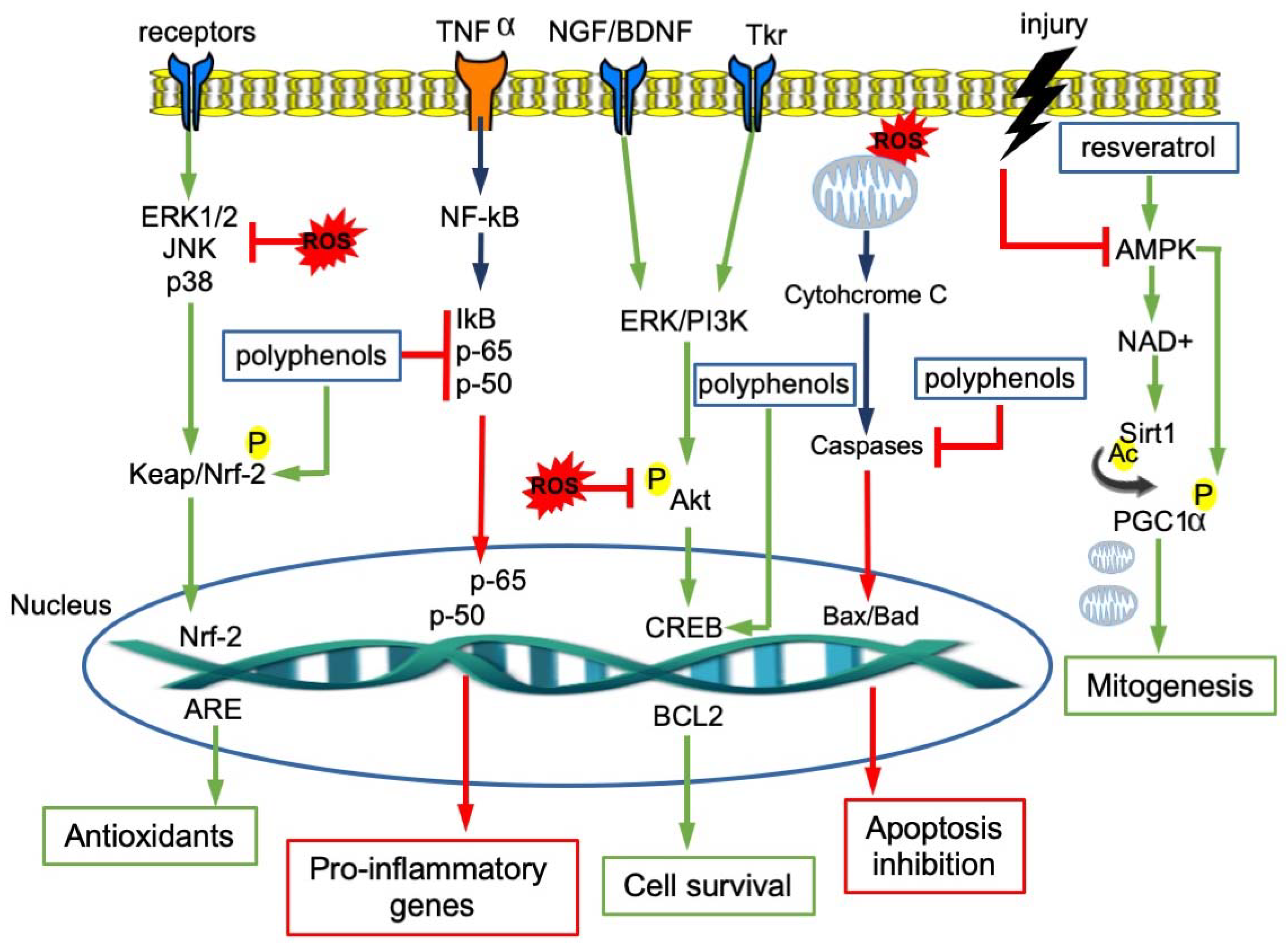
4. Molecular Pathways Involved in Neuroprotection of Polyphenols

Although cells are equipped with a high variety of antioxidants, some tissues and organs are much more vulnerable than others to oxidative stress, probably because of their elevated consumption of oxygen and the consequent generation of large amounts of ROS. Several observations suggest that the brain is particularly vulnerable to oxidative stress [32] and oxidative stress has been implicated in the pathogenesis of many clinical conditions and in the process of aging.
Oxidative stress has been found to be increased in several human age-related degenerative diseases, including genetically-linked neurodegenerative diseases, like Alzheimer’s disease (AD), Parkinson’s disease (PD), and Huntington’s disease (HD) [33]. However, whether oxidative stress is a primary cause or a downstream consequence of these neuropathological conditions or, more in general, of the aging process is still an open question [34].
Neurons are particularly sensitive to oxidative stress because, being postmitotic cells, they will not be replaced. Moreover, the ability of cells to respond to oxidative protein damage also seems to decline with age [35]. Since the antioxidant systems are overwhelmed in pathological conditions, the use of natural molecules like polyphenols can be viewed as a novel antioxidant therapeutic strategy to reduce neuronal ROS and ameliorate the neurodegenerative process.
Polyphenols, due to their ability to modulate multiple cellular processes including redox balance, have invaluable potential as antioxidant agents.
Polyphenols such as resveratrol from grapes and wine, curcumin from tumeric, and epigallocatechin-3-gallate from green tea (EGCG), are able to protect against neurodegenerative diseases by activating the protein kinases signaling molecular pathways such as Keap1/Nrf-2/ARE, the major protective pathway against endogenous and exogenous ROS [36,37]. The interaction between Keap1 and bioactive molecules leads to the disruption of the Keap1/Nrf2 complex, allowing Nrf2 to translocate to the nucleus where it binds adenylate and uridylate (AU)-rich elements (AREs) and triggers the expression of antioxidant proteins such as heme oxygenase-1 [38].
Polyphenols also stimulate neurotrophic receptor factors with a pivotal role in the maintenance of neuronal health. Examples are BDNF, which is involved in learning and memory [39], NGF, which is crucial for the survival of brain neurons [40], and GDNF, which regulates cell survival and synaptic plasticity [41]. Several studies reported that different flavonoids, including resveratrol, are able to enhance the expression of these factors. Quercetin and genistein were shown to stimulate NGF-induced neurite outgrowth [42,43]. Resveratrol is able to induce the release of BDNF and GDNF, thus protecting cells from neurotoxicity [44,45].
The neuroprotective role of polyphenols is carried out by activating the pathways that, in cells, regulate transcription, translation, proliferation, growth and survival, such as PI3K/Akt, PKC-ERK1/2, Akt-ERK1/2 and MAPK (Mitogen-activated protein kinase). Their binding to Trk receptors (tyrosine receptor kinases) activates the protein kinase cascades and, finally, cAMP (3’,5’-cyclic adenosine monophosphate)response element-binding protein (CREB), increasing the expression of Bcl-2, Bcl-xL and of neurotrophic factors [46]. In addition, this binding promotes receptor dimerization and autophosphorylation by activating the downstream signaling cascades and promoting the survival of motor neurons, hippocampal neuronal cells, and spinal ganglion neurons [47].
A flavone derivative has been reported to act as a TrkB agonist and to activate the downstream signaling pathway [48]. More recently, resveratrol was shown to exert neuroprotection through its interaction with Trk receptors [49].
In the brain, the anti-inflammatory action of polyphenols leads to neuroprotective effects. In fact, polyphenols inhibit the release of cytokines from activated glia and they also downregulate pro-inflammatory transcription factors such as NF-κB [50]. For example, quercetin determines a strong neuroprotective effect, repressing NF-κB [51]; catechin, after stress injury, increases cell survival by downregulating the NF-κB and MAP kinase pathways [52].
The molecular mechanisms modulated by polyphenols are summarized in Figure 2.

5. Effect of Polyphenols on Neurodegenerative Disorders

Despite the variability in the clinical picture of neurodegenerative disorders, these diseases share common molecular traits. Inflammation and oxidative stress are responsible for the disruption of the functions of the neurovascular units in AD, PD, HD and dementia. ROS are able to interact with different neuronal signaling pathways, such as protein kinase and lipid kinase signaling cascades [53,54] (Table 1).

5.1. Alzheimer’s Disease

AD is the most frequent form of dementia in the elderly population [63], with progressive neurodegeneration. It is characterized by the deposition of Amyloid β (Aβ) peptides as Aβ plaques and intracellular neurofibrillary tangles [64,65] that ultimately lead to a gradual deterioration in brain structure and to the loss of intellectual function [66].
The presence of amyloid beta (Aβ) peptides in Alzheimer’s disease confers oxidative insult on neurons and glial cells, leading to a change in synaptic plasticity [67]. The neuronal cytotoxicity in AD seems to be imputable to N-methyl- D-aspartic acid (NMDA) receptor activation coupled with ROS production [68]. This mechanism, through the PKC/MAPK pathways, leads to the release of arachidonic acid, involved in AD neuron apoptosis. Aβ peptides and ROS promote neurotoxicity in AD, inactivating PI3k/Akt pathways. The Akt inactivation regulates various pro-apoptotic mediators [56]. Several studies indicated that a diet rich in polyphenols inhibits the above-mentioned pathways [69]. Curcumin displays anti-amyloidogenic properties, preventing the neurodegenerative process in AD through the inhibition of MAPK and PI-3K pathways [70]. In addition, curcumin is able to block the BDNF decrease in rats inoculated with Aβ peptide, modulating the Akt/GSK-3β signaling pathway, thus determining a cognitive improvement [71]. Recently, it has been reported in an AD mouse model that curcumin reduced Aβ production through the inhibition of β-secretase (BACE-1), the enzyme that is responsible for the proteolytic processing of the amyloid precursor protein [72].
The antioxidant activity of resveratrol protects the memory decline in AD. In cells, it has been reported that resveratrol suppresses Aβ-induced ROS generation and apoptosis [73]. Another study reported that resveratrol exerts a neuroprotective role through its modulation of the PI3K/Akt signaling pathway [74]. SIRT1 (Sirtuin 1) in brain has been shown to be protective against neurodegeneration by deacetylating several transcription factors involved in neuronal protection and stress resistance [75]. Dietary resveratrol protects against Aβ formation and oxidative stress by modulating SIRT1 expression [76,77]. In particular, this ability is linked to the deacetylation of PGC-1α, to the presence of active proliferator-activated receptor-γ (PPAR-γ), and to the protection against mitochondrial damage by Bcl-2 upregulation [55]. EGCG was described as active towards AD in animal studies, being able to significantly reduce the cognitive decline and Aβ peptides, and to upregulate proteins related to synaptic plasticity [78]. EGCG prevents neuronal apoptosis from neurotoxic processes inhibiting BACE-1 [79]. In addition, EGCG significantly improves neuronal survival and hippocampal neurogenesis by activating the PI3K/Akt signaling pathway and inhibiting the MAPK pathway [80]. In vitro and in vivo studies reported the neuroprotective role of quercetin against Aβ toxicity, showing that this polyphenol determined cell viability increase with a reduction in neuronal oxidative stress [81]. Quercetin seems to inhibit the Aβ plaque aggregation and the formation of neurofibrillary tangles probably increasing the levels of apolipoprotein E, that has a key role in the clearance of Aβ [82]. A study performed in a triple transgenic mouse model of AD showed that quercetin, after three months treatment, was able to reduce the amount of the β-amyloid fibers with improvement of cognitive performances [83]. More recently, it was shown that a quercetin-enriched diet affected the latency in APP/PS1 mice only when administered at early stage. The effect was due to the inhibition of amyloidogenic processing through the reduction in the BACE-1 enzyme [84]. Finally, quercetin treatments in Drosophila models of AD indicated that this flavonoid could restore Aβ-induced perturbation by acting on cell cycle signaling pathways and DNA replication [85].

5.2. Parkinson’s Disease

PD is a chronic and long-term degenerative disorder, characterized by the loss of dopaminergic neurons in the substantia nigra, which determines clinical motor deficits such as rigidity, bradykinesia and tremors [57].
Oxidative stress remains the strongest leading theory to explain the progressive loss of dopaminergic neurons in substantia nigra in PD patients [58]. Microglia produce high levels of ROS through NADPH oxidase, which induces the PKC delta and ERK1/2 pathways’ activation of genes involved in apoptosis [59]. The neuroprotective effect of polyphenols, has been linked to their free radical scavenging and anti-inflammatory properties, both in cellular and animal models [60,61]. They are able to reduce neurotoxicity by interacting with protein aggregates such as α-synuclein [62].
Different studies using in vitro and in vivo models of toxin- induced PD indicated that curcumin reduces oxidative stress with a reduction in apoptosis through the Akt/Nrf2 signaling pathway, since an increase in antioxidant enzyme activity via Nrf2 transcription was detected [86,87,88]. Several experiments also reported that curcumin in PD is able to counteract the decrease in the enzyme tyrosine hydroxylase—the rate-limiting enzyme involved in dopamine synthesis—which has been suggested to be causative in the onset of PD [89]. Curcumin exerts neuroprotective effects in PD by inhibiting oxidative stress through the decrease in ROS, TNF-α and IL-6 and the concomitant increase in Glutathione (GSH) levels [90]. On the other hand, in PD, curcumin not only acts as an antioxidant, but also as anti-inflammatory, reducing the production of TNF-α and Interleukin-6 (IL-6). The beneficial effect of curcumin in the pathophysiology of PD can be also linked to the ability to decrease toll-like receptor 4 (TLR4) and its downstream effectors (NF-κB, IRF3 (Interferon Regulatory Factor 3) and MyD88 (Myeloid differentiation primary response 88) [91].
Resveratrol seems to be a direct modulator in PD-affected pathways. Numerous in vitro studies have demonstrated that this molecule is able to prevent the rotenone-induced autophagic dysfunction by promoting the degradation of α-synuclein [92]. Other studies showed that resveratrol has a protective effect acting on the AKT/GSK-3β signaling pathway [93] or by inhibiting apoptosis through the upregulation of antioxidant enzymes [94]. In addition, resveratrol neurotrophic effects are accomplished through CREB activation in the hippocampus and amygdala neurons, thus reducing oxidative damage induced by neurotoxins [11]. The antioxidant effects of quercetin in PD have not been fully elucidated. Some studies reported that quercetin protected neurons in a rotenone-induced rat model of PD in a dose-dependent manner, upregulating mitochondrial complex-I activity. This molecular mechanism strongly suggests that quercetin has the ability to repair defective mitochondrial electron transport, a hallmark of PD. In addition, quercetin decreases glutathione levels and increases catalase and superoxide dismutase [95]. EGCG in PD exerts neuroprotective effects through AMPK activation, which positively regulates the mitochondrial biogenesis needed for dopaminergic neuronal survival. EGCG in vivo has been reported to preserve the loss of dopaminergic neurons by inhibiting neuronal nitric oxide synthase [96]. EGCG was also shown to prevent striatal dopamine depletion and dopaminergic neuron loss in substantia nigra [97].

5.3. Huntington’s Disease and Vascular Dementia

In addition to AD and PD, two other neurodegenerative disorders, Huntington’s disease and dementia, are characterized by a total compromise in cognition. The role of polyphenols in prevention/treatment is not so extensively studied in these two neurodegenerative diseases.
HD is a dominantly inherited neurodegenerative disorder characterized by progressive striatal and cortical neurodegeneration with associated motor and cognitive defects. The disease-causing mutation is an expansion of a CAG trinucleotide repeat (>36 repeats) encoding a polyglutamine stretch in the N-terminal region of the huntingtin protein, a ubiquitous protein whose function is still unclear [98]. Mutated Huntington is expressed not only in the brain neurons, but also in the enteric neurons [99,100]. HD has also been associated with mitochondrial dysfunction and oxidative stress, as possible disease mechanisms [101,102]. HD cellular models displayed a deregulation in mitochondrial membrane potential and respiration, implicating a decline in mitochondrial function. It has been reported that resveratrol in HD increases the transcription of genes associated to mitochondrial function [103].
To date, only a few studies have analyzed the effects of curcumin in HD. It has been described that curcumin treatment in a rat model of HD reduced mitochondrial damage and exerted antioxidant effects by activating the Nrf2 pathway [104]. A different study using Drosophila melanogaster as an HD model showed that curcumin protects against neurodegeneration, suppressing polyglutamine cytotoxicity and cell death [105]. The neuroprotective role of curcumin has been recently reported in an HD transgenic animal model. This study evidenced that curcumin protected the brain from neuropathological and phenotypic complications associated to the disease [106]. It has been reported that resveratrol in HD increases the transcription of genes associated to mitochondrial function [103]. Resveratrol in HD seems to modulate SIRT. Resveratrol in mouse models of HD has been shown to strongly increase transcription of mitochondrial genes and to enhance mitochondrial function. This activity determines an improvement of motor function in HD transgenic mice [103]. Quercetin was able to reduce mitochondrial oxidative stress in HD. This effect leads to an increase in motor skills and coordination, as reported in a drug-induced HD model. Indeed, a reduction in the neuro-inflammatory response and an increased number of astrocytes and decreased microglial proliferation were observed in core lesions [107,108].
Vascular dementia arises from chronic vascular damages in the brain. Cerebral ischemia, increasing ROS production, induces neuronal injury accompanied by a progressive decline in memory and cognitive function [109]. In dementia, curcumin, resveratrol and catechins act as free radical scavengers, as well as natural anti-inflammatory agents, by suppressing the TNF-mediated NF-κB activation. In addition, they act to upregulate endogenous antioxidant enzymes and downregulate enzymes involved in the production of ROS [110]. Curcumin was shown to restore memory deficit in an induced mouse model of memory impairment, thanks to its antioxidant action and to the improvement of cerebral circulation [111]. Resveratrol was neuroprotective against vascular dementia by reducing cell death in the hippocampus and preventing the loss of reference memory [112]. In a rat model of vascular dementia, resveratrol was able to restore the cognitive deficits, to reverse oxidative stress levels and BDNF depletion [113]. In a mouse model of dementia, treatment with quercetin restored cognitive deficit and energy metabolism by directly scavenging superoxide, hydroxyl radicals and by inhibiting various oxidases [114]. A summary of the effects of polyphenols in neurodegenerative diseases is reported in Table 2.

6. Bioactivity of Polyphenols and Gut Microbiota Interplay

Most of the polyphenols from plants ingested in the diet undergo intestinal transformation before being absorbed by enterocytes and colonocytes. The modifications can be either by enzymes produced by the enterocytes themselves or by the microbiota present in the intestine (Figure 3).
The bioavailability of native polyphenols is very low after recruitment at circulating levels. Polyphenols, to be effective, must be transformed into potentially more bioactive compounds, such as low-molecular weight metabolites [116].
Gut microbiota is essential in transforming numerous compounds that reach the colon, thanks to the capacity of microorganisms to produce a huge and varied range of enzymes.
Enzymatic conjugation processes are needed to reduce the potential toxic effects of polyphenols. These modifications result in the formation of polyphenol metabolites that show new biological activities [117].
The two main factors determining the modification of dietary polyphenols in the gastrointestinal tract are the chemical polyphenols’ structure and the individual variety of gut microbiota composition. In fact, the structural subfamily and its scaffold enable only some transformations, thus reducing the kind (class, species) of final bioactive compounds that are produced. Some modifications of polyphenols can occur through enzymes produced by the majority of bacteria, while others require enzymes produced by specific bacterial species. The presence or absence of these bacteria in the individual’s microbiota will cause different biotransformation of dietary polyphenols. This means that beneficial effects for consumers depend on the dietary polyphenols and on the individual’s microbiota composition.
Polyphenols are molecules with high complexity and for this reason they generally reach the large intestine without modifications. The most complex oligomers are not absorbed in the small intestine, but are processed in the colon by microbiota [118].
EGCG, for example, is transformed in aglycones and gallate, which is further decarboxylated into pyrogallol. In vitro pyrogallol drastically inhibits monocyte migration by reducing levels of inflammatory macrophage differentiation. In addition, pyrogallol increases the phosphorylation of PI3K-AKT and AMPK and reduces caspase levels. In this way, it inhibits monocyte-associated cell death [119]. Pyrogallol is also a GPR35 agonist. GPR35 is an orphan G protein-coupled receptor and is associated to inflammation, cardiovascular diseases, metabolic disorders, Parkinson’s disease and other neuronal disorders. GPR35 agonists, as catechol-O-methyl transferase inhibitors, are commonly used for the treatment of Parkinson’s disease [120]. Pyrogallol can also block aggregate formation in α-synuclein [121]. The presence of aberrant soluble oligomeric conformations of α-synuclein may contribute to PD pathogenesis. Another example is curcumin that is modified in the colon tract by a specific enzyme produced by E. coli. This bacterium converts curcumin in its active metabolite, tetrahydrocurcumin, in a two-step reduction reaction. This metabolite shows both greater in vitro and in vivo antioxidant activity than curcumin [122]. Resveratrol bioavailability is greatly increased by gut microbiota metabolism. Resveratrol in plants is present as a glycosidic form of piceid, that is a stilbenoid glucoside. Two different bacteria—Bifidobacteria infantis and Lactobacillus acidophilus—convert piceid into resveratrol [123]. Then, resveratrol is further metabolized to obtain the active aglycone form by Slackia equolifaciens and Adlercreutzia equolifaciens [124].
Bacteria in the gut can cleave the ring structure of several flavonoids into short-chain fatty acids (SCFAs) like hydroxyphenylacetic and hydroxyphenylpropionic acids, as well as into acetate and butyrate [125]. Some polyphenols, like quercetin, are not modified in SCFAs; nonetheless, they can enhance the production of SCFAs, especially butyric acid [126].
Dietary polyphenols show neuroprotective potential, but their selective permeability across the blood–brain barrier (BBB) limits their bioavailability, thus limiting their protective efficacy. The BBB is a dynamic interface that regulates molecular interactions between the blood and the neuronal tissue, having an essential role in providing nutrients and other molecules and regulating the access of compounds to the brain. After intestinal absorption, some polyphenol metabolites can reach concentrations in the bloodstream that can exert effects in vivo. However, the effective brain uptake of these polyphenols’ metabolites, and their possible direct neuroprotective potential, is still controversial, given that the exact mechanisms by which they may permeate the BBB are not completely understood. Nerveless, polyphenol microbial metabolites largely showed greater permeability through gut and blood–brain barriers compared to their parent compounds. For example, 5-(hydroxyphenyl)-γ-valerolactone-O-sulfate, a secondary microbial metabolite of the flavan-3-ols, is able to reach the brain and cross the BBB in in silico, in vitro and in vivo models [127].
Microbial metabolites derived from dietary lignans, a class of polyphenols, like equol and enterolactone, passively cross both the gut and BBB barriers and show protective ability against inflammation in microglia [128]. A recent study demonstrates that microbial polyphenol metabolites could be transported across the BBB endothelium [129]. In vitro experiments showed that some polyphenols’ metabolites cross BBB by transmembrane diffusion and their lipophilicity can determine greater or minor uptake [130]. Nevertheless, it is not clear whether the primary route by which polyphenols’ metabolites cross the BBB happens by simple diffusion or by carrier-mediated transport.
Gut microbiota and polyphenols influence each other. Gut microbiota transforms polyphenols, improving bioavailability and health effects. At the same time, dietary polyphenols regulate microbiota composition, favoring the growth of some bacteria and avoiding the growth of pathogens. Specifically, dietary polyphenols have shown the ability to modulate gut microbiota composition and function, interfering with bacterial quorum sensing, membrane permeability, as well as sensitizing bacteria to xenobiotics.
Specific polyphenols modulate microbiota community composition, modifying the ratio of bacteroides/firmicutes, the most frequent bacteria genera present in the distal gut [131]. For example, resveratrol presented a significant antibacterial activity towards clinically important bacteria, such as Salmonella enterica, Enterococcus faecalis, and Escherichia coli [132]. The effects of polyphenols on gut microbiota have been shown in human studies. Dietary polyphenols’ mechanism of action is different in Gram-positive and Gram-negative bacteria due to changes in cell membrane structure. Polyphenols bind bacterial cell membranes in a concentration-dependent manner; thus, they modify their membrane and alter their growth. Catechins reacted with the dissolved oxygen in aqueous solution, resulting in the generation of hydrogen peroxide. H2O2 modifies the permeability of the microbial cell membrane, thus sensitizing the bacteria to the effects of antibiotics [133].
Thus, different polyphenols can change the composition of the microbiota (Table 3), and different bacterial populations possess different enzymes that can change the metabolism of dietary polyphenols in different ways. For example, the intake of catechins, which occur in green tea and black tea, considerably boosted the growth and development of members of E. coli. As mentioned above, E. coli is able to modify curcumin in its active metabolite, tetrahydrocurcumin, which has to be anti-inflammatory and neuroprotective. So, polyphenols can modify the composition of gut microbiota which, in turn, produce secondary metabolites that have neuroprotective effects.
However, gut microbiota can have a direct influence on brain function and alterations of the microbiota composition have been found in some neurodegenerative diseases, including PD and AD [140,141]. In addition, microbiota can exert neuroprotective effects by producing neurotransmitters and neuropeptides [142]. It has been shown that Streptococcus, Escherichia and Enterococcus spp. produce serotonin [143,144] and that Bifidobacterium infantis can modulate central serotonin transmission by increasing plasma tryptophan levels [145]. Meanwhile, different Lactobacillus and Bifidobacterium species are able to produce γ-aminobutyric acid (GABA), the principal inhibitory neurotransmitter in the central nervous system [146]. Therefore, gut microbiota can directly or indirectly contribute to neuroprotection.

7. Conclusions

The continuous increase in life expectancy is inversely correlated with quality of life during aging. During life there is, particularly in the brain, an accumulation of damage and a decrease in all of the mechanisms needed for cell repair. These processes can induce cell death.
Aging is one of the leading “risk factors” in the development of neurodegenerative processes. Indeed, neurodegenerative diseases are caused by nervous system dysfunction resulting from neuronal cell failure or cell death.
Neurodegenerative diseases increase with age and are becoming a big challenge for modern societies. Actually, neurodegenerative disorders in developed regions will have a strong increasing impact on medical and socio-economic conditions, since the population is becoming older. Therefore, it is essential to find strategies that help to prevent cognitive decline and improve the life quality of people living with dementia.
For thousands of years, it has been well known that food and health are related to each other. Indeed, even Hippocrates (400 BC), emphasized the importance of nutrition to prevent or cure diseases. In more recent years, different epidemiological studies have confirmed this thought, evidencing a strong link between the consumption of polyphenols and neurocognitive protection and suggesting that a polyphenol-rich diet can be an effective strategy to improve cognitive function in elderly populations.
Polyphenols are bioactive compounds contained in in food and beverages that are able to modulate the metabolic process, thus promoting health and preventing the age-related decline of cognitive, motor and sensory activities. Being antioxidants, polyphenols are able to counteract the oxidative damage accumulated in the cells. Moreover, they modulate different cellular signaling pathways, protecting cells from stress injury. Therefore, understanding the mechanisms by which polyphenols act at a molecular level is crucial in order to use them as dietary supplements to prevent neurodegenerative disorders.
Although polyphenols are abundant in fruits and vegetables and their intake can be increased in the diet, their bioavailability is also influenced by their chemical structure. Unlike in vitro, polyphenols in vivo have limited availability and they need to be metabolized rapidly in order to overcome biological barriers. Thus, gut microbiota has a key role in the production of specific bioactive polyphenols’ metabolites that are responsible for these health effects. Moreover, it is emerging that polyphenols can also modulate the bacterial composition of the gut microbiota, thus influencing the production of specific metabolites that act in modulating brain functions.
There is a growing attention being paid to food diversity, to the consumption of food rich in antioxidants and to developing novel food rich in the different bioactive nutrient groups. To date, the analysis of the impact of these products on neurodegeneration is limited, thus there is a need for an increasing number of studies to better evaluate the dose–effect relationship.
In conclusion, polyphenols and their metabolites are essential compounds with multiple biological activities. Their efficacy as antioxidants and their capacity to modulate pro-survival or anti-apoptotic signaling pathways are essential in preventing and slowing down neurodegenerative disorders. Moreover, considering that they are safe and have a very low toxicity, it is easy to test their efficacy in pre-clinical and clinical studies for the treatment of neurodegenerative diseases. Finally, different polyphenols, or their synthetic derivatives, have been patented as drugs against various human diseases in recent years.

Author Contributions

All authors actively participated in the review design, preparation and organization of the manuscript. They also extensively discussed the topics presented, giving positive criticism about the work. All authors have read and agreed to the published version of the manuscript.

Funding

This work was supported by the project PO FESR 2014-2020–Regione Campania, Asse 1–obiettivo specifico 1.2, Progetto “Sviluppo di nanotecnologie Orientate alla Rigenerazione e Ricostruzione tissutale, Implantologia e Sensoristica in Odontoiatria/oculistica (SORRISO)” (Grant Number: pdt1-000410) amd by CNR-DISBA project NutrAge.

Acknowledgments

FDM’s PhD fellowship in Biology is supported by MIUR project PON "Dottorati Innovativi con caratterizzazione industriale" 2017–2018. We thank Chiara Nobile and Valentina Brasiello for their editing assistance. We thank Maria Rosaria Aletta for her bibliographic assistance and Maria Rosaria Chiummo for her help in reviewing the manuscript.

Conflicts of Interest

The authors confirm that there are no conflicts of interest.

References

  1. Pandey, K.B.; Rizvi, S.I. Plant polyphenols as dietary antioxidants in human health and disease. Oxid. Med. Cell. Longev. 2009, 2, 270–278. [Google Scholar] [CrossRef] [PubMed] [Green Version]
  2. Fernandez-Fernandez, L.; Comes, G.; Bolea, I.; Valente, T.; Ruiz, J.; Murtra, P.; Ramirez, B.; Angles, N.; Reguant, J.; Morello, J.; et al. LMN diet, rich in polyphenols and polyunsaturated fatty acids, improves mouse cognitive decline associated with aging and Alzheimer’s disease. Behav. Brain Res. 2012, 228, 261–271. [Google Scholar] [CrossRef] [PubMed]
  3. Liu, P.; Kemper, L.; Wang, J.; Zahs, K.; Ashe, K.; Pasinetti, G. Grape seed polyphenolic extract specifically decreases Aβ* 56 in the brains of Tg2576 mice. J. Alzheimers Dis. 2011, 26, 657–666. [Google Scholar] [CrossRef] [PubMed]
  4. Sandoval-Acuña, C.; Ferreira, J.; Speisky, H. Polyphenols and mitochondria: An update on their increasingly emerging ROS-scavenging independent actions. Arch. Biochem. Biophys. 2014, 559, 75–90. [Google Scholar] [CrossRef] [PubMed]
  5. Smolensky, D.; Rhodes, D.; McVey, D.S.; Fawver, Z.; Perumal, R.; Herald, T.; Noronha, L. High-polyphenol sorghum bran extract inhibits cancer cell growth through ROS induction, cell cycle arrest, and apoptosis. J. Med. Food 2018, 21, 990–998. [Google Scholar] [CrossRef] [PubMed]
  6. Mello-Filho, A.C.; Meneghini, R. Iron is the intracellular metal involved in the production of DNA damage by oxygen radicals. Mutat. Res. 1991, 251, 109–113. [Google Scholar] [CrossRef]
  7. Gupta, S.; Hastak, K.; Afaq, F.; Ahmad, N.; Mukhtar, H. Essential role of caspases in epigallocatechin-3-gallate-mediated inhibition of nuclear factor kappa B and induction of apoptosis. Oncogene 2004, 23, 2507–2522. [Google Scholar] [CrossRef] [Green Version]
  8. Kinarivala, N.; Patel, R.; Boustany, R.M.; Al-Ahmad, A.; Trippier, P.C. Discovery of aromatic carbamates that confer neuroprotective activity by enhancing autophagy and inducing the anti-apoptotic protein B-cell lymphoma 2 (Bcl-2). J. Med. Chem. 2017, 60, 9739–9756. [Google Scholar] [CrossRef]
  9. Nakaso, K.; Ito, S.; Nakashima, K. Caffeine activates the PI3K/Akt pathway and prevents apoptotic cell death in a Parkinson’s disease model of SH-SY5Y cells. Neurosci. Lett. 2008, 432, 146–150. [Google Scholar] [CrossRef]
  10. Vauzour, D.; Vafeiadou, K.; Rice-Evans, C.; Williams, R.J.; Spencer, J.P. Activation of pro-survival Akt and ERK1/2 signalling pathways underlie the anti-apoptotic effects of flavanones in cortical neurons. J. Neurochem. 2007, 103, 1355–1367. [Google Scholar] [CrossRef]
  11. Moosavi, F.; Hosseini, R.; Saso, L.; Firuzi, O. Modulation of neurotrophic signaling pathways by polyphenols. Drug Des. Dev. 2016, 10, 23–42. [Google Scholar]
  12. Di Meo, F.; Donato, S.; Di Pardo, A.; Maglione, V.; Filosa, S.; Crispi, S. New therapeutic drugs from bioactive natural molecules: The role of gut microbiota metabolism in neurodegenerative diseases. Curr. Drug Metab. 2018, 19, 478–489. [Google Scholar] [CrossRef] [PubMed]
  13. Filosa, S.; Fico, A.; Paglialunga, F.; Balestrieri, M.; Crooke, A.; Verde, P.; Abrescia, P.; Bautista, J.M.; Martini, G. Failure to increase glucose consumption through the pentose-phosphate pathway results in the death of glucose-6-phosphate dehydrogenase gene-deleted mouse embryonic stem cells subjected to oxidative stress. Biochem. J. 2003, 370, 935–943. [Google Scholar] [CrossRef] [PubMed]
  14. Fico, A.; Paglialunga, F.; Cigliano, L.; Abrescia, P.; Verde, P.; Martini, G.; Iaccarino, I.; Filosa, S. Glucose-6-phosphate dehydrogenase plays a crucial role in protection from redox-stress-induced apoptosis. Cell Death Differ. 2004, 11, 823–831. [Google Scholar] [CrossRef] [PubMed]
  15. Redza-Dutordoir, M.; Averill-Bates, D.A. Activation of apoptosis signalling pathways by reactive oxygen species. Biochim. Biophys. Acta 2016, 1863, 2977–2992. [Google Scholar] [CrossRef]
  16. Martindale, J.L.; Holbrook, N.J. Cellular response to oxidative stress: Signaling for suicide and survival. J. Cell Physiol. 2002, 192, 1–15. [Google Scholar] [CrossRef]
  17. Young, I.S.; Woodside, J.V. Antioxidants in health and disease. J. Clin. Pathol. 2001, 54, 176–186. [Google Scholar] [CrossRef] [PubMed] [Green Version]
  18. Halliwell, B. Antioxidant characterization. Methodology and mechanism. Biochem. Pharm. 1995, 49, 1341–1348. [Google Scholar] [CrossRef]
  19. Ward, R.J.; Dexter, D.T.; Crichton, R.R. Ageing, neuroinflammation and neurodegeneration. Front. Biosci. 2015, 7, 189–204. [Google Scholar] [CrossRef]
  20. Matés, J.M. Effects of antioxidant enzymes in the molecular control of reactive oxygen species toxicology. Toxicology 2000, 153, 83–104. [Google Scholar] [CrossRef]
  21. Qin, S.; Hou, D.X. Multiple regulations of Keap1/Nrf2 system by dietary phytochemicals. Mol. Nutr. Food Res. 2016, 60, 1731–1755. [Google Scholar] [CrossRef] [PubMed]
  22. Zhang, Y.J.; Gan, R.Y.; Li, S.; Zhou, Y.; Li, A.N.; Xu, D.P.; Li, H.B. Antioxidant phytochemicals for the prevention and treatment of chronic diseases. Molecules 2015, 20, 21138–21156. [Google Scholar] [CrossRef] [PubMed]
  23. Pisoschi, A.M.; Pop, A. The role of antioxidants in the chemistry of oxidative stress: A review. Eur. J. Med. Chem. 2015, 97, 55–74. [Google Scholar] [CrossRef] [PubMed]
  24. Kurutas, E.B. The importance of antioxidants which play the role in cellular response against oxidative/nitrosative stress: Current state. Nutr. J. 2016, 15, 71. [Google Scholar] [CrossRef] [Green Version]
  25. Shan, B.; Cai, Y.Z.; Sun, M.; Corke, H. Antioxidant capacity of 26 spice extracts and characterization of their phenolic constituents. J. Agric. Food Chem. 2005, 53, 7749–7759. [Google Scholar] [CrossRef]
  26. Lü, J.M.; Lin, P.H.; Yao, Q.; Chen, C. Chemical and molecular mechanisms of antioxidants: Experimental approaches and model systems. J. Cell Mol. Med. 2010, 14, 840–860. [Google Scholar] [CrossRef]
  27. Gheldof, N.; Engeseth, N.J. Antioxidant capacity of honeys from various floral sources based on the determination of oxygen radical absorbance capacity and inhibition of in vitro lipoprotein oxidation in human serum samples. J. Agric. Food Chem. 2002, 50, 3050–3055. [Google Scholar] [CrossRef]
  28. Del Bano, M.J.; Lorente, J.; Castillo, J.; Benavente-Garcia, O.; del Rio, J.A.; Ortuno, A.; Quirin, K.W.; Gerard, D. Phenolic diterpenes, flavones, and rosmarinic acid distribution during the development of leaves, flowers, stems, and roots of Rosmarinus officinalis. Antioxid. Act. J. Agric. Food Chem. 2003, 51, 4247–4253. [Google Scholar] [CrossRef]
  29. Amorati, R.; Foti, M.C.; Valgimigli, L. Antioxidant activity of essential oils. J. Agric. Food Chem. 2013, 61, 10835–10847. [Google Scholar] [CrossRef]
  30. Tungmunnithum, D.; Thongboonyou, A.; Pholboon, A.; Yangsabai, A. Flavonoids and other phenolic compounds from medicinal plants for pharmaceutical and medical aspects: An overview. Medicines 2018, 5, 93. [Google Scholar] [CrossRef]
  31. Ak, T.; Gülçin, I. Antioxidant and radical scavenging properties of curcumin. Chem. Biol. Interact. 2008, 174, 27–37. [Google Scholar] [CrossRef] [PubMed]
  32. Foley, T.D. Reductive reprogramming: A not-so-radical hypothesis of neurodegeneration linking redox perturbations to neuroinflammation and excitotoxicity. Cell Mol. Neurobiol. 2019, 39, 577–590. [Google Scholar] [CrossRef]
  33. Cui, K.; Luo, X.; Xu, K.; Ven Murthy, M.R. Role of oxidative stress in neurodegeneration: Recent developments in assay methods for oxidative stress and nutraceutical antioxidants. Prog. Neuropsychopharmacol. Biol. Psychiatry 2004, 28, 771–799. [Google Scholar] [CrossRef]
  34. Andersen, J.K. Oxidative stress in neurodegeneration: Cause or consequence? Nat. Med. 2004, 10, S18–S25. [Google Scholar] [CrossRef]
  35. Mattson, M.P.; Magnus, T. Ageing and neuronal vulnerability. Nat. Rev. Neurosci. 2006, 7, 278–294. [Google Scholar] [CrossRef] [PubMed]
  36. Lim, H.J.; Lee, K.S.; Lee, S.; Park, J.H.; Choi, H.E.; Go, S.H.; Kwak, H.J.; Park, H.Y. 15d-PGJ2 stimulates HO-1 expression through p38 MAP kinase and Nrf-2 pathway in rat vascular smooth muscle cells. Toxicol. Appl. Pharm. 2007, 223, 20–27. [Google Scholar] [CrossRef] [PubMed]
  37. Kim, S.J.; Son, T.G.; Park, H.R.; Park, M.; Kim, M.S.; Kim, H.S.; Chung, H.Y.; Mattson, M.P.; Lee, J. Curcumin stimulates proliferation of embryonic neural progenitor cells and neurogenesis in the adult hippocampus. J. Biol. Chem. 2008, 283, 14497–14505. [Google Scholar] [CrossRef] [PubMed] [Green Version]
  38. Bhakkiyalakshmi, E.; Dineshkumar, K.; Karthik, S.; Sireesh, D.; Hopper, W.; Paulmurugan, R.; Ramkumar, K.M. Pterostilbene-mediated Nrf2 activation: Mechanistic insights on Keap1: Nrf2 interface. Bioorg. Med. Chem. 2016, 24, 3378–3386. [Google Scholar] [CrossRef]
  39. Lu, B.; Chow, A. Neurotrophins and hippocampal synaptic transmission and plasticity. J. Neurosci. Res. 1999, 58, 76–87. [Google Scholar] [CrossRef]
  40. Chao, M.V.; Rajagopal, R.; Lee, F.S. Neurotrophin signalling in health and disease. Clin. Sci. 2006, 110, 167–173. [Google Scholar] [CrossRef]
  41. Sariola, H.; Saarma, M. Novel functions and signalling pathways for GDNF. J. Cell Sci. 2003, 116, 3855–3862. [Google Scholar] [CrossRef] [Green Version]
  42. Nakajima, K.; Niisato, N.; Marunaka, Y. Quercetin stimulates NGF-induced neurite outgrowth in PC12 cells via activation of Na(+)/K(+)/2Cl(−) cotransporter. Cell Physiol. Biochem. 2011, 28, 147–156. [Google Scholar] [CrossRef]
  43. Nakajima, K.; Niisato, N.; Marunaka, Y. Genistein enhances the NGF-induced neurite outgrowth. Biomed. Res. 2011, 32, 351–356. [Google Scholar] [CrossRef] [Green Version]
  44. Zhang, F.; Wang, Y.Y.; Liu, H.; Lu, Y.F.; Wu, Q.; Liu, J.; Shi, J.S. Resveratrol produces neurotrophic effects on cultured dopaminergic neurons through prompting astroglial BDNF and GDNF release. Evid. Based Complement. Altern. Med. 2012, 2012, 937605. [Google Scholar] [CrossRef]
  45. Yuan, H.; Zhang, J.; Liu, H.; Li, Z. The protective effects of resveratrol on Schwann cells with toxicity induced by ethanol in vitro. Neurochem. Int. 2013, 63, 146–153. [Google Scholar] [CrossRef]
  46. Huang, E.J.; Reichardt, L.F. TRK receptors: Roles in neuronal signal transduction. Annu. Rev. Biochem. 2003, 72, 609–642. [Google Scholar] [CrossRef] [Green Version]
  47. Liu, C.; Chan, C.B.; Ye, K. 7,8-dihydroxyflavone, a small molecular TrkB agonist, is useful for treating various BDNF-implicated human disorders. Transl. Neurodegener. 2016, 5, 2. [Google Scholar] [CrossRef] [Green Version]
  48. Jang, S.W.; Liu, X.; Yepes, M.; Shepherd, K.R.; Miller, G.W.; Liu, Y.; Wilson, W.D.; Xiao, G.; Blanchi, B.; Sun, Y.E.; et al. A selective TrkB agonist with potent neurotrophic activities by 7,8-dihydroxyflavone. Proc. Natl. Acad. Sci. USA 2010, 107, 2687–2692. [Google Scholar] [CrossRef] [Green Version]
  49. Guo, Z.; Liu, Y.; Cheng, M. Resveratrol protects bupivacaine-induced neuro-apoptosis in dorsal root ganglion neurons via activation on tropomyosin receptor kinase A. Biomed. Pharm. 2018, 103, 1545–1551. [Google Scholar] [CrossRef]
  50. Spencer, J.P.; Vafeiadou, K.; Williams, R.J.; Vauzour, D. Neuroinflammation: Modulation by flavonoids and mechanisms of action. Mol. Asp. Med. 2012, 33, 83–97. [Google Scholar] [CrossRef]
  51. Si, T.L.; Liu, Q.; Ren, Y.F.; Li, H.; Xu, X.Y.; Li, E.H.; Pan, S.Y.; Zhang, J.L.; Wang, K.X. Enhanced anti-inflammatory effects of DHA and quercetin in lipopolysaccharide-induced RAW264.7 macrophages by inhibiting NF-κB and MAPK activation. Mol. Med. Rep. 2016, 14, 499–508. [Google Scholar] [CrossRef] [PubMed] [Green Version]
  52. Baur, J.A.; Pearson, K.J.; Price, N.L.; Jamieson, H.A.; Lerin, C.; Kalra, A.; Prabhu, V.V.; Allard, J.S.; Lopez-Lluch, G.; Lewis, K.; et al. Resveratrol improves health and survival of mice on a high-calorie diet. Nature 2006, 444, 337–342. [Google Scholar] [CrossRef]
  53. Zhu, X.; Raina, A.K.; Lee, H.G.; Casadesus, G.; Smith, M.A.; Perry, G. Oxidative stress signalling in Alzheimer’s disease. Brain Res. 2004, 1000, 32–39. [Google Scholar] [CrossRef]
  54. Di Pardo, A.; Amico, E.; Scalabrì, F.; Pepe, G.; Castaldo, S.; Elifani, F.; Capocci, L.; De Sanctis, C.; Comerci, L.; Pompeo, F.; et al. Impairment of blood-brain barrier is an early event in R6/2 mouse model of Huntington’s disease. Sci. Rep. 2017, 7, 41316. [Google Scholar] [CrossRef]
  55. Knutson, M.D.; Leeuwenburgh, C. Resveratrol and novel potent activators of SIRT1: Effects on aging and age-related diseases. Nutr. Rev. 2008, 66, 591–596. [Google Scholar] [CrossRef]
  56. Polivka, J.; Janku, F. Molecular targets for cancer therapy in the PI3K/AKT/mTOR pathway. Pharmacol. Therap. 2014, 142, 164–175. [Google Scholar] [CrossRef]
  57. Alexander, G.E. Biology of Parkinson’s disease: Pathogenesis and pathophysiology of a multisystem neurodegenerative disorder. Dialogues Clin. Neurosci. 2004, 6, 259–280. [Google Scholar]
  58. Miller, R.L.; James-Kracke, M.; Sun, G.Y.; Sun, A.Y. Oxidative and inflammatory pathways in Parkinson’s disease. Neurochem. Res. 2009, 34, 55–65. [Google Scholar] [CrossRef]
  59. Miller, R.L.; Sun, G.Y.; Sun, A.Y. Cytotoxicity of paraquat in microglial cells: Involvement of PKCδ- and ERK1/2-dependent NADPH oxidase. Brain Res. 2007, 1167, 129–139. [Google Scholar] [CrossRef] [Green Version]
  60. Weinreb, O.; Mandel, S.; Amit, T.; Youdim, M.B. Neurological mechanisms of green tea polyphenols in Alzheimer’s and Parkinson’s diseases. J. Nutr. Biochem. 2004, 15, 506–516. [Google Scholar] [CrossRef]
  61. Mercer, L.D.; Kelly, B.L.; Horne, M.K.; Beart, P.M. Dietary polyphenols protect dopamine neurons from oxidative insults and apoptosis: Investigations in primary rat mesencephalic cultures. Biochem. Pharm. 2005, 69, 339–345. [Google Scholar] [CrossRef] [PubMed]
  62. Masuda, M.; Suzuki, N.; Taniguchi, S.; Oikawa, T.; Nonaka, T.; Iwatsubo, T.; Hisanaga, S.; Goedert, M.; Hasegawa, M. Small molecule inhibitors of alpha-synuclein filament assembly. Biochemistry 2006, 45, 6085–6094. [Google Scholar] [CrossRef] [PubMed]
  63. Andrieu, S.; Coley, N.; Lovestone, S.; Aisen, P.S.; Vellas, B. Prevention of sporadic Alzheimer’s disease: Lessons learned from clinical trials and future directions. Lancet Neurol. 2015, 14, 926–944. [Google Scholar] [CrossRef]
  64. Haass, C.; Selkoe, D.J. Soluble protein oligomers in neurodegeneration: Lessons from the Alzheimer’s amyloid beta-peptide. Nat. Rev. Mol. Cell Biol. 2007, 8, 101–112. [Google Scholar] [CrossRef]
  65. Querfurth, H.W.; LaFerla, F.M. Alzheimer’s disease. N. Engl. J. Med. 2010, 362, 329–344. [Google Scholar] [CrossRef] [Green Version]
  66. Selkoe, D.J. Alzheimer’s disease: Genes, proteins, and therapy. Physiol. Rev. 2001, 81, 741–766. [Google Scholar] [CrossRef]
  67. Selkoe, D.J. Alzheimer’s disease results from the cerebral accumulation and cytotoxicity of amyloid beta-protein. J. Alzheimers Dis. 2001, 3, 75–80. [Google Scholar] [CrossRef]
  68. Snyder, E.M.; Nong, Y.; Almeida, C.G.; Paul, S.; Moran, T.; Choi, E.Y.; Nairn, A.C.; Salter, M.W.; Lombroso, P.J.; Gouras, G.K.; et al. Regulation of NMDA receptor trafficking by amyloid-beta. Nat. Neurosci. 2005, 8, 1051–1058. [Google Scholar] [CrossRef]
  69. Flanagan, E.; Müller, M.; Hornberger, M.; Vauzour, D. Impact of flavonoids on cellular and molecular mechanisms underlying age-related cognitive decline and neurodegeneration. Curr. Nutr. Rep. 2018, 7, 49–57. [Google Scholar] [CrossRef] [Green Version]
  70. Wang, R.; Li, Y.H.; Xu, Y.; Li, Y.B.; Wu, H.L.; Guo, H.; Zhang, J.Z.; Zhang, J.J.; Pan, X.Y.; Li, X.J. Curcumin produces neuroprotective effects via activating brain-derived neurotrophic factor/TrkB-dependent MAPK and PI-3K cascades in rodent cortical neurons. Prog. Neuropsychopharmacol. Biol. Psychiatry 2010, 34, 147–153. [Google Scholar] [CrossRef]
  71. Zhang, L.; Fang, Y.; Xu, Y.; Lian, Y.; Xie, N.; Wu, T.; Zhang, H.; Sun, L.; Zhang, R.; Wang, Z. Curcumin improves amyloid β-peptide (1–42) induced spatial memory deficits through BDNF-ERK signaling pathway. PLoS ONE 2015, 10, e0131525. [Google Scholar] [CrossRef] [Green Version]
  72. Zheng, K.; Dai, X.; Xiao, N.; Wu, X.; Wei, Z.; Fang, W.; Zhu, Y.; Zhang, J.; Chen, X. Curcumin ameliorates memory decline via inhibiting BACE1 expression and β-amyloid pathology in 5 × FAD transgenic mice. Mol. Neurobiol. 2017, 54, 1967–1977. [Google Scholar] [CrossRef]
  73. Zhao, Y.N.; Li, W.F.; Li, F.; Zhang, Z.; Dai, Y.D.; Xu, A.L.; Qi, C.; Gao, J.M.; Gao, J. Resveratrol improves learning and memory in normally aged mice through microRNA-CREB pathway. Biochem. Biophys. Res. Commun. 2013, 435, 597–602. [Google Scholar] [CrossRef] [Green Version]
  74. Fu, Z.; Yang, J.; Wei, Y.; Li, J. Effects of piceatannol and pterostilbene against β-amyloid-induced apoptosis on the PI3K/Akt/Bad signaling pathway in PC12 cells. Food Funct. 2016, 7, 1014–1023. [Google Scholar] [CrossRef]
  75. Donmez, G.; Outeiro, T.F. SIRT1 and SIRT2: Emerging targets in neurodegeneration. EMBO Mol. Med. 2013, 5, 344–352. [Google Scholar] [CrossRef]
  76. Karuppagounder, S.S.; Pinto, J.T.; Xu, H.; Chen, H.L.; Beal, M.F.; Gibson, G.E. Dietary supplementation with resveratrol reduces plaque pathology in a transgenic model of Alzheimer’s disease. Neurochem. Int. 2009, 54, 111–118. [Google Scholar] [CrossRef] [Green Version]
  77. Wang, R.; Zhang, Y.; Li, J.; Zhang, C. Resveratrol ameliorates spatial learning memory impairment induced by Aβ. Neuroscience 2017, 344, 39–47. [Google Scholar] [CrossRef]
  78. Rezai-Zadeh, K.; Arendash, G.W.; Hou, H.; Fernandez, F.; Jensen, M.; Runfeldt, M.; Shytle, R.D.; Tan, J. Green tea epigallocatechin-3-gallate (EGCG) reduces beta-amyloid mediated cognitive impairment and modulates tau pathology in Alzheimer’s transgenic mice. Brain Res. 2008, 1214, 177–187. [Google Scholar] [CrossRef]
  79. Evin, G.; Barakat, A.; Masters, C.L. BACE: Therapeutic target and potential biomarker for Alzheimer’s disease. Int. J. Biochem. Cell Biol. 2010, 42, 1923–1926. [Google Scholar] [CrossRef]
  80. Ortiz-López, L.; Márquez-Valadez, B.; Gómez-Sánchez, A.; Silva-Lucero, M.D.; Torres-Pérez, M.; Téllez-Ballesteros, R.I.; Ichwan, M.; Meraz-Ríos, M.A.; Kempermann, G.; Ramírez-Rodríguez, G.B. Green tea compound epigallo-catechin-3-gallate (EGCG) increases neuronal survival in adult hippocampal neurogenesis in vivo and in vitro. Neuroscience 2016, 322, 208–220. [Google Scholar] [CrossRef]
  81. Li, Y.; Zhou, S.; Li, J.; Sun, Y.; Hasimu, H.; Liu, R.; Zhang, T. Quercetin protects human brain microvascular endothelial cells from fibrillar β-amyloid1–40-induced toxicity. Acta Pharm. Sin. B 2015, 5, 47–54. [Google Scholar] [CrossRef] [Green Version]
  82. Zhang, X.; Hu, J.; Zhong, L.; Wang, N.; Yang, L.; Liu, C.C.; Li, H.; Wang, X.; Zhou, Y.; Zhang, Y.; et al. Quercetin stabilizes apolipoprotein E and reduces brain Aβ levels in amyloid model mice. Neuropharmacology 2016, 108, 179–192. [Google Scholar] [CrossRef]
  83. Sabogal-Guáqueta, A.M.; Muñoz-Manco, J.I.; Ramírez-Pineda, J.R.; Lamprea-Rodriguez, M.; Osorio, E.; Cardona-Gómez, G.P. The flavonoid quercetin ameliorates Alzheimer’s disease pathology and protects cognitive and emotional function in aged triple transgenic Alzheimer’s disease model mice. Neuropharmacology 2015, 93, 134–145. [Google Scholar] [CrossRef] [Green Version]
  84. Lu, Y.; Liu, Q.; Yu, Q. Quercetin enrich diet during the early-middle not middle-late stage of Alzheimer’s disease ameliorates cognitive dysfunction. Am. J. Transl. Res. 2018, 10, 1237–1246. [Google Scholar]
  85. Kong, Y.; Li, K.; Fu, T.; Wan, C.; Zhang, D.; Song, H.; Zhang, Y.; Liu, N.; Gan, Z.; Yuan, L. Quercetin ameliorates Aβ toxicity in Drosophila AD model by modulating cell cycle-related protein expression. Oncotarget 2016, 7, 67716–67731. [Google Scholar] [CrossRef] [Green Version]
  86. Cui, Q.; Li, X.; Zhu, H. Curcumin ameliorates dopaminergic neuronal oxidative damage via activation of the Akt/Nrf2 pathway. Mol. Med. Rep. 2016, 13, 1381–1388. [Google Scholar] [CrossRef] [Green Version]
  87. Wu, J.; Li, Q.; Wang, X.; Yu, S.; Li, L.; Wu, X.; Chen, Y.; Zhao, J.; Zhao, Y. Neuroprotection by curcumin in ischemic brain injury involves the Akt/Nrf2 pathway. PLoS ONE 2013, 8, e59843. [Google Scholar] [CrossRef] [Green Version]
  88. Ramkumar, M.; Rajasankar, S.; Swaminathan Johnson, W.M.; Prabu, K.; Venkatesh Gobi, V. Demethoxycurcumin ameliorates rotenone-induced toxicity in rats. Front. Biosci. 2019, 11, 1–11. [Google Scholar]
  89. Dauer, W.; Przedborski, S. Parkinson’s disease: Mechanisms and models. Neuron 2003, 39, 889–909. [Google Scholar] [CrossRef] [Green Version]
  90. Wang, Y.L.; Ju, B.; Zhang, Y.Z.; Yin, H.L.; Liu, Y.J.; Wang, S.S.; Zeng, Z.L.; Yang, X.P.; Wang, H.T.; Li, J.F. Protective Effect of Curcumin Against Oxidative Stress-Induced Injury in Rats with Parkinson’s Disease Through the Wnt/ β-Catenin Signaling Pathway. Cell. Physiol. Biochem. 2017, 43, 2226–2241. [Google Scholar] [CrossRef]
  91. Yu, S.; Wang, X.; He, X.; Wang, Y.; Gao, S.; Ren, L.; Shi, Y. Curcumin exerts anti-inflammatory and antioxidative properties in 1-methyl-4-phenylpyridinium ion (MPP(+))-stimulated mesencephalic astrocytes by interference with TLR4 and downstream signaling pathway. Cell Stress Chaperones 2016, 21, 697–705. [Google Scholar] [CrossRef] [Green Version]
  92. Potdar, S.; Parmar, M.S.; Ray, S.D.; Cavanaugh, J.E. Protective effects of the resveratrol analog piceid in dopaminergic SH-SY5Y cells. Arch. Toxicol. 2018, 92, 669–677. [Google Scholar] [CrossRef]
  93. Zeng, W.; Zhang, W.; Lu, F.; Gao, L.; Gao, G. Resveratrol attenuates MPP. Neurosci. Lett. 2017, 637, 50–56. [Google Scholar] [CrossRef]
  94. Gaballah, H.H.; Zakaria, S.S.; Elbatsh, M.M.; Tahoon, N.M. Modulatory effects of resveratrol on endoplasmic reticulum stress-associated apoptosis and oxido-inflammatory markers in a rat model of rotenone-induced Parkinson’s disease. Chem. Biol. Interact. 2016, 251, 10–16. [Google Scholar] [CrossRef]
  95. Karuppagounder, S.S.; Madathil, S.K.; Pandey, M.; Haobam, R.; Rajamma, U.; Mohanakumar, K.P. Quercetin up-regulates mitochondrial complex-I activity to protect against programmed cell death in rotenone model of Parkinson’s disease in rats. Neuroscience 2013, 236, 136–148. [Google Scholar] [CrossRef]
  96. Choi, J.Y.; Park, C.S.; Kim, D.J.; Cho, M.H.; Jin, B.K.; Pie, J.E.; Chung, W.G. Prevention of nitric oxide-mediated 1-methyl-4-phenyl-1,2,3,6-tetrahydropyridine-induced Parkinson’s disease in mice by tea phenolic epigallocatechin 3-gallate. Neurotoxicology 2002, 23, 367–374. [Google Scholar] [CrossRef]
  97. Levites, Y.; Weinreb, O.; Maor, G.; Youdim, M.B.; Mandel, S. Green tea polyphenol(−)-epigallocatechin-3-gallate prevents N-methyl-4-phenyl-1,2,3,6-tetrahydropyridine-induced dopaminergic neurodegeneration. J. Neurochem. 2001, 78, 1073–1082. [Google Scholar] [CrossRef]
  98. Walker, F.O. Huntington’s Disease. Semin. Neurol. 2007, 27, 143–150. [Google Scholar] [CrossRef]
  99. Strong, T.V.; Tagle, D.A.; Valdes, J.M.; Elmer, L.W.; Boehm, K.; Swaroop, M.; Kaatz, K.W.; Collins, F.S.; Albin, R.L. Widespread expression of the human and rat Huntington’s disease gene in brain and nonneural tissues. Nat. Genet. 1993, 5, 259–265. [Google Scholar] [CrossRef]
  100. Sciacca, S.; Favellato, M.; Madonna, M.; Metro, D.; Marano, M.; Squitieri, F. Early enteric neuron dysfunction in mouse and human Huntington’s disease. Parkinsonism Relat. Disord. 2017, 34, 73–74. [Google Scholar] [CrossRef]
  101. Siddiqui, A.; Rivera-Sánchez, S.; Castro, M.e.R.; Acevedo-Torres, K.; Rane, A.; Torres-Ramos, C.A.; Nicholls, D.G.; Andersen, J.K.; Ayala-Torres, S. Mitochondrial DNA damage is associated with reduced mitochondrial bioenergetics in Huntington’s disease. Free Radic. Biol. Med. 2012, 53, 1478–1488. [Google Scholar] [CrossRef] [Green Version]
  102. Xun, Z.; Rivera-Sánchez, S.; Ayala-Peña, S.; Lim, J.; Budworth, H.; Skoda, E.M.; Robbins, P.D.; Niedernhofer, L.J.; Wipf, P.; McMurray, C.T. Targeting of XJB-5–131 to mitochondria suppresses oxidative DNA damage and motor decline in a mouse model of Huntington’s disease. Cell Rep. 2012, 2, 1137–1142. [Google Scholar] [CrossRef] [Green Version]
  103. Naia, L.; Rosenstock, T.R.; Oliveira, A.M.; Oliveira-Sousa, S.I.; Caldeira, G.L.; Carmo, C.; Laço, M.N.; Hayden, M.R.; Oliveira, C.R.; Rego, A.C. Comparative mitochondrial-based protective effects of resveratrol and nicotinamide in Huntington’s disease models. Mol. Neurobiol. 2017, 54, 5385–5399. [Google Scholar] [CrossRef]
  104. Sandhir, R.; Yadav, A.; Mehrotra, A.; Sunkaria, A.; Singh, A.; Sharma, S. Curcumin nanoparticles attenuate neurochemical and neurobehavioral deficits in experimental model of Huntington’s disease. Neuromol. Med. 2014, 16, 106–118. [Google Scholar] [CrossRef]
  105. Chongtham, A.; Agrawal, N. Curcumin modulates cell death and is protective in Huntington’s disease model. Sci. Rep. 2016, 6, 18736. [Google Scholar] [CrossRef] [Green Version]
  106. Elifani, F.; Amico, E.; Pepe, G.; Capocci, L.; Castaldo, S.; Rosa, P.; Montano, E.; Pollice, A.; Madonna, M.; Filosa, S.; et al. Curcumin dietary supplementation ameliorates disease phenotype in an animal model of Huntington’s disease. Hum. Mol. Genet. 2019, 28, 4012–4021. [Google Scholar] [CrossRef]
  107. Sandhir, R.; Mehrotra, A. Quercetin supplementation is effective in improving mitochondrial dysfunctions induced by 3-nitropropionic acid: Implications in Huntington’s disease. Biochim. Biophys. Acta 2013, 1832, 421–430. [Google Scholar] [CrossRef] [Green Version]
  108. Chakraborty, J.; Singh, R.; Dutta, D.; Naskar, A.; Rajamma, U.; Mohanakumar, K.P. Quercetin improves behavioral deficiencies, restores astrocytes and microglia, and reduces serotonin metabolism in 3-nitropropionic acid-induced rat model of Huntington’s disease. CNS Neurosci. 2014, 20, 10–19. [Google Scholar] [CrossRef]
  109. Jellinger, K.A. The enigma of vascular cognitive disorder and vascular dementia. Acta Neuropathol. 2007, 113, 349–388. [Google Scholar] [CrossRef]
  110. Molino, S.; Dossena, M.; Buonocore, D.; Ferrari, F.; Venturini, L.; Ricevuti, G.; Verri, M. Polyphenols in dementia: From molecular basis to clinical trials. Life Sci. 2016, 161, 69–77. [Google Scholar] [CrossRef]
  111. Awasthi, H.; Tota, S.; Hanif, K.; Nath, C.; Shukla, R. Protective effect of curcumin against intracerebral streptozotocin induced impairment in memory and cerebral blood flow. Life Sci. 2010, 86, 87–94. [Google Scholar] [CrossRef]
  112. Anastácio, J.R.; Netto, C.A.; Castro, C.C.; Sanches, E.F.; Ferreira, D.C.; Noschang, C.; Krolow, R.; Dalmaz, C.; Pagnussat, A. Resveratrol treatment has neuroprotective effects and prevents cognitive impairment after chronic cerebral hypoperfusion. Neurol. Res. 2014, 36, 627–633. [Google Scholar] [CrossRef]
  113. Shen, D.; Tian, X.; Sang, W.; Song, R. Effect of melatonin and resveratrol against memory impairment and hippocampal damage in a rat model of vascular dementia. Neuroimmunomodulation 2016, 23, 318–331. [Google Scholar] [CrossRef]
  114. Tota, S.; Awasthi, H.; Kamat, P.K.; Nath, C.; Hanif, K. Protective effect of quercetin against intracerebral streptozotocin induced reduction in cerebral blood flow and impairment of memory in mice. Behav. Brain Res. 2010, 209, 73–79. [Google Scholar] [CrossRef]
  115. Jayaraj, R.; Elangovan, N.; Manigandan, K.; Singh, S.; Shukla, S. CNB-001 a novel curcumin derivative, guards dopamine neurons in MPTP model of Parkinson’s disease. BioMed Res. Int. 2014. [Google Scholar] [CrossRef] [Green Version]
  116. Selma, M.V.; Espín, J.C.; Tomás-Barberán, F.A. Interaction between phenolics and gut microbiota: Role in human health. J. Agric. Food Chem. 2009, 57, 6485–6501. [Google Scholar] [CrossRef]
  117. Aura, A. Microbial metabolism of dietary phenolic compounds in the colon. Phytochem. Rev. 2008, 7, 407–429. [Google Scholar] [CrossRef]
  118. Deprez, S.; Mila, I.; Huneau, J.F.; Tome, D.; Scalbert, A. Transport of proanthocyanidin dimer, trimer, and polymer across monolayers of human intestinal epithelial Caco-2 cells. Antioxid. Redox. Signal. 2001, 3, 957–967. [Google Scholar] [CrossRef]
  119. Kang, S.M.; Lee, S.H.; Heo, S.J.; Kim, K.N.; Jeon, Y.J. Evaluation of antioxidant properties of a new compound, pyrogallol-phloroglucinol-6,6’-bieckol isolated from brown algae, Ecklonia cava. Nutr. Res. Pract. 2011, 5, 495–502. [Google Scholar] [CrossRef] [Green Version]
  120. Deng, H.; Fang, Y. The three catecholics benserazide, catechol and pyrogallol are GPR35 agonists. Pharmaceuticals 2013, 6, 500–509. [Google Scholar] [CrossRef]
  121. Di Giovanni, S.; Eleuteri, S.; Paleologou, K.E.; Yin, G.; Zweckstetter, M.; Carrupt, P.A.; Lashuel, H.A. Entacapone and tolcapone, two catechol O-methyltransferase inhibitors, block fibril formation of alpha-synuclein and beta-amyloid and protect against amyloid-induced toxicity. J. Biol. Chem. 2010, 285, 14941–14954. [Google Scholar] [CrossRef] [Green Version]
  122. Di Meo, F.; Margarucci, S.; Galderisi, U.; Crispi, S.; Peluso, G. Curcumin, gut microbiota, and neuroprotection. Nutrients 2019, 11, 2426. [Google Scholar] [CrossRef] [PubMed] [Green Version]
  123. Basholli-Salihu, M.; Schuster, R.; Mulla, D.; Praznik, W.; Viernstein, H.; Mueller, M. Bioconversion of piceid to resveratrol by selected probiotic cell extracts. Bioprocess. Biosyst. Eng. 2016, 39, 1879–1885. [Google Scholar] [CrossRef] [PubMed] [Green Version]
  124. Bode, L.M.; Bunzel, D.; Huch, M.; Cho, G.S.; Ruhland, D.; Bunzel, M.; Bub, A.; Franz, C.M.; Kulling, S.E. In vivo and in vitro metabolism of trans-resveratrol by human gut microbiota. Am. J. Clin. Nutr. 2013, 97, 295–309. [Google Scholar] [CrossRef]
  125. Blaut, M.; Schoefer, L.; Braune, A. Transformation of flavonoids by intestinal microorganisms. Int. J. Vitam. Nutr. Res. 2003, 73, 79–87. [Google Scholar] [CrossRef]
  126. Zheng, C.J.; Liu, R.; Xue, B.; Luo, J.; Gao, L.; Wang, Y.; Ou, S.; Li, S.; Peng, X. Impact and consequences of polyphenols and fructooligosaccharide interplay on gut microbiota in rats. Food Funct. 2017, 8, 1925–1932. [Google Scholar] [CrossRef]
  127. Angelino, D.; Carregosa, D.; Domenech-Coca, C.; Savi, M.; Figueira, I.; Brindani, N.; Jang, S.; Lakshman, S.; Molokin, A.; Urban, J.F.; et al. 5-(hydroxyphenyl)-γ-valerolactone-sulfate, a key microbial metabolite of flavan-3-ols, is able to reach the brain: Evidence from different in. Nutrients 2019, 11, 2678. [Google Scholar] [CrossRef] [Green Version]
  128. Johnson, S.L.; Kirk, R.D.; DaSilva, N.A.; Ma, H.; Seeram, N.P.; Bertin, M.J. Polyphenol microbial metabolites exhibit gut and blood-brain barrier permeability and protect murine microglia against LPS-induced inflammation. Metabolites 2019, 9, 78. [Google Scholar] [CrossRef] [Green Version]
  129. Figueira, I.; Garcia, G.; Pimpão, R.C.; Terrasso, A.P.; Costa, I.; Almeida, A.F.; Tavares, L.; Pais, T.F.; Pinto, P.; Ventura, M.R.; et al. Polyphenols journey through blood-brain barrier towards neuronal protection. Sci. Rep. 2017, 7, 11456. [Google Scholar] [CrossRef]
  130. Youdim, K.A.; Qaiser, M.Z.; Begley, D.J.; Rice-Evans, C.A.; Abbott, N.J. Flavonoid permeability across an in situ model of the blood-brain barrier. Free Radic. Biol. Med. 2004, 36, 592–604. [Google Scholar] [CrossRef]
  131. Filosa, S.; Di Meo, F.; Crispi, S. Polyphenols-gut microbiota interplay and brain neuromodulation. Neural. Regen. Res. 2018, 13, 2055–2059. [Google Scholar]
  132. Bird, J.K.; Raederstorff, D.; Weber, P.; Steinert, R.E. Cardiovascular and antiobesity effects of resveratrol mediated through the gut microbiota. Adv. Nutr. 2017, 8, 839–849. [Google Scholar] [CrossRef]
  133. Arakawa, H.; Maeda, M.; Okubo, S.; Shimamura, T. Role of hydrogen peroxide in bactericidal action of catechin. Biol. Pharm. Bull. 2004, 27, 277–281. [Google Scholar] [CrossRef] [Green Version]
  134. Tzounis, X.; Vulevic, J.; Kuhnle, G.G.; George, T.; Leonczak, J.; Gibson, G.R.; Kwik-Uribe, C.; Spencer, J.P. Flavanol monomer-induced changes to the human faecal microflora. Br. J. Nutr. 2008, 99, 782–792. [Google Scholar] [CrossRef] [Green Version]
  135. Yamakoshi, J.; Tokutake, S.; Kikuchi, M.; Kubota, Y.; Konishi, H.; Mitsuoka, T. Effect of Proanthocyanidin-Rich Extract from Grape Seeds on Human Fecal Flora and Fecal Odor. Microb. Ecol. Health Dis. 2001, 13, 25–31. [Google Scholar] [CrossRef]
  136. García-Mediavilla, M.V.; Sánchez-Campos, S.; Tuñón, M.J. Fruit polyphenols, immunity and inflammation. Br. J. Nutr. 2010, 104, S15–S27. [Google Scholar]
  137. Tzounis, X.; Rodriguez-Mateos, A.; Vulevic, J.; Gibson, G.R.; Kwik-Uribe, C.; Spencer, J.P. Prebiotic evaluation of cocoa-derived flavanols in healthy humans by using a randomized, controlled, double-blind, crossover intervention study. Am. J. Clin. Nutr. 2011, 93, 62–72. [Google Scholar] [CrossRef] [Green Version]
  138. Queipo-Ortuño, M.I.; Boto-Ordóñez, M.; Murri, M.; Gomez-Zumaquero, J.M.; Clemente-Postigo, M.; Estruch, R.; Cardona Diaz, F.; Andrés-Lacueva, C.; Tinahones, F.J. Influence of red wine polyphenols and ethanol on the gut microbiota ecology and biochemical biomarkers. Am. J. Clin. Nutr. 2012, 95, 1323–1334. [Google Scholar] [CrossRef]
  139. Brasili, E.; Hassimotto, N.M.A.; Del Chierico, F.; Marini, F.; Quagliariello, A.; Sciubba, F.; Miccheli, A.; Putignani, L.; Lajolo, F. Daily consumption of orange juice from Citrus sinensis L. Osbeck cv. Cara Cara and cv. Bahia differently affects gut microbiota profiling as unveiled by an integrated meta-omics approach. J. Agric. Food Chem. 2019, 67, 1381–1391. [Google Scholar] [CrossRef]
  140. Gerhardt, S.; Mohajeri, M.H. Changes of colonic bacterial composition in Parkinson’s disease and other neurodegenerative diseases. Nutrients 2018, 10, 708. [Google Scholar] [CrossRef] [Green Version]
  141. Sun, M.F.; Shen, Y.Q. Dysbiosis of gut microbiota and microbial metabolites in Parkinson’s disease. Ageing Res. Rev. 2018, 45, 53–61. [Google Scholar] [CrossRef]
  142. Hsiao, E.Y.; McBride, S.W.; Hsien, S.; Sharon, G.; Hyde, E.R.; McCue, T.; Codelli, J.A.; Chow, J.; Reisman, S.E.; Petrosino, J.F.; et al. Microbiota modulate behavioral and physiological abnormalities associated with neurodevelopmental disorders. Cell 2013, 155, 1451–1463. [Google Scholar] [CrossRef] [Green Version]
  143. Lyte, M. Probiotics function mechanistically as delivery vehicles for neuroactive compounds: Microbial endocrinology in the design and use of probiotics. Bioessays 2011, 33, 574–581. [Google Scholar] [CrossRef]
  144. Nzakizwanayo, J.; Dedi, C.; Standen, G.; Macfarlane, W.M.; Patel, B.A.; Jones, B.V. Escherichia coli Nissle 1917 enhances bioavailability of serotonin in gut tissues through modulation of synthesis and clearance. Sci. Rep. 2015, 5, 17324. [Google Scholar] [CrossRef] [Green Version]
  145. O’Mahony, S.M.; Clarke, G.; Borre, Y.E.; Dinan, T.G.; Cryan, J.F. Serotonin, tryptophan metabolism and the brain-gut-microbiome axis. Behav. Brain Res. 2015, 277, 32–48. [Google Scholar] [CrossRef]
  146. Yunes, R.A.; Poluektova, E.U.; Dyachkova, M.S.; Klimina, K.M.; Kovtun, A.S.; Averina, O.V.; Orlova, V.S.; Danilenko, V.N. GABA production and structure of gadB/gadC genes in Lactobacillus and Bifidobacterium strains from human microbiota. Anaerobe 2016, 42, 197–204. [Google Scholar] [CrossRef]
Figure 1. Typical representatives of antioxidant polyphenol classes with their basic chemical structure.
Ijms 21 02564 g001
Figure 2. Intracellular signaling pathways involved in neuroprotection and modulated by polyphenols.
Ijms 21 02564 g002
Figure 3. Dietary polyphenol metabolism in small and large intestine. In the small intestine, low molecular weight polyphenols, monomers or dimers, can be absorbed directly or after phase II reaction metabolic conversion. In the large intestine, high molecular weight and conjugated polyphenols are absorbed after transformation processes by enzymes produced by bacteria.
Ijms 21 02564 g003
Table 1. Signaling pathways activated by polyphenols in neurodegenerative diseases.
Polyphenol Signaling Pathway References
ResveratrolSIRT1/PGC-1 [52,55]
PI3K/Akt
CurcuminAMPK/NF-kB [56,57,58]
PI3K/Akt/GSK-3β
QuercetinMAPK/AKT/ PI3K [59]
ERK/CREB
Catechins (EGCG)PKC/MAPK/PI3K/Akt [60,61,62]
MEK/ERK1/2
Table 2. Summary of the effects of polyphenol treatment from in vitro and in vivo studies.
PathologyType of StudyPolyphenols
(dose)
TimeEffectReferences
Alzheimer’s diseasemicegrape extract
(5–20 μM)
5 monthsInhibition of Aβ oligomerization[3]
Parkinson’s diseaseneuroblastoma cell linecaffeic acid
(10–100 μM)
1 hourPrevention of apoptotic cell death [9]
Neurodegenerative disordersneonatal mouse cerebellum cellscurcumin
(1–50 μM)
24 hoursEnhancement and repair of neural plasticity[37]
Alzheimer’s diseaseratscurcumin
(50–200 mg/kg)
7 daysImprovement of cognitive deficits[71,72]
CNS disorders miceResveratrol
(20 mg/kg)
7 daysRegulation of pathway involved in CNS disorder and aging[73]
Alzheimer’s diseasemice
Resveratrol
(24 mg/kg)
45 daysAnti-oxidant effect against beta-amyloid
plaque formation
[76,77]
Alzheimer’s diseasemiceECGC
(50 mg/kg)
6 monthsReduction in A-β deposition[78]
Alzheimer’s diseasehuman brain
microvascular
endothelial cells
Quercetin
(0.3–30 μmol/L)
24 hoursIncrease in cell viability and of antioxidant activity[81]
Parkinson’s diseaseprimary rat
mesencephalic cells
Catechin, quercetin
(40 μM)
48 hoursProtective effect on DA neurons under oxidative stress[61,70]
Parkinson’s diseaserodent modelCurcumin
30 mg/kg)
4 daysNeuroprotective actions (anti-inflammatory and anti-oxidative)[88,91,115]
Parkinson- like diseasedopaminergic-like cellsResveratrol
(50–200 μM)
12 hoursNeuroprotective effects by inhibiting apoptosis caused by oxidative stress[92]
Parkinson’s diseaseratsResveratrol
(20 mg/kg)
21 daysPrevention of neuronal death[94,95]
Parkinson’s diseaseratsQuercetin
(25–75 mg/kg)
4 daysNeuroprotective effect observed in neurotoxin-induced Parkinsonism[95]
Parkinson’s diseasemiceECGC
(20 mg/kg)
5 daysPreventive effects on NOS[96,97]
Huntington’s diseasemiceECGC
(1 mg/kg)
28 daysImprovement of gene transcription associated to mitochondrial function[103]
Huntington’s diseaseratsCurcumin
(40 mg/kg)
7 daysAmelioration of mitochondrial dysfunctions[104,105]
Huntington’s diseasemiceCurcumin
(20–40 mg/kg)
7 daysAlleviation of debilitating symptoms associated with the disease[106]
Huntington’s diseaseratsQuercetin
(25–50 mg/kg)
4 daysPotential use for inflammatory damages[107,108]
Memory and cerebral blood flow miceCurcumin
(10–50 mg/kg)
21 daysBeneficial effects of oxidative stress associated with neurodegenerative disorders[111]
Dementia ratsResveratrol (10–20 mg/kg) 4 daysNeurorestorative effects[113]
Memory dysfunction miceQuercetin
(2.5–10 mg/kg)
21 daysProtective toward off dementia and neurodegenerative disorders[114]
Table 3. Summary of the main findings of clinical trial studies related to the effects of polyphenols on gut microbiota by increase (+) or decrease (−) of specific strains.
Polyphenols BacteriaReferences
Catechin and epicatechin+Clostridium coccoides–Eubacterium rectale[134]
+E. coli
Clostridium histolyticum
Proanthocyanidin+Bifidobacteria[135]
Pomegranate extract +Odonbacter[136]
+Faecalibacterium
+Butyricicoccus
+Butyricimonas
+Bacteroides
Parvimonas
Metanobrevibacter
Metanosphaera
Cocoa flavonols +Bifidobacterium[137]
Lactobacillus
Clostridia
Red wine +Enterococcus[138]
+Prevotella
+Bacteroides
+Bifidobacterium
+Enterococcus
+Bacteroides uniformis
+Eggerthella lenta
+Blautia coccoides
Orange juice+Mogibacteriaceae[139]
+Tissierellaceae
+Veillonellaceae
+Odoribacteraceae
+Ruminococcaceae

