Article

New Insight into Mechanisms of Protein Adaptation to High Temperatures: A Comparative Molecular Dynamics Simulation Study of Thermophilic and Mesophilic Subtilisin-Like Serine Proteases

1
College of Agriculture and Biological Science, Dali University, Dali 671000, China
2
State Key Laboratory for Conservation and Utilization of Bio-Resources in Yunnan, Yunnan University, Kunming 650000, China
*
Authors to whom correspondence should be addressed.
Int. J. Mol. Sci. 2020, 21(9), 3128; https://0-doi-org.brum.beds.ac.uk/10.3390/ijms21093128
Submission received: 28 February 2020 / Revised: 19 April 2020 / Accepted: 27 April 2020 / Published: 28 April 2020
(This article belongs to the Section Molecular Biophysics)

Abstract

:
In high-temperature environments, thermophilic proteins must possess enhanced thermal stability in order to maintain their normal biological functions. However, the physicochemical basis of the structural stability of thermophilic proteins at high temperatures remains elusive. In this study, we performed comparative molecular dynamics simulations on thermophilic serine protease (THM) and its homologous mesophilic counterpart (PRK). The comparative analyses of dynamic structural and geometrical properties suggested that THM adopted a more compact conformation and exhibited more intramolecular interactions and lower global flexibility than PRK, which could be in favor of its thermal stability in high-temperature environments. Comparison between protein solvent interactions and the hydrophobicity of these two forms of serine proteases showed that THM had more burial of nonpolar areas, and less protein solvent hydrogen bonds (HBs), indicating that solvent entropy maximization and mobility may play a significant role in THM’s adaption to high temperature environments. The constructed funnel-like free energy landscape (FEL) revealed that, in comparison to PRK, THM had a relatively flat and narrow free energy surface, and a lower minimum free energy level, suggesting that the thermophilic form had lower conformational diversity and flexibility. Combining the FEL theory and our simulation results, we conclude that the solvent (entropy force) plays a significant role in protein adaption at high temperatures.

1. Introduction

Thermophiles should have a set of adaptive mechanisms to ensure the stability (homeostasis) of their physiological functions under extreme high temperature conditions, including cell integrity, sufficient metabolic rate, and stability of protein and genetic material [1]. How to discover, describe, and explain these mechanisms is a priority in order for human beings to begin to understand extreme life forms as well as the driving force behind biodiversity [2]. Enzymes are proteins that are needed for a majority of biological activities and physiological processes within organisms. Therefore, secretion of enzymes that are capable of performing normal catalytic functions at high temperatures is a key prerequisite for thermophiles to adapt to high temperature conditions.
Temperature is the key external factor that determines protein dynamics and enzyme catalytic efficiency [3,4,5]. Increases in temperature will increase the thermal or kinetic energy of atoms, which could further lead to the increase of protein mobility and instability. Therefore, in order for thermophilic enzymes to maintain normal catalytic function under extreme high temperature conditions, thermophilic enzymes must possess a unique set of dynamic properties to ensure their structural stability and conformational flexibility [6,7]. In theory, thermophilic enzymes require higher structural rigidity and more compact conformation to ensure that they are stable enough at high temperature conditions (for example, 60 °C or higher) without thermal denaturation. Current research suggests that salt bridges provide greater stabilization to the thermophilic than mesophilic enzymes, which indicate that electrostatic interactions play a significant role in protein adaptation to high temperatures [8,9]. In addition, thermophilic enzymes tend to have more non-covalent interactions (enthalpy force) when compared with psychrophilic or mesophilic enzymes in the same family [10,11]. However, thermophilic enzymes from different families may adopt different enthalpy force factors (hydrogen bonds, vander Waals interactions, metal ion binding, aromatic ring stacking, cation-π interactions, etc.) to ensure overall structural stability at high temperatures. This indicates that non-covalent interactions may not be the determinant or physicochemical reason for enzymes’ adaptation to high temperature. In addition, several experimental and theoretical studies have shown that the extra thermal stability of thermophilic enzymes can be achieved by decreasing the conformational entropy gain associated with denaturation [12]. Accordingly, the entropy effect should be taken into account when exploring the physicochemical basis for the increased thermal stability of thermophilic enzymes apart from the electrostatic interactions and other non-covalent interactions.
The interaction between proteins and solvents plays a key role in protein folding, structural stability, flexibility and function [2,13,14]. For example, the hydrophobic effect (solvent entropy maximization or force) has been considered as the most important driving force of protein folding, and the most important factor in determining protein structural stability [15,16]. Changes in temperature will lead to changes in the physicochemical properties and mobility of the solvent, thus affecting the interaction between protein and solvent molecules [17,18]. As temperature increases, solvent entropy will increase due to the weakened hydrogen bond strength between the solvent molecules. This could further squeeze the exposed hydrophobic groups into the protein interior, thus making the stability of the protein enhanced to a certain extent [16]. Nevertheless, when the denatured temperature is reached, the extremely intense mobility of the solvent molecules will destroy all kinds of enthalpy forces that maintain the protein structure stability, which could expose the hydrophobic groups embedded in the protein interior, and eventually lead to hydrophobic group entropy increase and protein thermal denaturation [19]. Although there are only minute conformational differences between homologous thermophilic and mesophilic static structures, the difference in their amino-acid composition could cause them to have varied surface composition polarity, which would cause them to undergo different interactions with solvent molecules. Therefore, it is necessary to explore how thermophilic enzyme utilize or resist the effect of solvent entropy increase to maintain structural stability under high temperature conditions, which is an important step in starting to elucidate the adaptation mechanism of thermophilic enzymes.
At present, the protein solvent system free-energy landscape (FEL) theory provides a theoretical basis for the in-depth understanding of protein dynamics and its interactions with solvents [14,20,21]. The FEL defines two dynamic properties of the protein solvent system: the thermodynamic property (i.e., the relative probabilities of the protein conformational states) and the kinetic property (i.e., the free-energy barrier between these conformational states). Under the FEL theory, entropy change, especially entropy change of solvent molecules, is considered to be the most fundamental cause of the fluctuation of the free-energy of the system [14]. Since temperature determines the mobility/entropy of the solvent, it also plays an important role, through affecting the protein solvent interactions, in shaping the profile of the funnel-like FEL, including the width, depth, and ruggedness of the funnel. Based on the FEL theory, D’Amico et al. proposed an ideal FEL model to rationalize the stability-flexibility relationships in extremophilic enzymes’ adaptation to extreme temperatures [22]. This model shows that the FEL of thermophilic enzymes can be depicted as having a single or only a few minima with high energy barriers between them, thus making thermophilic enzymes have lower conformational flexibility and more stability, when compared to the psychrophilic and/or mesophilic enzymes. Although this model elaborates on the physicochemical basis of the extremophilic enzymes’ stability-flexibility relationships, there have not been any true, feasible FEL reported at present.
Serine proteases are enzymes that can hydrolyze peptide bonds in proteins, and they present ubiquitously in almost all organisms [23]. Due to their ubiquity and clear catalytic mechanism, serine proteases are often deemed as excellent objects in which to study enzyme temperature adaptation. In this study, thermophilic serine protease (THM) from Thermoactinomyces vulgaris and its homologous mesophilic counterpart (PRK) from Tritirachium album (Figure 1) were used as starting structures for comparative molecular dynamics (MD) simulations. To explore the physicochemical basis for enzyme adaption at high temperatures, the dynamic structural and geometrical properties including intramolecular and protein solvent interactions as well as protein hydrophobicity were compared between these two forms of serine proteases. The FELs of THM and PRK were also constructed and compared in order to explore the mechanism underlying enzyme adaptation to high temperature.

2. Results

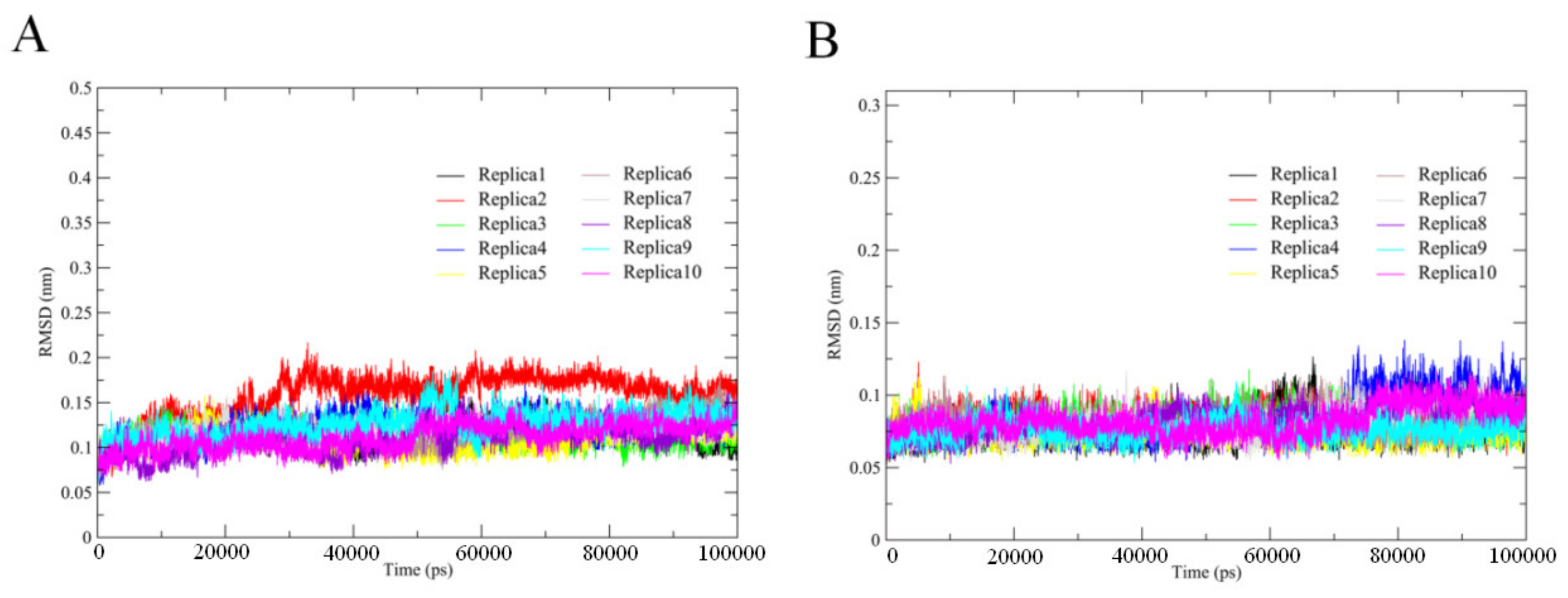
2.1. Trajectory Stability and Evaluation of Conformation Sampling

We used MD simulations to investigate and compare thermophilic and mesophilic serine proteases. In order to determine the stability/equilibrium of our simulations, we calculated the backbone root mean square deviation (RMSD) for each replica of the two simulation systems as a function of time. We observed that the backbone RMSD of each replica steadily increased for ~30 ns from the start of the simulation and then stabilized (Figure 2). Therefore, for each replica, we concatenated the MD trajectories from 30–100 ns to obtain a single 700-ns trajectory that was used for subsequent determination of the structural properties and essential dynamics (ED) analyses.
We next evaluated the convergence of our conformational sampling. We computed cosine content values from the first few eigenvectors obtained from the ED analyses of the replicas and joined trajectories (Table 1). Random diffusion protein dynamics resulted in a high cosine content value (i.e., close to 1) and indicates insufficient sampling relative to the simulation timescale. Conversely, converged sampling yielded a cosine content value close to 0. The eigenvector cosine content values were significantly higher than the corresponding single joined trajectory values for each simulation replica. These results demonstrate that a high degree of sampling convergence can be achieved by having multiple simulation replicas.

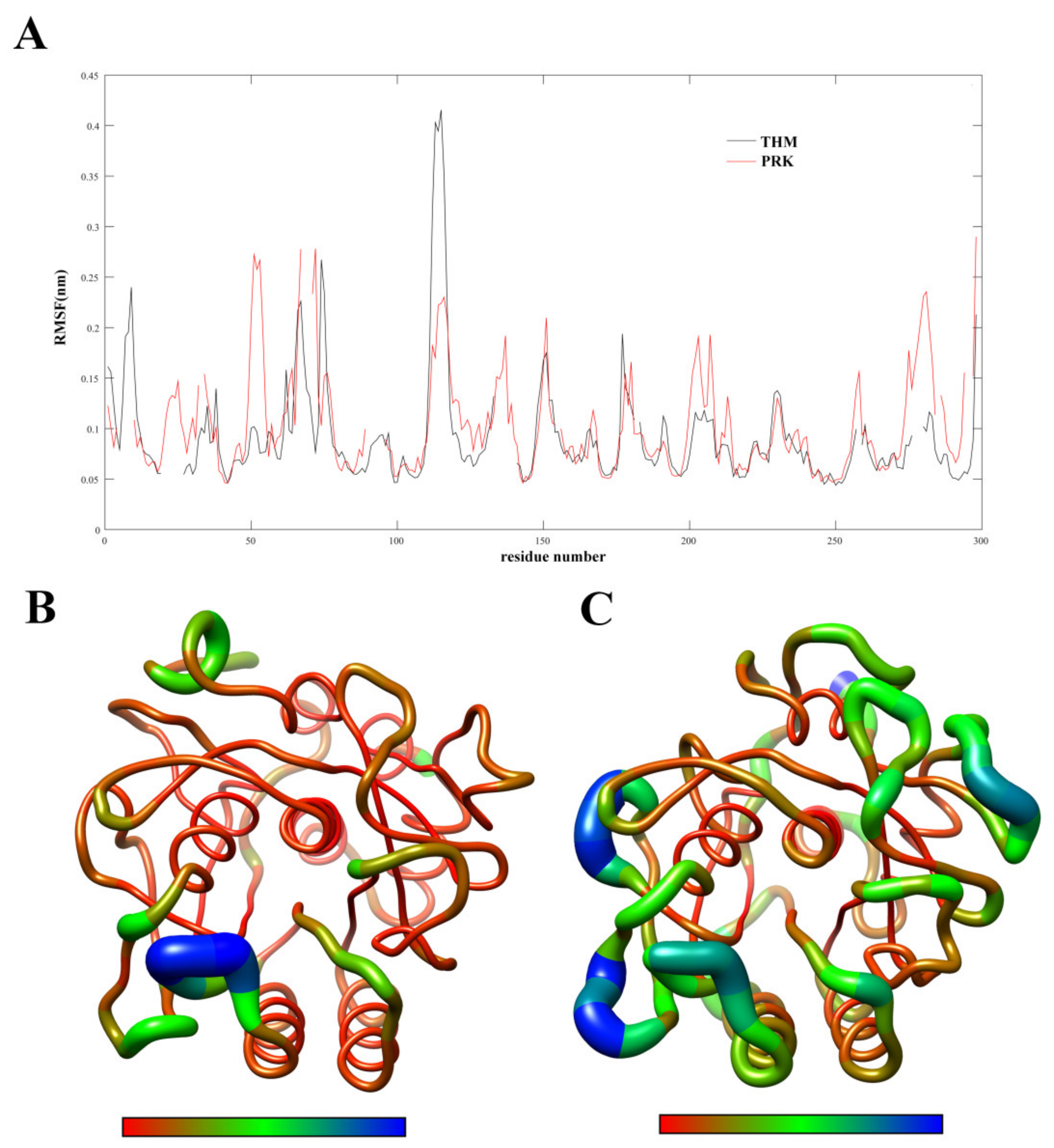
2.2. Flexibility Analysis

In order to examine the molecular flexibility of these two proteases, we computed the RMSF values of their Cα atoms from the joined MD trajectories. Figure 3A shows the Cα RMSF values as a function of residue number. Figure 3B,C are the three-dimensional (3D) backbone representations with the thickness/color of the worm corresponding to RMSF values. As shown in Figure 3, although many features are common to both proteases (e.g., the surface-exposed loops and N-, C- termini exhibit high RMSF values and the regular secondary structure elements and buried hydrophobic core exhibit low RMSF values), most structural regions are characterized by higher RMSF values in PRK compared to THM. Furthermore, the average RMSF values calculated over all the Cα atoms of THM and PRK are 0.81 and 0.92 Å, respectively, indicating a higher global rigidity of the thermophilic THM compared to the mesophilic PRK. The higher rigidity of THM can be expected to enhance its thermostability in high-temperature environments.

2.3. Geometrical and Structural Properties

For these two proteases, various geometrical and structural properties, including NHB, SASA, NSB, NNC, and Rg, were calculated based on the joined MD trajectories. Table 2 lists the average values and standard deviations of these properties. For all properties listed, the average values are very similar between the two proteases, with only minor differences observed. The subtle differences in these values can still reflect the changes in structure and stability between THM and PRK. For example, THM has more NNC, NSB and static HBs than PRK, indicating a higher number of stabilizing interactions or contacts in THM during simulation. When compared to PRK, THM has lower average values of Rg and SASA, revealing that THM is on average more tightly packed than PRK. This may also explain why THM has more static HBs and inter-atomic contacts than PRK.
We also computed the number of dynamic HBs (i.e., total number of HBs formed in a simulation) formed within the protein and between the protein and the solvent. There are 1592 and 1952 intramolecular dynamic HBs in the THM and PRK trajectories, respectively, as well as 328,834 and 331,932 protein solvent dynamic HBs in the THM and PPK trajectories, respectively. Since the dynamic HBs reflect the competitive HB interactions, a higher number means a larger freedom of competition and hence greater protein flexibility. The above results suggest that the number of dynamic HBs has been optimized to lower the conformational flexibility of THM for its adaptation to high-temperature.

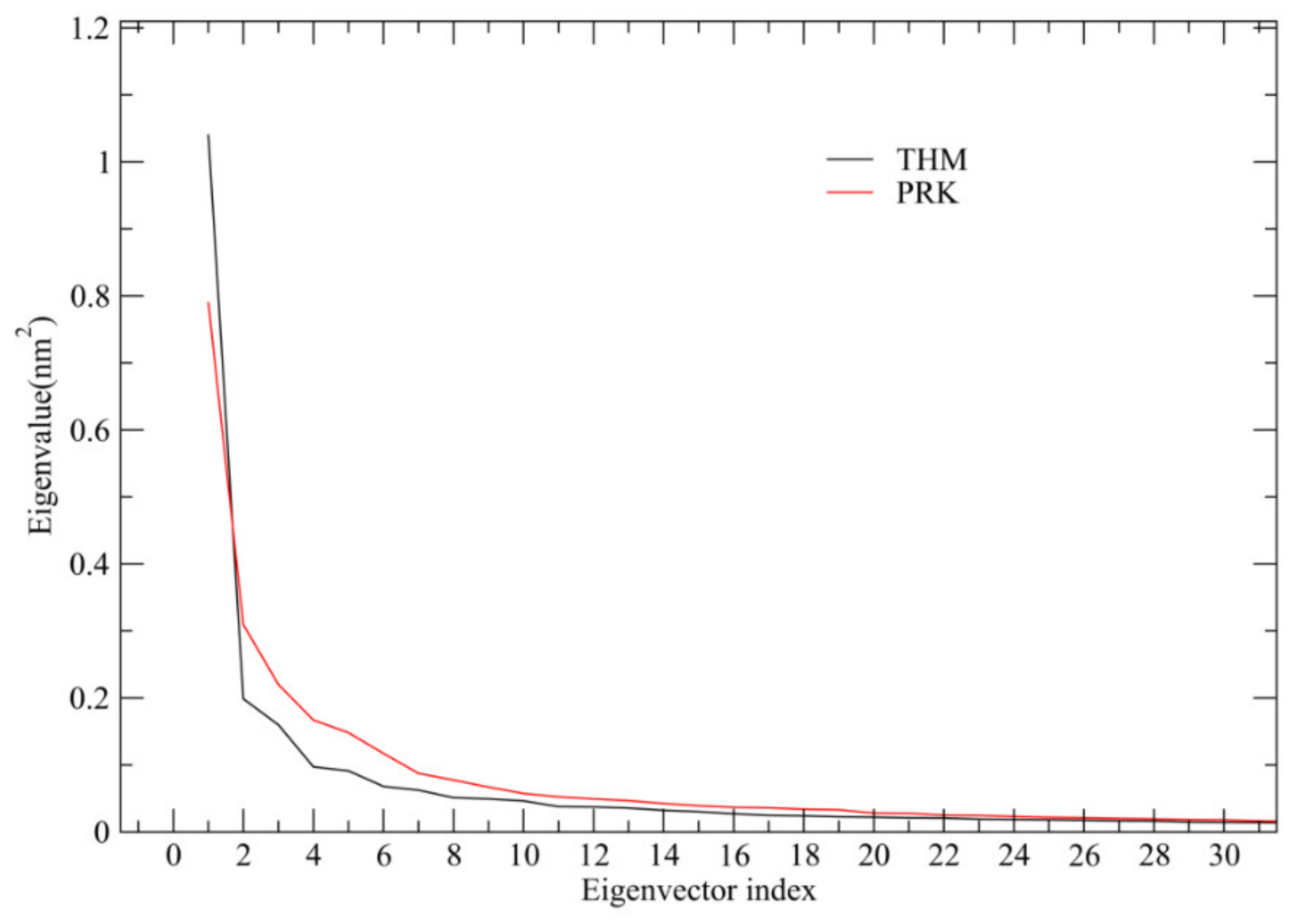
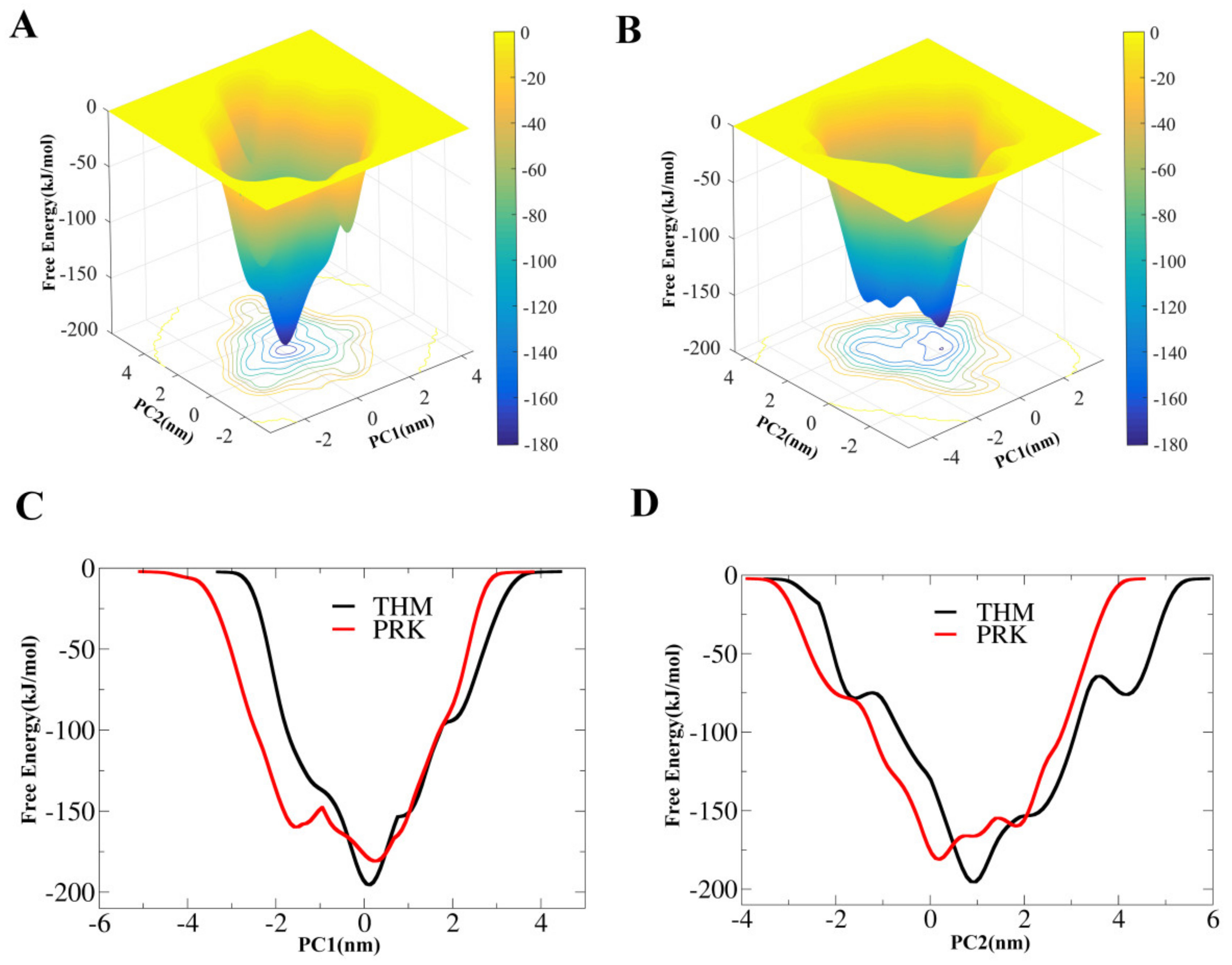
2.4. Essential Dynamics (ED) Analyses and Free-energy Landscapes

After diagonalization of the covariance matrix built from the concatenated trajectories, ED provides not only a set of eigenvectors and eigenvalues but also the value of total mean square fluctuations (TMSF). For THM and PRK, the TMSF values are 3.16 and 3.59 nm2, respectively, suggesting that THM experienced decreased amplitude of atomic fluctuations during simulation. Thus THM exhibited a lower overall flexibility compared to PRK, in agreement with the result of the RMSF analysis. The eigenvalues as a function of the eigenvector index was plotted to evaluate the amplitude of fluctuations along eigenvectors. As shown in Figure 4, for both proteases, only in the case of the first feweigenvectors can the significantly large eigenvalues be observed. Notably, the first two eigenvectors contribute 39.2% and 30.6% of THM and PRK, respectively to the TMSF values. This indicates that these TMSF values account for a significant fraction of the overall atomic fluctuations. Therefore, the projection of the first two eigenvectors/PCs can be used to approximately characterize the collective molecular motions of THM and PRK and, consequently, distinguish amongst different conformational substates.
For the two proteases, their FELs were constructed based on well-tempered metadynamics simulations using the projections of the first (PC1) and second (PC2) eigenvectors as the CV1 and CV2, respectively. Figure 5A,B show the 2D FELs of THM and PRK as a function of PC1 and PC2, respectively. Although both FELs present a funnel-like shape, there is only one main free-energy well/basin at the bottom of FEL (i.e., the global free-energy minimum region) for THM but three main wells/basins at the bottom of FEL for PRK, indicating that PRK has more stable conformational substates than THM. Figure 5C,D show the 1D free-energy profiles of these two proteases along PC1 and PC2, respectively. It is clear that THM has a lower global minimum free-energy value (or deeper funnel depth) compared to PRK. Furthermore, the free-energy profiles appear to be narrower, particularly at the lower half of the funnel for THM than for PRK.

3. Discussion

In order to understand the structure-function relationship of thermophilic enzymes and elucidate the mechanism by which they have adapted to high-temperature, it was necessary to perform a comparative study of the dynamic properties between thermophilic enzymes and their homologous mesophilic counterparts. Further, the FELs of these two form of homologous enzymes needed to be constructed and compared in order to evaluate the influence of solvent molecules on the conformational state with regards to protein dynamics. To explore the physicochemical basis of structural stability on thermophilic enzymes at high-temperatures, we performed MD simulations on thermophilic and mesophilic subtilisin-like serine proteases. Since our previous study showed that the mesophilic PRK did not unfold below 373 K [16], we selected 340 K as the temperature for MD simulations in this study. This was because 340 K is the optimal growth temperature for Thermoactinomyces vulgaris when it is secreting thermophilic THM [9]. The difference in temperature selection for MD simulations was decided on in an effort to emphasize the differences between these two types of enzymes in high-temperature tolerance. Nevertheless, in order to obtain more robust conclusions, we also performed 100 ns MD simulations at 300 K for PRK and THM, respectively (Figures S1–S3, Table S1). Comparisons of the dynamic structural and geometrical properties and the results of the ED analyses in both the 340 K and 300 K simulations pointed to a common conclusion – that thermophilic THM adopted a more compact conformation and exhibited more intramolecular interactions (including electrostatic interactions) and less global flexibility compared with mesophilic PRK. This finding is corroborated by the thermophilic THM’s thermal stability in high-temperature environments.
Under the FEL theory, solvent entropy maximization (hydrophobic effect) is considered to be the driving force in protein folding, and solvent entropy maximization is a critical factor in determining protein structural stability [15]. To elucidate the physicochemical basis as well as the role that solvents play in protein adaption in high-temperature environments, we computed the hydrophobicity (defined as the fraction of the buried non-polar area vs. the total nonpolar area) of these two forms of serine proteases. For THM and PRK, the estimated hydrophobicities were 40% and 38%, respectively, indicating that the hydrophobicity of thermophilic THM is slightly stronger than that of mesophilic PRK. It is widely accepted that when the hydrophobicity of a protein is stronger, its structure is often more compacted, and it has a higher capacity for resisting heat denaturation [26,27]. This can be exemplified by THM having more intramolecular contacts (NNC, NHB), smaller Rg, and smaller total SASA compared to PRK. This reflects a more compact structural packing of THM compared to that of PRK. Therefore, our results indicate that THM’s more stable and compact packing structure is most likely to arise from its stronger hydrophobicity.
A well-balanced compromise of stability and flexibility is considered to be the central issue in the adaptation of biomolecules to extreme conditions [28]. It is widely accepted that the thermostability of a protein is often associated with its decreased flexibility (or enhanced rigidity), implying that the suppression of protein internal fluctuations and mobility could be the reason it’s able to resist heat denaturation [29,30,31]. In this study, we observed that the overall fluctuation of thermophilic THM is lower than that of its homologous counterpart, supporting the viewpoint that thermal tolerance of a protein is usually correlated with reduced conformational flexibility. According to the FEL theory, protein and solvent entropy maximization (or atom mobility) are the driving forces behind the overall fluctuation of protein [14]. In particular, solvent entropy maximization has an overwhelming effect on protein internal flexibility and mobility [4,17]. Although entropy is hard to quantify, the number of dynamic HBs can be considered representative of atom mobility and competitive interactions [32]. In our study, the number of dynamic HBs within the protein and between the protein and solvent were both computed for THM and PRK, and the corresponding values were 1592 vs. 1952 and 328,834 vs. 331,932, respectively, indicating a lower level of atom mobility and competitive interactions for the thermophilic THM compared to that of the mesophilic PRK. Therefore, it is reasonable to conclude that the observed, less favorable protein solvent interactions for THM rather than for PRK are responsible for the lower global structural flexibility of THM.
Assessment of the differences between our constructed FELs revealed that, in comparison to PRK, THM has a narrower, relatively “flat” free-energy surface, and a lower minimum free-energy level, suggesting that the thermophilic form had fewer conformational substates. Since the size of these two proteins are very similar, their difference in free-energy surface could be related to their conformational flexibility, which is determined by the intramolecular competitive interactions as mentioned above. Our results have verified the ideal FEL model proposed by D’Amico et al. [22], which rationalizes the stability-flexibility relationships in extremophilic enzymes’ adaptation to extreme temperatures.

4. Materials and Methods

4.1. Preparation of the Simulated System

The high-resolution crystal structures of the thermophilic (PDB code: 1THM [33]) and mesophilic (PDB code: 1IC6 [34]) subtilisin-like serine proteases were used as starting models for MD simulations. Before simulations, all hetero atoms and crystallographic water were removed but the metal ions were retained.

4.2. MD Simulations

Energy minimizations and MD simulations were performed with Amber99SB-ILDN force field [35] using GROMACS software [36]. Each structural model was centered in a dodecahedron box with a 1.0 nm minimum distance between the protein and the edge of the box. Each model was then dissolved using TIP3P water model [37] along with three additional chloride ions, which were added in order to neutralize the net charge of the system. To minimize the simulation time, we used an energy minimization algorithm with the steepest descent, and we performed two continuous 500-ps position restraint simulations of 1000 kJ/mol/nm2 in the NVT and NPT ensembles. Conformational sampling efficiency was confirmed using ten independent 100-ns production MD simulations. A Maxwell distribution was used to determine the initial atomic velocities of each simulation. Replica 1 to 10 refer to MD trajectories with different initial velocities obtained by the same system.
During production MD runs, the system was coupled to 340 K with a 0.1 ps constant using a v-rescale [38] thermostat, and the system pressure was maintained at 1 atm with a 0.5 ps coupling constant using a weakly coupled external pressure bath [39]. The Particle-mesh Ewald (PME) method [40] was used to calculate the electrostatic interactions using an interpolation order of 4, Fourier grid spacing of 0.135 nm, and a Coulomb radius of 1.0 nm. Lennard-Jones potentials were used to model van der Waals (VDW) interactions with a cut-off distance of 1 nm. Water molecules and non-water bonds were constrained using the SETTLE [41] and LINCS [42] algorithms, respectively. Center-of-mass motion was removed at every time step, non-bonded pairs were updated every 10 time steps, and structural coordinates were saved every 10 ps.

4.3. Structural and Geometrical Properties

We used the following GROMACS tools to perform structural and geometrical analyses of MD trajectories: ‘gmx rms’ to determine backbone root mean square deviation (RMSD) relative to the starting structure, ‘gmx rmsf’ to determine per-residue Cα root mean square fluctuation (RMSF), ‘gmx sasa’ to determine solvent accessible surface area (SASA), ‘gmx mindist’ to calculate the number of native contacts (NNC), ‘gmx gyrate’ to measure the radius of gyration (Rg) and ‘gmx saltbr’ to calculate the number of salt bridge (NSB). The ‘gmx hbond’ tool and the ‘Hydrogen Bonds’ plugin within VMD [43] were used to determine prtein internal HBs and protein-solvent HBs, respectively. The number of existing HBs at a given snapshot of the MD trajectory is referred to as the static HB number, while the number of HBs that have ever existed in a trajectory is referred to as the dynamic HB number. To calculate the folded state buried area, the average protein SASA during the equilibrium portion of the MD trajectory was subtracted from the SASA of an extended polypeptide chain corresponding to the ideal unfolded protein state. For a given residue X, the values of the polar, nonpolar, and total SASAs were obtained from Lins et al. [44], in which they were estimated by calculating the corresponding SASAs in the Gly–X–Gly tripeptide with extended conformation.

4.4. Essential Dynamics (ED)

The ED method, also known as the principal component analysis (PCA) in mathematics, is widely used to extract the largest amplitude protein motions (also called collective motions or large-scale concerted motions) from an MD trajectory. A detailed mathematical description of the ED method is given by Ref. [45] Briefly, ED analysis is mainly based on the diagonalization of the covariance matrix constructed from atomic fluctuations in a trajectory, thus obtaining a set of eigenvectors (or principal components; PCs) and corresponding eigenvalues. The eigenvector is the direction in the conformational space and describes the protein collective motion during a simulation; the eigenvalue is the mean square atomic fluctuations and describes the amplitude of fluctuations along the corresponding eigenvector. The protein motion along an eigenvector can be derived by projecting the MD trajectory onto the eigenvector.
In this study, ED analyses were performed on the concatenated trajectories of THM and PRK using the GROMACS tools gmx covar and gmx anaeig. Only the Cα atoms were included in the ED analyses.

4.5. Free-Energy Calculations

In order to reconstruct the FELs of the thermophilic and mesophilic serine proteases, the well-tempered metadynamics simulations [46] were performed. Using metadynamics, the addition of an external history dependent potential that acts on properly chosen degrees of freedom (also called collective variables, CV) can accelerate the detection of rare events and help the system escape from local free-energy minima [47]. In our analyses, we used well-tempered metadynamics, a newly developed variant of metadynamics which enables avoiding the exploration of unphysically high free-energy regions such that the variations in the bias potential overtime are decreased, guaranteeing a smooth simulation convergence [46]. Coupling well-tempered metadynamics with a set of CVs (i.e., PCs) determined using the ED method has been shown to efficiently reconstruct a protein’s FEL [48,49]. Here, we chose projections of trajectory onto the eigenvectors 1 (PC1) and 2 (PC2) as CV1 and CV2, respectively.
We used the following parameters for the well-tempered metadynamics simulations: 0.4 kJ/mol initial Gaussian height, added every 2 ps; 0.35 nm Gaussian width; and a bias factor of 10. The other simulation parameters and conditions were the same as those used in the standard MD simulations. The starting structure for a metadynamics simulation was the final snapshot of the joined standard MD trajectory. The well-tempered metadynamics simulations were run for 500 ns for both THM and PRK using GROMACS and Plumed [49].

Supplementary Materials

The following are available online at https://0-www-mdpi-com.brum.beds.ac.uk/1422-0067/21/9/3128/s1. Figure S1. Time evolutions of the backbone RMSD values of the THM (black line) and PRK (red line) with respect to their starting structures during MD simulations at 300K; Figure S2. 3D backbone representations of protein structures mapped with per-residue average backbone RMSF values during MD simulations at 300K. (A) THM; (B) PRK; Figure S3. Eigenvalues as a function of eigenvector index. Only those of the first 30 eigenvectors are shown; Table S1. Structural and geometrical properties (standard deviations are in parentheses) of THM and PRK during MD simulations at 300K.

Author Contributions

P.S. performed the experiments, analyzed the data, and participated in designing the study and drafting the manuscript. S.-Q.L. and L.-Q.Y. designed the study, coordinated the work, and wrote and revised the manuscript. All authors contributed to and approved the final version of the manuscript.

Funding

This study was funded by grants (31660015, 31860243 and 31960198) from the National Natural Sciences Foundation of China, the Yunnan Applied Basic Research Projects (2017FH001-032 and 2017FB024).

Acknowledgments

We thank High Performance Computer Center of Dali University for computational support.

Conflicts of Interest

The authors declare no conflicts of interest for this work.

References

  1. Babu, P.; Chandel, A.K.; Singh, O.V. Survival mechanisms of extremophiles. In Extremophiles and Their Applications in Medical Processes; Springer International Publishing: Cham, Switzerland, 2015. [Google Scholar]
  2. Sang, P.; Du, X.; Yang, L.-Q.; Meng, Z.-H.; Liu, S.-Q. Molecular motions and free-energy landscape of serine proteinase K in relation to its cold-adaptation: A comparative molecular dynamics simulation study and the underlying mechanisms. RSC Adv. 2017, 7, 28580–28590. [Google Scholar] [CrossRef] [Green Version]
  3. Tilton, R.F., Jr.; Dewan, J.C.; Petsko, G.A. Effects of temperature on protein structure and dynamics: X-ray crystallographic studies of the protein ribonuclease-A at nine different temperatures from 98 to 320K. Biochemistry 1992, 31, 2469–2481. [Google Scholar] [CrossRef] [PubMed]
  4. Sang, P.; Yang, Q.; Du, X.; Yang, N.; Yang, L.-Q.; Ji, X.-L.; Fu, Y.-X.; Meng, Z.-H.; Liu, S.-Q. Effect of the solvent temperatures on dynamics of serine protease proteinase K. Int. J. Mol. Sci. 2016, 17, 254. [Google Scholar] [CrossRef] [PubMed] [Green Version]
  5. Lonhienne, T.; Gerday, C.; Feller, G. Psychrophilic enzymes: Revisiting the thermodynamic parameters of activation may explain local flexibility. Biochim. Biophys. Acta Protein Struct. Mol. Enzymol. 2000, 1543, 1–10. [Google Scholar] [CrossRef]
  6. Papaleo, E.; Tiberti, M.; Invernizzi, G.; Pasi, M.; Ranzani, V. Molecular determinants of enzyme cold adaptation: Comparative structural and computational studies of cold-and warm-adapted enzymes. Curr. Protein Pept. Sci. 2011, 12, 657–683. [Google Scholar] [CrossRef]
  7. Sterner, R.; Liebl, W. Thermophilic adaptation of proteins. Crit. Rev. Biochem. Mol. Biol. 2001, 36, 39–106. [Google Scholar] [CrossRef]
  8. Xia, Y.-L.; Sun, J.-H.; Ai, S.-M.; Li, Y.; Du, X.; Sang, P.; Yang, L.-Q.; Fu, Y.-X.; Liu, S.-Q. Insights into the role of electrostatics in temperature adaptation: A comparative study of psychrophilic, mesophilic, and thermophilic subtilisin-like serine proteases. RSC Adv. 2018, 8, 29698–29713. [Google Scholar] [CrossRef] [Green Version]
  9. Tiberti, M.; Papaleo, E. Dynamic properties of extremophilic subtilisin-like serine-proteases. J. Struct. Biol. 2011, 174, 69–83. [Google Scholar] [CrossRef]
  10. Kumar, S.; Nussinov, R. How do thermophilic proteins deal with heat? Cell. Mol. Life Sci. CMLS 2001, 58, 1216–1233. [Google Scholar] [CrossRef]
  11. Vieille, C.; Zeikus, G.J. Hyperthermophilic enzymes: Sources, uses, and molecular mechanisms for thermostability. Microbiol. Mol. Biol. Rev. 2001, 65, 1–43. [Google Scholar] [CrossRef] [Green Version]
  12. Pica, A.; Graziano, G. Shedding light on the extra thermal stability of thermophilic proteins. Biopolymers 2016, 105, 856–863. [Google Scholar] [CrossRef] [PubMed]
  13. Mattos, C. Protein–water interactions in a dynamic world. Trends Biochem. Sci. 2002, 27, 203–208. [Google Scholar] [CrossRef]
  14. Li, H.; Xie, Y.; Liu, C.; Liu, S. Physicochemical bases for protein folding, dynamics, and protein-ligand binding. Sci. China Life Sci. 2014, 57, 287–302. [Google Scholar] [CrossRef] [PubMed]
  15. Dill, K.A. Dominant forces in protein folding. Biochemistry 1990, 29, 7133–7155. [Google Scholar] [CrossRef] [PubMed]
  16. Du, X.; Sang, P.; Xia, Y.-L.; Li, Y.; Liang, J.; Ai, S.-M.; Ji, X.-L.; Fu, Y.-X.; Liu, S.-Q. Comparative thermal unfolding study of psychrophilic and mesophilic subtilisin-like serine proteases by molecular dynamics simulations. J. Biomol. Struct. Dyn. 2017, 35, 1500–1517. [Google Scholar] [CrossRef] [PubMed] [Green Version]
  17. Vitkup, D.; Ringe, D.; Petsko, G.A.; Karplus, M. Solvent mobility and the protein’glass’ transition. Nat. Struct. Mol. Biol. 2000, 7, 34. [Google Scholar]
  18. Finkelstein, A.V.; Ptitsyn, O. Protein Physics: A Course of Lectures; Elsevier: Amsterdam, The Netherlands, 2016. [Google Scholar]
  19. Chaplin, M.F. Water’s hydrogen bond strength. arXiv 2007, arXiv:preprint cond-mat/0706.1355. [Google Scholar]
  20. Yang, L.-Q.; Ji, X.-L.; Liu, S.-Q. The free energy landscape of protein folding and dynamics: A global view. J. Biomol. Struct. Dyn. 2013, 31, 982–992. [Google Scholar] [CrossRef] [Green Version]
  21. Yang, L.-Q.; Sang, P.; Tao, Y.; Fu, Y.-X.; Zhang, K.-Q.; Xie, Y.-H.; Liu, S.-Q. Protein dynamics and motions in relation to their functions: Several case studies and the underlying mechanisms. J. Biomol. Struct. Dyn. 2014, 32, 372–393. [Google Scholar] [CrossRef] [Green Version]
  22. D’Amico, S.; Marx, J.-C.; Gerday, C.; Feller, G. Activity-stability relationships in extremophilic enzymes. J. Biol. Chem. 2003, 278, 7891–7896. [Google Scholar] [CrossRef] [Green Version]
  23. Hedstrom, L. Serine protease mechanism and specificity. Chem. Rev. 2002, 102, 4501–4524. [Google Scholar] [CrossRef] [PubMed]
  24. The PyMOL Molecular Graphics System, Version 1.8.; Schrodinger, LLC: New York, NY, USA, 2015.
  25. Pettersen, E.F.; Goddard, T.D.; Huang, C.C.; Couch, G.S.; Greenblatt, D.M.; Meng, E.C.; Ferrin, T.E. UCSF Chimera—A visualization system for exploratory research and analysis. J. Comput. Chem. 2004, 25, 1605–1612. [Google Scholar] [CrossRef] [PubMed] [Green Version]
  26. Pace, C.N. Contribution of the hydrophobic effect to globular protein stability. J. Mol. Biol. 1992, 226, 29–35. [Google Scholar] [CrossRef]
  27. Reed, C.J.; Lewis, H.; Trejo, E.; Winston, V.; Evilia, C. Protein adaptations in archaeal extremophiles. Archaea 2013, 2013, 373275. [Google Scholar] [CrossRef] [PubMed] [Green Version]
  28. Jaenicke, R. Protein stability and molecular adaptation to extreme conditions. In EJB Reviews 1991; Springer: Berlin, Germany, 1991; pp. 291–304. [Google Scholar]
  29. Quezada, A.G.; Díaz-Salazar, A.J.; Cabrera, N.; Pérez-Montfort, R.; Piñeiro, Á.; Costas, M. Interplay between protein thermal flexibility and kinetic stability. Structure 2017, 25, 167–179. [Google Scholar] [CrossRef] [PubMed] [Green Version]
  30. Kamerzell, T.J.; Middaugh, C.R. The complex inter-relationships between protein flexibility and stability. J. Pharm. Sci. 2008, 97, 3494–3517. [Google Scholar] [CrossRef] [PubMed]
  31. Karshikoff, A.; Nilsson, L.; Ladenstein, R. Rigidity versus flexibility: The dilemma of understanding protein thermal stability. FEBS J. 2015, 282, 3899–3917. [Google Scholar] [CrossRef]
  32. Xie, B.-B.; Bian, F.; Chen, X.-L.; He, H.-L.; Guo, J.; Gao, X.; Zeng, Y.-X.; Chen, B.; Zhou, B.-C.; Zhang, Y.-Z. Cold adaptation of zinc metalloproteases in the thermolysin family from deep sea and arctic sea ice bacteria revealed by catalytic and structural properties and molecular dynamics new insights into relationship between conformational flexibility and hydrogen bonding. J. Biol. Chem. 2009, 284, 9257–9269. [Google Scholar]
  33. Teplyakov, A.V.; Kuranova, I.P.; Harutyunyan, E.H.; Vainshtein, B.K.; Fro, C.; Ho, W.E.; Wilson, K.S. Crystal structure of thermitase at 1.4 A resolution. J. Mol. Biol. 1990, 214, 261–279. [Google Scholar] [CrossRef]
  34. Betzel, C.; Gourinath, S.; Kumar, P.; Kaur, P.; Perbandt, M.; Eschenburg, S.; Singh, T.P. Structure of a serine protease proteinase K from Tritirachium album limber at 0.98 Å resolution. Biochemistry 2001, 40, 3080–3088. [Google Scholar] [CrossRef]
  35. Lindorff-Larsen, K.; Piana, S.; Palmo, K.; Maragakis, P.; Klepeis, J.L.; Dror, R.O.; Shaw, D.E. Improved side-chain torsion potentials for the Amber ff99SB protein force field. Proteins Struct. Funct. Bioinform. 2010, 78, 1950–1958. [Google Scholar] [CrossRef] [PubMed] [Green Version]
  36. Hess, B.; Kutzner, C.; Van Der Spoel, D.; Lindahl, E. GROMACS 4: Algorithms for highly efficient, load-balanced, and scalable molecular simulation. J. Chem. Theory Comput. 2008, 4, 435–447. [Google Scholar] [CrossRef] [PubMed] [Green Version]
  37. Jorgensen, W.L.; Chandrasekhar, J.; Madura, J.D.; Impey, R.W.; Klein, M.L. Comparison of simple potential functions for simulating liquid water. J. Chem. Phys. 1983, 79, 926–935. [Google Scholar] [CrossRef]
  38. Bussi, G.; Donadio, D.; Parrinello, M. Canonical sampling through velocity rescaling. J. Chem. Phys. 2007, 126, 014101. [Google Scholar] [CrossRef] [Green Version]
  39. Berendsen, H.J.; Postma, J.; van Gunsteren, W.F.; DiNola, A.; Haak, J. Molecular dynamics with coupling to an external bath. J. Chem. Phys. 1984, 81, 3684–3690. [Google Scholar] [CrossRef] [Green Version]
  40. Essmann, U.; Perera, L.; Berkowitz, M.L.; Darden, T.; Lee, H.; Pedersen, L.G. A smooth particle mesh Ewald method. J. Chem. Phys. 1995, 103, 8577–8593. [Google Scholar] [CrossRef] [Green Version]
  41. Miyamoto, S.; Kollman, P.A. Settle: An analytical version of the SHAKE and RATTLE algorithm for rigid water models. J. Comput. Chem. 1992, 13, 952–962. [Google Scholar] [CrossRef]
  42. Hess, B.; Bekker, H.; Berendsen, H.J.; Fraaije, J.G. LINCS: A linear constraint solver for molecular simulations. J. Comput. Chem. 1997, 18, 1463–1472. [Google Scholar] [CrossRef]
  43. Humphrey, W.; Dalke, A.; Schulten, K. VMD: Visual molecular dynamics. J. Mol. Graph. 1996, 14, 33–38. [Google Scholar] [CrossRef]
  44. Lins, L.; Thomas, A.; Brasseur, R. Analysis of accessible surface of residues in proteins. Protein Sci. 2003, 12, 1406–1417. [Google Scholar] [CrossRef]
  45. Amadei, A.; Linssen, A.B.; Berendsen, H.J. Essential dynamics of proteins. Proteins Struct. Funct. Bioinform. 1993, 17, 412–425. [Google Scholar] [CrossRef] [PubMed]
  46. Barducci, A.; Bussi, G.; Parrinello, M. Well-tempered metadynamics: A smoothly converging and tunable free-energy method. Phys. Rev. Lett. 2008, 100, 020603. [Google Scholar] [CrossRef] [PubMed] [Green Version]
  47. Laio, A.; Gervasio, F.L. Metadynamics: A method to simulate rare events and reconstruct the free energy in biophysics, chemistry and material science. Rep. Prog. Phys. 2008, 71, 126601. [Google Scholar] [CrossRef]
  48. Sicard, F.; Senet, P. Reconstructing the free-energy landscape of Met-enkephalin using dihedral principal component analysis and well-tempered metadynamics. J. Chem. Phys. 2013, 138, 06B610_1. [Google Scholar] [CrossRef] [Green Version]
  49. Spiwok, V.; Lipovová, P.; Králová, B. Metadynamics in essential coordinates: Free energy simulation of conformational changes. J. Phys. Chem. B 2007, 111, 3073–3076. [Google Scholar] [CrossRef]
Figure 1. Ribbon representation of thermophilic (A) and mesophilic (B) serine proteases. These structures were obtained from PDB with PDB codes 1THM and 1IC6. α-helices, β-strands, and loops are colored red, yellow, and green, respectively. The catalytic triad residues are shown as stick models. Ca2+ and Na+ are shown as blue spheres. (A) and (B) were generated using Pymol [24].
Ijms 21 03128 g001
Figure 2. Evolutions of the backbone RMSD values of the THM and PRK with respect to their starting structures during the 10 independent MD simulations (Replicas 1–10). (A) THM; (B) PRK.
Ijms 21 03128 g002
Figure 3. Comparison between the structural flexibility of THM and PRK. (A) Per-residue average backbone RMSF profiles calculated from MD trajectories of THM (black line) and PRK (red line). The gap of the curves corresponds to the insertion or deletion in the structural alignment. The residues are numbered according to the PRK structure. (B,C) are 3D backbone representations of the THM and PRK structures mapped with per-residue average backbone RMSF values, respectively. The backbone color ranges from red to blue and corresponds to a line from thin to thick and denotes that the backbone RMSF varies from the lowest to the highest values. (B,C) were generated using UCSF Chimera [25].
Ijms 21 03128 g003
Figure 4. Eigenvalues as a function of the eigenvector index. Only those of the first 30 eigenvectors are shown.
Ijms 21 03128 g004
Figure 5. Constructed free-energy landscapes (FELs) of THM and PRK. (A) and (B) are 2D FELs for THM and PRK as a function of the first (PC1) and second (PC2) eigenvectors, respectively. The color bar represents the free-energy value in kJmol−1. (C) and (D) are 1D free-energy profiles for THM and PRK as a function of PC1 and PC2, respectively.
Ijms 21 03128 g005
Table 1. Cosine content values of the first two eigenvectors calculated from the ten independent equilibrium MD trajectories (30–100 ns; Replicas 1–10) and the single 700-ns joined trajectory.
TrajectoryTHMPRK
eig.1eig.2eig.1eig.2
10.74910.38620.49350.3572
20.25270.64820.15790.5803
30.57230.37650.20150.1097
40.43250.25130.86050.2531
50.17080.25170.37010.2017
60.73650.68040.49710.0671
70.03760.40860.20360.2471
80.36920.89740.19120.4076
90.67020.39750.60810.2992
100.60910.48390.35820.5703
joined0.00190.02570.03090.0093
Table 2. Structural and geometrical properties (standard deviations are in parentheses) of THM and PRK during MD simulations.
ProteinNNC aSASA b2)NSB cRg d (Å)NHB e
Stat fDyna g
THM135,765 (1139)10,334 (191)14 (0.08)16.5 (0.05)208 (7.7)1592
PRK133,764 (982)11,007 (222)10 (0.07)16.7 (0.05)202 (7.6)1952
a Number of native contacts. A native contact is considered to exist if the distance between two atoms is less than 6 Å; b Total solvent accessible surface area; c Radius of gyration; d Number of salt bridges. Only salt bridges with persistence greater than 20% were counted in; e Number of corresponding HBs; f Static HB number averaged over all frames; g Dynamic HB number average over all single trajectories.

