Article

Proteomic Characterization of Antibiotic Resistance in Listeria and Production of Antimicrobial and Virulence Factors

1
Departamento de Microbiología y Parasitología, Facultad de Farmacia, Campus Sur 15782, Universidad de Santiago de Compostela, 15782 Santiago de Compostela, Spain
2
Marine Research Institute (IIM), Spanish National Research Council (CSIC), Eduardo Cabello 6, 36208 Vigo, Spain
3
Agroalimentary Technological Center of Lugo, Montirón 154, 27002 Lugo, Spain
4
Departamento de Química Analítica, Nutrición y Bromatología, Área de Tecnología de los Alimentos, Facultad de Veterinaria, Campus Lugo, Universidad de Santiago de Compostela, 27002 Santiago de Compostela, Spain
5
Sydney School of Veterinary Science, Faculty of Science, University of Sydney, Sydney, NSW 2006, Australia
*
Authors to whom correspondence should be addressed.
Int. J. Mol. Sci. 2021, 22(15), 8141; https://0-doi-org.brum.beds.ac.uk/10.3390/ijms22158141
Submission received: 17 May 2021 / Revised: 25 July 2021 / Accepted: 26 July 2021 / Published: 29 July 2021
(This article belongs to the Special Issue Molecular Analysis of the Resistome in Food)

Abstract

:
Some Listeria species are important human and animal pathogens that can be found in contaminated food and produce a variety of virulence factors involved in their pathogenicity. Listeria strains exhibiting multidrug resistance are known to be progressively increasing and that is why continuous monitoring is needed. Effective therapy against pathogenic Listeria requires identification of the bacterial strain involved, as well as determining its virulence factors, such as antibiotic resistance and sensitivity. The present study describes the use of liquid chromatography–electrospray ionization tandem mass spectrometry (LC–ESI–MS/MS) to do a global shotgun proteomics characterization for pathogenic Listeria species. This method allowed the identification of a total of 2990 non-redundant peptides, representing 2727 proteins. Furthermore, 395 of the peptides correspond to proteins that play a direct role in Listeria pathogenicity; they were identified as virulence factors, toxins and anti-toxins, or associated with either antibiotics (involved in antibiotic-related compounds production or resistance) or resistance to toxic substances. The proteomic repository obtained here can be the base for further research into pathogenic Listeria species and facilitate the development of novel therapeutics for these pathogens.

Graphical Abstract

1. Introduction

Listeria are Gram-positive facultative anaerobic bacteria that form small rods with low GC content; the species do not form spores or capsules and are motile at temperatures between 10 °C and 25 °C [1]. Listeria colonize a wide range of different environments, and species of this genus can be isolated from foodstuffs such as milk, poultry, and meat. Species such as Listeria monocytogenes and Listeria ivanovii can be spread and transmitted through contaminated food. L. monocytogenes represents the main human and animal pathogen that causes listeriosis [2]; L. ivanovii was also described, although to a lesser extent, to cause stillbirths or premature births in ruminants [3], while two additional species (Listeria innocua or L. grayi), that were originally considered nonpathogenic, are reported to also infect humans [4]. Listeria contamination is common in the food industry, as these pathogens can colonize both the equipment and the environment, forming biofilms that can survive for months or even years [5]. Fortunately, listeriosis incidence is low in the general population, as the syndrome presents with a high mortality rate (20–30%) in susceptible individuals with severe symptoms [6]. Four L. monocytogenes serovars—13, 1/2a, 1/2b, and 4b—are known to cause listeriosis, with 4b as the most frequent serovar found in humans [6].
Listeria species are currently still sensitive to a variety of antibiotics, such as penicillin, fluoroquinolones, macrolides, and vancomycin [4]. Antibiotic sensitivity has been mainly studied on L. monocytogenes, and the species is susceptible to a variety of antimicrobials, with the exception of cephalosporins and sulfonamides. The treatment of choice for listeriosis is currently either ampicillin or penicillin combined with aminoglycosides, with trimethoprim–sulfamethoxazole as an alternative. Recent studies have stablished that resistance to single antibiotics is more common than multiple resistance; tetracycline resistance is the most frequent, although some bacterial strains are resistant to other antibiotics [2,6,7]. As is generally the case for bacteria, the prevalence of antibiotic resistance, including multiple antibiotic resistances, within the genus Listeria spp. is currently increasing. A recent study demonstrated that a variety of Listeria species (i.e., L. murrayi, L. welshimeri or L. gravi) are sensitive to semisynthetic penicillins and vancomycin, but resistant to nalidixic acid [4]. The increase in antimicrobial resistance in bacteria present in food has been explained as follows: while antibiotic-resistant bacteria present in foods are initially saprophytic or commensal, their resistance genes are sporadically transferred to other foodborne microorganisms, thus contributing to worsen this problem. It is therefore currently urgent to develop an in-depth knowledge of how antibiotic resistance is disseminated among bacteria, in order to control this major threat to human and animal health and welfare [2].
Infection with pathogenic Listeria species involves at least eight known virulence factors. Bacterial invasion and breach of the gut epithelial barrier, after ingestion of contaminated food, is mediated by the bacterial surface proteins known as internalines (InlA and InlB). Escape from the host phagocytic vacuole is mediated by secreted bacterial proteins, such as PlcA, pore-forming toxin listeriolysin O (LLO), phosphatidylcholine-specific phospholipase, and phosphatidylinositol-specific phospholipase C. Intracellular actin-based motility (ActA) and cell-to-cell spread (without actual exit to the interstitial liquid) is accomplished by Hly and PlcB. All these genes are clustered into two pathogenicity islands, and the expression of the main virulence gene is controlled by transcription of the regulator PrfA, which is absent in L. innocua [3,6,8,9,10,11]. An additional 47 genes, scattered throughout the bacterial genome, have been identified to affect virulence [12].
Listeria detection and identification has usually focused on colony morphology, sugar fermentation and hemolytic properties; however, the methodology involved is time consuming and not appropriate to test food with a short shelf life. The food industry requires fast and unequivocal methods to quickly detect and identify pathogens present in foodstuffs. Although a number of procedures, such as molecular-based techniques (DNA hybridization, polymerase chain reaction (PCR), and microarray), enzyme-linked immuno-sorbent assay (ELISA), and flow cytometry, have been recently developed [13], these techniques need long enrichment time, expensive chemicals, and specialized equipment. Other methods developed for Listeria spp. detection and characterization include biosensors [14] and target proteomic methods [15,16,17]. In addition, matrix-assisted laser desorption/ionization time-of-flight mass spectrometry (MALDI-TOF MS) [15,16] and liquid chromatography coupled to tandem mass spectrometry (LC–MS/MS) against a spectral library construction, were used to stablish genotype–proteotype–phenotype relationships among the bacterial strains [17]. Another approach involved the development of shotgun proteomics protocols for the determination of virulence factors, such as adhesins, in L. monocytogenes, as well as the identification of relevant transport and metabolic pathways using LC–ESI–MS/MS [18,19,20]. Additional studies involve the use of LC–ESI–MS/MS to research the proteomics of antibiotic resistance and the production of antimicrobials and other virulence factors, in order to correctly identify the different streptococcal species associated to mastitis [21].
The characterization of virulence factors, pathogenicity, strain variation, and pathogenic serotypes is an essential part of epidemiologic investigations and food safety monitoring. The objectives of this study were to identify bacterial peptides corresponding to specific virulence factors, such as the production of antimicrobials, toxins and antimicrobial resistance, using advanced shotgun proteomic methods, to determine the diversity of the Listeria pathogens isolated from food.

2. Results

2.1. Listeria spp. Proteomics Data Repository

Nine different Listeria strains were studied (Table 1). Bacterial peptides were prepared by treating the protein mixtures with trypsin and analyzed by LC–ESI–MS/MS, as described previously [21,22,23]. A total of 2990 non-redundant peptides were identified which corresponded to 2727 annotated proteins from Listeria (see complete non-redundant dataset in Excel Supplemental Data). Accordingly, the virulence factors were identified by comparison with both the “Virulence Factors of Pathogenic Bacteria Database” (VFDB; http://www.mgc.ac.cn/VFs/, accessed on 26 July 2020), and with previously reported data [1,6,10,24,25].
In the group of non-redundant peptides, 395 were identified unequivocally as virulence factors, and included proteins such as the internalins involved in eukaryotic cell colonization and immune evasion, and which are specific for Listeria. Additional polypeptides (toxins and antitoxins), together with polypeptides so far involved in antibiotic resistance, were also found. The 395 virulent factors identified for listerial species are displayed in Tables S1–S6 in Supplemental Data and organized in groups according to the main role they play; this includes toxins, antibiotic resistance peptides, and other tolerance proteins involved in resistance to toxic substances, colonization and immune evasion factors, antimicrobial compounds, ABC and other transporters associated with virulence factors, and alternative virulence factors. In addition, the main antibiotic resistance proteins, antimicrobial related proteins, and other virulence factors are summarized in Table 2.
Two Listeria strains, Li4 and Li2 (with 73 and 116 peptides, respectively), L. monocyogenes and Listeria seeligeri, respectively, contained the highest number of peptides related to virulence. Taken together, all these results it suggests that the two species are probably the most pathogenic ones, as compared other species. Li2 strain, from L. seeligeri, was described as an avirulent Risk group 1 bacterium by Rocourt and Grimont (1983), it was also identified as non-pathogenic for holoxenic pathogen-free mice (50% lethal dose, more than 10′ colony-forming units per mL) [26]. In addition, strains Li1 (Listeria welshimeri) and Li3 (Listeria innocua) are also classified as risk Group 1; they are represented with 17 and 55 peptides related to virulence factors, respectively. The remaining strains analyzed belong to either risk group 1 or to different serotypes and are represented in Table 1 with a number of peptides related to virulence factor proteins that ranges from 15 to 37.

2.2. Proteins Involved in Bacterial Resistance to Antibiotics and Other Toxic Substances

In the present study 82 peptides, present in Listeria strains and involved in resistance to either antibiotics or toxic substances, were identified (Table S1). Forty-nine of the proteins were associated with antibiotic resistance, whereas the remaining 33 related to other tolerances.
Five peptides were characterized as belonging to the beta-lactamase family, while three peptides corresponded to a MarR family of transcriptional regulators, and one to a M56 peptidase family. An additional set of 15 peptides were characterized as penicillin-binding proteins, these exhibiting high affinity for the antibiotic [27]. MarR acts as a regulator for proteins involved in resistance against several antibiotics [28], while the M56 family protein is a metallopeptidase involved in beta-lactamase transduction [29].
Two peptides were characterized as aminoglycoside N(3)-acetyltransferases [30]. This enzyme can provide resistance against a variety of antibiotics, such as gentamicin, kanamycin, tobramycin, neomycin, and apramycin, which contain 2-deoxystreptamine rings; these rings acting as acceptors for the acetyltransferase activity. Two peptides were identified as part of proteins belonging to the family of fosfomycin resistance thiol transferases FosX/FosE/FosI and the tetronasin and bleomycin resistance, respectively. FosX proteins are Mn(II)-dependent fosfomycin-specific epoxide hydrolase and inhibit fosfomycin by hydration [31]. Four additional peptides were found to correspond to the GNAT family of acetyltransferases which confer antibiotic resistance by catalyzing the acetylation of amino groups in aminoglycoside antibiotics [32]. Two PhzF family phenazine biosynthesis protein were also identified by analysis of their peptides, and as it is known many phenazines exhibit broad-spectrum antibiotic activity against a variety of organisms, such as bacteria, parasites and fungi [33]. Ten peptides belonged to the TetR family of regulators (TFRs); the TetR proteins play a role in the regulation of genes encoding small-molecule exporters and antibiotic resistance. However, they also contribute to both antibiotic and quorum sensing production [34]. A peptide of a VanZ family protein O was also identified; these proteins confer resistance to teicoplanin, a glycopeptide antibiotic [35]. The last of the antibiotic resistance peptides presented in Table S1 is YdeI, a member of the YjbR/CyaY-like superfamily; this protein is located in the bacterial periplasm and interacts with OmpD/NmpC, playing an important role in antimicrobial peptide resistance [36]. The peptides associated with antibiotic resistance were detected in eight out of the nine Listeria strains studied (Li1, Li4, Li5, Li2, Li6, Li3, Li7, and Li8).
Bacterial tolerance refers to the ability of bacterial populations to survive treatment with antimicrobial agents, without developing resistance; many of the mechanisms governing bacterial tolerance directly influence the virulence of the strain. This study revealed peptides with a role in bacteria resistance and tolerance related proteins. One peptide interestingly related to an AIPR protein which is involved in resistance to abortive phage infection, and often found in restriction modification system operons [37]. Three peptides were identified as TelA, a protein belonging to the toxic anionic resistance family; TelA confers tellurite resistance [38]. Two further peptides corresponded to a Cass 2 protein and a copper resistance protein, respectively. Cass2 is an integron-associated protein that binds cationic drug compounds with sub-micromolar affinity [39]; while the copper resistance protein C (CopC) provides resistance to the metal by binding one atom of copper per protein molecule [40]. Four peptides were related to mercury resistance MerR proteins; these are metal ion sensing regulators that create a mercury-resistant phenotype via transcription of several Mer genes [41]. A peptide of an organic hydroperoxide resistance (Ohr) protein was also identified, this protein is involved in fatty acid peroxidation and the peroxynitrite bacterial response [42]. Two additional peptides represented a two-component sensor histidine kinase response to antimicrobials and a SugE protein, respectively; SugE is a small multidrug resistance protein [43]. Another peptide was identified as belonging to the quinolone efflux pump NorB [44]; efflux pumps play a role in the resistance of L. monocytogenes strains to antibiotics such as fluoroquinolones [14].
Additional peptides corresponded to proteins involved in other important bacterial tolerances, such as thermotolerance, osmotolerance, and stress tolerance. In addition to the major virulence factors involved in the pathogenicity of Listeria, a number of polypeptides, such as stress proteins, are also important, as they facilitate bacterial persistence in the environment as well as in the infectious process, and as matter of fact a number of publications indicate that both acid and oxidative stress proteins are produced following Listeria infection of human epithelial cells [45]. Accordingly, this study identified peptides belonging to heat shock proteins, such as the molecular chaperone Hsp70, Hsp20/alpha crystallin family of proteins and polypeptides such as DnaJ and ClpB, that are also chaperones involved in thermotolerance phenotypes. Both heat shock proteins (HSPs) and molecular chaperones DnaK and GroESL are induced during listerial infection of human epithelial cells [45,46]. In the case of bacterial heat damage, the heat-shock proteins DnaK, DnaJ and GrpE jointly induce ClpB synthesis; ClpB plays a role in thermotolerance, allowing L. monocytogenes to survive under increased, putative lethal, temperatures [45]. A total of three peptides were identified as general stress proteins, including a phosphoserine phosphatase RsbU. This protein dephosphorylates RsbV in response to environmental stress, acting as a general stress regulator of Gram-positive organisms [47]. Moreover, one of the peptides analyzed corresponded to proteins peroxiredoxin OsmC required for osmotic tolerance, an osmotically inducible protein [48]. One of the peptides constituted phenolic acid stress response proteins was also identified, PadR, an environmental sensor that acts as repressor of padA gene expression in the phenolic acid stress response [49]. Finally, all of the bacterial strains included in the present study contained two peptides corresponding to universal stress proteins.
Many bacterial transporters, such as the ABC transporter, play a role in either antibiotic and other resistances or tolerances; transporter proteins are described in Section 2.6.

2.3. Antibacterial Compounds and Proteins Involved in Antibacterial Production

This study identified two antibacterial proteins, from all the bacterial strains analyzed, which are present in the proteomic repository for Listeria spp. (Table S2); one of the proteins is involved in antibiotic production (antibiotic biosynthesis monooxygenase) found in strains L9 of L. monocytogenes, while the second one is a bacteriocin that belongs to the Lactococcin 972 family (found in strain L4 of L. seeligeri). A recent study demonstrated that L.monocytogenes strain EGD-e contains the lmo2776 gene in its genome; lmo2776 is homologous to the lactococcin 972, that produces a protein (Lcn972) that is secreted by Lactococcus lactis, Streptococcus iniae, Streptococcus pneumoniae, and Staphylococcus aureus; interestingly, the gene is absent in the non-pathogenic species Listeria innocua [50].

2.4. Proteins Involved in Bacterial Toxicity

The present study identified, by LC–ESI-MS/MS, 12 peptides involved in bacterial toxicity and recorded them in the proteomic repository for the bacteria (Table S3). The peptides correspond to proteins toxin Zeta, Type II toxin-antitoxin system, LXG domain-containing protein, and HicA toxin. Toxin zeta is a member of the epsilon/zeta TA family, described to stabilize resistance plasmids in major human pathogens, and regulated by the repressor protein omega; the toxin is released from epsilon by continuous antitoxin degradation through AAA+ proteases and causes cell death [51]. Members of the HicA toxin family contain a double-stranded RNA-binding domain, that functions as a translation-independent mRNA interferase; while HicB is the antitoxin which inhibits their mRNA interferase activity [52,53]. RelE/ParE family toxin is a member of the Type II toxin-antitoxin system that was also identified in this study. RelE toxins are mRNA interferases, while ParE toxins inhibit gyrase activity [54]. One of the peptides characterized corresponds to a Type II toxin–antitoxin system PemK/MazF family toxin; this family of proteins contains a toxin and an antitoxin gene pair as part of a post-segregation killing system, where gene loss results in the toxin attacking the cell. MazE, is the antitoxin that inhibits toxin MazF and, under stress conditions, mazEF transcription is reduced leading to the degradation of MazE, thus inhibiting cell division and resulting in cell death [55]. Seven peptides that belong to a LXG domain-containing protein, were identified in the Listeria strains (Li4, Li2, Li3, and Li7); this domain is present is the N-terminal region of a group of polymorphic toxin proteins.

2.5. Proteins Involved in Host Colonization and Immune Evasion

A total of 131 peptides were identified as belonging to proteins that play a role in Listeria colonization and immune evasion (Table S4).
These proteins are important for bacterial internalization into the mammalian host intestinal epithelial, thus facilitating listerial infection and propagation. These peptides represent proteins, such as internalin A (InlA) and internalin B (InlB), that facilitate bacterial entry into the host vacuole [11,56]. Additional bacterial surface proteins, such as adhesins, engage host receptors and also enable bacterial internalization [57]. This study identified so far fourteen peptides corresponding to internalins, five adhesin peptides, and six peptides representing Cna proteins. The adhesins identified included a polysaccharide intercellular polypeptide; this protein is encoded within the intercellular adhesin (ica) locus, involved in biofilm formation [58]. Cna is a collagen-binding MSCRAMM (Microbial Surface Component Recognizing Adhesive Matrix Molecules). To complete its infection cycle, L. monocitogenes must escape the host phagosome and reach the cytoplasm, and this role is facilitated by the competence (Com) system proteins [59].
Two peptides obtained from two of the Listeria strains analyzed were identified as ComGA (L. welshmeri) and ComEC in L. monocytogenes Li6 strain, respectively, being both proteins part of the Com system. Furthermore, one additional peptide was characterized as a phosphatidylinositol-specific phospholipase C (PI-PLC), which is used by L. monocytogenes uses the PI-PLC to destroy the membrane of the host vacuole and concomitantly releasing the bacterial cells into the cytoplasm [11]. Additional peptides included the chemotaxis protein CheA, which was present only in L. monocitogenes Li4 strain. As it is known, this protein facilitates bacterial intracellular movement, as it belongs to the Che group of proteins involved in flagellar rotation [56]. A total of 39 peptides were identified as peptidases, including members of the P60, M23, M22, M42, and M75 peptidase families, invasion associated secreted endopeptidase and viral enhancing peptidases. Particularly, the 60-kDa extracellular protein (p60) is a member of the P60 family that is encoded by the iap gene and participates in the host invasion process [60]. Peptidase C60 is also a sortase, a protein that plays a role in pili assembly by attaching secreted proteins to the cell wall; this peptidase is also involved in the infection process, rendering a good putative antibacterial target [61]. Two of the Listeria peptides were part of an autoinducer (AI-2) transporter (YdgG), that controls the transport of AI-2, the quorum sensing signal, and, consequently, represses biofilm formation [62].
A peptide of a peptidoglycan O-acetyltransferase was also identified; This protein is involved in avoiding eukaryotic lysozyme recognition by a versatile mechanism consisting on peptidoglycan acetylation and deacetylation [63]. Moreover, nine peptides belonging to the CLp ATase family were detected; CLp proteins are formed by a CLp ATase and a peptidase, the latter hydrolyzes the proteins controlling the modulation of virulence factors, such as biofilm formation [64,65].
Although Listeria species, as mentioned above, are described as non-capsulated, Smith and Metzger reported in 1962 that some strains of L. monocytogenes were able to form such a polysaccharide structure that can be related to virulence [66], although under certain circumstances, an unambiguous coat can be added to the bacterial wall which can be misidentified as a capsule [67]. This study characterized three peptides as related to the capsular exopolysaccharide protein family, and capsule biosynthesis was determined for tree of the strains analyzed (Li4, Li2, and Li9; L. monocytogenes, L. seelingeri, and L.ivanovi, respectively). In addition, three N-acetylmuramoyl-L-alanine amidase peptides and three D-alanyl-D-alanine carboxypeptidase peptides were identified also in this study, and the enzyme has been recorded as playing a role in virulence [68], N-acetylmuramoyl-L-alanine amidases are autolysins involved in bacterial adherence to eukaryotic cells. Some reports indicate that Listeria N-acetylmuramoyl-L-alanine amidases originate from phage endolysins [69,70]. Additional lysis proteins identified include muramidases and LytC, which are the main enzymes responsible for bacterial cell lysis during active growth [71]. Six listerial peptides contained the LysM domain.; this is a protein module, originally found in enzymes that degrade bacterial cell walls, present in many bacterial proteins involved in pathogenesis [72].
Transcriptional regulators, controlling virulence factors, were also identified for Listeria species; they include three peptides identified as LysR, five peptides corresponding to LytR and two peptides representing the LytTR transcriptional regulators. LysR contributes to L. monocytogenes virulence by controlling multiple pathways that include cationic antimicrobial peptide (CAMP) resistance, fructose and mannose metabolism, and beta-lactam resistance [24]. LytR and LytTR proteins regulate additional virulence factors, such as extracellular polysaccharides, toxins and bacteriocins, the polypeptides bind specific DNA sequences and act as transcriptional activators [8,73].
Two superoxide dismutase enzymes (SOD) peptides were also identified in the Listeria strains. SOD is a metalloenzyme that catalyzes superoxide radicals produced by host macrophages and neutrophils thus helping to fight the bacterial infection; in this sense the enzyme would facilitate the Listeria infection [74]. An additional group of four peptides corresponded to a Mga protein, another DNA-binding protein that regulates the expression of virulence genes; they included the M protein family of polypeptides (emm, mrp, and enn), C5a peptidase (scpA), and collagen-like protein 1 (scl1), that plays an important role in colonization and immune evasion [75]. Five of the peptides identified belonged to the well characterized type VI and type VII secretion systems and included an EssA peptide and an EssB peptide, both belonging to the Type VII system (ESS). This system facilitates the secretion of extracellular proteins across the cytoplasmic membrane and is involved in host infection [76]. A VirB/VirD4 component was also identified, this is a component of the type IV secretion system (T4SS) that is essential for both establishing intra- erythrocytic infection, as well as translocation of effector molecules into the host cells, thus causing a variety of effects [77].
The present study also identified one ESAT-6 peptide; which is a protein that plays an important role in virulence and immune evasion in Mycobacterium tuberculosis, with polypeptide homologues present in other Gram-positive bacterial species [78]. Moreover, one peptide was found to correspond to an insulinase; these are proteases that contains the M16 domain (metallopeptidases) and specifically cleave small proteins and peptides during bacterial invasion [79].
The most abundant glycopolymers in Listeria cell walls are teichoic acids (TAs) [80]. In addition, lipoteichoic acid (LTA) was reported to be involved in a wide range of inflammatory diseases; LTA is recognized by the eukaryotic Toll-like receptor 2 (TLR2), which triggers the innate immune responses [81]. Five TAs, LTA and related biosynthesis proteins were identified among the peptides obtained from Listeria strains. GtcA is protein that plays a role in teichoic acid glycosylation, that appears to be serotype-specific in some L. innocua and L. monocytogenes strains [82,83,84]. Another peptide was identified as protein DltD, a polypeptide involved in D-alanyl-lipoteichoic acid biosynthesis [81].
Finally, three Listeria peptides were identified as the transcriptional regulator Rgg, also called RopB. Rgg modulates the transcription in Gram-positive bacteria and is also required for secretion of several virulence-associated proteins. Additional, yet uncharacterized, Rgg-like proteins are encoded by the genomes of Streptococcus spp. and L. monocytogenes [85].

2.6. Transporters Associated to Virulence Factors

Several ABC-type transporters are involved in virulence and play an important role in the bacterial propagation during infection [86,87]. Putative ABC transporters representing virulence factors were identified in 113 of the Listeria peptides obtained, in addition to 12 peptides corresponding to a variety of transporters that facilitate bacterial virulence strategies (Table S5).
It is well established that bacteria, and in particular pathogenic strains, exhibit complex strategies in order to survive nutrient deprivation and in order to continue to growth, they respond to hardship conditions by releasing a variety of stress proteins and by immune evasion mechanisms. The LC–ESI-MS/MS analyses carried out here identified several peptides corresponding to proteins that are required for the uptake of metals, such as zinc and ferrous ion. Furthermore, some of peptides identified as ABC transporters are involved in the transport of fluoroquinolones, siderophores [88], oligopeptides ad peptides, amino acids, glycine/betaine [86], macrolide, spermidine, putrescine, bacitracin, petrobactin and other antibiotics, pheromones, and multidrug transporters. Multidrug ABC efflux transporters extrude antibiotics out of the bacterial cells thus allowing pathogenic bacteria to resist antimicrobial treatment [89]. The macrolide ABC transporters protect bacteria from macrolide drug treatment and are involved in both colistin and bacitracin resistance [90].
Additional, non-ABC, transporters related to virulence were also identified among the peptides analyzes. These peptides belong to a variety of transporters including cadmium, zinc and cobalt, putrescine, arsenical, multidrug MFS Mepa and SMR transporters, as well as chloramphenicol/florfenicol drug efflux MFS transporters. MFS (Major Facilitator Superfamily) is one of the largest groups of solute transporters [91], while the SMR (Small Multidrug Resistance) transporter family confers bacterial resistance to a variety of toxic elements, that are eliminated from the cells [92]. On the other hand, MepA is a member of the Multi Antimicrobial Extrusion (MATE) family, which acts as a drug/sodium antiporter through H+ or Na+ exchange. These proteins mediate resistance to a range of cationic drugs, such as fluroquinolones, aminoglycosides, norfloxacin, and ethidium bromide [93]. Finally, an additional multiple drug transporter which acts like an efflux pump across the cell envelope in bacteria is the resistance-nodulation-cell division (RND) protein family [94].

2.7. Other Bacterial Virulence Factors

The presence of mobile genetic elements is considered as perhaps the major mechanism of obtaining antibiotic resistances in L. monocytogenes [7,95]. These mobile elements may be either plasmids or viral DNA fragments, and provide a wide range of genes that encode proteins involved in antibiotic resistance and virulence determinants, as well as additional polypeptides playing a role in a variety of metabolic pathways [11,96]. Many peptides corresponding to proteins involved in the acquisition of these mobile elements were identified in the current analysis, including recombinases, integrases, and a phage infection protein (YhgE); additional peptides represented specific plasmid proteins, such as plasmid mobilization relaxosome protein MobC and transposases corresponding to different transposons, all of them related to a possible mechanism(s) of horizontal gene transfer among bacteria, were also found (Table S6). Four of the peptides characterized (Table S6) are part of the Tn3 transposase; this family of transposases can carry a variety of transient genes conferring antibiotic or heavy metal resistances, virulence and toxin–antitoxin (TA) system often associated with plasmid maintenance [30]. A peptide corresponding to a mutator family protein was also identified (Table S6); this polypeptide belongs to the class II DNA transposable elements (TEs) family that can exchange ectopic genomic sequences, leading to the formation of new gene arrangements [97]. The Tn554-like transposons introduces the arsenate resistance operon (arsCBADR) into the L. monocytogenes chromosome, but, as yet, it has not been characterized into a deeper in detail [98].

3. Discussion

Protein extracts from nine isolated Listeria species were analyzed and characterized by LC– ESI-MS/MS. The analysis identified 2990 non-redundant peptides, of which 395 represent proteins that act as either listerial virulence factors, toxins, or antibiotic resistance peptides, as well as tolerance proteins involved in bacterial resistance to toxic substances. Other proteins identified include colonization and immune evasion factors, polypeptides associated with antimicrobial production, ABC transporters, and other transporters associated with virulence factors. The Listeria strains used were previously characterized, in several studies, using proteomic techniques involving MALDI–TOF MS and LC– ESI-MS/MS [15,16,18,19,20]. In addition, the analyses here reported involved an in-depth study of the antimicrobial and virulence factors present in the strains, using shotgun proteomics tools. The rapid and accurate identification and potential pathogenicity characterization of pathogenic bacteria, including listerial species, is an essential issue to maintain a good quality food chain [99].
As mentioned above, Li2 and Li4 are the Listeria strains containing the highest number of virulence-related peptides identified (with 116 and 73 peptides, respectively), indicating that these represent the most pathogenic bacteria, as compared with the other strains analyzed. The methodology described here allows determination of specific virulence factors as displayed by the bacteria in situ, within a particular environment; hence it permits determination of the actual virulence in the bacterial strains contaminating the food chain. On the other hand, strain isolation and subsequent growth in laboratory conditions does not always reflect the characteristics displayed by a particular strain in a different environment.
All pathogen adaptations to either environmental stresses or sublethal concentrations of antimicrobial agents contribute to the development of antimicrobial resistances. In fact, understanding the full mechanism that permits L. monocytogenes to survive these adverse conditions, including pathogen virulence, antimicrobial resistance mechanisms and adaptation to environmental stress, is vital for the management and control of this pathogen, as it would aid in the development of novel, efficient, antimicrobial agents and in the development of biocontrol methods [100]. Unfortunately, the last decades have seen an increase in antibiotic resistance in listerial strains isolated from food products. Based on the clinical experience accumulated from other virulent bacterial strains, such as Staphylococcus and Pseudomonas, L. monocytogenes would likely become, in the near future, another multiresistant bacteria causing multitude of human deaths and increasing the rate of hospitalization. Although the number of Listeria strains exhibiting multidrug resistance is currently low, they are known to be progressively increase, by reasons yet unknown [7]. That is why continuous monitoring, and source tracking are currently imperative [101].
The studied Listeria strains contain many peptides involved in their resistome, including resistances to penicillin that are mediated by beta-lactamases and MarR protein, M56 peptidase family and penicillin-binding proteins. A variety of different drugs transports were also found, such as the multidrug ABC transporters, multidrug efflux MFS transporter, RND superfamily drug exporter, multidrug export protein mepA, and multidrug efflux SMR transporter. More peptides involved in other resistances such as gentamicin, kanamycin, tobramycin, neomycin, fosfomycin and apramycin were also found. As mentioned above, the prevalence of antibiotic resistances, including multiple antibiotic resistances, within the genus Listeria spp. is most concerning. Moreover, resistance to penicillins is increasing all over the world [102]; this makes it even more essential that pathogenic bacterial strains are quickly characterized and identified in foodstuffs as well as in clinics, in order to provide the appropriate antimicrobial treatment. This publication shows that bacterial characterization can be quickly achieved by the use of LC–ESI–MS/MS. As a matter of fact, there is an urgent need for novel therapies to both treatment and listeriosis prevention. As it is known, bacteriocins are active against antibiotic resistant bacterial strains, and accordingly they could be used, either independently or in combination with other antimicrobials. Moreover, a bacteriocin (belonging to the Lactococcin 972 family) has been found here. Deeper analysis should be performed however in the field of novel antimicrobials against pathogenic Listerial species.
In addition, some studies have demonstrated that an average of 54% of the L. monocytogenes strains may harbor plasmids. Schmitz-Esser and coworkers have described how plasmids were significantly more abundant in L. monocytogenes strains isolated from food and/or food production environments, compared to clinical strains, and in turn providing important advantages for survival in food or associated environments [103]. As the presence of mobile genetic elements is considered a major mechanism of antibiotic resistance acquisition for L. monocytogenes [7], the characterization of many resistances and tolerances that are maintained by plasmids in Listeria were determined from food products. A multidrug resistant (MDR) foodborne L. monocytogenes isolate was recovered by Li and colleges in 2016. It was observed how a plasmid carrying cat, ermB, and tetS genes, increased susceptibility to several antibiotics. This study demonstrated that the multi-drug resistant plasmid from L. monocytogenes is susceptible to be uptaken by Streptococcus mutans in a natural transformation fashion, and this resulted in the acquiring of multiple resistances as far as the transconjugants are concerned [104]. Besides, benzalkonium chloride (BC) and cadmium (Cd) tolerances are produced by bcrABC and cadAC genes-harboring plasmid in L. monocytogenes, isolated from food products of animal origin. The study also demostrated the potential horizontal transferability of this bcrABC cassette-harboring plasmid from a Gram-positive bacterium such as L. monocytogenes to the Gram-negative E. coli. Moreover, sub-lethal concentration of benzalkonium chloride (BC) may act as a selective pressure marker for L. monocytogenes strains that exhibited higher tolerance to some antibiotics as well as other toxic compounds [105]. In addition, it has been determined that a non-conjugative multiresistance plasmid (pNH1) from L. monocytogenes harbored nine antimicrobial resistance genes, including a multiresistance gene cluster. Sequence analysis revealed that the multiresistance gene cluster is part of a novel transposon, designated as Tn665. The association of the multiresistance gene cluster with an active transposon will support its persistence and dissemination among L. monocytogenes and potentially also other Gram-positive bacteria [106]. Other transposons determined for Listeria strains in this study such as Tn3 [30] and Tn554 [98] and Tn555 have been determined to be involved in antibiotic resistances and others.
Finally, many peptides corresponding to proteins that play a role in colonization and immune evasion of pathogenic Listeria strains have been identified in this study. These proteins play a crucial role in bacterial internalization into mammalian cells, during the course of infection, thus identification of microbial peptides may provide a way of characterization of the pathogen, even at the serotype level. MALDI-TOF MS has been used to identify the lineage of L. monocytogenes strains, but the method did not display enough potential to be useful in serotyping [98]. The GtcA protein (identified here in Li5 strain, serovar 1/2) has been reported to be serotype-specific for some of the L. innocua and L. monocytogenes strains [82,83,84]. Within L. monocytogenes, the gltA-gltB cassette is found only in strains belonging to serotypes 4b complex, 4d and 4e. Although gtcA was originally believed to be exclusive of serogroup 4, it was soon discovered that it was also present in serotype 1/2a, which harbors a similar, yet divergent, allelic form (sharing a homology of 80% at the nucleotide level and 82% in the amino acid sequence) and that is also located in an equivalent locus [100].
The precise proteomic method implemented in the present study represents a useful step for further analyses of pathogenic bacteria, as it offers considerable advantages over traditional approaches, in terms of speed and reliability, without the need for full genomic sequencing and analysis.

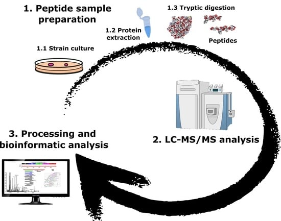
4. Materials and Methods

4.1. Bacterial Strains

Table 1 summarizes the nine Listeria strains used in this study, as well as their classification as either pathogenic or spoilage species. Eight reference bacterial strains were obtained from the Spanish Type Culture Collection, while the FBUNI strain was procured from the bacterial collection belonging to the Department of Microbiology, Faculty of Pharmacy, University of Santiago de Compostela (USC). The strains were previously analyzed by both MALDI-TOF MS and phylogenetic analyses on the 16S rRNA gene, and the genetic results compared to the proteomic data [107]. The Listeria strains were grown in Brain Heart Infusion (BHI, Oxoid Ltd., Hampshire, UK), at 31 °C for 24 h. Bacterial cultures were then transferred to plate count agar (PCA, Oxoid Ltd., Hampshire, UK) and subjected to further incubation at 31 °C for 24 h.

4.2. Protein Extraction

Protein extraction was done as indicated previously [23,108]. In short, a fresh inoculation loop of bacterial culture was resuspended in 100 μL of a solution with 50% acetonitrile (ACN; Merck, Darmstadt, Germany) and 1% aqueous trifluoroacetic acid (TFA; Acros Organics, NJ, USA). After vortexing and centrifuging, the supernatant was further treated with a lysis buffer consisting of 60 mM Tris-HCl pH 7.5, 1% lauryl maltoside, 5 mM phenylmethanesulfonyl fluoride (PMSF) and 1% dithiothreitol (DTT). The supernatant was transferred to a new tube and the amount of protein determined by the bicinchoninic acid method (Sigma Chemical Co. St. Louis, MO, USA). All experiments were performed in triplicate.

4.3. Peptide Sample Preparation

Protein extracts were solubilized and further digested with trypsin, as reported previously [109]. For doing so, 100 μg of protein was dried in a SpeedVac (CentriVap, Labconco Co., Kansas, MO, USA), resuspended in 25 μL of denaturation buffer, (8 M urea in 25 mM ammonium bicarbonate, pH 8.0), and sonicated for 5 min. Then, the addition of DTT followed, at a final concentration of 10 mM, and incubated at 37 °C for 1 h. Alkylation was easily achieved by addition of the appropriate amount of iodoacetamide (IAA), to a final concentration of 50 mM; the solution was dark-incubated for 1 additional hour at room temperature. The sample was diluted with 4 volumes of 25 mM ammonium bicarbonate (pH 8.0), in order to reduce the urea concentration. The final step included trypsin digestion (Promega, Madison, WI, USA), being the protease:protein ratio of 1:100. The incubation was performed overnight at 37 °C.

4.4. Shotgun LC–ESI–MS/MS Analysis

The peptide digests prepared as shown before were treated with formic acid (FA) (~pH 2), desalted in a C18 MicroSpin™ column (The Nest Group, Southborough, MA, USA) and finally analyzed by LC–ESI–MS/MS [104], using a Proxeon EASY-nLC II Nanoflow system (Thermo Fisher Scientific, San Jose, CA, USA) coupled to an LTQ-Orbitrap XL mass spectrometer (Thermo Fisher Scientific) [23,110]. Peptide separation (2 μg) was done in a reverse-phase (RP) column (EASY-Spray column, 50 cm × 75 μm ID, PepMap C18, 2 μm particles, 100 Å pore size, Thermo Fisher Scientific) equipped with a 10 mm precolumn (Accucore XL C18, Thermo Fisher Scientific). Elution from the column was accom by means of a linear gradient from 5 to 35% solvent B (solvent A: 98% water, 2% ACN, 0.1% FA; solvent B: 98% ACN, 2% water, 0.1% FA), for 120 min at a flow rate of 300 nL/min. Electrospray ionization was carried out with a spray voltage of 1.95 kV at a capillary temperature of 230 °C. Peptides were analyzed in positive mode (1 μscan; 400 to 1600 amu), followed by 10 data-dependent collision-induced dissociation (CID) MS/MS scans (1 μscan), using an isolation width of 3 amu and a normalized collision energy of 35%. After the second fragmentation event, dynamic exclusion was set for 30 s, and ions with unassigned charge state were excluded from MS/MS analysis.

4.5. LC–ESI–MS/MS Data Processing

The MS/MS spectra obtained by LC–ESI–MS/MS were analyzed using the program SEQUEST-HT (Proteome Discoverer 2.4, Thermo Fisher Scientific) and compared to the Listeriaceae UniProt/TrEMBL protein database (containing 267,496 protein sequence entries). MS/MS spectra were searched using fully tryptic cleavage constraints, and up to two missed cleavage sites were allowed. Tolerance windows were set at 10 ppm, for precursor ions, and 0.06 Da for MS/MS fragment ions. The variable modifications allowed were: (M*) methionine oxidation (+15.99 Da) and protein N-terminal acetylation (+42.0106 Da). Carbamidomethylation of Cys (+57.02 Da) (C*) was considered as fix modification. Percolator algorithm [111] was used to validate the results as well as for statistical analysis. The peptide false discovery rate (FDR was always kept at less than 1%.

Supplementary Materials

The following are available online at https://0-www-mdpi-com.brum.beds.ac.uk/article/10.3390/ijms22158141/s1, Excel data sheet in Supplementary Data S1: Complete list of non-redundant peptides identified for the complete dataset and for each Listeria strain. Tables S1–S6: Peptides corresponding to virulence factors, identified in the analyzed Listeria strains.

Author Contributions

All authors have made a substantial, direct, and intellectual contribution to the work presented here. All authors have read and agreed to the published version of the manuscript.

Funding

This work has received financial support from the Xunta de Galicia and the European Union (European Social Fund—ESF), the Spanish Ministry of Economy and Competitivity Project AGL 2.013-48.244-R and the European Regional Development Fund (ERDF) (2007–2013). The study was also supported by the GAIN-Xunta de Galicia Project (IN607D 2017/01) and the Spanish AEI/EU-FEDER PID2019-103845RB-C21 project.

Institutional Review Board Statement

Not applicable.

Informed Consent Statement

Not applicable.

Data Availability Statement

Not applicable.

Conflicts of Interest

The authors declare no conflict of interest.

References

  1. Vázquez-Boland, J.A.; Kuhn, M.; Berche, P.; Chakraborty, T.; Domínguez-Bernal, G.; Goebel, W.; González-Zorn, B.; Wehland, J.; Kreft, J. Listeria pathogenesis and molecular virulence determinants. Clin. Microbiol. Rev. 2001, 14, 584–640. [Google Scholar] [CrossRef] [Green Version]
  2. Walsh, D.; Duffy, G.; Sheridan, J.J.; Blair, I.S.; McDowell, D.A. Antibiotic resistance among Listeria, including Listeria monocytogenes, in retail foods. J. Appl. Microbiol. 2001, 90, 517–522. [Google Scholar] [CrossRef]
  3. Rocha, C.E.; Mol, J.P.S.; Garcia, L.N.N.; Costa, L.F.; Santos, R.L.; Paixão, T.A. Comparative experimental infection of Listeria monocytogenes and Listeria ivanovii in bovine trophoblasts. PLoS ONE 2017, 12, e0176911. [Google Scholar] [CrossRef] [Green Version]
  4. Vygovska, L.M. Determination Listeria spp. (L. welshimeri, L. grayi, L. murrayi, L. innocua). J. Vet. Med. Biotechnol. Biosaf. 2018, 4, 33–36. [Google Scholar]
  5. Iannetti, L.; Schirone, M.; Neri, D.; Visciano, P.; Acciari, V.A.; Centorotola, G.; Mangieri, M.S.; Torresi, M.; Santarelli, G.A.; Di Marzio, V.; et al. Listeria monocytogenes in poultry: Detection and strain characterization along an integrated production chain in Italy. Food Microbiol. 2020, 91, 103533. [Google Scholar] [CrossRef]
  6. Arslan, S.; Baytur, S. Prevalence and antimicrobial resistance of Listeria species and subtyping and virulence factors of Listeria monocytogenes from retail meat. J. Food Saf. 2019, 39, 1–10. [Google Scholar] [CrossRef] [Green Version]
  7. Olaimat, A.N.; Al-Holy, M.A.; Shahbaz, H.M.; Al-Nabulsi, A.A.; Abu Ghoush, M.H.; Osaili, T.M.; Ayyash, M.M.; Holley, R.A. Emergence of antibiotic resistance in Listeria monocytogenes isolated from food products: A Comprehensive Review. Compr. Rev. Food Sci. Food Saf. 2018, 17, 1277–1292. [Google Scholar] [CrossRef] [Green Version]
  8. Trost, M.; Wehmhöner, D.; Kärst, U.; Dieterich, G.; Wehland, J.; Jänsch, L. Comparative proteome analysis of secretory proteins from pathogenic and nonpathogenic Listeria species. Proteomics 2005, 5, 1544–1557. [Google Scholar] [CrossRef] [PubMed]
  9. Johansson, J.; Freitag, N.E. Regulation of Listeria monocytogenes Virulence. Microbiol. Spectr. 2019, 7, 836–850. [Google Scholar] [CrossRef] [PubMed]
  10. Lee, S. Bacteriocins of Listeria monocytogenes and their potential as a virulence factor. Toxins 2020, 12, 103. [Google Scholar] [CrossRef] [PubMed] [Green Version]
  11. Coelho, C.; Brown, L.; Maryam, M.; Vij, R.; Smith, D.F.Q.; Burnet, M.C.; Kyle, J.E.; Heyman, H.M.; Ramirez, J.; Prados-Rosales, R.; et al. Listeria monocytogenes virulence factors, including listeriolysin O, are secreted in biologically active extracellular vesicles. J. Biol. Chem. 2019, 294, 1202–1217. [Google Scholar] [CrossRef] [Green Version]
  12. Toledo-Arana, A.; Dussurget, O.; Nikitas, G.; Sesto, N.; Guet-Revillet, H.; Balestrino, D.; Loh, E.; Gripenland, J.; Tiensuu, T.; Vaitkevicius, K.; et al. The Listeria transcriptional landscape from saprophytism to virulence. Nature 2009, 459, 950–956. [Google Scholar] [CrossRef] [PubMed]
  13. Chen, J.Q.; Healey, S.; Regan, P.; Laksanalamai, P.; Hu, Z. PCR-based methodologies for detection and characterization of Listeria monocytogenes and Listeria ivanovii in foods and environmental sources. Food Sci. Hum. Wellness 2017, 6, 39–59. [Google Scholar] [CrossRef]
  14. Soni, D.K.; Ahmad, R.; Dubey, S.K. Biosensor for the detection of Listeria monocytogenes: Emerging trends. Crit. Rev. Microbiol. 2018, 44, 590–608. [Google Scholar] [CrossRef] [PubMed]
  15. Jadhav, S.R.; Shah, R.M.; Palombo, E.A. Maldi-tof ms: A rapid methodology for identifying and subtyping Listeria monocytogenes. Methods Mol. Biol. 2021, 2220, 17–29. [Google Scholar] [PubMed]
  16. Ojima-Kato, T.; Yamamoto, N.; Takahashi, H.; Tamura, H. Matrix-assisted Laser Desorption Ionization-Time of Flight Mass Spectrometry (MALDI-TOF MS) can precisely discriminate the lineages of Listeria monocytogenes and species of Listeria. PLoS ONE 2016, 11, e0159730. [Google Scholar] [CrossRef] [Green Version]
  17. Varadarajan, A.R.; Goetze, S.; Pavlou, M.P.; Grosboillot, V.; Shen, Y.; Loessner, M.J.; Christian, H.A.; Wollcheid, B. A proteogenomic resource enabling integrated analysis of Listeria genotype-proteotype-phenotype relationships. J. Proteome Res. 2020, 19, 1647–1662. [Google Scholar] [CrossRef]
  18. Tiong, H.K.; Hartson, S.; Muriana, P.M. Comparison of five methods for direct extraction of surface proteins from Listeria monocytogenes for proteomic analysis by orbitrap mass spectrometry. J. Microbiol. Methods 2015, 110, 54–60. [Google Scholar] [CrossRef]
  19. Calvo, E.; Pucciarelli, M.G.; Bierne, H.; Cossart, P.; Albar, J.P.; García-Del Portillo, F. Analysis of the Listeria cell wall proteome by two-dimensional nanoliquid chromatography coupled to mass spectrometry. Proteomics 2005, 5, 433–443. [Google Scholar] [CrossRef]
  20. Esbelin, J.; Santos, T.; Ribière, C.; Desvaux, M.; Viala, D.; Chambon, C.; Hébraud, M. Comparison of three methods for cell surface proteome extraction of Listeria monocytogenes biofilms. OMICS J. Integr. Biol. 2018, 22, 779–787. [Google Scholar] [CrossRef] [Green Version]
  21. Abril, A.G.; Carrera, M.; Böhme, K.; Barros-Velázquez, J.; Rama, J.L.R.; Calo-Mata, P.; Sánchez-Pérez, A.; Villa, T.G. Proteomic characterization of antibiotic resistance, and production of antimicrobial and virulence factors in Streptococcus species associated with bovine mastitis. Could enzybiotics represent novel therapeutic agents against these pathogens? Antibiotics 2020, 9, 302. [Google Scholar] [CrossRef]
  22. Abril, A.G.; Ortea, I.; Carrera, M.; Böhme, K.; Barros-Velázquez, J.; Cañas, B.; Villa, T.G.; Calo-Mata, P. Characterization biomarker peptides for the Identification of Bacillus species by LC-ESI-MS/MS. In Exploring Microorganisms: Recent Advances in Applied Microbiology; Mendez-Vilas, A., Ed.; Universal Publishers Inc.: Boca Raton, FL, USA, 2018; pp. 74–77. ISBN1 1-62734-623-6. ISBN2 978-1-62734-623-8. [Google Scholar]
  23. Carrera, M.; Böhme, K.; Gallardo, J.M.; Barros-Velázquez, J.; Cañas, B.; Calo-Mata, P. Characterization of foodborne strains of Staphylococcus aureus by shotgun proteomics: Functional networks, virulence factors and species-specific peptide biomarkers. Front. Microbiol. 2017, 8, 2458. [Google Scholar] [CrossRef] [PubMed] [Green Version]
  24. Abdelhamed, H.; Ramachandran, R.; Narayanan, L.; Ozdemir, O.; Cooper, A.; Olivier, A.K.; Karsi, A.; Lawrence, M.L. Contributions of a LysR transcriptional regulator to Listeria monocytogenes virulence and identification of its regulons. J. Bacteriol. 2020, 202. [Google Scholar] [CrossRef] [PubMed]
  25. Curtis, T.D.; Takeuchi, I.; Gram, L.; Knudsen, G.M. The influence of the Toxin/antitoxin mazEF on growth and survival of Listeria monocytogenes under stress. Toxins 2017, 9, 31. [Google Scholar] [CrossRef] [PubMed] [Green Version]
  26. Rocourt, J.; Grimont, P.A.D. Listeria welshimeri sp. nov. and Listeria seeligeri sp. nov. Int. J. Syst. Bacteriol. 1983, 33, 866–869. [Google Scholar] [CrossRef] [Green Version]
  27. Dowson, C.G.; Hutchison, A.; Brannigan, J.A.; George, R.C.; Hansman, D.; Linares, J.; Tomasz, A.; Smith, J.M.; Spratt, B.G. Horizontal transfer of penicillin-binding protein genes in penicillin-resistant clinical isolates of Streptococcus pneumoniae. Proc. Natl. Acad. Sci. USA 1989, 86, 8842–8846. [Google Scholar] [CrossRef] [Green Version]
  28. Grove, A. Quick guide MarR family transcription factors. Curr. Biol. 2013, 23, R142–R143. [Google Scholar] [CrossRef] [Green Version]
  29. López-pelegrín, M.; Cerdà-costa, N.; Martínez-jiménez, F.; Cintas-pedrola, A.; Canals, A.; Peinado, J.R.; Marti-renom, M.A.; López-otín, C.; Arolas, J.L.; Gomis-rüth, F.X. A Novel family of soluble minimal scaffolds provides structural insight into the catalytic domains of integral membrane metallopeptidases. J. Biol. Chem. 2013, 288, 21279–21294. [Google Scholar] [CrossRef] [PubMed] [Green Version]
  30. Luque-Sastre, L.; Arroyo, C.; Fox, E.M.; McMahon, B.J.; Bai, L.; Li, F.; Fanning, S. Antimicrobial resistance in Listeria species. In Antimicrobial Resistance in Bacteria from Livestock and Companion Animals; ASM Press: Washington, DC, USA, 2018; pp. 237–259. [Google Scholar]
  31. Fillgrove, K.L.; Pakhomova, S.; Schaab, M.R.; Newcomer, M.E.; Armstrong, R.N. Structure and mechanism of the genomically encoded fosfomycin resistance protein, FosX, from Listeria monocytogenes. Biochemistry 2007, 46, 8110–8120. [Google Scholar] [CrossRef] [PubMed]
  32. Vetting, M.W.; Luiz, L.P.; Yu, M.; Hegde, S.S.; Magnet, S.; Roderick, S.L.; Blanchard, J.S. Structure and functions of the GNAT superfamily of acetyltransferases. Arch. Biochem. Biophys. 2005, 433, 212–226. [Google Scholar] [CrossRef] [PubMed]
  33. Blankenfeldt, W.; Kuzin, A.P.; Skarina, T.; Korniyenko, Y.; Tong, L.; Bayer, P.; Janning, P.; Thomashow, L.S.; Mavrodi, D.V. Structure and function of the phenazine biosynthetic protein PhzF from Pseudomonas fluorescens. Proc. Natl. Acad. Sci. USA 2004, 101, 16431–16436. [Google Scholar] [CrossRef] [PubMed] [Green Version]
  34. Cuthbertson, L.; Nodwell, J.R. The TetR Family of Regulators. Microbiol. Mol. Biol. Rev. 2013, 77, 440–475. [Google Scholar] [CrossRef] [PubMed] [Green Version]
  35. Arthur, M.; Depardieu, F.; Molinas, C.; Reynolds, P.; Courvalin, P. The vanZ gene of Tn1546 from Enterococcus faecium BM4147 confers resistance to teicoplanin. Gene 1995, 154, 87–92. [Google Scholar] [CrossRef]
  36. Matson, J.S.; Livny, J.; DiRita, V.J. A putative Vibrio cholerae two-component system controls a conserved periplasmic protein in response to the antimicrobial peptide polymyxin B. PLoS ONE 2017, 12, e0186199. [Google Scholar] [CrossRef] [Green Version]
  37. Iyer, L.M.; Abhiman, S.; Aravind, L. MutL homologs in restriction-modification systems and the origin of eukaryotic MORC ATPases. Biol. Direct 2008, 3, 8. [Google Scholar] [CrossRef] [Green Version]
  38. Suprenant, K.A.; Bloom, N.; Fang, J.; Lushington, G. The major vault protein is related to the toxic anion resistance protein (TelA) family. J. Exp. Biol. 2007, 210, 946–955. [Google Scholar] [CrossRef] [Green Version]
  39. Deshpande, C.N.; Harrop, S.J.; Boucher, Y.; Hassan, K.A.; Leo, R.D.; Xu, X.; Cui, H.; Savchenko, A.; Chang, C.; Labbate, M.; et al. Crystal structure of an integron gene cassette-associated protein from Vibrio cholerae identifies a cationic drug-binding module. PLoS ONE 2011, 6, e16934. [Google Scholar] [CrossRef] [PubMed] [Green Version]
  40. Cha, J.S.; Cooksey, D.A. Copper resistance in Pseudomonas syringae mediated by periplasmic and outer membrane proteins. Proc. Natl. Acad. Sci. USA 1991, 88, 8915–8919. [Google Scholar] [CrossRef] [Green Version]
  41. Hobman, J.L.; Wilkie, J.; Brown, N.L. A design for life: Prokaryotic metal-binding MerR family regulators. Biometals 2005, 18, 429–436. [Google Scholar] [CrossRef] [PubMed]
  42. Domingos, R.M.; Teixeira, R.D.; Zeida, A.; Agudelo, W.A.; Alegria, T.G.P.; Da Silva Neto, J.F.; Vieira, P.S.; Murakami, M.T.; Farah, C.S.; Estrin, D.A.; et al. Substrate and product-assisted catalysis: Molecular aspects behind structural switches along organic hydroperoxide resistance protein catalytic cycle. ACS Catal. 2020, 10, 6587–6602. [Google Scholar] [CrossRef]
  43. Son, M.S.; Del Castilho, C.; Duncalf, K.A.; Carney, D.; Weiner, J.H.; Turner, R.J. Mutagenesis of SugE, a small multidrug resistance protein. Biochem. Biophys. Res. Commun. 2003, 312, 914–921. [Google Scholar] [CrossRef]
  44. Haubert, L.; Kremer, F.S.; da Silva, W.P. Whole-genome sequencing identification of a multidrug-resistant Listeria monocytogenes serotype 1/2a isolated from fresh mixed sausage in southern Brazil. Infect. Genet. Evol. 2018, 65, 127–130. [Google Scholar] [CrossRef] [PubMed]
  45. Chastanet, A.; Derre, I.; Nair, S.; Msadek, T. clpB, a Novel Member of the Listeria monocytogenes CtsR regulon, is involved in virulence but not in general stress tolerance. J. Bacteriol. 2004, 186, 1165–1174. [Google Scholar] [CrossRef] [PubMed] [Green Version]
  46. Bukau, B.; Horwich, A.L. The Hsp70 and Hsp60 chaperone machines. Cell 1998, 92, 351–366. [Google Scholar] [CrossRef] [Green Version]
  47. Delumeau, O.; Dutta, S.; Brigulla, M.; Kuhnke, G.; Hardwick, S.W.; Völker, U.; Yudkin, M.D.; Lewis, R.J. Functional and structural characterization of RsbU, a stress signaling protein phosphatase 2C. J. Biol. Chem. 2004, 279, 40927–40937. [Google Scholar] [CrossRef] [PubMed] [Green Version]
  48. Dubbs, J.M.; Mongkolsuk, S. Peroxiredoxins in Bacterial Antioxidant Defense. In Subcellular Biochemistry; Springer: New York, NY, USA, 2007; Volume 44, pp. 143–193. [Google Scholar]
  49. Park, S.C.; Kwak, Y.M.; Song, W.S.; Hong, M.; Yoon, S. Il Structural basis of effector and operator recognition by the phenolic acid-responsive transcriptional regulator PadR. Nucleic Acids Res. 2017, 45, 13080–13093. [Google Scholar] [CrossRef] [Green Version]
  50. Rolhion, N.; Chassaing, B.; Nahori, M.-A.; de Bodt, J.; Moura, A.; Lecuit, M.; Dussurget, O.; Bérard, M.; Marzorati, M.; Fehlner-Peach, H.; et al. Specific targeting of intestinal Prevotella copri by a Listeria monocytogenes bacteriocin. bioRxiv 2019, 680801. [Google Scholar] [CrossRef] [Green Version]
  51. Mutschler, H.; Gebhardt, M.; Shoeman, R.L.; Meinhart, A. A Novel mechanism of programmed cell death in bacteria by toxin–antitoxin systems corrupts peptidoglycan synthesis. PLoS Biol. 2011, 9, e1001033. [Google Scholar] [CrossRef]
  52. Jørgensen, M.G.; Pandey, D.P.; Jaskolska, M.; Gerdes, K. HicA of Escherichia coli defines a novel family of translation-independent mRNA interferases in bacteria and archaea. J. Bacteriol. 2009, 191, 1191–1199. [Google Scholar] [CrossRef] [Green Version]
  53. Makarova, K.S.; Grishin, N.V.; Koonin, E.V. The HicAB cassette, a putative novel, RNA-targeting toxin-antitoxin system in archaea and bacteria. Bioinformatics 2006, 22, 2581–2584. [Google Scholar] [CrossRef]
  54. Lima-Mendez, G.; Alvarenga, D.O.; Ross, K.; Hallet, B.; Van Melderen, L.; Varani, A.M.; Chandler, M. Toxin-antitoxin gene pairs found in Tn3 family transposons appear to be an integral part of the transposition module. MBio 2020, 11, e00452-20. [Google Scholar] [CrossRef] [PubMed] [Green Version]
  55. Szczepanowski, R.; Braun, S.; Riedel, V.; Schneiker, S.; Krahn, I.; Pühler, A.; Schlüter, A. The 120 592 bp IncF plasmid pRSB107 isolated from a sewage-treatment plant encodes nine different antiobiotic-resistance determinants, two iron-acquisition systems and other putative virulence-associated functions. Microbiology 2005, 151, 1095–1111. [Google Scholar] [CrossRef] [Green Version]
  56. Santos, T.; Viala, D.; Chambon, C.; Esbelin, J.; Hébraud, M. Listeria monocytogenes biofilm adaptation to different temperatures seen through shotgun proteomics. Front. Nutr. 2019, 6, 89. [Google Scholar] [CrossRef] [PubMed]
  57. Bonazzi, M.; Lecuit, M.; Cossart, P. Listeria monocytogenes internalin and E-cadherin: From bench to bedside. Cold Spring Harb. Perspect. Biol. 2009, 1, a0030807. [Google Scholar] [CrossRef] [Green Version]
  58. Jefferson, K.K.; Pier, D.B.; Goldmann, D.A.; Pier, G.B. The Teicoplanin-Associated Locus Regulator (TcaR) and the intercellular adhesin locus regulator (IcaR) are transcriptional inhibitors of the ica locus in Staphylococcus aureus. J. Bacteriol. 2004, 186, 2449–2456. [Google Scholar] [CrossRef] [Green Version]
  59. Molloy, S. Bacterial pathogenicity: A competent escape for Listeria. Nat. Rev. Microbiol. 2012, 10, 670. [Google Scholar] [CrossRef]
  60. Schubert, K.; Bichlmaier, A.M.; Mager, E.; Wolff, K.; Ruhland, G.; Fiedler, F. P45, an extracellular 45 kDa protein of Listeria monocytogenes with similarity to protein p60 and exhibiting peptidoglycan lyric activity. Arch. Microbiol. 2000, 173, 21–28. [Google Scholar] [CrossRef] [PubMed]
  61. Jacobitz, A.W.; Kattke, M.D.; Wereszczynski, J.; Clubb, R.T. Sortase Transpeptidases: Structural biology and catalytic mechanism. In Advances in Protein Chemistry and Structural Biology; Academic Press Inc.: Cambridge, MA, USA, 2017; Volume 109, pp. 223–264. [Google Scholar]
  62. Herzberg, M.; Kaye, I.K.; Peti, W.; Wood, T.K. YdgG (TqsA) controls biofilm formation in Escherichia coli K-12 through autoinducer 2 transport. J. Bacteriol. 2006, 188, 587–598. [Google Scholar] [CrossRef] [Green Version]
  63. Vollmer, W.; Tomasz, A. Peptidoglycan N-Acetylglucosamine deacetylase, a putative virulence factor in Streptococcus pneumoniae. Infect. Immun. 2002, 70, 7176–7178. [Google Scholar] [CrossRef] [PubMed] [Green Version]
  64. Kwon, H.; Ogunniyi, A.D.; Choi, M.; Pyo, S.; Rhee, D.; Paton, J.C. The ClpP Protease of Streptococcus pneumoniae modulates virulence gene expression and protects against fatal Pneumococcal challenge. Infect. Immun. 2004, 72, 5646–5653. [Google Scholar] [CrossRef] [PubMed] [Green Version]
  65. Kajfasz, J.K.; Martinez, A.R.; Rivera-ramos, I.; Abranches, J.; Koo, H.; Quivey, R.G. Role of Clp proteins in expression of virulence properties of Streptococcus mutans. J. Bacteriol. 2009, 191, 2060–2068. [Google Scholar] [CrossRef] [PubMed] [Green Version]
  66. Smith, C.W.; Metzger, J.F. Demonstration of a capsular structure on Listeria monocytogenes. Pathol. Microbiol. 1962, 25, 499–506. [Google Scholar] [CrossRef] [PubMed]
  67. Edwards, M.R.; Stevens, R.W. Fine structure of Listeria monocytogenes. J. Bacteriol. 1963, 86, 414–428. [Google Scholar] [CrossRef] [PubMed] [Green Version]
  68. Kijek, T.M.; Mou, S.; Bachert, B.A.; Kuehl, K.A.; Williams, J.A.; Daye, S.P.; Worsham, P.L.; Bozue, J.A. The D-alanyl-D-alanine carboxypeptidase enzyme is essential for virulence in the Schu S4 strain of Francisella tularensis and a dacD mutant is able to provide protection against a pneumonic challenge. Microb. Pathog. 2019, 137, 103742. [Google Scholar] [CrossRef] [PubMed]
  69. Tomazi, T.; de Souza Filho, A.F.; Heinemann, M.B.; dos Santos, M.V. Molecular characterization and antimicrobial susceptibility pattern of Streptococcus agalactiae isolated from clinical mastitis in dairy cattle. PLoS ONE 2018, 13, e0199561. [Google Scholar] [CrossRef]
  70. Schmelcher, M.; Waldherr, F.; Loessner, M.J. Listeria bacteriophage peptidoglycan hydrolases feature high thermoresistance and reveal increased activity after divalent metal cation substitution. Appl. Microbiol. Biotechnol. 2012, 93, 633–643. [Google Scholar] [CrossRef] [Green Version]
  71. Danevčič, T.; Borić Vezjak, M.; Tabor, M.; Zorec, M.; Stopar, D. Prodigiosin induces autolysins in actively grown Bacillus subtilis Cells. Front. Microbiol. 2016, 7, 27. [Google Scholar] [CrossRef] [Green Version]
  72. Bateman, A.; Bycroft, M. The structure of a LysM domain from E. coli membrane-bound lytic murein transglycosylase D (MltD). J. Mol. Biol. 2000, 299, 1113–1119. [Google Scholar] [CrossRef]
  73. McGowan, S.; Lucet, I.S.; Cheung, J.K.; Awad, M.M.; Whisstock, J.C.; Rood, J.I. The FxRxHrS motif: A conserved region essential for DNA binding of the VirR response regulator from Clostridium perfringens. J. Mol. Biol. 2002, 322, 997–1011. [Google Scholar] [CrossRef]
  74. Broxton, C.N.; Culotta, V.C. SOD Enzymes and microbial pathogens: Surviving the oxidative storm of infection. PLoS Pathog. 2016, 12, e1005295. [Google Scholar] [CrossRef] [PubMed] [Green Version]
  75. Ribardo, D.A.; Lambert, T.J.; Mciver, K.S. Role of Streptococcus pyogenes Two-Component response regulators in the temporal control of Mga and the Mga-Regulated virulence gene emm. Infect. Immun. 2004, 72, 3668–3673. [Google Scholar] [CrossRef] [Green Version]
  76. Warne, B.; Harkins, C.P.; Harris, S.R.; Vatsiou, A.; Stanley-Wall, N.; Parkhill, J.; Peacock, S.J.; Palmer, T.; Holden, M.T.G. The Ess/Type VII secretion system of Staphylococcus aureus shows unexpected genetic diversity. BMC Genom. 2016, 17, 222. [Google Scholar] [CrossRef] [Green Version]
  77. Schulein, R.; Dehio, C. The VirB/VirD4 type IV secretion system of Bartonella is essential for establishing intraerythrocytic infection. Mol. Microbiol. 2002, 46, 1053–1067. [Google Scholar] [CrossRef]
  78. Pallen, M.J. The ESAT-6/WXG100 superfamily and a new Gram-positive secretion system? Trends Microbiol. 2002, 10, 209–212. [Google Scholar] [CrossRef]
  79. Zhang, S.; Wang, Y.; Wu, H.; Li, N.; Jiang, J.; Guo, Y.; Feng, Y.; Xiao, L. Characterization of a species-specific insulinase-like protease in Cryptosporidium parvum. Front. Microbiol. 2019, 10, 354. [Google Scholar] [CrossRef] [PubMed] [Green Version]
  80. Sumrall, E.T.; Keller, A.P.; Shen, Y.; Loessner, M.J. Structure and function of Listeria teichoic acids and their implications. Mol. Microbiol. 2020, 113, 627–637. [Google Scholar] [CrossRef] [PubMed] [Green Version]
  81. Kang, S.S.; Sim, J.R.; Yun, C.H.; Han, S.H. Lipoteichoic acids as a major virulence factor causing inflammatory responses via Toll-like receptor 2. Arch. Pharm. Res. 2016, 39, 1519–1529. [Google Scholar] [CrossRef] [PubMed]
  82. Lan, Z.; Fiedler, F.; Kathariou, S. A sheep in wolf’s clothing: Listeria innocua strains with teichoic acid-associated surface antigens and genes characteristic of Listeria monocytogenes serogroup 4. J. Bacteriol. 2000, 182, 6161–6168. [Google Scholar] [CrossRef] [PubMed] [Green Version]
  83. Murazumi, N.; Kumita, K.; Yoshio, A.; Ito, E. Partial purification and properties of UDP-N-Acetylmannosamine:N-Acetylgiucosaminyl Pyrophosphorylundecaprenol N-Acetylmannosaminyltransferase from Bacillus subtilis. J. Biochem. 1988, 104, 980–984. [Google Scholar] [CrossRef]
  84. Shen, Y.; Boulos, S.; Sumrall, E.; Gerber, B.; Julian-Rodero, A.; Eugster, M.R.; Fieseler, L.; Nyström, L.; Ebert, M.O.; Loessner, M.J. Structural and functional diversity in Listeria cell wall teichoic acids. J. Biol. Chem. 2017, 292, 17832–17844. [Google Scholar] [CrossRef] [Green Version]
  85. Loughman, J.A.; Caparon, M.G. Contribution of invariant residues to the function of Rgg family transcription regulators. J. Bacteriol. 2007, 189, 650–655. [Google Scholar] [CrossRef] [Green Version]
  86. Price, C.T.D.; Bukka, A.; Cynamon, M.; Graham, J.E.; Al, P.E.T.; Acteriol, J.B. Glycine Betaine uptake by the ProXVWZ ABC transporter contributes to the ability of Mycobacterium tuberculosis to initiate growth in human macrophages. J. Bacteriol. 2008, 190, 3955–3961. [Google Scholar] [CrossRef] [Green Version]
  87. Tanaka, K.J.; Song, S.; Mason, K.; Pinkett, H.W. Selective substrate uptake: The role of ATP-binding cassette (ABC) importers in pathogenesis. BBA Biomembr. 2018, 1860, 868–877. [Google Scholar] [CrossRef]
  88. Naka, H.; Liu, M.; Crosa, J.H. Two ABC transporter systems participate in siderophore transport in the marine pathogen Vibrio anguillarum 775 (pJM1). FEMS Microbiol. Lett. 2013, 341, 79–86. [Google Scholar] [CrossRef] [Green Version]
  89. Orelle, C.; Mathieu, K.; Jault, J.M. Multidrug ABC transporters in bacteria. Res. Microbiol. 2019, 170, 381–391. [Google Scholar] [CrossRef] [PubMed]
  90. Greene, N.P.; Kaplan, E.; Crow, A.; Koronakis, V. Antibiotic resistance mediated by the MacB ABC transporter family: A structural and functional perspective. Front. Microbiol. 2018, 9, 950. [Google Scholar] [CrossRef] [PubMed] [Green Version]
  91. Kumar, S.; Mukherjee, M.M.; Varela, M.F. Modulation of bacterial multidrug resistance efflux pumps of the major facilitator superfamily. Int. J. Bacteriol. 2013, 2013, 204141. [Google Scholar] [CrossRef] [PubMed] [Green Version]
  92. Morimyo, M.; Hongo, E.; Hama-inaba, H.; Machida, I. Cloning and characterization of the mvrC gene of Escherichia coli K-12 which confers resistance against methyl viologen toxicity. Nucleic Acids Res. 1992, 20, 3159–3165. [Google Scholar] [CrossRef] [PubMed] [Green Version]
  93. Omote, H.; Hiasa, M.; Matsumoto, T.; Otsuka, M.; Moriyama, Y. The MATE proteins as fundamental transporters of metabolic and xenobiotic organic cations. Trends Pharmacol. Sci. 2006, 27, 587–593. [Google Scholar] [CrossRef]
  94. Hernandez-Mendoza, A.; Quinto, C.; Segovia, L.; Perez-Rueda, E. Ligand-binding prediction in the resistance-nodulation-cell division (RND) proteins. Comput. Biol. Chem. 2007, 31, 115–123. [Google Scholar] [CrossRef] [PubMed]
  95. Craig, N.L.; Chandler, M.; Gellert, M.; Lambowitz, A.M.; Rice, P.A.; Sandmeyer, S.B. Mobile DNA III, 3rd ed.; John Wiley & Sons: Washington, DC, USA, 2020. [Google Scholar]
  96. Robinson, K.M.; Dunning Hotopp, J.C. Mobile elements and viral integrations prompt considerations for bacterial DNA integration as a novel carcinogen. Cancer Lett. 2014, 352, 137–144. [Google Scholar] [CrossRef] [PubMed] [Green Version]
  97. Wang, J.; Yu, Y.; Tao, F.; Zhang, J.; Copetti, D.; Kudrna, D.; Talag, J.; Lee, S.; Wing, R.A.; Fan, C. DNA methylation changes facilitated evolution of genes derived from Mutator-like transposable elements. Genome Biol. 2016, 17, 92. [Google Scholar] [CrossRef] [Green Version]
  98. Wang, Y.; Li, X.; Osmundson, T.; Shi, L.; Yan, H. Comparative Genomic Analysis of a Multidrug-Resistant Listeria monocytogenes ST477 Isolate. Foodborne Pathog. Dis. 2019, 16, 604–615. [Google Scholar] [CrossRef] [PubMed]
  99. Koudelka, Š.; Gelbíčová, T.; Procházková, M.; Karpíšková, R. Lineage and serotype identification of Listeria monocytogenes by Matrix-assisted Laser Desorption Ionization-time of Flight Mass Spectrometry. Czech J. Food Sci. 2018, 36, 452–458. [Google Scholar] [CrossRef] [Green Version]
  100. Cheng, Y.; Promadej, N.; Kim, J.W.; Kathariou, S. Teichoic acid glycosylation mediated by gtcA is required for phage adsorption and susceptibility of Listeria monocytogenes serotype 4b. Appl. Environ. Microbiol. 2008, 74, 1653–1655. [Google Scholar] [CrossRef] [Green Version]
  101. Matereke, L.T.; Okoh, A.I. Listeria monocytogenes virulence, antimicrobial resistance and environmental persistence: A review. Pathogens 2020, 9, 528. [Google Scholar] [CrossRef]
  102. Dams, D.; Briers, Y. Enzybiotics: Enzyme-based antibacterials as therapeutics. Adv. Exp. Med. Biol. 2019, 1148, 233–253. [Google Scholar] [PubMed]
  103. Schmitz-Esser, S.; Anast, J.M.; Cortes, B.W. A large-scale sequencing-based survey of plasmids in Listeria monocytogenes reveals global dissemination of plasmids. Front. Microbiol. 2021, 12, 510. [Google Scholar] [CrossRef]
  104. Li, L.; Olsen, R.H.; Shi, L.; Ye, L.; He, J.; Meng, H. Characterization of a plasmid carrying cat, ermB and tetS genes in a foodborne Listeria monocytogenes strain and uptake of the plasmid by cariogenic Streptococcus mutans. Int. J. Food Microbiol. 2016, 238, 68–71. [Google Scholar] [CrossRef] [PubMed]
  105. Xu, D.; Nie, Q.; Wang, W.; Shi, L.; Yan, H. Characterization of a transferable bcrABC and cadAC genes-harboring plasmid in Listeria monocytogenes strain isolated from food products of animal origin. Int. J. Food Microbiol. 2016, 217, 117–122. [Google Scholar] [CrossRef]
  106. Yan, H.; Yu, R.; Li, D.; Shi, L.; Schwarz, S.; Yao, H.; Li, X.; Du, X. A novel multiresistance gene cluster located on a plasmid-borne transposon in Listeria monocytogenes. J. Antimicrob. Chemother. 2020, 75, 868–872. [Google Scholar] [CrossRef] [PubMed]
  107. Böhme, K.; Fernández-No, I.C.; Barros-Velázquez, J.; Gallardo, J.M.; Cañas, B.; Calo-Mata, P. Rapid species identification of seafood spoilage and pathogenic Gram-positive bacteria by MALDI-TOF mass fingerprinting. Electrophoresis 2011, 32, 2951–2965. [Google Scholar] [CrossRef] [PubMed]
  108. Böhme, K.; Fernández-No, I.C.; Barros-Velázquez, J.; Gallardo, J.M.; Calo-Mata, P.; Cañas, B. Species differentiation of seafood spoilage and pathogenic gram-negative bacteria by MALDI-TOF mass fingerprinting. J. Proteome Res. 2010, 9, 3169–3183. [Google Scholar] [CrossRef]
  109. Carrera, M.; Cañas, B.; Gallardo, J.M. The sarcoplasmic fish proteome: Pathways, metabolic networks and potential bioactive peptides for nutritional inferences. J. Proteomics 2013, 78, 211–220. [Google Scholar] [CrossRef]
  110. Calo-mata, P.; Carrera, M.; Böhme, K.; Caamaño-Antelo, S.; Gallardo, J.M.; Barros-Velázquez, J.; Cañas, B. Novel peptide biomarker discovery for detection and identification of bacterial pathogens by LC-ESI-MS/MS. J. Anal. Bioanal. Tech. 2016, 7, 296. [Google Scholar]
  111. Käll, L.; Canterbury, J.D.; Weston, J.; Noble, W.S.; MacCoss, M.J. Semi-supervised learning for peptide identification from shotgun proteomics datasets. Nat. Methods 2007, 4, 923–925. [Google Scholar] [CrossRef] [PubMed]
Table 1. Listeria strains used in this study. Total virulence factor peptides represent the number of peptides identified by LC–ESI-MS/MS. Pathogenicity data corresponds to the risk group of each analyzed strain. ATCC—American Type Culture Collection; CECT—Spanish Type Culture Collection; NCTC—The National Collection of Type Cultures of Public Health England.
SampleSpeciesStrainCulture Collection NumberSourcePathogenicityVarietyNCBI Accession NumberTotal Virulence Factor Peptides
Li1Listeria welshimeriATCC 35897CECT 919Decaying vegetationGroup 1Serovar 6aNZ_LT906444.117
Li2Listeria seeligeriATCC 35967CECT 917SoilAvirulent
Group 1
Serovar 1/2bDQ065845
NC_013891.1
116
Li3Listeria innocuaATCC 33090CECT 910Cow brainGroup 1Serovar 6aX9852755
Li4Listeria monocytogenesLIS FBUNIUSCDairy product---73
Li5Listeria monocytogenesATCC 15313CECT 4031RabbitGroup 2Serovar 1/2AJ515512
NC_003210.1
15
Li6Listeria monocytogenesATCC 19114CECT 934Brain of sheep with circling diseaseGroup 2Serovar 4aJF967620.118
Li7Listeria monocytogenesATCC 13932CECT 935Spinal fluid of child with meningitisGroup 2Serovar 4bJF967617.133
Li8Listeria monocytogenesNCTC 11994CECT 4032Associated with case of meningitis after eating soft cheeseGroup 2Serovar 4bAJ508749.137
Li9Listeria ivanoviiATCC 19119CECT 913SheepGroup 2Serovar 5X98528
NZ_CP009577.1
31
Table 2. Proteins corresponding to bacterial resistance to antibiotics, antimicrobial related proteins and other virulence factors, identified in the Listeria strains analyzed.
FunctionProtein
Antibiotic resistanceBeta-lactamase
Metallo-beta-lactamase
PbpX beta-lactamase-like superfamily
Bleomycin resistance protein
Aminoglycoside N(3)-acetyltransferase
Glyoxalase/bleomycin resistance protein/dioxygenase
MarR family transcriptional regulator
Peptidase M56
FosX/FosE/FosI family fosfomycin resistance thiol transferase
GNAT family acetyltransferase
PhzF family phenazine biosynthesis protein
TetR
VanZ family protein O
YdeI (YjbR/CyaY-like superfamily)
Tetronasin resistance transmembrane protein
Penicillin-binding protein
Additional resistances and tolerancesAIPR protein
Chaperone protein ClpB
Chaperone protein DnaJ
Tellurite resistance protein TelA
Cass2 domain-containing protein
Copper resistance protein
MerR family transcriptional regulator
Organic hydroperoxide resistance protein
Two-component sensor histidine kinase response to antimicrobials
Quaternary ammonium compound-resistance protein sugE
Quinolone resistance protein norB
Heat shock protein 70
Hsp20/alpha crystallin family protein
General stress protein
OsmC family protein
PadR family transcriptional regulator
RsbU protein
Universal stress protein
Antimicrobial compounds productionLactococcin 972 family bacteriocin
Antibiotic biosynthesis monooxygenase
ToxinsToxin zeta
LXG domain-containing protein
HicA toxin
Type II toxin-antitoxin system RelE/ParE family toxin
Type II toxin-antitoxin system PemK/MazF family toxin
Host colonization and immune evasionAutoinducer 2 transporter
Viral enhancin protein
Invasion associated secreted endopeptidase
Peptidase_C1B
Aminopeptidase ysdC
Beta-Ala-Xaa dipeptidase
Peptidase M23
Peptidase M16
Peptidase M23
M23 family metallopeptidase
Peptidoglycan DD-metalloendopeptidase family protein M22
Peptidoglycan DD-metalloendopeptidase family protein M23
Peptidase_M22
M42 family peptidase
Enhancing factor (Viral) Peptidase M60
Peptidase M60
Peptidase T
Peptidase T
Peptidase_M75
Peptidase C60 sortase
Membrane protein with peptidase activity M56
Tripeptide aminopeptidase M20
Peptidase SA1530
Peptidase SA1531
NLP/P60 family domain
Invasion-associated endopeptidase p60
Peptidoglycan endopeptidase P60
Peptidase P60
Competence protein ComEC/Rec2
Protein ComGA
Peptidoglycan O-acetyltransferase
ClpX protease
ClpP protease
ClpY protease
Internalin A
Internalin
Internalin
Internalin-J
Class 1 internalin InlJ
Class 3 internalin InlC
Internalin B
Capsular polysaccharide synthesis enzyme
Capsular biosynthesis protein
Capsular exopolysaccharide family protein
1-phosphatidylinositol- specific phospholipase C (PI-PLC)
Chemotaxis protein CheA
Cna B-type domain-containing protein
N-acetylmuramoyl-L-alanine amidase
D-alanyl-D-alanine carboxypeptidase
Muramidase-2; Autolysin
LysM domain-containing protein
LysR family transcriptional regulator
Autolysin LytC
Lysis protein
Superoxide dismutase
Type VI secretion protein
Type VII secretion protein EssA
Type VII secretion protein EssB
Putative type IV secretion system VirB4 component
Mga protein
Collagen-binding protein (adhesin)
Adhesion lipoprotein
Adhesin
Intercellular adhesion protein R
ESAT-6-like protein
Insulinase
Rgg family transcriptional regulator
Protein DltD D-alanyl-lipoteichoic acid biosynthesis protein DltD
Putative glycosyl/glycerophosphate transferases involved in teichoic acid biosynthesis
Wall teichoic acid glycosylation protein GtcA
Teichoic acid biosynthesis domain-containing protein
Teichoic acid/polysaccharide export protein
ABC transportersFluoroquinolones export ATP-binding protein Rv2688c/MT2762
Antibiotic ABC transporter ATP-binding protein
Petrobactin ABC transporter substrate-binding protein YclQ
Bacitracin ABC transporter BceA
Antibiotic ABC_2_transport permease
Multidrug ABC export ATP-binding/permease protein
Multidrug ABC transporter permease/ATP-binding protein
Multidrug resistance ABC transporter ATP-binding/permease protein YheI
Multidrug ABC transporter ATP-binding protein
Spermidine/putrescine ABC Transporter PotA
Siderophore ABC transporter
ABC transporter ATP-binding protein
ABC transporter domain-containing protein
ABC transporter permease
ABC transporter substrate-binding protein
ABC transporter permease
ABC-2 family transporter protein
Peptide ABC transporter
Oligopeptide ABC transporter binding protein AppA
ABC-type methionine transporter MetN
Amino acid ABC transporter
Di-tripeptide-proton ABC symporter
Glutamine ABC transporter ATP-binding protein
Glycine/betaine ABC transporter
Amino acid ABC transporter ATP-binding protein
Iron compound ABC transporter permease
Macrolide ABC transporter
Manganese ABC transporter
Metal ABC transporter ATP-binding protein
Zinc ABC transporter substrate-binding protein AdcA
Putative high-affinity zinc ABC transporter (Zn(II)-binding lipoprotein)
Pheromone ABC transporter
Thiol reductant ABC exporter subunit CydC
Other transportersArsenic transporter
Arsenical pump-driving ATPase
Cadmium, zinc, and cobalt-transporting ATPase
Chloramphenicol/florfenicol efflux MFS transporter
EmrB multiple drug resistance efflux pump MFS transporter
Multidrug efflux SMR transporter
Multidrug export protein mepA
Multidrug efflux MFS transporter
RND superfamily drug exporter
Putrescine importer PuuP
Integrase
Recombinase
IS21 family transposase
IS3 family transposase
IS30 family transposase
Transposase B of Tn554
Transposase B of Tn555
Tn3 family transposase
IS200/IS605 family transposase
IS30 family transposase
Transposase IS116/IS110/IS902 family protein
Mutator family transposase
Plasmid recombination enzyme
Plasmid pRiA4b ORF-3 family protein
Plasmid mobilization relaxosome protein MobC
Bacillus transposase protein
Phage infection protein, YhgE
Publisher’s Note: MDPI stays neutral with regard to jurisdictional claims in published maps and institutional affiliations.

