
The Impact of Vitamin D on Skin Aging


1
Department of Pharmacology and Toxicology, Medical University of Sofia, 1431 Sofia, Bulgaria
2
Department of Dermatology, University of Alabama at Birmingham, Birmingham, AL 35294, USA
3
Graduate Biomedical Sciences Program, University of Alabama at Birmingham, Birmingham, AL 35294, USA
4
Comprehensive Cancer Center, Cancer Chemoprevention Program, University of Alabama at Birmingham, Birmingham, AL 35294, USA
5
Veteran Administration Medical Center, Birmingham, AL 35294, USA
*
Authors to whom correspondence should be addressed.
Int. J. Mol. Sci. 2021, 22(16), 9097; https://0-doi-org.brum.beds.ac.uk/10.3390/ijms22169097
Submission received: 4 August 2021
/
Revised: 20 August 2021
/
Accepted: 21 August 2021
/
Published: 23 August 2021
(This article belongs to the Special Issue Recent Advances in Biology of Ultraviolet Radiation (UVR) and Vitamin D)
Abstract
:The active metabolites of vitamin D3 (D3) and lumisterol (L3) exert a variety of antiaging and photoprotective effects on the skin. These are achieved through immunomodulation and include anti-inflammatory actions, regulation of keratinocytes proliferation, and differentiation programs to build the epidermal barrier necessary for maintaining skin homeostasis. In addition, they induce antioxidative responses, inhibit DNA damage and induce DNA repair mechanisms to attenuate premature skin aging and cancerogenesis. The mechanism of action would involve interaction with multiple nuclear receptors including VDR, AhR, LXR, reverse agonism on RORα and -γ, and nongenomic actions through 1,25D3-MARRS receptor and interaction with the nongenomic binding site of the VDR. Therefore, active forms of vitamin D3 including its canonical (1,25(OH)2D3) and noncanonical (CYP11A1-intitated) D3 derivatives as well as L3 derivatives are promising agents for the prevention, attenuation, or treatment of premature skin aging. They could be administrated orally and/or topically. Other forms of parenteral application of vitamin D3 precursor should be considered to avoid its predominant metabolism to 25(OH)D3 that is not recognized by CYP11A1 enzyme. The efficacy of topically applied vitamin D3 and L3 derivatives needs further clinical evaluation in future trials.
1. Introduction
Skin, like any other organs, undergoes progressive decline in its physiological, morphological, and functional features during aging [1,2,3,4]. The phenomenon of aging is natural and genetically predisposed. The functions of the skin are crucial for the homeostasis and survival. As the largest organ in the human body, the skin, together with the hypodermis (subcutaneous fat), is both the source and the target for several hormones and neuromediators [5,6,7,8,9,10,11,12,13,14,15,16,17], making it an independent peripheral endocrine organ [5,18]. The skin has also the capacity of producing the prohormone vitamin D and transforming it to active metabolites [19,20,21,22,23,24,25], which can exert several different effects on the main skin cells (keratinocytes and fibroblasts) [20,25,26,27,28,29] and immune cells [4,28,30,31] via the activation of the nuclear vitamin D receptor (VDR) [29,32,33,34,35]. Vitamin D plays a pivotal role in skin homeostasis contributing to its barrier function [20,29,36,37,38]. Moreover, as an essential part of a functioning immune system, active forms of vitamin D modulate the cutaneous immunity [8,30,39].
The gold standard of analyzing a vitamin D status is by measuring its major circulating metabolite, 25-hydroxyvitammin D3 (25(OH)D3), via high-performance liquid chromatography (HPLC) or liquid chromatography tandem–mass spectrometry (LC-MS/MS) [40,41,42]. Recently, a novel sensitive and specific LC-MS/MS method of the simultaneous measurement of 13 circulating metabolites of vitamin D3 and D2 was presented [43].
Importantly, subclinical (30–50 nmol/L) and clinical vitamin D deficiency (<30 nmol/L) in the general population has become a global problem worldwide [44,45,46,47,48]. Several physiological factors may influence vitamin D status, such as age, body mass index (BMI), skin type, pregnancy, and exclusive breastfeeding [49,50,51,52,53]. The genetic polymorphisms of some genes involved in skin pigmentation (TYR, TYRP1, EXOC2, and DCT) are also associated with 25(OH)D3 serum concentration [54]. Many environmental factors contribute to vitamin D deficiency, such as the winter season, inadequate sun exposure, and high latitude location [55,56]. Sun avoidance and air pollution are the main factors leading to insufficient UVB exposure. Moreover, ozone and particulate matter (PM) can directly affect the cutaneous production of vitamin D [57]. Additionally, air pollutants, persistent organic pollutants, and heavy metals can behave like endocrine-disrupting chemicals (EDCs), which may cause vitamin D deficiency directly or indirectly. The latter would be secondary to a weight gain, the dysregulation of parathyroid hormone and calcium homeostasis, and a thyroid dysfunction [57,58]. Increasing evidence suggests that smoking can also decrease the serum levels of 25(OH)D3 [57].
With advancing age, the capacity of the skin to produce vitamin D3 decreases (irrespective of the season), and the degradation of its active forms increases [59,60]. It was found that the concentration of the precursor of vitamin D3 in the skin, 7-dehydrocholesterol (7-DHC), declines approximately by 50% from age 20 to age 80 years [59]. Several other factors contribute to the vitamin D deficiency state in accelerated age, including limited sun exposure, insufficient dietary intake of vitamin D, or diseases causing malabsorption. The vitamin D deficiency, which is common in advanced age, can decrease the important physiological functions of the skin such as protection from the environment and prevention of cancer development [25,39,61,62,63,64,65,66].
In this review, we aimed to discuss the significance of vitamin D in the skin aging process.
2. Skin Aging—Your Skin Can Reveal Stories
Skin aging is a complex process that is influenced by the total exposure of both intrinsic and extrinsic factors over the human lifespan (skin exposome), which is responsible for the progressive morphological and functional alterations of the aged skin [1,67,68,69]. The main internal factors affecting the chronological (physiological) skin aging include a hormonal decline and changes in gene expression with advancing age [1]. In addition, the cutaneous regenerative potential decreases with the age due to the excessive senescence of keratinocytes, fibroblasts, and melanocytes over time, which contributes to skin aging [70,71,72]. The aged skin is characterized by fine wrinkles and atrophy with reduced elasticity. This chronological aging affects all skin areas but shows phenotypic differences among different anatomical regions, and it varies significantly within different populations [67,73]. The single nucleotide polymorphisms (SNPs) of the MC1R gene (main regulator of melanin pigmentation [74]) are significantly linked to a perceived facial age, providing a new molecular base of youthful looks [75].
The most prominent external stressors affecting skin and causing its premature aging include ultraviolet (UV) radiation [76,77], ambient pollutants [78,79,80,81,82], and smoking [57,83]. The continuous exposure of the skin to these environmental insults stimulates the production of reactive oxygen species (ROS) and generate oxidative stress [84,85]. The environmental factors can also cause an impairment of the epidermal barrier function [69] and alterations in skin microflora [86,87], leading to significant morbidity [2,88].
Ultraviolet radiation (UVR) is the most harmful external factor contributing to the cutaneous photodamage on the sun-exposed areas. Photoaged skin is presented as a dry, deep-wrinkled skin with rough texture, dyschromia and senile lentigines, vascular complications, etc. [68,89]. UVR decreases the expression of filaggrin that contributes to epidermal hydration, so its downregulation from UVR could explain the skin xerosis in photoaging [90]. Both UVA (315–400 nm) and UVB (280–315 nm) have been shown to contribute to photoaging, either by imbalanced ROS production or by direct DNA damage [83]. However, UVA is considered to play a major role in the aging process. UVA represents more than 80% of total daily UV irradiation and can penetrate 5–10 times deeper into the reticular dermis, where it can damage the extracellular matrix (ECM) more significantly than UVB [91]. Moreover, UVA exposure increases the expression of matrix metalloproteinases (MMPs), especially the expression of the collagenolytic enzyme MMP-1 in dermal fibroblasts, which acts as an important regulator in photoaging [92,93]. Additionally, the chronic UVA irradiation inhibits hyaluronan synthesis, thus altering the composition of proteoglycans in the dermis [94]. A long-term exposure of UVA is related to photoaging and photocancer due to an overproduction of ROS and reactive nitrogen species (RNS), which can disrupt both the nuclear and mitochondrial DNA [95,96]. UVB can penetrate only through the epidermis but is biologically more active. It also induces the transformation of 7-DHC to vitamin D3 [97,98]. UVB absorbed by DNA and RNA induces a formation of cyclobutane pyrimidine dimers (CPDs) and other photoproducts [99], thus inducing various solar signature mutations in specific genes, including the tumor suppressor gene p53 [100,101]. UVR induces an accumulation of p53 protein in the nucleus that in turn activates the transcription of genes responsible for cell cycle arrest allowing DNA repair, as well as causing an induction of apoptosis of the cells with unrepaired DNA damage [102,103]. Specific p53 mutations can be found in high rates, not only in actinic keratosis (precancerous state) and squamous cell carcinomas (60–90%) with typical UV signature, but also in the normal appearance of UV-exposed skin (about 75%), compared with a much lower rate of such mutations in healthy sun-protected skin (5% of all cases) [104].
Chronic sunlight exposure, together with the persistence of cellular senescence, can drive an impaired regenerative capacity of the skin, chronic inflammation, and photoaging, which correlates with enhanced cancer risk [77,105,106,107]. Thereby, photoaging results in premature skin aging. Although some aging mechanisms share several similarities or overlapping, photoaged skin differs from physiologically aged skin in the ECM changes. Photoaged skin is characterized by degraded collagen and accumulated aberrant elastin fibers and glycosaminoglycans, whereas physiologically aged skin is presented by the atrophy of dermal structures [108].
The negative impact of ambient pollutants on human health and the human skin is of growing concern [109]. Ozone (O3) from the smog and PM, primarily contacting with the skin, is capable to stimulate ROS production and generate oxidative stress, leading to phenotypic features of extrinsic aging [69]. It was found that chronic exposure to PM leads to pigment spots and deep nasolabial folds [110,111]. Moreover, ultrafine particles (<0.1 μm) can penetrate tissues and localize in the mitochondria, causing an aberrant mitochondrial function because of the oxidative processes [112]. Additionally, the photo-pollution exposure may aggravate UVR-mediated skin aging [113].
UVR, predominantly UVA, by the excessive amount of ROS activates the mitogen-activated protein kinases (MAPKs) and transcription factors such as nuclear factor erythroid 2-like (Nrf2), c-Jun-N-terminal kinase (JNK), and nuclear factor-κβ (NF-κB), and increases the transcription of MMPs [114]. Activated MMPs, together with the decreased expression of MMP inhibitors (TIMP), cause a dysregulation of the ECM homeostasis and a progressive damage of collagen and elastin [115]. Additionally, UVR impairs the endogenous antioxidant enzymes, leading to an increased oxidative damage of collagen. The destruction of ECM integrity is visualized as wrinkle appearance in photo-damaged skin [93]. The activation of redox-sensitive transcription factors, the activator protein-1 (AP-1) and NF-κβ, involved in wrinkle formation and inflammation, plays crucial roles in skin aging [88]. Both factors, NF-κβ and AP-1, are elevated within hours after cutaneous exposure to low-dose UVB. The upregulation of AP-1 suppresses the transforming growth factor β (TGF-β) receptors, which further blocks the synthesis of procollagen [116,117]. Additionally, activated AP-1 stimulates the degradation of collagen by MMPs and triggers the main activator of the inflammatory response NF-κβ [118]. NF-κβ signaling is a well-known regulator of tissue homeostasis, and its central role in skin aging was recently underlined [119]. The ROS-induced activation of NF-κβ drives an increase of proinflammatory cytokines and MMPs and decreases TGF-β and type I collagen synthesis [119]. The proinflammatory cytokines (interleukin (IL)-1, IL-6, and tumor necrosis factor (TNF)-α) stimulate inflammatory responses and enhance the activation of NF-κβ [93]. It was found that NF-κβ expression could increase in mitochondrial DNA (mtDNA)-depleter mice and after restoring the mitochondrial function, the NF-κβ expression could be reduced. These data confirm that NF-κβ signaling is a key mechanism contributing to the skin and hair follicle pathologies [120]. Due to the longer wavelength, UVA reaches the dermal fibroblasts in vivo with the activation of the Nrf2-mediated expression of antioxidant genes. Unlike UVA, UVB does not activate Nrf2 in skin cells or even appears to have an inhibitory effect [121]. However, vitamin D3 derivatives, which are products of UVB action, do activate Nrf2 signaling [122]. The endogenous Nrf2 is essential for the protection of skin cells against oxidative insults and for regulating the redox balance during skin aging [123,124]. Many in vitro and in vivo studies confirmed the importance of the transcription factor Nrf2 and its downstream signaling in UV protection [125,126].
Indeed, the human skin aging is mainly driven by oxidative events. An extensive ROS production and insufficient scavenging activity or a mitochondrial dysfunction are crucial events in oxidative stress-induced skin aging. The high levels of ROS lead to oxidative damage of lipids, proteins, genomic, and mtDNA, and also can deplete and damage the antioxidant defense systems of the skin (both non-enzymatic and enzymatic one) [85,127].
Accumulating evidence support a strong link between the mitochondrial dysfunction and the aging process [126]. Many studies demonstrate a decrease in mtDNA content and mitochondrial number during aging. It is thought that mitochondrial dysfunction plays a role in accelerated cellular senescence, seen in advancing age [128,129,130]. Furthermore, mitochondria are believed to contribute to 90% of generated ROS in the cells [95]. mtDNA, as an important target for ROS, is highly vulnerable to the oxidative damage and possesses inefficient DNA repair mechanisms [96,131]. The functional decline of the mitochondria leads to vicious cycle effect contributing to further enhancement of ROS production [127,132].
3. Effects of Vitamin D3 on the Skin
3.1. Impact Paths on the Skin
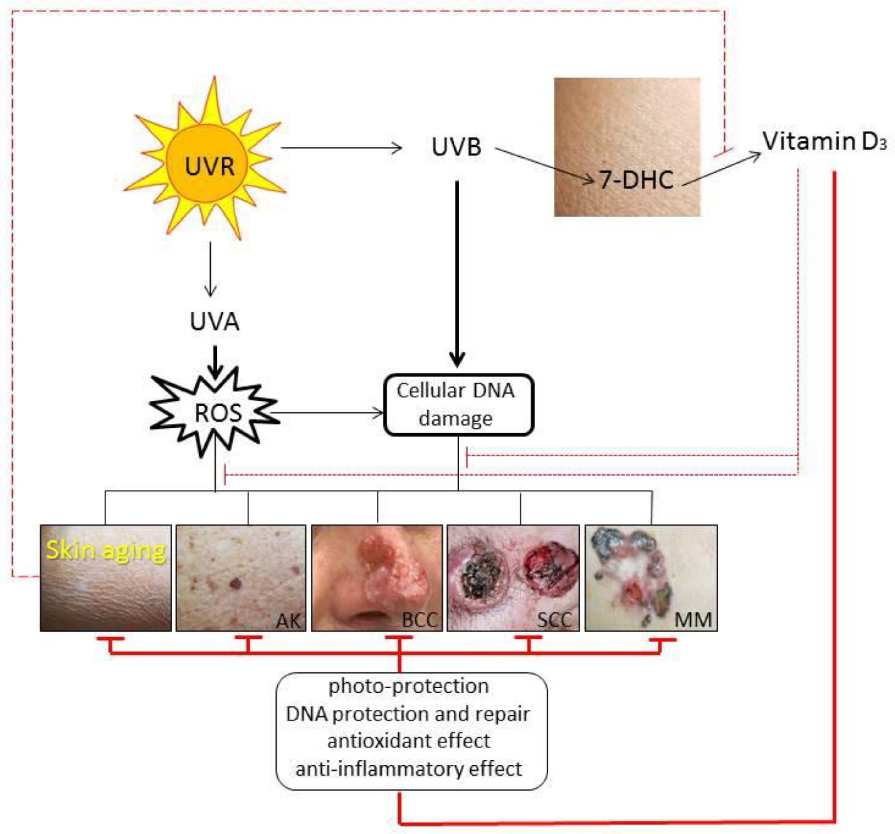
Excessive exposure to solar UVR accelerates skin aging and could trigger cutaneous cancerogenesis [133]. However, UVR plays a beneficial role in the regulation of many skin functions [56,77,134]. The same UVB, responsible for the increase of non-melanoma skin cancer, is required for vitamin D3 production in the skin that supplies more than 90% of the vitamin D3 body’s requirement [44,55,135]. In the skin, vitamin D3 is essential for the formation of the epidermal barrier and hair follicles, and its deficiency has been linked to many proliferative and inflammatory cutaneous disorders [20,29,44,136].
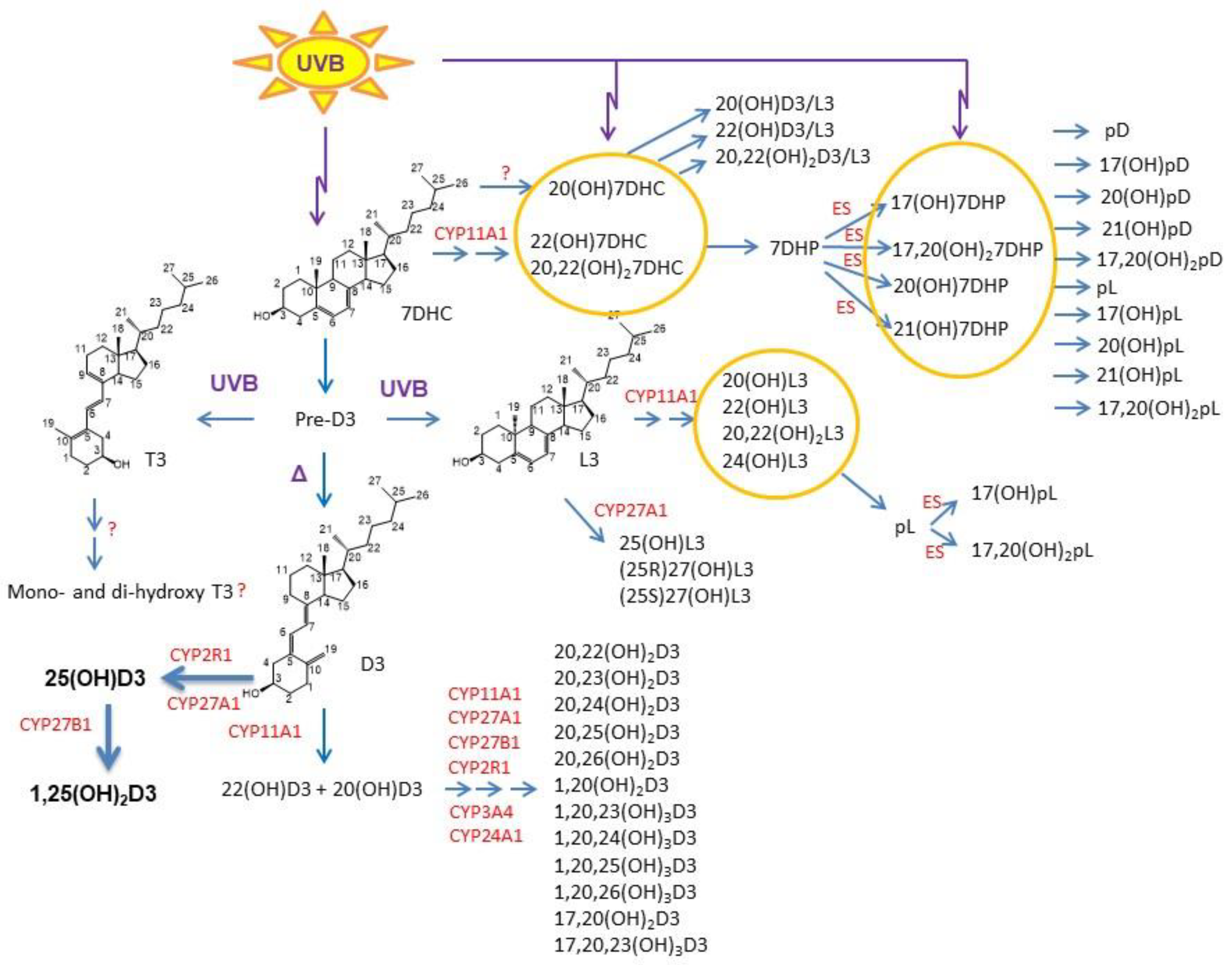
Upon the absorption of UVB, 7-DHC is transformed to vitamin D3 in the skin, a process accelerated by thermal energy. Prolonged UVB exposure can also generate tachysterol (T3) and lumisterol (L3) [24,97]. These reactions are non-enzymatic and dependent on the UVB dose and the temperature. Vitamin D3 can be activated through canonical and non-canonical pathways with similar activation of L3 to biologically active metabolites (Figure 1). In the classical pathway, vitamin D3 is hydroxylated to 25-hydroxyvitamin D3 (25(OH)D3) by CYP2R1 and/or CYP27A1 in the liver with further hydroxylation by CYP27B1 in the kidney, skin, and other tissues to its biologically active metabolite 1,25(OH)2D3 [20,21,137].
In the alternative (non-canonical) pathway, vitamin D3 can be activated by CYP11A1 with further modification by other cytochrome enzymes leading to production of large number of metabolites in humans [21,36,138,139,140,141,142] (Figure 1), some of which are non- or low-calcemic at high, therapeutic, doses [143,144,145,146]. The major CYP11A1-derived vitamin D3 products are 20(OH)D3 and 20,23(OH)2D3 [23,139,147,148]. In addition, 20(OH)D3 can be defined also as a natural product because its presence in the honey [149]. The L3 can also be metabolized to biologically active derivatives [150,151,152], which are not recognized by the 7-DHC reductase [153].
The main genomic effects and biological responses of vitamin D metabolites in the skin are mediated through their binding to the nuclear VDR [32,61,154,155,156]. Notably, VDR has been reported to regulate about 3% of mammalian genome due to its broad expression in all tissues [4,34,157,158]. The skin also expresses the VDR and serves not only as a source but also as a site for the action of vitamin D3 [28,39]. Additionally, the VDR activated by classical 1,25(OH)2D3 can induce rapid response signaling through a non-genomic, membrane-associated mechanism based on an alternative ligand-binding site [159] or through action on 1,25D3-MARRS receptor [156,160,161]. Similar non-genomic activities for CYP11A1-derived hydroxyderivatives are still not established. SNPs can affect VDR activity favoring a development of melanoma and non-melanoma skin tumors [162,163]. VDR functions as a tumor suppressor [164] and a decrease in its expression is associated with progression of cutaneous melanoma [165,166]. On the opposite, the nuclear VDR expression has been found significantly elevated (moderate to strong) in squamous cell carcinomas (SCCs) and basal cell carcinomas (BCCs) compared to in normal skin [167,168]. Thus, targeting VDR with vitamin D secosteroids (especially low calcemic ones) would be rational in skin cancer prevention, attenuation, or therapy [62,64,169].
Figure 1.
Noncanonical pathways of vitamin D3 and lumisterol (L3) activation (reprinted from [61] with the permission from Springer). D3, L3, and 7-DHC are substrates for CYP11A1 that produces the corresponding hydroxyderivatives. In the case of L3 and 7-DHC, the side chain can be cleaved by CYP11A1 to produce 7DHP or pL that can be further metabolized by steroidogenic enzymes (ES). In the skin, UVB acting on 5,7-dienes can lead to the production of D3, L3, and T3 derivatives with a full-length side chain and pD, pL, and pT derivatives with a shortened side chain. While the cut-off for UVC/UVB is 280 nm, we show the range of 290–315 nm because wavelengths below 290 nm are filtered by the ozone layer and no additional pre-D3 is produced above 315 nm [170]. 7DHC, 7-dehydrocholesterol; 7DHP, 7-dehydropregnenolone; pD, preganacalciferol; pL, preganalumisterol; D3, vitamin D3; L3, lumisterol; T3, tachysterol; OH, hydroxyl group; number before OH, carbon number with OH; number in subscripts after (OH), number of hydroxyl groups.

CYP11A1-derived hydroxyderivatives can regulate some skin functions through other nuclear receptor such as retinoic acid-related orphan receptors (ROR) α and γ, which expressed in the skin [171]. The endogenously produced nonclassical vitamin D3 hydroxyderivatives, 20(OH)D3 and 20,23(OH)2D3 can act as weak RORα and RORγ inverse agonists [155,171,172]. Moreover, these hydroxyderivatives could exert anti-inflammatory effect and could inhibit tumor progression in the skin via RORγ-mediated mechanism [173].
Alternative, the classical 1,25(OH)2D3 and CYP11A1-initiated vitamin D3 derivatives can act as agonists on aryl hydrocarbon receptor (AhR) [174] and liver X receptors (LXR) [175]. Moreover, the activation of AhR is the top canonical pathway for 20,23(OH)2D3 [174]. This receptor regulates cellular proliferation, inflammation, and melanogenesis in the skin [176]. Although many different ligands can target AhR, some functional studies and molecular modeling can predict that secosteroidal signal transduction further leads to the downregulation of proinflammatory responses [177], detoxification, and antioxidative action [61,174].
Summarizing, the biologically active classical and novel vitamin D3 metabolites exert different affinities to multiple receptors in the skin and through their modulation they can influence different cutaneous pathologies. In addition to act on the VDR, the active forms of vitamin D can act on alternative nuclear receptors including RORs, AhR, LXR, and 1,25D3-MARRS receptor. The active forms of vitamin D3 have various functions, which partially overlap in their anti-inflammatory, antimicrobial, antiproliferative, prodifferentiation, antifibrotic, and antioxidative effects on the skin [20,38,63,141,145,178]. Along with the best characterized 1,25(OH)2D3, CYP11A1-derived products of vitamin D3 and L3 exhibit photoprotective properties against UVR-induced skin damage (Figure 2) [37,61,179,180,181,182,183].
3.2. Effects on the Cutaneous Immune Function
Vitamin D3 and its analogues and precursors play an important complex role in the regulation of both the innate and adaptive immune systems, including in the skin [8,184,185]. There is a clear connection between vitamin D deficiency and incidences of infections and immune-mediated skin diseases [31,186,187,188]. The expression of the VDR has been found in almost all immune cells including T- and B-lymphocytes (Lym), macrophages, mast cells, natural killer (NK) cells, and regulatory T cells (Tregs), but it is differently regulated [189]. Monocytes, for example, lose VDR expression levels during differentiation towards macrophages and dendritic cells (DCs) [190,191]. Immunomodulatory actions of active vitamin D3 metabolites include the induction of Tregs [192] and Thelper-2 (Th2)-Lym, together with the downregulation of proinflammatory Th1/Th17/Th9-Lym [193]. 1,25(OH)2D3 can have both direct and indirect effects on T-Lym [194]. The indirect effect is based on DC-derived cytokines, which modulate Th-Lym response [30]. Directly, 1,25(OH)2D3 can suppress the immune cell production of inflammatory cytokines such as interferon gamma (IFN-γ), TNF-α, and IL-2 (Th1 cytokines), IL-17/21 (Th17 cytokines), and Th9 cytokines [193,195,196], while it enhances either the levels of anti-inflammatory IL-10 from Tregs [197] or Th2-derived IL-4 [198]. As a result, vitamin D shifts Th1 inflammatory response towards more tolerogenic Th2 response with an increase of CD4 + CD25 + Tregs reflected to a change in cytokine profile in the skin [199]. Additionally, 1,25(OH)2D3 influences the activated B-Lym by inducing apoptosis [200], suppressing immunoglobulin E (IgE)-dependent mast cell activation [201,202], and upregulating IL-10 production [203]. Enhanced IL-10 synthesis contributes to a suppressed mast cell-mediated inflammation and IgE-related allergic reactions [201]. 1,25(OH)2D3 and its analogues directly regulate antimicrobial peptide (AMP) gene expression in innate immune cells [204,205]. Moreover, it has been found that vitamin D is able to induce essential for antimicrobial defense, production of cathelicidin (LL37) [204] and to modulate the phagocytic activity of macrophages and NK cells [193]. Additionally, vitamin D exerts immunosuppressive effects through the modulation of epidermal Langerhans cells [206] and the proliferation of the Tregs number [184,185,197].
CYP11A1 is expressed also in immune cells [207], where vitamin D can be further metabolized to biologically active hydroxyderivatives [31,39]. Via the activation of VDR or through the inhibition of RORγ-mediated activation, 20(OH)D3 and 20,23(OH)2D3, like 1,25(OH)2D3, can attenuate Th17 differentiation, as well as the formation and activity of inflammatory cytokine IL-17 by immune cells [155,208,209]. Thus, Th17-related cutaneous inflammation could be successfully modulated via RORγ inverse agonists such as CYP11A1-derived D3-hydroxyderivatives, causing the regulation of the immune system and a resistance against autoimmunity [210,211]. The most recently inhibition of collagen-induced autoimmune arthritis by CYP11A1-derived 20(OH)D3 was reported [212].
3.3. Impact on Skin Aging
The normal vitamin D3 status is important for a general prevention of premature aging maintaining a healthful skin aging [213,214]. Vitamin D3 metabolites including its classical (1,25(OH)2D3) and novel (CYP11A1-intitated) D3 hydroxyderivatives exert many beneficial protective effects on the skin, which could influence the process of premature aging via many different mechanisms, leading to a delay or attenuation of both chronological skin aging and photoaging. Skin-resident cells (keratinocytes, fibroblasts, and sebocytes) are capable of locally activating vitamin D3 [23,36,215] and exhibiting a diverse biological effect such as photoprotection and immunosuppression, similar to the UVR-induced one [179,216].
The process of chronological aging is associated with immunological alteration and the imbalance between inflammatory and anti-inflammatory mechanisms, leading to a chronic low-grade inflammation, known as “inflammaging” state [217,218]. The “inflammaging” phenotype of the skin and hair follicles is a result of both chronic antigen stimulation and continued exposure to oxidative stress caused by ROS and RNS [219,220]. With advancing age, skin is affected by the profound remodeling of the immune system, leading to a decline in its adaptive capacity [221,222]. Th1- and Th17-related markers, together with the number of epidermal DCs are increased as a function of age [223,224,225]. DCs during aging appear to be functionally impaired, which contributes to an initiation of inflammatory and autoimmune skin disorders and a loss of their protective role against cutaneous infections. The active forms of vitamin D3 are able to decrease the proliferation and cytotoxicity of T-Lym, as well as to suppress the differentiation of B-Lym and the maturation of DCs [193]. Therefore, vitamin D3 hydroxyderivatives exert potent anti-inflammatory activities including the inhibition of TNF-α, INF-γ, and IL-1/6/9/17 production [4,38,185], suggesting their implication in the modulation of skin inflammation. Moreover, the noncalcemic and nontoxic 20(OH)D3 has shown a similar anti-inflammatory property in vivo to 1,25(OH)2D3 (hypercalcemic in high doses) through the suppression of the immune responses by T- and B-lym [155,212].
Active vitamin D3 metabolites can protect skin against the hazardous effects of skin aging-triggering agents, including UVR, pollution, and microbial infections [179,226,227,228,229,230]. It has been shown that the oral administration of high-dose vitamin D3 shortly after UVB exposure could reverse rapidly the photo-induced cutaneous damage by decreasing the inflammation and induction of the repair mechanisms of the epidermal barrier [38]. There is strong experimental evidence that active vitamin D3 and L3 hydroxyderivatives can induce, in a dose-dependent manner, antioxidative responses and reverse the UVB-mediated ROS production in keratinocytes by the activation of Nrf2 that works for cytoprotection and detoxification, thus attenuating photoaging [122]. Therefore, they serve as protective agents against UVB-induced oxidative stress in cells, pre-treated with each of these active metabolites for 24 h prior to UVB irradiation (50 mJ/cm2) [122]. These hydroxyderivatives stimulate the expression of antioxidant-response genes downstream of Nrf2 (GR, HO-1, CAT, SOD-1, and SOD-2) as well as the expression of HO-1, CAT, and MnSOD at the protein level [122]. The transcription factor Nrf2 plays an important role in the detection of excessive ROS and RNS and in the induction of mechanisms counteracting the oxidative damage and skin pigmentations produced by UVA [121,125,231].
Chronic UVR irradiation, mainly UVB [232] and UVA [233], induces DNA damage and the formation of CPDs that potentially lead to premature skin aging and carcinogenesis. CYP11A1-derived D3 and L3 hydroxyderivatives, along with 1,25(OH)2D3, demonstrate photoprotective and reparative properties by increasing the expression and phosphorylation of p53 with its translocation to the nucleus [61,229,234,235]. The P53 gene family, in particular its isoform p63, might be an important molecular target for vitamin D action in premature aging and cancer [236], which are promoted by similar mechanisms [237].
Moreover, 1,25(OH)2D3 and 1,25(OH)2L3 inhibit DNA damage and facilitate DNA repair by the reduction of CPDs [182,235,238,239] and RNS [178,234]. The photoprotection by 20(OH)D3 and 20,23(OH)2D3 is comparable to 1,25(OH)2D3 reduction of UVB-induced CPDs and DNA fragmentation in vivo [181,182] and in vitro [178]. In addition, both 20(OH)D3 and 20,23(OH)2D3 stimulate differentiation, inhibit proliferation and downregulate proinflammatory responses in keratinocytes via the decrease of NFκβ activity [240,241]. It was shown recently that not only the pretreatment, but also the post-treatment of keratinocytes with CYP11A1-derived D3 and L3 derivatives can reverse their UVB-induced damage [37,230].
Additionally, 1,25(OH)2D3 can induce rapid and dose-dependent reduction in skin cell apoptosis, and it can increase CPDs repair and decrease the oxidative DNA damage through non-genomic energy-conserving autophagy and mitophagy [227], thus contributing to the intrinsic skin photoprotection mechanism [242].
4. Conclusions and Future Perspectives
Vitamin D3 and its active metabolites exert a variety of antiaging and (photo) protective effects on the skin. These are achieved through immunomodulation that include anti-inflammatory actions and regulation of keratinocytes proliferation and differentiation program to build the epidermal barrier necessary to maintain skin homeostasis. In addition, they induce antioxidative responses, inhibit DNA damage and induce DNA repair mechanisms to attenuate premature skin aging and cancerogenesis. Similar actions can be assigned to lumisterol metabolites. Therefore, active forms of vitamin D3 including its canonical (1,25(OH)2D3) and noncanonical (CYP11A1-intitated) D3-hydroxyderivatives as well as L3-derivatives are promising agents for the prevention, attenuation, or treatment of premature skin aging, when applied topically. It is expected that they will attenuate photoaging and perhaps repair the existing damage induced by external stressors. The mechanism of action would involve interaction with nuclear receptors including VDR, AhR, LXR, reverse agonism on RORα and RORγ, and nongenomic actions through 1,25D3-MARRS receptor and interaction with the nongenomic binding site of the VDR. The regulatory mechanism affected by D3 and L3 derivatives would include the activation of Nrf2 and p53 and the downregulation of NFκβ signaling pathways or the regulation of mitochondrial functions. To prevent skin aging, vitamin D3 and lumisterol or their derivatives could be administrated orally and/or topically. Other forms of parenteral application of the vitamin D3 precursor should be considered to avoid channeling its metabolism to 25(OH)D3, which is not recognized by CYP11A1 enzyme [243]. The efficacy of topically applied vitamin D3 and L3 derivatives needs further clinical evaluation in future trials.
Funding
We acknowledge the support of NIH grants (1R01AR073004 and R01AR071189), a VA merit grant 1I01BX004293 to A.T.S., a grant R21 AI152047 to A.T.S. and C.R., and a grant R21 MD015319 to C.R.
Institutional Review Board Statement
Not applicable.
Informed Consent Statement
Not applicable.
Data Availability Statement
Not applicable.
Acknowledgments
We thank Carlton Philips and Craig Elmets from the department of dermatology, UAB, for the pictures of BCC and SCC.
Conflicts of Interest
The authors declare no conflict of interest.
References
- Bocheva, G.; Slominski, R.M.; Slominski, A.T. Neuroendocrine Aspects of Skin Aging. Int. J. Mol. Sci. 2019, 20, 2798. [Google Scholar] [CrossRef] [PubMed] [Green Version]
- Bocheva, G.S.; Slominski, R.M.; Slominski, A.T. Immunological Aspects of Skin Aging in Atopic Dermatitis. Int. J. Mol. Sci. 2021, 22, 5729. [Google Scholar] [CrossRef]
- Zouboulis, C.C.; Adjaye, J.; Akamatsu, H.; Moe-Behrens, G.; Niemann, C. Human skin stem cells and the ageing process. Exp. Gerontol. 2008, 43, 986–997. [Google Scholar] [CrossRef] [Green Version]
- Reichrath, J.; Zouboulis, C.C.; Vogt, T.; Holick, M.F. Targeting the vitamin D endocrine system (VDES) for the management of inflammatory and malignant skin diseases: An historical view and outlook. Rev. Endocr. Metab. Disord. 2016, 17, 405–417. [Google Scholar] [CrossRef]
- Slominski, A.; Wortsman, J. Neuroendocrinology of the skin. Endocr. Rev. 2000, 21, 457–487. [Google Scholar] [CrossRef] [PubMed]
- Slominski, A.T.; Zmijewski, M.A.; Skobowiat, C.; Zbytek, B.; Slominski, R.M.; Steketee, J.D. Sensing the environment: Regulation of local and global homeostasis by the skin’s neuroendocrine system. Adv. Anat. Embryol. Cell Biol. 2012, 212, 1–115. [Google Scholar]
- Slominski, A.T.; Zmijewski, M.A.; Zbytek, B.; Tobin, D.J.; Theoharides, T.C.; Rivier, J. Key role of CRF in the skin stress response system. Endocr. Rev. 2013, 34, 827–884. [Google Scholar] [CrossRef] [PubMed]
- Slominski, A.; Zbytek, B.; Nikolakis, G.; Manna, P.R.; Skobowiat, C.; Zmijewski, M.; Li, W.; Janjetovic, Z.; Postlethwaite, A.; Zouboulis, C.C.; et al. Steroidogenesis in the skin: Implications for local immune functions. J. Steroid Biochem. Mol. Biol. 2013, 137, 107–123. [Google Scholar] [CrossRef] [PubMed] [Green Version]
- Slominski, A.; Wortsman, J.; Tuckey, R.C.; Paus, R. Differential expression of HPA axis homolog in the skin. Mol. Cell. Endocrinol. 2007, 265–266, 143–149. [Google Scholar] [CrossRef] [PubMed] [Green Version]
- Slominski, A.; Ermak, G.; Hwang, J.; Chakraborty, A.; Mazurkiewicz, J.E.; Mihm, M. Proopiomelanocortin, corticotropin releasing hormone and corticotropin releasing hormone receptor genes are expressed in human skin. FEBS Lett. 1995, 374, 113–116. [Google Scholar] [CrossRef] [Green Version]
- Slominski, A.; Wortsman, J.; Pisarchik, A.; Zbytek, B.; Linton, E.A.; Mazurkiewicz, J.E.; Wei, E.T. Cutaneous expression of corticotropin-releasing hormone (CRH), urocortin, and CRH receptors. FASEB J. 2001, 15, 1678–1693. [Google Scholar] [CrossRef] [PubMed]
- Slominski, A.; Szczesniewski, A.; Wortsman, J. Liquid chromatography-mass spectrometry detection of corticotropin-releasing hormone and proopiomelanocortin-derived peptides in human skin. J. Clin. Endocrinol. Metab. 2000, 85, 3582–3588. [Google Scholar] [CrossRef] [PubMed]
- Ito, N.; Ito, T.; Betterman, A.; Paus, R. The human hair bulb is a source and target of CRH. J. Investig. Dermatol. 2004, 122, 235–237. [Google Scholar] [CrossRef] [PubMed] [Green Version]
- Slominski, A.; Wortsman, J.; Luger, T.; Paus, R.; Solomon, S. Corticotropin releasing hormone and proopiomelanocortin involvement in the cutaneous response to stress. Physiol. Rev. 2000, 80, 979–1020. [Google Scholar] [CrossRef] [PubMed] [Green Version]
- Kono, M.; Nagata, H.; Umemura, S.; Kawana, S.; Osamura, R.Y. In situ expression of corticotropin-releasing hormone (CRH) and proopiomelanocortin (POMC) genes in human skin. FASEB J. 2001, 15, 2297–2299. [Google Scholar] [CrossRef] [Green Version]
- Slominski, A.; Wortsman, J.; Tobin, D.J. The cutaneous serotoninergic/melatoninergic system: Securing a place under the sun. FASEB J. 2005, 19, 176–194. [Google Scholar] [CrossRef]
- Slominski, A.; Pisarchik, A.; Semak, I.; Sweatman, T.; Wortsman, J.; Szczesniewski, A.; Slugocki, G.; McNulty, J.; Kauser, S.; Tobin, D.J.; et al. Serotoninergic and melatoninergic systems are fully expressed in human skin. FASEB J. 2002, 16, 896–898. [Google Scholar] [CrossRef] [PubMed] [Green Version]
- Zouboulis, C.C. Human skin: An independent peripheral endocrine organ. Horm. Res. 2000, 54, 230–242. [Google Scholar] [CrossRef]
- Reichrath, J.; Lehmann, B.; Carlberg, C.; Varani, J.; Zouboulis, C.C. Vitamins as hormones. Horm. Metab. Res. 2007, 39, 71–84. [Google Scholar] [CrossRef] [Green Version]
- Bikle, D.D. Vitamin D: Newer Concepts of Its Metabolism and Function at the Basic and Clinical Level. J. Endocr. Soc. 2020, 4, bvz038. [Google Scholar] [CrossRef]
- Tuckey, R.C.; Cheng, C.Y.S.; Slominski, A.T. The serum vitamin D metabolome: What we know and what is still to discover. J. Steroid Biochem. Mol. Biol. 2019, 186, 4–21. [Google Scholar] [CrossRef]
- Slominski, A.; Kim, T.K.; Zmijewski, M.A.; Janjetovic, Z.; Li, W.; Chen, J.; Kusniatsova, E.I.; Semak, I.; Postlethwaite, A.; Miller, D.D.; et al. Novel vitamin D photoproducts and their precursors in the skin. Dermatoendocrinology 2013, 5, 7–19. [Google Scholar] [CrossRef] [Green Version]
- Slominski, A.T.; Kim, T.K.; Li, W.; Tuckey, R.C. Classical and non-classical metabolic transformation of vitamin D in dermal fibroblasts. Exp. Dermatol. 2016, 25, 231–232. [Google Scholar] [CrossRef] [PubMed]
- Holick, M.F. Vitamin D: A millenium perspective. J. Cell Biochem. 2003, 88, 296–307. [Google Scholar] [CrossRef] [PubMed]
- Reichrath, J.; Saternus, R.; Vogt, T. Endocrine actions of vitamin D in skin: Relevance for photocarcinogenesis of non-melanoma skin cancer, and beyond. Mol. Cell Endocrinol. 2017, 453, 96–102. [Google Scholar] [CrossRef] [PubMed]
- Bikle, D.D. Vitamin D and the skin. J. Bone Miner. Metab. 2010, 28, 117–130. [Google Scholar] [CrossRef]
- Luderer, H.F.; Demay, M.B. The vitamin D receptor, the skin and stem cells. J. Steroid Biochem. Mol. Biol. 2010, 121, 314–316. [Google Scholar] [CrossRef]
- Bikle, D.D. Vitamin D and the skin: Physiology and pathophysiology. Rev. Endocr. Metab. Disord. 2012, 13, 3–19. [Google Scholar] [CrossRef] [Green Version]
- Oda, Y.; Hu, L.; Nguyen, T.; Fong, C.; Zhang, J.; Guo, P.; Bikle, D.D. Vitamin D Receptor Is Required for Proliferation, Migration, and Differentiation of Epidermal Stem Cells and Progeny during Cutaneous Wound Repair. J. Investig. Dermatol. 2018, 138, 2423–2431. [Google Scholar] [CrossRef] [Green Version]
- Martens, P.J.; Gysemans, C.; Verstuyf, A.; Mathieu, A.C. Vitamin D’s Effect on Immune Function. Nutrients 2020, 12, 1248. [Google Scholar] [CrossRef]
- Hewison, M. Vitamin D and immune function: An overview. Proc. Nutr. Soc. 2012, 71, 50–61. [Google Scholar] [CrossRef] [Green Version]
- Lin, Z.; Chen, H.; Belorusova, A.Y.; Bollinger, J.C.; Tang, E.K.Y.; Janjetovic, Z.; Kim, T.K.; Wu, Z.; Miller, D.D.; Slominski, A.T.; et al. 1α,20S-Dihydroxyvitamin D3 Interacts with Vitamin D Receptor: Crystal Structure and Route of Chemical Synthesis. Sci. Rep. 2017, 7, 10193. [Google Scholar] [CrossRef]
- Pike, J.W.; Meyer, M.B.; Bishop, K.A. Regulation of target gene expression by the vitamin D receptor—An update on mechanisms. Rev. Endocr. Metab. Disord. 2012, 13, 45–55. [Google Scholar] [CrossRef]
- Bouillon, R.; Marcocci, C.; Carmeliet, G.; Bikle, D.; White, J.H.; Dawson-Hughes, B.; Lips, P.; Munns, C.F.; Lazaretti-Castro, M.; Giustina, A.; et al. Skeletal and Extraskeletal Actions of Vitamin D: Current Evidence and Outstanding Questions. Endocr. Rev. 2018, 40, 1109–1151. [Google Scholar] [CrossRef] [Green Version]
- Bikle, D.D. The vitamin D receptor: A tumor suppressor in skin. Adv. Exp. Med. Biol. 2014, 810, 282–302. [Google Scholar]
- Slominski, A.T.; Kim, T.K.; Li, W.; Yi, A.K.; Postlethwaite, A.; Tuckey, R.C. The role of CYP11A1 in the production of vitamin D metabolites and their role in the regulation of epidermal functions. J. Steroid Biochem. Mol. Biol. 2014, 144PA, 28–39. [Google Scholar] [CrossRef] [PubMed] [Green Version]
- Chaiprasongsuk, A.; Janjetovic, Z.; Kim, T.K.; Schwartz, C.J.; Tuckey, R.C.; Tang, E.K.Y.; Raman, C.; Panich, U.; Slominski, A.T. Hydroxylumisterols, Photoproducts of Pre-Vitamin D3, Protect Human Keratinocytes against UVB-Induced Damage. Int. J. Mol. Sci. 2020, 21, 9374. [Google Scholar] [CrossRef]
- Scott, J.F.; Das, L.M.; Ahsanuddin, S.; Qiu, Y.; Binko, A.M.; Traylor, Z.P.; Debanne, S.M.; Cooper, K.D.; Boxer, R.; Lu, K.Q. Oral Vitamin D Rapidly Attenuates Inflammation from Sunburn: An Interventional Study. J. Investig. Dermatol. 2017, 137, 2078–2086. [Google Scholar] [CrossRef] [PubMed] [Green Version]
- Bikle, D.; Christakos, S. New aspects of vitamin D metabolism and action—Addressing the skin as source and target. Nat. Rev. Endocrinol. 2020, 16, 234–252. [Google Scholar] [CrossRef] [PubMed]
- Binkley, N.; Dawson-Hughes, B.; Durazo-Arvizu, R.; Thamm, M.; Tian, L.; Merkel, J.M.; Jones, J.C.; Carter, G.D.; Sempos, C.T. Vitamin D measurement standardization: The way out of the chaos. J. Steroid Biochem. Mol. Biol. 2017, 173, 117–121. [Google Scholar] [CrossRef] [PubMed]
- Schöttker, B.; Jansen, E.H.; Haug, U.; Schomburg, L.; Köhrle, J.; Brenner, H. Standardization of misleading immunoassay based 25-hydroxyvitamin D levels with liquid chromatography tandem-mass spectrometry in a large cohort study. PLoS ONE 2012, 7, e48774. [Google Scholar] [CrossRef]
- Jenkinson, C. The vitamin D metabolome: An update on analysis and function. Cell Biochem. Funct. 2019, 37, 408–423. [Google Scholar] [CrossRef]
- Jenkinson, C.; Desai, R.; Slominski, A.T.; Tuckey, R.C.; Hewison, M.; Handelsman, D.J. Simultaneous measurement of 13 circulating vitamin D3 and D2 mono and dihydroxy metabolites using liquid chromatography mass spectrometry. Clin. Chem. Lab. Med. 2021. [Google Scholar] [CrossRef]
- Holick, M.F. Vitamin D deficiency. N. Engl. J. Med. 2007, 357, 266–281. [Google Scholar] [CrossRef]
- Palacios, C.; Gonzalez, L. Is vitamin D deficiency a major global public health problem? J. Steroid Biochem. Mol. Biol. 2014, 144, 138–145. [Google Scholar] [CrossRef] [PubMed] [Green Version]
- Kmieć, P.; Żmijewski, M.; Waszak, P.; Sworczak, K.; Lizakowska-Kmieć, M. Vitamin D deficiency during winter months among an adult, predominantly urban, population in Northern Poland. Endokrynol. Pol. 2014, 65, 105–113. [Google Scholar] [CrossRef] [PubMed] [Green Version]
- Borissova, A.M.; Shinkov, A.; Vlahov, J.; Dakovska, L.; Todorov, T.; Svinarov, D.; Kassabova, L. Vitamin D status in Bulgaria—Winter data. Arch. Osteoporos. 2013, 8, 133. [Google Scholar] [CrossRef]
- Holick, M.F. The vitamin D deficiency pandemic: Approaches for diagnosis, treatment and prevention. Rev. Endocr. Metab. Disord. 2017, 18, 153–165. [Google Scholar] [CrossRef] [PubMed]
- Serdar, M.A.; Batu Can, B.; Kilercik, M.; Durer, Z.A.; Aksungar, F.B.; Serteser, M.; Coskun, A.; Ozpinar, A.; Unsal, I. Analysis of Changes in Parathyroid Hormone and 25 (OH) Vitamin D Levels with Respect to Age, Gender and Season: A Data Mining Study. J. Med. Biochem. 2017, 36, 73–83. [Google Scholar] [CrossRef] [PubMed] [Green Version]
- Shinkov, A.; Borissova, A.M.; Dakovska, L.; Vlahov, J.; Kassabova, L.; Svinarov, D. Winter 25-hydroxyvitamin D levels in young urban adults are affected by smoking, body mass index and educational level. Eur. J. Clin. Nutr. 2015, 69, 355–360. [Google Scholar] [CrossRef]
- Holick, M.F. Bioavailability of vitamin D and its metabolites in black and white adults. N. Engl. J. Med. 2013, 369, 2047–2048. [Google Scholar] [CrossRef]
- Kiely, M.; Hemmingway, A.; O’Callaghan, K.M. Vitamin D in pregnancy: Current perspectives and future directions. Ther. Adv. Musculoskelet. Dis. 2017, 9, 145–154. [Google Scholar] [CrossRef]
- Lagunova, Z.; Porojnicu, A.C.; Lindberg, F.; Hexeberg, S.; Moan, J. The dependency of vitamin D status on body mass index, gender, age and season. Anticancer Res. 2009, 29, 3713–3720. [Google Scholar] [CrossRef]
- Rossberg, W.; Saternus, R.; Wagenpfeil, S.; Kleber, M.; März, W.; Reichrath, S.; Vogt, T.; Reichrath, J. Human Pigmentation, Cutaneous Vitamin D Synthesis and Evolution: Variants of Genes (SNPs) Involved in Skin Pigmentation Are Associated with 25(OH)D Serum Concentration. Anticancer Res. 2016, 36, 1429–1437. [Google Scholar] [PubMed]
- Wacker, M.; Holick, M.F. Sunlight and Vitamin D: A global perspective for health. Dermatoendocrinology 2013, 5, 51–108. [Google Scholar] [CrossRef] [PubMed] [Green Version]
- Grant, W.B.; Bhattoa, H.P.; Boucher, B.J. Seasonal variations of U.S. mortality rates: Roles of solar ultraviolet-B doses, vitamin D, gene exp ression, and infections. J. Steroid Biochem. Mol. Biol. 2017, 173, 5–12. [Google Scholar] [CrossRef] [PubMed] [Green Version]
- Mousavi, S.E.; Amini, H.; Heydarpour, P.; Amini Chermahini, F.; Godderis, L. Air pollution, environmental chemicals, and smoking may trigger vitamin D deficiency: Evidence and potential mechanisms. Environ. Int. 2019, 122, 67–90. [Google Scholar] [CrossRef]
- Boas, M.; Feldt-Rasmussen, U.; Main, K.M. Thyroid effects of endocrine disrupting chemicals. Mol. Cell. Endocrinol. 2012, 355, 240–248. [Google Scholar] [CrossRef]
- MacLaughlin, J.; Holick, M.F. Aging decreases the capacity of human skin to produce vitamin D3. J. Clin. Investig. 1985, 76, 1536–1538. [Google Scholar] [CrossRef] [PubMed] [Green Version]
- Nowak, J.; Hudzik, B.; Jagielski, P.; Kulik-Kupka, K.; Danikiewicz, A.; Zubelewicz-Szkodzińska, B. Lack of Seasonal Variations in Vitamin D Concentrations among Hospitalized Elderly Patients. Int. J. Environ. Res. Public Health 2021, 18, 1676. [Google Scholar]
- Slominski, A.T.; Chaiprasongsuk, A.; Janjetovic, Z.; Kim, T.K.; Stefan, J.; Slominski, R.M.; Hanumanthu, V.S.; Raman, C.; Qayyum, S.; Song, Y.; et al. Photoprotective Properties of Vitamin D and Lumisterol Hydroxyderivatives. Cell Biochem. Biophys. 2020, 78, 165–180. [Google Scholar] [CrossRef] [PubMed]
- Slominski, A.T.; Brożyna, A.A.; Skobowiat, C.; Zmijewski, M.A.; Kim, T.K.; Janjetovic, Z.; Oak, A.S.; Jozwicki, W.; Jetten, A.M.; Mason, R.S.; et al. On the role of classical and novel forms of vitamin D in melanoma progression and management. J. Steroid Biochem. Mol. Biol. 2018, 177, 159–170. [Google Scholar] [CrossRef] [PubMed] [Green Version]
- Slominski, A.T.; Janjetovic, Z.; Kim, T.K.; Wright, A.C.; Grese, L.N.; Riney, S.J.; Nguyen, M.N.; Tuckey, R.C. Novel vitamin D hydroxyderivatives inhibit melanoma growth and show differential effects on normal melanocytes. Anticancer Res. 2012, 32, 3733–3742. [Google Scholar] [PubMed]
- Slominski, A.T.; Brożyna, A.A.; Zmijewski, M.A.; Janjetovic, Z.; Kim, T.K.; Slominski, R.M.; Tuckey, R.C.; Mason, R.S.; Jetten, A.M.; Guroji, P.; et al. The Role of Classical and Novel Forms of Vitamin D in the Pathogenesis and Progression of Nonmelanoma Skin Cancers. Adv. Exp. Med. Biol. 2020, 1268, 257–283. [Google Scholar]
- Tongkao-On, W.; Gordon-Thomson, C.; Dixon, K.M.; Song, E.J.; Luu, T.; Carter, S.E.; Sequeira, V.B.; Reeve, V.E.; Mason, R.S. Novel vitamin D compounds and skin cancer prevention. Dermatoendocrinology 2013, 5, 20–33. [Google Scholar] [CrossRef] [PubMed] [Green Version]
- Panich, U.; Slominski, A.T. Editorial: Redox Biology of Skin Aging and Carcinogenesis: The Role of Natural Antioxidants as Potential Protective Agents. Front. Pharmacol. 2020, 11, 249. [Google Scholar] [CrossRef]
- Farage, M.A.; Miller, K.W.; Elsner, P.; Maibach, H.I. Intrinsic and extrinsic factors in skin ageing: A review. Int. J. Cosmet. Sci. 2008, 30, 87–95. [Google Scholar] [CrossRef]
- Fisher, G.J.; Kang, S.; Varani, J.; Bata-Csorgo, Z.; Wan, Y.; Datta, S.; Voorhees, J.J. Mechanisms of photoaging and chronological skin aging. Arch. Dermatol. 2002, 138, 1462–1470. [Google Scholar] [CrossRef]
- Krutmann, J.; Bouloc, A.; Sore, G.; Bernard, B.A.; Passeron, T. The skin aging exposome. J. Dermatol. Sci. 2017, 85, 152–161. [Google Scholar] [CrossRef] [Green Version]
- Orioli, D.; Dellambra, E. Epigenetic Regulation of Skin Cells in Natural Aging and Premature Aging Diseases. Cells 2018, 7, 268. [Google Scholar] [CrossRef] [Green Version]
- van Deursen, J.M. The role of senescent cells in ageing. Nature 2014, 509, 439–446. [Google Scholar] [CrossRef] [Green Version]
- Campisi, J. Aging, cellular senescence, and cancer. Annu. Rev. Physiol. 2013, 75, 685–705. [Google Scholar] [CrossRef] [PubMed] [Green Version]
- Ortonne, J.P. Photoprotective properties of skin melanin. Br. J. Dermatol. 2002, 146, 7–10. [Google Scholar] [CrossRef]
- Slominski, A.; Tobin, D.J.; Shibahara, S.; Wortsman, J. Melanin pigmentation in mammalian skin and its hormonal regulation. Physiol. Rev. 2004, 84, 1155–1228. [Google Scholar] [CrossRef] [PubMed]
- Liu, F.; Hamer, M.A.; Deelen, J.; Lall, J.S.; Jacobs, L.; van Heemst, D.; Murray, P.G.; Wollstein, A.; de Craen, A.J.; Uh, H.W.; et al. The MC1R Gene and Youthful Looks. Curr. Biol. 2016, 26, 1213–1220. [Google Scholar] [CrossRef] [PubMed] [Green Version]
- Rittié, L.; Fisher, G.J. UV-light-induced signal cascades and skin aging. Ageing Res. Rev. 2002, 1, 705–720. [Google Scholar] [CrossRef]
- Slominski, A.T.; Zmijewski, M.A.; Plonka, P.M.; Szaflarski, J.P.; Paus, R. How UV Light Touches the Brain and Endocrine System Through Skin, and Why. Endocrinology 2018, 159, 1992–2007. [Google Scholar] [CrossRef] [Green Version]
- Parrado, C.; Mercado-Saenz, S.; Perez-Davo, A.; Gilaberte, Y.; Gonzalez, S.; Juarranz, A. Environmental Stressors on Skin Aging. Mechanistic Insights. Front. Pharmacol. 2019, 10, 759. [Google Scholar] [CrossRef]
- Damevska, K.; Nikolovska, S.; Kazandjieva, J.; Kotevska, B.; Bocheva, G. Skin and pollution. In Advances in Integrative Dermatology; Franҫa, K., Lotti, T., Eds.; Wiley, J. & Sons, Ltd.: Hoboken, NJ, USA, 2019; pp. 379–392. [Google Scholar]
- Damevska, K.; Boev, B.; Mirakovski, D.; Petrov, A.; Darlenski, R.; Simeonovski, V. How to prevent skin damage from air pollution. Part 1: Exposure assessment. Dermatol. Ther. 2020, 33, e13171. [Google Scholar] [CrossRef] [Green Version]
- Green, A.C.; Wallingford, S.C.; McBride, P. Childhood exposure to ultraviolet radiation and harmful skin effects: Epidemiological evidence. Prog. Biophys. Mol. Biol. 2011, 107, 349–355. [Google Scholar] [CrossRef] [Green Version]
- Kennedy, C.; Bastiaens, M.T.; Bajdik, C.D.; Willemze, R.; Westendorp, R.G.; Bouwes Bavinck, J.N. Leiden Skin Cancer Study. Effect of smoking and sun on the aging skin. J. Investig. Dermatol. 2003, 120, 548–554. [Google Scholar] [CrossRef] [Green Version]
- Koohgoli, R.; Hudson, L.; Naidoo, K.; Wilkinson, S.; Chavan, B.; Birch-Machin, M.A. Bad air gets under your skin. Exp. Dermatol. 2017, 26, 384–387. [Google Scholar] [CrossRef] [PubMed]
- Naidoo, K.; Birch-Machin, M.A. Oxidative Stress and Ageing: The Influence of Environmental Pollution, Sunlight and Diet on Skin. Cosmetics 2017, 4, 4. [Google Scholar] [CrossRef]
- Kammeyer, A.; Luiten, R.M. Oxidation events and skin aging. Ageing Res. Rev. 2015, 21, 16–29. [Google Scholar] [CrossRef] [PubMed]
- Patra, V.; Gallais Sérézal, I.; Wolf, P. Potential of Skin Microbiome, Pro- and/or Pre-Biotics to Affect Local Cutaneous Responses to UV Exposure. Nutrients 2020, 12, 1795. [Google Scholar] [CrossRef]
- Burns, E.M.; Ahmed, H.; Isedeh, P.N.; Kohli, I.; Van Der Pol, W.; Shaheen, A.; Muza_ar, A.F.; Al-Sadek, C.; Foy, T.M.; Abdelgawwad, M.S.; et al. Ultraviolet radiation, both UVA and UVB, influences the compositionof the skin microbiome. Exp. Dermatol. 2019, 28, 136–141. [Google Scholar] [CrossRef]
- Bickers, D.R.; Athar, M. Oxidative stress in the pathogenesis of skin disease. J. Investig. Dermatol. 2006, 126, 2565–2575. [Google Scholar] [CrossRef] [Green Version]
- Imokawa, G.; Ishida, K. Biological mechanisms underlying the ultraviolet radiation-induced formation of skin wrinkling and sagging I: Reduced skin elasticity, highly associated with enhanced dermal elastase activity, triggers wrinkling and sagging. Int. J. Mol. Sci. 2015, 16, 7753–7775. [Google Scholar] [CrossRef] [Green Version]
- Tončić, R.J.; Kezić, S.; Hadžavdić, S.L.; Marinović, B. Skin barrier and dry skin in the mature patient. Clin. Dermatol. 2017, 36, 109–115. [Google Scholar] [CrossRef]
- Marionnet, C.; Tricaud, C.; Bernerd, F. Exposure to non-extreme solar UV daylight: Spectral characterization, effects on skin and photoprotection. Int. J. Mol. Sci. 2014, 16, 68–90. [Google Scholar] [CrossRef]
- Battie, C.; Jitsukawa, S.; Bernerd, F.; Del Bino, S.; Marionnet, C.; Verschoore, M. New insights in photoaging, UVA induced damage and skin types. Exp. Dermatol. 2014, 23, 7–12. [Google Scholar] [CrossRef]
- Chung, J.H.; Seo, J.Y.; Choi, H.R.; Lee, M.K.; Youn, C.S.; Rhie, G.; Cho, K.H.; Kim, K.H.; Park, K.C.; Eun, H.C. Modulation of skin collagen metabolism in aged and photoaged human skin in vivo. J. Investig. Dermatol. 2001, 117, 1218–1224. [Google Scholar] [CrossRef] [PubMed] [Green Version]
- Tigges, J.; Krutmann, J.; Fritsche, E.; Haendeler, J.; Schaal, H.; Fischer, J.W.; Kalfalah, F.; Reinke, H.; Reifenberger, G.; Stühler, K.; et al. The hallmarks of fibroblast ageing. Mech. Ageing Dev. 2014, 138, 26–44. [Google Scholar] [CrossRef] [PubMed]
- Baier, J.; Maisch, T.; Maier, M.; Landthaler, M.; Bäumler, W. Direct detection of singlet oxygen generated by UVA irradiation in human cells and skin. J. Investig. Dermatol. 2007, 127, 1498–1506. [Google Scholar] [CrossRef] [PubMed]
- Birch-Machin, M.A.; Bowman, A. Oxidative stress and ageing. Br. J. Dermatol. 2016, 175, 26–29. [Google Scholar] [CrossRef] [Green Version]
- Holick, M.F.; MacLaughlin, J.A.; Doppelt, S.H. Regulation of cutaneous previtamin D3 photosynthesis in man: Skin pigment is not an essential regulator. Science 1981, 211, 590–593. [Google Scholar] [CrossRef]
- Holick, M.F.; Clark, M.B. The photobiogenesis and metabolism of vitamin D. Fed. Proc. 1978, 37, 2567–2574. [Google Scholar]
- Setlow, R.B.; Carrier, W.L. Pyrimidine dimers in ultraviolet-irradiated DNA’s. J. Mol. Biol. 1966, 17, 237–254. [Google Scholar] [CrossRef]
- Hussein, M.R. Ultraviolet radiation and skin cancer: Molecular mechanisms. J. Cutan. Pathol. 2005, 32, 191–205. [Google Scholar] [CrossRef]
- Brash, D.E. UV signature mutations. Photochem. Photobiol. 2015, 91, 15–26. [Google Scholar] [CrossRef] [Green Version]
- Decraene, D.; Agostinis, P.; Pupe, A.; de Haes, P.; Garmyn, M. Acute response of human skin to solar radiation: Regulation and function of the p53 protein. J. Photochem. Photobiol. B 2001, 63, 78–83. [Google Scholar] [CrossRef]
- Ouhtit, A.; Muller, H.K.; Davis, D.W.; Ullrich, S.E.; McConkey, D.; Ananthaswamy, H.N. Temporal events in skin injury and the early adaptive responses in ultraviolet-irradiated mouse skin. Am. J. Pathol. 2000, 156, 201–207. [Google Scholar] [CrossRef] [Green Version]
- Nakazawa, H.; English, D.; Randell, P.L.; Nakazawa, K.; Martel, N.; Armstrong, B.K.; Yamasaki, H. UV and skin cancer: Specific p53 gene mutation in normal skin as a biologically relevant exposure measurement. Proc. Natl. Acad. Sci. USA 1994, 91, 360–364. [Google Scholar] [CrossRef] [Green Version]
- Fitsiou, E.; Pulido, T.; Campisi, J.; Alimirah, F.; Demaria, M. Cellular Senescence and the Senescence-Associated Secretory Phenotype as Drivers of Skin Photoaging. J. Investig. Dermatol. 2021, 141, 1119–1126. [Google Scholar] [CrossRef]
- Wendt, J.; Schanab, O.; Binder, M.; Pehamberger, H.; Okamoto, I. Site-dependent actinic skin damage as risk factor for melanoma in a central European population. Pigment Cell Melanoma Res. 2012, 25, 234–242. [Google Scholar] [CrossRef]
- Reichrath, J.; Rass, K. Ultraviolet damage, DNA repair and vitamin D in nonmelanoma skin cancer and in malignant melanoma: An update. Adv. Exp. Med. Biol. 2014, 810, 208–233. [Google Scholar]
- Scharffetter-Kochanek, K.; Brenneisen, P.; Wenk, J.; Herrmann, G.; Ma, W.; Kuhr, L.; Meewes, C.; Wlaschek, M. Photoaging of the skin from phenotype to mechanisms. Exp. Gerontol. 2000, 35, 307–316. [Google Scholar] [CrossRef]
- Mancebo, S.E.; Wang, S.Q. Recognizing the impact of ambient air pollution on skin health. J. Eur. Acad. Dermatol. Venereol. 2015, 29, 2326–2332. [Google Scholar] [CrossRef] [Green Version]
- Vierkötter, A.; Schikowski, T.; Ranft, U.; Sugiri, D.; Matsui, M.; Krämer, U.; Krutmann, J. Airborne particle exposure and extrinsic skin aging. J. Investig. Dermatol. 2010, 130, 2719–2726. [Google Scholar] [CrossRef] [PubMed] [Green Version]
- Peng, F.; Xue, C.H.; Hwang, S.K.; Li, W.H.; Chen, Z.; Zhang, J.Z. Exposure to fine particulate matter associated with senile lentigo in Chinese women: A cross-sectional study. J. Eur. Acad. Dermatol. Venereol. 2017, 31, 355–360. [Google Scholar] [CrossRef] [PubMed]
- Li, N.; Sioutas, C.; Cho, A.; Schmitz, D.; Misra, C.; Sempf, J.; Wang, M.; Oberley, T.; Froines, J.; Nel, A. Ultrafine particulate pollutants induce oxidative stress and mitochondrial damage. Environ. Health Perspect. 2003, 111, 455–460. [Google Scholar] [CrossRef] [PubMed]
- Soeur, J.; Belaïdi, J.P.; Chollet, C.; Denat, L.; Dimitrov, A.; Jones, C.; Perez, P.; Zanini, M.; Zobiri, O.; Mezzache, S.; et al. Photo-pollution stress in skin: Traces of pollutants (PAH and particulate matter) impair redox homeostasis in keratinocytes exposed to UVA1. J. Dermatol Sci. 2017, 86, 162–169. [Google Scholar] [CrossRef] [PubMed]
- Chung, J.H.; Kang, S.; Varani, J.; Lin, J.; Fisher, G.J.; Voorhees, J.J. Decreased extracellular-signal-regulated kinase and increased stress-activated MAP kinase activities in aged human skin in vivo. J. Investig. Dermatol. 2000, 115, 177–182. [Google Scholar] [CrossRef] [PubMed] [Green Version]
- Christensen, L.; Suggs, A.; Baron, E. Ultraviolet photobiology in dermatology. Adv. Exp. Med. Biol. 2017, 996, 89–104. [Google Scholar]
- Quan, C.; Cho, M.K.; Perry, D.; Quan, T. Age-associated reduction of cell spreading induces mitochondrial DNA common deletion by oxidative stress in human skin dermal fibroblasts: Implication for human skin connective tissue aging. J. Biomed. Sci. 2015, 2822, 62. [Google Scholar] [CrossRef] [Green Version]
- Quan, T.; He, T.; Kang, S.; Voorhees, J.J.; Fisher, G.J. Solar ultraviolet irradiation reduces collagen in photoaged human skin by blocking transforming growth factor-beta type II receptor/Smad signaling. Am. J. Pathol. 2004, 165, 741–751. [Google Scholar] [CrossRef]
- Poljšak, B.; Dahmane, R. Free radicals and extrinsic skin aging. Dermatol. Res. Pract. 2012, 2012, 135206. [Google Scholar] [CrossRef] [Green Version]
- Haustead, D.J.; Stevenson, A.; Saxena, V.; Marriage, F.; Firth, M.; Silla, R.; Martin, L.; Adcroft, K.F.; Rea, S.; Day, P.J.; et al. Transcriptome analysis of human ageing in male skin shows mid-life period of variability and central role of NF-κB. Sci. Rep. 2016, 6, 26846. [Google Scholar] [CrossRef] [Green Version]
- Singh, B.; Schoeb, T.R.; Bajpai, P.; Slominski, A.; Singh, K.K. Reversing wrinkled skin and hair loss in mice by restoring mitochondrial function. Cell Death Dis. 2018, 9, 735. [Google Scholar] [CrossRef]
- Schäfer, M.; Werner, S. Nrf2—A regulator of keratinocyte redox signaling. Free Radic. Biol. Med. 2015, 88, 243–252. [Google Scholar] [CrossRef]
- Chaiprasongsuk, A.; Janjetovic, Z.; Kim, T.K.; Jarrett, S.G.; D’Orazio, J.A.; Holick, M.F.; Tang, E.K.Y.; Tuckey, R.C.; Panich, U.; Li, W.; et al. Protective effects of novel derivatives of vitamin D3 and lumisterol against UVB-induced damage in human keratinocytes involve activation of Nrf2 and p53 defense mechanisms. Redox Biol. 2019, 24, 101206. [Google Scholar] [CrossRef]
- Battino, M.; Giampieri, F.; Pistollato, F.; Sureda, A.; de Oliveira, M.R.; Pittalà, V.; Fallarino, F.; Nabavi, S.F.; Atanasov, A.G.; Nabavi, S.M. Nrf2 as regulator of innate immunity: A molecular Swiss army knife! Biotechnol. Adv. 2018, 36, 358–370. [Google Scholar] [CrossRef]
- Hirota, A.; Kawachi, Y.; Yamamoto, M.; Koga, T.; Hamada, K.; Otsuka, F. Acceleration of UVB-induced photoageing in nrf2 gene-deficient mice. Exp. Dermatol. 2011, 20, 664–668. [Google Scholar] [CrossRef] [PubMed]
- Chaiprasongsuk, A.; Lohakul, J.; Soontrapa, K.; Sampattavanich, S.; Akarasereenont, P.; Panich, U. Activation of Nrf2 Reduces UVA-Mediated MMP-1 Upregulation via MAPK/AP-1 Signaling Cascades: The Photoprotective Effects of Sulforaphane and Hispidulin. J. Pharmacol. Exp. Ther. 2017, 360, 388–398. [Google Scholar] [CrossRef] [PubMed] [Green Version]
- Rojo de la Vega, M.; Krajisnik, A.; Zhang, D.D.; Wondrak, G.T. Targeting NRF2 for Improved Skin Barrier Function and Photoprotection: Focus on the Achiote-Derived Apocarotenoid Bixin. Nutrients 2017, 9, 1371. [Google Scholar] [CrossRef] [PubMed] [Green Version]
- Burke, K.E. Mechanisms of aging and development-A new understanding of environmental damage to the skin and prevention with topical antioxidants. Mech. Ageing Dev. 2018, 172, 123–130. [Google Scholar] [CrossRef] [PubMed]
- Zhang, R.; Wang, Y.; Ye, K.; Picard, M.; Gu, Z. Independent impacts of aging on mitochondrial DNA quantity and quality in humans. BMC Genom. 2017, 18, 890. [Google Scholar] [CrossRef] [PubMed] [Green Version]
- Bowman, A.; Birch-Machin, M.A. Age-Dependent Decrease of Mitochondrial Complex II Activity in Human Skin Fibroblasts. J. Investig. Dermatol. 2016, 136, 912–919. [Google Scholar] [CrossRef]
- Bratic, A.; Larsson, N.G. The role of mitochondria in aging. J. Clin. Investig. 2013, 123, 951–957. [Google Scholar] [CrossRef] [PubMed] [Green Version]
- Birch-Machin, M.A.; Russell, E.V.; Latimer, J.A. Mitochondrial DNA damage as a biomarker for ultraviolet radiation exposure and oxidative stress. Br. J. Dermatol. 2013, 169, 9–14. [Google Scholar] [CrossRef] [PubMed]
- Naidoo, K.; Hanna, R.; Birch-Machin, M.A. What is the role of mitochondrial dysfunction in skin photoaging? Exp. Dermatol. 2018, 27, 124–128. [Google Scholar] [CrossRef] [Green Version]
- Kennedy, C.; Bajdik, C.D.; Willemze, R.; De Gruijl, F.R.; Bouwes Bavinck, J.N. The influence of painful sunburns and lifetime of sun exposure on the risk of actinic keratoses, seborrheic warts, melanocytic nevi, atypical nevi and skin cancer. J. Investig. Dermatol. 2003, 120, 1087–1093. [Google Scholar] [CrossRef] [Green Version]
- Skobowiat, C.; Dowdy, J.C.; Sayre, R.M.; Tuckey, R.C.; Slominski, A. Cutaneous hypothalamic-pituitary-adrenal axis homolog: Regulation by ultraviolet radiation. Am. J. Physiol. Endocrinol. Metab. 2011, 301, 484–493. [Google Scholar] [CrossRef] [Green Version]
- Bikle, D.D. Vitamin D: An ancient hormone. Exp. Dermatol. 2011, 20, 7–13. [Google Scholar] [CrossRef]
- Reichrath, J.; Saternus, R.; Vogt, T. Challenge and perspective: The relevance of ultraviolet (UV) radiation and the vitamin D endocrine system (VDES) for psoriasis and other inflammatory skin diseases. Photochem. Photobiol. Sci. 2017, 16, 433–444. [Google Scholar] [CrossRef]
- Bikle, D.D. Vitamin D metabolism and function in the skin. Mol. Cell Endocrinol. 2011, 347, 80–99. [Google Scholar] [CrossRef] [Green Version]
- Guryev, O.; Carvalho, R.A.; Usanov, S.; Gilep, A.; Estabrook, R.W. A pathway for the metabolism of vitamin D3: Unique hydroxylated metabolites formed during catalysis with cytochrome P450scc (CYP11A1). Proc. Natl. Acad. Sci. USA 2003, 100, 14754–14759. [Google Scholar] [CrossRef] [PubMed] [Green Version]
- Slominski, A.; Semak, I.; Zjawiony, J.; Wortsman, J.; Li, W.; Szczesniewski, A.; Tuckey, R.C. The cytochrome P450scc system opens an alternate pathway of vitamin D3 metabolism. FEBS J. 2005, 272, 4080–4090. [Google Scholar]
- Slominski, A.T.; Kim, T.K.; Li, W.; Postlethwaite, A.; Tieu, E.W.; Tang, E.K.Y.; Tuckey, R.C. Detection of novel CYP11A1-derived secosteroids in the human epidermis and serum and pig adrenal gland. Sci. Rep. 2015, 5, 14875. [Google Scholar] [CrossRef] [PubMed] [Green Version]
- Slominski, R.M.; Raman, C.; Elmets, C.; Jetten, A.M.; Slominski, A.T.; Tuckey, R.C. The Significance of CYP11A1 Expression in Skin Physiology and Pathology. Mol. Cell Endocrinol. 2021, 530, 111238. [Google Scholar] [CrossRef] [PubMed]
- Slominski, A.T.; Kim, T.K.; Shehabi, H.Z.; Semak, I.; Tang, E.K.; Nguyen, M.N.; Benson, H.A.; Korik, E.; Janjetovic, Z.; Chen, J.; et al. In vivo evidence for a novel pathway of vitamin D3 metabolism initiated by P450scc and modified by CYP27B1. FASEB J. 2012, 26, 3901–3915. [Google Scholar] [CrossRef] [Green Version]
- Chen, J.; Wang, J.; Kim, T.K.; Tieu, E.W.; Tang, E.K.; Lin, Z.; Kovacic, D.; Miller, D.D.; Postlethwaite, A.; Tuckey, R.C.; et al. Novel vitamin D analogs as potential therapeutics: Metabolism, toxicity profiling, and antiproliferative activity. Anticancer Res. 2014, 34, 2153–2163. [Google Scholar] [PubMed]
- Slominski, A.T.; Janjetovic, Z.; Fuller, B.E.; Zmijewski, M.A.; Tuckey, R.C.; Nguyen, M.N.; Sweatman, T.; Li, W.; Zjawiony, J.; Miller, D.; et al. Products of vitamin D3 or 7-dehydrocholesterol metabolism by cytochrome P450scc show anti-leukemia effects, having low or absent calcemic activity. PLoS ONE 2010, 5, e9907. [Google Scholar] [CrossRef] [PubMed]
- Slominski, A.T.; Janjetovic, Z.; Tuckey, R.C.; Nguyen, M.N.; Bhattacharya, K.G.; Wang, J.; Li, W.; Jiao, Y.; Gu, W.; Brown, M.; et al. 20S-hydroxyvitamin D3, noncalcemic product of CYP11A1 action on vitamin D3, exhibits potent antifibrogenic activity in vivo. J. Clin. Endocrinol. Metab. 2013, 98, 298–303. [Google Scholar] [CrossRef] [PubMed] [Green Version]
- Wang, J.; Slominski, A.; Tuckey, R.C.; Janjetovic, Z.; Kulkarni, A.; Chen, J.; Postlethwaite, A.E.; Miller, D.; Li, W. 20-hydroxyvitamin D3 inhibits proliferation of cancer cells with high efficacy while being non-toxic. Anticancer Res. 2012, 32, 739–746. [Google Scholar]
- Slominski, A.T.; Kim, T.K.; Chen, J.; Nguyen, M.N.; Li, W.; Yates, C.R.; Sweatman, T.; Janjetovic, Z.; Tuckey, R.C. Cytochrome P450scc-dependent metabolism of 7-dehydrocholesterol in placenta and epidermal keratinocytes. Int. J. Biochem. Cell Biol. 2012, 44, 2003–2018. [Google Scholar] [CrossRef] [Green Version]
- Tuckey, R.C.; Li, W.; Zjawiony, J.K.; Zmijewski, M.A.; Nguyen, M.N.; Sweatman, T.; Miller, D.; Slominski, A. Pathways and products for the metabolism of vitamin D3 by cytochrome P450scc. FEBS J. 2008, 275, 2585–2596. [Google Scholar] [CrossRef] [Green Version]
- Kim, T.K.; Atigadda, V.; Brzeminski, P.; Fabisiak, A.; Tang, E.K.Y.; Tuckey, R.C.; Slominski, A.T. Detection of 7-Dehydrocholesterol and Vitamin D3 Derivatives in Honey. Molecules 2020, 25, 2583. [Google Scholar] [CrossRef]
- Tuckey, R.C.; Slominski, A.T.; Cheng, C.Y.; Chen, J.; Kim, T.K.; Xiao, M.; Li, W. Lumisterol is metabolized by CYP11A1: Discovery of a new pathway. Int. J. Biochem. Cell Biol. 2014, 55, 24–34. [Google Scholar] [CrossRef] [Green Version]
- Slominski, A.T.; Kim, T.K.; Hobrath, J.V.; Janjetovic, Z.; Oak, A.S.W.; Postlethwaite, A.; Lin, Z.; Li, W.; Takeda, Y.; Jetten, A.M.; et al. Characterization of a new pathway that activates lumisterol in vivo to biologically active hydroxylumisterols. Sci. Rep. 2017, 7, 11434. [Google Scholar] [CrossRef] [Green Version]
- Tuckey, R.C.; Li, W.; Ma, D.; Cheng, C.Y.S.; Wang, K.M.; Kim, T.K.; Jeayeng, S.; Slominski, A.T. CYP27A1 acts on the pre-vitamin D3 photoproduct, lumisterol, producing biologically active hydroxy-metabolites. J. Steroid Biochem. Mol. Biol. 2018, 181, 1–10. [Google Scholar] [CrossRef] [PubMed]
- Tuckey, R.C.; Tang, E.K.Y.; Chen, Y.A.; Slominski, A.T. Selective ability of rat 7-Dehydrocholesterol reductase (DHCR7) to act on some 7-Dehydrocholesterol metabolites but not on lumisterol metabolites. J. Steroid Biochem. Mol. Biol. 2021, 212, 105929. [Google Scholar] [CrossRef]
- Haussler, M.R.; Whitfield, G.K.; Kaneko, I.; Haussler, C.A.; Hsieh, D.; Hsieh, J.C.; Jurutka, P.W. Molecular mechanisms of vitamin D action. Calcif. Tissue Int. 2013, 92, 77–98. [Google Scholar] [CrossRef] [PubMed]
- Lin, Z.; Marepally, S.R.; Goh, E.S.Y.; Cheng, C.Y.S.; Janjetovic, Z.; Kim, T.K.; Miller, D.D.; Postlethwaite, A.E.; Slominski, A.T.; Tuckey, R.C.; et al. Investigation of 20S-hydroxyvitamin D3 analogs and their 1α-OH derivatives as potent vitamin D receptor agonists with anti-inflammatory activities. Sci. Rep. 2018, 8, 1478. [Google Scholar] [CrossRef] [Green Version]
- Zmijewski, M.A.; Carlberg, C. Vitamin D receptor(s): In the nucleus but also at membranes? Exp. Dermatol. 2020, 29, 876–884. [Google Scholar] [CrossRef] [PubMed]
- Bikle, D.D.; Oda, Y.; Tu, C.L.; Jiang, Y. Novel mechanisms for the vitamin D receptor (VDR) in the skin and in skin cancer. J. Steroid Biochem. Mol. Biol. 2015, 148, 47–51. [Google Scholar] [CrossRef] [PubMed] [Green Version]
- Carlberg, C. Vitamin D Genomics: From In Vitro to In Vivo. Front. Endocrinol. (Lausanne) 2018, 9, 250. [Google Scholar] [CrossRef]
- Mizwicki, M.T.; Keidel, D.; Bula, C.M.; Bishop, J.E.; Zanello, L.P.; Wurtz, J.M.; Moras, D.; Norman, A.W. Identification of an alternative ligand-binding pocket in the nuclear vitamin D receptor and its functional importance in 1alpha,25(OH)2-vitamin D3 signaling. Proc. Natl. Acad. Sci. USA 2004, 101, 12876–12881. [Google Scholar] [CrossRef] [PubMed] [Green Version]
- Nemere, I.; Garbi, N.; Hammerling, G.; Hintze, K.J. Role of the 1,25D3-MARRS receptor in the 1,25(OH)2D3-stimulated uptake of calcium and phosphate in intestinal cells. Steroids 2012, 77, 897–902. [Google Scholar] [CrossRef]
- Nemere, I.; Garbi, N.; Winger, Q. The 1,25D3 -MARRS receptor/PDIA3/ERp57 and lifespan. J. Cell. Biochem. 2015, 116, 380–385. [Google Scholar] [CrossRef]
- Denzer, N.; Vogt, T.; Reichrath, J. Vitamin D receptor (VDR) polymorphisms and skin cancer: A systematic review. Dermatoendocrinology 2011, 3, 205–210. [Google Scholar] [CrossRef]
- Köstner, K.; Denzer, N.; Koreng, M.; Reichrath, S.; Gräber, S.; Klein, R.; Tilgen, W.; Vogt, T.; Reichrath, J. Association of genetic variants of the vitamin D receptor (VDR) with cutaneous squamous cell carcinomas (SCC) and basal cell carcinomas (BCC): A pilot study in a German population. Anticancer Res. 2012, 32, 327–333. [Google Scholar] [PubMed]
- Bikle, D.D. The Vitamin D Receptor as Tumor Suppressor in Skin. Adv. Exp. Med. Biol. 2020, 1268, 285–306. [Google Scholar] [PubMed]
- Brożyna, A.A.; Jozwicki, W.; Janjetovic, Z.; Slominski, A.T. Expression of vitamin D receptor decreases during progression of pigmented skin lesions. Hum. Pathol. 2011, 42, 618–631. [Google Scholar] [CrossRef] [PubMed] [Green Version]
- Brożyna, A.A.; Jóźwicki, W.; Slominski, A.T. Decreased VDR expression in cutaneous melanomas as marker of tumor progression: New data and analyses. Anticancer Res. 2014, 34, 2735–2743. [Google Scholar] [PubMed]
- Reichrath, J.; Kamradt, J.; Zhu, X.H.; Kong, X.F.; Tilgen, W.; Holick, M.F. Analysis of 1,25-dihydroxyvitamin D(3) receptors (VDR) in basal cell carcinomas. Am. J. Pathol. 1999, 155, 583–589. [Google Scholar] [CrossRef] [Green Version]
- Reichrath, J.; Rafi, L.; Rech, M.; Mitschele, T.; Meineke, V.; Gärtner, B.C.; Tilgen, W.; Holick, M.F. Analysis of the vitamin D system in cutaneous squamous cell carcinomas. J. Cutan. Pathol. 2004, 31, 224–231. [Google Scholar] [CrossRef]
- Bikle, D.D.; Elalieh, H.; Welsh, J.; Oh, D.; Cleaver, J.; Teichert, A. Protective role of vitamin D signaling in skin cancer formation. J. Steroid Biochem. Mol. Biol. 2013, 136, 271–279. [Google Scholar] [CrossRef] [Green Version]
- MacLaughlin, J.A.; Anderson, R.R.; Holick, M.F. Spectral character of sunlight modulates photosynthesis of previtamin D3 and its photoisomers in human skin. Science 1982, 216, 1001–1003. [Google Scholar] [CrossRef] [Green Version]
- Slominski, A.T.; Kim, T.K.; Takeda, Y.; Janjetovic, Z.; Brozyna, A.A.; Skobowiat, C.; Wang, J.; Postlethwaite, A.; Li, W.; Tuckey, R.C.; et al. RORα and ROR γ are expressed in human skin and serve as receptors for endogenously produced noncalcemic 20-hydroxy- and 20,23-dihydroxyvitamin D. FASEB J. 2014, 28, 2775–2789. [Google Scholar] [CrossRef] [Green Version]
- Slominski, A.T.; Kim, T.K.; Hobrath, J.V.; Oak, A.S.W.; Tang, E.K.Y.; Tieu, E.W.; Li, W.; Tuckey, R.C.; Jetten, A.M. Endogenously produced nonclassical vitamin D hydroxy-metabolites act as "biased" agonists on VDR and inverse agonists on RORα and RORγ. J. Steroid Biochem. Mol. Biol. 2017, 173, 42–56. [Google Scholar] [CrossRef] [PubMed] [Green Version]
- Brożyna, A.A.; Jóźwicki, W.; Skobowiat, C.; Jetten, A.; Slominski, A.T. RORα and RORγ expression inversely correlates with human melanoma progression. Oncotarget 2016, 7, 63261–63282. [Google Scholar] [CrossRef] [PubMed]
- Slominski, A.T.; Kim, T.K.; Janjetovic, Z.; Brożyna, A.A.; Żmijewski, M.A.; Xu, H.; Sutter, T.R.; Tuckey, R.C.; Jetten, A.M.; Crossman, D.K. Differential and Overlapping Effects of 20,23(OH)2D3 and 1,25(OH)2D3 on Gene Expression in Human Epidermal Keratinocytes: Identification of AhR as an Alternative Receptor for 20,23(OH)2D3. Int. J. Mol. Sci. 2018, 19, 3072. [Google Scholar] [CrossRef] [Green Version]
- Slominski, A.T.; Kim, T.K.; Qayyum, S.; Song, Y.; Janjetovic, Z.; Oak, A.S.W.; Slominski, R.M.; Raman, C.; Stefan, J.; Mier-Aguilar, C.A.; et al. Vitamin D and lumisterol derivatives can act on liver X receptors (LXRs). Sci. Rep. 2021, 11, 8002. [Google Scholar] [CrossRef] [PubMed]
- Esser, C.; Lawrence, B.P.; Sherr, D.H.; Perdew, G.H.; Puga, A.; Barouki, R.; Coumoul, X. Old Receptor, New Tricks-The Ever-Expanding Universe of Aryl Hydrocarbon Receptor Functions. Report from the 4th AHR Meeting, 29–31 August 2018 in Paris, France. Int. J. Mol. Sci. 2018, 19, 3603. [Google Scholar] [CrossRef] [Green Version]
- Gutiérrez-Vázquez, C.; Quintana, F.J. Regulation of the Immune Response by the Aryl Hydrocarbon Receptor. Immunity 2018, 48, 19–33. [Google Scholar] [CrossRef] [Green Version]
- Wasiewicz, T.; Piotrowska, A.; Wierzbicka, J.; Slominski, A.T.; Zmijewski, M.A. Antiproliferative Activity of Non-Calcemic Vitamin D Analogs on Human Melanoma Lines in Relation to VDR and PDIA3 Receptors. Int. J. Mol. Sci. 2018, 19, 2583. [Google Scholar] [CrossRef] [Green Version]
- Slominski, A.T.; Janjetovic, Z.; Kim, T.K.; Wasilewski, P.; Rosas, S.; Hanna, S.; Sayre, R.M.; Dowdy, J.C.; Li, W.; Tuckey, R.C. Novel non-calcemic secosteroids that are produced by human epidermal keratinocytes protect against solar radiation. J. Steroid Biochem. Mol. Biol. 2015, 148, 52–63. [Google Scholar] [CrossRef] [Green Version]
- Dixon, K.M.; Deo, S.S.; Norman, A.W.; Bishop, J.E.; Halliday, G.M.; Reeve, V.E.; Mason, R.S. In vivo relevance for photoprotection by the vitamin D rapid response pathway. J. Steroid Biochem. Mol. Biol. 2007, 103, 451–456. [Google Scholar] [CrossRef]
- Gordon-Thomson, C.; Tongkao-on, W.; Song, E.J.; Carter, S.E.; Dixon, K.M.; Mason, R.S. Protection from ultraviolet damage and photocarcinogenesis by vitamin D compounds. Adv. Exp. Med. Biol. 2014, 810, 303–328. [Google Scholar] [PubMed]
- Tongkao-On, W.; Carter, S.; Reeve, V.E.; Dixon, K.M.; Gordon-Thomson, C.; Halliday, G.M.; Tuckey, R.C.; Mason, R.S. CYP11A1 in skin: An alternative route to photoprotection by vitamin D compounds. J. Steroid Biochem. Mol. Biol. 2015, 148, 72–78. [Google Scholar] [CrossRef]
- Song, E.J.; Gordon-Thomson, C.; Cole, L.; Stern, H.; Halliday, G.M.; Damian, D.L.; Reeve, V.E.; Mason, R.S. 1α,25-Dihydroxyvitamin D3 reduces several types of UV-induced DNA damage and contributes to photoprotection. J. Steroid Biochem. Mol. Biol. 2013, 136, 131–138. [Google Scholar] [CrossRef]
- Chun, R.F.; Liu, P.T.; Modlin, R.L.; Adams, J.S.; Hewison, M. Impact of vitamin D on immune function: Lessons learned from genome-wide analysis. Front. Physiol. 2014, 5, 151. [Google Scholar] [CrossRef] [Green Version]
- Wobke, T.K.; Sorg, B.L.; Steinhilber, D. Vitamin D in inflammatory diseases. Front. Physiol. 2014, 5, 244. [Google Scholar]
- Lipinska-Opalka, A.; Wawrzyniak, A.; Lewicki, S.; Zdanowski, R.; Kalicki, B. Evaluation of immune indices and serum vitamin D content in children with atopic dermatitis. Adv. Exp. Med. Biol. 2017, 1020, 81–89. [Google Scholar] [PubMed]
- Amon, U.; Baier, L.; Yaguboglu, R.; Ennis, M.; Holick, M.F.; Amon, J. Serum 25-hydroxyvitamin D levels in patients with skin diseases including psoriasis, infections, and atopic dermatitis. Dermatoendocrinology 2018, 10, e1442159. [Google Scholar] [CrossRef] [Green Version]
- Qayyum, S.; Mohammad, T.; Slominski, R.M.; Hassan, I.; Tuckey, R.; Raman, C.; Slominski, A.T. Vitamin D and lumisterol novel metabolites can inhibit SARS-COV-2 replication machinery enzymes. Am. J. Physiol. Endocrinol. Metab. 2021, 321, E246–E251. [Google Scholar] [CrossRef] [PubMed]
- Veldman, C.M.; Cantorna, M.T.; DeLuca, H.F. Expression of 1,25-dihydroxyvitamin D(3) receptor in the immune system. Arch. Biochem. Biophys. 2000, 374, 334–338. [Google Scholar] [CrossRef]
- Hewison, M.; Freeman, L.; Hughes, S.V.; Evans, K.N.; Bland, R.; Eliopoulos, A.G.; Kilby, M.D.; Moss, P.A.; Chakraverty, R. Differential regulation of vitamin D receptor and its ligand in human monocyte-derived dendritic cells. J. Immunol. 2003, 170, 5382–5390. [Google Scholar] [CrossRef] [PubMed] [Green Version]
- Adorini, L.; Penna, G. Dendritic cell tolerogenicity: A key mechanism in immunomodulation by vitamin D receptor agonists. Hum. Immunol. 2009, 70, 345–352. [Google Scholar] [CrossRef]
- Unger, W.W.; Laban, S.; Kleijwegt, F.S.; van der Slik, A.R.; Roep, B.O. Induction of Treg by monocyte-derived DC modulated by vitamin D3 or dexamethasone: Differential role for PD-L1. Eur. J. Immunol. 2009, 39, 3147–3159. [Google Scholar] [CrossRef]
- Skrobot, A.; Demkow, U.; Wachowska, M. Immunomodulatory Role of Vitamin D: A Review. Adv. Exp. Med. Biol. 2018, 1108, 13–23. [Google Scholar]
- Baeke, F.; Korf, H.; Overbergh, L.; van Etten, E.; Verstuyf, A.; Gysemans, C.; Mathieu, C.; Human, T. lymphocytes are direct targets of 1,25-dihydroxyvitamin D3 in the immune system. J. Steroid Biochem. Mol. Biol. 2010, 121, 221–227. [Google Scholar] [CrossRef]
- Gorman, S.; Geldenhuys, S.; Weeden, C.E.; Grimbaldeston, M.A.; Hart, P.H. Investigating the roles of regulatory T cells, mast cells and interleukin-9 in the control of skin inflammation by vitamin D. Arch. Dermatol. Res. 2018, 310, 221–230. [Google Scholar] [CrossRef] [PubMed]
- Lemire, J.M.; Archer, D.C.; Beck, L.; Spiegelberg, H.L. Immunosuppressive actions of 1,25-dihydroxyvitamin D3: Preferential inhibition of Th1 functions. J. Nutr. 1995, 6 (Suppl. S36), 1704S–1708S. [Google Scholar]
- Fisher, S.A.; Rahimzadeh, M.; Brierley, C.; Gration, B.; Doree, C.; Kimber, C.E.; Plaza Cajide, A.; Lamikanra, A.A.; Roberts, D.J. The role of vitamin D in increasing circulating T regulatory cell numbers and modulating T regulatory cell phenotypes in patients with inflammatory disease or in healthy volunteers: A systematic review. PLoS ONE 2019, 14, e0222313. [Google Scholar]
- Cantorna, M.T.; Snyder, L.; Lin, Y.D.; Yang, L. Vitamin D and 1,25(OH)2D regulation of T cells. Nutrients 2015, 7, 3011–3021. [Google Scholar] [CrossRef] [Green Version]
- Sassi, F.; Tamone, C.; D’Amelio, P. Vitamin D: Nutrient, Hormone, and Immunomodulator. Nutrients 2018, 10, 1656. [Google Scholar] [CrossRef] [Green Version]
- Chen, S.; Sims, G.P.; Chen, X.X.; Gu, Y.Y.; Chen, S.; Lipsky, P.E. Modulatory effects of 1,25-dihydroxyvitamin D3 on human B cell differentiation. J. Immunol. 2007, 179, 1634–1647. [Google Scholar] [CrossRef] [Green Version]
- Yip, K.H.; Kolesnikoff, N.; Yu, C.; Hauschild, N.; Taing, H.; Biggs, L.; Goltzman, D.; Gregory, P.A.; Anderson, P.H.; Samuel, M.S.; et al. Mechanisms of vitamin D3 metabolite repression of IgE-dependent mast cell activation. J. Allergy Clin. Immunol. 2014, 133, 1356–1364.e14. [Google Scholar] [CrossRef] [Green Version]
- Liu, Z.Q.; Li, X.X.; Qiu, S.Q.; Yu, Y.; Li, M.G.; Yang, L.T.; Li, L.J.; Wang, S.; Zheng, P.Y.; Liu, Z.G.; et al. Vitamin D contributes to mast cell stabilization. Allergy 2017, 72, 1184–1192. [Google Scholar] [CrossRef]
- Heine, G.; Niesner, U.; Chang, H.D.; Steinmeyer, A.; Zügel, U.; Zuberbier, T.; Radbruch, A.; Worm, M. 1,25-dihydroxyvitamin D(3) promotes IL-10 production in human B cells. Eur. J. Immunol. 2008, 38, 2210–2218. [Google Scholar] [CrossRef]
- Wang, T.T.; Nestel, F.P.; Bourdeau, V.; Nagai, Y.; Wang, Q.; Liao, J.; Tavera-Mendoza, L.; Lin, R.; Hanrahan, J.W.; Mader, S.; et al. Cutting edge: 1,25-dihydroxyvitamin D3 is a direct inducer of antimicrobial peptide gene expression. J. Immunol. 2004, 173, 2909–2912. [Google Scholar] [CrossRef] [PubMed] [Green Version]
- Svensson, D.; Nebel, D.; Nilsson, B.O. Vitamin D3 modulates the innate immune response through regulation of the hCAP-18/LL-37 gene expression and cytokine production. Inflamm. Res. 2016, 65, 25–32. [Google Scholar] [CrossRef] [PubMed] [Green Version]
- Penna, G.; Adorini, L. 1 Alpha,25-dihydroxyvitamin D3 inhibits differentiation, maturation, activation, and survival of dendritic cells leading to impaired alloreactive T cell activation. J. Immunol. 2000, 164, 2405–2411. [Google Scholar] [CrossRef] [Green Version]
- Slominski, R.M.; Tuckey, R.C.; Manna, P.R.; Jetten, A.M.; Postlethwaite, A.; Raman, C.; Slominski, A.T. Extra-adrenal glucocorticoid biosynthesis: Implications for autoimmune and inflammatory disorders. Genes Immun. 2020, 21, 150–168. [Google Scholar] [CrossRef] [PubMed]
- Chang, S.H.; Chung, Y.; Dong, C. Vitamin D suppresses Th17 cytokine production by inducing C/EBP homologous protein (CHOP) expression. J. Biol. Chem. 2010, 285, 38751–38755. [Google Scholar] [CrossRef] [Green Version]
- Fawaz, L.; Mrad, M.F.; Kazan, J.M.; Sayegh, S.; Akika, R.; Khoury, S.J. Comparative effect of 25(OH)D3 and 1,25(OH)2D3 on Th17 cell differentiation. Clin. Immunol. 2016, 166, 59–71. [Google Scholar] [CrossRef]
- Jetten, A.M.; Cook, D.N. (Inverse) Agonists of Retinoic Acid-Related Orphan Receptor γ: Regulation of Immune Responses, Inflammation, and Autoimmune Disease. Annu. Rev. Pharmacol. Toxicol. 2020, 60, 371–390. [Google Scholar] [CrossRef]
- Jetten, A.M.; Takeda, Y.; Slominski, A.; Kang, H.S. Retinoic acid-related Orphan Receptor γ (RORγ): Connecting sterol metabolism to regulation of the immune system and autoimmune disease. Curr. Opin. Toxicol. 2018, 8, 66–80. [Google Scholar] [CrossRef]
- Postlethwaite, A.E.; Tuckey, R.C.; Kim, T.K.; Li, W.; Bhattacharya, S.K.; Myers, L.K.; Brand, D.D.; Slominski, A.T. 20S-Hydroxyvitamin D3, a Secosteroid Produced in Humans, Is Anti-Inflammatory and Inhibits Murine Autoimmune Arthritis. Front. Immunol. 2021, 12, 678487. [Google Scholar] [CrossRef]
- Haussler, M.R.; Haussler, C.A.; Whitfield, G.K.; Hsieh, J.C.; Thompson, P.D.; Barthel, T.K.; Bartik, L.; Egan, J.B.; Wu, Y.; Kubicek, J.L.; et al. The nuclear vitamin D receptor controls the expression of genes encoding factors which feed the "Fountain of Youth" to mediate healthful aging. J. Steroid Biochem. Mol. Biol. 2010, 121, 88–97. [Google Scholar] [CrossRef] [Green Version]
- Keisala, T.; Minasyan, A.; Lou, Y.R.; Zou, J.; Kalueff, A.V.; Pyykkö, I.; Tuohimaa, P. Premature aging in vitamin D receptor mutant mice. J. Steroid Biochem. Mol. Biol. 2009, 115, 91–97. [Google Scholar] [CrossRef]
- Krämer, C.; Seltmann, H.; Seifert, M.; Tilgen, W.; Zouboulis, C.C.; Reichrath, J. Characterization of the vitamin D endocrine system in human sebocytes in vitro. J. Steroid Biochem. Mol. Biol. 2009, 113, 9–16. [Google Scholar] [PubMed]
- Kuritzky, L.A.; Finlay-Jones, J.J.; Hart, P.H. The controversial role of vitamin D in the skin: Immunosuppression vs. photoprotection. Clin. Exp. Dermatol. 2008, 33, 167–170. [Google Scholar] [CrossRef] [PubMed]
- Cevenini, E.; Monti, D.; Franceschi, C. Inflamm-ageing. Curr. Opin. Clin. Nutr. Metab. Care 2013, 16, 14–20. [Google Scholar] [CrossRef] [PubMed]
- Pilkington, S.M.; Bulfone-Paus, S.; Griffiths, C.E.M.; Watson, R.E.B. Inflammaging and the Skin. J. Investig. Dermatol. 2021, 141, 1087–1095. [Google Scholar] [CrossRef] [PubMed]
- Lee, Y.I.; Choi, S.; Roh, W.S.; Lee, J.H.; Kim, T.G. Cellular Senescence and Inflammaging in the Skin Microenvironment. Int. J. Mol. Sci. 2021, 22, 3849. [Google Scholar] [CrossRef]
- Arck, P.C.; Overall, R.; Spatz, K.; Liezman, C.; Handjiski, B.; Klapp, B.F.; Birch-Machin, M.A.; Peters, E.M. Towards a “free radical theory of graying”: Melanocyte apoptosis in the aging human hair follicle is an indicator of oxidative stress induced tissue damage. FASEB J. 2006, 20, 1567–1569. [Google Scholar] [CrossRef] [PubMed]
- Sunderkötter, C.; Kalden, H.; Luger, T.A. Aging and the skin immune system. Arch. Dermatol. 1997, 133, 1256–1262. [Google Scholar] [CrossRef]
- Nguyen, A.V.; Soulika, A.M. The Dynamics of the Skin’s Immune System. Int. J. Mol. Sci. 2019, 20, 1811. [Google Scholar] [CrossRef] [PubMed] [Green Version]
- Agrawal, A.; Sridharan, A.; Prakash, S.; Agrawal, H. Dendritic cells and aging: Consequences for autoimmunity. Expert Rev. Clin. Immunol. 2012, 8, 73–80. [Google Scholar] [CrossRef] [PubMed] [Green Version]
- Gittler, J.K.; Shemer, A.; Suárez-Fariñas, M.; Fuentes-Duculan, J.; Gulewicz, K.J.; Wang, C.Q.; Mitsui, H.; Cardinale, I.; de Guzman Strong, C.; Krueger, J.G.; et al. Progressive activation of T(H)2/T(H)22 cytokines and selective epidermal proteins characterizes acute and chronic atopic dermatitis. J. Allergy Clin. Immunol. 2012, 130, 1344–1354. [Google Scholar] [CrossRef] [Green Version]
- Gupta, S. Role of dendritic cells in innate and adaptive immune response in human aging. Exp. Gerontol. 2014, 54, 47–52. [Google Scholar] [CrossRef]
- Tremezaygues, L.; Sticherling, M.; Pföhler, C.; Friedrich, M.; Meineke, V.; Seifert, M.; Tilgen, W.; Reichrath, J. Cutaneous photosynthesis of vitamin D: An evolutionary highly-conserved endocrine system that protects against environmental hazards including UV-radiation and microbial infections. Anticancer Res. 2006, 26, 2743–2748. [Google Scholar]
- Reichrath, J. Unravelling of hidden secrets: The role of vitamin D in skin aging. Dermatoendocrinology 2012, 4, 241–244. [Google Scholar] [CrossRef] [PubMed] [Green Version]
- Rybchyn, M.S.; De Silva, W.G.M.; Sequeira, V.B.; McCarthy, B.Y.; Dilley, A.V.; Dixon, K.M.; Halliday, G.M.; Mason, R.S. Enhanced Repair of UV-Induced DNA Damage by 1,25-Dihydroxyvitamin D3 in Skin Is Linked to Pathways that Control Cellular Energy. J. Investig. Dermatol. 2018, 138, 1146–1156. [Google Scholar] [CrossRef] [Green Version]
- Chaiprasongsuk, A.; Janjetovic, Z.; Kim, T.K.; Tuckey, R.C.; Li, W.; Raman, C.; Panich, U.; Slominski, A.T. CYP11A1-derived vitamin D3 products protect against UVB-induced inflammation and promote keratinocytes differentiation. Free Radic. Biol. Med. 2020, 155, 87–98. [Google Scholar] [CrossRef] [PubMed]
- De Silva, W.G.M.; Abboud, M.; Yang, C.; Dixon, K.M.; Rybchyn, M.S.; Mason, R.S. Protection from Ultraviolet Damage and Photocarcinogenesis by Vitamin D Compounds. Adv. Exp. Med. Biol. 2020, 1268, 227–253. [Google Scholar]
- Kerns, M.L.; Miller, R.J.; Mazhar, M.; Byrd, A.S.; Archer, N.K.; Pinkser, B.L.; Lew, L.; Dillen, C.A.; Wang, R.; Miller, L.S.; et al. Pathogenic and therapeutic role for NRF2 signaling in ultraviolet light-induced skin pigmentation. JCI Insight 2020, 5, 139342. [Google Scholar] [CrossRef]
- Dixon, K.M.; Tongkao-On, W.; Sequeira, V.B.; Carter, S.E.; Song, E.J.; Rybchyn, M.S.; Gordon-Thomson, C.; Mason, R.S. Vitamin D and death by sunshine. Int. J. Mol. Sci. 2013, 14, 1964–1977. [Google Scholar] [CrossRef] [Green Version]
- Mouret, S.; Baudouin, C.; Charveron, M.; Favier, A.; Cadet, J.; Douki, T. Cyclobutane pyrimidine dimers are predominant DNA lesions in whole human skin exposed to UVA radiation. Proc. Natl. Acad. Sci. USA 2006, 103, 13765–13770. [Google Scholar] [CrossRef] [PubMed] [Green Version]
- Gupta, R.; Dixon, K.M.; Deo, S.S.; Holliday, C.J.; Slater, M.; Halliday, G.M.; Reeve, V.E.; Mason, R.S. Photoprotection by 1,25 dihydroxyvitamin D3 is associated with an increase in p53 and a decrease in nitric oxide products. J. Investig. Dermatol. 2007, 127, 707–715. [Google Scholar] [CrossRef] [Green Version]
- Dixon, K.M.; Sequeira, V.B.; Deo, S.S.; Mohan, R.; Posner, G.H.; Mason, R.S. Differential photoprotective effects of 1,25-dihydroxyvitamin D3 and a low calcaemic deltanoid. Photochem. Photobiol. Sci. 2012, 11, 1825–1830. [Google Scholar] [CrossRef] [PubMed]
- Paris, M.; Rouleau, M.; Pucéat, M.; Aberdam, D. Regulation of skin aging and heart development by TAp63. Cell Death Differ. 2012, 19, 186–193. [Google Scholar] [CrossRef] [PubMed] [Green Version]
- Kudryavtseva, A.V.; Krasnov, G.S.; Dmitriev, A.A.; Alekseev, B.Y.; Kardymon, O.L.; Sadritdinova, A.F.; Fedorova, M.S.; Pokrovsky, A.V.; Melnikova, N.V.; Kaprin, A.D.; et al. Mitochondrial dysfunction and oxidative stress in aging and cancer. Oncotarget 2016, 7, 44879–44905. [Google Scholar] [CrossRef] [PubMed] [Green Version]
- Mason, R.S.; Sequeira, V.B.; Dixon, K.M.; Gordon-Thomson, C.; Pobre, K.; Dilley, A.; Mizwicki, M.T.; Norman, A.W.; Feldman, D.; Halliday, G.M.; et al. Photoprotection by 1alpha,25-dihydroxyvitamin D and analogs: Further studies on mechanisms and implications for UV-damage. J. Steroid Biochem. Mol. Biol. 2010, 121, 164–168. [Google Scholar] [CrossRef] [PubMed]
- Sequeira, V.B.; Rybchyn, M.S.; Gordon-Thomson, C.; Tongkao-On, W.; Mizwicki, M.T.; Norman, A.W.; Reeve, V.E.; Halliday, G.M.; Mason, R.S. Opening of chloride channels by 1α,25-dihydroxyvitamin D3 contributes to photoprotection against UVR-induced thymine dimers in keratinocytes. J. Investig. Dermatol. 2013, 133, 776–782. [Google Scholar] [CrossRef] [Green Version]
- Janjetovic, Z.; Tuckey, R.C.; Nguyen, M.N.; Thorpe, E.M., Jr.; Slominski, A.T. 20,23-dihydroxyvitamin D3, novel P450scc product, stimulates differentiation and inhibits proliferation and NF-kappaB activity in human keratinocytes. J. Cell Physiol. 2010, 223, 36–48. [Google Scholar]
- Janjetovic, Z.; Zmijewski, M.A.; Tuckey, R.C.; DeLeon, D.A.; Nguyen, M.N.; Pfeffer, L.M.; Slominski, A.T. 20-Hydroxycholecalciferol, product of vitamin D3 hydroxylation by P450scc, decreases NF-kappaB activity by increasing IkappaB alpha levels in human keratinocytes. PLoS ONE 2009, 4, e5988. [Google Scholar] [CrossRef] [Green Version]
- Wong, G.; Gupta, R.; Dixon, K.M.; Deo, S.S.; Choong, S.M.; Halliday, G.M.; Bishop, J.E.; Ishizuka, S.; Norman, A.W.; Posner, G.H.; et al. 1,25-Dihydroxyvitamin D and three low-calcemic analogs decrease UV-induced DNA damage via the rapid response pathway. J. Steroid Biochem. Mol. Biol. 2004, 89–90, 567–570. [Google Scholar] [CrossRef] [PubMed]
- Slominski, R.M.; Stefan, J.; Athar, M.; Holick, M.F.; Jetten, A.M.; Raman, C.; Slominski, A.T. COVID-19 and Vitamin D: A lesson from the skin. Exp. Dermatol. 2020, 29, 885–890. [Google Scholar] [CrossRef] [PubMed]
Figure 2.
Photoprotective effects of vitamin D3 in premature skin aging and cutaneous cancerogenesis. Abbreviations: 7-DHC, 7-dehydrocholesterol; AK, actinic keratosis; BCC, basal cell carcinoma; SCC, squamous cell carcinoma; MM, malignant melanoma.
Figure 2.
Photoprotective effects of vitamin D3 in premature skin aging and cutaneous cancerogenesis. Abbreviations: 7-DHC, 7-dehydrocholesterol; AK, actinic keratosis; BCC, basal cell carcinoma; SCC, squamous cell carcinoma; MM, malignant melanoma.

Publisher’s Note: MDPI stays neutral with regard to jurisdictional claims in published maps and institutional affiliations. |
© 2021 by the authors. Licensee MDPI, Basel, Switzerland. This article is an open access article distributed under the terms and conditions of the Creative Commons Attribution (CC BY) license (https://creativecommons.org/licenses/by/4.0/).
Share and Cite
MDPI and ACS Style
Bocheva, G.; Slominski, R.M.; Slominski, A.T. The Impact of Vitamin D on Skin Aging. Int. J. Mol. Sci. 2021, 22, 9097. https://0-doi-org.brum.beds.ac.uk/10.3390/ijms22169097
AMA Style
Bocheva G, Slominski RM, Slominski AT. The Impact of Vitamin D on Skin Aging. International Journal of Molecular Sciences. 2021; 22(16):9097. https://0-doi-org.brum.beds.ac.uk/10.3390/ijms22169097
Chicago/Turabian StyleBocheva, Georgeta, Radomir M. Slominski, and Andrzej T. Slominski. 2021. "The Impact of Vitamin D on Skin Aging" International Journal of Molecular Sciences 22, no. 16: 9097. https://0-doi-org.brum.beds.ac.uk/10.3390/ijms22169097
Note that from the first issue of 2016, this journal uses article numbers instead of page numbers. See further details here.