
Weak Interactions of the Isomers of Phototrexate and Two Cavitand Derivatives


Abstract
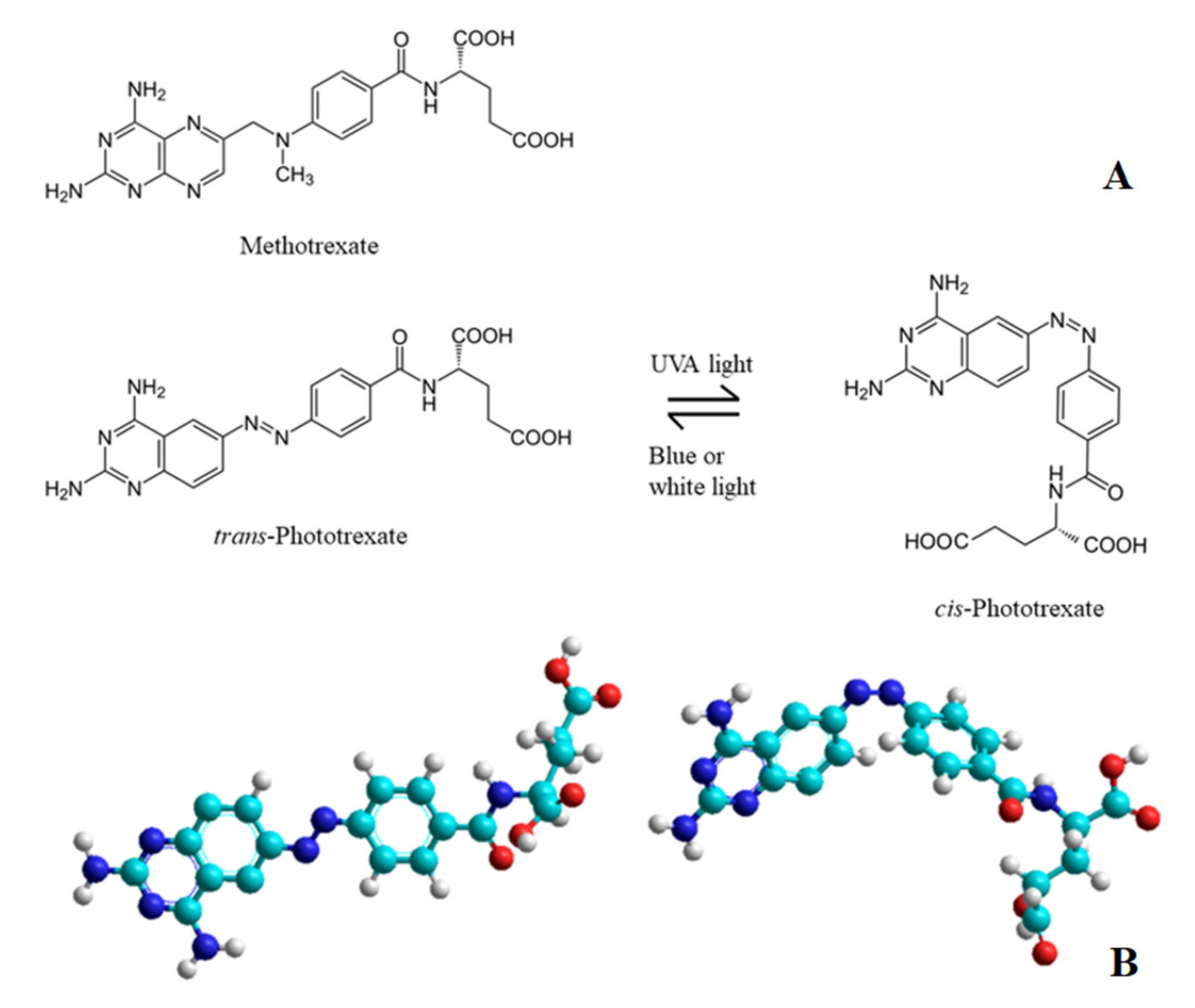
:1. Introduction
2. Results
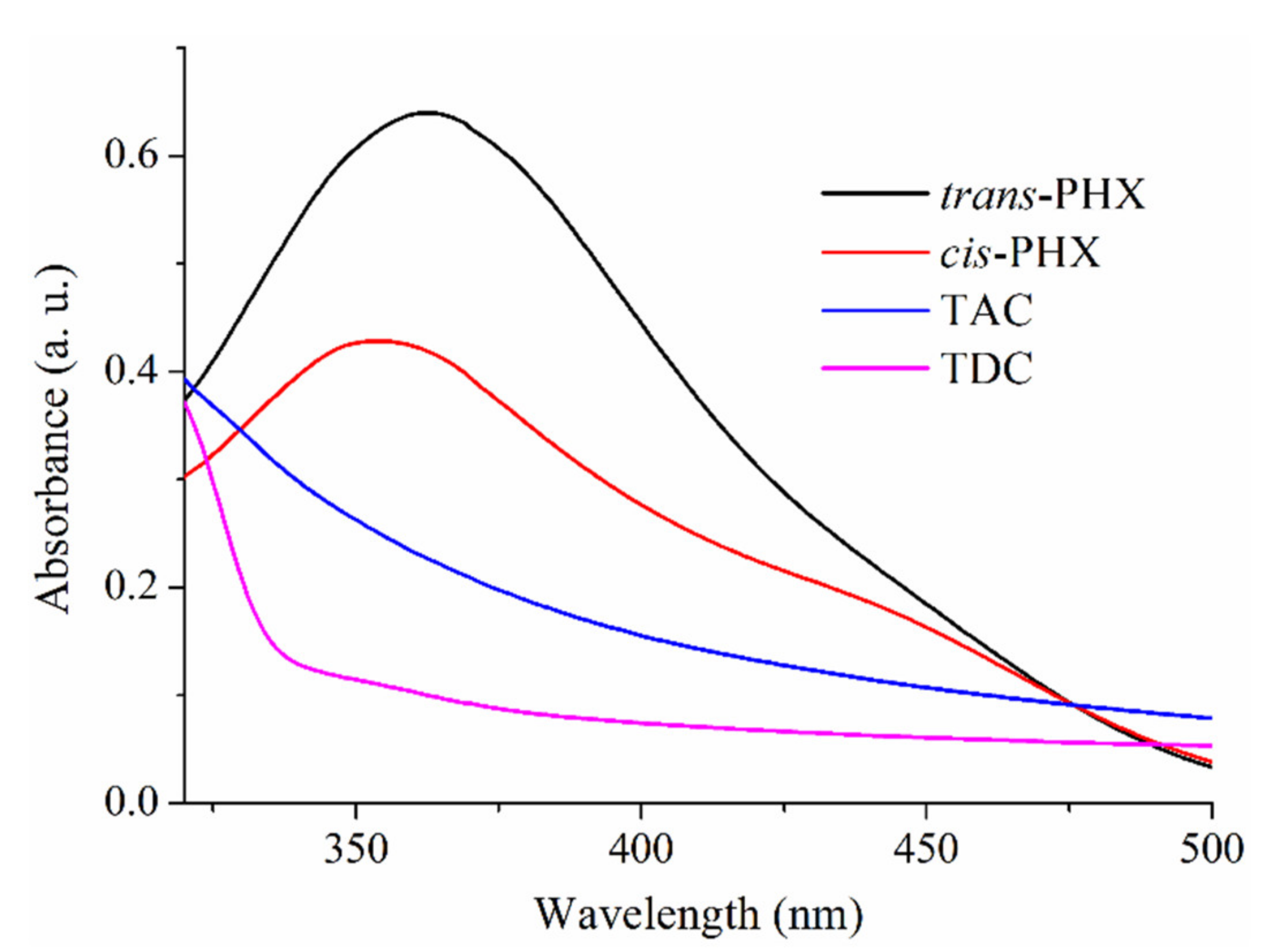
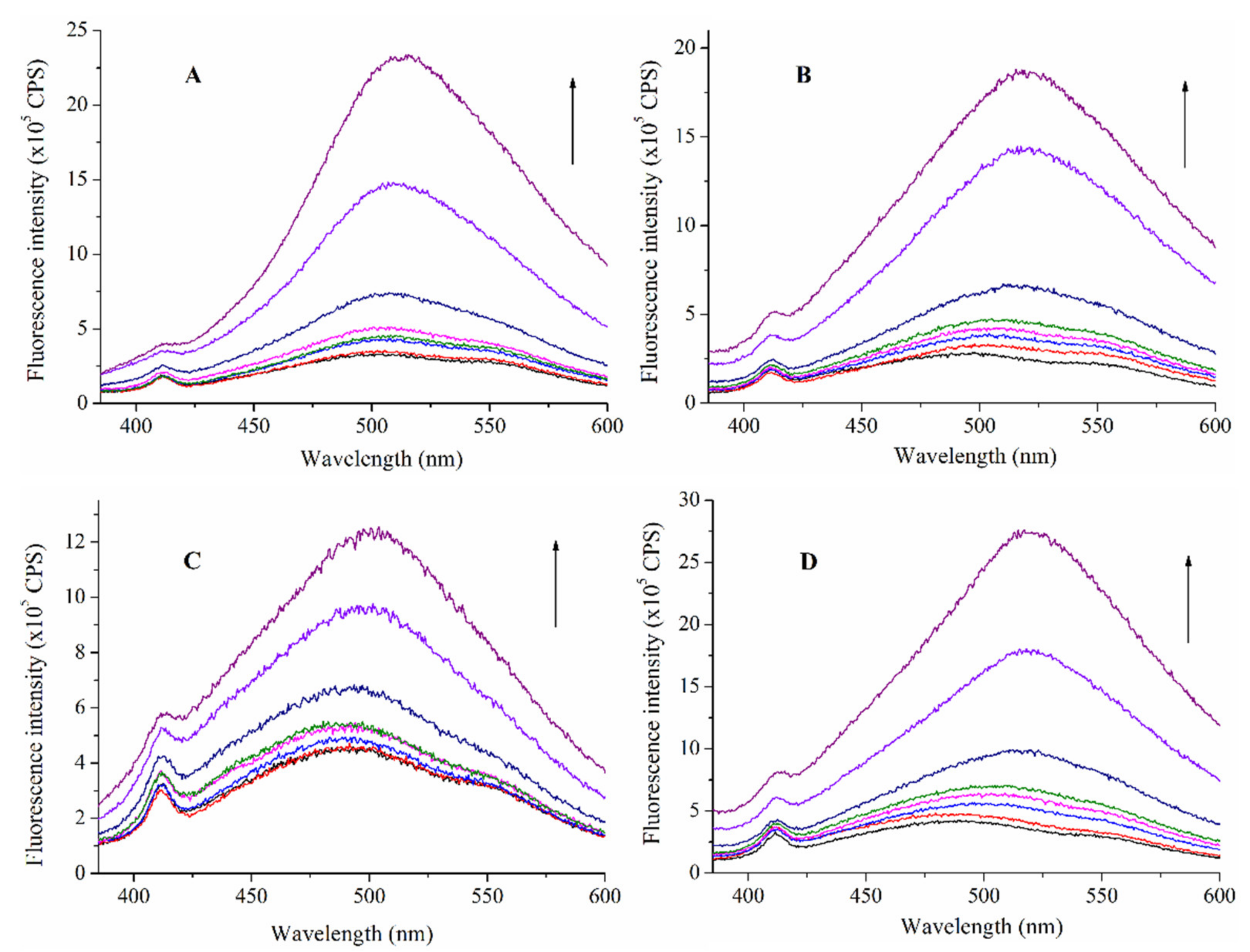
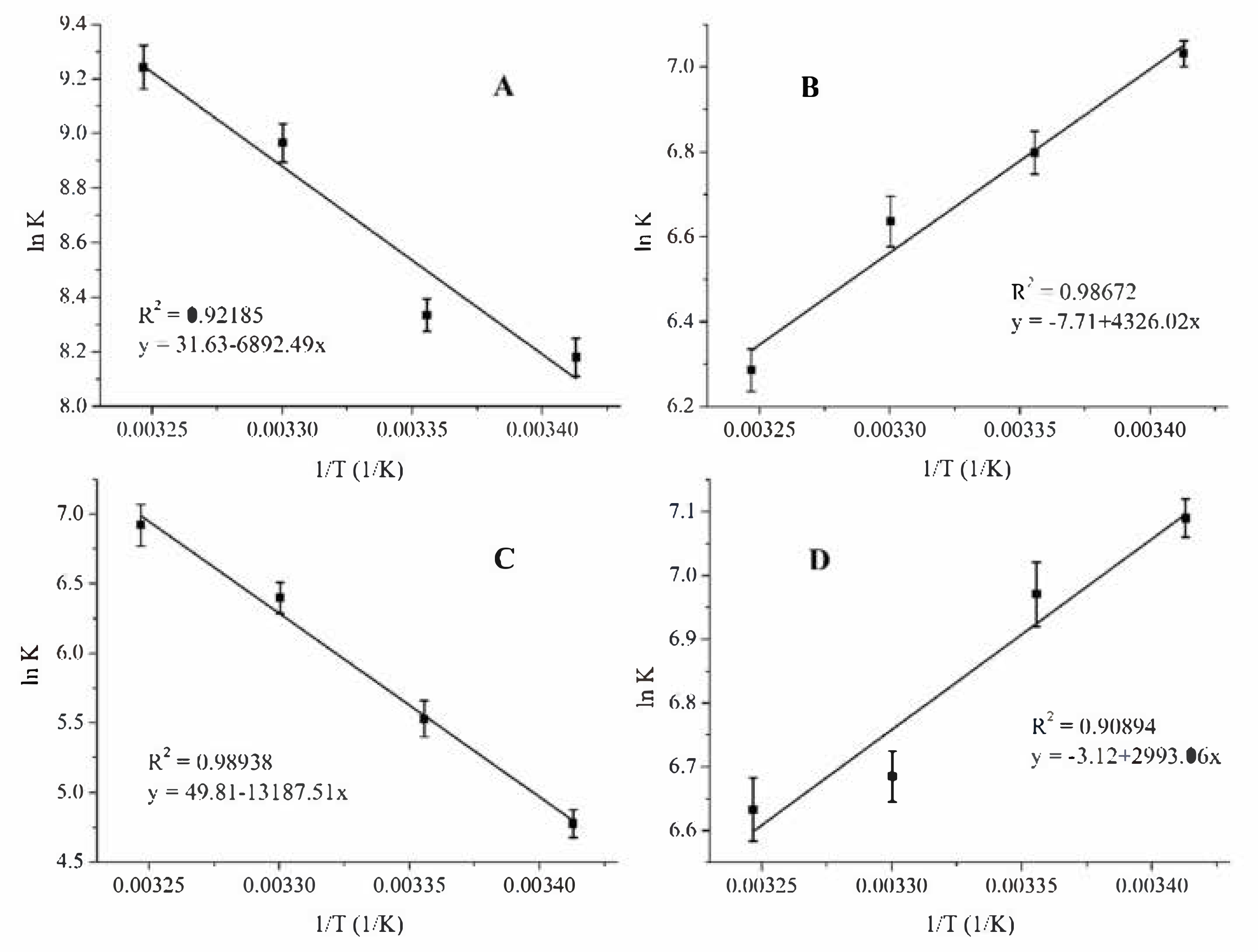
2.1. Determination of the Association Constants
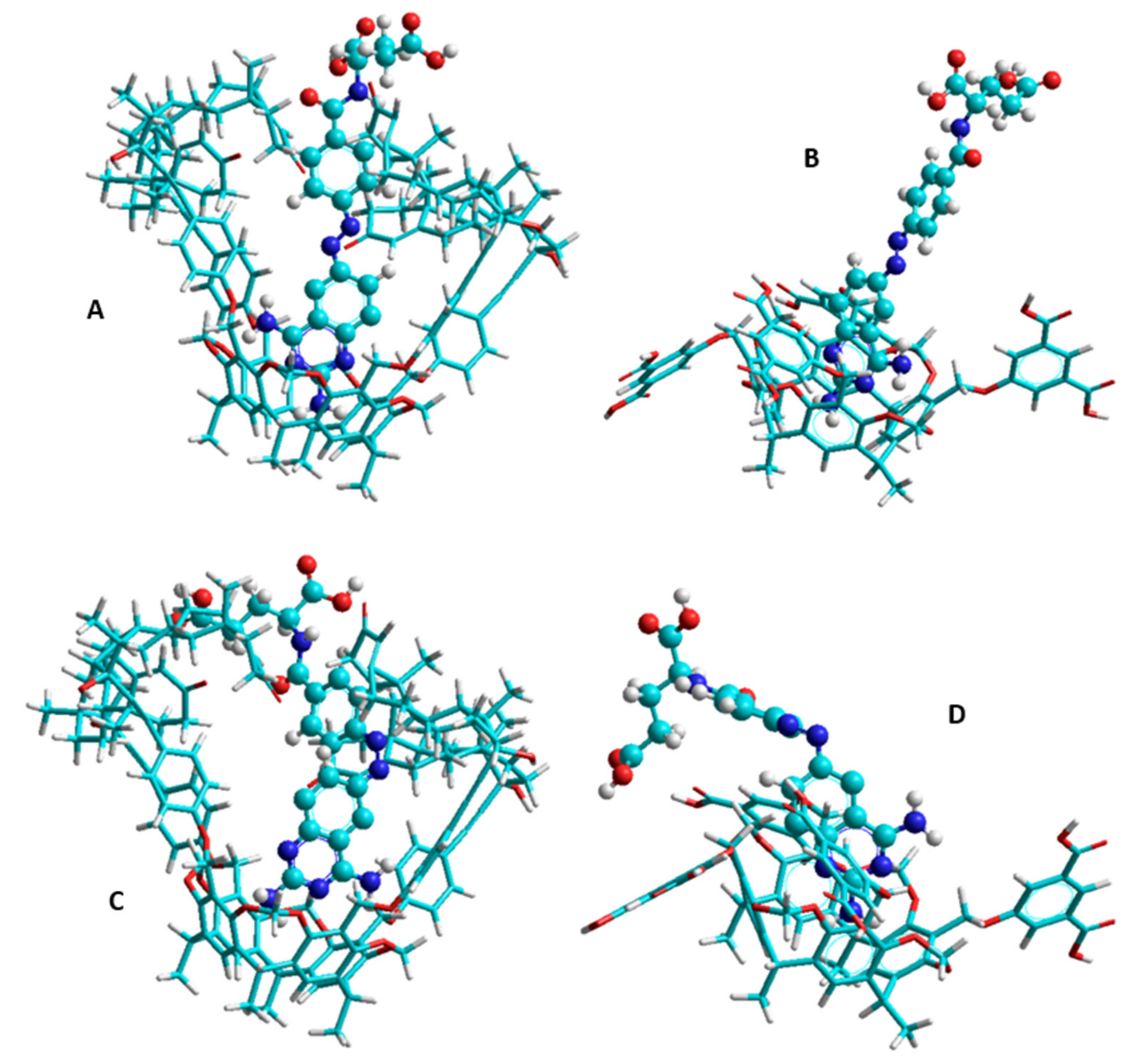
2.2. Modelling Studies
3. Discussion
4. Materials and Methods
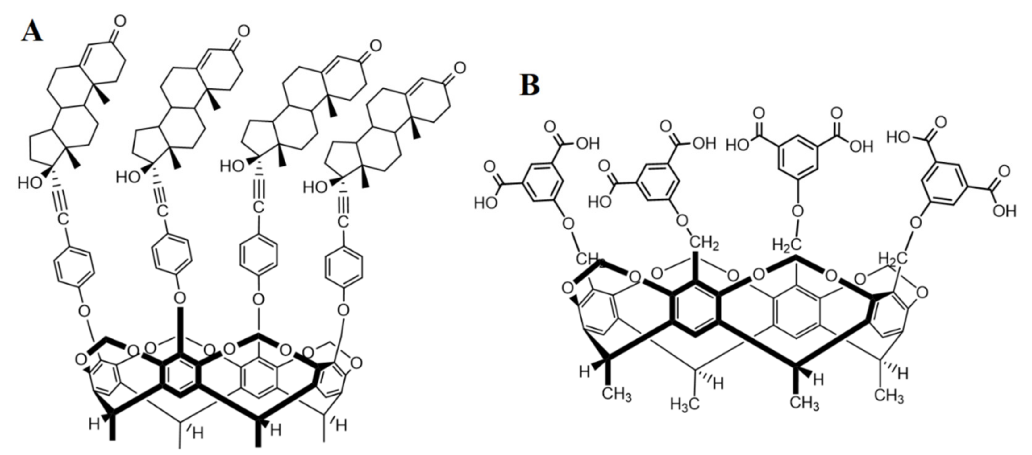
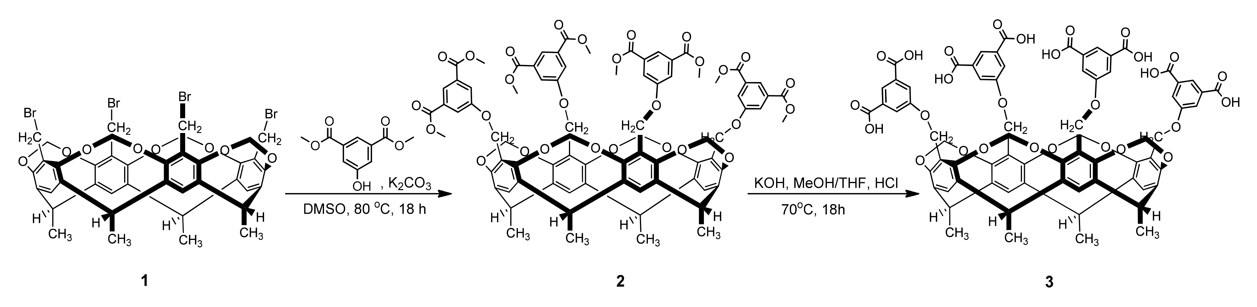
4.1. Synthesis of Tetrakis(3,5-dicarboxylatophenoxy)-cavitand (TDC, 3)
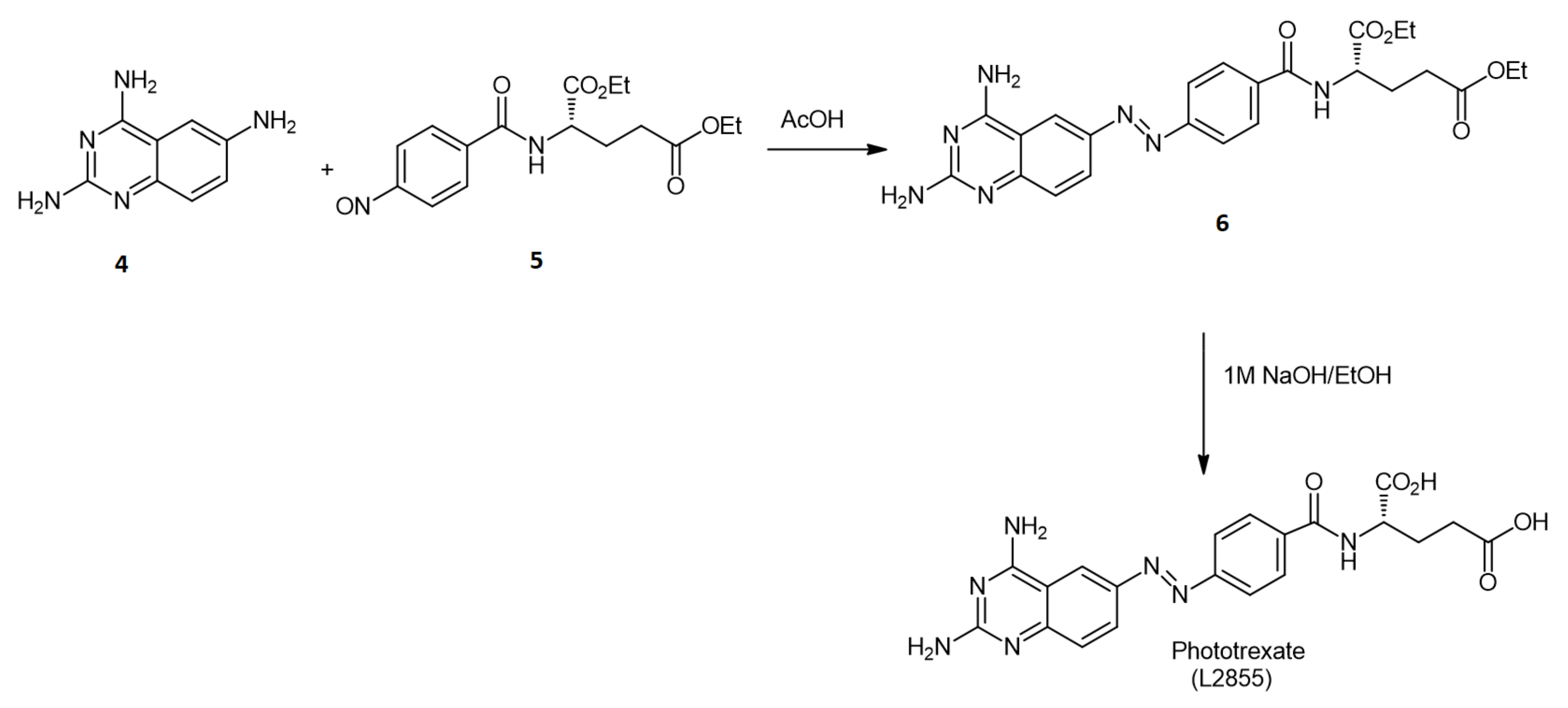
4.2. Synthesis of PHX
4.3. Other Chemicals and Instruments
4.4. Data Evaluation
4.5. Modelling
5. Conclusions
Author Contributions
Funding
Institutional Review Board Statement
Informed Consent Statement
Data Availability Statement
Acknowledgments
Conflicts of Interest
References
- Chabner, B.A.; Roberts, T.G. Chemotherapy and the war on cancer. Nat. Rev. Cancer 2005, 5, 65–72. [Google Scholar] [CrossRef] [PubMed]
- Shams, S.; Martinez, J.M.; Dawson, J.R.D.; Flores, J.; Gabriel, M.; Garcia, G.; Guevara, A.; Murray, K.; Pacifici, N.; Vargas, M.V.; et al. The Therapeutic Landscape of Rheumatoid Arthritis: Current Stateand Future Directions. Front. Pharmacol. 2021, 12, 1233. [Google Scholar] [CrossRef] [PubMed]
- Tian, H.; Cronstein, B.N. Understanding the mechanisms of action of methotrexate. Bull. NYU Hosp. Jt. Dis. 2007, 65, 168–173. Available online: http://metoject.ru/wp-content/uploads/files/public_en/34.pdf (accessed on 3 October 2021). [PubMed]
- Solomon, D.H.; Kremer, J.M.; Fisher, M.; Curtis, J.R.; Furer, V.; Harrold, L.R.; Hochberg, M.C.; Reed, G.; Tsao, P.; Greenberg, J.D. Comparative cancer risk associated with methotrexate, other non-biologic and biologic disease-modifying anti-rheumatic drugs. Semin. Arthritis Rheu. 2014, 43, 489–497. [Google Scholar] [CrossRef] [PubMed]
- Senapati, S.; Mahanta, A.K.; Kumar, S.; Maiti, P. Controlled drug delivery vehicles for cancer treatment and their performance. Signal Transduct. Target. Ther. 2018, 3, 7. [Google Scholar] [CrossRef] [Green Version]
- Baudino, T.A. Targeted Cancer Therapy: The Next Generation of Cancer Treatment. Curr. Drug Discov. Technol. 2015, 12, 3–20. [Google Scholar] [CrossRef]
- Broichhagen, J.; Frank, J.A.; Trauner, D. A Roadmap to Success in Photopharmacology. Acc. Chem. Res. 2015, 48, 1947–1960. [Google Scholar] [CrossRef]
- Reessing, F.; Szymanski, W. Beyond Photodynamic Therapy: Light-Activated Cancer Chemotherapy. Curr. Med. Chem. 2017, 24, 4905–4950. [Google Scholar] [CrossRef] [PubMed]
- Szymanski, W.; Beierle, J.M.; Kistemaker, H.A.; Velema, W.A.; Feringa, B.L. Reversible photocontrol of biological systems by the incorporation of molecular photoswitches. Chem. Rev. 2013, 113, 6114–6178. [Google Scholar] [CrossRef] [Green Version]
- Banghart, M.R.; Mourot, A.; Fortin, D.L.; Yao, J.Z.; Kramer, R.H.; Trauner, D. Photochromic blockers of voltage-gated potassium channels. Angew. Chem. Int. Ed. 2009, 48, 9097–9101. [Google Scholar] [CrossRef] [Green Version]
- Matera, C.; Gomila, A.M.J.; Camarero, N.; Libergoli, M.; Soler, C.; Gorostiza, P. Photoswitchable Antimetabolite for Targeted Photoactivated Chemotherapy. J. Am. Chem. Soc. 2018, 140, 15764–15773. [Google Scholar] [CrossRef] [PubMed] [Green Version]
- Mashita, T.; Kowada, T.; Takahashi, H.; Matsui, T.; Mizukami, S. Light-wavelength based Quantitative Control of Dihydrofolate Reductase Activity Using Photochromic Isostere of Inhibitor. ChemBioChem 2019, 20, 1382–1386. [Google Scholar] [CrossRef] [PubMed]
- Fahmy, S.A.; Brüßler, J.; Alawak, M.; El-Sayed, M.M.H.; Bakowsky, U.; Shoeib, T. Chemotherapy Based on Supramolecular Chemistry: A Promising Strategy in Cancer Therapy. Pharmaceutics 2019, 11, 292. [Google Scholar] [CrossRef] [PubMed] [Green Version]
- Hoskins, C.; Curtis, A.D.M. Simple Calix[n]arenes and Calix[4]resorcinarenes as Drug Solubilizing Agents. J. Nanomed. Res. 2015, 2, 00028. [Google Scholar] [CrossRef]
- Neda, I.; Vollbrecht, A.; Grunenberg, J.; Schmutzler, R. Functionalization of the Periphery of Calix[4]resorcinarenes with P(III)-containing Substituents via Hydroxy-, Trimethylsiloxy- and Ethoxy-Tethered Trimethylsiloxy Intermediates. Heteroat. Chem. 1998, 9, 553–558. [Google Scholar] [CrossRef]
- Franz, M.H.; Iorga, M.; Maftei, C.V.; Maftei, E.; Neda, I. Studies on the constituents of Helleborus purpurascens: Use of derivatives from calix[6]arene, homooxa-calix[3]arene and homoazacalix[3]arene as extractant agents for amino acids from the aqueous extract. Amino Acids 2020, 52, 55–72. [Google Scholar] [CrossRef]
- Maftei, V.; Fodor, E.; Jones, P.G.; Franz, M.H.; Davidescu, C.M.; Neda, I. Asymmetric Calixarene Derivatives as Potential Hosts in Chiral Recognition Processes. Pure Appl. Chem. 2015, 87, 415–439. [Google Scholar] [CrossRef]
- Dieleman, B.; Matt, D.; Neda, I.; Schmutzler, R.; Harriman, A.; Yaftian, R. Hexahomotrioxacalix[3]arene: A scaffold for a C3-symmetric phosphine ligand that traps a hydro-rhodium fragment inside a molecular funnel. Chem. Commun. 1999, 1911–1912. [Google Scholar] [CrossRef]
- Kunze, C.; Selent, D.; Neda, I.; Schmuzler, R.; Spannenberg, A.; Börner, A. Synthesis of New Calix[4]arene-Based Phosphorus Ligands and Their Application in the Rh(I) Catalyzed Hydroformylation of 1-Octene. Heteroat. Chem. 2001, 12, 577–585. [Google Scholar] [CrossRef]
- Preisz, Z.; Nagymihály, Z.; Lemli, B.; Kollár, L.; Kunsági-Máté, S. Weak interaction of the antimetabolite drug methotrexate with a cavitand derivative. Int. J. Mol. Sci. 2020, 21, 4345. [Google Scholar] [CrossRef]
- Csók, Z.; Kégl, T.; Párkányi, L.; Varga, Á.; Kunsági-Máté, S.; Kollár, L. Facile, high yielding synthesis of deepened cavitands: A synthetic and theoretical study. Supramol. Chem. 2011, 23, 710–719. [Google Scholar] [CrossRef]
- Sorrell, T.N.; Pigge, F.C. A convenient synthesis of functionalized cavitands via free-radical bromination. J. Org. Chem. 1993, 58, 784–785. [Google Scholar] [CrossRef]
- Preisz, Z.; Hartvig, N.; Bognár, B.; Kálai, T.; Kunsági-Máté, S. Comparative EPR Study on the Scavenging Effect of Methotrexate with the Isomers of Its Photoswitchable Derivative. Pharmaceuticals 2021, 14, 665. [Google Scholar] [CrossRef] [PubMed]


| Temperature (K) | log K | |||
|---|---|---|---|---|
| trans-PHX-TAC | trans-PHX-TDC | cis-PHX-TAC | cis-PHX-TDC | |
| 293 | 3.55 ± 0.19 | 3.05 ± 0.15 | 2.07 ± 0.17 | 3.08 ± 0.25 |
| 298 | 3.62 ± 0.21 | 2.95 ± 0.19 | 2.40 ± 0.21 | 3.03 ± 0.21 |
| 303 | 3.89 ± 0.18 | 2.88 ± 0.17 | 2.78 ± 0.22 | 2.90 ± 0.22 |
| 308 | 4.01 ± 0.20 | 2.73 ± 0.16 | 3.01 ± 0.20 | 2.88 ± 0.18 |
| ΔH (kJ·mol−1) | ΔS (J·K−1·mol−1) | ΔG (kJ·mol−1) | |
|---|---|---|---|
| trans-PHX-TAC | 57.30 ± 2.84 | 262.97 ± 11.36 | −21.10 ± 4.03 |
| trans-PHX-TDC | −35.97 ± 3.69 | −64.10 ± 10.43 | −16.85 ± 3.21 |
| cis-PHX-TAC | 109.65 ± 6.93 | 414.12 ± 17.51 | −13.83 ± 3.35 |
| cis-PHX-TDC | −24.88 ± 3.20 | −25.94 ± 4.72 | −17.15 ± 3.62 |
| ΔH (kJ·mol−1) | ΔS (J·K−1·mol−1) | ΔG (kJ·mol−1) | |
|---|---|---|---|
| trans-PHX-TAC | 42.13 | 210.99 | −20.89 |
| trans-PHX-TDC | −33.63 | −58.13 | −16.29 |
| cis-PHX-TAC | 101.11 | 392.09 | −15.79 |
| cis-PHX-TDC | −23.54 | −22.93 | −16.70 |
Publisher’s Note: MDPI stays neutral with regard to jurisdictional claims in published maps and institutional affiliations. |
© 2021 by the authors. Licensee MDPI, Basel, Switzerland. This article is an open access article distributed under the terms and conditions of the Creative Commons Attribution (CC BY) license (https://creativecommons.org/licenses/by/4.0/).
Share and Cite
Preisz, Z.; Nagymihály, Z.; Kollár, L.; Kálai, T.; Kunsági-Máté, S. Weak Interactions of the Isomers of Phototrexate and Two Cavitand Derivatives. Int. J. Mol. Sci. 2021, 22, 10764. https://0-doi-org.brum.beds.ac.uk/10.3390/ijms221910764
Preisz Z, Nagymihály Z, Kollár L, Kálai T, Kunsági-Máté S. Weak Interactions of the Isomers of Phototrexate and Two Cavitand Derivatives. International Journal of Molecular Sciences. 2021; 22(19):10764. https://0-doi-org.brum.beds.ac.uk/10.3390/ijms221910764
Chicago/Turabian StylePreisz, Zsolt, Zoltán Nagymihály, László Kollár, Tamás Kálai, and Sándor Kunsági-Máté. 2021. "Weak Interactions of the Isomers of Phototrexate and Two Cavitand Derivatives" International Journal of Molecular Sciences 22, no. 19: 10764. https://0-doi-org.brum.beds.ac.uk/10.3390/ijms221910764