Review

Neuron-Derived Estrogen—A Key Neuromodulator in Synaptic Function and Memory

1
Department of Neuroscience and Regenerative Medicine, Medical College of Georgia, Augusta University, Augusta, GA 30912, USA
2
Department of Neurosurgery, Medical College of Georgia, Augusta University, Augusta, GA 30912, USA
3
Department of Obstetrics and Gynecology, University of Texas Health, San Antonio, TX 78229, USA
4
Department of Neurology, Louisiana State University Health, Shreveport, LA 71103, USA
5
Audie L. Murphy Division, South Texas Veterans Health Care System, San Antonio, TX 78229, USA
*
Author to whom correspondence should be addressed.
Int. J. Mol. Sci. 2021, 22(24), 13242; https://0-doi-org.brum.beds.ac.uk/10.3390/ijms222413242
Submission received: 11 November 2021 / Revised: 29 November 2021 / Accepted: 4 December 2021 / Published: 8 December 2021
(This article belongs to the Special Issue Steroids, Central Neurotransmission, Mood, Memory and Mental State)

Abstract

:
In addition to being a steroid hormone, 17β-estradiol (E2) is also a neurosteroid produced in neurons in various regions of the brain of many species, including humans. Neuron-derived E2 (NDE2) is synthesized from androgen precursors via the action of the biosynthetic enzyme aromatase, which is located at synapses and in presynaptic terminals in neurons in both the male and female brain. In this review, we discuss evidence supporting a key role for NDE2 as a neuromodulator that regulates synaptic plasticity and memory. Evidence supporting an important neuromodulatory role of NDE2 in the brain has come from studies using aromatase inhibitors, aromatase overexpression in neurons, global aromatase knockout mice, and the recent development of conditional forebrain neuron-specific knockout mice. Collectively, these studies demonstrate a key role of NDE2 in the regulation of synapse and spine density, efficacy of excitatory synaptic transmission and long-term potentiation, and regulation of hippocampal-dependent recognition memory, spatial reference memory, and contextual fear memory. NDE2 is suggested to achieve these effects through estrogen receptor-mediated regulation of rapid kinase signaling and CREB-BDNF signaling pathways, which regulate actin remodeling, as well as transcription, translation, and transport of synaptic proteins critical for synaptic plasticity and function.

1. Introduction

17β-Estradiol (E2) is a steroid hormone synthesized from androgen precursors by the enzyme aromatase (Figure 1), which targets many different organs and tissues in the body, including the central nervous system (CNS) [1]. The neuromodulatory actions of E2 in the brain have been widely documented, including regulation of reproduction, sexual behavior, neuroplasticity, and cognition, as well as inflammation and neuroprotection [2,3,4,5,6,7,8,9]. Classically, the female ovary is considered as the main source of circulating E2 in the bloodstream. However, there are now abundant studies showing significant expression and activity of aromatase in different brain regions of most species studied to date, including rodents, monkeys, birds, amphibians, and humans [7,10,11,12,13]. Several approaches have been used to study the role of brain-derived estrogen (BDE2), including systemic or intracerebral administration of aromatase inhibitors (AI) (Figure 1), intracerebral administration of aromatase antisense oligonucleotides, global aromatase knockout mice, novel forebrain neuron-specific aromatase knockout (FBN-ARO-KO) mice, and astrocyte-specific aromatase knockout (GFAP-ARO-KO) mice. These studies have demonstrated that BDE2 regulates many brain functions and processes, including sexual differentiation, reproduction, socio-sexual behavior, synaptic plasticity, cognition, neuroinflammation, and neuroprotection. In this review, we discuss the evidence supporting a role for neuron-derived E2 (NDE2) in synaptic plasticity and memory.

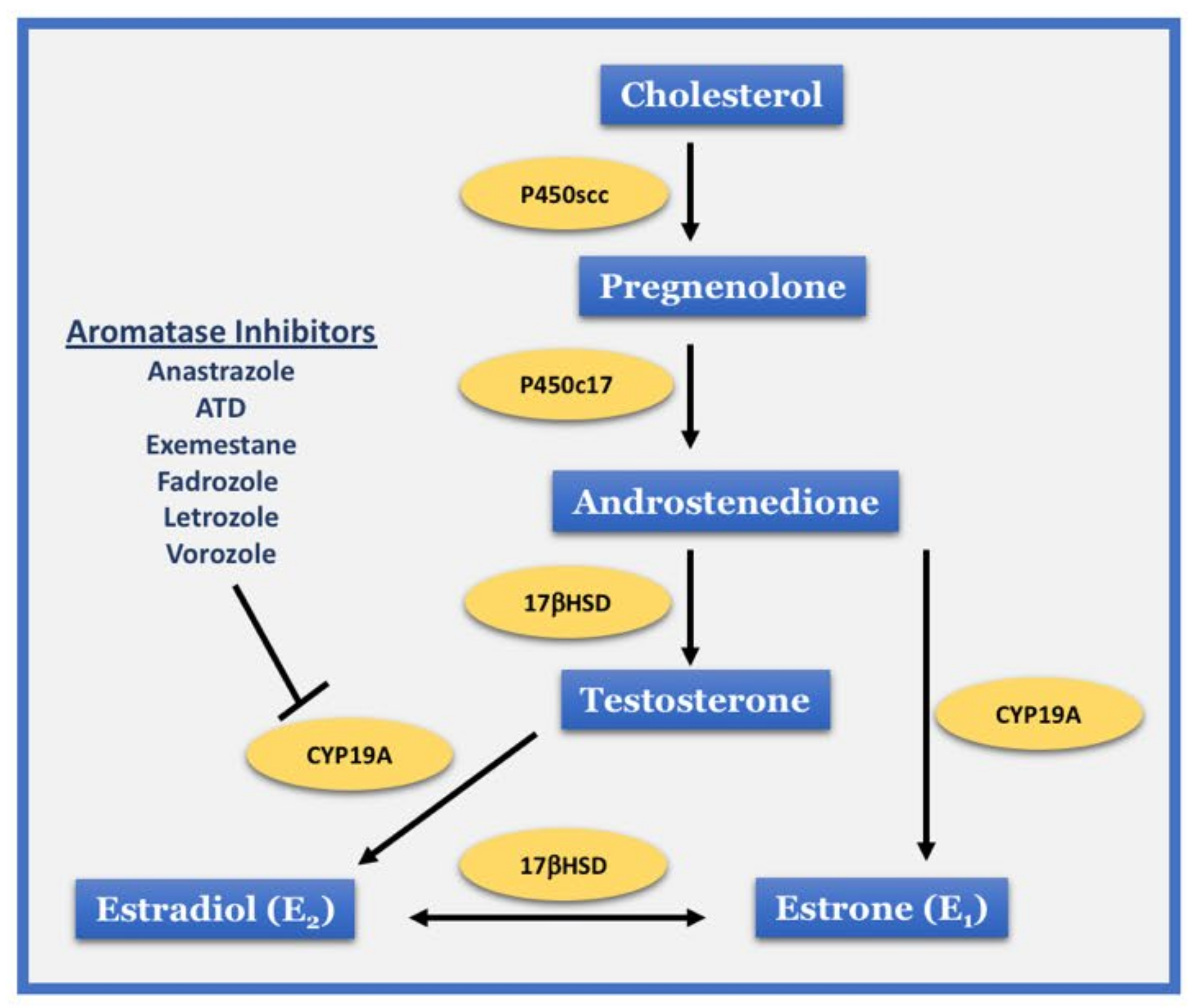
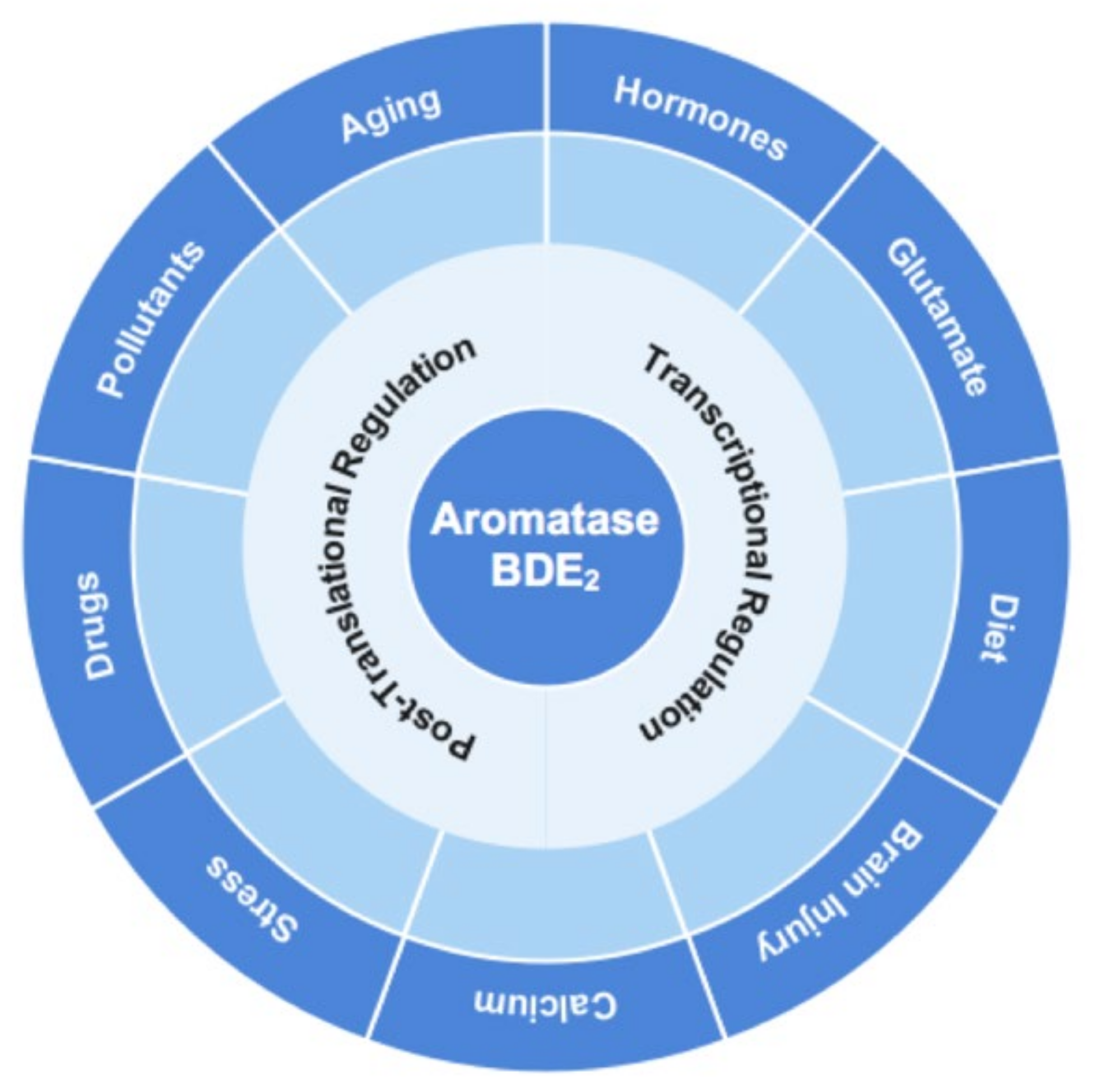
2. Aromatase Localization and Regulation in Neurons

Localization of aromatase in various brain areas has been examined by many groups since the early 1970s through the use of aromatase activity assays, RT-PCR, in situ hybridization, Western blot analysis, immunohistochemistry, and positron emission tomography (PET) imaging. These studies have revealed that, under normal physiological conditions, aromatase is expressed in neurons in many brain regions, including the hippocampus, hypothalamus, amygdala, thalamus, and cortex of both males and females in a variety of species [7,10,11,12,13,14,15,16,17]. In contrast, aromatase is essentially undetectable in astrocytes under basal conditions, but can be induced by cerebral ischemia, trauma, or neuroinflammation [8,18,19,20,21,22]. PET imaging in humans using labeled AI has further confirmed the localization of aromatase in the amygdala, thalamus, preoptic area, hippocampus, cortex, cerebellum, putamen, and white matter of both sexes in humans [23]. Additional work has shown that hippocampal neurons can generate significant quantities of E2, as evidenced by concentrations of E2 in the rat hippocampus ranging from 1 to 8 nM, which is significantly greater than E2 levels in the blood [11,24]. Aromatase has been shown to be localized throughout the neuronal cell body, including in dendrites and axons [15,25], as well as at synapses and presynaptic terminals in many species [11,15,24,26]. The synaptic localization of aromatase has led to the suggestion that NDE2 may act as a neurotransmitter/neuromodulator in the brain.
Multiple processes and factors regulate brain aromatase activity and NDE2 production (Figure 2).
Phosphorylation of aromatase has been proposed to be a key mechanism for rapid regulation of brain aromatase activity. Human aromatase has 19 putative phosphorylation sites, and aromatase activity in various species can be either increased or decreased rapidly by phosphorylation, with the effect observed depending upon the particular site phosphorylated [27,28,29,30]. Evidence for Ca2+-dependent phosphorylation of brain aromatase was first shown in quail and was associated with a rapid decrease of aromatase activity in hypothalamic homogenates and explants [31,32,33]. Furthermore, depletion of Ca2+ stores in rat hippocampal neurons and zebra finch forebrain resulted in an increase of E2 [34,35]. The neurotransmitter, glutamate has also been shown to rapidly regulate brain aromatase activity and local E2 levels, although divergent results have been reported in different species [11,36,37,38]. For instance, administration of glutamate or its agonists rapidly decreased both aromatase activity in quail hypothalamic explants [36], and local E2 levels in the caudomedial nidopallium of the zebra finch [37]. In contrast, in vivo administration of the glutamate agonist kainate doubled E2 release in the rat hippocampus [38], while the glutamate agonist NMDA strongly increased E2 release in rat hippocampal slices in vitro, an effect that was blocked by a NMDA receptor antagonist and dependent upon Ca2+ influx [11]. As the major excitatory neurotransmitter in the brain, glutamate regulation of NDE2 thus provides a mechanism for rapid changes in NDE2 that could serve to facilitate synaptic function and memory, which will be discussed in the next section. Finally, in contrast to glutamate regulation, cholinergic inputs to the hippocampus appear to have no role in the regulation of hippocampal NDE2, as cholinergic lesions and administration of cholinesterase inhibitors have no effect upon aromatase expression or activity in the hippocampus [39].
In addition to post-translational regulation, brain aromatase is also regulated at the transcriptional level. Binding sites for many regulatory factors have been identified in the brain aromatase gene, including for transcription factors such as the retinoic acid-related orphan receptor-alpha (RORA) [40], ARP-1 [41], Lhx2 [42], and retinoid X receptor [43]. Knockdown of the ARP-1 or Lhx2 gene in mouse neurons has been shown to significantly decrease aromatase expression [41,42]. Furthermore, overexpression of RORA increased aromatase expression ~10-fold in neuroblastoma cells [40], while treatment with bexarotene, a retinoid X receptor agonist, likewise significantly increased aromatase expression and E2 levels in hippocampal slices [43]. Hormones can also regulate aromatase at the transcriptional level. Studies using a mouse hypothalamic neuronal cell line revealed that estrogen receptor α (ERα) interacts with the brain-specific 1.f aromatase promoter, and that E2 treatment increases aromatase expression in an ER-dependent manner [44]. Furthermore, testosterone has been shown to increase aromatase mRNA levels and activity in the brain of multiple species [45,46,47,48,49,50]. Interestingly, glucocorticoids have been shown to increase aromatase mRNA and protein levels almost 100-fold in mouse hypothalamic neurons [51]. This may explain why stress, which increases release of the endogenous adrenal glucocorticoid, corticosterone, has likewise been shown to increase aromatase expression and local E2 levels in the hypothalamus of female rats [52]. In addition to steroid hormones, both in vitro and in vivo studies have demonstrated that the peptide hormone, the gonadotropin-releasing hormone (GnRH), can also increase E2 synthesis in the rat hippocampus [53]. The upregulation of NDE2 appears to underlie GnRH effects to enhance synaptic spine density and memory, as administration of letrozole blocks the plasticity and memory-enhancing effects of GnRH [54,55].
There is also evidence that factors such as diet, aging, drugs, and environmental pollutants can regulate brain aromatase. With respect to diet, flavonoids have been suggested to enhance aromatase levels and activity in the brain. Red wine, a well-known flavonoid, increased aromatase expression and activity in the hippocampus following chronic 8-week treatment in rats [56]. On the other hand, phytoestrogens, which are E2-like compounds found in plants, were found to have no effect on brain aromatase expression in rats [57,58]. Regarding aging, several studies have found that brain aromatase and E2 levels decline with aging. For instance, PET imaging revealed the decreased uptake of the labelled AI, 11C-vorozole, in the brains of aged men and women, which suggests that aging decreases aromatase levels in the human brain [59]. Similarly, aged female rats and mice were shown to have a significant reduction in aromatase expression and E2 levels in the hippocampus, as compared to young animals [60,61]. With respect to drug regulation of brain aromatase, nicotine has been shown to decrease forebrain aromatase activity in rats [62]. Furthermore, PET imaging using 11C-vorozole uptake in the brain suggests that nicotine also decreases aromatase in the non-human primate brain [63,64]. This finding raises the possibility that smoking may decrease brain E2 levels, although this remains to be determined. Antipsychotic drugs such as haloperidol and clozapine, but not olanzapine, were also found to decrease aromatase mRNA but not protein levels in the male rat brain after chronic treatment [65]. In contrast, long-term treatment with olanzapine and haloperidol increased aromatase expression in the male rat brainstem [66]. Since antipsychotics can regulate synaptic plasticity [67], it is possible regulation of NDE2 could contribute to antipsychotic effects upon synaptic plasticity. However, further studies are needed to measure antipsychotic effects upon local E2 levels and examine causality. Finally, a number of studies have found that environmental pollutants such as dioxin, tributyltin, and bisphenol A can increase brain aromatase expression in several species [68,69,70]. The reason for the increase in aromatase expression after environmental pollutant exposure is not clear, but it may be a protective mechanism against neuronal damage by the pollutants, as BDE2 has been shown to be neuroprotective against a variety of neural insults [8,20,21,71,72].

3. NDE2 Is an Important Modulator of Synaptic Plasticity and Cognition

3.1. Evidence from Aromatase Inhibitor Studies

Evidence for a role of NDE2 in synaptic plasticity was first suggested based on the results of administration of the AI, letrozole, which led to a significant decrease in hippocampal spine synapses and presynaptic boutons in female rat hippocampal slice cultures [73,74]. Additional studies using cultured rat hippocampal neurons confirmed that letrozole decreased spine density in rat hippocampal neurons [73] and in mouse mHippoE-14 hippocampal cells [75]. NDE2 may also have a role in preserving dendritic spines and mitochondrial structure in neurodegenerative disorders, as Aβ1−42-induced defects in dendritic spines, synaptic proteins, and mitochondrial structure were significantly exacerbated in hippocampal slice cultures when letrozole was combined with Aβ1−42 treatment [76]. Interestingly, some in vivo studies found AI effects upon hippocampal spine synapses in females, but not in males [77,78]. For example, systemic treatment with letrozole was reported to decrease hippocampal spine synapses in cycling and ovariectomized female mice and rats, but no significant effect was observed in males [77,78]. However, Zhao et al. [79] reported that letrozole treatment did significantly decrease hippocampal spines, synapses, and synaptic proteins in male mice, and the effect was correlated with a defect in spatial memory. Zhao et. al. [79] used a higher dose of letrozole than the previous studies, which may account for the difference in results.
AI treatment has also been shown to reduce the amplitude of long-term potentiation (LTP) in hippocampal slices from both male and female rats [80,81,82,83], as well as in slices from male rat striatum [84], brainstem [82], and cerebellum [85]. As an electrophysiological parameter of memory function, decreased LTP indicates impaired cognitive function. In support of this, several studies have reported that postmenopausal women with breast cancer who received letrozole treatment displayed a series of memory impairments, including deficits in executive function, processing speed, and verbal and visual learning and memory [86,87]. Furthermore, impairments in hippocampal-dependent memory after AI treatment in women was associated with decreased hippocampal activity during encoding [88]. Similar cognitive defects have also been reported with AI administration in other species. For example, inhibition of hippocampal E2 production by central AI administration in adult male zebra finch resulted in worse spatial memory acquisition, along with reduced levels of postsynaptic protein PSD95 [89,90,91]. Likewise, AI treatment in male and female mice and rats was associated with impairment of spatial learning and memory [53,79,92,93]. Additionally, a recent study examining recognition memory in mice found that hippocampal E2 levels are elevated within 30 min of novel object training, and letrozole treatment blocked this increase of local E2 levels and resulted in impaired hippocampal memory consolidation [94]. Notably, global aromatase knockout mice also display a pronounced cognitive defect, although the actions of ovarian and neuronal-derived estrogen cannot be easily differentiated in this model [95,96].

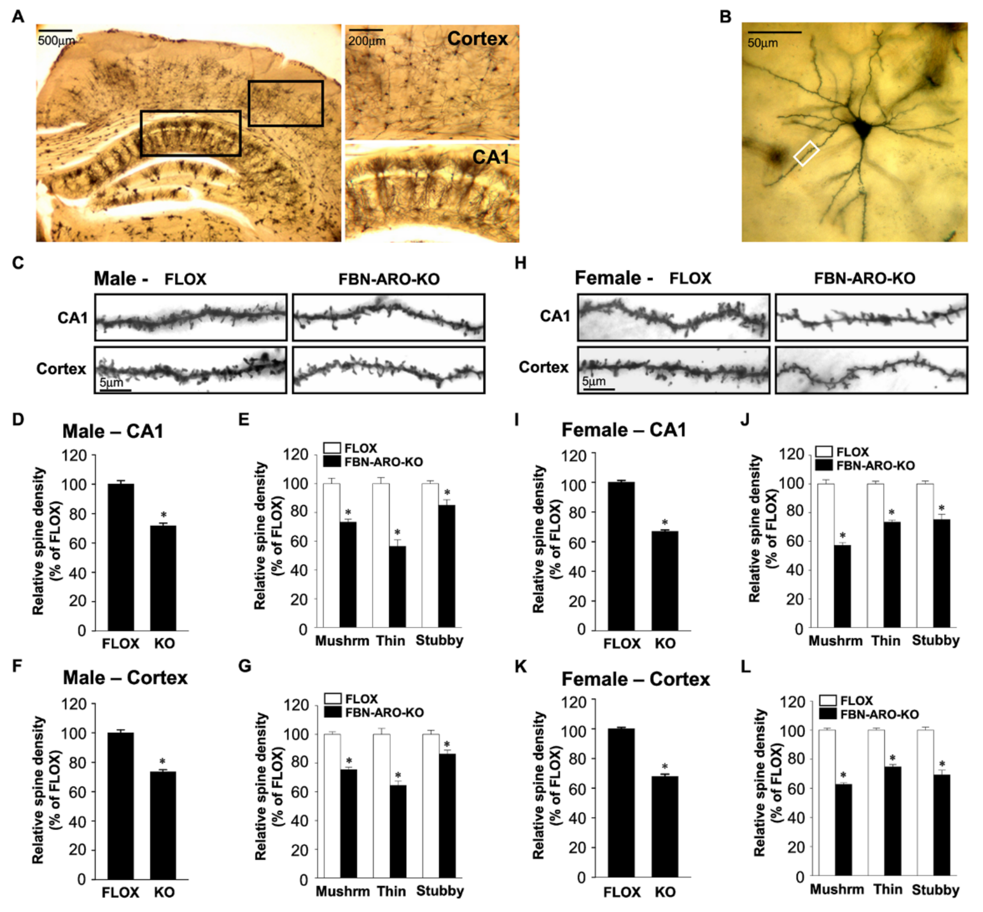
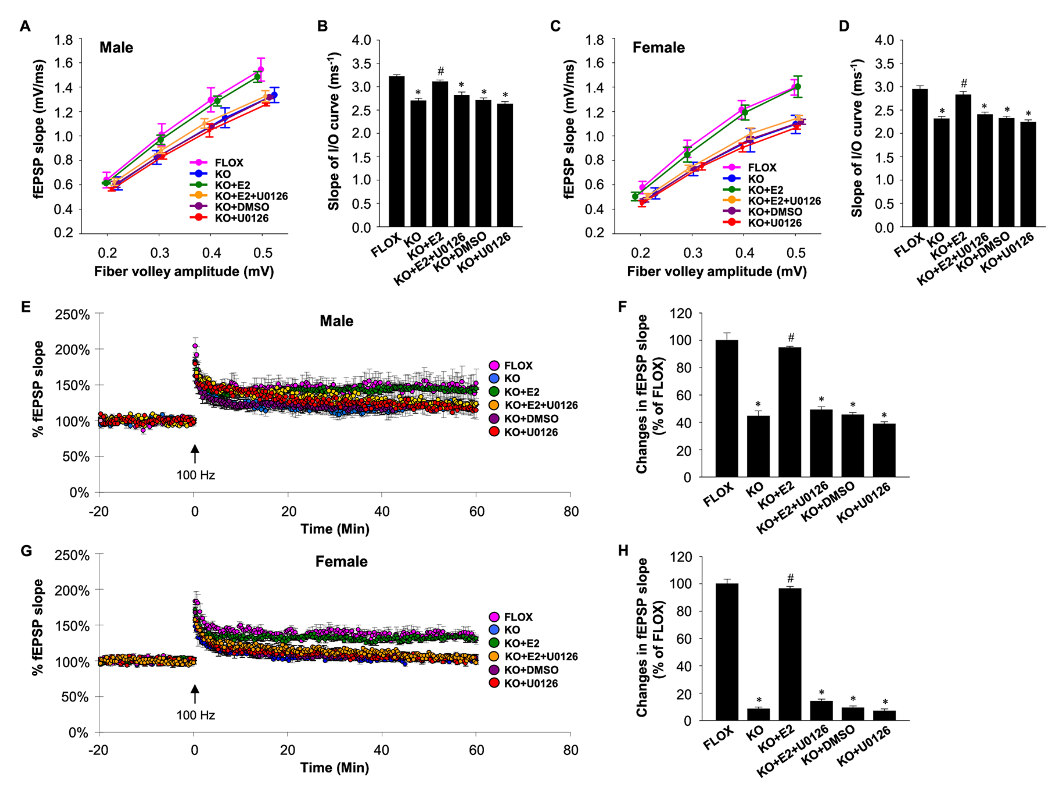
3.2. Evidence from Conditional Knockout Mouse Studies

To extend the previous AI studies and further elucidate the specific roles of NDE2 in the forebrain, our group created a forebrain neuron-specific aromatase knockout (FBN-ARO-KO) mouse model using cre-loxP recombination system in technology in which Cre is expressed under the control of the CaMKIIα promoter exclusively in forebrain excitatory neurons [7]. The conditional KO mice exhibited a robust 65%–80% decrease of aromatase and E2 levels in the hippocampus and cerebral cortex in both sexes, which was specific for the forebrain as there was no change of aromatase expression in the hindbrain or ovaries, and no change in E2 serum levels. As shown in Figure 3, detailed examination of FBN-ARO-KO mice revealed a significant decrease in spine density in the hippocampus and cerebral cortex of both intact male and ovariectomized female FBN-ARO-KO mice as compared to FLOX control mice [7].
Interestingly, of the three major spine types (thin, mushroom, and stubby), thin spines showed the highest decrease in the hippocampus and cerebral cortex of male FBN-ARO-KO mice, while mushroom spines were decreased the most in the ovariectomized FBN-ARO-KO female mice. Further work showed that synapse density was also significantly impaired in the hippocampal CA1 region and cerebral cortex of both intact male and ovariectomized female FBN-ARO-KO mice [7].
We further demonstrated that depletion of NDE2 in the forebrain of the FBN-ARO-KO male and ovariectomized female mice led to a decrease in efficacy of excitatory synaptic transmission and decreased LTP amplitude in hippocampal slices, which could be rapidly reinstated within minutes by E2 replacement [7] (Figure 4). While the decrease in LTP amplitude was highly significant in both sexes, the decrease was greatest in ovariectomized female mice as compared to male mice (e.g., 91% decrease in ovariectomized females versus 57% decrease in males as compared to FLOX controls) [7].
The defect in LTP amplitude in the FBN-ARO-KO mice was correlated with significant defects in hippocampal-dependent spatial memory, recognition memory, and contextual fear memory in intact male, ovariectomized female, and intact female FBN-ARO-KO mice [7]. The cognitive defects appeared to be due to loss of NDE2 as they could be rescued by reinstatement of forebrain E2 levels in the FBN-ARO-KO mice [7]. In contrast to the role of NDE2 in cognitive function, astrocyte-derived E2 (ADE2) does not appear to have a significant role in learning and memory as we found that male and female astrocyte-specific aromatase knockout (GFAP-ARO-KO) mice had normal hippocampal-dependent spatial and recognition memory and long-term fear memory [20].

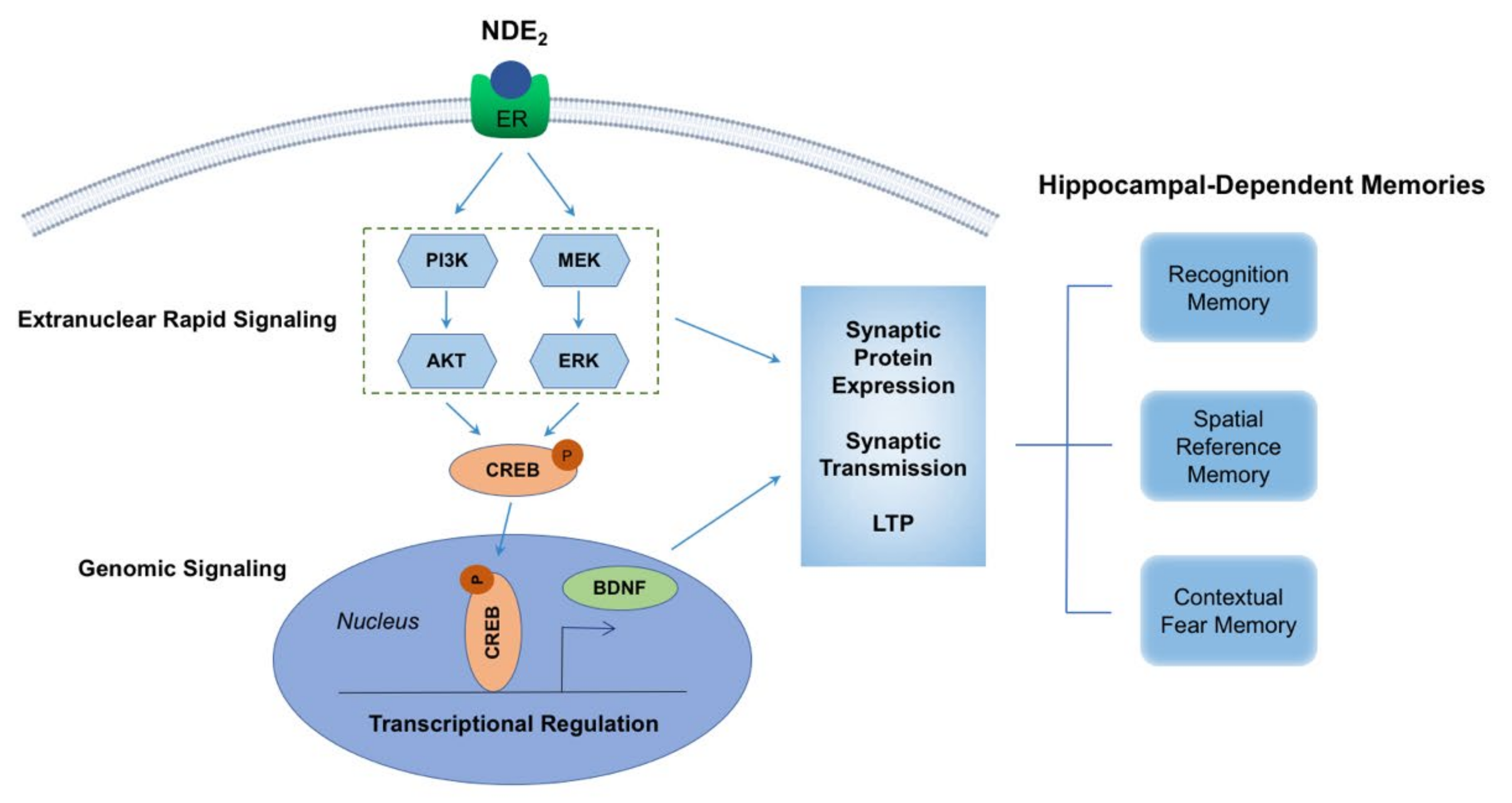
4. Mechanisms Underlying NDE2 Effects on Synaptic Plasticity and Memory

Several mechanisms have been implicated to underlie NDE2 regulation of synaptic plasticity and memory, including (1) regulation of actin cytoskeleton polymerization/depolymerization and PSD proteins, (2) enhancement of rapid MAPK/ERK and PI3K-AKT signaling, (3) regulation of CREB-BDNF signaling, and (4) mediation by estrogen receptors and the steroid nuclear receptor co-activator, SRC-1. In support of NDE2 regulation of actin polymerization/depolymerization, which is critical for spine formation, several studies found that letrozole treatment in mice resulted in disruption of actin cytoskeleton polymerization dynamics, which was associated with loss of hippocampal spine formation [75,77,97]. Letrozole was also shown to downregulate the actin remodeling proteins profilin-1, phospho-cofilin, and rictor in the mouse hippocampus and in mHippoE-14 hippocampal cells in vitro [75], indicating that NDE2 regulates actin polymerization dynamics through regulating expression and/or activity of these key actin remodeling proteins. Furthermore, aromatase overexpression in mHippoE-14 hippocampal cells was shown to induce a significant increase in actin polymerization and the PSD proteins, AMPA GluR1 receptor, and PSD95 [79], further supporting a role of NDE2 in regulating actin remodeling and PSD proteins. The aromatase overexpression effects on actin remodeling and PSD proteins were blocked by knockdown of SRC-1, a coactivator of steroid nuclear receptors [75,98], which suggests that the effects of NDE2 involve mediation by SRC-1. In addition, the involvement of SRC-1 suggests that steroid nuclear receptors play a role in mediating the plasticity effects of NDE2. In support of this suggestion, agonists of estrogen receptor α (ERα) enhanced spine synapse formation in rat hippocampal slices that were treated with letrozole [74]. In contrast, estrogen receptor β (ERβ) agonists reduced spine synapse formation, which suggests a possible antagonistic relationship may exist in the ER subtype control of hippocampal spine synapses [74]. Interestingly, we observed reduced ERα and elevated ERβ levels in the hippocampus and cerebral cortex of FBN-ARO-KO mice, which correlated with reduced spine and synapse density in the hippocampus of these mice [7]. Reinstatement of forebrain E2 levels significantly reversed these changes in ERα and ERβ in the hippocampus and cortex of FBN-ARO-KO mice, and rescued defects in synaptic proteins and cognitive function [7]. Since the ER changes in FBN-ARO-KO mice provide only correlational evidence of a role of ERs, more causative-type studies are needed, such as examining rescue effects of ER-selective agonists on synaptic plasticity and memory in FBN-ARO-KO mice.
Using FBN-ARO-KO mice, we further demonstrated that NDE2 is critical for activation of PI3K-AKT and MAPK/ERK rapid kinase signaling in the hippocampus and cerebral cortex of both male and female mice [7]. Furthermore, letrozole treatment has been shown to significantly decrease phospho-AKT levels in the hippocampus of mice, and this effect was reversed by E2 replacement [75]. Interestingly, both MAPK/ERK and PI3K-AKT signaling have been implicated to be key mediators of hippocampal neuroplasticity via their roles in the regulation of protein synthesis and transport of synaptic proteins that are necessary for synaptic plasticity [99,100,101,102,103,104]. Thus, NDE2 regulation of these rapid kinase signaling pathways is suggested to underlie its plasticity and memory effects. In support of this suggestion, we found that rapid ERK signaling is indeed critical for E2 rescue of LTP in FBN-ARO-KO mice, as administration of a MEK-ERK inhibitor blocked the ability of E2 to rescue LTP in hippocampal slices from FBN-ARO-KO mice (Figure 4) [7]. In addition, reinstatement of forebrain E2 levels rescued defects in phospho-AKT and phospho-ERK levels in the hippocampus and cerebral cortex of FBN-ARO-KO mice, and this effect was correlated with rescue of synaptic density [7]. While NDE2 is thought to exert its actions predominantly by extranuclear rapid signaling, findings in FBN-ARO-KO mice demonstrate that NDE2 also promotes CREB activation, which transactivates BDNF transcription [7]. As an important neurotrophin, BDNF is critical for synaptic protein expression and regulation of LTP via BDNF-TrkB signaling [105], and knockdown or knockout of BDNF has been shown to impair learning and memory [106,107,108]. While the above implicated signaling pathways could exist in parallel, it is most likely that the pathways crosstalk to mediate NDE2 actions and effects in the brain. Indeed, interactions and crosstalk are well known to exist between the pathways. For instance, BDNF is well known to enhance MAPK/ERK and AKT signaling in the brain to help exert its actions, while MAPK/ERK signaling in turn can phosphorylate CREB, which regulates transcription of many genes, including BDNF. A summary diagram of the proposed mechanisms underlying NDE2 regulation of synaptic plasticity and memory is provided in Figure 5.

5. Conclusions

In conclusion, studies discussed in this paper suggest that NDE2 functions as a neuromodulator to regulate neuronal synaptic plasticity and cognitive function. Despite some studies reporting sex differences, increasing evidence supports that NDE2 plays similar roles in both the male and female brain. NDE2 actions on synaptic plasticity and memory are suggested to be mediated via rapid kinase and CREB-BDNF signaling, which regulates actin remodeling, as well as transcription, translation, and transport of synaptic proteins involved in synaptic plasticity and cognitive function. While much has been learned regarding the roles and actions of NDE2 in the brain, much remains to be elucidated. Specifically, it is expected that continued advancement in technology and animal models will allow for a greater understanding of mechanisms underlying regulation of NDE2 in the brain as well as how it exerts its key effects on synaptic plasticity and memory. Furthermore, while most studies have focused on excitatory neurons and the hippocampus and cortex, NDE2 is also produced in other brain regions including the amygdala, cerebellum, and hypothalamus, as well as in inhibitory neurons, and thus studies to elucidate NDE2 roles and actions in these areas and neurons, as well as associated behavioral outcomes, are urgently needed.

Author Contributions

D.W.B. drafting and writing of manuscript, editing and revising manuscript, funding acquisition, and prepared figures. Y.L. drafting, editing and revising manuscript, and prepared figures; J.W. edited and revised manuscript; G.R.S. edited and revised manuscript; U.P.P. edited and revised manuscript; Q.Z. edited and revised manuscript; R.R.T. edited and revised manuscript. R.K.V. edited and revised manuscript and performed funding acquisition. All authors have read and agreed to the published version of the manuscript.

Funding

The authors research described in this review article was supported by Research Grant NS088058 from the National Institutes of Neurological Disorders and Stroke, National Institutes of Health.

Conflicts of Interest

The authors declare no conflict of interest.

References

  1. Nelson, L.R.; Bulun, S.E. Estrogen production and action. J. Am. Acad. Dermatol. 2001, 45 (Suppl. 3), S116–S124. [Google Scholar] [CrossRef]
  2. Spencer, J.L.; Waters, E.M.; Romeo, R.D.; Wood, G.E.; Milner, T.A.; McEwen, B.S. Uncovering the mechanisms of estrogen effects on hippocampal function. Front. Neuroendocrinol. 2008, 29, 219–237. [Google Scholar] [CrossRef] [Green Version]
  3. Terasawa, E. Neuroestradiol in regulation of GnRH release. Horm. Behav. 2018, 104, 138–145. [Google Scholar] [CrossRef]
  4. Simpkins, J.W.; Singh, M.; Brock, C.; Etgen, A.M. Neuroprotection and estrogen receptors. Neuroendocrinology 2012, 96, 119–130. [Google Scholar] [CrossRef]
  5. Brann, D.W.; Dhandapani, K.; Wakade, C.; Mahesh, V.B.; Khan, M.M. Neurotrophic and neuroprotective actions of estrogen: Basic mechanisms and clinical implications. Steroids 2007, 72, 381–405. [Google Scholar] [CrossRef] [Green Version]
  6. Mahesh, V.B.; Brann, D.W. Neuroendocrine mechanisms underlying the control of gonadotropin secretion by steroids. Steroids 1998, 63, 252–256. [Google Scholar] [CrossRef]
  7. Lu, Y.; Sareddy, G.R.; Wang, J.; Wang, R.; Li, Y.; Dong, Y.; Zhang, Q.; Liu, J.; O’Connor, J.C.; Xu, J.; et al. Neuron-Derived Estrogen Regulates Synaptic Plasticity and Memory. J. Neurosci. 2019, 39, 2792–2809. [Google Scholar] [CrossRef]
  8. Lu, Y.; Sareddy, G.R.; Wang, J.; Zhang, Q.; Tang, F.L.; Pratap, U.P.; Tekmal, R.R.; Vadlamudi, R.K.; Brann, D.W. Neuron-Derived Estrogen Is Critical for Astrocyte Activation and Neuroprotection of the Ischemic Brain. J. Neurosci. 2020, 40, 7355–7374. [Google Scholar] [CrossRef]
  9. Pedersen, A.L.; Nelson, L.H.; Saldanha, C.J. Centrally Synthesized Estradiol Is a Potent Anti-Inflammatory in the Injured Zebra Finch Brain. Endocrinology 2016, 157, 2041–2051. [Google Scholar] [CrossRef] [Green Version]
  10. Callard, G.V.; Petro, Z.; Ryan, K.J. Phylogenetic distribution of aromatase and other androgen-converting enzymes in the central nervous system. Endocrinology 1978, 103, 2283–2290. [Google Scholar] [CrossRef]
  11. Hojo, Y.; Hattori, T.A.; Enami, T.; Furukawa, A.; Suzuki, K.; Ishii, H.T.; Mukai, H.; Morrison, J.H.; Janssen, W.G.; Kominami, S.; et al. Adult male rat hippocampus synthesizes estradiol from pregnenolone by cytochromes P45017alpha and P450 aromatase localized in neurons. Proc. Natl. Acad. Sci. USA 2004, 101, 865–870. [Google Scholar] [CrossRef] [PubMed] [Green Version]
  12. MacLusky, N.J.; Naftolin, F.; Goldman-Rakic, P.S. Estrogen formation and binding in the cerebral cortex of the developing rhesus monkey. Proc. Natl. Acad. Sci. USA 1986, 83, 513–516. [Google Scholar] [CrossRef] [PubMed] [Green Version]
  13. Naftolin, F.; Ryan, K.J.; Petro, Z. Aromatization of androstenedione by limbic system tissue from human foetuses. J. Endocrinol. 1971, 51, 795–796. [Google Scholar] [CrossRef]
  14. Azcoitia, I.; Yague, J.G.; Garcia-Segura, L.M. Estradiol synthesis within the human brain. Neuroscience 2011, 191, 139–147. [Google Scholar] [CrossRef]
  15. Naftolin, F.; Horvath, T.L.; Jakab, R.L.; Leranth, C.; Harada, N.; Balthazart, J. Aromatase immunoreactivity in axon terminals of the vertebrate brain. An. immunocytochemical study on quail, rat, monkey and human tissues. Neuroendocrinology 1996, 63, 149–155. [Google Scholar] [CrossRef] [PubMed]
  16. Saldanha, C.J.; Tuerk, M.J.; Kim, Y.H.; Fernandes, A.O.; Arnold, A.P.; Schlinger, B.A. Distribution and regulation of telencephalic aromatase expression in the zebra finch revealed with a specific antibody. J. Comp. Neurol. 2000, 423, 619–630. [Google Scholar] [CrossRef]
  17. Stoffel-Wagner, B.; Watzka, M.; Schramm, J.; Bidlingmaier, F.; Klingmuller, D. Expression of CYP19 (aromatase) mRNA in different areas of the human brain. J. Steroid Biochem. Mol. Biol. 1999, 70, 237–241. [Google Scholar] [CrossRef]
  18. Negri Cesi, P.; Melcangi, R.C.; Celotti, F.; Martini, L. Aromatase activity in cultured brain cells: Difference between neurons and glia. Brain Res. 1992, 589, 327–332. [Google Scholar] [CrossRef]
  19. Negri Cesi, P.; Melcangi, R.C.; Celotti, F.; Martini, L. Distribution of aromatase activity in cultured neurons and glia cells. J. Steroid Biochem. Mol. Biol. 1993, 44, 637–639. [Google Scholar] [CrossRef]
  20. Wang, J.; Sareddy, G.R.; Lu, Y.; Pratap, U.P.; Tang, F.; Greene, K.M.; Meyre, P.L.; Tekmal, R.R.; Vadlamudi, R.K.; Brann, D.W. Astrocyte-Derived Estrogen Regulates Reactive Astrogliosis and is Neuroprotective following Ischemic Brain Injury. J. Neurosci. 2020, 40, 9751–9771. [Google Scholar] [CrossRef]
  21. Garcia-Segura, L.M.; Wozniak, A.; Azcoitia, I.; Rodriguez, J.R.; Hutchison, R.E.; Hutchison, J.B. Aromatase expression by astrocytes after brain injury: Implications for local estrogen formation in brain repair. Neuroscience 1999, 89, 567–578. [Google Scholar] [CrossRef]
  22. Pedersen, A.L.; Brownrout, J.L.; Saldanha, C.J. Neuroinflammation and neurosteroidogenesis: Reciprocal modulation during injury to the adult zebra finch brain. Physiol. Behav. 2018, 187, 51–56. [Google Scholar] [CrossRef] [PubMed]
  23. Biegon, A. In Vivo visualization of aromatase in animals and humans. Front. Neuroendocrinol. 2016, 40, 42–51. [Google Scholar] [CrossRef] [PubMed] [Green Version]
  24. Mukai, H.; Tsurugizawa, T.; Ogiue-Ikeda, M.; Murakami, G.; Hojo, Y.; Ishii, H.; Kimoto, T.; Kawato, S. Local neurosteroid production in the hippocampus: Influence on synaptic plasticity of memory. Neuroendocrinology 2006, 84, 255–263. [Google Scholar] [CrossRef] [PubMed]
  25. Naftolin, F. Brain aromatization of androgens. J. Reprod. Med. 1994, 39, 257–261. [Google Scholar] [PubMed]
  26. Srivastava, D.P.; Woolfrey, K.M.; Liu, F.; Brandon, N.J.; Penzes, P. Estrogen receptor-b activity modulates synaptic signaling and structure. J. Neurosci. 2010, 30, 13454–13460. [Google Scholar] [CrossRef] [PubMed] [Green Version]
  27. Ghosh, D.; Egbuta, C.; Kanyo, J.E.; Lam, T.T. Phosphorylation of human placental aromatase CYP19A1. Biochem. J. 2019, 476, 3313–3331. [Google Scholar] [CrossRef] [PubMed]
  28. Ghosh, D.; Griswold, J.; Erman, M.; Pangborn, W. Structural basis for androgen specificity and oestrogen synthesis in human aromatase. Nature 2009, 457, 219–223. [Google Scholar] [CrossRef] [Green Version]
  29. Miller, T.W.; Shin, I.; Kagawa, N.; Evans, D.B.; Waterman, M.R.; Arteaga, C.L. Aromatase is phosphorylated in situ at serine-118. J. Steroid Biochem. Mol. Biol. 2008, 112, 95–101. [Google Scholar] [CrossRef] [Green Version]
  30. Hovsepian-Ruby, L. Phosphorylation of Aromatase in the Zebra Finch Brain; University of California: Los Angeles, CA, USA, 2017; p. 19. [Google Scholar]
  31. Balthazart, J.; Baillien, M.; Ball, G.F. Interactions between kinases and phosphatases in the rapid control of brain aromatase. J. Neuroendocrinol. 2005, 17, 553–559. [Google Scholar] [CrossRef] [Green Version]
  32. Balthazart, J.; Baillien, M.; Ball, G.F. Phosphorylation processes mediate rapid changes of brain aromatase activity. J. Steroid Biochem. Mol. Biol. 2001, 79, 261–277. [Google Scholar] [CrossRef]
  33. Balthazart, J.; Baillien, M.; Ball, G.F. Rapid and reversible inhibition of brain aromatase activity. J. Neuroendocrinol. 2001, 13, 63–73. [Google Scholar] [PubMed]
  34. Fester, L.; Brandt, N.; Windhorst, S.; Prols, F.; Blaute, C.; Rune, G.M. Control. of aromatase in hippocampal neurons. J. Steroid Biochem. Mol. Biol. 2016, 160, 9–14. [Google Scholar] [CrossRef]
  35. Remage-Healey, L.; Dong, S.; Maidment, N.T.; Schlinger, B.A. Presynaptic control of rapid estrogen fluctuations in the songbird auditory forebrain. J. Neurosci. 2011, 31, 10034–10038. [Google Scholar] [CrossRef] [Green Version]
  36. Balthazart, J.; Baillien, M.; Ball, G.F. Rapid control of brain aromatase activity by glutamatergic inputs. Endocrinology 2006, 147, 359–366. [Google Scholar] [CrossRef]
  37. Remage-Healey, L.; Maidment, N.T.; Schlinger, B.A. Forebrain steroid levels fluctuate rapidly during social interactions. Nat. Neurosci. 2008, 11, 1327–1334. [Google Scholar] [CrossRef] [PubMed]
  38. Sato, S.M.; Woolley, C.S. Acute inhibition of neurosteroid estrogen synthesis suppresses status epilepticus in an animal model. Elife 2016, 5, e12917. [Google Scholar] [CrossRef] [Green Version]
  39. Li, J.; Rao, D.; Gibbs, R.B. Effects of Cholinergic Lesions and Cholinesterase Inhibitors on Aromatase and Estrogen Receptor Expression in Different Regions of the Rat Brain. Neuroscience 2018, 384, 203–213. [Google Scholar] [CrossRef]
  40. Sarachana, T.; Xu, M.; Wu, R.C.; Hu, V.W. Sex. hormones in autism: Androgens and estrogens differentially and reciprocally regulate RORA, a novel candidate gene for autism. PLoS ONE 2011, 6, e17116. [Google Scholar] [CrossRef]
  41. Honda, S.I.; Harada, N. ARP-1 Regulates the Transcriptional Activity of the Aromatase Gene in the Mouse Brain. Front. Endocrinol. 2020, 11, 306. [Google Scholar] [CrossRef]
  42. Honda, S.; Kozako, T.; Shimeno, H.; Soeda, S.; Harada, N. LIM-homeodomain transcription factor, Lhx2, is involved in transcriptional control of brain-specific promoter/exon 1f of the mouse aromatase gene. J. Neuroendocrinol. 2012, 24, 1367–1374. [Google Scholar] [CrossRef] [PubMed]
  43. Ishihara, Y.; Sakurai, H.; Oguro, A.; Tsuji, M.; Vogel, C.F.A.; Yamazaki, T. Retinoid X receptor-mediated neuroprotection via CYP19 upregulation and subsequent increases in estradiol synthesis. J. Steroid Biochem. Mol. Biol. 2019, 193, 105421. [Google Scholar] [CrossRef] [PubMed]
  44. Yilmaz, M.B.; Wolfe, A.; Cheng, Y.H.; Glidewell-Kenney, C.; Jameson, J.L.; Bulun, S.E. Aromatase promoter I.f is regulated by estrogen receptor alpha (ESR1) in mouse hypothalamic neuronal cell lines. Biol. Reprod. 2009, 81, 956–965. [Google Scholar] [CrossRef] [PubMed]
  45. Abdelgadir, S.E.; Resko, J.A.; Ojeda, S.R.; Lephart, E.D.; McPhaul, M.J.; Roselli, C.E. Androgens regulate aromatase cytochrome P450 messenger ribonucleic acid in rat brain. Endocrinology 1994, 135, 395–401. [Google Scholar] [CrossRef]
  46. Harada, N.; Yamada, K.; Foidart, A.; Balthazart, J. Regulation of aromatase cytochrome P-450 (estrogen synthetase) transcripts in the quail brain by testosterone. Brain Res. Mol. Brain Res. 1992, 15, 19–26. [Google Scholar] [CrossRef]
  47. Roselli, C.E.; Ellinwood, W.E.; Resko, J.A. Regulation of brain aromatase activity in rats. Endocrinology 1984, 114, 192–200. [Google Scholar] [CrossRef]
  48. Roselli, C.E.; Resko, J.A. Androgens regulate brain aromatase activity in adult male rats through a receptor mechanism. Endocrinology 1984, 114, 2183–2189. [Google Scholar] [CrossRef]
  49. Vockel, A.; Prove, E.; Balthazart, J. Effects of castration and testosterone treatment on the activity of testosterone-metabolizing enzymes in the brain of male and female zebra finches. J. NeuroBiol. 1990, 21, 808–825. [Google Scholar] [CrossRef]
  50. Roselli, C.E.; Resko, J.A. Testosterone regulates aromatase activity in discrete brain areas of male rhesus macaques. Biol. Reprod. 1989, 40, 929–934. [Google Scholar] [CrossRef] [Green Version]
  51. Brooks, D.C.; Zhao, H.; Yilmaz, M.B.; Coon, V.J.; Bulun, S.E. Glucocorticoid-induction of hypothalamic aromatase via its brain-specific promoter. Mol. Cell. Endocrinol. 2012, 362, 85–90. [Google Scholar] [CrossRef] [PubMed] [Green Version]
  52. Liu, J.; Hu, P.; Qi, X.R.; Meng, F.T.; Kalsbeek, A.; Zhou, J.N. Acute restraint stress increases intrahypothalamic oestradiol concentrations in conjunction with increased hypothalamic oestrogen receptor beta and aromatase mRNA expression in female rats. J. Neuroendocrinol. 2011, 23, 435–443. [Google Scholar] [CrossRef] [PubMed]
  53. Marbouti, L.; Zahmatkesh, M.; Riahi, E.; Sadr, S.S. Inhibition of brain 17beta-estradiol synthesis by letrozole induces cognitive decline in male and female rats. NeuroBiol. Learn. Mem. 2020, 175, 107300. [Google Scholar] [CrossRef] [PubMed]
  54. Nelson, B.S.; Black, K.L.; Daniel, J.M. Circulating Estradiol Regulates Brain-Derived Estradiol via Actions at GnRH Receptors to Impact Memory in Ovariectomized Rats. eNeuro 2016, 3. [Google Scholar] [CrossRef] [Green Version]
  55. Prange-Kiel, J.; Dudzinski, D.A.; Prols, F.; Glatzel, M.; Matschke, J.; Rune, G.M. Aromatase Expression in the Hippocampus of AD Patients and 5xFAD Mice. Neural Plast. 2016, 2016, 9802086. [Google Scholar] [CrossRef] [Green Version]
  56. Monteiro, R.; Faria, A.; Mateus, N.; Calhau, C.; Azevedo, I. Red wine interferes with oestrogen signalling in rat hippocampus. J. Steroid Biochem. Mol. Biol. 2008, 111, 74–79. [Google Scholar] [CrossRef]
  57. Lephart, E.D.; Adlercreutz, H.; Lund, T.D. Dietary soy phytoestrogen effects on brain structure and aromatase in Long-Evans rats. Neuroreport 2001, 12, 3451–3455. [Google Scholar] [CrossRef] [PubMed]
  58. Weber, K.S.; Setchell, K.D.; Lephart, E.D. Maternal and perinatal brain aromatase: Effects of dietary soy phytoestrogens. Brain Res. Dev. Brain Res. 2001, 126, 217–221. [Google Scholar] [CrossRef]
  59. Biegon, A.; Alexoff, D.L.; Kim, S.W.; Logan, J.; Pareto, D.; Schlyer, D.; Wang, G.J.; Fowler, J.S. Aromatase imaging with [N-methyl-11C]vorozole PET in healthy men and women. J. Nucl. Med. 2015, 56, 580–585. [Google Scholar] [CrossRef] [Green Version]
  60. Chamniansawat, S.; Sawatdiyaphanon, C. Age-Related Memory Impairment Associated With Decreased Endogenous Estradiol in the Hippocampus of Female Rats. Int. J. Toxicol. 2018, 37, 207–215. [Google Scholar] [CrossRef]
  61. Zhao, Y.; He, L.; Zhang, Y.; Zhao, J.; Liu, Z.; Xing, F.; Liu, M.; Feng, Z.; Li, W.; Zhang, J. Estrogen receptor alpha and beta regulate actin polymerization and spatial memory through an SRC-1/mTORC2-dependent pathway in the hippocampus of female mice. J. Steroid Biochem. Mol. Biol. 2017, 174, 96–113. [Google Scholar] [CrossRef] [PubMed]
  62. Von Ziegler, N.I.; Schlumpf, M.; Lichtensteiger, W. Prenatal nicotine exposure selectively affects perinatal forebrain aromatase activity and fetal adrenal function in male rats. Brain Res. Dev. Brain Res. 1991, 62, 23–31. [Google Scholar] [CrossRef]
  63. Biegon, A.; Alia-Klein, N.; Fowler, J.S. Potential contribution of aromatase inhibition to the effects of nicotine and related compounds on the brain. Front. Pharm. 2012, 3, 185. [Google Scholar] [CrossRef] [PubMed] [Green Version]
  64. Biegon, A.; Kim, S.W.; Logan, J.; Hooker, J.M.; Muench, L.; Fowler, J.S. Nicotine blocks brain estrogen synthase (aromatase): In Vivo positron emission tomography studies in female baboons. Biol. Psychiatry 2010, 67, 774–777. [Google Scholar] [CrossRef] [Green Version]
  65. Bogus, K.; Palasz, A.; Suszka-Switek, A.; Worthington, J.J.; Krzystanek, M.; Wiaderkiewicz, R. Chronic Antipsychotic Treatment Modulates Aromatase (CYP19A1) Expression in the Male Rat Brain. J. Mol. Neurosci. 2019, 68, 311–317. [Google Scholar] [CrossRef] [Green Version]
  66. Bogus, K.; Zarczynska, M.; Palasz, A.; Suszka-Switek, A.; Worthington, J.J.; Krzystanek, M.; Zarczynski, P. Antipsychotics increase steroidogenic enzyme gene expression in the rat brainstem. Mol. Biol. Rep. 2021. Online ahead of print. [Google Scholar] [CrossRef]
  67. Dzyubenko, E.; Juckel, G.; Faissner, A. The antipsychotic drugs olanzapine and haloperidol modify network connectivity and spontaneous activity of neural networks in vitro. Sci. Rep. 2017, 7, 11609. [Google Scholar] [CrossRef] [Green Version]
  68. Munetsuna, E.; Hattori, M.; Yamazaki, T. Stimulation of estradiol biosynthesis by tributyltin in rat hippocampal slices. Endocr. Res. 2014, 39, 168–172. [Google Scholar] [CrossRef] [PubMed]
  69. Tan, W.; Wong, T.Y.; Wang, Y.; Huang, J.; Leung, L.K. CYP19 expression is induced by 2,3,7,8-tetrachloro-dibenzo-para-dioxin in human glioma cells. Mol. Cell. Endocrinol. 2013, 375, 106–112. [Google Scholar] [CrossRef]
  70. Chung, E.; Genco, M.C.; Megrelis, L.; Ruderman, J.V. Effects of bisphenol A and triclocarban on brain-specific expression of aromatase in early zebrafish embryos. Proc. Natl. Acad. Sci. USA 2011, 108, 17732–17737. [Google Scholar] [CrossRef] [Green Version]
  71. Garcia-Segura, L.M.; Veiga, S.; Sierra, A.; Melcangi, R.C. Aromatase: A neuroprotective enzyme. Prog. NeuroBiol. 2003, 71, 31–41. [Google Scholar] [CrossRef] [PubMed] [Green Version]
  72. Duncan, K.A.; Saldanha, C.J. Central aromatization: A dramatic and responsive defense against threat and trauma to the vertebrate brain. Front. Neuroendocrinol. 2020, 56, 100816. [Google Scholar] [CrossRef] [PubMed]
  73. Kretz, O.; Fester, L.; Wehrenberg, U.; Zhou, L.; Brauckmann, S.; Zhao, S.; Prange-Kiel, J.; Naumann, T.; Jarry, H.; Frotscher, M.; et al. Hippocampal synapses depend on hippocampal estrogen synthesis. J. Neurosci. 2004, 24, 5913–5921. [Google Scholar] [CrossRef] [Green Version]
  74. Zhou, L.; Fester, L.; Haghshenas, S.; de Vrese, X.; von Hacht, R.; Gloger, S.; Brandt, N.; Bader, M.; Vollmer, G.; Rune, G.M. Oestradiol-induced synapse formation in the female hippocampus: Roles of oestrogen receptor subtypes. J. Neuroendocrinol. 2014, 26, 439–447. [Google Scholar] [CrossRef]
  75. Zhao, Y.; Yu, Y.; Zhang, Y.; He, L.; Qiu, L.; Zhao, J.; Liu, M.; Zhang, J. Letrozole regulates actin cytoskeleton polymerization dynamics in a SRC-1 dependent manner in the hippocampus of mice. J. Steroid Biochem. Mol. Biol. 2017, 167, 86–97. [Google Scholar] [CrossRef] [PubMed] [Green Version]
  76. Chang, P.K.; Boridy, S.; McKinney, R.A.; Maysinger, D. Letrozole Potentiates Mitochondrial and Dendritic Spine Impairments Induced by beta Amyloid. J. Aging Res. 2013, 2013, 538979. [Google Scholar] [CrossRef] [PubMed] [Green Version]
  77. Fester, L.; Prange-Kiel, J.; Zhou, L.; Blittersdorf, B.V.; Bohm, J.; Jarry, H.; Schumacher, M.; Rune, G.M. Estrogen-regulated synaptogenesis in the hippocampus: Sexual dimorphism in vivo but not in vitro. J. Steroid Biochem. Mol. Biol. 2012, 131, 24–29. [Google Scholar] [CrossRef] [PubMed]
  78. Zhou, L.; Fester, L.; Haghshenas, S.; de Vrese, X.; von Hacht, R.; Gloger, S.; Brandt, N.; Bader, M.; Vollmer, G.; Rune, G.M. Aromatase inhibitors induce spine synapse loss in the hippocampus of ovariectomized mice. Endocrinology 2010, 151, 1153–1160. [Google Scholar] [CrossRef] [Green Version]
  79. Zhao, J.; Bian, C.; Liu, M.; Zhao, Y.; Sun, T.; Xing, F.; Zhang, J. Orchiectomy and letrozole differentially regulate synaptic plasticity and spatial memory in a manner that is mediated by SRC-1 in the hippocampus of male mice. J. Steroid Biochem. Mol. Biol. 2018, 178, 354–368. [Google Scholar] [CrossRef] [PubMed]
  80. Di Mauro, M.; Tozzi, A.; Calabresi, P.; Pettorossi, V.E.; Grassi, S. Neo-synthesis of estrogenic or androgenic neurosteroids determine whether long-term potentiation or depression is induced in hippocampus of male rat. Front. Cell. Neurosci. 2015, 9, 376. [Google Scholar] [CrossRef] [Green Version]
  81. Di Mauro, M.; Tozzi, A.; Calabresi, P.; Pettorossi, V.E.; Grassi, S. Different synaptic stimulation patterns influence the local androgenic and estrogenic neurosteroid availability triggering hippocampal synaptic plasticity in the male rat. Eur. J. Neurosci. 2017, 45, 499–509. [Google Scholar] [CrossRef] [PubMed]
  82. Grassi, S.; Tozzi, A.; Costa, C.; Tantucci, M.; Colcelli, E.; Scarduzio, M.; Calabresi, P.; Pettorossi, V.E. Neural 17beta-estradiol facilitates long-term potentiation in the hippocampal CA1 region. Neuroscience 2011, 192, 67–73. [Google Scholar] [CrossRef] [PubMed]
  83. Vierk, R.; Brandt, N.; Rune, G.M. Hippocampal estradiol synthesis and its significance for hippocampal synaptic stability in male and female animals. Neuroscience 2014, 274, 24–32. [Google Scholar] [CrossRef] [PubMed]
  84. Tozzi, A.; de Iure, A.; Tantucci, M.; Durante, V.; Quiroga-Varela, A.; Giampa, C.; Di Mauro, M.; Mazzocchetti, P.; Costa, C.; Di Filippo, M. Endogenous 17beta-estradiol is required for activity-dependent long-term potentiation in the striatum: Interaction with the dopaminergic system. Front. Cell. Neurosci. 2015, 9, 192. [Google Scholar] [CrossRef] [Green Version]
  85. Dieni, C.V.; Ferraresi, A.; Sullivan, J.A.; Grassi, S.; Pettorossi, V.E.; Panichi, R. Acute inhibition of estradiol synthesis impacts vestibulo-ocular reflex adaptation and cerebellar long-term potentiation in male rats. Brain Struct. Funct. 2018, 223, 837–850. [Google Scholar] [CrossRef] [PubMed] [Green Version]
  86. Lee, P.E.; Tierney, M.C.; Wu, W.; Pritchard, K.I.; Rochon, P.A. Endocrine treatment-associated cognitive impairment in breast cancer survivors: Evidence from published studies. Breast Cancer Res. Treat. 2016, 158, 407–420. [Google Scholar] [CrossRef]
  87. Underwood, E.A.; Rochon, P.A.; Moineddin, R.; Lee, P.E.; Wu, W.; Pritchard, K.I.; Tierney, M.C. Cognitive sequelae of endocrine therapy in women treated for breast cancer: A meta-analysis. Breast Cancer Res. Treat. 2018, 168, 299–310. [Google Scholar] [CrossRef]
  88. Bayer, J.; Rune, G.; Schultz, H.; Tobia, M.J.; Mebes, I.; Katzler, O.; Sommer, T. The effect of estrogen synthesis inhibition on hippocampal memory. Psychoneuroendocrinology 2015, 56, 213–225. [Google Scholar] [CrossRef]
  89. Bailey, D.J.; Ma, C.; Soma, K.K.; Saldanha, C.J. Inhibition of hippocampal aromatization impairs spatial memory performance in a male songbird. Endocrinology 2013, 154, 4707–4714. [Google Scholar] [CrossRef] [Green Version]
  90. Bailey, D.J.; Makeyeva, Y.V.; Paitel, E.R.; Pedersen, A.L.; Hon, A.T.; Gunderson, J.A.; Saldanha, C.J. Hippocampal Aromatization Modulates Spatial Memory and Characteristics of the Synaptic Membrane in the Male Zebra Finch. Endocrinology 2017, 158, 852–859. [Google Scholar] [CrossRef] [Green Version]
  91. Bailey, D.J.; Saldanha, C.J. The importance of neural aromatization in the acquisition, recall, and integration of song and spatial memories in passerines. Horm. Behav. 2015, 74, 116–124. [Google Scholar] [CrossRef]
  92. Rosenfeld, C.S.; Shay, D.A.; Vieira-Potter, V.J. Cognitive Effects of Aromatase and Possible Role in Memory Disorders. Front. Endocrinol. 2018, 9, 610. [Google Scholar] [CrossRef] [PubMed]
  93. Zameer, S.; Vohora, D. Effect of aromatase inhibitors on learning and memory and modulation of hippocampal dickkopf-1 and sclerostin in female mice. Pharm. Rep. 2017, 69, 1300–1307. [Google Scholar] [CrossRef]
  94. Tuscher, J.J.; Szinte, J.S.; Starrett, J.R.; Krentzel, A.A.; Fortress, A.M.; Remage-Healey, L.; Frick, K.M. Inhibition of local estrogen synthesis in the hippocampus impairs hippocampal memory consolidation in ovariectomized female mice. Horm. Behav. 2016, 83, 60–67. [Google Scholar] [CrossRef] [PubMed] [Green Version]
  95. Fisher, C.R.; Graves, K.H.; Parlow, A.F.; Simpson, E.R. Characterization of mice deficient in aromatase (ArKO) because of targeted disruption of the cyp19 gene. Proc. Natl. Acad. Sci. USA 1998, 95, 6965–6970. [Google Scholar] [CrossRef] [PubMed] [Green Version]
  96. Martin, S.; Jones, M.; Simpson, E.; van den Buuse, M. Impaired spatial reference memory in aromatase-deficient (ArKO) mice. NeuroReport 2003, 14, 1979–1982. [Google Scholar] [CrossRef] [PubMed]
  97. Liu, M.; Huangfu, X.; Zhao, Y.; Zhang, D.; Zhang, J. Letrozole induces worse hippocampal synaptic and dendritic changes and spatial memory impairment than ovariectomy in adult female mice. Neurosci. Lett. 2019, 706, 61–67. [Google Scholar] [CrossRef]
  98. Liu, M.; Huangfu, X.; Zhao, Y.; Zhang, D.; Zhang, J. Steroid receptor coactivator-1 mediates letrozole induced downregulation of postsynaptic protein PSD-95 in the hippocampus of adult female rats. J. Steroid Biochem. Mol. Biol. 2015, 154, 168–175. [Google Scholar] [CrossRef]
  99. Kelleher, R.J., III; Govindarajan, A.; Jung, H.Y.; Kang, H.; Tonegawa, S. Translational control by MAPK signaling in long-term synaptic plasticity and memory. Cell 2004, 116, 467–479. [Google Scholar] [CrossRef] [Green Version]
  100. Sarbassov, D.D.; Ali, S.M.; Sabatini, D.M. Growing roles for the mTOR pathway. Curr. Opin. Cell Biol. 2005, 17, 596–603. [Google Scholar] [CrossRef]
  101. Thomas, G.M.; Huganir, R.L. MAPK cascade signalling and synaptic plasticity. Nat. Rev. Neurosci. 2004, 5, 173–183. [Google Scholar] [CrossRef]
  102. Yoshii, A.; Constantine-Paton, M. BDNF induces transport of PSD-95 to dendrites through PI3K-AKT signaling after NMDA receptor activation. Nat. Neurosci. 2007, 10, 702–711. [Google Scholar] [CrossRef] [PubMed]
  103. Sweatt, J.D. The neuronal MAP kinase cascade: A biochemical signal integration system subserving synaptic plasticity and memory. J. Neurochem. 2001, 76, 1–10. [Google Scholar] [CrossRef]
  104. Levenga, J.; Wong, H.; Milstead, R.A.; Keller, B.N.; LaPlante, L.E.; Hoeffer, C.A. AKT isoforms have distinct hippocampal expression and roles in synaptic plasticity. Elife 2017, 6, e30640. [Google Scholar] [CrossRef] [Green Version]
  105. Leal, G.; Comprido, D.; Duarte, C.B. BDNF-induced local protein synthesis and synaptic plasticity. Neuropharmacology 2014, 76, 639–656. [Google Scholar] [CrossRef] [Green Version]
  106. Cunha, C.; Brambilla, R.; Thomas, K.L. A simple role for BDNF in learning and memory? Front. Mol. Neurosci. 2010, 3, 1. [Google Scholar] [CrossRef] [PubMed] [Green Version]
  107. Gorski, J.A.; Balogh, S.A.; Wehner, J.M.; Jones, K.R. Learning deficits in forebrain-restricted brain-derived neurotrophic factor mutant mice. Neuroscience 2003, 121, 341–354. [Google Scholar] [CrossRef]
  108. Mizuno, M.; Yamada, K.; Olariu, A.; Nawa, H.; Nabeshima, T. Involvement of brain-derived neurotrophic factor in spatial memory formation and maintenance in a radial arm maze test in rats. J. Neurosci. 2000, 20, 7116–7121. [Google Scholar] [CrossRef] [PubMed] [Green Version]
Figure 1. Simplified representation of the pathway for estrogen biosynthesis. Estrogen synthesis begins with conversion of cholesterol to pregnenolone. Through multiple steps, pregnenolone is converted into androstenedione, which is converted into testosterone and estrone (E1). Testosterone is then converted into 17β-estradiol (E2) through the action of aromatase (CYP19A). Brain aromatase can be inhibited by various aromatase inhibitors.
Ijms 22 13242 g001
Figure 2. Brain aromatase is regulated by multiple factors. Multiple factors and processes have been implicated to regulate brain aromatase expression and activity. See text for full description and discussion. BDE2 = brain-derived 17β-estradiol.
Ijms 22 13242 g002
Figure 3. Dendritic spine density was decreased in both male and ovariectomized female FBN-ARO-KO mice. (A) Representative images of coronal sections subjected to Golgi staining from the cortex and hippocampal CA1 region. (B) Representative single neuron morphology with Golgi staining and the framed apical dendrite section for spine density analysis. (C) Representative images of selected dendritic segments from the hippocampal CA1 region and cortex of male FLOX and FBN-ARO-KO mice. (D) Quantitative analysis of mean spine density in the hippocampal CA1 region from FLOX and FBN-ARO-KO male mice. (E) Quantitative analysis for the changes of different spines, which were classified into mushroom, thin, and stubby based on their morphology. (F,G) Quantitative analysis of both (F) mean spine density and (G) different spine morphologies in the cortex of FBN-ARO-KO mice. (H) Representative images of dendritic spines from hippocampal CA1 pyramidal and cortical neurons of ovx female FLOX and FBN-ARO-KO mice. (I,J) Quantitative analysis of (I) mean spine density and (J) the classified spines in the hippocampal CA1 region from ovx female FLOX and FBN-ARO-KO mice. (K,L) Group data of (K) mean spine density and (L) classified spines in cortex of ovx female FLOX and FBN-ARO-KO mice. Values are means ± SEM. N = 5. * p < 0.05 vs. FLOX group. Mushrm = Mushroom. Adapted from [7].
Ijms 22 13242 g003
Figure 4. Functional synaptic plasticity is impaired in both male and ovariectomized female FBN-ARO-KO mice and rescued by acute 17β-estradiol (E2) treatment. (A) Examination of the alteration in excitatory synaptic transmission in male FBN-ARO-KO mice compared with the FLOX control, and the rescue effect by acute E2 treatment (1 nM). In another group, U0126 (10 μM), a MEK/ERK inhibitor was co-administered with E2 to determine the role of MEK-ERK signaling in the regulation of acute E2 benefit on synaptic transmission. (B) Quantification of the slopes of I/O curve obtained from linear regression of the I/O curves in A. (C,D) Analysis of excitatory synaptic transmission in ovariectomized female mice and the rescue effect by acute E2 administration (1 nM) on FBN-ARO-KO brain slices. (C) In another group, U0126 (10 μM) was co-administered with E2 to determine the role of MEK-ERK signaling in E2 rescue. (D) The corresponding changes in slope of I/O curve were quantified in. (E) Long-term potentiation (LTP) recording of Schaffer collateral synapses in above male groups by HFS (100 Hz, 1 s) stimulation, with the fEPSP slope measured for 60 min after LTP induction. (F) Quantitative analysis for changes in mean fEPSP slope between 50 and 60 min after LTP induction indicated in E. (G) LTP recording in ovariectomized female mice and (H) the analysis for fEPSP slope in each group. Values are means ± SEM of determinations from each group. N = 6 slices. * p < 0.05 vs. FLOX group, # p < 0.05 vs. KO group. Adapted from [7].
Ijms 22 13242 g004
Figure 5. Schematic illustration for the proposed mechanisms underlying NDE2 regulation of neuroplasticity and hippocampal-dependent memory. NDE2 regulates hippocampal neuroplasticity predominantly through the activation of extranuclear rapid signaling, which includes PI3K-AKT and MEK-ERK pathways. Both PI3K-AKT and MEK/ERK signaling have been implicated in rapidly mediating synaptic plasticity by enhancing synaptic transmission and long-term potentiation (LTP). In addition, PI3K-AKT and MEK/ERK signaling also facilitates the synthesis and transport of synaptic proteins that are necessary for synaptic plasticity. NDE2 can also promote CREB activation, which transactivates BDNF transcription via genomic signaling, which can crosstalk with the rapid signaling pathways to mediate synaptic functions and hippocampus-dependent memory, such as recognition memory, spatial reference memory, and contextual fear memory.
Ijms 22 13242 g005
Publisher’s Note: MDPI stays neutral with regard to jurisdictional claims in published maps and institutional affiliations.

