Article

Molecular Characterization of Paralichthys olivaceus MAF1 and Its Potential Role as an Anti-Viral Hemorrhagic Septicaemia Virus Factor in Hirame Natural Embryo Cells

1
Biotechnology Research Division, National Institute of Fisheries Science, Busan 46083, Korea
2
Fish Genetics and Breeding Research Center, National Institute of Fisheries Science, Geoje 53334, Korea
3
Department of Molecular Biology, Pusan National University, Busan 46241, Korea
*
Authors to whom correspondence should be addressed.
Int. J. Mol. Sci. 2021, 22(3), 1353; https://0-doi-org.brum.beds.ac.uk/10.3390/ijms22031353
Submission received: 28 December 2020 / Revised: 25 January 2021 / Accepted: 26 January 2021 / Published: 29 January 2021
(This article belongs to the Special Issue Fish Immunology 2.0)

Abstract

:
MAF1 is a global suppressor of RNA polymerase III-dependent transcription, and is conserved from yeast to human. Growing evidence supports the involvement of MAF1 in the immune response of mammals, but its biological functions in fish are unknown. We isolated and characterized Maf1 from the olive flounder Paralichthys olivaceus (PoMaf1). The coding region of PoMaf1 comprised 738 bp encoding a 245-amino-acid protein. The deduced PoMAF1 amino acid sequence shared features with those of MAF1 orthologues from vertebrates. PoMaf1 mRNA was detected in all tissues examined, and the levels were highest in eye and muscle tissue. The PoMaf1 mRNA level increased during early development. In addition, the PoMaf1 transcript level decreased during viral hemorrhagic septicemia virus (VHSV) infection of flounder hirame natural embryo (HINAE) cells. To investigate the role of PoMaf1 in VHSV infection, single-cell-derived PoMaf1 knockout HINAE cells were generated using the clustered regularly interspaced short palindromic repeats/CRISPR-associated-9 (CRISPR/Cas9) system, and cell clones with complete disruption of PoMaf1 were selected. PoMaf1 disruption increased the VHSV glycoprotein (G) mRNA levels during VHSV infection of HINAE cells, implicating PoMAF1 in the immune response to VSHV infection. To our knowledge, this is the first study to characterize fish Maf1, which may play a role in the response to viral infection.

1. Introduction

MAF1, a central RNA polymerase (pol) III-associated transcription repressor, is highly conserved from yeast to human [1]. MAF1 was discovered in the yeast, Saccharomyces cerevisiae [2]. MAF1 represses RNA pol III in response to unfavorable growth conditions, such as oxidative stress, DNA damage, rapamycin or chlorpromazine treatment, and secretory pathway inhibition [3]. In mammals, MAF1 represses not only RNA pol III-dependent transcription, but also RNA pol II-dependent transcription of the transcription initiation factor, TATA-binding protein (TBP) [4]. Because TBP is required for the function of RNA pol I, MAF1 can impact transcriptional activity directly and indirectly [5]. Moreover, phosphorylated MAF1 is localized at the promoters of tRNA and 5S rRNA genes, and represses their expression [6].
Recent studies have focused on the physiological role of MAF1, mainly in lipid metabolism and tumorigenesis. MAF1 controls intracellular lipid accumulation and de novo lipogenesis by repressing transcription of the lipogenesis-related genes FASN and ACC1 [7]. Whole-body knockout (KO) of MAF1 in mice confers resistance to diet-induced obesity and nonalcoholic fatty liver disease [8]. MAF1 also inhibits cancer proliferation by suppressing AKT-mTOR signaling through activation of PTEN transcription [9]. MAF1 inhibits ERK1/2 signaling, a key component of the MAPK signaling pathway [10]. mTOR, which interacts with MAF1, is targeted by pathogens to promote their replication and plays important roles in the innate and adaptive immune response [11]. However, whether MAF1 is involved in the immune response is unclear. In addition, despite the diverse functions of MAF1 in a range of species, the roles of fish MAF1 are unknown.
The olive flounder Paralichthys olivaceus is an economically important marine teleost in Korea, China, and Japan. Viral hemorrhagic septicemia (VHS) is an OIE-listed disease that causes significant mortality in farmed fish, including P. olivaceus, rainbow trout (Oncorhynchus mykiss), and a broad range of wild freshwater and marine species worldwide [12,13]. The causative agent of this contagious disease is viral hemorrhagic septicemia virus (VHSV), a negative-sense single-stranded RNA virus of the genus Novirhabdovirus, family Rhabdoviridae [14]. Despite much research, a VHS vaccine is not yet commercially available [15]. VHSV is sensitive to common disinfectants, but these do not inactivate the virus [16]. Studies aiming to improve the management of VHSV infection are underway.
The RNA-guided clustered regularly interspaced short palindromic repeats/CRISPR-associated 9 (CRISPR/Cas9) system enables genome editing in diverse species, including human, monkey, mouse, rat, fly, and zebrafish [17,18]. CRISPR/Cas9 enables genome editing to generate KO or knock-in models with higher precision and efficiency than other genome-engineering tools, such as zinc-finger nucleases and transcription activator-like effector nuclease [19]. The CRISPR/Cas9 system uses a single-guide RNA (sgRNA) complementary to the target DNA sequence, and the Cas9 nuclease-sgRNA complex is programmed to cleave the target genomic locus bearing a protospacer adjacent motif (PAM) [20]. Genome editing using the CRISPR/Cas9 system has been performed in a variety of fish species, including Atlantic salmon (Salmo salar), channel catfish (Ictalurus punctatus), common carp (Cyprinus carpio), red sea bream (Pagrus major), and P. olivaceus, as well as in fish cells in vitro [21,22,23]. CRISPR/Cas9-based genome editing enables functional discovery of genes and genetic improvement of aquatic animals [18].
In this study, we isolated P. olivaceus Maf1 (PoMaf1) cDNA to assess the role of MAF1 in the teleost fish P. olivaceus. The ORF sequence of PoMaf1 was characterized, and multiple alignments of the deduced amino acid sequences of PoMAF1 and MAF1 orthologues from various species were conducted. The tissue- and developmental stage-specific PoMaf1 mRNA profiles were determined in P. olivaceus. The PoMaf1 mRNA level was investigated in flounder Hirame Natural Embryo (HINAE) cells during VHSV infection. To explore the role of PoMaf1 in VHSV infection in P. olivaceus, we generated single-cell-derived PoMaf1 KO HINAE cells using the CRISPR/Cas9 system and evaluated the effect of PoMaf1 disruption on VHSV replication and the expression levels of immune response genes.

2. Results and Discussion

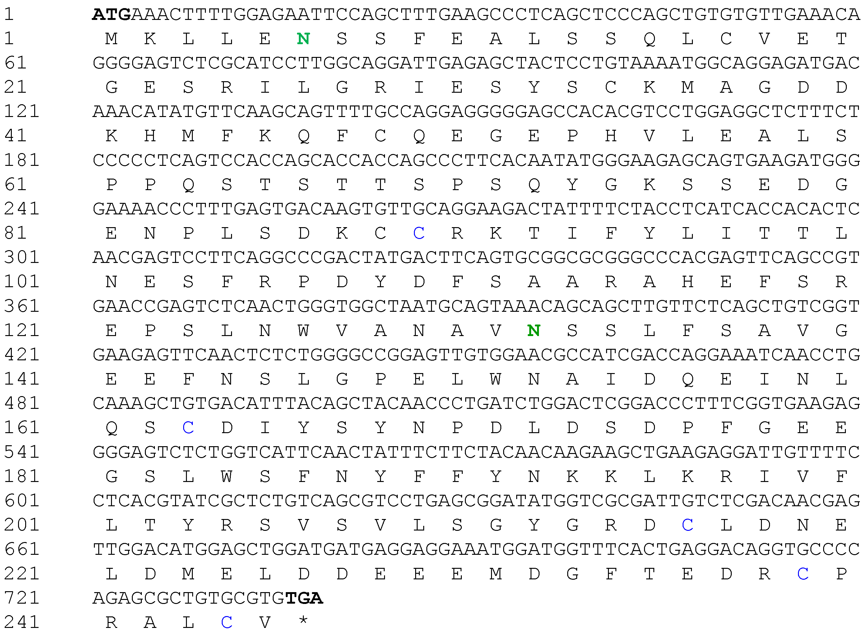
2.1. Isolation and Characterization of Maf1 Gene from P. olivaceus

The PoMaf1 full-length coding region (738 bp) encoding 245 amino acids was obtained from PoMaf1 cDNA (GenBank accession No. MW417124) (Figure 1). The putative molecular weight (MW) and isoelectric point (pI) of PoMAF1 were 27,805.74 Da and 4.39, respectively. Potential N-glycosylation sites and a disulfide bond were predicted in PoMaf1. The deduced amino acid sequence of PoMAF1 lacked a signal peptide sequence, such as a nuclear localization sequence (NLS). Yeast MAF1 has two conserved NLSs at the N- and C-termini (residues 205–208 and 328–332) [24]. In contrast, there is no canonical NLS in MAF1 of higher eukaryotes, including humans, similar to PoMAF1 [25]. MAF1 of all species localizes to both the nucleus and cytoplasm, and is expected to translocate into the nucleus as a negative regulator of transcription [26].

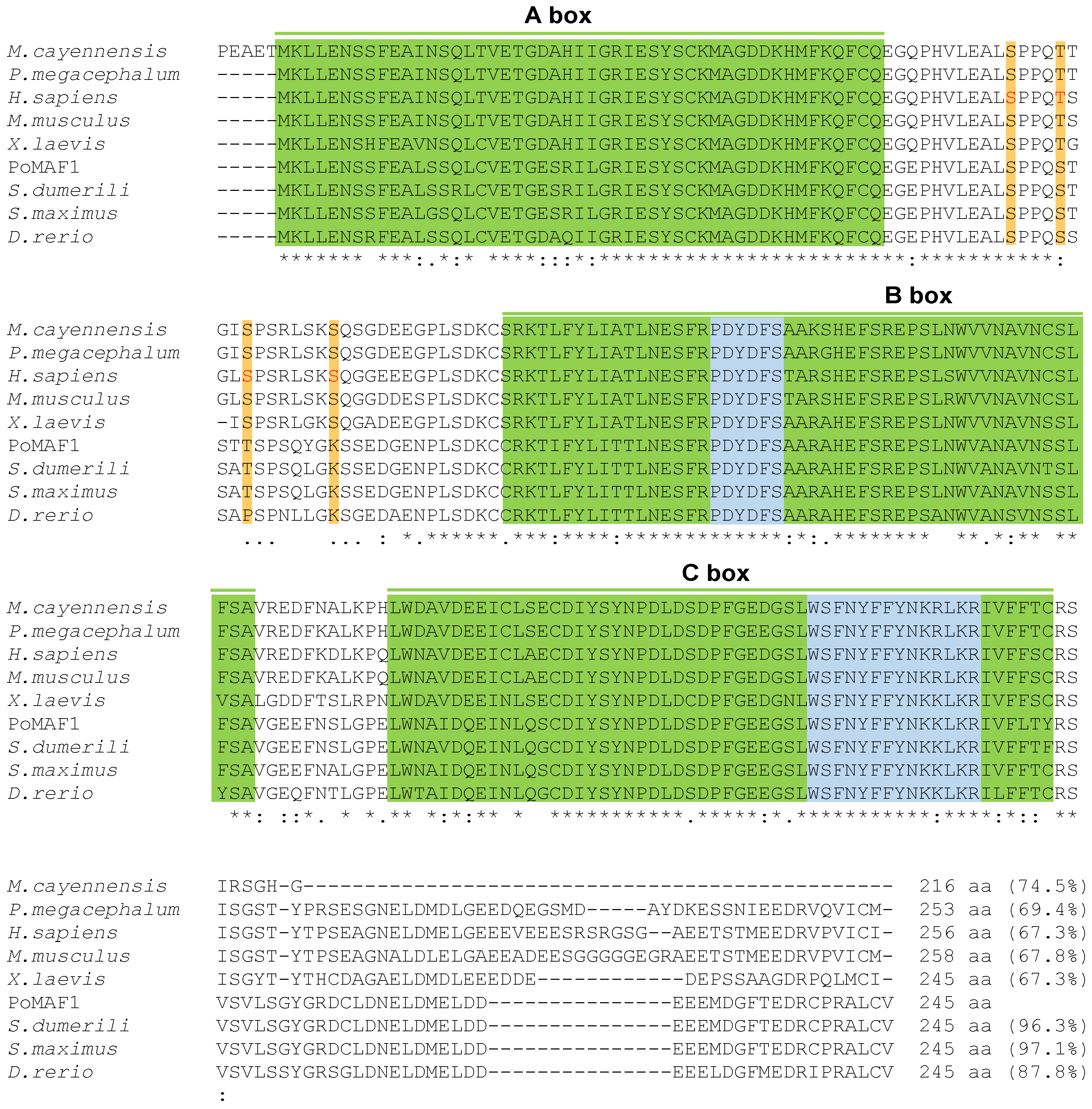
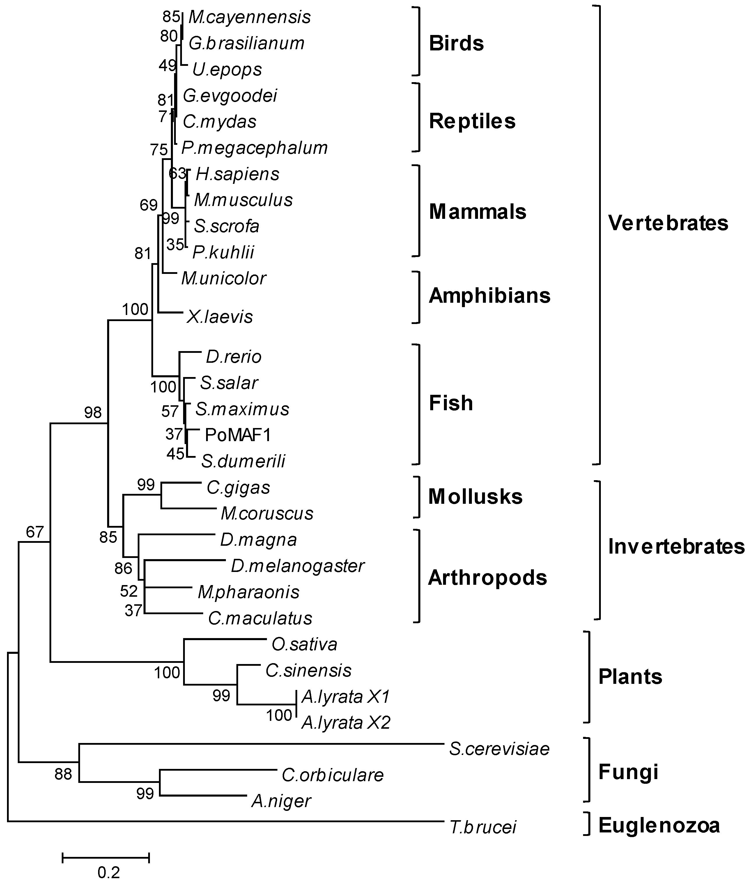
2.2. Amino Acids Sequence and Phylogenetic Analysis of PoMAF1

The deduced amino acid sequence of PoMaf1 was aligned with those of orthologues in mammals, reptiles, amphibians, birds, and fish (Figure 2). PoMAF1 exhibited the highest similarity (97.1%) with Black-Sea turbot S. maximus, a flatfish. The multiple sequence alignment revealed that PoMAF1 has three regions of high similarity to MAF1 orthologues: the A-, B- and C-boxes. The two MAF1 signature sequences (PDXDFS/T and WSXXYFFYNKKXKR) were present within domains B and C of PoMAF1, respectively. Phosphorylation sites (human residues Ser-60, Thr-64, Ser-68, and Ser-75) between the A- and B-boxes, which are directly phosphorylated by mTORC1 and required for mTORC1-mediated control of RNA pol III transcription, were found in PoMAF1 [27]. The A- and B-boxes are associated with RNA pol III subunits and Brf1, respectively [28]. The YSY motif (human residues 166–168) in the C-box of human MAF1 is critical for its stability and regulation [29]. Conserved elements in PoMAF1 suggest a role for PoMAF1 as a repressor of RNA pol III in P. olivaceus. Using multiple alignments, a neighbor-joining phylogenic tree was constructed based on the deduced PoMAF1 amino acid sequence and MAF1 sequences of 29 species (Figure 3). PoMAF1 was divisible into 10 main clusters: fish, amphibians, mammals, reptiles, birds, mollusks, arthropods, plants, fungi, and euglenozoa. PoMAF1 was grouped with orthologues from other teleost species and was most closely related to S. dumerili MAF1.

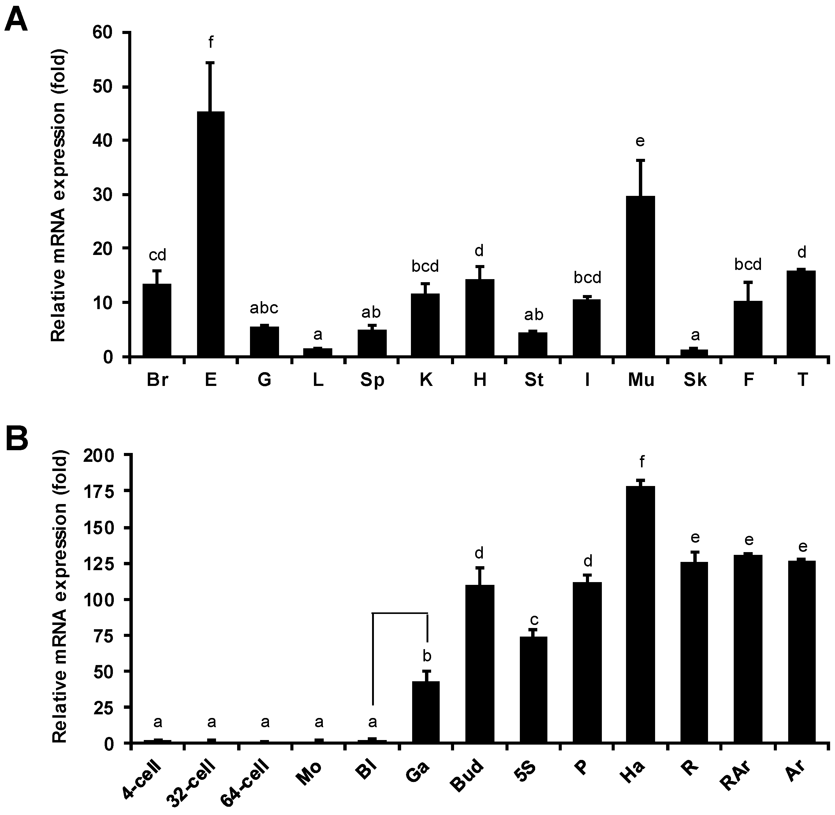
2.3. Tissue Distribution and Developmental Stage-Specific Expression of PoMaf1

The mRNA levels of PoMaf1 in 12 P. olivaceus tissues were determined by quantitative real-time PCR (Figure 4A). PoMaf1 mRNA was ubiquitously detected in the tissues, and the highest levels were in eye and muscle tissue. In humans, the Maf1 mRNA level is high in muscle [30]. In pigs, the mRNA expression profile of skeletal muscle changed in response to nutrient intake. MAF1 is a nutrient- and stress-sensitive global repressor of RNA pol III [31,32]. Therefore, MAF1 might be involved in the nutrient and stress responses of P. olivaceus. The high PoMaf1 mRNA level in the eye suggests that PoMAF1 has diverse biological functions in various tissues.
The mRNA level of PoMaf1 in P. olivaceus, from the four-cell stage to the feeding stage to 27 days post-hatching (dph), was evaluated by quantitative real-time PCR (Figure 4B). The mRNA level of PoMaf1 remained low until the blastula stage, and increased 18-fold at the gastrula stage compared to the blastula stage. The PoMaf1 mRNA level increased gradually and was highest in hatched larva. In the feeding stage, the PoMaf1 mRNA level decreased by 70% and then remained constant. The PoMaf1 mRNA level fluctuated markedly during early development, indicating a role for PoMAF1. MAF1 is a regulator of Akt/mTOR signaling in dendritic morphogenesis during neuron development [33]. This is in accordance with the marked increase in PoMaf1 mRNA level during gastrulation, which is immediately followed by neurulation [34]. The expression profile of PoMaf1 suggests that further investigation of the role of PoMAF1 is warranted.

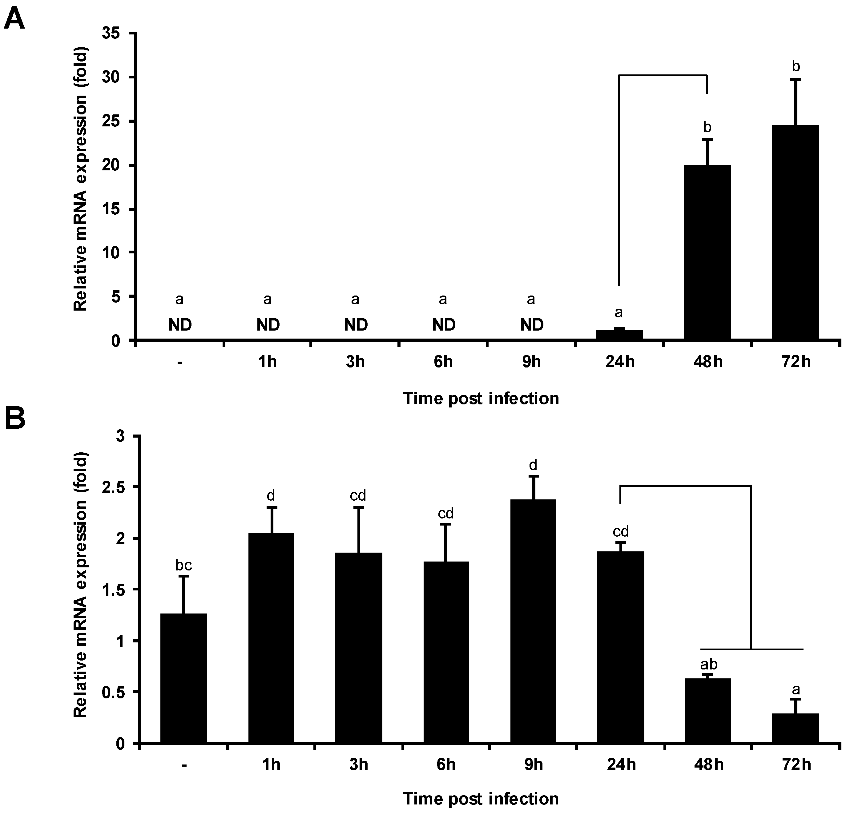
2.4. VHSV Infection Decreases PoMaf1 mRNA Level

To determine the physiological relevance of VHSV and PoMAF1, quantitative real-time PCR of VHSV-infected HINAE cells was performed (Figure 5). During VHSV infection, the PoMaf1 mRNA level increased moderately from 1 to 24 h, but was markedly decreased at 48 and 72 h. In addition, the VHSV glycoprotein (G) mRNA level significantly increased at 48 and 72 h post-infection. The decrease in PoMaf1 mRNA level with increasing VHSV expression is in agreement with the postulated relationship between viral replication and MAF1 expression. MAF1 is a new target of PTEN that negatively regulates lipid metabolism, and Dengue virus (DENV) infection inhibits MAF1 via Akt/FoxO1/Maf1 signaling, which is mediated by PTEN, to regulate lipid metabolism and thus promote DENV replication [7,35]. Akt in the PI3K/Akt intracellular signaling pathway is activated in acute and persistent viral infection to suppress apoptosis and prolong viral replication [36]. The fish virus, Siniperca chuatsi rhabdovirus (SCRV), activates the PI3K/Akt pathway, thus inducing autophagy [37]. PI3K/Akt/FoxO1 signaling modulates MAF1 abundance, and PI3K/Akt/mTORC1 signaling regulates MAF1 in a posttranslational manner, such as by phosphorylation [38]. Therefore, PoMaf1 expression might be related to the immune response to VHSV.

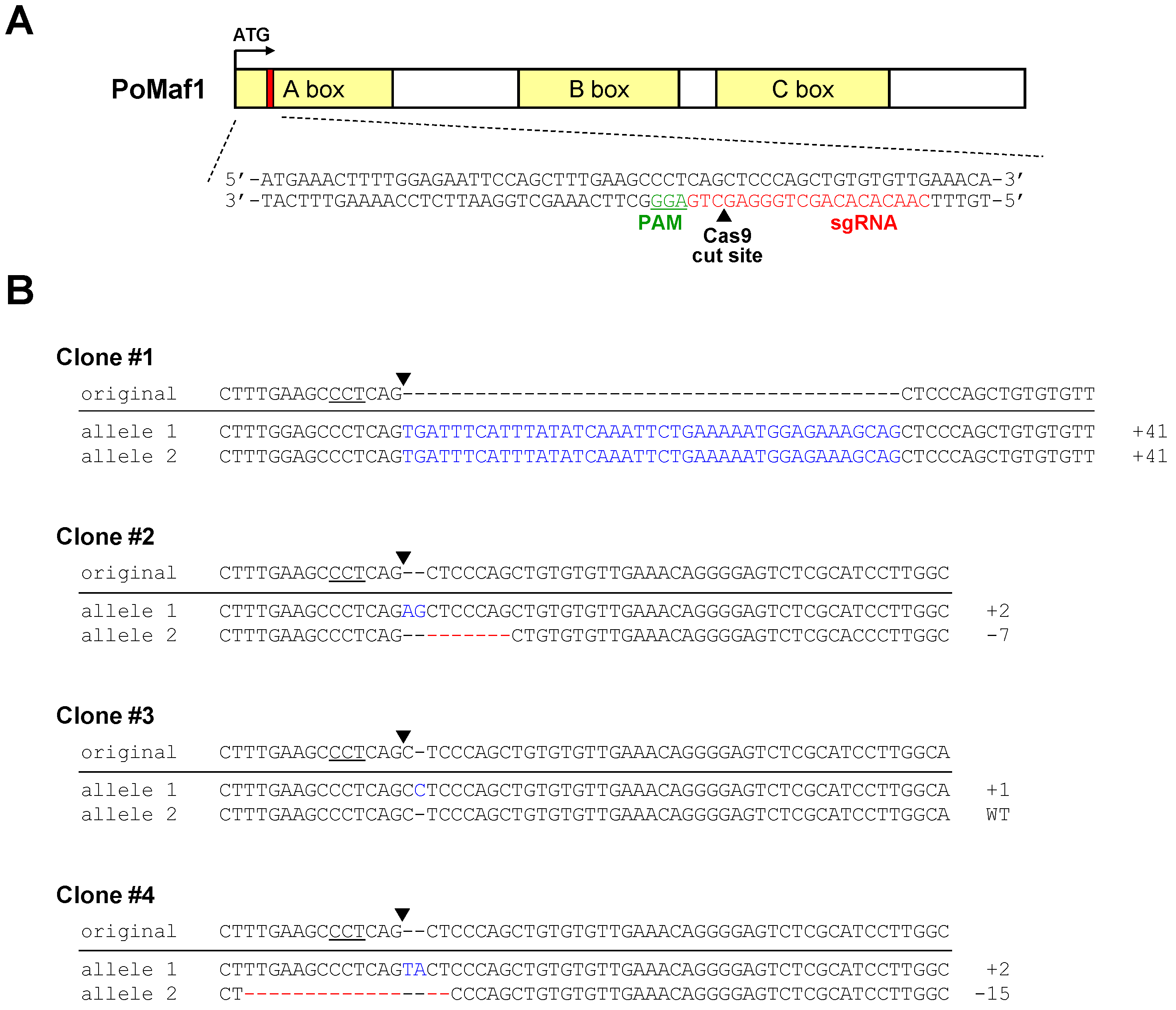
2.5. Disruption of PoMaf1 in HINAE Cells Using CRISPR/Cas9 System

To explore the function of PoMaf1 in olive flounder HINAE cells, we knocked out PoMaf1 using the CRISPR/Cas9 system to generate homogeneous mutant cell clones and overcome the low transfection efficiency of fish cell lines. Based on the PoMaf1 genomic DNA sequence, the CRISPR target sequence was selected from several candidates to design a sgRNA (Figure 6A). The target sequence did not span an exon–exon junction and matched the PoMaf1 genomic DNA sequence. A Cas9 plasmid (pX458) containing the U6 promoter-driven PoMaf1 sgRNA expression cassette (pSpCas9(BB)-2A-GFP-PoMaf1 sgRNA) was transfected into HINAE cells and cells expressing GFP were sorted. The sequencing results of representative mutant clones are shown in Figure 6B. Clone #1, with a 41 bp insertion in both alleles, was a homozygous mutant clone. Clone #2, with a 2 bp insertion and 7 bp deletion in each allele, was a heterozygous biallelic mutant clone. Clone #3, with a 1 bp insertion in one allele, was a heterozygous monoallelic mutant clone. Clone #4, with a 2 bp insertion and 15 bp deletion in each allele, was a heterozygous biallelic mutant. Mutations of the 41 bp insertion, 2 bp insertion, 7 bp deletion, 1 bp insertion, and 2 bp insertion led to frameshift and nonsense mutations by out-of-frame indels, resulting in a shortened and possibly dysfunctional PoMAF1 protein. The 15 bp deletion mutation caused a non-frameshift mutation. Therefore, the CRISPR/Cas9 system enabled targeting and disruption of PoMaf1 in HINAE cells.

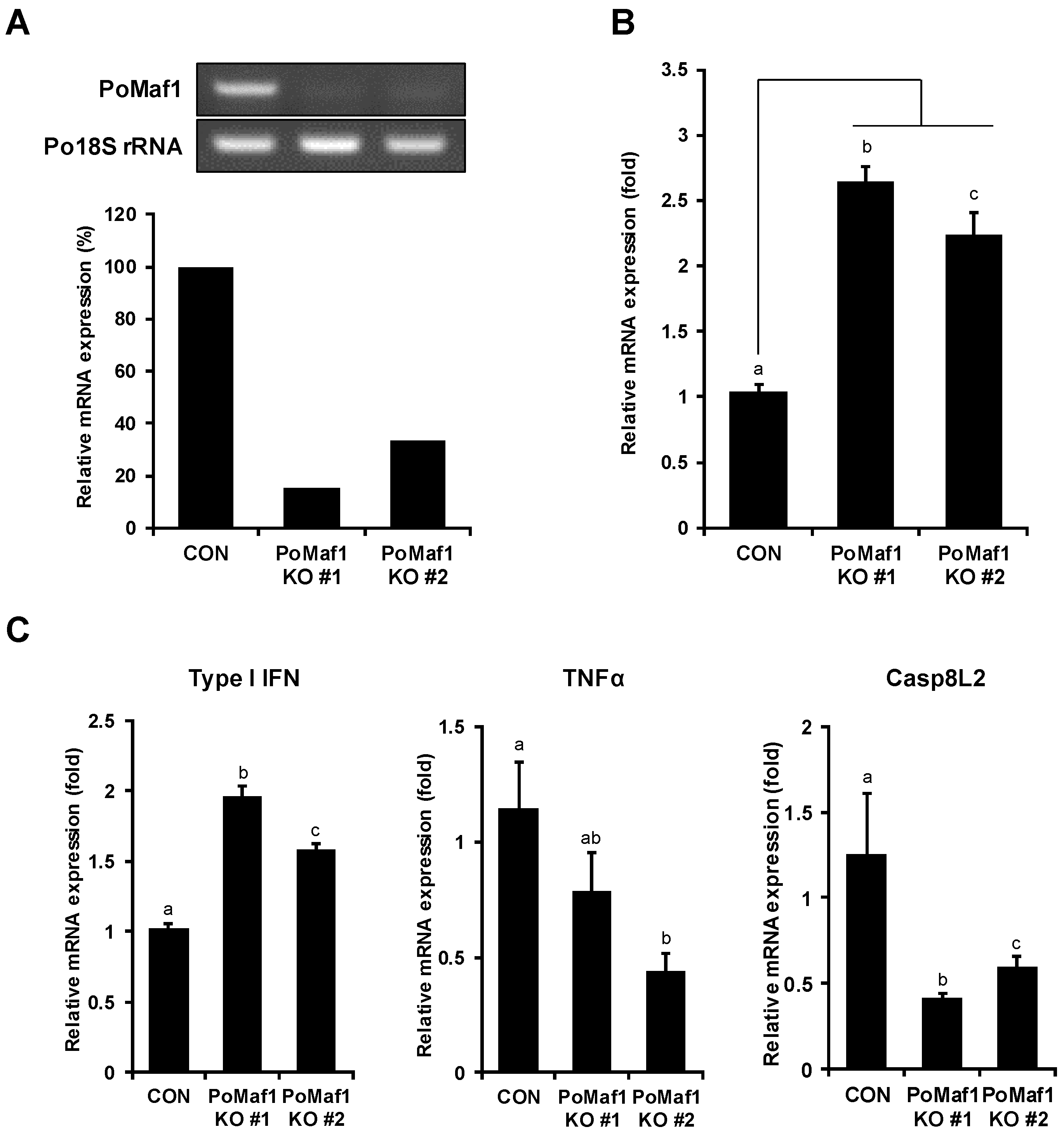
2.6. PoMaf1 Disruption Enhances VHSV G mRNA Level in HINAE Cells

To determine the effect of PoMaf1 KO on VHSV replication, PoMaf1 KO cells were infected with VHSV, and viral gene expression was analyzed. Among the cell clones harboring indels in the PoMaf1 locus, two biallelic mutant clones (clones #1 and #2) producing truncated PoMAF1 protein from both alleles were subjected to analysis of the PoMaf1 mRNA level (Figure 7A). The PoMaf1 RT-PCR primers were designed to span the indel junction of mutant alleles so that they bind only intact mRNA of PoMaf1. The PoMaf1 transcript level was significantly reduced, by 85% and 67%, in PoMaf1 KO clones #1 and #2, respectively, compared to control HINAE cells transfected with empty Cas9 vector (pX458). The higher PoMaf1 mRNA level in PoMaf1 KO clone #2 compared to clone #1 was a result of detection of the target PoMaf1 cDNA sequence with a 2 bp deletion in one allele. In PoMaf1 KO clones #1 and #2, the VHSV G mRNA level increased 2.2 to 2.5-fold compared to control HINAE cells transfected with pX458, indicating that PoMaf1 KO promoted VHSV infection of HINAE cells (Figure 7B). In addition, PoMaf1 expression was inhibited by VHSV infection, and disruption of PoMaf1 enhanced VHSV expression. Taken together, these results suggest that PoMAF1 inhibits VHSV, and that VHSV downregulates PoMaf1 to promote its replication.
These observations raise the question as to how PoMAF1 regulates VHSV replication. Regarding the function of MAF1 as a repressor of RNA pol, several RNA viruses—including influenza virus and hepatitis delta virus—have been reported to require host RNA pol for their replication [4,39,40]. However, whether VHSV interacts with host RNA pol, and whether MAF1 suppresses viral RNA pol, is unclear. Production of DENV, another RNA virus, is regulated by Akt/FoxO1/Maf1 signaling [35]. In this study, a reduction in PoMaf1 expression promoted VHSV expression; the underlying mechanism warrants further investigation.
RNA pol III activity is closely related to the immune response, suggesting that MAF1 may have a role in immunity [41,42]. MAF1 plays an important role in normal physiology and disease, and is involved in tumor immunity in colorectal cancer [5,43]. To investigate VHSV-induced immune response in PoMaf1 KO cells, the transcription levels of innate immune response genes such as type I IFN, TNFα and Caspase 8 like 2 (Casp8L2) were analyzed in PoMaf1 KO cells (Figure 7C). The type I IFN mRNA level increased in both PoMaf1 KO cells against VHSV infection, indicating that PoMaf1 disruption led to increased IFN and VHSV G expression level. IFN is a major immune response gene induced in VHSV-infected P. olivaceus and HINAE cells, suggesting that PoMaf1 is involved in immune response [44,45]. Focusing on the function of MAF1 as a repressor of RNA pol III, type I IFN is induced by RNA pol III through the RIG-I pathway [42], suggesting that PoMaf1 disruption enhances the IFN expression level by activating RNA pol III. However, it is unclear how enhanced IFN levels matched increased VHSV G expression. Recently, it has been reported that a high-virulence VHSV strain induced higher levels of IFN 1 compared to a low-virulence VHSV strain, suggesting that the IFN response in VHSV infection correlates with virus titers but does not necessarily correlate with protection [45]. On the other hand, mRNA expression levels of pro-inflammatory cytokine TNFα and apoptosis initiator Casp8L2 decreased in PoMaf1 KO cells during VHSV infection. In zebrafish, PoMAF1 overexpression upregulated the mRNA levels of key regulators of the innate immune response (unpublished data). Therefore, PoMAF1 is involved in the immune response to VHSV infection, although further studies are needed.
PoMAF1 is a potential therapeutic target VHSV. The CRISPR/Cas9 system enables genome editing and has revolutionized genetic improvement of fish, so we can apply it to upregulate PoMaf1. To enhance target gene expression, regulatory elements are inserted upstream of an endogenous gene by a CRISPR/Cas9-mediated knock-in strategy [46]. In contrast, cis-regulatory elements (CREs) capable of silencing a gene promoter region can be targeted by the CRISPR/Cas9 system without adding exogenous genes [47]. The CRISPR/Cas9 system in combination with multiple sgRNAs can cause large deletions in the target region [48]. CRISPR/Cas9-based genome editing has been accomplished in both P. olivaceus-derived cell lines and P. olivaceus, leading to investigation of gene function and improvement of the traits of P. olivaceus [22].

3. Materials and Methods

3.1. Fish and Sample Preparation

Olive flounder Paralichthys olivaceus were raised at the National Institute of Fisheries Sciences (NIFS; Busan, Korea) and maintained in 1-ton flow-through tanks at 19 ± 0.3 °C under a natural photoperiod using fluorescent lighting and an electronic timer (Yuwon Engineering Co., Hwaseong, Korea). Adults were fed to satiation with commercial extruded pellets (Daebong LS Co., Ltd., Jeju, Korea) twice daily to ensure a sufficient food supply. For RNA extraction, tissues were dissected from three healthy P. olivaceus, immediately frozen in liquid nitrogen, and stored at −80 °C before use. Animal experiments were conducted in accordance with the Animal Protection Act of the Republic of Korea, and approved by the Institutional Animal Care and Use Committee of the NIFS (2019-NIFS-IACUC-10, 1 February 2019).

3.2. Cloning and Sequencing of PoMaf1 from P. olivaceus

To amplify PoMaf1, the primers PoMaf1-ORF-F (5′-CAA TGA AAC TTT TGG AGA ATT CC-3′) and PoMaf1-ORF-R (5′-CAT CTC GAG TCA CAC GCA CAG CGC-3′) were designed based on P. olivaceus transcriptome analysis. The P. olivaceus cDNA library was used as the template for reverse transcription-polymerase chain reaction (PCR) to amplify the coding region of putative PoMaf1. The PCR fragments were purified using a gel extraction kit (Qiagen, Venlo, The Netherlands), and cloned into the pGEM®-T easy vector (Promega, Madison, WI, USA) according to the manufacturer’s instructions. Putative PoMaf1 was sequenced using the universal primer T7 promoter, SP6, on an ABI3730xl automatic sequencer (Applied Biosystems, Inc., Foster City, CA, USA). The PoMaf1 sequence is deposited in GenBank with the accession number MW417124.

3.3. Amino Acid Sequence Analysis

The pI and MW of the deduced PoMAF1 protein were computed on the ExPASy (http://web.expasy.org/compute_pi/). The signal sequence was predicted using SignalP (http://www.cbs.dtu.dk/services/SignalP/). Disulfide bonds were predicted using DiANNA (http://clavius.bc.edu/~clotelab/DiANNA). Potential N-glycosylation sites were assessed using the NetNGlyc1.0 Server (http://www.cbs.dtu.dk/services/NetNGlyc/).

3.4. Multiple Alignment and Phylogenetic Analysis

Coding sequence homology was compared in BLASTX (http://0-www-ncbi-nlm-nih-gov.brum.beds.ac.uk/BLAST/). Multiple alignments were generated with CLUSTALW (http://www.genome.jp/tools-bin/clustalw) and used for assessing similarities among the aligned sequences with MEGA software (ver. 5; Arizona State University, AZ, USA; [49]). An unrooted phylogenetic tree based on the deduced amino acid sequences was constructed using the neighbor-joining method. The bootstrap resampling was repeated 1000 times to determine the reliability of the phylogenetic tree.

3.5. RNA Isolation, Reverse Transcriptase PCR and Quantitative Real-Time PCR

Total RNA was isolated using TRIzol™ reagent (Invitrogen, Carlsbad, CA, USA) according to the manufacturer’s instructions, and treated with DNase (New England BioLabs, Beverly, MA, USA). RNA samples (500 ng) were used to synthesize first-strand cDNA with the Transcriptor First Strand cDNA Synthesis Kit (Roche, Basel, Switzerland). RT-PCR was performed using Ex Taq™ polymerase (TaKaRa Bio Inc., Shiga, Japan) and gene-specific primers: PoMaf1-F (5′-AGC CCT CAG CTC CCA GCT-3′), PoMaf1-R (5′-GTC CTG GAG GCT CTT TCT CC-3′), Po18S-RT-F (5′-ATG GCC GTT CTT AGT TGG TG-3′) and Po18S-RT-R (5′-CAC ACG CTG ATC CAG TCA GT-3′). The PCR reaction conditions were denaturation for 5 min at 95 °C; followed by 25 or 35 cycles of 95 °C for 30 s, 60 °C for 30 s, and 95 °C for 30 s; and a final extension at 72 °C for 10 min. Quantitative real-time PCR was carried out using Fast SYBR Green Master Mix (Applied Biosystems, Inc.) and the following primers for PoMaf1, VHSV G, type I IFN, TNFα and Casp8L2: PoMaf1-RT-F (5′-GGG AAA ACC CTC TGA GTG ACA A-3′), PoMaf1-RT-R (5′-CGC CGC ACT GAA GTC ATA GTC-3′), VHSV-G-RT-F (5′-AGA TGA GGG GAG CCA CAG AC-3′), VHSV-G-RT-R (5′-GGG ATG ATC AAT TTG TCC CC-3′), PoIFN1-RT-F (5′-GGC CAC ATT CAC GCA ATC AC-3′), PoIFN2-RT-R (5′-TGC AGG TGT CTA TGT GGC TA-3′), PoTNFα-RT-F (5′-CCG ACT GGA TGT GTA AGG TG-3′), PoTNFα-RT-R (5′-GTT GTG GGG TTC TGT TTT CTC-3′), PoCasp8L2-RT-F (5′-ACC ACG TCT TCC ATG AGA CC-3′), PoCasp8L3-RT-R (5′-GCC CAG CCA CTT AAA CAC AT-3′). The primers for P. olivaceus 18S rRNA were the same as those for RT-PCR. Amplification and detection were performed using the ABI 7500 Real-Time PCR System (Applied Biosystems, Inc.) with the following steps: 50 °C for 2 min, 95 °C for 10 min, and 40 cycles of 95 °C for 15 s and 60 °C for 30 s. Quantitative real-time PCR data were analyzed using the comparative threshold cycle (Ct) method (2−ΔΔCT method) for relative quantification.

3.6. sgRNA Design and Vector Construction for the CRISPR/Cas9 System

Using the CRISPR RGEN Tools (www.rgenome.net/cas-designer), a single guide RNA (sgRNA) targeting PoMaf1 was designed (5′-CAA CAC ACA GCT GGG AGC TGA GG-3′; PAM, underlined). A pair of oligonucleotides corresponding to the target sgRNA sequence (oligo-1, 5′-CAC CGC AAC ACA CAG CTG GGA GCT G-3′; oligo-2, 5′-AAA CCA GCT CCC AGC TGT GTG TTG-3′) was inserted into the sgRNA/Cas9 dual expression vector pSpCas9(BB)-2A-GFP (also known as PX458; Addgene, Watertown, MA, USA) according to the protocol of Zhang [50]. Briefly, the pX458 vector was digested with BbsI and gel-purified. The dsDNA with 4 bp overhangs on both ends was generated by annealing following phosphorylation, and ligated into the linearized pX458 vector.

3.7. Cell Culture and Transfection

HINAE olive flounder embryonic cells, a gift from Takashi Aoki, were maintained in Leibovitz’s L-15 medium (L-15; Gibco, Invitrogen, Carlsbad, CA, USA) with 10% heat-inactivated fetal bovine serum (FBS; Gibco) and 1% (v/v) antibiotic-antimycotic (AA; Gibco) at 20 °C. Medium was changed every third day. HINAE cells were transfected with ViaFect™ transfection reagent (Promega) according to the manufacturer’s instructions.

3.8. Production of Single-Cell Clones of KO Cells

To obtain single PoMaf1 KO cells, HINAE cells were transfected with Cas9/PoMaf1 sgRNA and EGFP co-expression vector (pSpCas9(BB)-2A-GFP-PoMaf1 sgRNA). At 48 h post transfection, cells were trypsinized for 3 min at room temperature and collected by centrifugation. Cells were resuspended in L-15 culture medium containing 15% heat-inactivated FBS and 1.5% (v/v) AA before being passed through a 35 μm cell strainer into tubes (BD Falcon, Franklin Lakes, NJ, USA). Cells positive for GFP (and therefore Cas9) expression were sorted and transferred to 96-well plates using a cell sorter (BD FACS Aria III; BD Biosciences, San Jose, CA, USA) following the manufacturer’s instructions. Control cells were transfected with the empty pX458 construct. After culture for one month, genomic DNA was extracted from the clones using PrimePrep Direct PCR Reagent (GeNetBio, Daejeon, Korea) following the manufacturer’s instructions, and the fragments containing the CRISPR target site were amplified by PCR using specific primers (PoMaf1-seq-F, 5′-TGT TTT GCA AGG TGA CTG TAC GT-3′; PoMaf1-seq-R, 5′-GGC GTG ACT TTT GTT GAG TAT TAA CT-3′). The PCR amplicons were purified and cloned into the pGEM®-T easy vector (Promega), and at least eight colonies per clone were sequenced to detect indels using an ABI3730xl Automatic Sequencer (Applied Biosystems, Inc.).

3.9. VHSV Preparation

VHSV isolated from a diseased P. olivaceus was propagated in a monolayer of HINAE cells at 20 °C [51]. When cytopathic effects became evident, the supernatants of VHSV-infected cell cultures were clarified by centrifugation and stored as aliquots at −80 °C. The tissue culture infective dose 50 (TCID50) was determined using the end-point dilution method in 96-well plates.

3.10. Statistical Analysis

All experiments were conducted using three biological replicates and results were represented as means ± standard deviation (SD) (n = 3). In Figure 4, Figure 5 and Figure 7, for a comparison of more than two groups, data were analyzed using one-way analysis of variance (ANOVA), followed by Duncan’s multiple range test. Statistical significance was considered at p ≤ 0.05. Statistical analyses were conducted using PASW Statistics (ver. 18.0; SPSS Inc., Chicago, IL, USA).

4. Conclusions

We characterized the full-length ORF of Maf1 in P. olivaceus (PoMaf1). Deduced amino acids of PoMAF1 had putative A, B, C, box and MAF1 signature sequences comparable with MAF1 orthologues from other vertebrate species. The PoMaf1 mRNA level was highest in eye tissue in P. olivaceus and markedly increased during early development. The PoMaf1 mRNA level in HINAE cells decreased during VHSV infection. To investigate the role of PoMAF1 in VHSV infection, single-cell-derived PoMaf1 KO cells were generated using the CRISPR/Cas9 system. In the absence of PoMAF1, the VHSV G mRNA level increased during VHSV infection, suggesting PoMAF1 inhibits VHSV replication. In addition, the VHSV-induced immune response in PoMaf1 KO cells was investigated. This study provides fundamental data on fish Maf1 and will facilitate the development of novel therapeutic approaches to VHSV infection. However, the mechanism underlying the inhibitory effect of PoMAF1 on VHSV infection warrants further investigation.

Author Contributions

Conceptualization, H.J.K. and J.C.; investigation, J.K. and J.Y.C.; software, J.-W.K. and D.-G.K.; validation, B.-S.K.; writing, J.K. and H.J.K.; resources, B.-H.N. and W.-J.K.; project administration, Y.-O.K. All authors have read and agreed to the published version of manuscript.

Funding

This research was funded by National Institute of Fisheries Science (NIFS), Republic of Korea, grant number R2021025.

Institutional Review Board Statement

Animal experiments were conducted in accordance with the Animal Protection Act of the Republic of Korea and approved by the Institutional Animal Care and Use Committee of the NIFS (2019-NIFS-IACUC-10, 1 February 2019).

Informed Consent Statement

Not applicable.

Data Availability Statement

The data presented in this study are available in this article.

Conflicts of Interest

The authors declare no conflict of interest.

References

  1. Vannini, A.; Ringel, R.; Kusser, A.G.; Berninghausen, O.; Kassavetis, G.A.; Cramer, P. Molecular Basis of RNA Polymerase III Transcription Repression by Maf1. Cell 2010, 143, 59–70. [Google Scholar] [CrossRef] [Green Version]
  2. Pluta, K.; Lefebvre, O.; Martin, N.C.; Smagowicz, W.J.; Stanford, D.R.; Ellis, S.R.; Hopper, A.K.; Sentenac, A.; Boguta, M. Maf1p, a Negative Effector of RNA Polymerase III in Saccharomyces cerevisiae. Mol. Cell. Biol. 2001, 21, 5031–5040. [Google Scholar] [CrossRef] [PubMed] [Green Version]
  3. Upadhya, R.; Lee, J.; Willis, I.M. Maf1 Is an Essential Mediator of Diverse Signals that Repress RNA Polymerase III Transcription. Mol. Cell 2002, 10, 1489–1494. [Google Scholar] [CrossRef] [PubMed]
  4. Johnson, S.S.; Zhang, C.; Fromm, J.; Willis, I.M.; Johnson, D.L. Mammalian Maf1 Is a Negative Regulator of Transcription by All Three Nuclear RNA Polymerases. Mol. Cell 2007, 26, 367–379. [Google Scholar] [CrossRef] [PubMed]
  5. Zhang, S.; Li, X.; Wang, H.Y.; Zheng, X.F.S. Beyond regulation of pol III: Role of MAF1 in growth, metabolism, aging and cancer. Biochim. Biophys. Acta Gene Regul. Mech. 2018, 1861, 338–343. [Google Scholar] [CrossRef] [PubMed]
  6. Kantidakis, T.; Ramsbottom, B.A.; Birch, J.L.; Dowding, S.N.; White, R.J. mTOR associates with TFIIIC, is found at tRNA and 5S rRNA genes, and targets their repressor Maf1. Proc. Natl. Acad. Sci. USA 2010, 107, 11823–11828. [Google Scholar] [CrossRef] [Green Version]
  7. Palian, B.M.; Rohira, A.D.; Johnson, S.A.S.; He, L.; Zheng, N.; Dubeau, L.; Stiles, B.L.; Johnson, D.L. Maf1 Is a Novel Target of PTEN and PI3K Signaling That Negatively Regulates Oncogenesis and Lipid Metabolism. PLoS Genet. 2014, 10, e1004789. [Google Scholar] [CrossRef] [Green Version]
  8. Bonhoure, N.; Byrnes, A.; Moir, R.D.; Hodroj, W.; Preitner, F.; Praz, V.; Marcelin, G.; Chua, S.C., Jr.; Martinez-Lopez, N.; Singh, R.; et al. Loss of the RNA polymerase III repressor MAF1 confers obesity resistance. Genes Dev. 2015, 29, 934–947. [Google Scholar] [CrossRef] [Green Version]
  9. Li, Y.; Tsang, C.K.; Wang, S.; Li, X.; Yang, Y.; Fu, L.; Huang, W.; Li, M.; Wang, H.; Zheng, X.S. MAF1 suppresses AKT-mTOR signaling and liver cancer through activation of PTEN transcription. Hepatology 2016, 63, 1928–1942. [Google Scholar] [CrossRef] [Green Version]
  10. Sun, Y.; Chen, C.; Xue, R.; Wang, Y.; Dong, B.; Li, J.; Chen, C.; Jiang, J.; Fan, W.; Liang, Z.; et al. Maf1 ameliorates cardiac hypertrophy by inhibiting RNA polymerase III through ERK1/2. Theranostics 2019, 9, 7268–7281. [Google Scholar] [CrossRef]
  11. Martin, S.; Saha, B.; Riley, J.L. The Battle over mTOR: An Emerging Theatre in Host–Pathogen Immunity. PLoS Pathog. 2012, 8, e1002894. [Google Scholar] [CrossRef] [PubMed] [Green Version]
  12. Skall, H.F.; Olesen, N.J.; Mellergaard, S. Viral haemorrhagic septicaemia virus in marine fish and its implications for fish farming—A review. J. Fish Dis. 2005, 28, 509–529. [Google Scholar] [CrossRef] [PubMed]
  13. Office International des Epizooties (OIE). Available online: https://www.oie.int/en/standard-setting/aquatic-manual/access-online/ (accessed on 10 December 2020).
  14. Takano, R.; Nishizawa1, T.; Arimoto, M.; Muroga, K. Isolation of viral haemorrhagic septicaemia virus (VHSV) from wild Jap-anese flounder, Paralichthys olivaceus. Bull. Eur. Assoc. Fish Pathol. 2000, 20, 186. [Google Scholar]
  15. Kim, M.S.; Kim, K.H. Genetically engineered viral hemorrhagic septicemia virus (VHSV) vaccines. Fish Shellfish Immunol. 2019, 95, 11–15. [Google Scholar] [CrossRef]
  16. Kurita, J.; Iida, Y.; Nakajima, K.; Inouye, K. Virucidal Effects of Various Disinfectants on Viral Hemorrhagic Septicemia Virus(VHSV) Isolated from Japanese Flounder. Fish Pathol. 2002, 37, 175–181. [Google Scholar] [CrossRef]
  17. Yan, M.; Li, J. The evolving CRISPR technology. Protein Cell 2019, 10, 783–786. [Google Scholar] [CrossRef] [Green Version]
  18. Gratacap, R.L.; Wargelius, A.; Edvardsen, R.B.; Houston, R.D. Potential of Genome Editing to Improve Aquaculture Breeding and Production. Trends Genet. 2019, 35, 672–684. [Google Scholar] [CrossRef] [PubMed] [Green Version]
  19. Bedell, V.M.; Wang, Y.; Campbell, J.M.; Poshusta, T.L.; Starker, C.G.; Krug, R.G., 2nd; Tan, W.; Penheiter, S.G.; Ma, A.C.; Leung, A.Y.; et al. In vivo genome editing using a high-efficiency TALEN system. Nat. Cell Biol. 2012, 491, 114–118. [Google Scholar] [CrossRef] [Green Version]
  20. O’Connell, M.R.; Oakes, B.L.; Sternberg, S.H.; East-Seletsky, A.; Kaplan, M.; Doudna, J.A. Programmable RNA recognition and cleavage by CRISPR/Cas9. Nat. Cell Biol. 2014, 516, 263–266. [Google Scholar] [CrossRef]
  21. Cleveland, B.M. A perspective of the future value and challenges of genetic engineering in aquaculture. J. World Aquac. Soc. 2018, 50, 890–893. [Google Scholar] [CrossRef]
  22. Kim, J.; Cho, J.Y.; Kim, J.-W.; Kim, H.-C.; Noh, J.K.; Kim, Y.-O.; Hwang, H.-K.; Kim, W.-J.; Yeo, S.-Y.; An, C.M.; et al. CRISPR/Cas9-mediated myostatin disruption enhances muscle mass in the olive flounder Paralichthys olivaceus. Aquaculture 2019, 512, 734336. [Google Scholar] [CrossRef]
  23. Dehler, C.E.; Boudinot, P.; Martin, S.A.M.; Collet, B. Development of an Efficient Genome Editing Method by CRISPR/Cas9 in a Fish Cell Line. Mar. Biotechnol. 2016, 18, 449–452. [Google Scholar] [CrossRef] [PubMed] [Green Version]
  24. Moir, R.D.; Lee, J.; Haeusler, R.A.; Desai, N.; Engelke, D.R.; Willis, I. Protein kinase A regulates RNA polymerase III transcription through the nuclear localization of Maf1. Proc. Natl. Acad. Sci. USA 2006, 103, 15044–15049. [Google Scholar] [CrossRef] [PubMed] [Green Version]
  25. Wei, Y.; Zheng, X.F.S. Maf1 regulation: A model of signal transduction inside the nucleus. Nucleus 2010, 1, 162–165. [Google Scholar] [CrossRef] [PubMed] [Green Version]
  26. Khanna, A.; Pradhan, A.; Curran, S.P. Emerging Roles for Maf1 beyond the Regulation of RNA Polymerase III Activity. J. Mol. Biol. 2015, 427, 2577–2585. [Google Scholar] [CrossRef] [PubMed] [Green Version]
  27. Shor, B.; Wu, J.; Shakey, Q.; Toral-Barza, L.; Shi, C.; Follettie, M.; Yu, K. Requirement of the mTOR Kinase for the Regulation of Maf1 Phosphorylation and Control of RNA Polymerase III-dependent Transcription in Cancer Cells. J. Biol. Chem. 2010, 285, 15380–15392. [Google Scholar] [CrossRef] [PubMed] [Green Version]
  28. Reina, J.H.; Azzouz, T.N.; Hernandez, N. Maf1, a New Player in the Regulation of Human RNA Polymerase III Transcription. PLoS ONE 2006, 1, e134. [Google Scholar] [CrossRef] [Green Version]
  29. Pradhan, A.; Hammerquist, A.M.; Khanna, A.; Curran, S.P. The C-Box Region of MAF1 Regulates Transcriptional Activity and Protein Stability. J. Mol. Biol. 2017, 429, 192–207. [Google Scholar] [CrossRef] [Green Version]
  30. Uhlen, M.; Fagerberg, L.; Hallström, B.M.; Lindskog, C.; Oksvold, P.; Mardinoglu, A.; Sivertsson, Å.; Kampf, C.; Sjöstedt, E.; Asplund, A.; et al. Tissue-based map of the human proteome. Science 2015, 347, 1260419. [Google Scholar] [CrossRef]
  31. Cardoso, T.F.; Quintanilla, R.; Tibau, J.; Gil, M.; Marmol-Sanchez, E.; González-Rodríguez, O.; González-Prendes, R.; Amills, M. Nutrient supply affects the mRNA expression profile of the porcine skeletal muscle. BMC Genom. 2017, 18, 1–11. [Google Scholar] [CrossRef] [Green Version]
  32. Willis, I.; Moir, R.D. Integration of nutritional and stress signaling pathways by Maf1. Trends Biochem. Sci. 2007, 32, 51–53. [Google Scholar] [CrossRef]
  33. Chen, K.; Zhu, L.; Guo, L.; Pan, Y.-B.; Feng, D.-F. Maf1 regulates dendritic morphogenesis and influences learning and memory. Cell Death Dis. 2020, 11, 1–18. [Google Scholar] [CrossRef] [PubMed]
  34. Hinaux, H.; Devos, L.; Blin, M.; Elipot, Y.; Bibliowicz, J.; Alié, A.; Rétaux, S. Sensory evolution in blind cavefish is driven by early embryonic events during gastrulation and neurulation. Development 2016, 143, 4521–4532. [Google Scholar] [CrossRef] [PubMed] [Green Version]
  35. Liu, B.; Gao, T.-T.; Fu, X.-Y.; Xu, Z.-H.; Ren, H.; Zhao, P.; Qi, Z.-T.; Qin, Z.-L. PTEN Lipid Phosphatase Activity Enhances Dengue Virus Production through Akt/FoxO1/Maf1 Signaling. Virol. Sin. 2020, 1–12. [Google Scholar] [CrossRef] [PubMed]
  36. Ji, W.-T. PI3K-Akt Signaling and Viral Infection. Recent Patents Biotechnol. 2008, 2, 218–226. [Google Scholar] [CrossRef]
  37. Fu, X.; Ming, Y.; Li, C.; Niu, Y.; Lin, Q.; Liu, L.; Liang, H.; Huang, Z.; Li, N.-Q. Siniperca chuatsi rhabdovirus (SCRV) induces autophagy via PI3K/Akt-mTOR pathway in CPB cells. Fish Shellfish Immunol. 2020, 102, 381–388. [Google Scholar] [CrossRef]
  38. Moir, R.D.; Willis, I.M. Regulating Maf1 Expression and Its Expanding Biological Functions. PLoS Genet. 2015, 11, e1004896. [Google Scholar] [CrossRef] [Green Version]
  39. Walker, A.P.; Fodor, E. Interplay between Influenza Virus and the Host RNA Polymerase II Transcriptional Machinery. Trends Microbiol. 2019, 27, 398–407. [Google Scholar] [CrossRef] [Green Version]
  40. Sikora, D.; Zhang, D.; Bojic, T.; Beeharry, Y.; Tanara, A.; Pelchat, M. Identification of a Binding Site for ASF/SF2 on an RNA Fragment Derived from the Hepatitis delta Virus Genome. PLoS ONE 2013, 8, e54832. [Google Scholar] [CrossRef] [Green Version]
  41. Graczyk, D.; White, R.J.; Ryan, K.M. Involvement of RNA Polymerase III in Immune Responses. Mol. Cell. Biol. 2015, 35, 1848–1859. [Google Scholar] [CrossRef] [PubMed] [Green Version]
  42. Chiu, Y.-H.; Macmillan, J.B.; Chen, Z. RNA Polymerase III Detects Cytosolic DNA and Induces Type I Interferons through the RIG-I Pathway. Cell 2009, 138, 576–591. [Google Scholar] [CrossRef] [Green Version]
  43. Hokonohara, K.; Nishida, N.; Miyoshi, N.; Takahashi, H.; Haraguchi, N.; Hata, T.; Matsuda, C.; Mizushima, T.; Doki, Y.; Mori, M. Involvement of MAF1 homolog, negative regulator of RNA polymerase III in colorectal cancer progression. Int. J. Oncol. 2019, 54, 1001–1009. [Google Scholar] [CrossRef] [PubMed] [Green Version]
  44. Ohtani, M.; Hikima, J.-I.; Kondo, H.; Hirono, I.; Jung, T.-S.; Aoki, T. Evolutional Conservation of Molecular Structure and Antiviral Function of a Viral RNA Receptor, LGP2, in Japanese Flounder, Paralichthys olivaceus. J. Immunol. 2010, 185, 7507–7517. [Google Scholar] [CrossRef] [PubMed] [Green Version]
  45. Hwang, J.Y.; Lee, U.H.; Heo, M.J.; Jeong, J.M.; Kwon, M.G.; Jee, B.Y.; Park, C.-I.; Park, J.W. RNA-seq transcriptome analysis in flounder cells to compare innate immune responses to low- and high-virulence viral hemorrhagic septicemia virus. Arch. Virol. 2021, 166, 191–206. [Google Scholar] [CrossRef] [PubMed]
  46. Osborn, M.J.; Lees, C.; McElroy, A.N.; Nielsen, S.C.; Eide, C.R.; Mathews, W.; Feser, C.J.; Tschann, M.; McElmurry, R.; Webber, B.R.; et al. CRISPR/Cas9-Based Cellular Engineering for Targeted Gene Overexpression. Int. J. Mol. Sci. 2018, 19, 946. [Google Scholar] [CrossRef] [PubMed] [Green Version]
  47. Li, X.; Xie, Y.; Zhu, Q.; Liu, Y. Targeted Genome Editing in Genes and cis-Regulatory Regions Improves Qualitative and Quantitative Traits in Crops. Mol. Plant 2017, 10, 1368–1370. [Google Scholar] [CrossRef] [Green Version]
  48. Song, Y.; Lai, L.; Li, Z. Large-scale genomic deletions mediated by CRISPR/Cas9 system. Oncotarget 2017, 8, 5647. [Google Scholar] [CrossRef]
  49. Kumar, S.; Tamura, K.; Nei, M. MEGA3: Integrated software for Molecular Evolutionary Genetics Analysis and sequence alignment. Briefings Bioinform. 2004, 5, 150–163. [Google Scholar] [CrossRef] [Green Version]
  50. Ran, F.A.; Hsu, P.D.; Wright, J.D.; Agarwala, V.; Scott, D.A.; Zhang, F. Genome engineering using the CRISPR-Cas9 system. Nat. Protoc. 2013, 8, 2281–2308. [Google Scholar] [CrossRef] [Green Version]
  51. Cho, H.K.; Kim, J.; Moon, J.Y.; Nam, B.-H.; Kim, Y.-O.; Kim, W.-J.; Park, J.Y.; An, C.M.; Cheong, J.; Kong, H.J. Microarray analysis of gene expression in olive flounder liver infected with viral haemorrhagic septicaemia virus (VHSV). Fish Shellfish Immunol. 2016, 49, 66–78. [Google Scholar] [CrossRef]
Figure 1. Coding sequence and deduced amino acid sequence of the P. olivaceus Maf1 (PoMaf1). Start and stop codons in the coding region are in bold. Nucleotide and deduced amino acid residues are numbered to the left. Potential N-glycosylation sites are denoted in bold green. The predicted disulfide bond is indicated in blue.
Ijms 22 01353 g001
Figure 2. Alignments of the deduced amino acid sequence of PoMaf1 with its orthologues in other vertebrates: Mesembrinibis cayennensis (NXL10327.1), Platysternon megacephalum (TFJ99060.1), Homo sapiens (AAH14082.1), Mus musculus (AAH16260.1), Xenopus laevis (NP_001080708.1), Seriola dumerili (XP_022622762.1), Scophthalmus maximus (XP_035474856.1), and Danio rerio (NP_001025410.1). Identical and similar residues are marked by asterisks (*) and colons (:), respectively. Residues that are somewhat similar are denoted by dots (.). A, B, and C boxes with high similarities in the MAF1 orthologues are boxed in green. The two MAF1 signature sequences, which are conserved in PoMAF1, are shown in blue. Thr and Ser residues that are phosphorylated in human MAF1 are indicated in red, and putative phosphorylated sites are boxed in orange. Similarity scores compared to PoMAF1 are shown at the end of the deduced amino acid sequences.
Ijms 22 01353 g002
Figure 3. Phylogenetic relationship of PoMAF1 with its orthologues. The tree is based on alignment of the amino acid sequences from vertebrates—birds, reptiles, mammals, amphibians and fish—and invertebrates, and shows that PoMAF1 clusters with MAF1s from fish species. Bootstrap values are shown as percentages at the nodes. GenBank accession numbers of PoMAF1 orthologues are provided in Table 1.
Ijms 22 01353 g003
Figure 4. Tissue distribution and developmental expression of PoMaf1 in P. olivaceus. (A) Tissue distribution of PoMaf1 mRNA in P. olivaceus. The PoMaf1 mRNA level was measured by quantitative real-time PCR. Values were normalized to that of 18S rRNA and are shown as fold relative values compared to skin. Br, brain; E, eye; G, gill; L, liver; Sp, spleen; K, kidney; H, heart; St, stomach; I, intestine; Mu, muscle; Sk, skin; F, fin; T, testis. (B) PoMaf1 mRNA level during early development of P. olivaceus. Embryo and larvae samples were harvested from the four-cell stage to 27 days post-hatching (dph). mRNA levels were normalized to that of 18S rRNA and 64-cell-stage values were compared to those at other stages. 4-cell, 4-cell stage; 32-cell, 32-cell stage; 64-cell, 64-cell stage; Mo, morula stage; Bl, blastula stage; Ga, gastrula stage; Bud, bud stage; 5S, 5-somite stage; P, pharyngula stage; Ha, hatched larva (0 dph); R, rotifer feeding stage (12 dph); RAr, rotifer and Artemia feeding stage (20 dph); Ar, Artemia feeding stage (27 dph). Means and standard deviations of three independent replicates are shown. Means with the same superscript letter are not significantly different (p ≤ 0.05).
Ijms 22 01353 g004
Figure 5. PoMaf1 expression was reduced by VHSV infection in flounder HINAE cells in a time-dependent manner. (A) Transcript level of VHSV glycoprotein (G) in HINAE cells infected with VHSV. ND, not detected. (B) Transcript level of PoMaf1 in HINAE cells infected with VHSV. HINAE cells were infected with VHSV at a MOI of 0.5 and samples were collected at the indicated time points. Transcript levels were analyzed by quantitative real-time PCR and are expressed as fold relative values over the control (0 h) normalized to 18S rRNA. Means and standard deviations of three independent replicates are shown. Means with the same superscript letter are not significantly different (p ≤ 0.05).
Ijms 22 01353 g005
Figure 6. Construction of single cell-derived PoMaf1 knockout (KO) clones using CRISPR/Cas9 system. (A) Simplified schematic of the Cas9/sgRNA-targeting site in the PoMaf1 coding region. sgRNA and protospacer-adjacent motif (PAM) sequences are labeled in red and green, respectively. The Cas9 cutting site is indicated by a black arrow. (B) Indel sequences of mutant alleles of four single-cell-derived clones are shown compared to the original PoMaf1 sequence. PAM sequences are underlined and the Cas9 cutting site is indicated by a black arrow. Insertions and deletions are indicated in blue and red, respectively. The size of each indel is indicated on the right. Genomic DNA of each clone was extracted and amplified by PCR. TA clones of the PCR products were subjected to Sanger sequencing.
Ijms 22 01353 g006
Figure 7. PoMaf1 KO activates VHSV G expression in VHSV-infected cells. (A) Relative PoMaf1 mRNA levels in control HINAE cells and PoMaf1 KO cells by RT-PCR. RT-PCR band intensities were measured using ImageJ software (ver. 1.53 g; NIH, Bethesda, MD, USA). (B) Relative VHSV G mRNA levels in PoMaf1 KO cells infected with VHSV. HINAE cells were infected with VHSV at a MOI of 0.5 and samples were collected at 48 h post infection. (C) Relative mRNA levels of type I IFN, TNFα and Caspase 8 like 2 (Casp8L2) in PoMaf1 KO cells infected with VHSV. HINAE cells were infected with VHSV at a MOI of 0.5 and samples were collected at 48 h post infection. Target mRNA levels in clones #1 and #2 revealed by quantitative real-time PCR, as compared to the cells transfected with vector control (CON). Expression levels were normalized to that of 18S rRNA. Means and standard deviations of three independent replicates are shown. Means with the same superscript letter are not significantly different (p ≤ 0.05).
Ijms 22 01353 g007
Table 1. GenBank accession numbers used for phylogenetic tree construction.
SpeciesAccession No.SpeciesAccession No.
Seriola dumeriliXP_022622762.1Oryza sativaXP_015636424.1
Scophthalmus maximusXP_035474856.1Drosophila melanogasterNP_001015167.2
Homo sapiensAAH14082.1Aspergillus nigerPYH58464.1
Danio rerioNP_001025410.1Citrus sinensisNP_001275774.1
Mus musculusAAH16260.1Upupa epopsNWU98758.1
Mesembrinibis cayennensisNXL10327.1Glaucidium brasilianumNXL38974.1
Platysternon megacephalumTFJ99060.1Microcaecilia unicolorXP_030076765.1
Salmo salarACI33843.1Xenopus laevisNP_001080708.1
Saccharomyces cerevisiaeQHB07477.1Gopherus evgoodeiXP_030409929.1
Trypanosoma bruceiRHW73349.1Chelonia mydasXP_027690307.1
Colletotrichum orbiculareTDZ26909.1Monomorium pharaonisXP_012522189.1
Arabidopsis lyrata (MAF1 isoform X1)XP_020878685.1Callosobruchus maculatusVEN63387.1
Arabidopsis lyrata (MAF1 isoform X2)XP_020878686.1Daphnia magnaXP_032783504.1
Sus scrofaXP_003125492.2Crassostrea gigasXP_011417596.2
Pipistrellus kuhliiKAF6276919.1Mytilus coruscusCAC5403110.1
Publisher’s Note: MDPI stays neutral with regard to jurisdictional claims in published maps and institutional affiliations.

