Article

Keratinocytes Migration Promotion, Proliferation Induction, and Free Radical Injury Prevention by 3-Hydroxytirosol

Department of Medicine, Surgery and Dentistry “Scuola Medica Salernitana”, University of Salerno, Via Salvador Allende, Baronissi, 84081 Salerno, Italy
*
Author to whom correspondence should be addressed.
These authors contributed equally to this work.
Int. J. Mol. Sci. 2021, 22(5), 2438; https://0-doi-org.brum.beds.ac.uk/10.3390/ijms22052438
Submission received: 11 February 2021 / Revised: 22 February 2021 / Accepted: 24 February 2021 / Published: 28 February 2021
(This article belongs to the Section Biochemistry)

Abstract

:
3-hydroxytyrosol (HT) is the main phenolic compound found in olive oil with known antioxidant, anti-inflammatory, and antimicrobial properties in several dermatological conditions, both when taken in the form of olive oil or pure in cosmeceutical formulations. To date, its direct effect on the wound healing process and the molecular mechanisms involved have not yet been elucidated. Thus, in the present study, we aimed to explore its effects in vitro in epidermal keratinocyte cultures focusing on the molecular mechanism implied. HT was able to induce keratinocyte proliferation in the low micromolar range, increasing the expression of cyclin dependent kinases fundamental for cell cycle progression such as CDK2 and CDK6. Furthermore, it increased cell migration through the activation of tissue remodeling factors such as matrix metalloproteinase-9 (MMP-9) protein. Then, we evaluated whether HT also showed antioxidant activity at this concentration range, protecting from H2O2-induced cytotoxicity. The HT prevented the activation of ATM serine/threonine kinase (ATM), Checkpoint kinase 1 (Chk1), Checkpoint kinase 2 (Chk2), and p53, reducing the number of apoptotic cells. Our study highlighted novel pharmacological properties of HT, providing the first evidence of its capability to induce keratinocyte migration and proliferation required for healing processes and re-epithelialization.

1. Introduction

Many beneficial properties have been attributed to the Mediterranean diet (MD) which, to date, represents the best diet for the prevention of numerous diseases. It is indeed well recognized that adherence to the MD is associated with greater longevity thanks to its cardioprotective and general anti-inflammatory properties and with a lower incidence of chronic degenerative diseases and cancer [1,2,3,4,5].
Over the years, scientific research has tried to identify which molecules enriched in foods that characterize MD pattern such as olive oil, vegetables, fruit, legumes, cereals, fish, and so on, are responsible for its beneficial effects [6,7,8]. At the same time, the identification of these bioactive molecules has increasingly attracted pharmaceutical companies toward the creation of nutraceuticals to be used in the prevention and treatment of numerous diseases.
To date, scientific research has shown that the polyphenols contained in olive oil, the main source of fat at the base of MD, have excellent pharmacological-nutritional properties and it is also thanks to these molecules that MD is so beneficial to health [9,10,11,12].
The main phenolic compound of olive oil is 3,4-dihydroxyphenylethanol (3,4-DHPEA), also known as 3-hydroxytyrosol (HT), which derives from the spontaneous chemical hydrolysis of oleuropein during olives ripening [13,14]. HT is a molecule of high interest for the pharmaceutical industry thanks to its strong antioxidant, anti-inflammatory, antimicrobial, cardioprotective, and neuroprotective properties [15,16,17]. In addition, the continuous research conducted in this field has also shown that this molecule has no undesirable effects even at high doses [18,19], so much so that the EFSA (European Food Safety Authority) recommends a daily intake of at least 5 mg of 3-hydroxytyrosol through extra virgin olive oil use at meals [20,21].
Historically, beyond its nutritional use, olive oil has been employed, along or as an alternative to other plant oils, topically applied on the skin for the treatment of numerous dermatological conditions. Its beneficial properties, beyond the moisturizing action, are due to the antioxidant and anti-inflammatory effects of its constituents such as phenolic compounds and tocopherols. Currently, the prevention and treatment of dermatological diseases and the protection of skin are assuming great importance due to exposure to sunlight without protective factors, pollution, or aging factors. Therefore, given the multiple beneficial activities of olive oil and HT in different physiological and pathological contexts, more and more studies are aimed at evaluating the benefits of such natural molecules [22,23,24].
To date, various scientific studies have shown beneficial properties on the skin of both olive oil and HT, derived from daily oral intake or after topical application. Owen et al. hypothesized and evaluated how dietary intake of polyphenols present in olive oil is associated with a reduction in the incidence of skin cancers [25]. Recent studies conducted in vivo on mice have shown that the daily oral intake of olive oil improves the healing of pressure ulcers, thanks to its anti-inflammatory properties, reducing oxidative damage and promoting dermal reconstruction, which are added to the reduction of nitric oxide synthesis and contrast of reactive oxygen species (ROS) [26]. Furthermore, oral administration of olive oil has been shown to accelerate collagen deposition, myofibroblastic differentiation, and wound contraction [26].
In addition to oral administration, many studies are evaluating the beneficial effects of olive oil on the skin when used in cosmetic formulations. Recent research shows that olive oil creams are effective for treating patients with chronic wounds, having the ability to reduce the size, depth, and edges of the wound, along with the associated pain [27]. Better wound healing has also been associated with the antibacterial effects of polyphenols present in the oil [28], configuring it as also useful in the treatment of burns [28,29]. Some studies suggest that the positive effects of olive oil in promoting wound healing are due to the modulation of early stages of the reparative process such as inflammation and stimulation of dermal reconstruction [30].
However, in addition to testing olive oil as a whole, other studies have been aimed to investigate the properties of its main components [14]. It has been shown that HT has great antioxidant properties, even higher than vitamin E, with remarkable radical-scavenging effects in vitro, responsible for the cytoprotective activity [22].
However, HT is extensively metabolized after oral administration, which leads to the formulation of HT in a topical vehicle to prolong its pharmacological action and at the same time provide a localized effect [31].
Thanks to these strong antioxidant activities, many pharmaceutical companies are currently trying to create cosmetic formulations based on HT-based antioxidant surfactant emulsions [32].
Recent studies have shown that HT may be a potential alternative therapeutic agent for the treatment of rheumatoid arthritis, in fact, it mitigates hydrocortisone systemic adverse effects when co-administered [33]. HT provides additional anti-inflammatory and antioxidant benefits in the treatment of atopic dermatitis and shows antipsoriasic-beneficial properties [18,34,35].
Despite HT action against aging or dermal damage, its direct effect on wound healing and the molecular mechanisms involved have not yet been elucidated.
To date, its wide range of biological properties has mainly been attributed to its strong antioxidant activity [36], but it is increasingly clear that so many biological activities observed in vivo cannot be traced back to antioxidant and anti-inflammatory activity alone.
In this study we analyzed the molecular mechanisms underlying the properties of HT using the human keratinocytes cell line HaCaT, a well characterized cell line with a wide use as an in vitro model of epidermal damage.
Our results demonstrate that HT is able to stimulate keratinocyte proliferation and migration by the activation of tissue remodeling factors, in addition to its free radical fighting activity, thus preventing skin aging.

2. Results

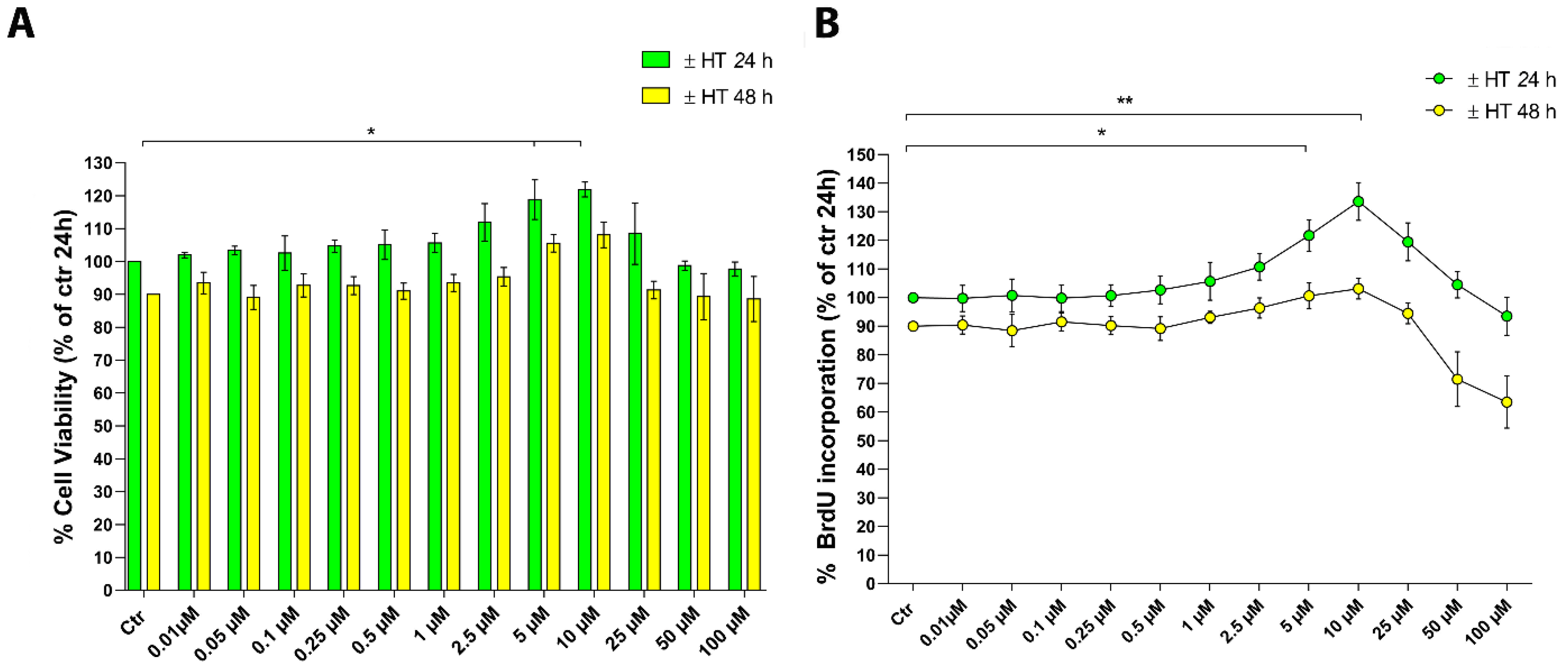
2.1. Evaluation of 3-Hydroxytyrosol (HT) Effect on Human Keratinocytes

The HaCaT cells were cultured with increasing concentrations of HT (0–100 µM) for 24 and 48 h and their cell viability was evaluated by the MTT (3-(4, 5-dimethylthiazolyl-2)-2, 5-diphenyltetrazolium bromide) assay. The results reported in Figure 1A show that HT has no influence on cell viability even at the highest doses. Interestingly, we identified an interval of concentrations (5 and 10 µM) where HT induced a little, but significant increase of cell viability at 24 h (* p < 0.05). Therefore, we evaluated whether the increase in cell viability was attributable to an increased number of cells, due to proliferation. HaCaT cells were cultured for the same times and with the same concentrations of HT and cell proliferation was assessed by BrdU incorporation (Figure 1B). HT 5 and 10 µM induced an increase in DNA synthesis only at 24 h, thus effectively stimulating cell proliferation (* p < 0.05 and ** p < 0.01) whereas at a longer time (48 h), the effect was not so marked, probably due to a depletion of nutrients in the culture medium.
We then decided to perform all subsequent experiments in the 1–10 µM range of concentrations and within 24 h.

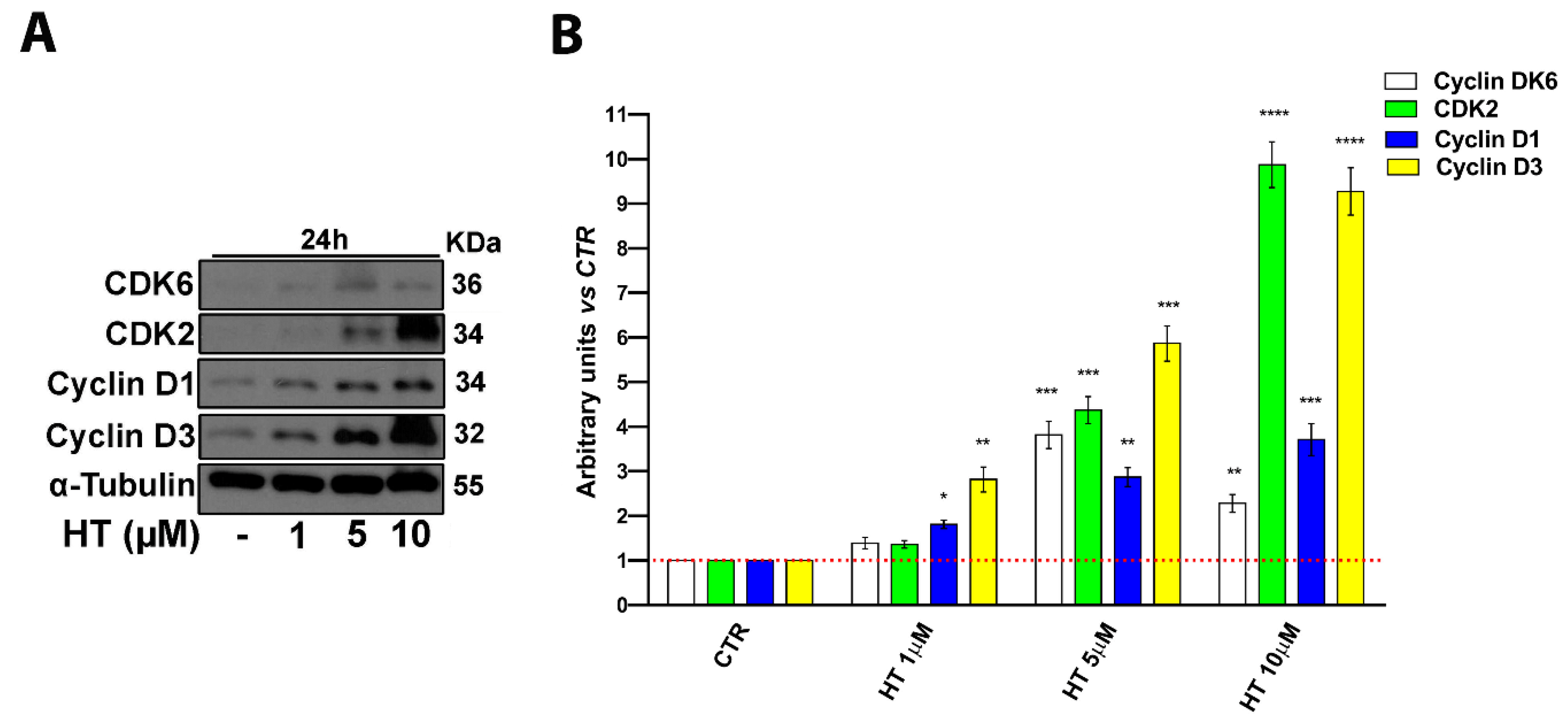
2.2. HT Induces Expression of Cell Cycle Control Proteins

We next investigated the influence of HT on cell cycle progression, focusing on the molecular pathways through which it carries out the observed biological effects. To this end, we determined by western blot analysis the status of the same proteins specifically involved in cell cycle progression (Figure 2). We treated cells with HT at the most effective doses, 5 and 10 µM for 24 h. According to the proliferation assay results, we observed that HT increased the expression of proteins involved in cell cycle transition from G1 to the S phase such as cyclin D1, D3, Cyclin Dependent Kinase 2 (CDK2), and Cyclin Dependent Kinase 6 (CDK6) (Figure 2A,B).

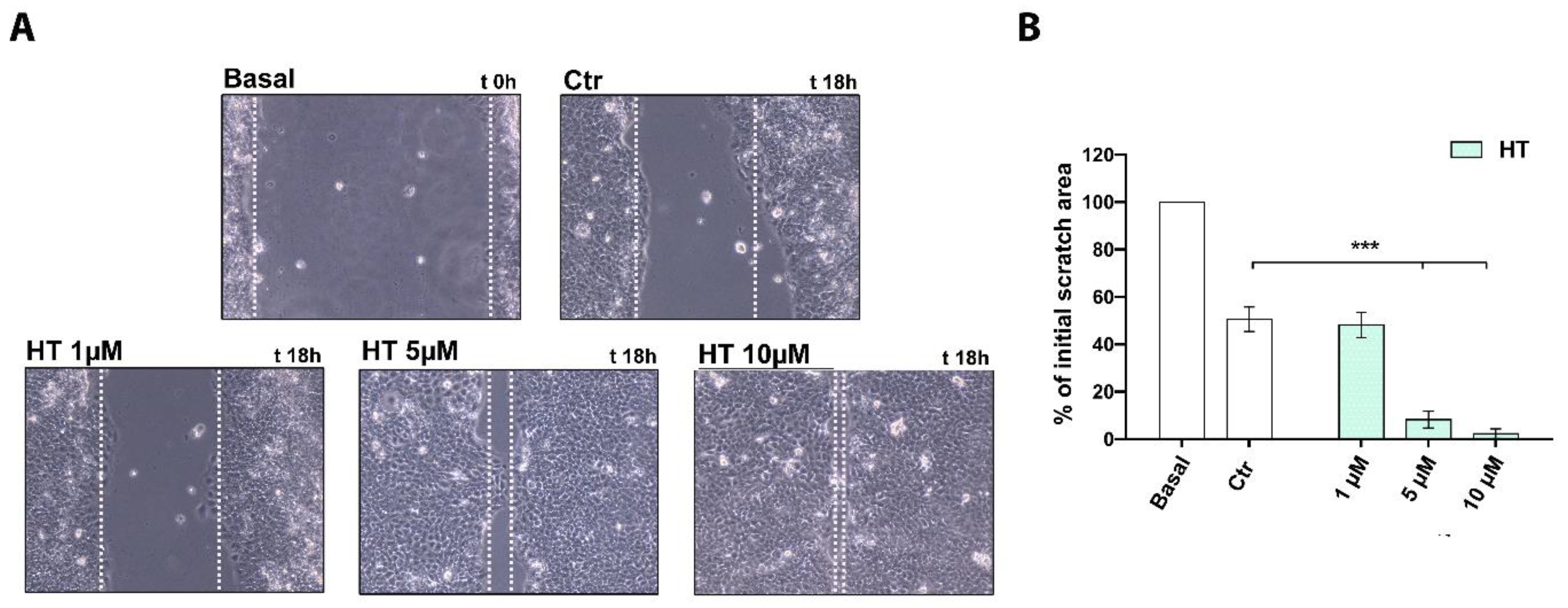
2.3. HT Improves the Migratory Capacity of HaCaT Keratinocytes

Keratinocyte cell migration is essential for the re-epithelialization both after superficial and deeper skin injury such as ulcers [37,38]. For this reason, in order to assess a potential effect of HT on the migratory function of HaCaT cells, we performed a scratch wound assay. The closure of the wounded area, carried out on a monolayer of confluent cells, was monitored over the time. After 18 h of treatment with HT, we observed an enhancement of wound healing at 5 and 10 µM (*** p < 0.001) with respect to the control and to a smaller concentration of HT (1 µM), as shown in Figure 3A. Indeed, the wound area, measured as the percentage of initial scratch at time 0, was almost completely closed after 18 h by HT at 5 and 10 µM with respect to untreated control (Figure 3B).

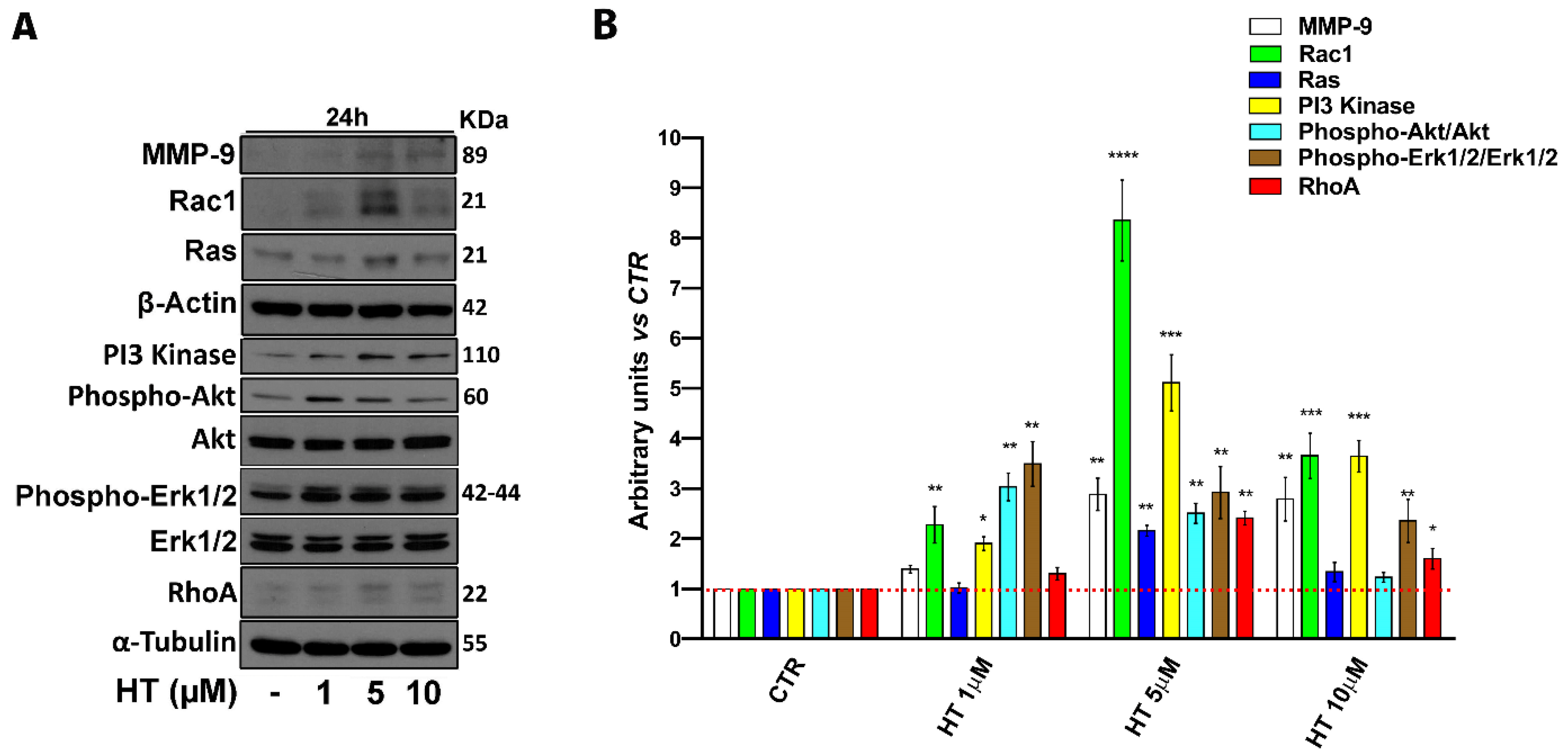
2.4. HT Induces Migration-Linked Proteins Expression

Regulation of epithelial cell migration involves several factors and for this process the activation of cytoskeletal remodeling signaling pathways is essential [38,39,40,41,42,43]. In order to establish the mechanism of HT action on the migration stimulation, we determined the expression of crucial proteins involved in the migration process by western blot. We treated cells with HT at concentrations of 1–10 µM for 24 h. We observed an upregulated expression of proteins that are implicated in cell adhesion, cytoskeletal dynamics, and migration such as extracellular regulated protein kinases (ERK), phosphatidylinositol 3-kinase (PI3 Kinase), protein kinase B (Akt), ras-related C3 botulinum toxin substrate 1 (Rac1), ras homolog family member A (RhoA), GTPase (Ras), but also the activation of matrix metalloproteinase-9 (MMP-9) required for the degradation of the extracellular matrix (Figure 4A,B). These data suggest that exposure to HT provided a driving force in human keratinocyte migration.

2.5. HT Contrasts Cytotoxicity and Apoptosis Induced by H2O2

To evaluate whether the HT concentrations identified in previous experiments (1–10 µM) could also show antioxidant activity in our experimental model, as previously reported in other cell lines for HT, we tested its effects against H2O2-induced cytotoxicity and apoptosis. HaCaT cells were exposed to H2O2 (0–800 µM) for 24 h, and the MTT assay was used as an indicator of cell viability (Figure 5A). We observed a dose-dependent decrease in cell viability following H2O2 treatment. Cell viability reduction was statistically significant already at 100 µM H2O2, whereas it was reduced to 65.1% at 200 µM H2O2 (Figure 5A). It is recognized that concentrations of H2O2 within 250 µM caused apoptotic death, while higher doses, up to 1000 µM of H2O2, resulted in an increase in overall cell death [44]. For this reason, we used 200 µM H2O2 in the subsequent experiments. We observed that 18 h pretreatment of HaCaT keratinocytes with HT at 5 and 10 µM significantly protected the cells from H2O2-induced cytotoxicity, improving cell viability. After exposure to 200 µM H2O2 for 6 h, cell viability dropped to 65.1% ± 5.7%, whereas it increased to 75.2% ± 3.3% and 83.2% ± 4.5% by pre-treatment with HT at 5 and 10 µM, respectively (Figure 5B). Subsequently, we performed a cell death analysis by annexin-V and propidium iodide double staining to corroborate the data obtained by the MTT assay. HT pretreatment before H2O2 exposure resulted in a significant reduction of apoptosis, in particular, inhibiting the percentage of early double positive apoptotic cells (Figure 5C).

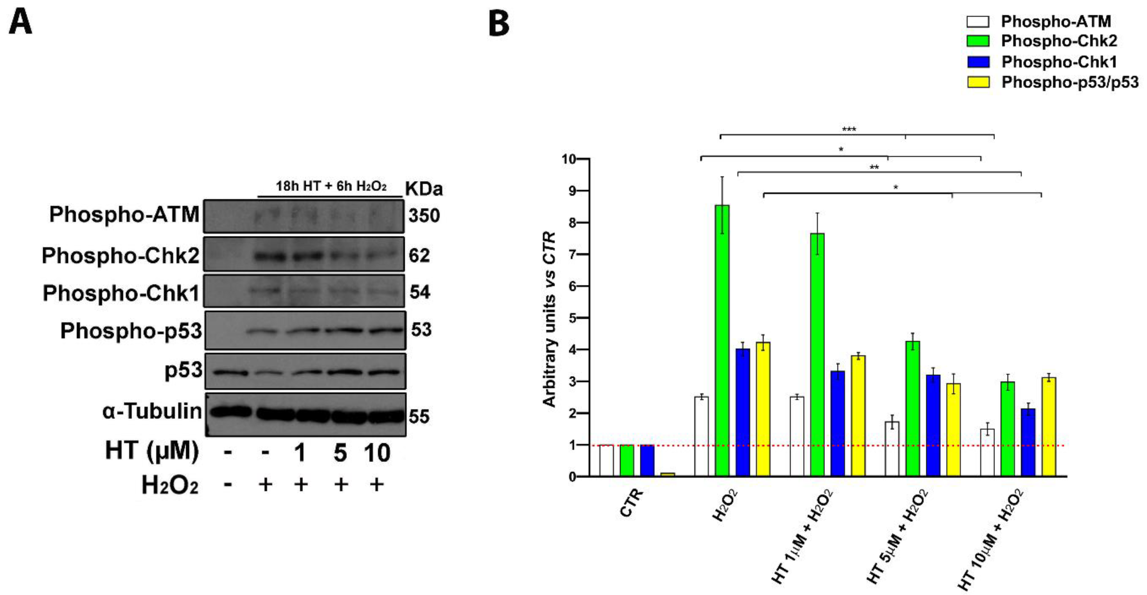
2.6. HT Prevents Activation of Key DNA Damage-Associated Proteins

ATM serine/threonine kinase (ATM), Checkpoint kinase 1 (Chk1), Checkpoint kinase 2 (Chk2), and p53 are known to play a pivotal role in the control of cell response to external damages, indeed, they are induced by free radicals and involved in apoptosis activation following DNA damage [45,46,47,48]. In order to elucidate the molecular pathways modulated by HT, using H2O2 to mimic oxidative stress-induced injury (OSI) within a short period, we performed a western blot analysis of ATM, Chk1, Chk2, and p53 proteins (Figure 6). We evaluated the phosphorylation status of these proteins in 18 h pretreated keratinocytes with HT at 1–10 µM, following H2O2 exposure (Figure 6).
Phospho-ATM, Phospho-Chk1, Phospho-Chk2, and p53 levels were increased by H2O2-treatment whereas HT treatment significantly reversed these effects at higher concentrations (5 and 10 µM) and the activation of these DNA damage signaling pathways (Figure 6A,B). The results showed that HT protects keratinocytes from the activation of the oxidative stress-induced DNA-damage pathway and subsequent apoptosis activation.

3. Discussion

HT, the most represented polyphenol contained in olive oil, has a strong biological relevance thanks to its antioxidant, anti-atherogenic, anti-inflammatory, neuroprotective, and antitumor activities, which have been demonstrated by accurate scientific studies both in vitro and in animal models, supporting its preventive and pharmacological potential [1,2,3,4,5]. With regard to dermatological diseases, recent studies have shown that the daily consumption of olive oil through the Mediterranean dietary pattern, is associated with a lower incidence of skin tumors, but also with a better healing of pressure ulcers, thanks to a reduction in oxidative damage, a better dermal reconstruction, and a reduction in collagen deposition time [25,26]. As already seen from a daily intake of olive oil, several scientific studies have reported that even olive oil creams applied topically have been proven to be effective for the treatment of patients with chronic wounds, having the ability to reduce the size, depth, and wound edges, along with associated pain, thanks to the antibacterial and anti-inflammatory effects of polyphenols [27,30]. In addition, it has been shown that HT may be a potential alternative therapeutic agent for the treatment of several autoimmune and inflammatory disorders. Indeed, in rheumatoid arthritis, it has the advantage of mitigating the systemic adverse effects of hydrocortisone [33]. Moreover, it displays anti-inflammatory and antioxidant benefits in the treatment of atopic dermatitis and psoriasis [35]. Despite its already known use in cosmetic formulations for antioxidant activities, the overall therapeutic potential of HT for the treatment of dermatological diseases has yet to be studied in detail, both in regard its beneficial activities and the molecular mechanisms implied.
Therefore, in our study, the effect of HT on human keratinocytes as an in vitro model was investigated in order to identify the dermatological properties of the molecule and provide solid biological bases for its transfer to the cosmetic or medical setting.
We assessed HT activity in wound healing phases that are not well-defined. The wound is a pathological condition with the destruction of the physiological microstructure and a loss of tissue function by a chemical, thermal, or physical injury [49,50]. The wound healing process is mediated by the activation of local and systemic cells that cooperate to restore skin integrity [51]. It is distinguished by three different phases: inflammatory, proliferative of reepithelization, and remodeling [52].
The first inflammatory phase is characterized by vasodilation, followed by neutrophils infiltration, monocyte activation, and macrophage penetration into the wound site [53]. A non-physiological inflammatory state, too long, can stop the process and block the transition to the second phase. Indeed, excessive levels of inflammatory cytokines such as tumor necrosis factor alpha (TNF-α) or interleukin 6 (IL-6), IL-8, and IL-1β are implicated in a wound healing delay [54]. In this first phase, HT acts as a powerful anti-inflammatory molecule that relieves the inflammatory state favoring the wound healing progression [50].
On the other hand, little is known about HT activity in the second phase of the wound healing process. Cell proliferation and migration are critical cellular events in the second phase. Fibroblasts, keratinocytes, and endothelial cells are the three main actors involved in the human skin regeneration. First, fibroblasts migrate to the wound site and degrade the temporary fibrin matrix laid by the inflammatory phase cells [51]. Then, fibroblasts proliferate to form the granulation tissue and the re-epithelialization of the wound by the epithelial cells takes place, which migrate to the new matrix and proliferate [55]. At the same time, during the second phase, a new blood vessel is formed by the endothelial cells [56]. An increase in the proliferation and migration rate of keratinocytes undoubtedly leads to fast wound healing. In this wound healing second phase, our data are relevant, indeed, we show how HT has proliferative and migratory properties on keratinocytes, which undoubtedly represent the predominant cell type in the epidermis and have a fundamental role for the process of complete healing through re-epithelization and dynamic skin renewal [57].
Once the lack of HT cytotoxicity was confirmed even in the keratinocyte cell line, we found for the first time that concentrations in a low micromolar range (5–10 μM) induce DNA synthesis, stimulating cell proliferation. Our first result indicates that HT induces keratinocyte proliferation by increasing the protein expression of cyclin-dependent kinase, mainly CDK2 and CDK6, key components of the cell cycle mechanism for the transition from the G1 to S phase [37]. It is important to note that even if we used HaCaT cells, an immortalized cell line, as the model, basal keratinocytes physiologically maintain the ability to proliferate and only most terminally differentiated keratinocytes of the suprabasal layer are in a quiescent state, but can proliferate under appropriate stimuli [40,55].
In addition to cell proliferation, an adequate cell migration is also required for the second phase of wound healing for minor, superficial, and basic skin lesions, but also for more complicated pathological states such as ulcers or pressure sores [38,39].
We investigated if HT treatment could also influence the cell migration process. Several signaling events have been described as involved in the regulation of cell migration, extension, and contraction of the cytoskeleton, and in our cell model, HT induced an increase in the migration rate supported by the activation of MMP-9, ERK, PI3 Kinase, Akt, Rac1, Ras, and RhoA important factors for stimulating cell migration and normal tissue remodeling [40,41,42,43,58]. Successful wound healing requires movement of keratinocytes through the extracellular matrix. This process is facilitated by the activation of the gelatinolytic activity of metalloproteases as MMP-9, which are nevertheless also involved in the stimulation of cell proliferation, downstream cytokines, and growth factors stimulated signaling pathways that converge on Ras and ERK activation. Our results suggest that HT could show beneficial effects in wound healing not only by increasing keratinocyte proliferation, but also inducing cell migration.
Oxidative stress is considered as a condition of imbalance in the reactive oxygen species (ROS) levels and the ability of biological systems to repair the resulting damage. Oxidative stress plays a crucial role in the onset of various diseases including allergic and inflammatory skin diseases such as atopic dermatitis, urticaria, and psoriasis [59,60] as well as in skin aging, and if it persists, it has been reported to inhibit cellular functions such as proliferation and migration, necessary for physiological wound healing [59].
Therefore, the regulation of skin ROS is essential for the maintenance of healthy skin homeostasis [60], but also for allowing a suitable wound healing [50]. We demonstrate the role of HT on the protection of cells by H2O2-induced DNA-damage and subsequent cell death. Our results confirm what is already reported in the scientific literature for the antioxidant activity of HT [36], but we demonstrated that this property is already evident at lower concentrations than previously reported (5 and 10 µM). Furthermore, as recent studies have revealed that HT has a very short half-life and the time required for its complete elimination is approximately 6 h [18,25], we believe that the effect shown by HT-treatment at 24 h (18 h + 6 h with H2O2) is not only due to the chemical effect of HT, but is attributable to the cellular response stimulated by HT. A rebalancing of the oxidative state is undoubtedly useful in reducing the destruction of the extracellular matrix (ECM) that provides nutrition for the microbes [61]. This scavenger activity of HT is added to the molecule’s ability to inhibit bacterial growth, as described in recent studies [50,62].
Furthermore, the second phase of the wound healing process includes a simultaneous angiogenesis necessary to restore the interrupted microcirculation necessary to support, with the supply of nutrients, the correct proliferation of the surrounding cells [56]. We and others have recently reported the proangiogenic activity of HT through the induction of cell proliferation and migration of vascular endothelial cells [63,64,65]. We observed that low concentrations of HT (1–5 µM), generally achieved after the ingestion of olive oil, are able to promote angiogenesis, inducing an increase in the expression of the vascular endothelial growth factor receptor (VEGF-R2) and activating the PI3K-Akt-eNOS protein pathway, recognized for its central role in angiogenesis [65]. In this context, it is conceivable that, beyond directly acting on keratinocytes and endothelial cells, HT could influence their reciprocal cross-talk in the multi-player milieu of wound healing, which depends on the integration of different signals and biological processes.
To date, nothing is known about the specific activity of HT in the last phase of the wound healing process, therefore further studies will be necessary in order to evaluate its potential in tissue remodeling and in maintaining balance between degradation and ECM synthesis. It will be necessary to investigate if HT also has a direct activity on fibroblasts and on keratinocyte–fibroblast interactions fundamental for the wound healing process. Moreover, it is important to determine if HT shows activity in the remodeling of the dermal structure during the formation of granulation tissue and if it plays a functional role in the transition from the fibroblast to the myofibroblast for the subsequent collagen deposition.
Our results allowed for a better understanding of HT beneficial activity in dermatological alterations using an in vitro model of keratinocyte. In addition to its anti-inflammatory and antioxidant activities, we provide, for the first time, evidence of increased cell proliferation and migration by identifying the proteins involved and the possible molecular mechanisms at the basis of these beneficial properties.
In addition, the results obtained have identified the best concentrations for its biological activities and have paved the way for the development of highly effective HT-based cosmeceuticals.
Further studies will undoubtedly be needed in order to design formulations that contain effective concentrations of HT and allow for a continuous presence of its active form after topical application as well as a proper permeation of all skin layers.

4. Materials and Methods

4.1. Chemicals and Materials

HT was purchased from Sigma-Aldrich Inc. (St Luis, MO, USA), solubilized in dimethyl sulfoxide (DMSO) (<0.001% in our assays). For western blot analysis, the following antibodies were used: mouse monoclonal antihuman CDK6, rabbit monoclonal antihuman CDK2, mouse monoclonal antihuman α-Tubulin, mouse monoclonal antihuman cyclin D3, rabbit monoclonal antihuman CD1, rabbit monoclonal anti-human phospho-ATM, rabbit monoclonal anti-human phospho-Chk1, rabbit monoclonal anti-human phospho-Chk2, rabbit anti-human phospho-p53, rabbit monoclonal anti-human phospho-p44/42 MAPK (p-Erk1/2; Thr202/Tyr204), rabbit monoclonal anti-human p44/42 MAPK (Erk1/2), rabbit monoclonal anti-human Phospho-Akt (p-Akt; Ser473), rabbit monoclonal anti-human Akt, rabbit monoclonal PI3-Kinase, rabbit monoclonal anti-human p53 (Cell Signaling Technology, Danvers, MA, USA). Mouse monoclonal MMP9, mouse monoclonal Rac1, rabbit polyclonal anti-human RhoA, rabbit monoclonal anti-human Ras, and rabbit polyclonal anti-human β-Actin were purchased from Abcam (Cambridge, UK). Secondary HRP-linked goat anti-mouse or goat anti-rabbit IgG were obtained from Cell Signaling Technology (Danvers, MA, USA).

4.2. Cells

Human immortalized keratinocytes (HaCaT) were grown in Dulbecco’s modified Eagle’s medium (DMEM, GIBCO, Grand Island, NY, USA) and supplemented as described in detail elsewhere [66]. Keratinocyte cell cultures were maintained at 37 °C in a humidified 5% CO2 atmosphere.

4.3. MTT Assay for Cell Viability Determination

HaCaT cells (6 × 10³/well) were cultured for 24 h into 96-well plates before the addition of HT at the indicated concentrations and were cultured for an additional 24–48 h at 37 °C. To examine cell viability, we employed the reduction of the MTT (3-(4, 5-dimethylthiazolyl-2)-2, 5-diphenyltetrazolium bromide) tetrazolium salts assay, as described in detail elsewhere [67]. All experiments were performed in triplicate, and the relative cell viability was expressed as percentage in comparison with the untreated control cells.

4.4. BrdU Assay for Cell Proliferation Determination

HaCaT cells (6 × 10³/well) were cultured for 24 h into 96-well plates before the addition of HT at the indicated concentrations and were cultured for an additional 24–48 h at 37 °C. BrdU incorporation into DNA (BrdU colorimetric assay kit; Roche Applied Science, South San Francisco, CA, USA) was used to measure cell proliferation, determined by an ELISA (enzyme-linked immunosorbent assay) plate reader (ThermoScientific, Waltham, MA, USA) at 450 nm as described in detail elsewhere [68]. All experiments were performed in triplicate, and the relative cell growth was expressed as a percentage in comparison with the untreated control cells.

4.5. Scratch Wound Healing Assay

For cell migration evaluation, HaCaT cells were plated in 6-well plates at a density of 5 × 10³ cells/well. When the confluent cells formed a homogeneous carpet and a vertical wound in the wells using a 200 µL tip was performed, culture medium containing HT at 1, 5, and 10 µM or the vehicle alone was added to the wells, after two washes to eliminate the cells detached. The wound area was recorded immediately and after 18 h through microscope analysis, as previously described [69] and quantified by Wimasis Image Analysis software (Onimagin Technologies Spa, Cordoba, Spain).

4.6. Apoptosis Analysis

Quantitative assessment of apoptosis of HaCaT cells was analyzed by anti-human annexin V (BioLegend, San Diego, CA, USA) using propidium iodide solution (PI) staining. Briefly, cells grown in p60 tissue culture plates for 24 h with HT at 1, 5, and 10 µM, H2O2 or combined as indicated, were harvested with trypsin, washed in phosphate buffer saline (PBS), and subjected to apoptosis determination by the procedure described in detail elsewhere [70].

4.7. Western Blot Analysis

Cells were grown in p100 tissue culture plates at a density of 2 × 10⁴ cells/cm2 for 24 h. Cells were then incubated with HT, H2O2 (for 6 h), or their combination (HT for 18 h and H2O2 for an additional 6 h) at the indicated concentrations. Subsequently, cells were washed with PBS, harvested, and lysed in ice-cold radioimmunoprecipitation assay (RIPA) lysis buffer (50 mM Tris-HCl, 150 mM NaCl, 0.5% Triton X-100, 0.5% deoxycholic acid, 10 mg/mL leupeptin, 2 mM phenylmethylsulfonyl fluoride, and 10 mg/mL aprotinin) and then assayed for Western Blot (WB) by the procedure described in detail elsewhere [71].

4.8. Statistical Analysis

Statistical analysis was performed by GraphPad prism 6.0 software for Windows (GraphPad software, San Diego, CA, USA). For each type of assay, data obtained from multiple experiments were calculated as mean ± (SD) and analyzed for statistical significance using the 2-tailed Student t-test for independent groups, or 2-way ANOVA followed by Tukey post-hoc correction for multiple comparisons. P values < 0.05 were considered significant. * p < 0.05, ** p < 0.01, *** p < 0.001, and **** p < 0.0001.

5. Conclusions

In conclusion, the novelty of our in vitro study is that we provide the first demonstration of how specific HT concentrations of 5 and 10 µM are able to promote keratinocyte cell migration and proliferation in vitro. HT has the potential to enhance re-epithelialization, at the same time, protecting the skin from the action of free radicals thanks to its anti-oxidant properties, also confirmed in our keratinocyte model. Our data highlight the great pharmaceutical potential for HT through multi-targeted actions, paving the way for possible cosmeceutical applications. Additional in vivo and clinical studies are required to develop and validate novel cosmeceuticals and pharmacological formulations based on HT.

Author Contributions

Conceptualization, M.A., S.P., and R.M.; Investigation, M.A. and M.C. (Marianna Citro); Resources R.M. and S.P.; Funding acquisition, R.M.; Writing—original draft, M.A. and S.P.; Writing—review and editing M.A., S.P., M.C. (Marianna Citro), M.C. (Mariella Caputo), and R.M. All authors have read and agreed to the published version of the manuscript.

Funding

This study was supported by funds provided by the University of Salerno (R.M. FARB 2017) and by the Associazione Italiana per la Ricerca sul Cancro—Fondazione Cariplo (S.P. AIRC TRIDEO 2015 No. 17216). S.P. and M.C. (Mariella Caputo) were supported by “Piattaforme Tecnologiche Di Ricerca Collaborativa Per La Lotta Alle Patologie Oncologiche” Por Campania Fesr 2014–2020 Asse 1 Obbiettivo Specifico 1.2 System Innovation For Cancer Early Diagnosis SICED.

Institutional Review Board Statement

Not applicable.

Informed Consent Statement

Not applicable.

Data Availability Statement

The data presented in this study are available on request from the corresponding author.

Conflicts of Interest

The authors declare no competing financial interest.

References

  1. Georgoulis, M.; Kontogianni, M.D.; Yiannakouris, N. Mediterranean diet and diabetes: Prevention and treatment. Nutrients 2014, 6, 1406–1423. [Google Scholar] [CrossRef] [Green Version]
  2. Mentella, M.C.; Scaldaferri, F.; Ricci, C.; Gasbarrini, A.; Miggiano, G.A.D. Cancer and Mediterranean Diet: A Review. Nutrients 2019, 11, 2059. [Google Scholar] [CrossRef] [Green Version]
  3. Martínez-González, M.A.; Gea, A.; Ruiz-Canela, M. The Mediterranean Diet and Cardiovascular Health. Circ. Res. 2019, 124, 779–798. [Google Scholar] [CrossRef]
  4. Finicelli, M.; Squillaro, T.; Di Cristo, F.; Di Salle, A.; Melone, M.A.B.; Galderisi, U.; Peluso, G. Metabolic syndrome, Mediterranean diet, and polyphenols: Evidence and perspectives. J. Cell Physiol. 2019, 234, 5807–5826. [Google Scholar] [CrossRef] [PubMed]
  5. Gardener, H.; Caunca, M.R. Mediterranean Diet in Preventing Neurodegenerative Diseases. Curr. Nutr. Rep. 2018, 7, 10–20. [Google Scholar] [CrossRef]
  6. Maruca, A.; Catalano, R.; Bagetta, D.; Mesiti, F.; Ambrosio, F.A.; Romeo, I.; Moraca, F.; Rocca, R.; Ortuso, F.; Artese, A.; et al. The Mediterranean Diet as source of bioactive compounds with multi-targeting anti-cancer profile. Eur. J. Med. Chem. 2019, 1, 111579. [Google Scholar] [CrossRef] [PubMed]
  7. Schwingshackl, L.; Morze, J.; Hoffmann, G. Mediterranean diet and health status: Active ingredients and pharmacological mechanisms. Br. J. Pharmacol. 2020, 177, 1241–1257. [Google Scholar] [CrossRef] [Green Version]
  8. Arauna, D.; Furrianca, M.; Espinosa-Parrilla, Y.; Fuentes, E.; Alarcón, M.; Palomo, I. Natural Bioactive Compounds As Protectors Of Mitochondrial Dysfunction In Cardiovascular Diseases And Aging. Molecules 2019, 24, 4259. [Google Scholar] [CrossRef] [Green Version]
  9. Jimenez-Lopez, C.; Carpena, M.; Lourenço-Lopes, C.; Gallardo-Gomez, M.; Lorenzo, J.M.; Barba, F.J.; Prieto, M.A.; Simal-Gandara, J. Bioactive Compounds and Quality of Extra Virgin Olive Oil. Foods 2020, 9, 1014. [Google Scholar] [CrossRef] [PubMed]
  10. Gorzynik-Debicka, M.; Przychodzen, P.; Cappello, F.; Kuban-Jankowska, A.; Marino Gammazza, A.; Knap, N.; Wozniak, M.; Gorska-Ponikowska, M. Potential Health Benefits of Olive Oil and Plant Polyphenols. Int. J. Mol. Sci. 2018, 19, 686. [Google Scholar] [CrossRef] [PubMed] [Green Version]
  11. Romani, A.; Ieri, F.; Urciuoli, S.; Noce, A.; Marrone, G.; Nediani, C.; Bernini, R. Health Effects of Phenolic Compounds Found in Extra-Virgin Olive Oil, By-Products, and Leaf of Olea europaea L. Nutrients 2019, 11, 1776. [Google Scholar] [CrossRef] [PubMed] [Green Version]
  12. Serreli, G.; Deiana, M. Extra Virgin Olive Oil Polyphenols: Modulation of Cellular Pathways Related to Oxidant Species and Inflammation in Aging. Cells 2020, 9, 478. [Google Scholar] [CrossRef] [Green Version]
  13. Leri, M.; Scuto, M.; Ontario, M.L.; Calabrese, V.; Calabrese, E.J.; Bucciantini, M.; Stefani, M. Healthy Effects of Plant Polyphenols: Molecular Mechanisms. Int. J. Mol. Sci. 2020, 21, 1250. [Google Scholar] [CrossRef] [Green Version]
  14. Hu, T.; He, X.W.; Jiang, J.G.; Xu, X.L. Hydroxytyrosol and its potential therapeutic effects. J. Agric. Food Chem. 2014, 62, 1449–1455. [Google Scholar] [CrossRef] [PubMed]
  15. Tejada, S.; Pinya, S.; Del Mar Bibiloni, M.; Tur, J.A.; Pons, A.; Sureda, A. Cardioprotective Effects of the Polyphenol Hydroxytyrosol from Olive Oil. Curr. Drug Targets 2017, 18, 1477–1486. [Google Scholar] [CrossRef]
  16. Karković Marković, A.; Torić, J.; Barbarić, M.; Jakobušić Brala, C. Hydroxytyrosol, Tyrosol and Derivatives and Their Potential Effects on Human Health. Molecules 2019, 24, 2001. [Google Scholar] [CrossRef] [Green Version]
  17. Peyrol, J.; Riva, C.; Amiot, M.J. Hydroxytyrosol in the Prevention of the Metabolic Syndrome and Related Disorders. Nutrients 2017, 9, 306. [Google Scholar] [CrossRef] [PubMed]
  18. Robles-Almazan, M.; Pulido-Moran, M.; Moreno-Fernandez, J.; Ramirez-Tortosa, C.; Rodriguez-Garcia, C.; Quiles, J.L.; Ramirez-Tortosa, M. Hydroxytyrosol: Bioavailability, toxicity, and clinical applications. Food Res. Int. 2018, 105, 654–667. [Google Scholar] [CrossRef]
  19. D’Angelo, S.; Manna, C.; Migliardi, V.; Mazzoni, O.; Morrica, P.; Capasso, G.; Pontoni, G.; Galletti, P.; Zappia, V. Pharmacokinetics and metabolism of hydroxytyrosol, a natural antioxidant from olive oil. Drug Metab. Dispos. 2001, 29, 1492–1498. [Google Scholar] [PubMed]
  20. European Commission Regulation EC No. 1924/2006 on Nutrition and Health Claims Made on Foods. Off. J. Eur. Union. 2012, L310, 36. Available online: https://eur-lex.europa.eu/legal-content/EN/TXT/PDF/?uri=CELEX:02006R1924-20121129&from=EN (accessed on 7 May 2020).
  21. European Food Safety Authority (EFSA). Safety of hydroxytyrosol as a novel food pursuant to Regulation (EC) No 258/97. EFSA J. 2017, 15, 4728. [Google Scholar]
  22. Visioli, F.; Bellomo, G.; Galli, C. Free radical-scavenging properties of olive oil polyphenols. Biochem. Biophys. Res. Commun. 1998, 247, 60–64. [Google Scholar] [CrossRef]
  23. Salucci, S.; Burattini, S.; Battistelli, M.; Buontempo, F.; Canonico, B.; Martelli, A.M.; Papa, S.; Falcieri, E. Tyrosol prevents apoptosis in irradiated keratinocytes. J. Dermatol. Sci. 2015, 80, 61–68. [Google Scholar] [CrossRef]
  24. Avola, R.; Graziano, A.C.E.; Pannuzzo, G.; Bonina, F.; Cardile, V. Hydroxytyrosol from olive fruits prevents blue-light-induced damage in human keratinocytes and fibroblasts. J. Cell Physiol. 2019, 234, 9065–9076. [Google Scholar] [CrossRef]
  25. Rodríguez-Morató, J.; Boronat, A.; Kotronoulas, A.; Pujadas, M.; Pastor, A.; Olesti, E.; Pérez-Mañá, C.; Khymenets, O.; Fitó, M.; Farré, M.; et al. Metabolic disposition and biological significance of simple phenols of dietary origin: Hydroxytyrosol and tyrosol. Drug Metab. Rev. 2016, 48, 218–236. [Google Scholar] [CrossRef] [Green Version]
  26. Donato-Trancoso, A.; Monte-Alto-Costa, A.; Romana-Souza, B. Olive oil-induced reduction of oxidative damage and inflammation promotes wound healing of pressure ulcers in mice. J. Dermatol. Sci. 2016, 83, 60–69. [Google Scholar] [CrossRef] [PubMed]
  27. Panahi, Y.; Izadi, M.; Sayyadi, N.; Rezaee, R.; Jonaidi-Jafari, N.; Beiraghdar, F.; Zamani, A.; Sahebkar, A. Comparative trial of Aloe vera/olive oil combination cream versus phenytoin cream in the treatment of chronic wounds. J. Wound Care 2015, 24, 459–460, 462–465. [Google Scholar] [CrossRef]
  28. Edraki, M.; Akbarzadeh, A.; Hosseinzadeh, M.; Tanideh, N.; Salehi, A.; Koohi-Hosseinabadi, O. Healing effect of sea buckthorn, olive oil, and their mixture on full-thickness burn wounds. Adv. Skin Wound Care 2014, 27, 317–323. [Google Scholar] [CrossRef] [PubMed]
  29. Zahmatkesh, M.; Manesh, M.J.; Babashahabi, R. Effect of Olea ointment and Acetate Mafenide on burn wounds-A randomized clinical trial. Iran J. Nurs. Midwifery Res. 2015, 20, 599–603. [Google Scholar]
  30. Poljšak, N.; Kreft, S.; Kočevar Glavač, N. Vegetable butters and oils in skin wound healing: Scientific evidence for new opportunities in dermatology. Phytother. Res. 2020, 34, 254–269. [Google Scholar] [CrossRef] [PubMed]
  31. Ng, S.F.; Tan, L.S.; Buang, F. Transdermal anti-inflammatory activity of bilayer film containing olive compound hydroxytyrosol: Physical assessment, in vivo dermal safety and efficacy study in Freund’s adjuvant-induced arthritic rat model. Drug Dev. Ind. Pharm. 2017, 43, 108–119. [Google Scholar] [CrossRef]
  32. Alonso, C.; Lucas, R.; Barba, C.; Marti, M.; Rubio, L.; Comelles, F.; Morales, J.C.; Coderch, L.; Parra, J.L. Skin delivery of antioxidant surfactants based on gallic acid and hydroxytyrosol. J. Pharm. Pharmacol. 2015, 67, 900–908. [Google Scholar] [CrossRef]
  33. Hussain, Z.; Katas, H.; Mohd Amin, M.C.; Kumolosasi, E.; Buang, F.; Sahudin, S. Self-assembled polymeric nanoparticles for percutaneous co-delivery of hydrocortisone/hydroxytyrosol: An ex vivo and in vivo study using an NC/Nga mouse model. Int. J. Pharm. 2013, 444, 109–119. [Google Scholar] [CrossRef]
  34. Siddique, M.I.; Katas, H.; Jamil, A.; Mohd Amin, M.C.I.; Ng, S.F.; Zulfakar, M.H.; Nadeem, S.M. Potential treatment of atopic dermatitis: Tolerability and safety of cream containing nanoparticles loaded with hydrocortisone and hydroxytyrosol in human subjects. Drug Deliv. Transl. Res. 2019, 9, 469–481. [Google Scholar] [CrossRef]
  35. Souto, E.B.; Dias-Ferreira, J.; Oliveira, J.; Sanchez-Lopez, E.; Lopez-Machado, A.; Espina, M.; Garcia, M.L.; Souto, S.B.; Martins-Gomes, C.; Silva, A.M. Trends in Atopic Dermatitis-From Standard Pharmacotherapy to Novel Drug Delivery Systems. Int. J. Mol. Sci. 2019, 20, 5659. [Google Scholar] [CrossRef] [Green Version]
  36. Serreli, G.; Deiana, M. Biological Relevance of Extra Virgin Olive Oil Polyphenols Metabolites. Antioidants (Basel) 2018, 7, 170. [Google Scholar] [CrossRef] [PubMed] [Green Version]
  37. Long, M.; Cai, L.; Li, W.; Zhang, L.; Guo, S.; Zhang, R.; Zheng, Y.; Liu, X.; Wang, M.; Zhou, X.; et al. DPP-4 Inhibitors Improve Diabetic Wound Healing via Direct and Indirect Promotion of Epithelial-Mesenchymal Transition and Reduction of Scarring. Diabetes 2018, 67, 518–531. [Google Scholar] [CrossRef] [PubMed] [Green Version]
  38. Kirfel, G.; Herzog, V. Migration of epidermal keratinocytes: Mechanisms, regulation, and biological significance. Protoplasma 2004, 223, 67–78. [Google Scholar] [CrossRef]
  39. Horikoshi, Y.; Kamizaki, K.; Hanaki, T.; Morimoto, M.; Kitagawa, Y.; Nakaso, K.; Kusumoto, C.; Matsura, T. α-Tocopherol promotes HaCaT keratinocyte wound repair through the regulation of polarity proteins leading to the polarized cell migration. Biofactors 2018, 44, 180–191. [Google Scholar] [CrossRef] [PubMed]
  40. Li, J.; Chen, L.; Xu, J.; Xie, Z.; Xu, Y.; Jiang, P.; Duan, B.; Huang, X.; Feng, F.; Liu, W. Effects of Periploca forrestii Schltr on wound healing by Src meditated Mek/Erk and PI3K/Akt signals. J. Ethnopharmacol. 2019, 237, 116–127. [Google Scholar] [CrossRef] [PubMed]
  41. Ritto, D.; Tanasawet, S.; Singkhorn, S.; Klaypradit, W.; Hutamekalin, P.; Tipmanee, V.; Sukketsiri, W. Astaxanthin induces migration in human skin keratinocytes via Rac1 activation and RhoA inhibition. Nutr. Res. Pract. 2017, 11, 275–280. [Google Scholar] [CrossRef] [PubMed] [Green Version]
  42. Charvat, S.; Chignol, M.C.; Souchier, C.; Le Griel, C.; Schmitt, D.; Serres, M. Cell migration and MMP-9 secretion are increased by epidermal growth factor in HaCaT-ras transfected cells. Exp. Dermatol. 1998, 7, 184–190. [Google Scholar] [CrossRef]
  43. Patruno, A.; Ferrone, A.; Costantini, E.; Franceschelli, S.; Pesce, M.; Speranza, L.; Amerio, P.; D’Angelo, C.; Felaco, M.; Grilli, A.; et al. Extremely low-frequency electromagnetic fields accelerates wound healing modulating MMP-9 and inflammatory cytokines. Cell. Prolif. 2018, 51, e12432. [Google Scholar] [CrossRef] [Green Version]
  44. Turner, N.A.; Xia, F.; Azhar, G.; Zhang, X.; Liu, L.; Wei, J.Y. Oxidative stress induces DNA fragmentation and caspase activation via the c-Jun NH2-terminal kinase pathway in H9c2 cardiac muscle cells. J. Mol. Cell Cardiol. 1998, 30, 1789–1801. [Google Scholar] [CrossRef]
  45. Guo, Z.; Kozlov, S.; Lavin, M.F.; Person, M.D.; Paull, T.T. ATM activation by oxidative stress. Science 2010, 330, 517–521. [Google Scholar] [CrossRef] [PubMed] [Green Version]
  46. Reissig, K.; Silver, A.; Hartig, R.; Schinlauer, A.; Walluscheck, D.; Guenther, T.; Siedentopf, S.; Ross, J.; Vo, D.K.; Roessner, A.; et al. Chk1 Promotes DNA Damage Response Bypass following Oxidative Stress in a Model of Hydrogen Peroxide-Associated Ulcerative Colitis through JNK Inactivation and Chromatin Binding. Oxid. Med. Cell Longev. 2017, 2017, 9303158. [Google Scholar] [CrossRef] [Green Version]
  47. Hsu, P.C.; Gopinath, R.K.; Hsueh, Y.A.; Shieh, S.Y. CHK2-mediated regulation of PARP1 in oxidative DNA damage response. Oncogene 2019, 38, 1166–1182. [Google Scholar] [CrossRef]
  48. Wang, P.; Zhang, S.D.; Jiao, J.; Wang, W.; Yu, L.; Zhao, X.L.; Wang, L.T.; Meng, D.; Fu, Y.J. ROS -mediated p53 activation by juglone enhances apoptosis and autophagy in vivo and in vitro. Toxicol. Appl. Pharmacol. 2019, 379, 114647. [Google Scholar] [CrossRef] [PubMed]
  49. Slominski, A.; Wortsman, J. Neuroendocrinology of the skin. Endocr. Rev. 2000, 21, 457–487. [Google Scholar] [CrossRef] [PubMed]
  50. Utami, N.D.; Nordin, A.; Katas, H.; Bt Hj Idrus, R.; Fauzi, M.B. Molecular Action of Hydroxytyrosol in Wound Healing: An In Vitro Evidence-Based Review. Biomolecules 2020, 10, 1397. [Google Scholar] [CrossRef]
  51. Martin, P.; Nunan, R. Cellular and molecular mechanisms of repair in acute and chronic wound healing. Br. J. Dermatol. 2015, 173, 370–378. [Google Scholar] [CrossRef]
  52. Broughton, G., II; Janis, J.E.; Attinger, C.E. Wound healing: An overview. Plast. Reconstr. Surg. 2006, 117, 1e-S–32e-S. [Google Scholar] [CrossRef] [PubMed]
  53. Larouche, J.; Sheoran, S.; Maruyama, K.; Martino, M.M. Immune regulation of skin wound healing: Mechanisms and novel therapeutic targets. Adv. Wound Care 2018, 7, 209–231. [Google Scholar] [CrossRef] [PubMed]
  54. Basso, F.G.; Pansani, T.N.; Turrioni, A.P.S.; Soares, D.G.; Costa, C.A.D.S.; Hebling, J. Tumor necrosis factor-alpha and interleukins IL-1ß, IL-6 and IL-8 impair in vitro migration and induce apoptosis of gingival fibroblasts and epithelial cells, delaying wound healing. J. Periodontol. 2016, 87, 990–996. [Google Scholar] [CrossRef]
  55. Landen, N.X.; Li, D.; Stahle, M. Transition from inflammation to proliferation: A critical step during wound healing. Cell. Mol. Life Sci. 2016, 73, 3861–3885. [Google Scholar] [CrossRef] [PubMed] [Green Version]
  56. Sorg, H.; Tilkorn, D.J.; Mirastschijski, U.; Hauser, J.; Kraemer, R. Pantha rei: Neovascularization, angiogenesis and nutritive perfusion in wound healing. Eur. Surg. Res. 2018, 59, 232–241. [Google Scholar] [CrossRef]
  57. Mi, B.; Liu, J.; Liu, G.; Zhou, W.; Liu, Y.; Hu, L.; Xiong, L.; Ye, S.; Wu, Y. Icariin promotes wound healing by enhancing the migration and proliferation of keratinocytes via the AKT and ERK signaling pathway. Int. J. Mol. Med. 2018, 42, 831–838. [Google Scholar] [CrossRef]
  58. Misiura, M.; Baszanowska, W.; Ościłowska, I.; Pałka, J.; Miltyk, W. Prolidase Stimulates Proliferation and Migration through Activation of the PI3K/Akt/mTOR Signaling Pathway in Human Keratinocytes. Int. J. Mol. Sci. 2020, 21, 9243. [Google Scholar] [CrossRef]
  59. Okayama, Y. Oxidative stress in allergic and inflammatory skin diseases. Curr. Drug Targets Inflamm. Allergy 2005, 4, 517–519. [Google Scholar] [CrossRef]
  60. Dunaway, S.; Odin, R.; Zhou, L.; Ji, L.; Zhang, Y.; Kadekaro, A.L. Natural Antioxidants: Multiple Mechanisms to Protect Skin From Solar Radiation. Front. Pharmacol. 2018, 9, 392. [Google Scholar] [CrossRef] [PubMed] [Green Version]
  61. Grant, S.S.; Hung, D.T. Persistent bacterial infections, antibiotic tolerance, and the oxidative stress response. Virulence 2013, 4, 273–283. [Google Scholar] [CrossRef] [Green Version]
  62. Crisante, F.; Taresco, V.; Donelli, G.; Vuotto, C.; Martinelli, A.; D’Illario, L.; Pietrelli, L.; Francolini, I.; Piozzi, A. Antioxidant hydroxytyrosol-based polyacrylate with antimicrobial and antiadhesive activity versus staphylococcus epidermidis. Adv. Microbiol. Infect. Dis. Public Health 2015, 901, 25–36. [Google Scholar]
  63. Zrelli, H.; Matsuoka, M.; Kitazaki, S.; Araki, M.; Kusunoki, M.; Zarrouk, M.; Miyazaki, H. Hydroxytyrosol induces proliferation and cytoprotection against oxidative injury in vascular endothelial cells: Role of Nrf2 activation and HO-1 induction. J. Agric. Food Chem. 2011, 59, 4473–4482. [Google Scholar] [CrossRef] [PubMed]
  64. Zrelli, H.; Kusunoki, M.; Miyazaki, H. Role of hydroxytyrosol-dependent regulation of HO-1 expression in promoting wound healing of vascular endothelial cells via Nrf2 de novo synthesis and stabilization. Phytother. Res. 2015, 29, 1011–1018. [Google Scholar] [CrossRef]
  65. Abate, M.; Pisanti, S.; Caputo, M.; Citro, M.; Vecchione, C.; Martinelli, R. 3-Hydroxytyrosol Promotes Angiogenesis In Vitro by Stimulating Endothelial Cell Migration. Int. J. Mol. Sci. 2020, 21, 3657. [Google Scholar] [CrossRef]
  66. Ciaglia, E.; Malfitano, A.M.; Laezza, C.; Fontana, A.; Nuzzo, G.; Cutignano, A.; Abate, M.; Pelin, M.; Sosa, S.; Bifulco, M.; et al. Immuno-Modulatory and Anti-Inflammatory Effects of Dihydrogracilin A, a Terpene Derived from the Marine Sponge Dendrilla membranosa. Int. J. Mol. Sci. 2017, 18, 1643. [Google Scholar] [CrossRef] [PubMed] [Green Version]
  67. Chicca, A.; Arena, C.; Bertini, S.; Gado, F.; Ciaglia, E.; Abate, M.; Digiacomo, M.; Lapillo, M.; Poli, G.; Bifulco, M.; et al. Polypharmacological profile of 1,2-dihydro-2-oxo-pyridine-3-carboxamides in the endocannabinoid system. Eur. J. Med. Chem. 2018, 154, 155–171. [Google Scholar] [CrossRef] [PubMed]
  68. Ciaglia, E.; Abate, M.; Laezza, C.; Pisanti, S.; Vitale, M.; Seneca, V.; Torelli, G.; Franceschelli, S.; Catapano, G.; Gazzerro, P.; et al. Antiglioma effects of N6-isopentenyladenosine, an endogenous isoprenoid end product, through the downregulation of epidermal growth factor receptor. Int. J. Cancer 2017, 140, 959–972. [Google Scholar] [CrossRef] [PubMed] [Green Version]
  69. Abate, M.; Pepe, G.; Randino, R.; Pisanti, S.; Basilicata, M.G.; Covelli, V.; Bifulco, M.; Cabri, W.; D’Ursi, A.M.; Campiglia, P.; et al. Ganoderma lucidum Ethanol Extracts Enhance Re-Epithelialization and Prevent Keratinocytes from Free-Radical Injury. Pharmaceuticals 2020, 13, 224. [Google Scholar] [CrossRef] [PubMed]
  70. Abate, M.; Laezza, C.; Pisanti, S.; Torelli, G.; Seneca, V.; Catapano, G.; Montella, F.; Ranieri, R.; Notarnicola, M.; Gazzerro, P.; et al. Deregulated expression and activity of Farnesyl Diphosphate Synthase (FDPS) in Glioblastoma. Sci. Rep. 2017, 7, 14123. [Google Scholar] [CrossRef] [Green Version]
  71. Ciaglia, E.; Grimaldi, M.; Abate, M.; Scrima, M.; Rodriquez, M.; Laezza, C.; Ranieri, R.; Pisanti, S.; Ciuffreda, P.; Manera, C.; et al. The isoprenoid derivative N6 -benzyladenosine CM223 exerts antitumor effects in glioma patient-derived primary cells through the mevalonate pathway. Br. J. Pharmacol. 2017, 174, 2287–2301. [Google Scholar] [CrossRef] [PubMed] [Green Version]
Figure 1. Evaluation of 3-hydroxytyrosol (HT) effect in HaCaT keratinocytes. Cells were cultured in the presence of the indicated HT concentrations (0–100 µM) for 24 or 48 h before the MTT assay (A) or BrdU incorporation (B). Results are expressed as mean (± SD) and are representative of four independent experiments carried out in triplicate. Data are reported as percentage vs. control (untreated cells at 24 h or 48 h) (2-way ANOVA, * p < 0.05, ** p < 0.01 vs. control).
Ijms 22 02438 g001
Figure 2. HT induced expression of cell cycle progression proteins. (A) Western blot analysis of cyclin D1, CDK6, CDK2, and cyclin D3 in whole cell extracts from HaCaT cells cultured for 24 h with HT 1–10 µM. α-Tubulin was used as a control for protein loading. The panel shows a representative western blot of three different experiments with similar results. (B) Histograms represent mean (±SD) in densitometry units of scanned immunoblots from three different experiments (2-way ANOVA, * p < 0.05, ** p < 0.01, *** p < 0.001, and **** p < 0.0001).
Ijms 22 02438 g002
Figure 3. Improvement of the migratory capacity of HaCaT cells exposed to HT. (A) Wound healing assay was carried out in HaCaT cells treated for 18 h with HT (1–10 µM) in complete medium. Light microscope images shown are representative of four independent experiments. Dotted white lines indicate the wounded area from the initial scratch. Magnification ×200; (B) Bar columns correspond to the mean scratch area expressed as percentage respect to basal time point area. The measurement was carried out in four different experiments. Results are shown as mean (± SD) (2-way ANOVA, *** p < 0.001).
Ijms 22 02438 g003
Figure 4. HT induces migration protein expression in HaCaT cells. (A) Western blot analysis of MMP-9, Phospho Erk1/2, Erk1/2, Phospho-Akt, Akt, PI3 Kinase, RhoA, Rac1, and Ras in whole cell extracts from HaCaT cells treated for 24 h with HT 1–10 µM. β-Actin and α-Tubulin were used as the control of protein loading. The panel shows a representative western blot of four different experiments with similar results. (B) Histograms represent mean (± SD) in densitometry units of scanned immunoblots from four different experiments (2-way ANOVA, * p < 0.05, ** p < 0.01, *** p < 0.001, and **** p < 0.0001).
Ijms 22 02438 g004
Figure 5. HT ameliorated cytotoxicity and apoptosis induced by H2O2 in keratinocytes. (A) HaCaT cells were cultured for 6 h in the presence of H2O2 (25–800 µM) before the MTT assay. The results are expressed as means ± SD of three independent experiments and reported as percentage vs. the untreated control (2-way ANOVA, * p < 0.05, ** p < 0.01, and *** p < 0.001). (B) HaCaT cells were cultured for 18 h in the presence of HT (1–10 µM) before treatment with H2O2 for 6 h. The results are expressed as means ± SD of three independent experiments reported as percentage vs. the untreated control (2-way ANOVA, * p < 0.05 and ** p < 0.01 vs. control). (C) Flow cytometric analysis of annexin V and propidium iodide (PI) double staining in HT-pretreated HaCaT cells for 18 h after H2O2 exposure for 6 h. Bars indicate the total percentage of early (annexin V-positive cells/PI-negative cells) and late apoptotic events (annexin V/PI-double positive cells) as well as necrotic cells (annexin V-negative cells/PI-positive cells). The results are representative of three independent experiments and expressed as mean ± SD (ANOVA, * p < 0.05 and ** p < 0.01).
Ijms 22 02438 g005
Figure 6. HT prevented the activation of cell death molecular pathways. (A) Western blot analysis of Phospho-ATM, Phospho-Chk1, Phospho-Chk2, and p53 (total and phosphorylated) in whole cell extracts from HaCaTs for 18 h in the presence of HT (1–10 µM) and H2O2 (200 µM) for 6 h. α-Tubulin was used as a control for protein loading. The panel shows a representative western blot of three different experiments performed with similar results. (B) Histograms represent mean (±SD) in densitometry units of scanned immunoblots of three different experiments (2-way ANOVA, * p < 0.05, ** p < 0.01, and *** p < 0.001).
Ijms 22 02438 g006
Publisher’s Note: MDPI stays neutral with regard to jurisdictional claims in published maps and institutional affiliations.

