
Secondary Metabolites of Plants as Modulators of Endothelium Functions


1
Department of Physiology, Faculty of Medicine, Masaryk University, 625 00 Brno, Czech Republic
2
International Clinical Research Center, St. Anne’s University Hospital Brno, 656 91 Brno, Czech Republic
*
Author to whom correspondence should be addressed.
Int. J. Mol. Sci. 2021, 22(5), 2533; https://0-doi-org.brum.beds.ac.uk/10.3390/ijms22052533
Submission received: 20 January 2021
/
Revised: 20 February 2021
/
Accepted: 25 February 2021
/
Published: 3 March 2021
(This article belongs to the Special Issue Animal and Plant Cell–Tissue, Organ Specialization and Function: Investigational, Experimental and Medical Aspects 2.0)
Abstract
:According to the World Health Organization, cardiovascular diseases are the main cause of death worldwide. They may be caused by various factors or combinations of factors. Frequently, endothelial dysfunction is involved in either development of the disorder or results from it. On the other hand, the endothelium may be disordered for other reasons, e.g., due to infection, such as COVID-19. The understanding of the role and significance of the endothelium in the body has changed significantly over time—from a simple physical barrier to a complex system encompassing local and systemic regulation of numerous processes in the body. Endothelium disorders may arise from impairment of one or more signaling pathways affecting dilator or constrictor activity, including nitric oxide–cyclic guanosine monophosphate activation, prostacyclin–cyclic adenosine monophosphate activation, phosphodiesterase inhibition, and potassium channel activation or intracellular calcium level inhibition. In this review, plants are summarized as sources of biologically active substances affecting the endothelium. This paper compares individual substances and mechanisms that are known to affect the endothelium, and which subsequently may cause the development of cardiovascular disorders.
1. Introduction
According to the World Health Organization (WHO), almost 18 million people died worldwide in 2017 due to cardiovascular disorders. Numerous experimental and clinical studies are, therefore, focused on the cardiovascular system under both physiological and pathological conditions.
The cardiovascular system consists of the heart and vessels of various types. Three layers form a typical vessel: the tunica intima, tunica media, and tunica adventitia. The thickness ratio of a vessel wall depends on the functional requirements of that particular part of circulation system. Nevertheless, endothelial cells are a standard part of the tunica intima in any vessel.
2. The Endothelium: From a Simple Barrier to a Specialized Organ
2.1. Morphology of the Endothelium
A single layer of flat endothelial cells covers the inner surface of a vessel, which is in direct contact with the blood. Thus, this inner lining provides an anticoagulant barrier between the vessel wall and blood. All endothelial cells form a large organ consisting of approximately 1–6 × 10¹³ of cells, a mass of almost one kilogram [1].
The endothelium originates from the splanchnopleuric mesoderm [1]. Vascular endothelial growth factor (VEGF) and its high-affinity flk-1 and flt-1 receptor tyrosine kinases represent a paracrine signaling system that is critical for endothelial cell differentiation and vascular system development [2,3]. It has been proven that VEGF is the only specific mitogen for endothelial cells. It stimulates their growth, inhibits apoptosis, increases vascular permeability in various tissues, and promotes vasculogenesis and angiogenesis. Angiogenesis plays a protective role in coronary artery disease and myocardial infarction [4].
Endothelial cells consist of four basic compartments: the glycocalyx, cell cortex, cytoplasm, and nucleus (Figure 1). The structure and mechanical properties of these compartments directly affect physiological processes [1]. The endothelial glycocalyx is a thick, carbohydrate-rich layer that surrounds the endothelial lumen surface; it is composed of proteoglycans and glycoproteins. On the inner side of a cell membrane, the cell cortex is found, containing actin organized in a dynamic net. Actin fibers represent a support network for the plasma membrane and membrane proteins. The cell is also penetrated by actin microtubules and intermediate filaments. All components of the cell cytoskeleton are associated with the nucleus. Mechanical stimuli perceived by actin fibers, microtubules, or intermediate filaments are integrated in the nucleus [5]. Endothelial cells contain so-called Weibel–Palade bodies, measuring 0.1 µm wide and 0.3 µm long. These membrane-bound structures are a kind of storage organelle for von Willebrand’s factor (vWf) (Figure 1) [1].
2.2. Physiological Roles of Endothelium
For a long time, the role of the simple barrier was attributed to the endothelium. Since then, its concept has changed significantly and new functions of endothelial cells have been reported. It is now considered a specialized organ with numerous physiological functions [1].
First of all, the barrier function of the endothelium is viewed in a less static way than in the original concept, where the endothelium was believed to simply separate blood from the surrounding tissues. Nowadays, it is considered a dynamic barrier, the integrity of which is essential for maintaining physiological blood flow. On the other hand, endothelial cells communicate among themselves on one side and with circulating blood elements on the other side; the latter involves thrombocytes and leukocytes. Communication with other cells, even distant ones, via various paracrine and endocrine substances has also been described. All of these cells, cooperatively with the blood flow, affect the behavior of the endothelium [6].
Based on the above, it can be presumed that both endothelial cell injury and its dysfunction may lead to a number of pathological situations. Endothelial dysfunction results in various seemingly unrelated pathological processes, such as loss of semipermeable membrane function, hyperlipoproteinemia (often accompanied by atherogenesis), diabetes mellitus, vascular spasms, and arterial hypertension. Together with certain risk factors (e.g., smoking), these processes progress to uniform vascular changes. Subsequent organ hypoperfusion leads to failure in the target structure, for example heart failure [1].
The basic humoral and metabolic functions of the endothelium are summarized in Figure 2. Various types of autocrine, paracrine, and endocrine communication systems are presented.
2.2.1. Vascular Tone Regulation
The endothelium is a site of production or modification of numerous vasodilatory and vasoconstrictory substances, which regulate the vascular tone via several pathways, namely nitric oxide–cyclic guanosine monophosphate (NO–cGMP) activation, prostacyclin–cyclic adenosine monophosphate (PGI2–cAMP) activation, inhibition of phosphodiesterase (PDE), and activation of K+ channels or inhibition of intracellular Ca2+ levels (Figure 3).
The endothelial cell reacts to physical and chemical stimuli from the circulation. Physical (hemodynamic) factors increase the sensory tension of endothelial cells, which depends on the blood flow velocity in the vessels. Chemical stimuli are represented by vasoactive substances (e.g., adenosine monophosphate, bradykinin, histamine), neurotransmitters (e.g., acetylcholine), hormones (e.g., antidiuretic hormone, angiotensin), coagulation factors, and substances produced by platelets (e.g., thrombin) [1].
In cases of locally increased blood flow, the local regulatory system is activated, which results in endothelium-mediated vasodilation. Nitric oxide (NO), prostacyclin (PGI2), or endothelium-derived hyperpolarization (EDH) is secreted from the endothelium due to the increased shear stress. This may be a form of endothelium protection, resulting from increased blood flow. In the case of a turbulent flow, the risk of damage to the endothelium and consequent thrombus formation increases. NO mainly regulates the tonus of relatively large conduit vessels. On the contrary, EDH mediates vasodilation, especially in small resistance vessels in the microcirculation. Prostacyclins play a small but constant role, independent of vessel size. Furthermore, metabolic regulation can occur when substances (e.g., O2) that are necessary to ensure metabolism or emerging catabolites (CO2, lactic acid, adenosine, and others) act on vascular smooth muscle and affect its tone, either directly or more often through endothelial receptors [7,8,9].
Angiotensin-Converting Enzyme
A detailed view of the intracellular mediation of the effects of vasoactive substances brings about a thought-provoking idea: a key player in this game is angiotensin-converting enzyme (ACE), also known as kininase II. It is produced by the vascular endothelium and plays a central role in the renin–angiotensin–aldosterone system (RAAS). ACE converts angiotensin I (AT I) to octapeptide angiotensin II (AT II), which is a very potent vasoconstrictor (Figure 4) [10]. AT II increases the production of reactive oxygen species (ROS) via increasing NADPH oxidase activity. Increased levels of endothelial ROS lead to rapid inactivation or degradation of NO, and at the same time to endothelial nitric oxide synthase (eNOS) and prostacyclin synthase (PGIS) inhibition [10,11,12,13,14]. It is important to mention that NADPH oxidase activation is one of the pathways involved in production of endothelium-derived H2O2 (E-D H2O2) hyperpolarizing factor, a substance with high vasodilating potency [7].
AT II itself increases blood pressure, not only through vasoconstriction, but also through stimulation of the sympathetic system via the synthesis of aldosterone. AT II also acts as an inducer of growth, cell migration, and cell mitosis in vascular smooth muscle. It also increases the synthesis of type I and III collagen in fibroblasts, resulting in thickening of the blood vessel wall and myocardium and fibrosis. These effects are mediated by receptor type I for angiotensin II (AT1R) and can be blocked by AT1R blockers known as the “sartan” family [15,16]. Receptor type II for AT II mediates the opposite effect, e.g., inhibition of cell proliferation in coronary endothelial cells [17]. AT II may trigger endothelial cell apoptosis, mediated either by generation of ROS or by inhibiting the function of the antiapoptotic protein B-cell lymphoma 2 [11]. The regulation of its effect is an essential part of the clinical practice of treating hypertension [10].
Moreover, ACE degrades kinins. Bradykinin stimulates NO and PGI2 release [10,11,12,14] and increases vascular permeability [18]. The effect of bradykinin on NO release is mediated by B2 receptor [10,11,12,14]. Angiotensin-converting enzyme inhibitors (iACEs) potentiate the actions of bradykinin by reducing its degradation [11], which leads to higher bradykinin levels. On the contrary, blocking the effect of AT II through AT1R does not affect the level of bradykinin [19].
At this point, we would like to emphasize that iACEs affect the delicate physiological balance between NO and EDH [7].
Nitric Oxide–Cyclic Guanosine Monophosphate Activation Pathway
Endothelium-derived relaxing factor (NO) is produced from the amino acid arginine, which is transferred into the amino acid citrulline. This reaction is catalyzed by the enzyme nitric oxide synthase (NOS).
Nitric oxide is one of the three gasotransmitters, along with carbon monoxide (CO) and hydrogen sulphide (H2S), which are critical for cardiovascular homeostasis [20]. NO acts as a mediator, having a local vasodilatory effect on vascular smooth muscle. NOS exists in three isoforms: endothelial (eNOS), neural (nNOS), and inducible (iNOS). Vascular tone regulation is primarily dependent on NO produced in the reaction catalyzed by eNOS [21,22]. Its production is regulated either at the level of its activity (increased by agonists such as CO, bradykinin, acetylcholine, substance P, thrombin, insulin, and shear stress) or gene expression [6,21,22,23,24,25]. NO stimulates the soluble receptor with guanylate cyclase activity (sGC) in a neighboring cell. This leads to an increase in the cyclic guanosine monophosphate (cGMP) concentration, and consequently to vasodilation (Figure 5). Another possible way to affect the NO–cGMP pathway is to modulate the activity or gene expression of sGC. Some substances activate the sGC [21,22].
Inhibitors of both eNOS and sGC are used in studies focusing on the NO–cGMP pathway. In the case of eNOS, NG-nitro-L-arginine methyl esters or NG-monomethyl-L-arginine are most often used; in the case of sGC, methylene blue or 1H-[1,2,4]oxadiazole[4,3-a]quinoxalin-1-one can be employed [21,22]. Another possible approach is the use of NO scavengers, e.g., hydroxocobalamin [26]. The plants are summarized in Table 1, the vasodilation effects of which are mediated via the NO–cGMP pathway. As examples, Cynara scolymus L. [27], Panax ginseng C. A. Meyer [28], and Theobroma cacao L. [29] can be mentioned.
Prostacyclin–Cyclic Adenosine Monophosphate Activation Pathway
Prostacyclin is an endogenous eicosanoid that relaxes vascular smooth muscle by stimulating the G-protein-coupled receptor. It is a vasodilator and platelet aggregation inhibitor, which activates adenylyl cyclase (AC), thereby increasing cyclic adenosine monophosphate (cAMP) levels. It also counterbalances the vasoconstrictor effect of thromboxane A2 (TXA2). Arachidonic acid (ARA) is metabolized by cyclooxygenase (COX) to form unstable prostaglandin H2 (PGH2). PGI2 release is further catalyzed by PGIS (Figure 6) [30,31,32]. Production of PGI2 is activated by endogenous substances, such as histamine, serotonin, bradykinin, and acetylcholine [32,33]. PGIS is activated by thrombin, cytokines, growth factors, and shear stress [31]. On the contrary, increased concentration of ROS inhibits PGIS activity, resulting in decreased PGI2 synthesis [30,31,32].
Numerous natural substances have been studied for their vasodilation effects mediated via the PGI2–cAMP pathway. Both AC inhibitor SQ22536 and protein kinase A inhibitor KT5720 can be employed to study this pathway. Another possibility is the use of analogues and antagonists of cyclic nucleotides or COX inhibitor indomethacin [26,32,33]. The plants’ vasodilation effects, which are mediated via the PGI2–cAMP pathway, are summarized in Table 2. A frequently mentioned representative of this group is Piper truncatum Vell [34,35].
Inhibition of Phosphodiesterase
Cyclic nucleotide phosphodiesterases (PDEs) are enzymes regulating cellular cAMP and cGMP levels by regulation of their degradation rate. Inhibition of the PDE enzyme leads to an increase of cyclic nucleotide levels and induces vasodilation (Figure 7). The change in PDE activity, as measured by radioenzymatic assays, can elucidate the role of PDEs in the vasodilation effects of compounds in this pathway [33]. The plant metabolites that cause vasodilation via inhibition of PDE are summarized in Table 3. A model representative of such plants is Epimedium L. [36,37].
Activation of K+ Channels or Inhibition of Intracellular Ca2+ Levels
Vascular smooth muscle cell (VSMC) relaxation can be directly regulated by specific ionic channels. An important role is played by K+ channels. In VSMC, four different types of K+ channels were characterized: voltage-dependent, Ca2+-activated, ATP-dependent, and inward rectifier [33,38].
K+ channels control the membrane potential in VSMC, thereby determining the activity of voltage-dependent Ca2+ channels (VDCC). A K+ channel opening leads to membrane hyperpolarization (Figure 8), resulting in closing of VDCC and preventing Ca2+ influx. The concentration of cytosolic Ca2+ is reduced, which leads to VSMC relaxation and consequent vasodilation [39]. A significant number of natural vasodilators at least partially utilize the mechanism of Ca2+-activated K+ channel activation [33,38].
Decreasing of the intracellular Ca2+ concentration is another possibility to induce vasodilation. Ca2+ enters cells through a receptor-operated Ca2+ channel (ROCC) or VDCC. Obstructing these channels or inhibition of Ca2+ release from intracellular stores lead to vasodilation [33].
Endothelium-derived hyperpolarization (EDH) represents a vasodilation system that is particularly important in small arteries, which are mostly dependent on Ca2+ influx during contraction. EDH is used to describe the endothelium-dependent relaxation that is non-NO and non-prostanoid in nature. This results in VSMC hyperpolarization via opening of K+-channels or activation of Na+–K+-ATPase [38,40].
Since 1988, several candidates have been identified as the driver of EDH, including H2O2 [7], H2S [20,41,42], epoxyeicosatrienoic acids, metabolites of ARA, K+ ions, electrical communication through gap junctions, and P450 epoxygenase pathway. Nowadays, E-D H2O2 is one of the major EDH in human vessels. It is generated by the dismutation of superoxide anions derived from various sources in the endothelium, including NADPH oxidase and eNOS [7]. Despite the fact that EDH evokes hyperpolarization and subsequent vasodilation (especially of small resistance vessels), higher concentrations of E-D H2O2 induce vasoconstriction by releasing COX-derived TXA2 [7,43].
As mentioned above, although a lot of attention is paid to NO-targeted therapy and ROS elimination (including iACEs), the evidence indicates the importance of maintaining the delicate balance between NO and EDH. Moreover, despite the fact that ROS have been considered primarily harmful for cells and tissues, physiological levels of ROS can serve as crucial signaling molecules [7].
The vasodilation is caused by either K+ channel activation or based on decreasing intracellular Ca2+ levels, which can be studied by using selective activators or blockers of specific ionic channels. Voltage–clamp or patch–clamp techniques help to elucidate the roles of particular channels and their activation or blocking in vasodilation processes. Another possibility is to study the vasodilation or vasoconstriction effect of a particular substance on isolated vessels or isolated aortic rings. Most of the present knowledge of the roles of ionic channels in vasodilation was gained in experiments using non-selective K+ channel blockers chloride tetraethylammonium and BaCl2, ATP-dependent K+ channel blocker glibenclamide, and voltage-dependent K+ channel blocker 4-aminopyridine. Various compounds affecting either Ca2+ influx across the plasmatic membrane via Ca2+ channels (such as cobalt or verapamil) or its release or re-uptake from or to the sarcoplasmic reticulum (SR Ca2+ channel opener ryanodine or SR Ca2+–ATPase blockers cyclopiazonic acid and thapsigargin) can be used in studies focusing on the changes of cytosolic Ca2+ availability and its impact on vascular tone [33]. The plants and their primary or secondary metabolites that lead to vasodilation via this pathway are summarized in Table 4.
All of the abovementioned substances are vasodilatory ones. Contrary to this, ET-1 and TXA2 are endothelium-produced vasoconstrictors. Next to them, AT II-mediated vasoconstriction is worth mentioning [32].
2.2.2. Other Endothelial Functions
In addition to the previously described functions, other endothelium functions should be mentioned, such as its role in hemostasis and coagulation. Endothelial and smooth muscle cells express a variety of proteins that act both pro- and antithrombotically (intact non-wettable endothelium is an important factor in preventing intravascular hemocoagulation). Endothelial cells also participate in the regulation of inflammation [6,44].
Another endothelium function is the transport of numerous substances dissolved in blood to the subendothelial space to meet the metabolic needs of the surrounding tissues [6].
Finally, the endothelium participates in lipid metabolism on one side, while circulating lipids (fatty acids, lipoproteins) alter endothelial function on the other side. This leads to certain endothelial changes that exacerbate inflammatory processes and may promote certain diseases, such as atherogenesis [45].
3. Substances Affecting Vascular Tone
3.1. Substances with Vasoconstriction Activity
Most research is focused on substances with vasodilatory potential, since these are of high clinical relevance. Although there are also some substances with vasoconstriction activity, research studies focus on them quite rarely. In folk medicine, some plants are used for their vasoconstriction activity, e.g., Cissus sicyoides L. (Vitaceae Juss.) [46], Nicotiana tabacum L. (Solanaceae Juss.) [47,48], Potentilla erecta (L.) Räusch. (Rosaceae L.) [49], Paspalidium flavidum (Retz.) A. Camus (Poaceae Barnhart) [50], and Haloxylon recurvum Bunge ex Boiss. (Amaranthaceae Juss.) [51,52].
3.1.1. Thromboxane A2
Thromboxane A2 (as well as PGI2) is a metabolite of ARA. For a long time, TXA2 was known to be released from platelets. Nowadays, it is known to be released by a variety of cells, including the endothelial ones. It stimulates platelet activation, aggregation, and proliferation, as well as vasoconstriction [53,54]. It counterbalances the effects of PGI2, especially in pathological situations, such as tissue injury and inflammation [54]. ARA is metabolised by COX to form unstable PGH2. PGH2 is further converted into TXA2 by thromboxane synthase (TXAS) [53]. TXA2 binds to TXA2–prostanoid receptor (TPR), resulting in an influx of Ca2+ ions and VSMC contraction [53,54]. Production of TXA2 can be evoked by acetylcholine, among others. TXA2 level reduction and TPR antagonism may be promising therapeutic targets to prevent cardiovascular disease [53,55].
As mentioned above, the production of synergic TXA2 and PGI2 is catalyzed by COX enzymes. The two COX isoforms, cyclooxygenase 1 (COX-1) and cyclooxygenase 2 (COX-2), metabolise ARA to PGH2, the common substrate for TXA2 and PGI2 synthesis. TXA2 is the predominant COX-1-derived product, in contrast to PGI2, which is synthetized as a result of COX-2 activation [32,56].
3.1.2. Endothelin
The common name endothelin (ET) is used for three peptides, namely endothelin-1, -2, and -3 (ET-1, ET-2, and ET-3). ET-1 is the most examined endothelin and is considered the most potent vasoconstrictive substance to date. Its expression is stimulated by shear stress, thrombin, insulin, adrenaline, AT II, cortisol, and also by hypoxia; it is inhibited by NO and natriuretic peptides. ET-1 is produced by endothelial cells, smooth muscle cells, macrophages, fibroblasts, cardiomyocytes, neurons, and endocrine pancreas cells. ET-2 is formed in the ovaries and intestinal epithelial cells. ET-3 is expressed in endothelial cells, placenta, brain neurons, melanocytes, and renal tubular epithelial cells [57,58,59,60,61].
Formation of the final, biologically active ET-1 is catalyzed by endothelin-converting enzymes 1–3 (ECE 1–3), each occurring in several isoforms. ECE-1 is the major enzyme, which catalyzes all endothelin isoform formation.
Endothelin receptors ETA, ETB1, ETB2, and ETC are G-protein-coupled receptors, differing in their affinity for individual ETs. ET-1 via ETA mediates vasoconstriction (ETA is expressed mainly in smooth muscle cells). Moreover, bronchoconstriction and secretion of aldosterone are mediated via ETA. ETB1 and ETB2 occur in both endothelial and smooth muscle cells. ETB1 agonist causes vasodilation by stimulating NO, PGI2, and EDH. On the contrary, ETB2 mediates vasoconstriction [57,58,59,60,61].
3.1.3. Platelet-Activating Factor
Platelet-activating factor (PAF) is a phospholipid mediator, synthesis and degradation of which are catalyzed enzymatically. PAF plays a role in numerous pathophysiological reactions—it potentiates aggregation and chemotaxis, as well as formation of neutrophils, eosinophils, and monocytes. In other words, by increasing vascular permeability, it induces local inflammatory processes and edema [62].
4. Exogenous Substances with Vasodilation Activity
Endogenous substances with vasodilatory potential were overviewed in previous chapters. This chapter is focused on plants with a potential vasodilating effect. Table 1 to Table 4 summarize plants and their primary or secondary metabolites, in which certain effects dominate a particular signaling pathway—in Table 1 it is the NO–cGMP activation pathway, in Table 2 it is the PGI2–cAMP activation pathway, in Table 3 it is inhibition of PDE, and in Table 4 it is activation of K+ channels or inhibition of intracellular Ca2+ levels.
Numerous plants exhibiting vasodilatory effects are reported to use more than one signaling pathway. In Table 5, plant metabolites with combined mechanisms and without a dominant mechanism are summarized. Table 6 presents the plant metabolites, the effects of which have not yet been fully elucidated. Most metabolites with vasodilatory activity belong to alkaloids, flavonoids, or terpenes; additionally, stilbenes, lignans, xanthones, and coumarins are reported to have vasoactive effects. Numerous studies suggest that the most common mechanisms are interactions with the NO–cGMP pathway [33].
5. Conclusions
The clinical relevance of endothelial dysfunction in patients with (not only) cardiovascular disorders remains subject to investigation. Although a number of vascular and non-vascular markers of endothelial dysfunction have been proposed, inexpensive, clinically accessible, optimal, and reproducible indicators still have not been found [247]. Nevertheless, it should always be considered that numerous plants and their metabolites may impact on the endothelium and affect its physiological functions. This may become even more important if the endothelium is disordered, as can be observed in numerous diseases. Therefore, patients should be actively informed about possible interactions between the prescribed medication and various dietary supplements or folk medicines containing substances with the potential to affect endothelial functions.
Further basic science and clinical studies are needed to better inform us about the therapeutic potential of and drug interferences from plant metabolites.
Author Contributions
A.B. was responsible for the literature search and writing the article. M.N. participated in the review and editing of the text. All authors have read and agreed to the published version of the manuscript.
Funding
This research was funded by Masaryk University as part of project numbers MUNI/A/1307/2019 and MUNI/A/1246/2020, with the support of the Specific University Research Grant, as provided by the Ministry of Education, Youth, and Sports of the Czech Republic in the years 2020 and 2021. This research was supported by project number LQ1605 from the National Program of Sustainability II (MEYS CR).
Institutional Review Board Statement
Not applicable.
Informed Consent Statement
Not applicable.
Data Availability Statement
No new data were created or analyzed in this study. Data sharing is not applicable to this article.
Acknowledgments
The authors wish to thank Petr Babula for creating a supportive atmosphere and for fruitful discussions over the manuscript.
Conflicts of Interest
The authors declare no conflict of interest.
Abbreviations
| AC | adenylyl cyclase |
| ACE | angiotensin-converting enzyme |
| ARA | arachidonic acid |
| AT I | angiotensin I |
| AT II | angiotensin II |
| AT1R | angiotensin II receptor type-1 |
| ATP | adenosine triphosphate |
| BK | bradykinin |
| C | calmodulin |
| cAMP | cyclic adenosine monophosphate |
| cGMP | cyclic guanosine monophosphate |
| CNP | natriuretic peptide C |
| CO | carbon monoxide |
| COX | cyclooxygenase |
| COX-1 | cyclooxygenase 1 |
| COX-2 | cyclooxygenase 2 |
| CSF | colony-stimulating factor |
| EC | endothelial cell |
| ECE-1 | endothelin-converting enzyme 1 |
| ECE-2 | endothelin-converting enzyme 2 |
| ECE-3 | endothelin-converting enzyme 3 |
| ECM | extracellular matrix |
| EDH | endothelium-derived hyperpolarization |
| E-D H2O2 | endothelium-derived H2O2 |
| eNOS | endothelial nitric oxide synthase |
| ER | endoplasmic reticulum |
| ET | endothelin |
| ET-1 | endothelin-1 |
| ET-2 | endothelin-2 |
| ET-3 | endothelin-3 |
| ETA | receptor A for endothelin |
| ETB1 | receptor B1 for endothelin |
| ETB2 | receptor B2 for endothelin |
| ETC | receptor C for endothelin |
| GTP | guanosine triphosphate |
| H2S | hydrogen sulphide |
| iACEs | angiotensin-converting enzyme inhibitors |
| i-eNOS | endothelial nitric oxide synthase inhibition |
| i-PGIS | prostacyclin synthase inhibition |
| IGF | insulin-like growth factor |
| iNOS | inducible nitric oxide synthase |
| KCa | Ca2+ activated K+ channels |
| L-arg | L-arginine |
| LDL-receptor | low-density lipoprotein receptor |
| MH | membrane hyperpolarization |
| MHC II | major histocompatibility complex type 2 |
| N | nucleus |
| nNOS | neural nitric oxide synthase |
| NO | nitric oxide |
| NO–cGMP | nitric oxide–cyclic guanosine monophosphate |
| NOS | nitric oxide synthase |
| PAF | platelet-activating factor |
| PAI | plasminogen activator inhibitor |
| PDE | phosphodiesterase |
| PGH2 | prostaglandin H2 |
| PGI2 | prostacyclin |
| PGI2–cAMP | prostacyclin–cyclic adenosine monophosphate |
| PGIS | prostacyclin synthase |
| R | receptor |
| RAAS | renin–angiotensin–aldosterone system |
| ROCC | receptor-operated Ca2+ channels |
| ROS | reactive oxygen species |
| sGC | soluble receptor with guanylate cyclase activity |
| SMC | smooth muscle cell |
| SO Ca2+ | store-operated Ca2+ channels |
| SR | sarcoplasmic reticulum |
| SS | shear stress |
| TGF | transforming growth factor |
| TPR | thromboxane A2–prostanoid receptor |
| TXA2 | thromboxane A2 |
| TXAS | thromboxane synthase |
| VDCC | voltage-dependent Ca2+ channels |
| VEGF | vascular endothelial growth factor |
| vIF | various inactive fragments |
| vRF | various reactive fragments |
| VSMC | vascular smooth muscle cell |
| vWf | von Willebrand’s factor |
References
- Sumpio, B.E.; Riley, J.T.; Dardik, A. Cells in focus: Endothelial cell. Int. J. Biochem. Cell Biol. 2002, 34, 1508–1512. [Google Scholar] [CrossRef]
- Roberts, D.M.; Kearney, J.B.; Johnson, J.H.; Rosenberg, M.P.; Kumar, R.; Bautch, V.L. The vascular endothelial growth factor (VEGF) receptor Flt-1 (VEGFR-1) modulates Flk-1 (VEGFR-2) signaling during blood vessel formation. Am. J. Pathol. 2004, 164, 1531–1535. [Google Scholar] [CrossRef] [Green Version]
- Patan, S. Vasculogenesis and angiogenesis. Cancer Treat. Res. 2004, 117, 3–32. [Google Scholar] [CrossRef] [PubMed]
- Kajdaniuk, D.; Marek, B.; Borgiel-Marek, H.; Kos-Kudła, B. Vascular endothelial growth factor (VEGF)—Part 1: In physiology and pathophysiology. Endokrynol. Pol. 2011, 62, 444–455. [Google Scholar]
- Fels, J.; Jeggle, P.; Liashkovich, I.; Peters, W.; Oberleithner, H. Nanomechanics of vascular endothelium. Cell Tissue Res. 2014, 355, 727–737. [Google Scholar] [CrossRef] [Green Version]
- Galley, H.F.; Webster, N.R. Physiology of the endothelium. Br. J. Anaesth. 2004, 93, 105–113. [Google Scholar] [CrossRef] [Green Version]
- Shimokawa, H.; Godo, S. Nitric oxide and endothelium-dependent hyperpolarization mediated by hydrogen peroxide in health and disease. Basic Clin. Pharmacol. Toxicol. 2020, 127, 92–101. [Google Scholar] [CrossRef] [PubMed]
- Laroia, S.T.; Ganti, A.K.; Laroia, A.T.; Tendulkar, K.K. Endothelium and the lipid metabolism: The current understanding. Int J. Cardiol. 2003, 88, 1–9. [Google Scholar] [CrossRef]
- Ballermann, B.J.; Dardik, A.; Eng, E.; Liu, A. Shear stress and the endothelium. Kidney Int. Suppl. 1998, 67, S100–S108. [Google Scholar] [CrossRef] [Green Version]
- Landmesser, U.; Drexler, H. Effect of angiotensin II type 1 receptor antagonism on endothelial function: Role of bradykinin and nitric oxide. J. Hypertens. Suppl. 2006, 24, S39–S43. [Google Scholar] [CrossRef]
- Watanabe, T.; Barker, T.A.; Berk, B.C. Angiotensin II and the endothelium: Diverse signals and effects. Hypertension 2005, 45, 163–169. [Google Scholar] [CrossRef]
- Parsaee, H.; McEwan, J.R.; MacDermot, J. Bradykinin-induced release of PGI2 from aortic endothelial cell lines: Responses mediated selectively by Ca2+ ions or a staurosporine-sensitive kinase. Br. J. Pharmacol. 1993, 110, 411–415. [Google Scholar] [CrossRef] [PubMed] [Green Version]
- Kumar, K.V.; Das, U.N. Are free radicals involved in the pathobiology of human essential hypertension? Free Radic. Res. Commun. 1993, 19, 59–66. [Google Scholar] [CrossRef]
- Barbosa-Filho, J.M.; Martins, V.K.M.; Rabelo, L.A.; Moura, M.D.; Silva, M.S.; Cunha, E.V.L.; Souza, M.F.V.; Almeida, R.N.; Medeiros, I.A. Natural products inhibitors of the angiotensin converting enzyme (ACE)s: A review between 1980–2000. Rev. Bras. Farmacogn. 2006, 16, 421–446. [Google Scholar] [CrossRef]
- Fyhrquist, F.; Metsärinne, K.; Tikkanen, I. Role of angiotensin II in blood pressure regulation and in the pathophysiology of cardiovascular disorders. J. Hum. Hypertens. 1995, 9 (Suppl. 5), S19–S24. [Google Scholar]
- Ferrario, C.M. Role of angiotensin II in cardiovascular disease therapeutic implications of more than a century of research. J. Renin. Angiotensin. Aldosterone Syst. 2006, 7, 3–14. [Google Scholar] [CrossRef] [PubMed] [Green Version]
- Stoll, M.; Steckelings, U.M.; Paul, M.; Bottari, S.P.; Metzger, R.; Unger, T. The angiotensin AT2-receptor mediates inhibition of cell proliferation in coronary endothelial cells. J. Clin. Invest. 1995, 95, 651–657. [Google Scholar] [CrossRef]
- Wong, M.K.S. Bradykinin. In Handbook of Hormones: Comparative Endocrinology for Basic and Clinical Research; Takei, Y., Ando, H., Tsutsui, K., Eds.; Academic Press Ltd-Elsevier Science Ltd.: London, UK, 2016; p. 274. [Google Scholar] [CrossRef]
- Tomiyama, H.; Kushiro, T.; Abeta, H.; Ishii, T.; Takahashi, A.; Furukawa, L.; Asagami, T.; Hino, T.; Saito, F.; Otsuka, Y. Kinins contribute to the improvement of insulin sensitivity during treatment with angiotensin converting enzyme inhibitor. Hypertension 1994, 23, 450–455. [Google Scholar] [CrossRef] [Green Version]
- Greaney, J.L.; Kutz, J.L.; Shank, S.W.; Jandu, S.; Santhanam, L.; Alexander, L.M. Impaired Hydrogen Sulfide-Mediated Vasodilation Contributes to Microvascular Endothelial Dysfunction in Hypertensive Adults. Hypertension 2017, 69, 902–909. [Google Scholar] [CrossRef] [PubMed] [Green Version]
- Vallance, P.; Hingorani, A. Endothelial nitric oxide in humans in health and disease. Int. J. Exp. Pathol. 1999, 80, 291–303. [Google Scholar] [CrossRef]
- Ghalayini, I.F. Nitric oxide-cyclic GMP pathway with some emphasis on cavernosal contractility. Int. J. Impot. Res. 2004, 16, 459–469. [Google Scholar] [CrossRef] [PubMed] [Green Version]
- Ahmad, A.; Khan, R.M.; Alkharfy, K.M. Effects of selected bioactive natural products on the vascular endothelium. J. Cardiovasc. Pharmacol. 2013, 62, 111–121. [Google Scholar] [CrossRef]
- Ozkor, M.A.; Quyyumi, A.A. Endothelium-derived hyperpolarizing factor and vascular function. Cardiol. Res. Pract. 2011, 2011, 156146. [Google Scholar] [CrossRef] [Green Version]
- Yang, P.M.; Huang, Y.T.; Zhang, Y.Q.; Hsieh, C.W.; Wung, B.S. Carbon monoxide releasing molecule induces endothelial nitric oxide synthase activation through a calcium and phosphatidylinositol 3-kinase/Akt mechanism. Vascul. Pharmacol. 2016, 87, 209–218. [Google Scholar] [CrossRef] [PubMed]
- Jankovic, G.; Marinko, M.; Milojevic, P.; Stojanovic, I.; Nenezic, D.; Kanjuh, V.; Yang, Q.; He, G.W.; Novakovic, A. Mechanisms of endothelium-dependent vasorelaxation induced by procyanidin B2 in venous bypass graft. J. Pharmacol. Sci. 2020, 142, 101–108. [Google Scholar] [CrossRef]
- Li, H.; Xia, N.; Brausch, I.; Yao, Y.; Förstermann, U. Flavonoids from artichoke (Cynara scolymus L.) up-regulate endothelial-type nitric-oxide synthase gene expression in human endothelial cells. J. Pharmacol. Exp. Ther. 2004, 310, 926–932. [Google Scholar] [CrossRef] [PubMed] [Green Version]
- Yu, J.; Eto, M.; Akishita, M.; Kaneko, A.; Ouchi, Y.; Okabe, T. Signaling pathway of nitric oxide production induced by ginsenoside Rb1 in human aortic endothelial cells: A possible involvement of androgen receptor. Biochem Biophys Res. Commun. 2007, 353, 764–769. [Google Scholar] [CrossRef]
- Fisher, N.D.; Hughes, M.; Gerhard-Herman, M.; Hollenberg, N.K. Flavanol-rich cocoa induces nitric-oxide-dependent vasodilation in healthy humans. J. Hypertens. 2003, 21, 2281–2286. [Google Scholar] [CrossRef] [PubMed]
- Moncada, S.; Vane, J.R. Interrelationships between prostacyclin and thromboxane A2. Ciba Found. Symp. 1980, 78, 165–183. [Google Scholar] [CrossRef] [PubMed]
- Majed, B.H.; Khalil, R.A. Molecular mechanisms regulating the vascular prostacyclin pathways and their adaptation during pregnancy and in the newborn. Pharmacol. Rev. 2012, 64, 540–582. [Google Scholar] [CrossRef] [PubMed] [Green Version]
- Sandoo, A.; van Zanten, J.J.; Metsios, G.S.; Carroll, D.; Kitas, G.D. The endothelium and its role in regulating vascular tone. Open Cardiovasc. Med. J. 2010, 4, 302–312. [Google Scholar] [CrossRef] [PubMed]
- Luna-Vázquez, F.J.; Ibarra-Alvarado, C.; Rojas-Molina, A.; Rojas-Molina, I.; Zavala-Sánchez, M.A. Vasodilator compounds derived from plants and their mechanisms of action. Molecules 2013, 18, 5814–5857. [Google Scholar] [CrossRef] [PubMed] [Green Version]
- Raimundo, J.M.; Trindade, A.P.; Velozo, L.S.; Kaplan, M.A.; Sudo, R.T.; Zapata-Sudo, G. The lignan eudesmin extracted from Piper truncatum induced vascular relaxation via activation of endothelial histamine H1 receptors. Eur. J. Pharmacol. 2009, 606, 150–154. [Google Scholar] [CrossRef] [PubMed]
- Raimundo, J.M.; de Almeida, R.R.; Velozo, L.S.; Kaplan, M.A.; Gattass, C.R.; Zapata-Sudo, G. In-vitro vasodilatory activity of the hexanic extract of leaves and stems from Piper truncatum Vell. in rats. J. Pharm. Pharmacol. 2004, 56, 1457–1462. [Google Scholar] [CrossRef]
- Chau, Y.; Li, F.S.; Levsh, O.; Weng, J.K. Exploration of icariin analog structure space reveals key features driving potent inhibition of human phosphodiesterase-5. PLoS ONE 2019, 14, e0222803. [Google Scholar] [CrossRef] [PubMed]
- Lan, T.H.; Chen, X.L.; Wu, Y.S.; Qiu, H.L.; Li, J.Z.; Ruan, X.M.; Xu, D.P.; Lin, D.Q. 3,7-Bis(2-hydroxyethyl)icaritin, a potent inhibitor of phosphodiesterase-5, prevents monocrotaline-induced pulmonary arterial hypertension via NO/cGMP activation in rats. Eur. J. Pharmacol. 2018, 829, 102–111. [Google Scholar] [CrossRef] [PubMed]
- Garland, C.J.; Hiley, C.R.; Dora, K.A. EDHF: Spreading the influence of the endothelium. Br. J. Pharmacol. 2011, 164, 839–852. [Google Scholar] [CrossRef]
- Knox, M.; Vinet, R.; Fuentes, L.; Morales, B.; Martínez, J.L. A Review of Endothelium-Dependent and -Independent Vasodilation Induced by Phytochemicals in Isolated Rat Aorta. Animals 2019, 9, 623. [Google Scholar] [CrossRef] [Green Version]
- Lumsden, N.G.; Khambata, R.S.; Hobbs, A.J. C-type natriuretic peptide (CNP): Cardiovascular roles and potential as a therapeutic target. Curr. Pharm. Des. 2010, 16, 4080–4088. [Google Scholar] [CrossRef]
- Mustafa, A.K.; Sikka, G.; Gazi, S.K.; Steppan, J.; Jung, S.M.; Bhunia, A.K.; Barodka, V.M.; Gazi, F.K.; Barrow, R.K.; Wang, R.; et al. Hydrogen sulfide as endothelium-derived hyperpolarizing factor sulfhydrates potassium channels. Circ. Res. 2011, 109, 1259–1268. [Google Scholar] [CrossRef]
- Bhatia, M. Hydrogen sulfide as a vasodilator. IUBMB Life 2005, 57, 603–606. [Google Scholar] [CrossRef]
- Mombouli, J.V.; Bissiriou, I.; Agboton, V.; Vanhoutte, P.M. Endothelium-derived hyperpolarizing factor: A key mediator of the vasodilator action of bradykinin. Immunopharmacology 1996, 33, 46–50. [Google Scholar] [CrossRef]
- Van Hinsbergh, V.W. Endothelium—Role in regulation of coagulation and inflammation. Semin. Immunopathol. 2012, 34, 93–106. [Google Scholar] [CrossRef] [PubMed] [Green Version]
- Goldberg, I.J.; Bornfeldt, K.E. Lipids and the endothelium: Bidirectional interactions. Curr. Atheroscler. Rep. 2013, 15, 365. [Google Scholar] [CrossRef] [Green Version]
- García, X.; Cartas-Heredia, L.; Lorenzana-Jímenez, M.; Gijón, E. Vasoconstrictor effect of Cissus sicyoides on guinea-pig aortic rings. Gen. Pharmacol. 1997, 29, 457–462. [Google Scholar] [CrossRef]
- Bull, H.A.; Pittilo, R.M.; Blow, D.J.; Blow, C.M.; Rowles, P.M.; Woolf, N.; Machin, S.J. The effects of nicotine on PGI2 production by rat aortic endothelium. Thromb. Haemost. 1985, 54, 472–474. [Google Scholar] [CrossRef] [PubMed]
- Oakes, J.M.; Xu, J.; Morris, T.M.; Fried, N.D.; Pearson, C.S.; Lobell, T.D.; Gilpin, N.W.; Lazartigues, E.; Gardner, J.D.; Yue, X. Effects of Chronic Nicotine Inhalation on Systemic and Pulmonary Blood Pressure and Right Ventricular Remodeling in Mice. Hypertension 2020, 75, 1305–1314. [Google Scholar] [CrossRef] [PubMed]
- Wölfle, U.; Hoffmann, J.; Haarhaus, B.; Rao Mittapalli, V.; Schempp, C.M. Anti-inflammatory and vasoconstrictive properties of Potentilla erecta—A traditional medicinal plant from the northern hemisphere. J. Ethnopharmacol. 2017, 204, 86–94. [Google Scholar] [CrossRef] [PubMed]
- Hayat-Malik, M.N.; Bashir, S.; Khan, I.U.; Karim, S.; Mushtaq, M.N.; Khan, H.U.; Rashid, M.; Naz, H.; Samreen, S. Cardiotonic and vasoconstriction effects of aqueous methanolic extract of Paspalidium flavidum L. Pak. J. Pharm. Sci. 2015, 28, 437–441. [Google Scholar]
- Gilani, A.U.H.; Shaheen, F. Vasoconstrictor and cardiotonic actions of Haloxylon-recurvum extract. Phytother. Res. 1994, 8, 115–117. [Google Scholar] [CrossRef]
- Wahab, A.; Ahmed, E.; Nawaz, S.A.; Sharif, A.; Ul Haq, R.; Malik, A.; Choudhary, M.I.; Raza, M. A pharmacological and toxicological evaluation of Haloxylon recurvum. Nat. Prod. Res. 2008, 22, 1317–1326. [Google Scholar] [CrossRef]
- Chen, H. Role of thromboxane A. Prostaglandins Other Lipid Med. 2018, 134, 32–37. [Google Scholar] [CrossRef]
- Rucker, D.; Dhamoon, A.S. Physiology, Thromboxane A2. In StatPearls; StatPearls Publishing: Treasure Island, FL, USA, 2020. [Google Scholar]
- Grann, M.; Comerma-Steffensen, S.; Arcanjo, D.D.; Simonsen, U. Mechanisms Involved in Thromboxane A. Basic Clin. Pharmacol. Toxicol. 2016, 119 (Suppl. 3), 86–95. [Google Scholar] [CrossRef] [Green Version]
- Caughey, G.E.; Cleland, L.G.; Penglis, P.S.; Gamble, J.R.; James, M.J. Roles of cyclooxygenase (COX)-1 and COX-2 in prostanoid production by human endothelial cells: Selective up-regulation of prostacyclin synthesis by COX-2. J. Immunol. 2001, 167, 2831–2838. [Google Scholar] [CrossRef] [PubMed]
- Davenport, A.P.; Hyndman, K.A.; Dhaun, N.; Southan, C.; Kohan, D.E.; Pollock, J.S.; Pollock, D.M.; Webb, D.J.; Maguire, J.J. Endothelin. Pharmacol. Rev. 2016, 68, 357–418. [Google Scholar] [CrossRef] [Green Version]
- Drawnel, F.M.; Archer, C.R.; Roderick, H.L. The role of the paracrine/autocrine mediator endothelin-1 in regulation of cardiac contractility and growth. Br. J. Pharmacol. 2013, 168, 296–317. [Google Scholar] [CrossRef] [PubMed] [Green Version]
- Barton, M.; Yanagisawa, M. Endothelin: 30 Years From Discovery to Therapy. Hypertension 2019, 74, 1232–1265. [Google Scholar] [CrossRef] [PubMed]
- Stow, L.R.; Jacobs, M.E.; Wingo, C.S.; Cain, B.D. Endothelin-1 gene regulation. FASEB J. 2011, 25, 16–28. [Google Scholar] [CrossRef] [PubMed] [Green Version]
- Unic, A.; Derek, L.; Hodak, N.; Marijancevic, D.; Ceprnja, M.; Serdar, T.; Krhac, M.; Romic, Z. Endothelins—Clinical perspectives. Biochem. Med. 2011, 21, 231–242. [Google Scholar] [CrossRef]
- Camussi, G.; Tetta, C.; Baglioni, C. The role of platelet-activating factor in inflammation. Clin. Immunol. Immunopathol. 1990, 57, 331–338. [Google Scholar] [CrossRef]
- Majewski, M. Allium sativum: Facts and myths regarding human health. Rocz. Panstw. Zakl. Hig. 2014, 65, 1–8. [Google Scholar]
- Ashraf, R.; Khan, R.A.; Ashraf, I.; Qureshi, A.A. Effects of Allium sativum (garlic) on systolic and diastolic blood pressure in patients with essential hypertension. Pak. J. Pharm. Sci. 2013, 26, 859–863. [Google Scholar]
- El-Saber Batiha, G.; Magdy Beshbishy, A.; G Wasef, L.; Elewa, Y.H.A.; A Al-Sagan, A.; Abd El-Hack, M.E.; Taha, A.E.; M Abd-Elhakim, Y.; Prasad Devkota, H. Chemical Constituents and Pharmacological Activities of Garlic (Allium sativum L.). Nutrients 2020, 12, 872. [Google Scholar] [CrossRef] [PubMed] [Green Version]
- Pedraza-Chaverrí, J.; Tapia, E.; Medina-Campos, O.N.; de los Angeles Granados, M.; Franco, M. Garlic prevents hypertension induced by chronic inhibition of nitric oxide synthesis. Life Sci. 1998, 62, PL71–PL77. [Google Scholar] [CrossRef]
- Victorio, C.P.; Kuster, R.M.; de Moura, R.S.; Lage, C.L.S. Vasodilator activity of extracts of field Alpinia purpurata (Vieill) K. Schum and A. zerumbet (Pers.) Burtt et Smith cultured in vitro. Braz. J. Pharm. Sci. 2009, 45, 507–514. [Google Scholar] [CrossRef] [Green Version]
- De Moura, R.S.; Emiliano, A.F.; de Carvalho, L.C.; Souza, M.A.; Guedes, D.C.; Tano, T.; Resende, A.C. Antihypertensive and endothelium-dependent vasodilator effects of Alpinia zerumbet, a medicinal plant. J. Cardiovasc. Pharmacol. 2005, 46, 288–294. [Google Scholar] [CrossRef]
- Afkir, S.; Nguelefack, T.B.; Aziz, M.; Zoheir, J.; Cuisinaud, G.; Bnouham, M.; Mekhfi, H.; Legssyer, A.; Lahlou, S.; Ziyyat, A. Arbutus unedo prevents cardiovascular and morphological alterations in L-NAME-induced hypertensive rats Part I: Cardiovascular and renal hemodynamic effects of Arbutus unedo in L-NAME-induced hypertensive rats. J. Ethnopharmacol. 2008, 116, 288–295. [Google Scholar] [CrossRef]
- Xie, Y.W.; Ming, D.S.; Xu, H.X.; Dong, H.; But, P.P. Vasorelaxing effects of Caesalpinia sappan involvement of endogenous nitric oxide. Life Sci. 2000, 67, 1913–1918. [Google Scholar] [CrossRef]
- Hu, C.M.; Kang, J.J.; Lee, C.C.; Li, C.H.; Liao, J.W.; Cheng, Y.W. Induction of vasorelaxation through activation of nitric oxide synthase in endothelial cells by brazilin. Eur. J. Pharmacol. 2003, 468, 37–45. [Google Scholar] [CrossRef]
- Cherkaoui-Tangi, K.; Lachkar, M.; Wibo, M.; Morel, N.; Gilani, A.H.; Lyoussi, B. Pharmacological studies on hypotensive, diuretic and vasodilator activities of chrysin glucoside from Calycotome villosa in rats. Phytother. Res. 2008, 22, 356–361. [Google Scholar] [CrossRef] [PubMed]
- Villar, I.C.; Vera, R.; Galisteo, M.; O‘Valle, F.; Romero, M.; Zarzuelo, A.; Duarte, J. Endothelial nitric oxide production stimulated by the bioflavonoid chrysin in rat isolated aorta. Planta Med. 2005, 71, 829–834. [Google Scholar] [CrossRef]
- Duarte, J.; Jiménez, R.; Villar, I.C.; Pérez-Vizcaíno, F.; Jiménez, J.; Tamargo, J. Vasorelaxant effects of the bioflavonoid chrysin in isolated rat aorta. Planta Med. 2001, 67, 567–569. [Google Scholar] [CrossRef] [PubMed]
- Assreuy, A.M.; Fontenele, S.R.; Pires, A.e.F.; Fernandes, D.C.; Rodrigues, N.V.; Bezerra, E.H.; Moura, T.R.; do Nascimento, K.S.; Cavada, B.S. Vasodilator effects of Diocleinae lectins from the Canavalia genus. Naunyn Schmiedebergs Arch. Pharmacol. 2009, 380, 509–521. [Google Scholar] [CrossRef] [PubMed]
- Barroso-Neto, I.L.; Simões, R.C.; Rocha, B.A.; Bezerra, M.J.; Pereira-Junior, F.N.; Silva Osterne, V.J.; Nascimento, K.S.; Nagano, C.S.; Delatorre, P.; Pereira, M.G.; et al. Vasorelaxant activity of Canavalia grandiflora seed lectin: A structural analysis. Arch. Biochem. Biophys. 2014, 543, 31–39. [Google Scholar] [CrossRef] [PubMed]
- Zhang, Y.; Cao, Y.; Duan, H.; Wang, H.; He, L. Imperatorin prevents cardiac hypertrophy and the transition to heart failure via NO-dependent mechanisms in mice. Fitoterapia 2012, 83, 60–66. [Google Scholar] [CrossRef]
- Bertin, R.; Chen, Z.; Martínez-Vázquez, M.; García-Argaéz, A.; Froldi, G. Vasodilation and radical-scavenging activity of imperatorin and selected coumarinic and flavonoid compounds from genus Casimiroa. Phytomedicine 2014, 21, 586–594. [Google Scholar] [CrossRef]
- Vinet, R.; Cortes, M.; Alvarez, R.; Guzman, L.; Flores, E. Centaurium cachanlahuen (Mol.) Robinson, a Chilean native plant with a vasodilatory effect. Bol. Latinoam. Caribe Plantas Med. Aromat. 2012, 11, 61–65. [Google Scholar]
- Belmokhtar, M.; Bouanani, N.E.; Ziyyat, A.; Mekhfi, H.; Bnouham, M.; Aziz, M.; Matéo, P.; Fischmeister, R.; Legssyer, A. Antihypertensive and endothelium-dependent vasodilator effects of aqueous extract of Cistus ladaniferus. Biochem. Biophys. Res. Commun. 2009, 389, 145–149. [Google Scholar] [CrossRef] [PubMed]
- Kosala, K.; Ismail, S.; Fikriah, I.; Magdaleni, A.R. In vitro Exploration of Vasodilation Activity of the Methanol Extract of the Coptosapelta flavescens Korth stem. J. Islam. Med. Res. 2017, 1, 10–14. [Google Scholar]
- Generalić Mekinić, I.; Blažević, I.; Mudnić, I.; Burčul, F.; Grga, M.; Skroza, D.; Jerčić, I.; Ljubenkov, I.; Boban, M.; Miloš, M.; et al. Sea fennel (Crithmum maritimum L.): Phytochemical profile, antioxidative, cholinesterase inhibitory and vasodilatory activity. J. Food Sci Technol 2016, 53, 3104–3112. [Google Scholar] [CrossRef] [Green Version]
- Guerrero, M.F.; Puebla, P.; Carrón, R.; Martín, M.L.; Arteaga, L.; Román, L.S. Assessment of the antihypertensive and vasodilator effects of ethanolic extracts of some Colombian medicinal plants. J. Ethnopharmacol. 2002, 80, 37–42. [Google Scholar] [CrossRef]
- Paez, M.T.; Rodriguez, D.C.; Lopez, D.F.; Castaneda, J.A.; Buitrago, D.M.; Cuca, L.E.; Guerrero, M.F. Croton schiedeanus Schltd prevents experimental hypertension in rats induced by nitric oxide deficit. Braz. J. Pharm. Sci. 2013, 49, 865–871. [Google Scholar] [CrossRef] [Green Version]
- Guerrero, M.F.; Puebla, P.; Carrón, R.; Martín, M.L.; San Román, L. Quercetin 3,7-dimethyl ether: A vasorelaxant flavonoid isolated from Croton schiedeanus Schlecht. J. Pharm. Pharmacol. 2002, 54, 1373–1378. [Google Scholar] [CrossRef] [PubMed]
- Mendes, L.J.; Capettini, L.S.; Lôbo, L.T.; da Silva, G.A.; Arruda, M.S.; Lemos, V.S.; Côrtes, S.F. Endothelial nitric oxide-dependent vasorelaxant effect of isotirumalin, a dihydroflavonol from Derris urucu, on the rat aorta. Biol. Pharm. Bull. 2011, 34, 1499–1500. [Google Scholar] [CrossRef] [PubMed] [Green Version]
- Lobo, L.T.; da Silva, G.A.; Ferreira, M.; da Silva, M.N.; Santos, A.S.; Arruda, A.C.; Guilhon, G.; Santos, L.S.; Borges, R.D.; Arruda, M.S.P. Dihydroflavonols from the leaves of Derris urucu (Leguminosae): Structural Elucidation and DPPH Radical-Scavenging Activity. J. Braz. Chem. Soc. 2009, 20, 1082–1088. [Google Scholar] [CrossRef] [Green Version]
- Rocha, A.P.; Carvalho, L.C.; Sousa, M.A.; Madeira, S.V.; Sousa, P.J.; Tano, T.; Schini-Kerth, V.B.; Resende, A.C.; Soares de Moura, R. Endothelium-dependent vasodilator effect of Euterpe oleracea Mart. (Açaí) extracts in mesenteric vascular bed of the rat. Vascul. Pharmacol. 2007, 46, 97–104. [Google Scholar] [CrossRef] [PubMed]
- Xie, Y.W.; Xu, H.X.; Dong, H.; Fiscus, R.R.; But, P.P. Role of nitric oxide in the vasorelaxant and hypotensive effects of extracts and purified tannins from Geum japonicum. J. Ethnopharmacol. 2007, 109, 128–133. [Google Scholar] [CrossRef]
- Chen, X.; Salwinski, S.; Lee, T.J.F. Extracts of Ginkgo biloba and ginsenosides exert cerebral vasorelaxation via a nitric oxide pathway. Clin. Exp. Pharm. Physiol. 1997, 24, 958–959. [Google Scholar] [CrossRef]
- Nishida, S.; Satoh, H. Mechanisms for the vasodilations induced by Ginkgo biloba extract and its main constituent, bilobalide, in rat aorta. Life Sci. 2003, 72, 2659–2667. [Google Scholar] [CrossRef]
- Hakkou, Z.; Maciuk, A.; Leblais, V.; Bouanani, N.E.; Mekhfi, H.; Bnouham, M.; Aziz, M.; Ziyyat, A.; Rauf, A.; Hadda, T.B.; et al. Antihypertensive and vasodilator effects of methanolic extract of Inula viscosa: Biological evaluation and POM analysis of cynarin, chlorogenic acid as potential hypertensive. Biomed. Pharmacother. 2017, 93, 62–69. [Google Scholar] [CrossRef]
- Del Valle-Mondragón, L.; Tenorio-López, F.A.; Zarco-Olvera, G.; Pastelín-Hernández, G. Vulgarenol, a sesquiterpene isolated from Magnolia grandiflora, induces nitric oxide synthases II and III overexpression in guinea pig hearts. Z. Naturforsch. C J. Biosci. 2007, 62, 725–730. [Google Scholar] [CrossRef]
- Zamblé, A.; Martin-Nizard, F.; Sahpaz, S.; Reynaert, M.L.; Staels, B.; Bordet, R.; Duriez, P.; Gressier, B.; Bailleul, F. Effects of Microdesmis keayana alkaloids on vascular parameters of erectile dysfunction. Phytother. Res. 2009, 23, 892–895. [Google Scholar] [CrossRef]
- Interaminense, L.F.; Leal-Cardoso, J.H.; Magalhães, P.J.; Duarte, G.P.; Lahlou, S. Enhanced hypotensive effects of the essential oil of Ocimum gratissimum leaves and its main constituent, eugenol, in DOCA-salt hypertensive conscious rats. Planta Med. 2005, 71, 376–378. [Google Scholar] [CrossRef]
- Pires, A.F.; Madeira, S.V.; Soares, P.M.; Montenegro, C.M.; Souza, E.P.; Resende, A.C.; Soares de Moura, R.; Assreuy, A.M.; Criddle, D.N. The role of endothelium in the vasorelaxant effects of the essential oil of Ocimum gratissimum in aorta and mesenteric vascular bed of rats. Can. J. Physiol. Pharmacol. 2012, 90, 1380–1385. [Google Scholar] [CrossRef]
- Yoo, M.Y.; Lee, B.H.; Choi, Y.H.; Lee, J.W.; Seo, J.H.; Oh, K.S.; Koo, H.N.; Seo, H.W.; Yon, G.H.; Kwon, D.Y.; et al. Vasorelaxant effect of the rootbark extract of Paeonia moutan on isolated rat thoracic aorta. Planta Med. 2006, 72, 1338–1341. [Google Scholar] [CrossRef]
- Kim, Y.M.; Namkoong, S.; Yun, Y.G.; Hong, H.D.; Lee, Y.C.; Ha, K.S.; Lee, H.; Kwon, H.J.; Kwon, Y.G. Water extract of Korean red ginseng stimulates angiogenesis by activating the PI3K/Akt-dependent ERK1/2 and eNOS pathways in human umbilical vein endothelial cells. Biol. Pharm. Bull. 2007, 30, 1674–1679. [Google Scholar] [CrossRef] [Green Version]
- Leung, K.W.; Cheng, Y.K.; Mak, N.K.; Chan, K.K.; Fan, T.P.; Wong, R.N. Signaling pathway of ginsenoside-Rg1 leading to nitric oxide production in endothelial cells. FEBS Lett. 2006, 580, 3211–3216. [Google Scholar] [CrossRef] [PubMed] [Green Version]
- Xia, N.; Bollinger, L.; Steinkamp-Fenske, K.; Förstermann, U.; Li, H. Prunella vulgaris L. Upregulates eNOS expression in human endothelial cells. Am. J. Chin. Med. 2010, 38, 599–611. [Google Scholar] [CrossRef] [PubMed]
- Gu, X.; Li, Y.; Mu, J.; Zhang, Y. Chemical constituents of Prunella vulgaris. J. Environ. Sci. 2013, 25 (Suppl. 1), S161–S163. [Google Scholar] [CrossRef]
- Sham, T.T.; Yuen, A.C.; Ng, Y.F.; Chan, C.O.; Mok, D.K.; Chan, S.W. A review of the phytochemistry and pharmacological activities of raphani semen. Evid. Based Complement. Alternat. Med. 2013, 2013, 636194. [Google Scholar] [CrossRef] [PubMed] [Green Version]
- Chung, D.H.; Kim, S.H.; Myung, N.; Cho, K.J.; Chang, M.J. The antihypertensive effect of ethyl acetate extract of radish leaves in spontaneously hypertensive rats. Nutr. Res. Pract. 2012, 6, 308–314. [Google Scholar] [CrossRef] [PubMed] [Green Version]
- Moon, M.K.; Kang, D.G.; Lee, J.K.; Kim, J.S.; Lee, H.S. Vasodilatory and anti-inflammatory effects of the aqueous extract of rhubarb via a NO-cGMP pathway. Life Sci. 2006, 78, 1550–1557. [Google Scholar] [CrossRef] [PubMed]
- Oh, K.S.; Ryu, S.Y.; Kim, Y.S.; Lee, B.H. Large conductance Ca2+-activated K+ (BKCa) channels are involved in the vascular relaxations elicited by piceatannol isolated from Rheum undulatum rhizome. Planta Med. 2007, 73, 1441–1446. [Google Scholar] [CrossRef]
- Yoo, M.Y.; Oh, K.S.; Lee, J.W.; Seo, H.W.; Yon, G.H.; Kwon, D.Y.; Kim, Y.S.; Ryu, S.Y.; Lee, B.H. Vasorelaxant effect of stilbenes from rhizome extract of rhubarb (Rheum undulatum) on the contractility of rat aorta. Phytother. Res. 2007, 21, 186–189. [Google Scholar] [CrossRef] [PubMed]
- Oh, K.S.; Choi, Y.H.; Ryu, S.Y.; Oh, B.K.; Seo, H.W.; Yon, G.H.; Kim, Y.S.; Lee, B.H. Cardiovascular effects of lignans isolated from Saururus chinensis. Planta Med. 2008, 74, 233–238. [Google Scholar] [CrossRef]
- Kang, D.G.; Yin, M.H.; Oh, H.; Lee, D.H.; Lee, H.S. Vasorelaxation by amentoflavone isolated from Selaginella tamariscina. Planta Med. 2004, 70, 718–722. [Google Scholar] [CrossRef]
- Yu, S.; Yan, H.; Zhang, L.; Shan, M.; Chen, P.; Ding, A.; Li, S.F. A Review on the Phytochemistry, Pharmacology, and Pharmacokinetics of Amentoflavone, a Naturally-Occurring Biflavonoid. Molecules 2017, 22, 299. [Google Scholar] [CrossRef] [Green Version]
- Vinet, R.; Alvarez, R.; Knox, M.; Guzman, L.; Martinez, J.L.; Flores, E. Vasodilatory properties of Solanum crispum Ruiz & Pav. a South American native plant. Bol. Latinoam. Caribe Plantas Med. Aromat 2016, 15, 94–98. [Google Scholar]
- Zaima, K.; Koga, I.; Iwasawa, N.; Hosoya, T.; Hirasawa, Y.; Kaneda, T.; Ismail, I.S.; Lajis, N.H.; Morita, H. Vasorelaxant activity of indole alkaloids from Tabernaemontana dichotoma. J. Nat. Med. 2013, 67, 9–16. [Google Scholar] [CrossRef] [Green Version]
- Rodrigues, A.M.; Guimarães, D.O.; Konno, T.U.; Tinoco, L.W.; Barth, T.; Aguiar, F.A.; Lopes, N.P.; Leal, I.C.; Raimundo, J.M.; Muzitano, M.F. Phytochemical Study of Tapirira guianensis Leaves Guided by Vasodilatory and Antioxidant Activities. Molecules 2017, 22, 304. [Google Scholar] [CrossRef] [PubMed] [Green Version]
- Schroeter, H.; Heiss, C.; Balzer, J.; Kleinbongard, P.; Keen, C.L.; Hollenberg, N.K.; Sies, H.; Kwik-Uribe, C.; Schmitz, H.H.; Kelm, M. (-)-Epicatechin mediates beneficial effects of flavanol-rich cocoa on vascular function in humans. Proc. Natl. Acad. Sci. USA 2006, 103, 1024–1029. [Google Scholar] [CrossRef] [Green Version]
- Grassi, D.; Necozione, S.; Lippi, C.; Croce, G.; Valeri, L.; Pasqualetti, P.; Desideri, G.; Blumberg, J.B.; Ferri, C. Cocoa reduces blood pressure and insulin resistance and improves endothelium-dependent vasodilation in hypertensives. Hypertension 2005, 46, 398–405. [Google Scholar] [CrossRef] [PubMed] [Green Version]
- Faridi, Z.; Njike, V.Y.; Dutta, S.; Ali, A.; Katz, D.L. Acute dark chocolate and cocoa ingestion and endothelial function: A randomized controlled crossover trial. Am. J. Clin. Nutr. 2008, 88, 58–63. [Google Scholar] [CrossRef] [Green Version]
- Karim, M.; McCormick, K.; Kappagoda, C.T. Effects of cocoa extracts on endothelium-dependent relaxation. J. Nutr. 2000, 130, 2105S–2108S. [Google Scholar] [CrossRef]
- Seya, K.; Furukawa, K.; Taniguchi, S.; Kodzuka, G.; Oshima, Y.; Niwa, M.; Motomura, S. Endothelium-dependent vasodilatory effect of vitisin C, a novel plant oligostilbene from Vitis plants (Vitaceae), in rabbit aorta. Clin. Sci. 2003, 105, 73–79. [Google Scholar] [CrossRef] [PubMed] [Green Version]
- Soares De Moura, R.; Costa Viana, F.S.; Souza, M.A.; Kovary, K.; Guedes, D.C.; Oliveira, E.P.; Rubenich, L.M.; Carvalho, L.C.; Oliveira, R.M.; Tano, T.; et al. Antihypertensive, vasodilator and antioxidant effects of a vinifera grape skin extract. J. Pharm. Pharmacol. 2002, 54, 1515–1520. [Google Scholar] [CrossRef] [PubMed]
- Leifert, W.R.; Abeywardena, M.Y. Cardioprotective actions of grape polyphenols. Nutr. Res. 2008, 28, 729–737. [Google Scholar] [CrossRef] [PubMed]
- Ito, J.; Niwa, M. Absolute structures of new hydroxystilbenoids, vitisin C and viniferal, from Vitis vinifera ‘Kyohou’. Tetrahedron 1996, 52, 9991–9998. [Google Scholar] [CrossRef]
- Da Costa, G.F.; Ognibene, D.T.; da Costa, C.A.; Teixeira, M.T.; Cordeiro, V.D.S.C.; de Bem, G.F.; Moura, A.S.; Resende, A.C.; de Moura, R.S.L. Grape Skin Extract Prevents Development of Hypertension and Altered Lipid Profile in Spontaneously Hypertensive Rats: Role of Oxidative Stress. Prev. Nutr. Food Sci. 2020, 25, 25–31. [Google Scholar] [CrossRef]
- Andriambeloson, E.; Stoclet, J.C.; Andriantsitohaina, R. Mechanism of endothelial nitric oxide-dependent vasorelaxation induced by wine polyphenols in rat thoracic aorta. J. Cardiovasc. Pharmacol. 1999, 33, 248–254. [Google Scholar] [CrossRef] [PubMed]
- Steinkamp-Fenske, K.; Bollinger, L.; Xu, H.; Yao, Y.; Horke, S.; Förstermann, U.; Li, H. Reciprocal regulation of endothelial nitric-oxide synthase and NADPH oxidase by betulinic acid in human endothelial cells. J. Pharmacol. Exp. Ther. 2007, 322, 836–842. [Google Scholar] [CrossRef] [Green Version]
- Othman, R.; Ibrahim, H.; Mohd, M.A.; Awang, K.; Gilani, A.U.; Mustafa, M.R. Vasorelaxant effects of ethyl cinnamate isolated from Kaempferia galanga on smooth muscles of the rat aorta. Planta Med. 2002, 68, 655–657. [Google Scholar] [CrossRef]
- De Oliveira, A.P.; Furtado, F.F.; da Silva, M.S.; Tavares, J.F.; Mafra, R.A.; Araújo, D.A.; Cruz, J.S.; de Medeiros, I.A. Calcium channel blockade as a target for the cardiovascular effects induced by the 8 (17), 12E, 14-labdatrien-18-oic acid (labdane-302). Vascul. Pharmacol. 2006, 44, 338–344. [Google Scholar] [CrossRef]
- Ribeiro, L.A.A.; Tavares, J.F.; Andrade, N.C.d.; Silva, M.S.d.; Silva, B.A.d. The (8)17,12E,14-labdatrien-18-oic acid (labdane302), labdane-type diterpene isolated from Xylopia langsdorffiana St. Hil. & Tul. (Annonaceae) relaxes the guinea-pig trachea Ácido (8)17,12E,14-labdatrieno-18-óico (labdano302), diterpeno tipo labdano isolado de Xylopia langsdorffiana St. Hil. & Tul. (Annonaceae) relaxa a traquéia isolada de cobaia. Rev. Bras. Farmacogn. 2007, 17, 197–203. [Google Scholar] [CrossRef] [Green Version]
- Rivedal, E.; Sanner, T. Caffeine and other phosphodiesterase inhibitors are potent inhibitors of the promotional effect of TPA on morphological transformation of hamster embryo cells. Cancer Lett. 1985, 28, 9–17. [Google Scholar] [CrossRef]
- Boswell-Smith, V.; Spina, D.; Page, C.P. Phosphodiesterase inhibitors. Br. J. Pharmacol. 2006, 147 (Suppl. 1), S252–S257. [Google Scholar] [CrossRef] [PubMed]
- Patay, É.; Bencsik, T.; Papp, N. Phytochemical overview and medicinal importance of Coffea species from the past until now. Asian Pac. J. Trop Med. 2016, 9, 1127–1135. [Google Scholar] [CrossRef] [Green Version]
- Shindel, A.W.; Xin, Z.C.; Lin, G.; Fandel, T.M.; Huang, Y.C.; Banie, L.; Breyer, B.N.; Garcia, M.M.; Lin, C.S.; Lue, T.F. Erectogenic and neurotrophic effects of icariin, a purified extract of horny goat weed (Epimedium spp.) in vitro and in vivo. J. Sex. Med. 2010, 7, 1518–1528. [Google Scholar] [CrossRef] [PubMed] [Green Version]
- Xu, H.B.; Huang, Z.Q. Icariin enhances endothelial nitric-oxide synthase expression on human endothelial cells in vitro. Vascul. Pharmacol. 2007, 47, 18–24. [Google Scholar] [CrossRef] [PubMed]
- Xu, H.B.; Huang, Z.Q. Vasorelaxant effects of icariin on isolated canine coronary artery. J. Cardiovasc. Pharmacol. 2007, 49, 207–213. [Google Scholar] [CrossRef] [PubMed]
- Takır, S.; Sezgi, B.; Süzgeç-Selçuk, S.; Eroğlu-Özkan, E.; Beukelman, K.J.; Mat, A.; Uydeş-Doğan, B.S. Endothelium-dependent vasorelaxant effect of Alchemilla vulgaris methanol extract: A comparison with the aqueous extract in rat aorta. Nat. Prod. Res. 2014, 28, 2182–2185. [Google Scholar] [CrossRef]
- Takır, S.; Altun, I.H.; Sezgi, B.; Süzgeç-Selçuk, S.; Mat, A.; Uydeş-Doǧan, B.S. Vasorelaxant and blood pressure lowering effects of alchemilla vulgaris: A comparative study of methanol and aqueous extracts. Pharmacogn. Mag. 2015, 11, 163–169. [Google Scholar] [CrossRef] [Green Version]
- Duarte, J.; Pérez-Vizcaíno, F.; Torres, A.I.; Zarzuelo, A.; Jiménez, J.; Tamargo, J. Vasodilator effects of visnagin in isolated rat vascular smooth muscle. Eur. J. Pharmacol. 1995, 286, 115–122. [Google Scholar] [CrossRef]
- Lima, T.C.; de Jesus Souza, R.; da Silva, F.A.; Biavatti, M.W. The genus Calea L.: A review on traditional uses, phytochemistry, and biological activities. Phytother. Res. 2018, 32, 769–795. [Google Scholar] [CrossRef] [PubMed]
- Somoza, B.; de Rojas, V.R.S.; Ortega, T.; Villar, A.M. Vasodilator effects of the extract of the leaves of Cistus populifolius on rat thoracic aorta. Phytother. Res. 1996, 10, 304–308. [Google Scholar] [CrossRef]
- Jiang, H.; Xia, Q.; Wang, X.; Song, J.; Bruce, I.C. Luteolin induces vasorelaxion in rat thoracic aorta via calcium and potassium channels. Pharmazie 2005, 60, 444–447. [Google Scholar] [PubMed]
- Janbaz, K.H.; Qayyum, A.; Saqib, F.; Imran, I.; Zia-Ul-Haq, M.; de Feo, V. Bronchodilator, vasodilator and spasmolytic activities of Cymbopogon martinii. J. Physiol. Pharmacol. 2014, 65, 859–866. [Google Scholar]
- Adaramoye, O.A.; Medeiros, I.A. Endothelium-independent vasodilation induced by kolaviron, a biflavonoid complex from Garcinia kola seeds, in rat superior mesenteric arteries. J. Smooth Muscle Res. 2009, 45, 39–53. [Google Scholar] [CrossRef] [Green Version]
- Chericoni, S.; Testai, L.; Calderone, V.; Flamini, G.; Nieri, P.; Morelli, I.; Martinotti, E. The xanthones gentiacaulein and gentiakochianin are responsible for the vasodilator action of the roots of Gentiana kochiana. Planta Med. 2003, 69, 770–772. [Google Scholar] [CrossRef] [PubMed]
- Wang, Y.; Shi, J.G.; Wang, M.Z.; Che, C.T.; Yeung, J.H. Vasodilatory actions of xanthones isolated from a Tibetan herb, Halenia elliptica. Phytomedicine 2009, 16, 1144–1150. [Google Scholar] [CrossRef]
- Zheoat, A.M.; Gray, A.I.; Igoli, J.O.; Ferro, V.A.; Drummond, R.M. Hibiscus acid from Hibiscus sabdariffa (Malvaceae) has a vasorelaxant effect on the rat aorta. Fitoterapia 2019, 134, 5–13. [Google Scholar] [CrossRef] [PubMed] [Green Version]
- Campos, M.G.; Oropeza, M.V.; Villanueva, T.; Aguilar, M.I.; Delgado, G.; Ponce, H.A. Xanthorrhizol induces endothelium-independent relaxation of rat thoracic aorta. Life Sci. 2000, 67, 327–333. [Google Scholar] [CrossRef]
- Kim, B.; Lee, K.; Chinannai, K.S.; Ham, I.; Bu, Y.; Kim, H.; Choi, H.Y. Endothelium-Independent Vasorelaxant Effect of Ligusticum jeholense Root and Rhizoma on Rat Thoracic Aorta. Molecules 2015, 20, 10721–10733. [Google Scholar] [CrossRef] [Green Version]
- El Bardai, S.; Morel, N.; Wibo, M.; Fabre, N.; Llabres, G.; Lyoussi, B.; Quetin-Leclercq, J. The vasorelaxant activity of marrubenol and marrubiin from Marrubium vulgare. Planta Med. 2003, 69, 75–77. [Google Scholar] [CrossRef] [PubMed] [Green Version]
- El-Bardai, S.; Wibo, M.; Hamaide, M.C.; Lyoussi, B.; Quetin-Leclercq, J.; Morel, N. Characterisation of marrubenol, a diterpene extracted from Marrubium vulgare, as an L-type calcium channel blocker. Br. J. Pharmacol. 2003, 140, 1211–1216. [Google Scholar] [CrossRef] [Green Version]
- Rendón-Vallejo, P.; Hernández-Abreu, O.; Vergara-Galicia, J.; Millán-Pacheco, C.; Mejía, A.; Ibarra-Barajas, M.; Estrada-Soto, S. Ex vivo study of the vasorelaxant activity induced by phenanthrene derivatives isolated from Maxillaria densa. J. Nat. Prod. 2012, 75, 2241–2245. [Google Scholar] [CrossRef]
- Gilani, A.H.; Mandukhail, S.U.; Iqbal, J.; Yasinzai, M.; Aziz, N.; Khan, A. Antispasmodic and vasodilator activities of Morinda citrifolia root extract are mediated through blockade of voltage dependent calcium channels. BMC Complement. Altern. Med. 2010, 10, 2. [Google Scholar] [CrossRef] [PubMed] [Green Version]
- Sanni, D.M.; Fatoki, T.H.; Kolawole, A.O.; Akinmoladun, A.C. Xeronine structure and function: Computational comparative mastery of its mystery. In Silico Pharmacol 2017, 5, 8. [Google Scholar] [CrossRef] [Green Version]
- Liew, S.Y.; Mukhtar, M.R.; Hadi, A.H.; Awang, K.; Mustafa, M.R.; Zaima, K.; Morita, H.; Litaudon, M. Naucline, a new indole alkaloid from the bark of Nauclea officinalis. Molecules 2012, 17, 4028–4036. [Google Scholar] [CrossRef] [Green Version]
- Ishizuka, M.; Koga, I.; Zaima, K.; Kaneda, T.; Hirasawa, Y.; Hadi, A.H.; Morita, H. Vasorelaxant effects on rat aortic artery by two types of indole alkaloids, naucline and cadamine. J. Nat. Med. 2013, 67, 399–403. [Google Scholar] [CrossRef]
- Berrougui, H.; Herrera-Gonzalez, M.D.; Marhuenda, E.; Ettaib, A.; Hmamouchi, M. Relaxant activity of methanolic extract from seeds of Peganum harmala on isolated rat aorta. Therapie 2002, 57, 236–241. [Google Scholar] [PubMed]
- Shi, C.C.; Liao, J.F.; Chen, C.F. Comparative study on the vasorelaxant effects of three harmala alkaloids in vitro. Jpn. J. Pharmacol. 2001, 85, 299–305. [Google Scholar] [CrossRef] [Green Version]
- Berrougui, H.; Martín-Cordero, C.; Khalil, A.; Hmamouchi, M.; Ettaib, A.; Marhuenda, E.; Herrera, M.D. Vasorelaxant effects of harmine and harmaline extracted from Peganum harmala L. seeds in isolated rat aorta. Pharmacol. Res. 2006, 54, 150–157. [Google Scholar] [CrossRef] [PubMed]
- Lin, L.L.; Huang, F.; Chen, S.B.; Yang, D.J.; Chen, S.L.; Yang, J.S.; Xiao, P.G. Xanthones from the roots of Polygala caudata and their antioxidation and vasodilatation activities in vitro. Planta Med. 2005, 71, 372–375. [Google Scholar] [CrossRef]
- Fang, L.H.; Mu, Y.M.; Lin, L.L.; Xiao, P.G.; Du, G.H. Vasorelaxant effect of euxanthone in the rat thoracic aorta. Vascul. Pharmacol. 2006, 45, 96–101. [Google Scholar] [CrossRef]
- Lee, K.; Ham, I.; Yang, G.; Lee, M.; Bu, Y.; Kim, H.; Choi, H.Y. Vasorelaxant effect of Prunus yedoensis bark. BMC Complement. Altern. Med. 2013, 13, 31. [Google Scholar] [CrossRef] [PubMed] [Green Version]
- Kim, B.; Jo, C.; Choi, H.Y.; Lee, K. Prunetin Relaxed Isolated Rat Aortic Rings by Blocking Calcium Channels. Molecules 2018, 23, 2372. [Google Scholar] [CrossRef] [PubMed] [Green Version]
- Ghayur, M.N.; Gilani, A.H. Studies on cardio-suppressant, vasodilator and tracheal relaxant effects of Sarcococca saligna. Arch. Pharm. Res. 2006, 29, 990–997. [Google Scholar] [CrossRef]
- Sargazi Zadeh, G.; Panahi, N. Endothelium-independent vasorelaxant activity of Trachyspermum ammi essential oil on rat aorta. Clin. Exp. Hypertens. 2017, 39, 133–138. [Google Scholar] [CrossRef] [PubMed]
- Zhang, W.B.; Chen, C.X.; Sim, S.M.; Kwan, C.Y. In vitro vasodilator mechanisms of the indole alkaloids rhynchophylline and isorhynchophylline, isolated from the hook of Uncaria rhynchophylla (Miquel). Naunyn Schmiedebergs Arch. Pharmacol. 2004, 369, 232–238. [Google Scholar] [CrossRef] [PubMed]
- Horie, S.; Yano, S.; Aimi, N.; Sakai, S.; Watanabe, K. Effects of hirsutine, an antihypertensive indole alkaloid from Uncaria rhynchophylla, on intracellular calcium in rat thoracic aorta. Life Sci. 1992, 50, 491–498. [Google Scholar] [CrossRef]
- Hernández-Abreu, O.; Castillo-España, P.; León-Rivera, I.; Ibarra-Barajas, M.; Villalobos-Molina, R.; González-Christen, J.; Vergara-Galicia, J.; Estrada-Soto, S. Antihypertensive and vasorelaxant effects of tilianin isolated from Agastache mexicana are mediated by NO/cGMP pathway and potassium channel opening. Biochem. Pharmacol. 2009, 78, 54–61. [Google Scholar] [CrossRef]
- Flores-Flores, A.; Hernández-Abreu, O.; Rios, M.Y.; León-Rivera, I.; Aguilar-Guadarrama, B.; Castillo-España, P.; Perea-Arango, I.; Estrada-Soto, S. Vasorelaxant mode of action of dichloromethane-soluble extract from Agastache mexicana and its main bioactive compounds. Pharm. Biol. 2016, 54, 2807–2813. [Google Scholar] [CrossRef] [PubMed] [Green Version]
- Wang, Z.T.; Lau, C.W.; Chan, F.L.; Yao, X.; Chen, Z.Y.; He, Z.D.; Huang, Y. Vasorelaxant effects of cardamonin and alpinetin from Alpinia henryi K. Schum. J. Cardiovasc. Pharmacol. 2001, 37, 596–606. [Google Scholar] [CrossRef] [PubMed]
- Fusi, F.; Cavalli, M.; Mulholland, D.; Crouch, N.; Coombes, P.; Dawson, G.; Bova, S.; Sgaragli, G.; Saponara, S. Cardamonin is a bifunctional vasodilator that inhibits Ca(v)1.2 current and stimulates K(Ca)1.1 current in rat tail artery myocytes. J. Pharmacol. Exp. Ther. 2010, 332, 531–540. [Google Scholar] [CrossRef] [Green Version]
- Channa, S.; Dar, A.; Ahmed, S. Evaluation of Alstonia scholaris leaves for broncho-vasodilatory activity. J. Ethnopharmacol. 2005, 97, 469–476. [Google Scholar] [CrossRef] [PubMed]
- Bello, I.; Usman, N.S.; Mahmud, R.; Asmawi, M.Z. Mechanisms underlying the antihypertensive effect of Alstonia scholaris. J. Ethnopharmacol. 2015, 175, 422–431. [Google Scholar] [CrossRef]
- Arai, H.; Zaima, K.; Mitsuta, E.; Tamamoto, H.; Saito, A.; Hirasawa, Y.; Rahman, A.; Kusumawati, I.; Zaini, N.C.; Morita, H. Alstiphyllanines I-O, ajmaline type alkaloids from Alstonia macrophylla showing vasorelaxant activity. Bioorg. Med. Chem. 2012, 20, 3454–3459. [Google Scholar] [CrossRef]
- Ozolua, R.I.; Adejayan, A.; Aigbe, O.P.; Uwaya, D.O.; Argawal, A. Some characteristic relaxant effects of aqueous leaf extract of Andrographis paniculata and andrographolide on guinea pig tracheal rings. Niger. J. Physiol. Sci. 2011, 26, 119–124. [Google Scholar]
- Zhang, C.Y.; Tan, B.K. Hypotensive activity of aqueous extract of Andrographis paniculata in rats. Clin. Exp. Pharmacol. Physiol. 1996, 23, 675–678. [Google Scholar] [CrossRef]
- Zhang, C.Y.; Tan, B.K. Vasorelaxation of rat thoracic aorta caused by 14-deoxyandrographolide. Clin. Exp. Pharmacol. Physiol. 1998, 25, 424–429. [Google Scholar] [CrossRef] [PubMed]
- Awang, K.; Abdullah, N.H.; Hadi, A.H.; Fong, Y.S. Cardiovascular activity of labdane diterpenes from Andrographis paniculata in isolated rat hearts. J. Biomed. Biotechnol. 2012, 2012, 876458. [Google Scholar] [CrossRef] [PubMed] [Green Version]
- Lee, K.; Shin, M.S.; Ham, I.; Choi, H.Y. Investigation of the mechanisms of Angelica dahurica root extract-induced vasorelaxation in isolated rat aortic rings. BMC Complement. Altern. Med. 2015, 15, 395. [Google Scholar] [CrossRef] [PubMed] [Green Version]
- Deng, G.G.; Wei, W.; Yang, X.W.; Zhang, Y.B.; Xu, W.; Gong, N.B.; Lü, Y.; Wang, F.F. New coumarins from the roots of Angelica dahurica var. formosana cv. Chuanbaizhi and their inhibition on NO production in LPS-activated RAW264.7 cells. Fitoterapia 2015, 101, 194–200. [Google Scholar] [CrossRef]
- He, J.Y.; Zhang, W.; He, L.C.; Cao, Y.X. Imperatorin induces vasodilatation possibly via inhibiting voltage dependent calcium channel and receptor-mediated Ca2+ influx and release. Eur. J. Pharmacol. 2007, 573, 170–175. [Google Scholar] [CrossRef]
- Nie, H.; Meng, L.Z.; Zhou, J.Y.; Fan, X.F.; Luo-, Y.; Zhang, G.W. Imperatorin is responsible for the vasodilatation activity of Angelica Dahurica var. Formosana regulated by nitric oxide in an endothelium-dependent manner. Chin. J. Integr. Med. 2009, 15, 442–447. [Google Scholar] [CrossRef] [PubMed]
- Rhyu, M.R.; Kim, J.H.; Kim, E.Y. Radix angelica elicits both nitric oxide-dependent and calcium influx-mediated relaxation in rat aorta. J. Cardiovasc. Pharmacol. 2005, 46, 99–104. [Google Scholar] [CrossRef]
- Matsuura, M.; Kimura, Y.; Nakata, K.; Baba, K.; Okuda, H. Artery relaxation by chalcones isolated from the roots of Angelica keiskei. Planta Med. 2001, 67, 230–235. [Google Scholar] [CrossRef] [PubMed]
- Zhang, Y.H.; Park, Y.S.; Kim, T.J.; Fang, L.H.; Ahn, H.Y.; Hong, J.T.; Kim, Y.; Lee, C.K.; Yun, Y.P. Endothelium-dependent vasorelaxant and antiproliferative effects of apigenin. Gen. Pharmacol. 2000, 35, 341–347. [Google Scholar] [CrossRef]
- Ko, F.N.; Huang, T.F.; Teng, C.M. Vasodilatory action mechanisms of apigenin isolated from Apium graveolens in rat thoracic aorta. Biochim. Biophys. Acta 1991, 1115, 69–74. [Google Scholar] [CrossRef]
- Ma, X.; He, D.; Ru, X.; Chen, Y.; Cai, Y.; Bruce, I.C.; Xia, Q.; Yao, X.; Jin, J. Apigenin, a plant-derived flavone, activates transient receptor potential vanilloid 4 cation channel. Br. J. Pharmacol. 2012, 166, 349–358. [Google Scholar] [CrossRef] [Green Version]
- Dar, A.; Channa, S. Calcium antagonistic activity of Bacopa monniera on vascular and intestinal smooth muscles of rabbit and guinea-pig. J. Ethnopharmacol. 1999, 66, 167–174. [Google Scholar] [CrossRef]
- Channa, S.; Dar, A.; Yaqoob, M.; Anjum, S.; Sultani, Z. Broncho-vasodilatory activity of fractions and pure constituents isolated from Bacopa monniera. J. Ethnopharmacol. 2003, 86, 27–35. [Google Scholar] [CrossRef]
- Kamkaew, N.; Paracha, T.U.; Ingkaninan, K.; Waranuch, N.; Chootip, K. Vasodilatory Effects and Mechanisms of Action of. Molecules 2019, 24, 2243. [Google Scholar] [CrossRef] [Green Version]
- Kamkaew, N.; Scholfield, C.N.; Ingkaninan, K.; Maneesai, P.; Parkington, H.C.; Tare, M.; Chootip, K. Bacopa monnieri and its constituents is hypotensive in anaesthetized rats and vasodilator in various artery types. J. Ethnopharmacol. 2011, 137, 790–795. [Google Scholar] [CrossRef]
- Ko, W.H.; Yao, X.Q.; Lau, C.W.; Law, W.I.; Chen, Z.Y.; Kwok, W.; Ho, K.; Huang, Y. Vasorelaxant and antiproliferative effects of berberine. Eur. J. Pharmacol. 2000, 399, 187–196. [Google Scholar] [CrossRef]
- Neag, M.A.; Mocan, A.; Echeverría, J.; Pop, R.M.; Bocsan, C.I.; Crişan, G.; Buzoianu, A.D. Berberine: Botanical Occurrence, Traditional Uses, Extraction Methods, and Relevance in Cardiovascular, Metabolic, Hepatic, and Renal Disorders. Front. Pharmacol. 2018, 9, 557. [Google Scholar] [CrossRef] [PubMed] [Green Version]
- Moore, R.J.; Jackson, K.G.; Minihane, A.M. Green tea (Camellia sinensis) catechins and vascular function. Br. J. Nutr. 2009, 102, 1790–1802. [Google Scholar] [CrossRef] [PubMed] [Green Version]
- Ghayur, M.N.; Khan, H.; Gilani, A.H. Antispasmodic, bronchodilator and vasodilator activities of (+)-catechin, a naturally occurring flavonoid. Arch. Pharm. Res. 2007, 30, 970–975. [Google Scholar] [CrossRef]
- Aggio, A.; Grassi, D.; Onori, E.; D’Alessandro, A.; Masedu, F.; Valenti, M.; Ferri, C. Endothelium/nitric oxide mechanism mediates vasorelaxation and counteracts vasoconstriction induced by low concentration of flavanols. Eur. J. Nutr. 2013, 52, 263–272. [Google Scholar] [CrossRef] [PubMed]
- Alvarez, E.; Campos-Toimil, M.; Justiniano-Basaran, H.; Lugnier, C.; Orallo, F. Study of the mechanisms involved in the vasorelaxation induced by (-)-epigallocatechin-3-gallate in rat aorta. Br. J. Pharmacol. 2006, 147, 269–280. [Google Scholar] [CrossRef] [PubMed] [Green Version]
- Romano, M.R.; Lograno, M.D. Epigallocatechin-3-gallate relaxes the isolated bovine ophthalmic artery: Involvement of phosphoinositide 3-kinase-Akt-nitric oxide/cGMP signalling pathway. Eur. J. Pharmacol. 2009, 608, 48–53. [Google Scholar] [CrossRef]
- Assaidi, A.; Dib, I.; Tits, M.; Angenot, L.; Bellahcen, S.; Bouanani, N.; Legssyer, A.; Aziz, M.; Mekhfi, H.; Bnouham, M.; et al. Chenopodium ambrosioides induces an endothelium-dependent relaxation of rat isolated aorta. J. Integr. Med. 2019, 17, 115–124. [Google Scholar] [CrossRef]
- Jiang, H.D.; Cai, J.; Xu, J.H.; Zhou, X.M.; Xia, Q. Endothelium-dependent and direct relaxation induced by ethyl acetate extract from Flos Chrysanthemi in rat thoracic aorta. J. Ethnopharmacol. 2005, 101, 221–226. [Google Scholar] [CrossRef] [PubMed]
- Affuso, F.; Mercurio, V.; Fazio, V.; Fazio, S. Cardiovascular and metabolic effects of Berberine. World J. Cardiol. 2010, 2, 71–77. [Google Scholar] [CrossRef]
- Wang, Y.; Huang, Y.; Lam, K.S.; Li, Y.; Wong, W.T.; Ye, H.; Lau, C.W.; Vanhoutte, P.M.; Xu, A. Berberine prevents hyperglycemia-induced endothelial injury and enhances vasodilatation via adenosine monophosphate-activated protein kinase and endothelial nitric oxide synthase. Cardiovasc. Res. 2009, 82, 484–492. [Google Scholar] [CrossRef] [PubMed] [Green Version]
- Gong, L.L.; Fang, L.H.; Qin, H.L.; Lv, Y.; Du, G.H. Analysis of the mechanisms underlying the vasorelaxant action of coptisine in rat aortic rings. Am. J. Chin. Med. 2012, 40, 309–320. [Google Scholar] [CrossRef]
- Tan, H.L.; Chan, K.G.; Pusparajah, P.; Duangjai, A.; Saokaew, S.; Mehmood Khan, T.; Lee, L.H.; Goh, B.H. Rhizoma Coptidis: A Potential Cardiovascular Protective Agent. Front. Pharmacol. 2016, 7, 362. [Google Scholar] [CrossRef] [Green Version]
- Qiao, M.M.; Liu, F.; Liu, Y.; Guo, L.; Zhou, Q.M.; Peng, C.; Xiong, L. Curcumane C and (±)-curcumane D, an unusual seco-cadinane sesquiterpenoid and a pair of unusual nor-bisabolane enantiomers with significant vasorelaxant activity from Curcuma longa. Bioorg. Chem. 2019, 92, 103275. [Google Scholar] [CrossRef]
- Yu, S.M.; Cheng, Z.J.; Kuo, S.C. Endothelium-dependent relaxation of rat aorta by butein, a novel cyclic AMP-specific phosphodiesterase inhibitor. Eur. J. Pharmacol. 1995, 280, 69–77. [Google Scholar] [CrossRef]
- Yu, S.M.; Kuo, S.C. Vasorelaxant effect of isoliquiritigenin, a novel soluble guanylate cyclase activator, in rat aorta. Br. J. Pharmacol. 1995, 114, 1587–1594. [Google Scholar] [CrossRef] [PubMed] [Green Version]
- Ma, F.Y.; Luo, M.; Zhao, C.J.; Li, C.Y.; Wang, W.; Gu, C.B.; Wei, Z.F.; Zu, Y.G.; Fu, Y.J. Simple and efficient preparation of biochanin A and genistein from Dalbergia odorifera T. Chen leaves using macroporous resin followed by flash chromatography. Sep. Purif. Technol. 2013, 120, 310–318. [Google Scholar] [CrossRef]
- Kumar, T.; Sharma, M.; Rana, A.; Lingaraju, M.C.; Parida, S.; Kumar, D.; Singh, T.U. Biochanin-A elicits relaxation in coronary artery of goat through different mechanisms. Res. Vet. Sci 2020, 131, 206–214. [Google Scholar] [CrossRef]
- Wang, H.P.; Mei, R.H.; Li, X.Y.; Zhao, M.H.; Lu, Y.; Xia, Q.; Bruce, I. Endothelium-independent Vasorelaxant Effect of the Phyto-oestrogen Biochanin A on Rat Thoracic Aorta. In Proceedings of the 2005 IEEE Engineering in Medicine and Biology 27th Annual Conference, Shanghai, China, 17–18 January 2006; Volume 2005, pp. 2244–2247. [Google Scholar] [CrossRef]
- Choi, S.; Jung, W.S.; Cho, N.S.; Ryu, K.H.; Jun, J.Y.; Shin, B.C.; Chung, J.H.; Yeum, C.H. Mechanisms of phytoestrogen biochanin A-induced vasorelaxation in renovascular hypertensive rats. Kidney Res. Clin. Pract. 2014, 33, 181–186. [Google Scholar] [CrossRef] [Green Version]
- Migkos, T.; Pourová, J.; Vopršalová, M.; Auger, C.; Schini-Kerth, V.; Mladěnka, P. Biochanin A, the Most Potent of 16 Isoflavones, Induces Relaxation of the Coronary Artery Through the Calcium Channel and cGMP-dependent Pathway. Planta Med. 2020, 86, 708–716. [Google Scholar] [CrossRef]
- Sá, R.e.C.; Almeida, R.N.; Bhattacharyya, J. Pharmaceutical properties and toxicology of Dioclea grandiflora. Pharm. Biol. 2013, 51, 659–667. [Google Scholar] [CrossRef] [Green Version]
- Trigueiro, F.; Cortes, S.F.; Almeida, R.N.; Lemos, V.S. Endothelium-independent vasorelaxant effect of dioclein, a new flavonoid isolated from Dioclea grandiflora, in the rat aorta. J. Pharm. Pharmacol. 2000, 52, 1431–1434. [Google Scholar] [CrossRef]
- Côrtes, S.F.; Rezende, B.A.; Corriu, C.; Medeiros, I.A.; Teixeira, M.M.; Lopes, M.J.; Lemos, V.S. Pharmacological evidence for the activation of potassium channels as the mechanism involved in the hypotensive and vasorelaxant effect of dioclein in rat small resistance arteries. Br. J. Pharmacol. 2001, 133, 849–858. [Google Scholar] [CrossRef] [PubMed]
- Gonçalves, R.L.; Lugnier, C.; Keravis, T.; Lopes, M.J.; Fantini, F.A.; Schmitt, M.; Cortes, S.F.; Lemos, V.S. The flavonoid dioclein is a selective inhibitor of cyclic nucleotide phosphodiesterase type 1 (PDE1) and a cGMP-dependent protein kinase (PKG) vasorelaxant in human vascular tissue. Eur. J. Pharmacol. 2009, 620, 78–83. [Google Scholar] [CrossRef] [PubMed]
- Lemos, V.S.; Côrtes, S.F.; dos Santos, M.H.; Ellena, J.; Moreira, M.E.; Doriguetto, A.C. Structure and vasorelaxant activity of floranol, a flavonoid isolated from the roots of Dioclea grandiflora. Chem. Biodivers. 2006, 3, 635–645. [Google Scholar] [CrossRef]
- Marques, A.M.; Provance, D.W.; Kaplan, M.A.C.; Figueiredo, M.R. Echinodorus grandiflorus: Ethnobotanical, phytochemical and pharmacological overview of a medicinal plant used in Brazil. Food Chem. Toxicol. 2017, 109, 1032–1047. [Google Scholar] [CrossRef]
- Tibiriçá, E.; Almeida, A.; Caillleaux, S.; Pimenta, D.; Kaplan, M.A.; Lessa, M.A.; Figueiredo, M.R. Pharmacological mechanisms involved in the vasodilator effects of extracts from Echinodorus grandiflorus. J. Ethnopharmacol. 2007, 111, 50–55. [Google Scholar] [CrossRef] [PubMed]
- Prando, T.B.; Barboza, L.N.; Araújo, V.e.O.; Gasparotto, F.M.; de Souza, L.M.; Lourenço, E.L.; Gasparotto Junior, A. Involvement of bradykinin B2 and muscarinic receptors in the prolonged diuretic and antihypertensive properties of Echinodorus grandiflorus (Cham. & Schltdl.) Micheli. Phytomedicine 2016, 23, 1249–1258. [Google Scholar] [CrossRef]
- Peng, H.; Xing, Y.; Gao, L.; Zhang, L.; Zhang, G. Simultaneous separation of apigenin, luteolin and rosmarinic acid from the aerial parts of the copper-tolerant plant Elsholtzia splendens. Environ. Sci. Pollut. Res. Int. 2014, 21, 8124–8132. [Google Scholar] [CrossRef] [PubMed]
- Wang, H.P.; Lu, J.F.; Zhang, G.L.; Li, X.Y.; Peng, H.Y.; Lu, Y.; Zhao, L.; Ye, Z.G.; Bruce, I.C.; Xia, Q.; et al. Endothelium-dependent and -independent vasorelaxant actions and mechanisms induced by total flavonoids of Elsholtzia splendens in rat aortas. Environ. Toxicol. Pharmacol. 2014, 38, 453–459. [Google Scholar] [CrossRef]
- Ferreira, H.C.; Serra, C.P.; Endringer, D.C.; Lemos, V.S.; Braga, F.C.; Cortes, S.F. Endothelium-dependent vasodilation induced by Hancornia speciosa in rat superior mesenteric artery. Phytomedicine 2007, 14, 473–478. [Google Scholar] [CrossRef] [PubMed]
- Chan, S.S.; Choi, A.O.; Jones, R.L.; Lin, G. Mechanisms underlying the vasorelaxing effects of butylidenephthalide, an active constituent of Ligusticum chuanxiong, in rat isolated aorta. Eur. J. Pharmacol. 2006, 537, 111–117. [Google Scholar] [CrossRef]
- Cao, Y.X.; Zhang, W.; He, J.Y.; He, L.C.; Xu, C.B. Ligustilide induces vasodilatation via inhibiting voltage dependent calcium channel and receptor-mediated Ca2+ influx and release. Vascul. Pharmacol. 2006, 45, 171–176. [Google Scholar] [CrossRef] [PubMed] [Green Version]
- Chan, S.S.; Cheng, T.Y.; Lin, G. Relaxation effects of ligustilide and senkyunolide A, two main constituents of Ligusticum chuanxiong, in rat isolated aorta. J. Ethnopharmacol. 2007, 111, 677–680. [Google Scholar] [CrossRef] [PubMed]
- Kim, E.Y.; Kim, J.H.; Rhyu, M.R. Endothelium-independent vasorelaxation by Ligusticum wallichii in isolated rat aorta: Comparison of a butanolic fraction and tetramethylpyrazine, the main active component of Ligusticum wallichii. Biol. Pharm. Bull. 2010, 33, 1360–1363. [Google Scholar] [CrossRef] [Green Version]
- Guedes, D.N.; Silva, D.F.; Barbosa-Filho, J.M.; de Medeiros, I.A. Endothelium-dependent hypotensive and vasorelaxant effects of the essential oil from aerial parts of Mentha x villosa in rats. Phytomedicine 2004, 11, 490–497. [Google Scholar] [CrossRef] [PubMed]
- Guedes, D.N.; Silva, D.F.; Barbosa-Filho, J.M.; Medeiros, I.A. Muscarinic agonist properties involved in the hypotensive and vasorelaxant responses of rotundifolone in rats. Planta Med. 2002, 68, 700–704. [Google Scholar] [CrossRef] [PubMed]
- Guedes, D.N.; Silva, D.F.; Barbosa-Filho, J.M.; Medeiros, I.A. Calcium antagonism and the vasorelaxation of the rat aorta induced by rotundifolone. Braz. J. Med. Biol. Res. 2004, 37, 1881–1887. [Google Scholar] [CrossRef]
- Silva, D.F.; Araújo, I.G.; Albuquerque, J.G.; Porto, D.L.; Dias, K.L.; Cavalcante, K.V.; Veras, R.C.; Nunes, X.P.; Barbosa-Filho, J.M.; Araújo, D.A.; et al. Rotundifolone-induced relaxation is mediated by BK(Ca) channel activation and Ca(v) channel inactivation. Basic Clin. Pharmacol. Toxicol. 2011, 109, 465–475. [Google Scholar] [CrossRef] [PubMed]
- Dongmo, A.; Kamanyi, M.A.; Tan, P.V.; Bopelet, M.; Vierling, W.; Wagner, H. Vasodilating properties of the stem bark extract of Mitragyna ciliata in rats and guinea pigs. Phytother. Res. 2004, 18, 36–39. [Google Scholar] [CrossRef] [PubMed]
- Zaima, K.; Takeyama, Y.; Koga, I.; Saito, A.; Tamamoto, H.; Azziz, S.S.; Mukhtar, M.R.; Awang, K.; Hadi, A.H.; Morita, H. Vasorelaxant effect of isoquinoline derivatives from two species of Popowia perakensis and Phaeanthus crassipetalus on rat aortic artery. J. Nat. Med. 2012, 66, 421–427. [Google Scholar] [CrossRef]
- Senejoux, F.; Girard-Thernier, C.; Berthelot, A.; Bévalot, F.; Demougeot, C. New insights into the mechanisms of the vasorelaxant effects of apocynin in rat thoracic aorta. Fundam. Clin. Pharmacol. 2013, 27, 262–270. [Google Scholar] [CrossRef]
- Perassa, L.A.; Graton, M.E.; Potje, S.R.; Troiano, J.A.; Lima, M.S.; Vale, G.T.; Pereira, A.A.; Nakamune, A.C.; Sumida, D.H.; Tirapelli, C.R.; et al. Apocynin reduces blood pressure and restores the proper function of vascular endothelium in SHR. Vascul. Pharmacol. 2016, 87, 38–48. [Google Scholar] [CrossRef] [PubMed] [Green Version]
- Luna-Vázquez, F.J.; Ibarra-Alvarado, C.; Rojas-Molina, A.; Romo-Mancillas, A.; López-Vallejo, F.H.; Solís-Gutiérrez, M.; Rojas-Molina, J.I.; Rivero-Cruz, F. Role of Nitric Oxide and Hydrogen Sulfide in the Vasodilator Effect of Ursolic Acid and Uvaol from Black Cherry Prunus serotina Fruits. Molecules 2016, 21, 78. [Google Scholar] [CrossRef] [PubMed] [Green Version]
- Park, J.Y.; Shin, H.K.; Lee, Y.J.; Choi, Y.W.; Bae, S.S.; Kim, C.D. The mechanism of vasorelaxation induced by Schisandra chinensis extract in rat thoracic aorta. J. Ethnopharmacol. 2009, 121, 69–73. [Google Scholar] [CrossRef]
- Park, J.Y.; Shin, H.K.; Choi, Y.W.; Lee, Y.J.; Bae, S.S.; Han, J.; Kim, C.D. Gomisin A induces Ca2+-dependent activation of eNOS in human coronary artery endothelial cells. J. Ethnopharmacol. 2009, 125, 291–296. [Google Scholar] [CrossRef] [PubMed]
- Park, J.Y.; Lee, S.J.; Yun, M.R.; Seo, K.W.; Bae, S.S.; Park, J.W.; Lee, Y.J.; Shin, W.J.; Choi, Y.W.; Kim, C.D. Gomisin A from Schisandra chinensis induces endothelium-dependent and direct relaxation in rat thoracic aorta. Planta Med. 2007, 73, 1537–1542. [Google Scholar] [CrossRef]
- Ding, L.; Jia, C.; Zhang, Y.; Wang, W.; Zhu, W.; Chen, Y.; Zhang, T. Baicalin relaxes vascular smooth muscle and lowers blood pressure in spontaneously hypertensive rats. Biomed. Pharmacother. 2019, 111, 325–330. [Google Scholar] [CrossRef]
- Lin, Y.L.; Dai, Z.K.; Lin, R.J.; Chu, K.S.; Chen, I.J.; Wu, J.R.; Wu, B.N. Baicalin, a flavonoid from Scutellaria baicalensis Georgi, activates large-conductance Ca2+-activated K+ channels via cyclic nucleotide-dependent protein kinases in mesenteric artery. Phytomedicine 2010, 17, 760–770. [Google Scholar] [CrossRef] [PubMed]
- Paredes, A.; Palacios, J.; Quispe, C.; Nwokocha, C.R.; Morales, G.; Kuzmicic, J.; Cifuentes, F. Hydroalcoholic extract and pure compounds from Senecio nutans Sch. Bip (Compositae) induce vasodilation in rat aorta through endothelium-dependent and independent mechanisms. J. Ethnopharmacol. 2016, 192, 99–107. [Google Scholar] [CrossRef]
- Auger, C.; Chabert, P.; Lugnier, C.; Mushtaq, M.N.; Schini-Kerth, V.B. Mechanisms underlying vasorelaxation induced in the porcine coronary arteries by Thymus linearis, Benth. J. Ethnopharmacol. 2018, 225, 211–219. [Google Scholar] [CrossRef]
- Getiye, Y.; Tolessa, T.; Engidawork, E. Antihypertensive activity of 80% methanol seed extract of Calpurnia aurea (Ait.) Benth. subsp. aurea (Fabaceae) is mediated through calcium antagonism induced vasodilation. J. Ethnopharmacol 2016, 189, 99–106. [Google Scholar] [CrossRef]
- Khan, M.; Gilani, A.H. Studies on Blood Pressure Lowering, Vasodilator and Cardiac Suppressant Activities of Vitex negundo: Involvement of K+ Channel Activation and Ca++ Channel Blockade. Int. J. Pharmacol. 2015, 11, 137–142. [Google Scholar] [CrossRef]
- Dongmo, A.B.; Ndom, J.C.; Massoma, L.D.; Dzikouk, D.G.; Fomani, M.; Bissoue, N.; Kamanyi, A.; Vierling, W. Vasodilating effect of the root bark extract of Ficus saussureana on guinea pig aorta. Pharmaceutical. Biol. 2003, 41, 371–374. [Google Scholar]
- Kim, B.; Kim, K.W.; Lee, S.; Jo, C.; Lee, K.; Ham, I.; Choi, H.Y. Endothelium-dependent vasorelaxant effect of Prunus persica branch on isolated rat thoracic aorta. Nutrients 2019, 11, 1816. [Google Scholar] [CrossRef] [Green Version]
- Ramón Sánchez de Rojas, V.; Somoza, B.; Ortega, T.; Villar, A.M.; Tejerina, T. Vasodilatory effect in rat aorta of eriodictyol obtained from Satureja obovata. Planta Med. 1999, 65, 234–238. [Google Scholar] [CrossRef]
- Derojas, V.R.S.; Ortega, T.; Villar, A. Pharmacological activity of the extracts of 2 Satureja obovata varieties on isolated smooth-muscle preparations. Phytother. Res. 1994, 8, 212–217. [Google Scholar] [CrossRef]
- Ch‘ng, Y.S.; Loh, Y.C.; Tan, C.S.; Ahmad, M.; Asmawi, M.Z.; Wan Omar, W.M.; Yam, M.F. Vasorelaxant properties of Vernonia amygdalina ethanol extract and its possible mechanism. Pharm. Biol. 2017, 55, 2083–2094. [Google Scholar] [CrossRef] [PubMed] [Green Version]
- Gkaliagkousi, E.; Gavriilaki, E.; Triantafyllou, A.; Douma, S. Clinical Significance of Endothelial Dysfunction in Essential Hypertension. Curr. Hypertens. Rep. 2015, 17, 85. [Google Scholar] [CrossRef] [PubMed]
Figure 1.
Endothelial cell structure.

Figure 2.
The basic humoral and metabolic functions of the endothelium. ACE: angiotensin converting enzyme; CSF: colony-stimulating factor; ECM: extracellular matrix; EDH: endothelium-derived hyperpolarization; IGF: insulin-like growth factor; LDL receptor: low-density lipoprotein receptor; MHC II: major histocompatibility complex type 2; PAF: platelet-activating factor; PAI: plasminogen activator inhibitor; ROS: reactive oxygen species; TGF: transforming growth factor; vWf: von Willebrand’s factor. Purple arrow: paracrine communication, red arrow: endocrine communication.

Figure 3.
Endothelial regulation of vascular tone via several pathways.

Figure 4.
Regulation of vascular tone via ACE pathway. AT I: angiotensin I; AT II: angiotensin II; ACE: angiotensin-converting enzyme; AT1R: angiotensin type-1 receptor; BK: bradykinin; EC: endothelial cell; eNOS: endothelial NO synthase; i-eNOS: endothelial NO synthase inhibition; i-PGIS: prostacyclin synthase inhibition; NO: nitric oxide; N: nucleus; ROS: reactive oxygen species; SMC: smooth muscle cell; vIF: various inactive fragments; vRF: various reactive fragments.

Figure 5.
Regulation of vascular tone via nitric oxide–cyclic guanosine monophosphate pathway. C: calmodulin; cGMP: cyclic guanosine monophosphate; EC: endothelial cell; E-D H2O2: endothelium-derived H2O2; eNOS: endothelial NO synthase; ER: endoplasmic reticulum; GTP: guanosine triphosphate; KCa: Ca2+-activated K+ channels; L-arg: L-arginine; MH: membrane hyperpolarization; N: nucleus; NO: nitric oxide; PDE: phosphodiesterase; R: receptor; sGC: soluble receptor with guanylate cyclase activity; SMC: smooth muscle cell; SO Ca2+: store-operated Ca2+ channels; SR: sarcoplasmic reticulum; SS: shear stress.

Figure 6.
Regulation of vascular tone via prostacyclin–cyclic adenosine monophosphate pathway. AC: adenylyl cyclase; ARA: arachidonic acid; ATP: adenosine triphosphate; cAMP: cyclic adenosine monophosphate; COX: cyclooxygenase; EC: endothelial cell; KCa: Ca2+-activated K+ channels; MH: membrane hyperpolarization; N: nucleus; PDE: phosphodiesterase; PGH2: prostaglandin H2; PGI2: prostacyclin; PGIS: prostacyclin synthase; SMC: smooth muscle cell; SS: shear stress; TxAS: thromboxane synthase.

Figure 7.
Regulation of vascular tone via inhibition of phosphodiesterase. AC: adenylyl cyclase; ARA: arachidonic acid; ATP: adenosine triphosphate; cAMP: cyclic adenosine monophosphate; COX: cyclooxygenase; EC: endothelial cell; N: nucleus; PDE: phosphodiesterase; PGH2: prostaglandin H2; PGI2: prostacyclin; PGIS: prostacyclin synthase; SMC: smooth muscle cell; SS: shear stress.

Figure 8.
Regulation of vascular tone via activation of K+ channels or inhibition of intracellular Ca2+ levels. ARA: arachidonic acid; ATP: adenosine triphosphate; cAMP: cyclic adenosine monophosphate; COX: cyclooxygenase; EC: endothelial cell; KCa: Ca2+-activated K+ channels; MH: membrane hyperpolarization; N: nucleus; PGH2: prostaglandin H2; PGI2: prostacyclin; PGIS: prostacyclin synthase; SMC: smooth muscle cell; SS: shear stress; TPR: thromboxane A2–prostanoid receptor; TxAS: thromboxane synthase.


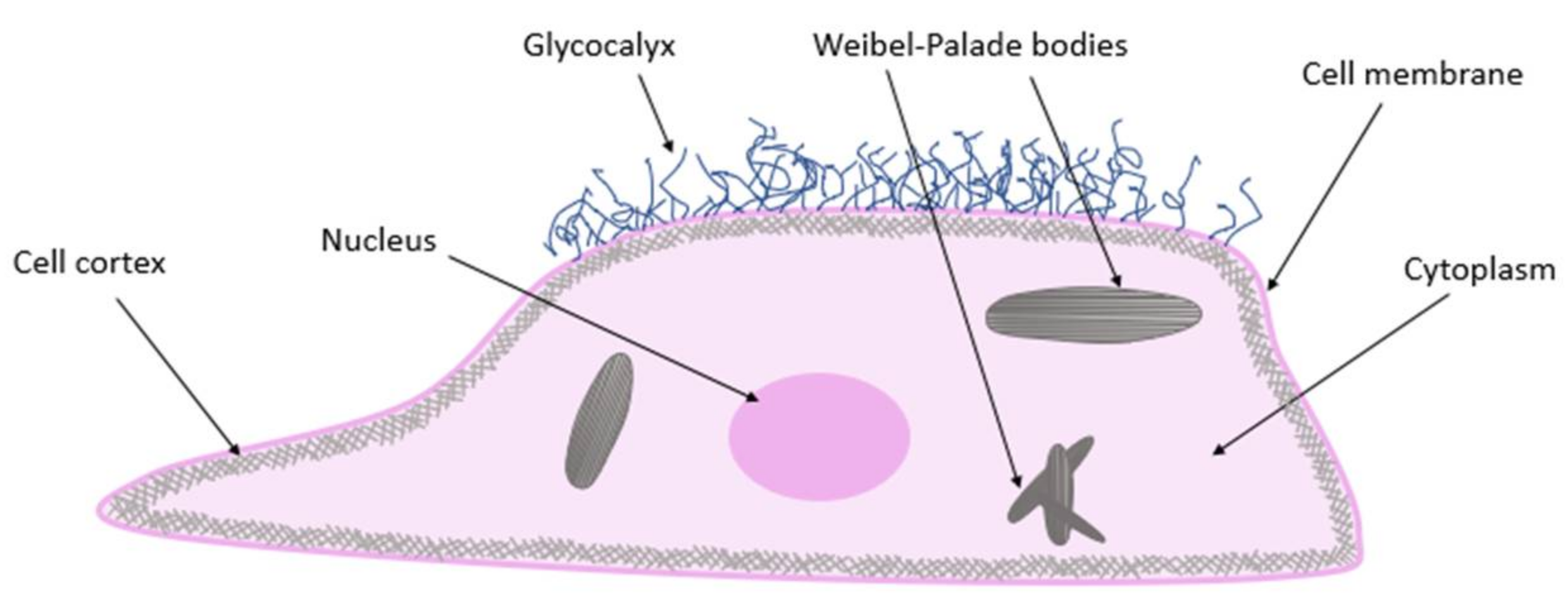
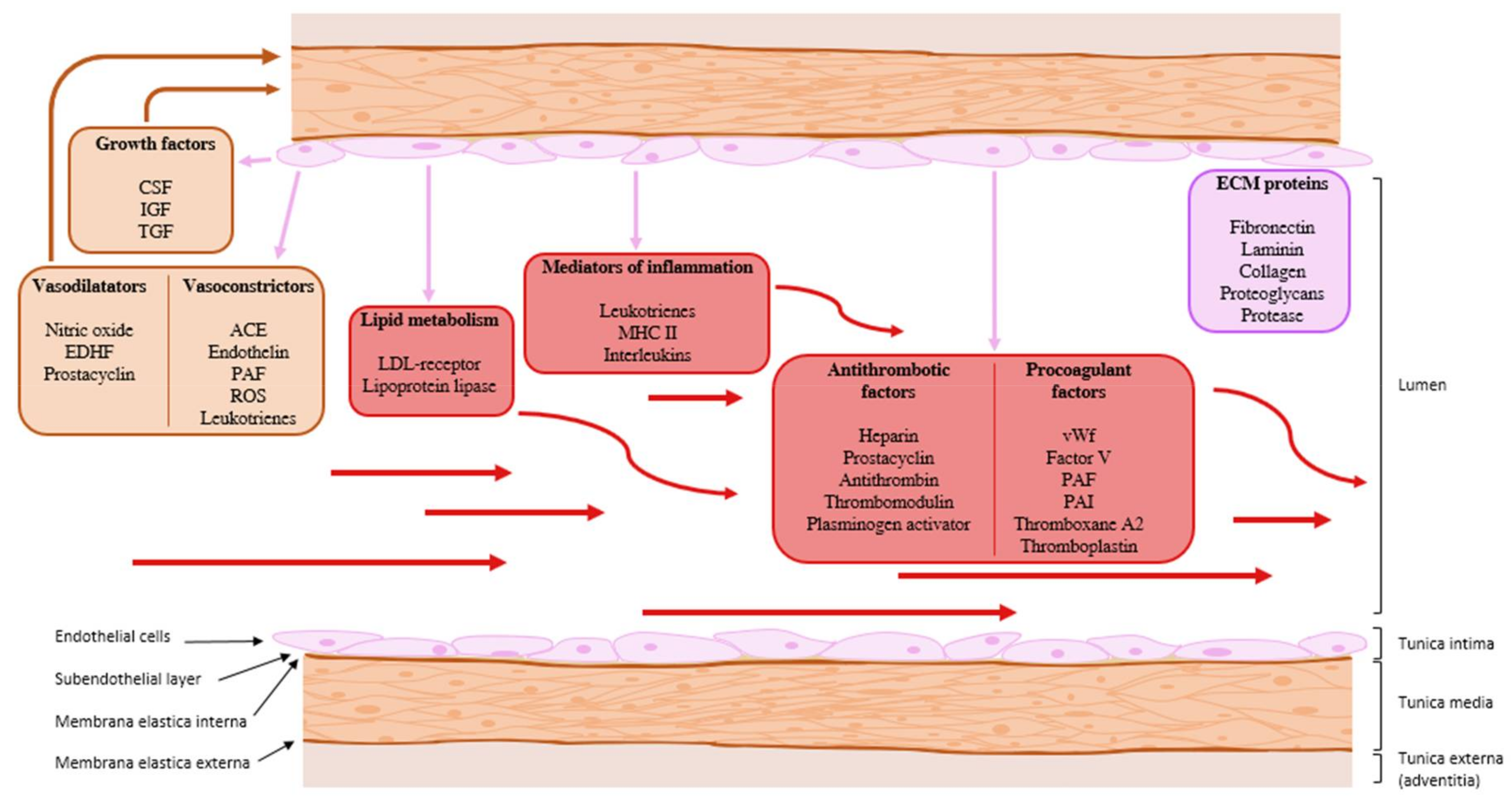
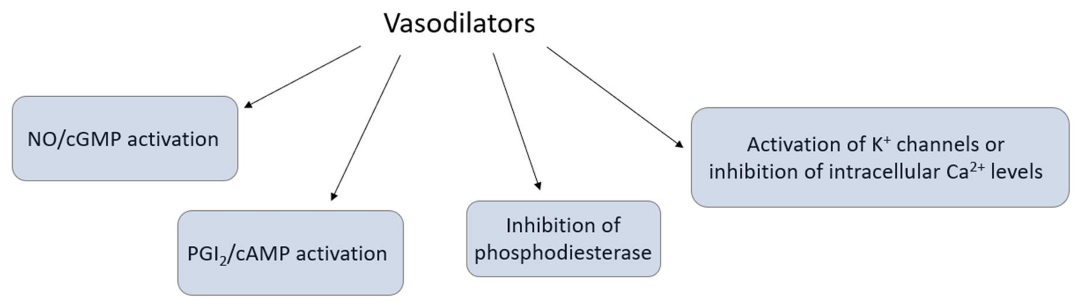
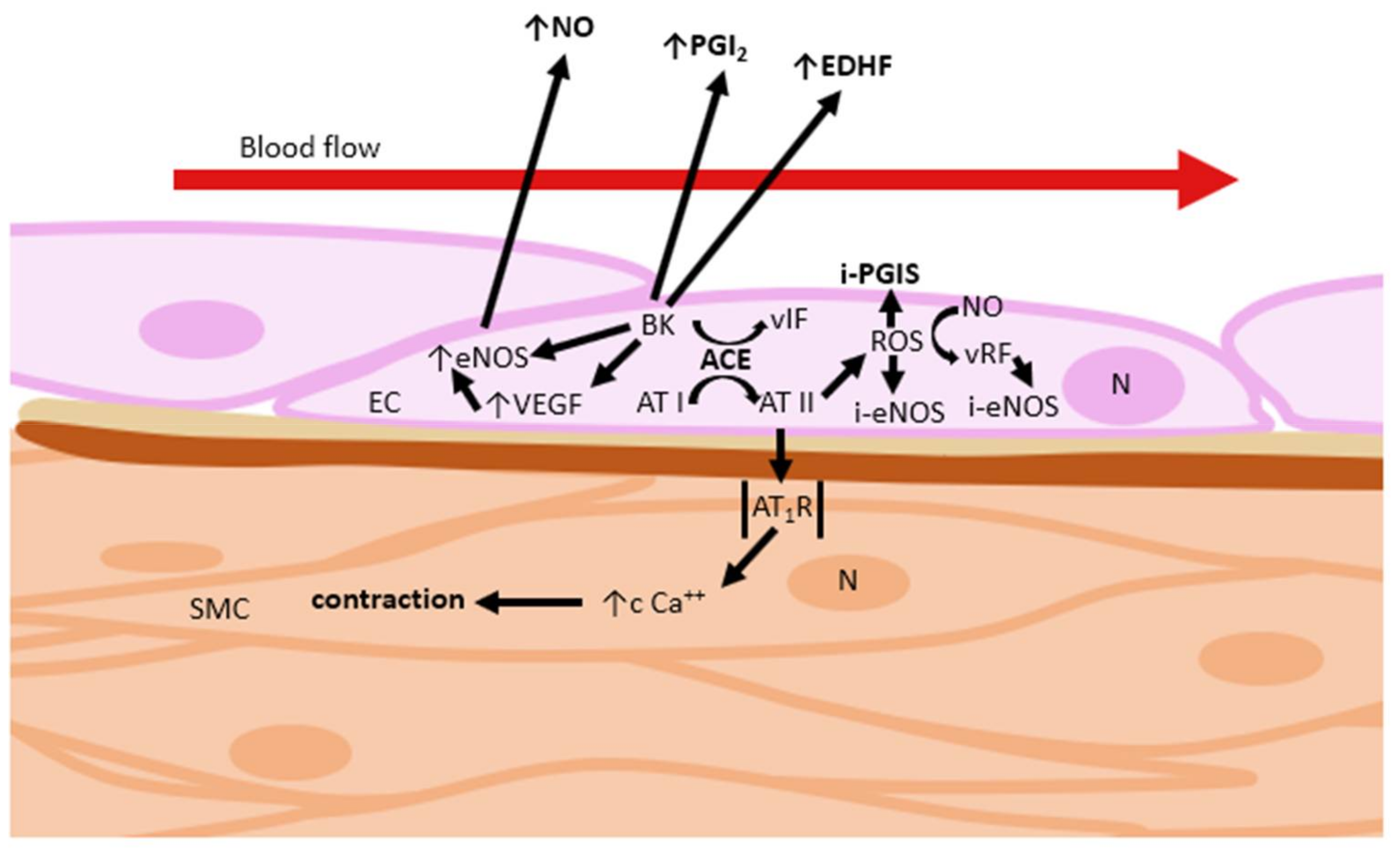
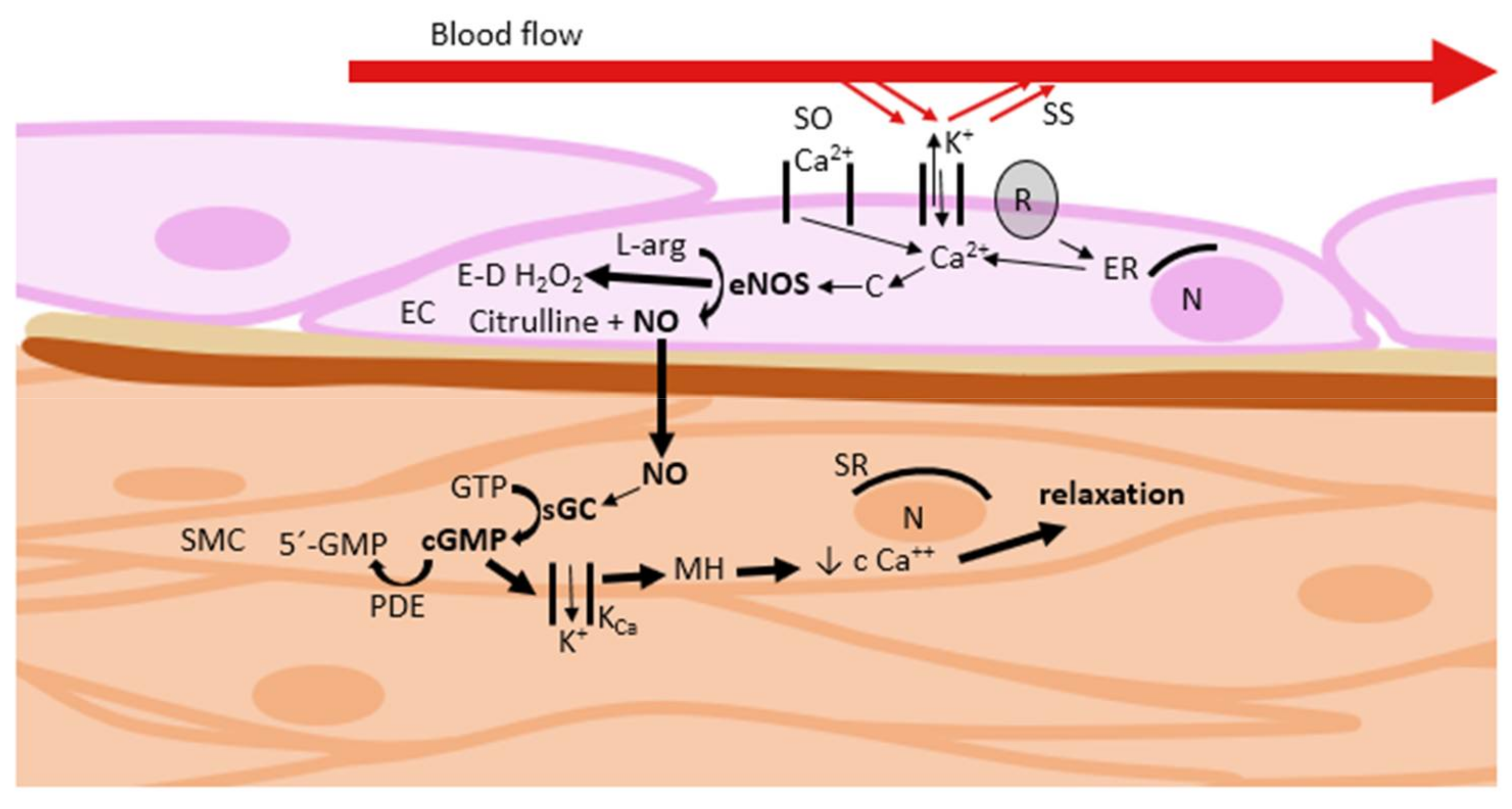
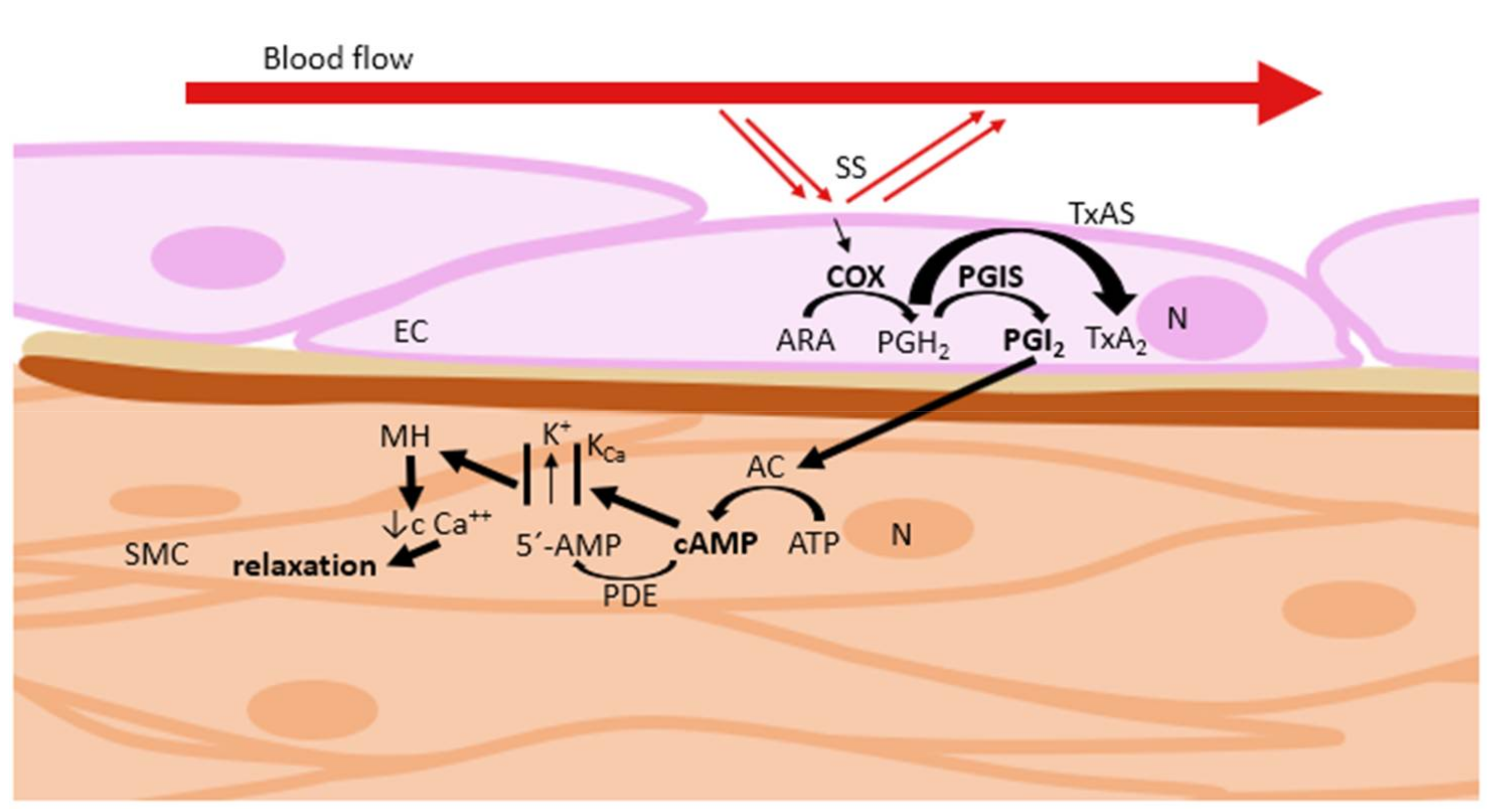
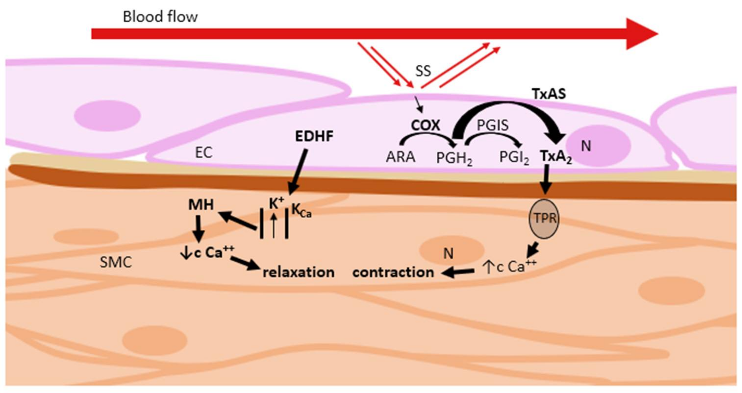
Table 1.
Nitric oxide–cyclic guanosine monophosphate activation pathway.
| Plant(s) | Primary/Secondary Metabolite(s) | Plant Part(s) Used | Citation(s) |
|---|---|---|---|
| Allium sativum L. (Amaryllidaceaee Jaume St.-Hil.) | alliin, allicin | aged garlic extract | [63,64,65,66] |
| Alpinia purpurata (Vieill.) K. Schum. (Zingiberaceae Lindl.) | rutin, quercetin, kaempferol-3-O-β-D-glucuronide | leaves | [67] |
| Alpinia zerumbet (Pers.) Burtt & R.M. Sm. (Zingiberaceae Lindl.) | catechin, epicatechin, rutin, quercetin, kaempferol 3-O-rutinoside, kaempferol 3-O-glucuronide, dihydro-5,6-dehydrokawain, 5,6-dehydrokawain | leaves | [67,68] |
| Arbutus unedo L. (Ericaceae) | tannins, afzeline, juglamine, avicularine, quercitroside, hyperoside | leaves, roots | [69] |
| Caesalpinia sappan L. (Fabaceae Lindl.) | brazilin and hematoxylin | heartwood | [70,71] |
| Calicotome villosa (Poir.) Link. (Fabaceae Lindl.) | chrysin | flowers, leaves | [72,73,74] |
| Canavalia DC. (Fabaceae Lindl.) | lectins | seeds | [75,76] |
| Casimiroa Llave & Lex (Casimirova edulis Llave & Lex and Casimiroa pubescens Ramírez) (Rutaceae Juss.) | hernianin, imperatorin, geranyloxypsoralen 5,6,2′,3′,4′-pentamethoxyflavon | seeds, leaves | [77,78] |
| Centaurium cachanlahuen (Mol.) Robinson (Gentianaceae Juss.) | xanthones | stems, flowers, leaves | [79] |
| Cistus ladaniferus L. (Cistaceae Juss.) | quercetin, kaempferol, myricetin | leaves | [80] |
| Coptosapelta flavescens Korth (Rubiaceae Juss.) | saponin, polyphenols | stems | [81] |
| Crithmum maritimum L. (Apiaceae Lindl.) | limonene, terpinen-4-ol, carvacrol, thymol, chlorogenic acid | flowers, stems, leaves | [82] |
| Croton schiedeanus Schlecht (Euphorbiaceae Juss.) | quercetin 3,7-dimethyl ether, diterpenoid and fenylbutanoid compounds | aerial parts | [83,84,85] |
| Cynara scolymus L. (Asteraceae Martinov) | cymaroside, luteolin, cynarin, chlorogenic acid | leaves | [27] |
| Derris (Lonchocarpus) urucu Killip & A. C. Smith (Fabaceae Lindl.) | isotirumalin | leaves | [86,87] |
| Euterpe oleracea C. Martius (Arecaceae Bercht & J. Presl) | cyanidin 3-O-arabinoside, cyanidin 3-O-glucoside, cyanidin 3-O-rutinoside, epicatechine, catechine homorientin, orientin, isovitexin, taxifolin deoxyhexose | fruits | [88] |
| Geum japonicum Thunberg (Rosaceae L.) | penta-O-galloyl-β-glucoside, casuariin, and 5-desgalloylstachyurin | whole plants | [89] |
| Ginkgo biloba L. (Ginkgoaceae) | bilobalide | leaves | [90,91] |
| Inula viscosa L. (Asteraceae Martinov) | cynarin, chlorogenic acid | leaves | [92] |
| Magnolia grandiflora L. (Magnoliaceae Juss.) | vulgarenol | flower petals | [93] |
| Microdesmis keayana J. Léonard (Pandanaceae) | keayanidin B, keayanin | roots | [94] |
| Ocimum gratissimum L. (Lamiaceae Lindl.) | eugenol | leaves | [95,96] |
| Paeonia sect. Moutan DC. (Paeoniaceae) | paeoniflorin, paeonidanin, methylpaeoniflorin, tetragalloylglucose, pentagalloylglucose | rootbark | [97] |
| Panax ginseng C. A. Meyer (Araliaceae Juss.) | ginsenoside-Rg1, ginsenoside Rb1 | roots | [28,98,99] |
| Prunella vulgaris L. (Lamiaceae Lindl.) | cynaroside, luteolin, ursolic acid, betulinic acid, quercetin | flowering spike | [100,101] |
| Raphanus sativus L. (Brassicaceae Burnett) | sinapine thiocyanate, glucosinolates, brassinosteroids, flavonoids | seeds, leaves | [102,103] |
| Rheum undulatum L. (Polygonaceae Juss.) | piceatannol, tetrahydroxystilbene, resveratrol, anthraquinone derivates | rhizomes | [104,105,106] |
| Saururus chinensis (SC) Baill. (Saururaceae) | saucerneol, saucerneol D, machilin D | roots | [107] |
| Selaginella tamariscina (Beauv.) Spring. (Selaginellaceae) | amentoflavone | whole plants | [108,109] |
| Solanum crispum Ruiz & Pav (Solanaceae Juss.) | alkaloids, flavonoids, resins, saponins, tannins | stems, leaves | [110] |
| Tabernaemontana dichotoma Roxb. ex Wall. (Apocynaceae Juss.) | 10-methoxyaffinisine, cathafoline, alstonisine | bark | [111] |
| Tapirira guianensis Aubl. (Anacardiaceae Lindl.) | triterpenoids, quercetin, myricetin glycoside, hyperoside, penta-O-galloyl-β-glucoside | leaves | [112] |
| Theobroma cacao L. (Malvaceae Juss.) | epicatechin, oligomeric procyanidins | seeds | [29,113,114,115,116] |
| Vitis labrusca L. (Vitaceae Juss.) | vitisin C, phenolic acids, anthocyanins, flavonoids | grape skin, stems | [117,118,119] |
| Vitis vinifera L. (Vitaceae Juss.) | vitisin C, phenolic acids, anthocyanins, flavonoids | grape skin, stems | [117,119,120,121,122] |
| Ziziphus jujuba (L.) Mill. (Rhamnaceae Juss.) | betulinic acid | seeds | [123] |
Table 2.
Prostacyclin–cyclic adenosine monophosphate activation pathway.
| Plant(s) | Primary/Secondary Metabolite(s) | Plant Part(s) Used | Citation(s) |
|---|---|---|---|
| Kaempferia galanga L. (Zingiberaceae Lindl.) | ethyl cinnamate | rhizomes | [124] |
| Piper truncatum Vell. (Piperaceae C. A. Agardh) | eudesmin | leaves, stems | [34,35] |
| Xylopia langdorffiana A.St.-Hil. & Tul. (Annonaceae Juss.) | labdane-302 | stems | [125,126] |
Table 3.
Inhibition of phosphodiesterase.
| Plant(s) | Primary/Secondary Metabolite(s) | Plant Part(s) Used | Citation(s) |
|---|---|---|---|
| Coffea arabica L. (Rubiaceae Juss.) | caffeine, theobromine, theophylline, chlorogenic acid, quercetin, ferulic acid, kaempferol, rutin | seeds | [127,128,129] |
| Epimedium L. (Berberidaceae Juss.) | icariin | young stems | [36,37,130,131,132] |
Table 4.
Activation of K+ channels or inhibition of intracellular Ca2+ levels.
| Plant(s) | Primary/Secondary Metabolite(s) | Plant Part(s) Used | Citation(s) |
|---|---|---|---|
| Alchemilla vulgaris L. (Rosaceae L.) | quercetin | aerial parts | [133,134] |
| Ammi visnaga (L.) Lam. (Apiaceae Lindl.) | visnagin | fruits | [135] |
| Calea glomerata Klatt. (Asteraceae Martinov) | flavonoids, terpenoids | aerial parts | [83,136] |
| Cistus populifolius L. (Cistaceae Juss.) | diterpenoids, luteolin | leaves | [137,138] |
| Cymbopogon martini (Roxb.) W.Watson (Poaceae Barnhart) | geraniol | leaves | [139] |
| Garcinia kola Heckel (Guttiferae Juss.) | kolaviron | seeds | [140] |
| Gentiana kochiana J.O.E. Perrier & Songeon (Gentianaceae Juss.) | gentiacaulein, gentiakochianin | roots | [141] |
| Halenia elliptica D. Don (Gentianaceae Juss.) | 1-hydroxy-2,3,5-trimethoxy-xanthone (HM-1) | whole plants | [142] |
| Hibiscus sabdariffa L. (Malvaceae Juss.) | hibiscus acid, garcinia acid | calyces | [143] |
| Iostephane heterophylla (Cav.) Benth. (Asteraceae Martinov) | xanthorrhizol | whole plants | [144] |
| Ligusticum jeholense Nakai et Kitagawa (Apiaceae Lindl.) | linoleic acid, ferulic acid, ligustilide | roots, rhizomes | [145] |
| Marrubium vulgare L. (Lamiaceae Lindl.) | marrubiin, marrubenol | aerial parts | [146,147] |
| Maxillaria densa Lindl. (Orchidaceae Juss.) | gymnopusin, fimbriol A, erianthridin | whole plants | [148] |
| Morinda citrifolia L. (Rubiaceae Juss.) | alkaloid xeronine, phenolic compounds, sterols, flavonoids, tannins, coumarins, anthraquinones | roots | [149,150] |
| Nauclea officinalis (Pierre ex Pit.) Merr. & Chun (Rubiaceae Juss.) | naucline, angustine, nauclefine, naucletine | bark | [151,152] |
| Peganum harmala L. (Zygophyllaceae) | harmaline, harmine, harmalol | seeds | [153,154,155] |
| Polygala caudata Rehder & E.H.Wilson (Polygalaceae Juss.) | euxanthone | roots | [156,157] |
| Prunus yedoensis Matsum (Rosaceae L.) | prunetin | bark | [158,159] |
| Sarcococca saligna (D. Don) Muell.-Arg. (Buxaceae Dumort.) | flavonoids | whole plants | [160] |
| Trachyspermum ammi (L.) Sprague (Apiaceae Lindl.) | thymol, gamma-terpinene, p-cymene | seeds | [161] |
| Uncaria rhynchophylla (Miquel) Jack (Rubiaceae Juss.) | rhynchophylline, isorhynchophylline, hirsutine | hooks | [162,163] |
Table 5.
Combination of mechanisms without a dominant one.
| Plant(s) | Primary/Secondary Metabolite(s) | Plant Part(s) Used | Citation(s) |
|---|---|---|---|
| Agastache Mexicana (Kunth.) Link. & Epling (Lamiaceae Lindl.) | tilianin, acecatin | aerial parts | [164,165] |
| Alpinia henryi K. Schum. (Zingiberaceae Lindl.) | cardamonin, alpinetin | seeds | [166,167] |
| Alstonia scholaris (L.) R. Br. (Apocynaceae Juss.) | picrinine, schloaricine, alstonamine, rhazimanine, botulin, ursolic acid, β-sitosterol | bark, leaves | [168,169] |
| Alstonia macrophylla Wall. ex G. Don (Apocynaceae Juss.) | vincamedine | leaves | [170] |
| Andrographis paniculata (burm. F.) Nees (Acanthaceae Juss.) | 14-deoxyandrographolide, 14-deoxy-11,12-dihydroandrographolide | leaves | [171,172,173,174] |
| Angelica dahurica Benthman et Hooker (Apiaceae Lindl.) | pyranocoumarin, biscoumarin, isoimperatorin, imperatorin, phellopterin, isodemethylfuropinarine, demethylfuropinarine, decursinol | roots, rhizomes | [175,176,177,178] |
| Angelica gigas Nakai (Apiaceae Lindl.) | ferulic acid | roots | [179] |
| Angelica keiskei Koidz. (Apiaceae Lindl.) | xanthoangelol, 4-hydroxyderricin, xanthoangelol B, xanthoangelol E, xanthoangelol F | roots | [180] |
| Apium graveolens L. var. dulce DC (Apiaceae Lindl.) | apigenin | leaves, roots | [181,182,183] |
| Bacopa monnieri (L.) Pennel (Plantaginaceae Juss.) | bacoside A, bacopaside I, luteolin, apigenin | whole plants | [184,185,186,187] |
| Berberis vulgaris L. (Berberidaceae Juss.) | berberine | fruits, stems bark, roots | [188,189] |
| Camellia sinensis (L.) Kunzte (Theaceae D. Don) | epigallocatechin-3-gallate, epicatechin, epigallocatechin, epicatechin-3-gallate | green tea (leaves) | [190,191,192,193,194] |
| Chenopodium ambrosioides L.(Amaranthaceae Juss.) | kaempferol, quercetin, isorhamnetin, catechins, delphinidin | leaves | [195] |
| Chrysanthemum morifolium Ramat (Asteraceae Martinov) | luteolin-7-O-β-d-glucoside, apigenin-7-O-β-d-glucoside, acacetin-7-O-β-d-glucoside | flowers | [196] |
| Coptis chinensis Franch. (Ranunculaceae Arnott) | berberine, coptisine | rhizomes | [197,198,199,200] |
| Curcuma longa L. (Zingiberaceae Lindl.) | curcumane C, curcumane D, 4,5-seco-cadinane sesquiterpenoid | rhizomes | [201] |
| Dalbergia odorifera T. Chen (Fabaceae Lindl.) | butein, isoliquiritigenin, biochanin A | roots, leaves | [202,203,204,205,206,207,208] |
| Dioclea grandiflora Mart. ex Benth (Fabaceae Lindl.) | dioclein, floranol | roots | [209,210,211,212,213] |
| Echinodorus grandiflorus (Cham. & Schltdl.) Micheli) (Alismataceae Vent.) | flavonoids, diterpenes, triterpenes | leaves | [214,215,216] |
| Elsholtzia splendens Nakai (Lamiaceae Lindl.) | apigenin, luteolin | aerial parts | [217,218] |
| Hancornia speciosa B. A. Gomes (Apocynaceae Juss.) | rutin | leaves | [219] |
| Liqusticum wallichii Franchat (Apiaceae Lindl.) | butylidenephthalide, ligustilide, senkyunolide A, tetramethylpyrazine | rhizomes | [220,221,222,223] |
| Mentha X villosa Hudson (Lamiaceae Lindl.) | rotundifolone | leaves | [224,225,226,227] |
| Mitragyna ciliata aubrev. & Pellegr.(Rubiaceae Juss.) | mitragynine, mitraphylline, rhynophylline, flavonoids | stem bark | [228] |
| Phaeanthus crassipetalus Becc. (Annonaceae Juss.) | limacine, pecrassipine A, backebergine | bark, leaves | [229] |
| Picrorhiza kurroa L. (Plantaginaceae Juss.) | apocynin | roots | [230,231] |
| Prunus serotina Ehrh (Rosaceae L.) | ursolic acid, uvaol | fruits | [232] |
| Schisandra chinensis (Turcz.) Baill. (Schisandraceae Bl.) | schizandrin, γ-schizandrin, gomisin A | fruits (seeds) | [233,234,235] |
| Scutellaria baicalensis Georgi (Lamiaceae Lindl.) | baicalin | roots | [236,237] |
| Senecio nutans Sch. Bip. (Asteraceae Martinov) | 4-hydroxy-3-(3-methyl-2-butenyl)acetophenone, 5-acetyl-6-hydroxy-2-isopropenyl-2,3-dihydrobenzofurane | aerial parts | [238] |
| Thymus linearis Benth. (Lamiaceae Lindl.) | thymol, carvacrol | aerial parts | [239] |
Table 6.
Not fully elucidated/not specified.
| Plant | Primary/Secondary Metabolite | Plant Part Used | Citation |
|---|---|---|---|
| Calpurnia aurea (Ait.) Benth. (Fabaceae Lindl.) | seeds | [240] | |
| Vitex negundo L. (Lamiaceae Lindl.) | aerial parts | [241] | |
| Ficcus saussureana DC (Moraceae Dumort.) | root bark | [242] | |
| Prunus persica (L.) (Rosaceae L.) | branches | [243] | |
| Satureja obovata Lag. (Lamiaceae Lindl.) | eriodictyol | [244,245] | |
| Vernonia amygdalina Del. (Asteraceae Martinov) | alkaloids, flavonoids, saponins | leaves | [246] |
Publisher’s Note: MDPI stays neutral with regard to jurisdictional claims in published maps and institutional affiliations. |
© 2021 by the authors. Licensee MDPI, Basel, Switzerland. This article is an open access article distributed under the terms and conditions of the Creative Commons Attribution (CC BY) license (http://creativecommons.org/licenses/by/4.0/).
Share and Cite
MDPI and ACS Style
Bartáková, A.; Nováková, M. Secondary Metabolites of Plants as Modulators of Endothelium Functions. Int. J. Mol. Sci. 2021, 22, 2533. https://0-doi-org.brum.beds.ac.uk/10.3390/ijms22052533
AMA Style
Bartáková A, Nováková M. Secondary Metabolites of Plants as Modulators of Endothelium Functions. International Journal of Molecular Sciences. 2021; 22(5):2533. https://0-doi-org.brum.beds.ac.uk/10.3390/ijms22052533
Chicago/Turabian StyleBartáková, Anna, and Marie Nováková. 2021. "Secondary Metabolites of Plants as Modulators of Endothelium Functions" International Journal of Molecular Sciences 22, no. 5: 2533. https://0-doi-org.brum.beds.ac.uk/10.3390/ijms22052533
Note that from the first issue of 2016, this journal uses article numbers instead of page numbers. See further details here.