Article

Molecular Basis of AmpC β-Lactamase Induction by Avibactam in Pseudomonas aeruginosa: PBP Occupancy, Live Cell Binding Dynamics and Impact on Resistant Clinical Isolates Harboring PDC-X Variants

by
Silvia López-Argüello
,
María Montaner
,
Antonio Oliver
and
Bartolome Moya
*
Servicio de Microbiología and Unidad de Investigación, Hospital Son Espases, Instituto de Investigación Sanitaria Illes Balears (IdISBa), 07120 Palma de Mallorca, Spain
*
Author to whom correspondence should be addressed.
Joint first authors with equal contributions.
Int. J. Mol. Sci. 2021, 22(6), 3051; https://0-doi-org.brum.beds.ac.uk/10.3390/ijms22063051
Submission received: 27 January 2021 / Revised: 10 March 2021 / Accepted: 12 March 2021 / Published: 17 March 2021

Abstract

:
Avibactam belongs to the new class of diazabicyclooctane β-lactamase inhibitors. Its inhibitory spectrum includes class A, C and D enzymes, including P. aeruginosa AmpC. Nonetheless, recent reports have revealed strain-dependent avibactam AmpC induction. In the present work, we wanted to assess the mechanistic basis underlying AmpC induction and determine if derepressed PDC-X mutated enzymes from ceftazidime/avibactam-resistant clinical isolates were further inducible. We determined avibactam concentrations that half-maximally inhibited (IC50) bocillin FL binding. Inducer β-lactams were also studied as comparators. Live cells’ time-course penicillin-binding proteins (PBPs) occupancy of avibactam was studied. To assess the ampC induction capacity of avibactam and comparators, qRT-PCR was performed in wild-type PAO1, PBP4, triple PBP4, 5/6 and 7 knockout derivatives and two ceftazidime/avibactam-susceptible/resistant XDR clinical isolates belonging to the epidemic high-risk clone ST175. PBP4 inhibition was observed for avibactam and β-lactam comparators. Induction capacity was consistently correlated with PBP4 binding affinity. Outer membrane permeability-limited PBP4 binding was observed in the live cells’ assay. As expected, imipenem and cefoxitin showed strong induction in PAO1, especially for carbapenem; avibactam induction was conversely weaker. Overall, the inducer effect was less remarkable in ampC-derepressed mutants and nonetheless absent upon avibactam exposure in the clinical isolates harboring mutated AmpC variants and their parental strains.

1. Introduction

The worldwide emergence of multidrug-resistant Gram-negative Pseudomonas aeruginosa isolates has caused a substantial public health problem, which is exacerbated by few therapeutic options remaining [1]. Owing to their extensively proven efficacy and safety, β-lactams have been the drug of choice to treat infections caused by P. aeruginosa [2]. All β-lactams bind to and inactivate multiple penicillin-binding proteins (PBPs) with different affinities. PBPs play different roles in peptidoglycan biosynthesis. Roughly, high-molecular-weight PBPs catalyze peptidoglycan polymerization and the cross-linking of glycan strands (transglycosylase (ponA and mrcB; PBP1a and 1b) and DD-transpeptidase (PBP1a, 1b, 2 and 3)), cell wall elongation (pbpA; PBP2) and septum formation (fstI; PBP3). Low-molecular-weight PBPs catalyze DD-endopeptidase (dac and pbpG; PBP4 and 7) and DD-carboxypeptidase (dacC; PBP5/6) activities, necessary for the correct peptidoglycan incorporation and cell growth. The bactericidal activity of β-lactams requires the saturation of different PBP receptors, causing the inhibition of the aforementioned biological processes. However, intrinsic and acquired β-lactam resistance is one of the striking features of this microorganism. The chromosomal cephalosporinase AmpC (or Pseudomonas-derived cephalosporinase 1 (PDC-1)) represents one of the most menacing weapons among the extensive P. aeruginosa enzymatic warfare [3]. AmpC is an inducible broad-spectrum Ambler class C β-lactamase capable of hydrolyzing most β-lactams, excluding carbapenems.
Inducible serine AmpC β-lactamases are a common resistance mechanism among many Enterobacterales (Enterobacter cloacae, Serratia marcescens and Citrobacter freundii) and P. aeruginosa isolates. The induction phenomenon occurs via transient ampC overexpression due to the resulting accumulation of the activator muropeptide anhNAM-P5 (1,6-anhydro-N-acetylmuramyl-pentapeptides) after peptidoglycan metabolism disruption. The fact that the same muropeptide has been found to be the underlying cause of AmpC constitutive hyperexpression in dacB (penicillin-binding protein 4 (PBP4)) mutants, endorses PBP4’s key role during the induction process [4,5,6]. Antipseudomonal penicillins (piperacillin) and cephalosporins (ceftazidime or ceftolozane) are active against P. aeruginosa because they are very weak inducers of this chromosomal β-lactamase.
In the face of the continuous upscaling of β-lactamase-driven multidrug resistance (MDR) in Gram-negatives, new β-lactam-β-lactamase inhibitor (BLI) treatment options have become available. However, AmpC inhibition is out of the spectrum of most BLIs. Novel combinations such as ceftazidime/avibactam, ceftolozane/tazobactam, meropenem/vaborbactam and cefepime/zidebactam have based their activity against P. aeruginosa upon different strategies [7,8]. The avibactam β-lactamase inhibitor spectrum includes class C enzymes, which protect the partner β-lactam from AmpC-related hydrolysis [9]. While tazobactam does not have a major impact on the activity of ceftolozane against P. aeruginosa, the latter retains its activity against extremely high ampC (PDC-1) expression in addition to its improved PBP-binding profile [10,11]. Zidebactam, in spite of its fair inhibition capability towards class C β-lactamases, focuses its activity on β-lactam-enhancer properties derived from targeting PBP2 [12,13]. Finally, vaborbactam, a cyclic boronic-based narrow-spectrum BLI, does not offer additional protection against AmpC hyperproduction; however, it seems to enhance meropenem activity against P. aeruginosa [7].
Unfortunately, clinical isolates of P. aeruginosa resistant to ceftolozane/tazobactam (TOL/TAZ) and ceftazidime/avibactam (CAZ/AVI), expressing mutant variants of AmpC (i.e., PDC-221, -222, -223 and -322), have already been reported [14,15,16]. The studied AmpC mutants showed structural mutations in the omega (Ω) loop or adjacent residues. Such mutations appear to exert a concurrent effect on the catalytic properties of AmpC, reducing its susceptibility towards avibactam inhibition and enhancing its catalytic efficiencies towards ceftolozane and ceftazidime hydrolysis [17].
Furthermore, clinically relevant BLIs such as clavulanic acid, tazobactam and sulbactam have been shown to induce ampC expression at clinically relevant concentrations [18,19]. Livermore et al. described a similar effect for the non-β-lactam BLI avibactam, showing significant induction at higher than clinically relevant concentrations (32 µg/mL) and profound strain-to-strain variability. However, according to their work, such induction capacity would become clinically significant only if two conditions were met for the AmpC enzyme: loss of inhibition by avibactam while retaining inducibility [20].
Under these circumstances, knowing whether a novel BLI would be able to induce AmpC expression in avibactam-insensitive fifth-generation cephalosporin-resistant strains would be of great interest. On that premise, we examined the basis for the underlying AmpC induction in the presence of clinically relevant avibactam concentrations (≈50% of Cmax after 500 mg standard dosage) [21] and the behavior of mutated AmpC enzymes (PDC-221 and -223) [15] upon induction in CAZ/AVI-resistant P. aeruginosa clinical isolates.

2. Results and Discussion

2.1. Molecular Basis of Avibactam AmpC Induction

To investigate whether the induction of ampC expression in the wild-type P. aeruginosa PAO1 strain was correlated to PBP4 inhibition, the fluorogenic bocillin FL PBP-binding assay was used. The PBP-binding IC50 values were determined by incubating PBP-containing membrane preparations in growing concentrations (0.25–32 µg/mL) of avibactam and two typical PBP4-binding AmpC inducers (cefoxitin and imipenem) and afterwards labeling with bocillin FL to determine the β-lactam concentrations that half-maximally inhibited (IC50) bocillin FL binding (Table 1). Cefoxitin and imipenem, as previously described, showed low PBP4 IC50 values, the lowest observed for imipenem (0.1 and 1.5 µg/mL, respectively) [11,22]. Imipenem bound to all PBPs with the highest efficiency with concentrations that ranged from 0.05 to 0.3 µg/mL, whereas cefoxitin binding values for non-PBP4 enzymes were >250-fold higher (range: 7.8–14.6 µg/mL), and no PBP1b binding was detected. Avibactam bound to PBP1b, 2, 4 and 5/6 (IC50 = 3.6, 4.2, 3.1 and 2.2 µg/mL, respectively). However, in contrast to previous works, no PBP1a and 3 binding was detected under our experimental conditions in the studied strain (Figure 1) [23]. Besides showing greater PBP4 affinity than previously reported values, avibactam effective concentration was the highest observed from all the three compounds tested (3.1 µg/mL) [23]. Consequently, the differential PBP affinities could be attributed to strain-to-strain variability [20].

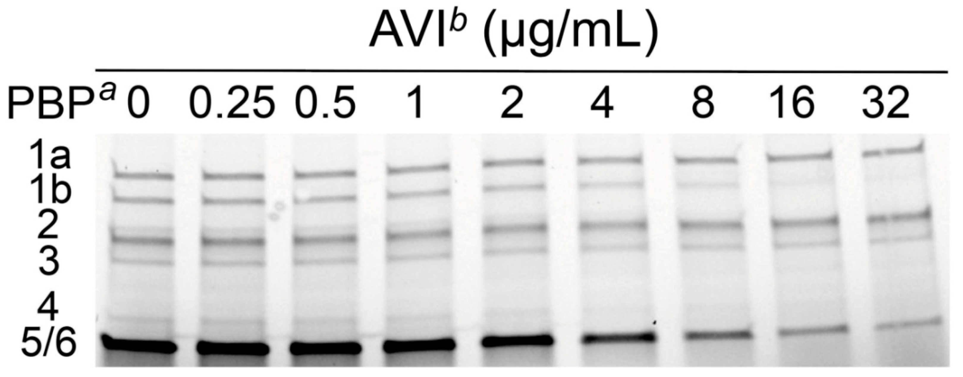
2.2. Avibactam Target Binding in Live P. aeruginosa Cells

To gather further information regarding the PBP4-related ampC induction by avibactam, the time-course of PBP-binding in live P. aeruginosa cells was determined as previously described with several modifications [24]. We used intact PAO1 cells in the late exponential growth phase with an initial inoculum of ≈4.0 × 10⁷ CFU/mL. To infer the outer membrane permeability-limited target binding, a matching experiment with isolated PBP-containing membranes (from previously lysed cells) was similarly performed in the presence of 4 µg/mL avibactam (AVI) (fixed concentration used for minimum inhibitory concentrations and pharmacokinetic model studies). Figure 2 shows the intact cells’ (Figure 2a) and isolated membranes’ (Figure 2b) binding affinities. No significant PBP1a, 3 and 5/6 binding was observed for the whole-cell assay, whereas 20% of PBP1b and 2 fractions and up to 50% of PBP4 were bound by avibactam after 60 min of incubation. Previously reported data showed that avibactam uptake was indeed limited by the outer membrane permeability barrier, and its activity could be further enhanced upon permeabilization in Gram-negatives, including P. aeruginosa [25]. Unsurprisingly, isolated membranes assay determined a more extensive inhibition of all the PBPs but PBP1a and 3, binding up to 68 and 82% of PBP4 and 5/6 total fractions respectively. As previously suggested by other authors, we confirmed PBP4 binding by avibactam in P. aeruginosa, in both whole cells and isolated membranes [20,26].

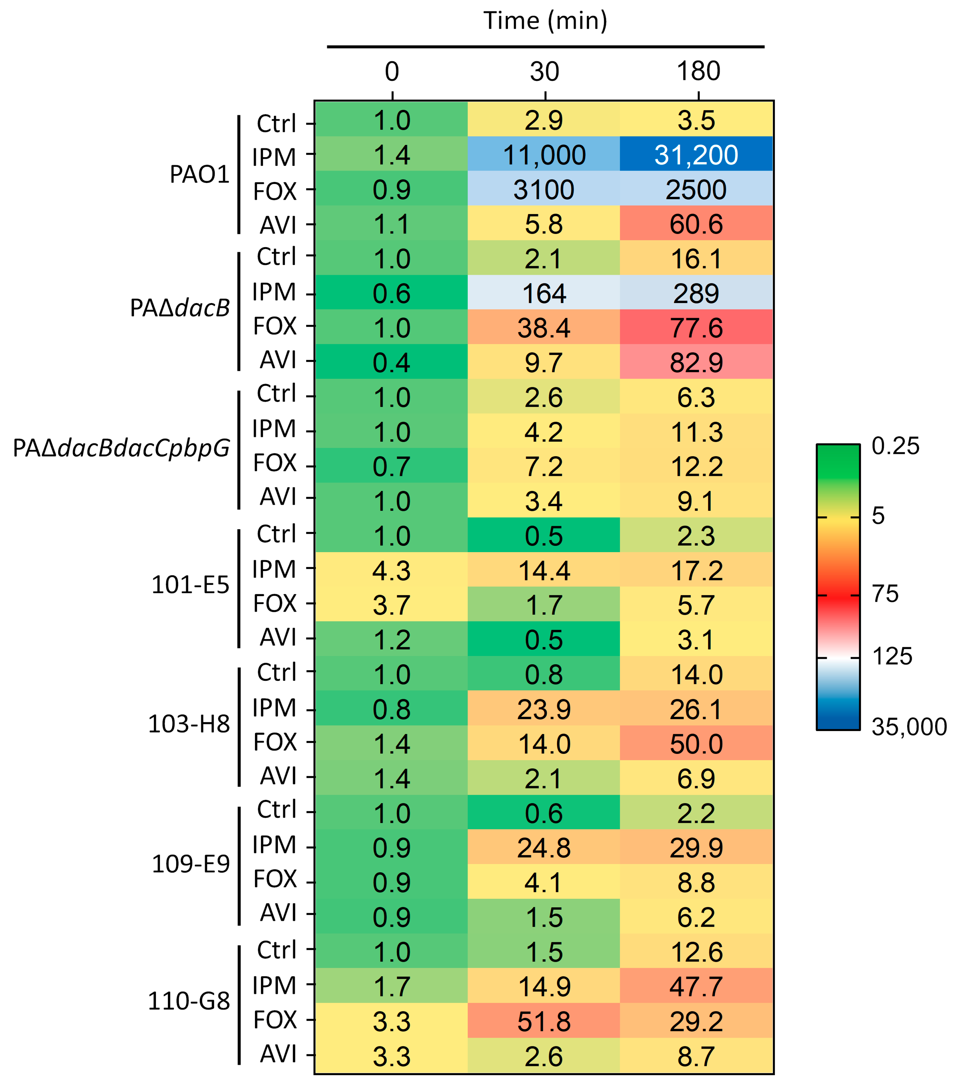
2.3. qRT-PCR-Based ampC Induction Assays

To investigate whether the binding of avibactam to PBP4 in live cells was correlated with ampC induction, qRT-PCR was performed to determine the time-course expression of β-lactamase mRNA (AVI 16 µg/mL; t = 0, 30 and 180 min). Typical AmpC inducers, imipenem and cefoxitin, were used as comparators (IPM 8 µg/mL, FOX 64 µg/mL). To assess the potential clinical implications of ampC induction in the clinical worst-case scenario, two clinical isolates belonging to the high-risk clone ST175, harboring AmpC structural modifications in the Ω loop or adjacent residues and their parental wild-type AmpC isolates, were also studied. Described AmpC mutations were previously shown to reduce susceptibility to avibactam inhibition and enhance ceftolozane and ceftazidime catalytic efficiencies simultaneously [15,17].
Table 2 shows the basal ampC mRNA expression levels for each of the strains studied relative to wild-type strain PAO1. As previously demonstrated by our group and other independent researchers, PBP4 inactivation (PAΔdacB) elicited significant ampC hyperexpression; the highest relative expression values were observed at the 180 min determination (1100 ± 243). Both initial ST175 isolates, 101-E5 and 109-E9 (wild-type AmpC), and their CAZ/AVI post-therapy resistant isolates, 103-H8 and 110-G8, showed an even more extensive ampC relative expression (2400 ± 1100, 1700 ± 382, 5400 ± 2900 and 3500 ± 1600, respectively) consistent with the ampR-G154R-resulting enhanced ampC expression observed in nearly all of the ST175 isolates [15,27,28]. Inactivation of the three low-molecular-weight (LMW) PBPs (PBP4, 5/6 and 7) in PAΔdacBdacCpbpG elicited the most remarkable cephalosporinase constitutive overexpression (58,000 ± 26,000) after 180 min.
The AmpC mRNA levels under basal conditions and after incubation with 8 µg/mL imipenem, 64 µg/mL cefoxitin and 16 µg/mL avibactam for 0, 30 and 180 min in strains PAO1, PAΔdacB, PAΔdacBdacCpbpG, 101-E5 and 109-E9 (OprD Q142X, AmpR G154R) CAZ/AVI sensitive and their 103-H8 (PDC-221; AmpC E247K) and 110-G8 (PDC-223; AmpC DelG229-E247) isogenic CAZ/AVI-resistant isolates are shown in Figure 3. The values represent each strain ampC relative expression normalized to its expression under basal conditions at t = 0 min. The most notable cephalosporinase induction was observed for the wild-type strain PAO1 upon imipenem incubation. The maximum values (31,200 ± 17,000) were observed by the end of the assay (180 min). As previously reported in works from our group and others, cefoxitin induction was less significant compared to imipenem, achieving maximum expression after 30 min incubation [11,20]. On the other hand, avibactam showed the lowest ampC induction capacity, however still significant (according to the >10 times threshold proposed by several authors) after 180 min incubation (60.6 ± 25.2) [20,29]. As opposed to observations in other species, avibactam is capable of inducing P. aeruginosa chromosomal cephalosporinase AmpC [20,26], although not as strongly as classical inducers such as imipenem or cefoxitin. Experimental induction capacity was consistently correlated with observed PBP4 inhibition values for each of the studied drugs (Table 1).
According to the premise that higher basal ampC expression levels (constitutive overexpression) determine lesser inducible endpoints, the PBP4 (dacB) mutant showed a much lower ampC inducibility compared to its parental strain PAO1 for both imipenem and cefoxitin for all the observations, 107- and 32-fold lower (289 ± 138 and 77.6 ± 35.7) for the knockout strain at 180 min, respectively [6]. Surprisingly, avibactam caused a slightly higher ampC expression increase in dacB mutant, especially towards the end of the assay (180 min). This observation could be attributed to the concomitant inhibition of LMW PBP5/6 and 7 (dacC, pbpG) beyond PBP4 inactivation [4,22]. In fact, regardless of a remarkably higher relative basal ampC expression compared to PAO1 (58,000 ± 26,000; t = 180 min), triple PAΔdacBdacCpbpG mutant showed no significant avibactam induction (9.1 ± 4.3) (Figure 3).
In addition to the lower induction capacity thoroughly observed for the AmpC-derepressed mutants, the lower imipenem β-lactamase induction effect in ST175 clinical isolates (101-E5, 109-E9, 103-H8 and 110-G8) could be further explained by the observed OprD mutations [15]; that is, restricting imipenem periplasmic uptake would reduce the number of molecules capable of binding to the low-molecular-weight PBPs, which, in turn, cause the β-lactamase expression induction [4,22]. Moreover, AmpC structural mutations have been shown to reduce imipenem resistance and presumably enzyme’s hydrolytic capacity towards this compound [30].
Regarding cefoxitin, all the ST175 clinical isolates showed a significantly diminished induction, virtually negligible in the wild-type AmpC carriers 101-E5 and 109-E9 (5.7 ± 2.8 and 8.8 ± 7.9, respectively), but still within positive induction threshold values at 180 min in the 103-H8 and 110-G8 resistant isolates (50 ± 36.5 and 29.2 ± 13.1, respectively); higher after 30 min incubation in the 110-G8 isolate (51.8 ± 13.2). This observation could be intimately linked to its otherwise ampR mutation-mediated high-level ampC derepression. Conversely, induction caused by clinically relevant avibactam exposure (16 µg/mL) [21] was not significant for any of the ST175 clinical isolates (3.1, 6.9, 6.2 and 8.7 at 180 min); indeed, mRNA expression was consistently lower than the observed for the control arms in the 103-H8 and 110-G8 CAZ/AVI-resistant mutants (14 and 12.6, respectively). Hence, the observed loss of inducibility appears to be linked to the transcriptional regulator AmpR mutation (G154R) [28].
In summary, the results presented in this work are in accordance with previous studies suggesting a link between avibactam ampC induction and PBP4 inhibition. Furthermore, we demonstrated “in vivo” PBP4-derived induction process and outer membrane permeability-limited avibactam target site penetration and binding. This may explain the added synergistic effects of CAZ/AVI when coadministered with an outer membrane permeabilizer [25]. What is more important is that, even when the foretold conditions for the clinical significance of avibactam induction are met (i.e., mutated AmpC with loss of affinity for avibactam while retaining inducibility) [20], ampC variants studied in this work (PDC-221 and -223) were not reactive to avibactam induction. In terms of bacterial physiology and fitness, it seems reasonable not to retain full inducibility once the derepressed enzyme expression has already been selected [6]. Understanding the dynamics of mutational ampC regulation and resistance to antimicrobials and β-lactamase inhibitors will definitely help the rational development of novel compounds and combinations that are less prone to select mutational resistance.

3. Methods

3.1. Bacterial Strains and Antibiotics

The wild-type reference strain P. aeruginosa PAO1 [31] was used for PBP-binding, IC50 determination and quantification of the AmpC induction assay. P. aeruginosa knockout strains PAΔdacB (PBP4) [6] and PAΔdacBdacCpbpG (PBP4, 5/6 and 7) [22], and two previously characterized [15] pairs of TOL/TAZ susceptible/resistant XDR clinical isolates belonging to the epidemic high-risk clone ST175, were also used for the quantification of AmpC induction.
The isogenic clinical isolates pairs were obtained from two patients treated with TOL/TAZ. Susceptible isolates 101-E5 and 109-E9 (OprD Q142X, AmpR G154R) were obtained before the therapy onset, and the resistant isolates 103-H8 and 110-G8 were obtained during or after the completion of therapy. Resistance development to TOL/TAZ was linked to additional AmpC structural modifications associated with mutations in the Ω loop or adjacent residues in 103-H8 (AmpC E247K; PDC-221) and 110-G8 (AmpC DelG229-E247; PDC-223) isolates. Mutant forms of AmpC rendered the isolates resistant to CAZ/AVI as well [15].
Imipenem (IPM) was obtained from Fresenius Kabi, Barcelona, Spain, cefoxitin (FOX) from Laboratorios Normon, Madrid, Spain and avibactam (AVI) from MedChem Express, Stockholm, Sweden.

3.2. Intact Cells’ Time-Course of PBP-Binding Assay

P. aeruginosa PAO1 cultures grown to the midexponential phase (7.6 log10 CFU/mL) were preincubated in cation-adjusted Mueller–Hinton broth (CAMHB) at 37 °C (180 rpm) for 20 min. Afterwards, 4  µg/mL of avibactam (AVI) was added, and the bacterial cultures were incubated (37 °C, 180 rpm). Control and treatment samples were taken after 15, 30 and 60 min incubation, kept in ice and centrifuged (3220× g for 10 min at 4 °C). The bacterial pellets were washed in 30 mL phosphate-buffered saline (PBS) (pH 7.5) four times, resuspended in 10 mL of PBS and sonicated by using a Digital Sonifier Unit model S-450D (Branson Ultrasonics Corp., Danbury, CT, USA) at 40% amplitude for three 2 min bursts (while immersed in an ice bath) and centrifuged at 3220× g for 15 min at 4 °C. Membranes containing the PBPs were isolated by ultracentrifugation at 150,000× g for 30 min at 4 °C using an Optima L100XP Ultra centrifuge (Beckman Coulter, Inc., Palo Alto, CA, USA) and were resuspended in 60 μL of PBS. Total protein content was measured using the Quick Start Bradford protein assay kit with bovine serum albumin as a standard (Bio-Rad Laboratories, Hercules, CA, USA), according to the manufacturer’s instructions. To determine the PBP fraction unbound, membranes containing PBPs (20 μL, at 0.5 mg/mL) were labeled with a 25 μM concentration of the fluorescent penicillin bocillin FL [32]. Labeled PBPs were denatured and separated through 4–15% SDS–polyacrylamide gel electrophoresis (Bio-Rad Laboratories, Hercules, CA, USA). Labeled PBPs were visualized using a Typhoon FLA 9500 biomolecular imager (GE Healthcare Bio-Sciences AB, Uppsala, Sweden) (excitation at 488 nm and emission at 530 nm), and binding to different PBPs was determined from at least three independent experiments using ImageQuant TL software v8.1.0.0 (GE Healthcare Bio-Sciences AB, Uppsala, Sweden).
In parallel to the aforementioned assay, we performed analogous experiments in which isolated PBP-containing membrane preparations instead of intact cells were pre-exposed to avibactam. Briefly, membrane preparations containing PBPs were obtained by following previously described protocols [8] and were incubated for 15, 30 and 60 min at 37 °C in the presence of 4 µg/mL avibactam and were afterwards labeled with bocillin FL (25 μM). Determination of the PBP fraction unbound was performed as described in the above section.

3.3. Determination of PBP-Binding Affinity (IC50)

PBP-binding affinity was determined using previously described methods [8]. Briefly, P. aeruginosa PAO1 cultures grown to the midexponential phase (7.6 log10 CFU/mL) were incubated in CAMHB at 37 °C and 180 rpm, washed and centrifuged (3220× g for 10 min at 4 °C). Bacterial pellets were washed in 30 mL of PBS buffer (pH 7.5), resuspended in 10 mL of PBS and sonicated at 40% amplitude for three 2 min bursts (while immersed in an ice bath) and centrifuged at 3220× g for 15 min at 4 °C. PBP-containing membranes were isolated by ultracentrifugation at 150,000× g for 30 min at 4 °C. Total protein concentrations were measured through the Bradford method.
To measure the 50% inhibitory concentrations (IC50), PBP-containing membrane preparations (0.5 mg/mL) were incubated at 37 °C for 30 min in the presence of increasing concentrations of avibactam, cefoxitin or imipenem (range of concentrations tested: 0.25–32 µg/mL). Subsequently, the fluorescent penicillin bocillin FL was added to a 25 μM final concentration [32]. Labeled PBPs were denatured and separated through 4–15% SDS–polyacrylamide gel electrophoresis. Labeled PBPs were visualized using a Typhoon FLA 9500 biomolecular imager (GE Healthcare Bio-Sciences AB, Uppsala, Sweden) (excitation at 488 nm and emission at 530 nm), and binding was determined from at least three independent experiments using ImageQuant TL software v8.1.0.0 (GE Healthcare Bio-Sciences AB, Uppsala, Sweden).

3.4. qRT-PCR Quantification of AmpC Induction

The induction of AmpC production by imipenem, cefoxitin and avibactam was determined by measuring the ampC mRNA levels by quantitative real-time reverse transcription-PCR (qRT-PCR) in wild-type PAO1, PAΔdacB (PBP4), PAΔdacBdacCpbpG (PBP4, 5/6 and 7), 101-E5 and 109-E9 (OprD Q142X, AmpR G154R) TOL/TAZ-sensitive clinical isolates and their respective isogenic resistant clinical isolates 103-H8 (AmpC E247K; PDC-221) and 110-G8 (AmpC DelG229–E247; PDC-223). To this end, late exponential-growing P. aeruginosa cultures of PAO1, PAΔdacB, PAΔdacBdacCpbpG, 101-E5, 109-E9, 103-H8 and 110-G8 (dilution: 1/100) were preincubated in CAMHB at 37 °C (180 rpm) for 20 min. Afterwards, imipenem (IPM; 8 µg/mL), cefoxitin (FOX; 64 µg/mL) or avibactam (AVI; 16 µg/mL) were added, and bacterial cultures were incubated at 37 °C, 180 rpm. Samples were taken at 0, 30 and 180 min. Total RNA was extracted with the RNeasy Mini Kit (Qiagen, Hilden, Germany) and treated with TURBO DNase (Thermo Fisher Scientific, Waltham, MA, USA) to remove contaminating DNA. Samples were afterwards normalized to 50 ng/µL.
Normalized samples were then used for one-step reverse transcription and real-time PCR amplification using the QuantiTect SYBR green qRT-PCR kit (Qiagen, Hilden, Germany) in a CFX Connect Real-Time PCR System (Bio-Rad, Hercules, California, USA). Previously described primer pairs ACrnaF/ACrnaR and rpsL-1/rpsL-2 were used for the amplification of ampC and housekeeping gene rpsL, respectively [3]. The mean values of relative mRNA expression (2−ΔΔCt) [33] obtained from at least two independent biological replicates and duplicate technical replicates were considered.

Author Contributions

B.M. and A.O. conceived and designed the experiments. S.L.-A. and M.M. performed the experiments and analyzed the data along with B.M. and A.O. B.M. and A.O. provided the reagents, materials, and analysis tools. S.L.-A., M.M., B.M. and A.O. drafted the manuscript, and all authors read and approved the final manuscript. S.L.-A. and M.M. equally contributed to the manuscript experiments and writing. All authors have read and agreed to the published version of the manuscript.

Funding

B.M. received funds from RADIX17/3-1 fellowship and RADIX17/3-2 grant, program within the FUTURMed project IdISBa Research Institute of Health Sciences of the Balearic Islands, Hospital Universitario Son Espases, Palma, Spain. Sustainable Tourism Tax, Govern de les Illes Balears and by the Miguel Servet Research Contract Program CP20/00138. A.O. received funds from the Ministerio de Economía y Competitividad of Spain, Instituto de Salud Carlos III, cofinanced by the European Regional Development Fund “A way to achieve Europe” ERDF, through the Spanish Network for the Research in Infectious Diseases (RD16/0016).

Institutional Review Board Statement

Not applicable.

Informed Consent Statement

Not applicable.

Data Availability Statement

The data presented in this study are openly available in Mendeley Data at doi: 10.17632/45x4wyf96f.1 (http://0-dx-doi-org.brum.beds.ac.uk/10.17632/45x4wyf96f.1).

Conflicts of Interest

A.O. has received fees as speaker and/or research grants from MSD, Pfizer and Wockhardt.

References

  1. Munoz-Price, L.S.; Poirel, L.; Bonomo, R.A.; Schwaber, M.J.; Daikos, G.L.; Cormican, M.; Cornaglia, G.; Garau, J.; Gniadkowski, M.; Hayden, M.K.; et al. Clinical epidemiology of the global expansion of Klebsiella pneumoniae carbapenemases. Lancet Infect. Dis. 2013, 13, 785–796. [Google Scholar] [CrossRef] [Green Version]
  2. Jiao, Y.; Moya, B.; Chen, M.J.; Zavascki, A.P.; Tsai, H.; Tao, X.; Sutaria, D.S.; Louie, A.; Boyce, J.D.; Deveson Lucas, D.; et al. Comparable Efficacy and Better Safety of Double β-Lactam Combination Therapy versus β-Lactam plus Aminoglycoside in Gram-Negative Bacteria in Randomized, Controlled Trials. Antimicrob. Agents Chemother. 2019, 63. [Google Scholar] [CrossRef] [Green Version]
  3. Juan, C.; Maciá, M.D.; Gutiérrez, O.; Vidal, C.; Pérez, J.L.; Oliver, A. Molecular mechanisms of beta-lactam resistance mediated by AmpC hyperproduction in Pseudomonas aeruginosa clinical strains. Antimicrob. Agents Chemother. 2005, 49, 4733–4738. [Google Scholar] [CrossRef] [PubMed] [Green Version]
  4. Sanders, C.C.; Bradford, P.A.; Ehrhardt, A.F.; Bush, K.; Young, K.D.; Henderson, T.A.; Sanders, W.E. Penicillin-binding proteins and induction of AmpC beta-lactamase. Antimicrob. Agents Chemother. 1997, 41, 2013–2015. [Google Scholar] [CrossRef] [Green Version]
  5. Torrens, G.; Hernández, S.B.; Ayala, J.A.; Moya, B.; Juan, C.; Cava, F.; Oliver, A. Regulation of AmpC-Driven β-Lactam Resistance in Pseudomonas aeruginosa: Different Pathways, Different Signaling. mSystems 2019, 4. [Google Scholar] [CrossRef] [Green Version]
  6. Moya, B.; Dotsch, A.; Juan, C.; Blazquez, J.; Zamorano, L.; Haussler, S.; Oliver, A. Beta-lactam resistance response triggered by inactivation of a nonessential penicillin-binding protein. PLoS Pathog. 2009, 5, e1000353. [Google Scholar] [CrossRef] [Green Version]
  7. Bhowmick, T.; Weinstein, M.P. Microbiology of Meropenem-Vaborbactam: A Novel Carbapenem Beta-Lactamase Inhibitor Combination for Carbapenem-Resistant Enterobacterales Infections. Infect. Dis. Ther. 2020, 9, 757–767. [Google Scholar] [CrossRef]
  8. Moya, B.; Bhagwat, S.; Cabot, G.; Bou, G.; Patel, M.; Oliver, A. Effective inhibition of PBPs by cefepime and zidebactam in the presence of VIM-1 drives potent bactericidal activity against MBL-expressing Pseudomonas aeruginosa. J. Antimicrob. Chemother. 2020, 75, 1474–1478. [Google Scholar] [CrossRef] [PubMed]
  9. Li, H.; Estabrook, M.; Jacoby, G.A.; Nichols, W.W.; Testa, R.T.; Bush, K. In vitro susceptibility of characterized β-lactamase-producing strains tested with avibactam combinations. Antimicrob. Agents Chemother. 2015, 59, 1789–1793. [Google Scholar] [CrossRef] [PubMed] [Green Version]
  10. Moya, B.; Zamorano, L.; Juan, C.; Perez, J.L.; Ge, Y.; Oliver, A. Activity of a new cephalosporin, CXA-101 (FR264205), against beta-lactam-resistant Pseudomonas aeruginosa mutants selected in vitro and after antipseudomonal treatment of intensive care unit patients. Antimicrob. Agents Chemother. 2010, 54, 1213–1217. [Google Scholar] [CrossRef] [PubMed] [Green Version]
  11. Moya, B.; Zamorano, L.; Juan, C.; Ge, Y.; Oliver, A. Affinity of the new cephalosporin CXA-101 to penicillin-binding proteins of Pseudomonas aeruginosa. Antimicrob. Agents Chemother. 2010, 54, 3933–3937. [Google Scholar] [CrossRef] [Green Version]
  12. Moya, B.; Barcelo, I.M.; Bhagwat, S.; Patel, M.; Bou, G.; Papp-Wallace, K.M.; Bonomo, R.A.; Oliver, A. WCK 5107 (Zidebactam) and WCK 5153 Are Novel Inhibitors of PBP2 Showing Potent “β-Lactam Enhancer” Activity against Pseudomonas aeruginosa, Including Multidrug-Resistant Metallo-β-Lactamase-Producing High-Risk Clones. Antimicrob. Agents Chemother. 2017, 61. [Google Scholar] [CrossRef] [Green Version]
  13. Papp-Wallace, K.M.; Nguyen, N.Q.; Jacobs, M.R.; Bethel, C.R.; Barnes, M.D.; Kumar, V.; Bajaksouzian, S.; Rudin, S.D.; Rather, P.N.; Bhavsar, S.; et al. Strategic Approaches to Overcome Resistance against Gram-Negative Pathogens Using β-Lactamase Inhibitors and β-Lactam Enhancers: Activity of Three Novel Diazabicyclooctanes WCK 5153, Zidebactam (WCK 5107), and WCK 4234. J. Med. Chem 2018, 61, 4067–4086. [Google Scholar] [CrossRef]
  14. MacVane, S.H.; Pandey, R.; Steed, L.L.; Kreiswirth, B.N.; Chen, L. Emergence of Ceftolozane-Tazobactam-Resistant Pseudomonas aeruginosa during Treatment Is Mediated by a Single AmpC Structural Mutation. Antimicrob. Agents Chemother. 2017, 61. [Google Scholar] [CrossRef] [PubMed] [Green Version]
  15. Fraile-Ribot, P.A.; Cabot, G.; Mulet, X.; Periañez, L.; Martín-Pena, M.L.; Juan, C.; Pérez, J.L.; Oliver, A. Mechanisms leading to in vivo ceftolozane/tazobactam resistance development during the treatment of infections caused by MDR Pseudomonas aeruginosa. J. Antimicrob. Chemother. 2018, 73, 658–663. [Google Scholar] [CrossRef] [PubMed] [Green Version]
  16. Haidar, G.; Philips, N.J.; Shields, R.K.; Snyder, D.; Cheng, S.; Potoski, B.A.; Doi, Y.; Hao, B.; Press, E.G.; Cooper, V.S.; et al. Ceftolozane-Tazobactam for the Treatment of Multidrug-Resistant Pseudomonas aeruginosa Infections: Clinical Effectiveness and Evolution of Resistance. Clin. Infect. Dis. 2017, 65, 110–120. [Google Scholar] [CrossRef] [PubMed]
  17. Slater, C.L.; Winogrodzki, J.; Fraile-Ribot, P.A.; Oliver, A.; Khajehpour, M.; Mark, B.L. Adding Insult to Injury: Mechanistic Basis for How AmpC Mutations Allow Pseudomonas aeruginosa To Accelerate Cephalosporin Hydrolysis and Evade Avibactam. Antimicrob. Agents Chemother. 2020, 64. [Google Scholar] [CrossRef]
  18. Lister, P.D.; Gardner, V.M.; Sanders, C.C. Clavulanate induces expression of the Pseudomonas aeruginosa AmpC cephalosporinase at physiologically relevant concentrations and antagonizes the antibacterial activity of ticarcillin. Antimicrob. Agents Chemother. 1999, 43, 882–889. [Google Scholar] [CrossRef] [Green Version]
  19. Weber, D.A.; Sanders, C.C. Diverse potential of beta-lactamase inhibitors to induce class I enzymes. Antimicrob. Agents Chemother. 1990, 34, 156–158. [Google Scholar] [CrossRef] [PubMed] [Green Version]
  20. Livermore, D.M.; Jamrozy, D.; Mushtaq, S.; Nichols, W.W.; Young, K.; Woodford, N. AmpC β-lactamase induction by avibactam and relebactam. J. Antimicrob. Chemother. 2017, 72, 3342–3348. [Google Scholar] [CrossRef]
  21. Tarral, A.; Merdjan, H. Effect of age and sex on the pharmacokinetics and safety of avibactam in healthy volunteers. Clin. Ther. 2015, 37, 877–886. [Google Scholar] [CrossRef]
  22. Ropy, A.; Cabot, G.; Sanchez-Diener, I.; Aguilera, C.; Moya, B.; Ayala, J.A.; Oliver, A. Role of Pseudomonas aeruginosa low-molecular-mass penicillin-binding proteins in AmpC expression, beta-lactam resistance, and peptidoglycan structure. Antimicrob. Agents Chemother. 2015, 59, 3925–3934. [Google Scholar] [CrossRef] [PubMed] [Green Version]
  23. Asli, A.; Brouillette, E.; Krause, K.M.; Nichols, W.W.; Malouin, F. Distinctive Binding of Avibactam to Penicillin-Binding Proteins of Gram-Negative and Gram-Positive Bacteria. Antimicrob. Agents Chemother. 2016, 60, 752–756. [Google Scholar] [CrossRef] [Green Version]
  24. Kocaoglu, O.; Carlson, E.E. Profiling of beta-lactam selectivity for penicillin-binding proteins in Escherichia coli strain DC2. Antimicrob. Agents Chemother. 2015, 59, 2785–2790. [Google Scholar] [CrossRef] [Green Version]
  25. Pagès, J.M.; Peslier, S.; Keating, T.A.; Lavigne, J.P.; Nichols, W.W. Role of the Outer Membrane and Porins in Susceptibility of β-Lactamase-Producing Enterobacteriaceae to Ceftazidime-Avibactam. Antimicrob. Agents Chemother. 2015, 60, 1349–1359. [Google Scholar] [CrossRef] [PubMed] [Green Version]
  26. Miossec, C.; Claudon, M.; Levasseur, P.; Black, M.T. The β-lactamase inhibitor avibactam (NXL104) does not induce ampC β-lactamase in Enterobacter cloacae. Infect. Drug Resist. 2013, 6, 235–240. [Google Scholar] [CrossRef] [Green Version]
  27. Cabot, G.; Ocampo-Sosa, A.A.; Domínguez, M.A.; Gago, J.F.; Juan, C.; Tubau, F.; Rodríguez, C.; Moyà, B.; Peña, C.; Martínez-Martínez, L.; et al. Genetic markers of widespread extensively drug-resistant Pseudomonas aeruginosa high-risk clones. Antimicrob. Agents Chemother. 2012, 56, 6349–6357. [Google Scholar] [CrossRef] [PubMed] [Green Version]
  28. Cabot, G.; López-Causapé, C.; Ocampo-Sosa, A.A.; Sommer, L.M.; Domínguez, M.; Zamorano, L.; Juan, C.; Tubau, F.; Rodríguez, C.; Moyà, B.; et al. Deciphering the Resistome of the Widespread Pseudomonas aeruginosa Sequence Type 175 International High-Risk Clone through Whole-Genome Sequencing. Antimicrob. Agents Chemother. 2016, 60, 7415–7423. [Google Scholar] [PubMed] [Green Version]
  29. Tam, V.H.; Chang, K.T.; Zhou, J.; Ledesma, K.R.; Phe, K.; Gao, S.; Van Bambeke, F.; Sánchez-Díaz, A.M.; Zamorano, L.; Oliver, A.; et al. Determining β-lactam exposure threshold to suppress resistance development in Gram-negative bacteria. J. Antimicrob. Chemother. 2017, 72, 1421–1428. [Google Scholar] [CrossRef]
  30. Fraile-Ribot, P.A.; Zamorano, L.; Orellana, R.; Del Barrio-Tofiño, E.; Sánchez-Diener, I.; Cortes-Lara, S.; López-Causapé, C.; Cabot, G.; Bou, G.; Martínez-Martínez, L.; et al. Activity of Imipenem-Relebactam against a Large Collection of Pseudomonas aeruginosa Clinical Isolates and Isogenic β-Lactam-Resistant Mutants. Antimicrob. Agents Chemother. 2020, 64. [Google Scholar] [CrossRef]
  31. Stover, C.K.; Pham, X.Q.; Erwin, A.L.; Mizoguchi, S.D.; Warrener, P.; Hickey, M.J.; Brinkman, F.S.; Hufnagle, W.O.; Kowalik, D.J.; Lagrou, M.; et al. Complete genome sequence of Pseudomonas aeruginosa PAO1, an opportunistic pathogen. Nature 2000, 406, 959–964. [Google Scholar] [CrossRef] [PubMed]
  32. Zhao, G.; Meier, T.I.; Kahl, S.D.; Gee, K.R.; Blaszczak, L.C. BOCILLIN FL, a sensitive and commercially available reagent for detection of penicillin-binding proteins. Antimicrob. Agents Chemother. 1999, 43, 1124–1128. [Google Scholar] [CrossRef] [PubMed] [Green Version]
  33. Livak, K.J.; Schmittgen, T.D. Analysis of relative gene expression data using real-time quantitative PCR and the 2(-Delta Delta C(T)) Method. Methods 2001, 25, 402–408. [Google Scholar] [CrossRef] [PubMed]
Figure 1. Illustrative example of a PBP-binding IC50 SDS–polyacrylamide gel. P. aeruginosa PAO1 cultures were grown to the midexponential phase (7.6 log10 CFU/mL), and membranes were isolated by ultracentrifugation. Isolated PBP-containing membrane preparations (0.5 mg/mL) were incubated in the presence of increasing concentrations of avibactam and afterwards were labeled with 25 µM bocillin FL. Labeled PBPs were separated by SDS-PAGE and detected using a Fluorimager. a Penicillin-binding proteins identified in P. aeruginosa PAO1. b AVI, avibactam, range of concentrations tested: 0.25–32 µg/mL.
Ijms 22 03051 g001
Figure 2. Time-course of intact cells and isolated PBP-binding assay. P. aeruginosa PAO1 cultures (a) and isolated PBP-containing membrane preparations (b) were incubated for 15, 30 and 60 min in the presence of 4 µg/mL avibactam (AVI). After isolating PBP-containing membranes, preparations were labeled with 25 µM bocillin FL. The graphs represent the PBP fraction unbound relative to each time control over time. The mean PBP fraction unbound values from at least three independent experiments ± standard deviations are shown.
Ijms 22 03051 g002
Figure 3. Relative ampC qRT-PCR expression under basal conditions (Ctrl) and after induction with 8 µg/mL imipenem (IPM), 64 µg/mL cefoxitin (FOX) and 16 µg/mL avibactam (AVI) in strains PAO1, PAΔdacB (PBP4 knockout mutant), PAΔdacBdacCpbpG (PBP4, 5/6 and 7 triple knockout mutant), 101-E5 and 109-E9 (OprD Q142X, AmpR G154R) CAZ/AVI sensitive ST175 high-risk clone clinical isolates and 103-H8 (AmpC E247K; PDC-221) and 110-G8 (AmpC DelG229–E247; PDC-223) CAZ/AVI-resistant ST175 high-risk clone clinical isolates. Differentially expressed ampC levels at times 0, 30 and 180 min relative to each strain basal initial determination (Ctrl; time = 0 min) are shown. Results are averages of at least two sets (biological replicates) of two technical replicates.
Ijms 22 03051 g003
Table 1. PBP-binding affinities (IC50) of imipenem, cefoxitin and avibactam for P. aeruginosa PAO1 isolated PBP-containing membranes.
P. aeruginosa PAO1 PBP aMean IC50 ± SD (µg/mL) b
ImipenemCefoxitinAvibactam
1a0.2 ± 0.067.8 ± 0.5>32
1b0.1 ± 0.03>323.6 ± 0.1
20.05 ± 0.0214.6 ± 2.24.2 ± 1.6
30.3 ± 0.0710.7 ± 1.7>32
40.1 ± 0.041.5 ± 0.53.1 ± 1.3
5/60.5 ± 0.29 ± 2.42.2 ± 0.7
P. aeruginosa PAO1 cultures were grown to the midexponential phase (7.6 log10 CFU/mL), and PBP-containing membranes were isolated by ultracentrifugation. Growing concentrations (range: 0.25–32 µg/mL) of the indicated compounds were added to the membrane preparations (0.5 mg/mL) during the 30 min binding reaction before labeling with 25 µM bocillin FL. Labeled PBPs were separated by SDS-PAGE and detected using a Fluorimager. a PBP, penicillin-binding protein. b Mean values ± standard deviations from at least 2 independent experiments are shown.
Table 2. Basal ampC qRT-PCR expression for the studied P. aeruginosa strains.
P. aeruginosa Strain aampC Expression (min) b
030180
PAO112.6 ± 0.55.8 ± 0.1
PAΔdacB110 ± 0.8228 ± 1171100 ± 243
PAΔdacBdacCpbpG7500 ± 380027,000 ± 940058,000 ± 26,000
101-E5554 ± 273466 ± 2722400 ± 1100
103-H8391 ± 345317 ± 21.25400 ± 2900
109-E9414 ± 122496 ± 2131700 ± 382
110-G8306 ± 31.2418 ± 1833500 ± 1600
a The strains studied are: wild-type PAO1, PAΔdacB (PBP4 knockout mutant), PAΔdacBdacCpbpG (PBP4, 5/6 and 7 triple knockout mutant) 101-E5 and 109-E9 (OprD Q142X, AmpR G154R) CAZ/AVI-sensitive ST175 high-risk clone clinical isolates and 103-H8 (AmpC E247K; PDC-221) and 110-G8 (AmpC DelG229–E247; PDC-223) CAZ/AVI-resistant ST175 high-risk clone clinical isolates. b Relative ampC mRNA expression (with respect to wild-type PAO1) without induction (basal) was assessed via qRT-PCR. The mean values from at least two sets (biological replicates) of two technical replicates ± standard deviation are shown.
Publisher’s Note: MDPI stays neutral with regard to jurisdictional claims in published maps and institutional affiliations.

