Article

Lithium Enhances the GABAergic Synaptic Activities on the Hypothalamic Preoptic Area (hPOA) Neurons

Department of Oral Physiology, School of Dentistry & Institute of Oral Bioscience, Jeonbuk National University, Jeonju 54896, Korea
*
Author to whom correspondence should be addressed.
Int. J. Mol. Sci. 2021, 22(8), 3908; https://0-doi-org.brum.beds.ac.uk/10.3390/ijms22083908
Submission received: 15 February 2021 / Revised: 6 April 2021 / Accepted: 8 April 2021 / Published: 9 April 2021
(This article belongs to the Special Issue Synaptic Transmission and Protein Interaction)

Abstract

:
Lithium (Li+) salt is widely used as a therapeutic agent for treating neurological and psychiatric disorders. Despite its therapeutic effects on neurological and psychiatric disorders, it can also disturb the neuroendocrine axis in patients under lithium therapy. The hypothalamic area contains GABAergic and glutamatergic neurons and their receptors, which regulate various hypothalamic functions such as the release of neurohormones, control circadian activities. At the neuronal level, several neurotransmitter systems are modulated by lithium exposure. However, the effect of Li+ on hypothalamic neuron excitability and the precise action mechanism involved in such an effect have not been fully understood yet. Therefore, Li+ action on hypothalamic neurons was investigated using a whole-cell patch-clamp technique. In hypothalamic neurons, Li+ increased the GABAergic synaptic activities via action potential independent presynaptic mechanisms. Next, concentration-dependent replacement of Na+ by Li+ in artificial cerebrospinal fluid increased frequencies of GABAergic miniature inhibitory postsynaptic currents without altering their amplitudes. Li+ perfusion induced inward currents in the majority of hypothalamic neurons independent of amino-acids receptor activation. These results suggests that Li+ treatment can directly affect the hypothalamic region of the brain and regulate the release of various neurohormones involved in synchronizing the neuroendocrine axis.

1. Introduction

Gamma-aminobutyric acid (GABA) is well known to be a major inhibitory neurotransmitter in the central nervous system (CNS) [1]. Excitation and inhibition of neuronal activities in the CNS are balanced by GABAergic transmission, whose impairment can result in various CNS disorders [2,3]. Various physiological and pathological conditions continuously modulate the strength and polarity of GABAergic transmission in the CNS [4]. The hypothalamic area of the CNS contains GABAergic and glutamatergic neurons and their receptors that regulate various hypothalamic functions such as the release of neurohormones, control circadian activities [5]. Additionally, some hypothalamus areas receive GABAergic and glycinergic innervations from other brain areas [6,7,8].
In the hypothalamic preoptic area (hPOA), GABA and glutamate mediate most of the fast postsynaptic potentials/events, indicating that neuronal communications in the hypothalamic area are due to amino acid neurotransmitters [9]. GABA in the hypothalamic area exerts multiple effects on the hypothalamic-pituitary system. It is involved in the physiological control of anterior pituitary hormones [10], and regulation of the neuroendocrine system [6]. The hPOA is critically involved in several homeostatic processes such as sleep, reproduction, osmolality, body temperature, and behavior process as most brain regions have interconnections to the hPOA [11].
Lithium-ion (Li+) is known to exhibit a therapeutic effect in the treatment of some neurological disorders such as Parkinson’s diseases, Alzheimer’s diseases, and bipolar disorder [12,13]. Neurotransmitters such as GABA, glutamate, dopamine, glycine, and acetylcholine are modulated by lithium [13]. It has been reported that Li+ suppresses dopamine and glutamate transmissions but increases GABA neurotransmission at the neuronal level [14]. For example, granule cells (GCs) in the hippocampal dentate gyrus show increased GABAergic synaptic inputs to GCs by lithium [15]. Besides, Li+ can act on the second-messenger system at the intracellular and molecular level, thus, regulating neurotransmission [14].
Both in vivo and in vitro studies have suggested that Li+ can alter the release of several hormones such as prolactin, growth hormone [16], corticotropin-releasing hormone [17], arginine vasopressin [18], and opioid peptides like β-endorphin, dynorphin, and met-enkephalin from the hypothalamus [19]. Furthermore, Li+ can suppress the secretion of gonadotropins and gonadal hormones [20], decrease testosterone levels in male rats, and increase estrogen levels in female rats [21]. Several clinical cases have reported that Li+ can impact hypothalamic-pituitary-adrenal (HPA) [22,23], hypothalamic-pituitary-thyroid (HPT) [24], and hypothalamic-pituitary-gonadal (HPG) [25] axes. However, the effect of Li+ on hypothalamic neuronal excitability and the precise action mechanism involved in such an effect have not been fully understood yet. Therefore, the objective of this study was to investigate Li+ action on hypothalamic neurons using the whole-cell patch-clamp technique.

2. Results

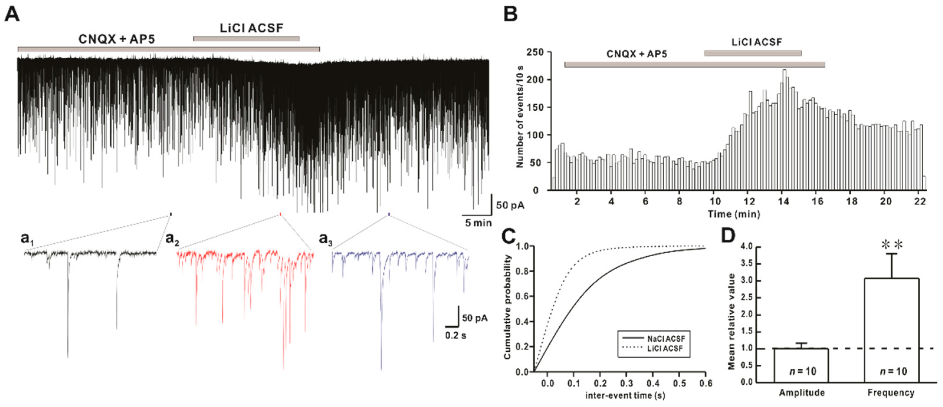
2.1. Li+ Enhances the Frequency of Spontaneous Inhibitory Postsynaptic Currents (sIPSCs)

sIPSCs were recorded from hPOA neurons in the presence of ionotropic glutamate receptor blockers CNQX and AP5. Mean frequency and amplitude of sIPSCs under normal artifical cerebrospinal fluid (ACSF) conditions were 3.02 ± 1.04 Hz (n = 10) and 80.5 ± 12.6 pA (n = 10), respectively. In the same neuron, perfusion of LiCl ACSF rapidly and reversibly increased the frequency of sIPSCs. Mean frequency and amplitude of sIPSCs in hPOA neurons during Li+ ACSF exposure were 7.05 ± 1.91 Hz (n = 10) and 73.9 ± 12.1 pA (n = 10), respectively (Figure 1A). The time-frequency histogram showed an immediate and reversible increase in the frequency of sIPSCs by Li+ (Figure 1B). The cumulative probability curve (Figure 1C) showed a progressive leftward shift in the inter-event interval (IEI) of sIPSCs events shown in Figure 1A. The frequency of sIPSCs was significantly increased by Li+ application (relative frequency compared to control: 3.08 ± 0.72, n = 10, p < 0.01). However, the amplitude of sPSCs was not affected by Li+ (relative amplitude: 1.01 ± 0.17, n = 10, p > 0.05) (Figure 1D).

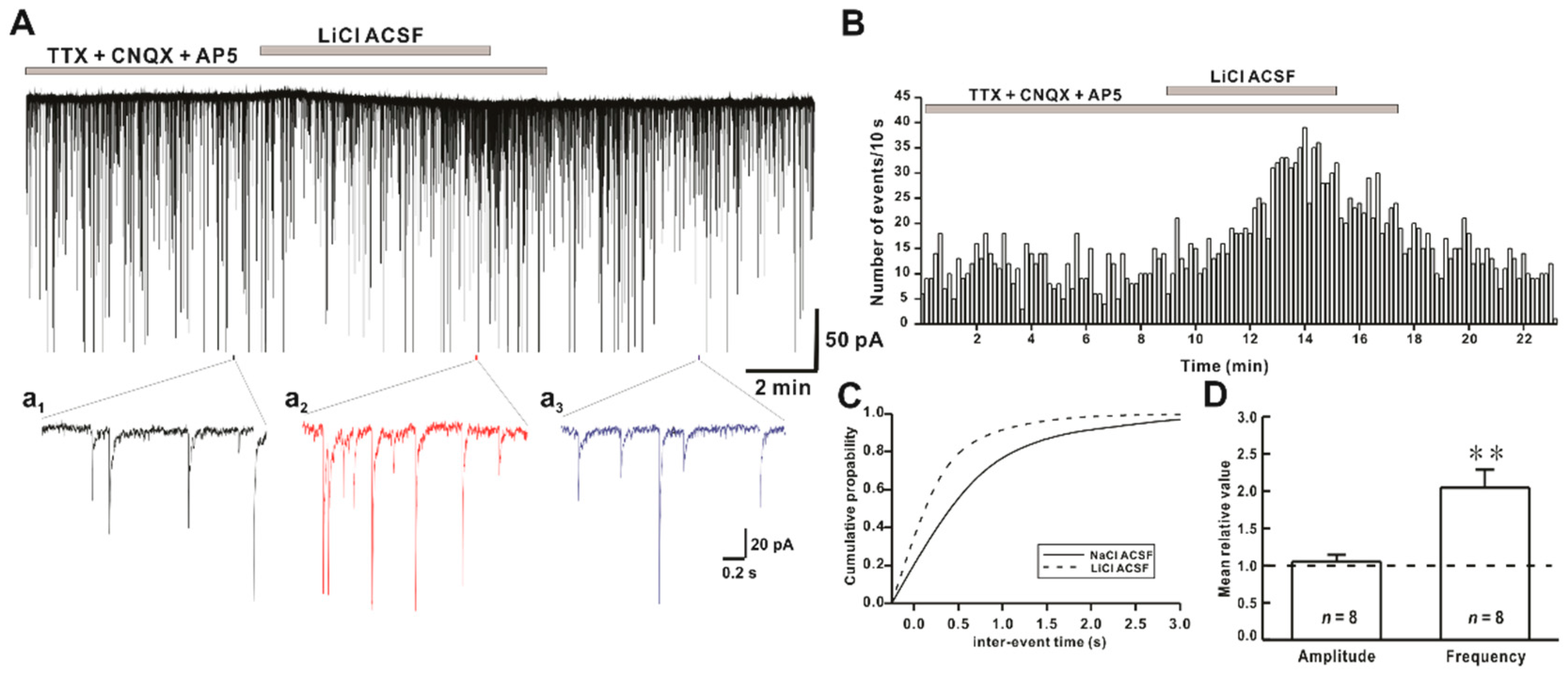
2.2. Li+ Enhances the Frequency of Miniature Inhibitory Postsynaptic Currents (mIPSCs)

To determine if Li+ ion could affect action potential-independent release of an inhibitory neurotransmitter from presynaptic axon terminal, mIPSCs were recorded in the presence of tetrodotoxin (TTX, a voltage-gated Na+ channel blocker) and ionotropic glutamate receptor blockers CNQX and AP5. The frequency of mIPSCs was increased by Li+ ACSF application in all tested neurons, as shown in Figure 2A. Mean frequency and amplitude of mIPSCs under a normal ACSF condition were 1.94 ± 0.42 Hz (n = 8) and 57.1 ± 10.5 pA (n = 8), respectively. In the presence of Li+ ACSF, mean frequency and amplitude of mIPSCs in hPOA neurons were 4.44 ± 1.29 Hz (n = 8) and 57.2 ± 9.59 pA (n = 8), respectively. Time-frequency histogram of mIPSCs showed an increase in the frequency on Li+ exposure (Figure 2B). Li+ significantly shifted the cumulative frequency curve to the left (Figure 2C). The frequency of mIPSCs was significantly increased by Li+ application (relative frequency compared to control: 2.04 ± 0.24, n = 8, p < 0.01). However, the amplitude of sPSCs was not affected by Li+ (relative amplitude: 1.05 ± 0.09, n = 8, p > 0.05) (Figure 2D).

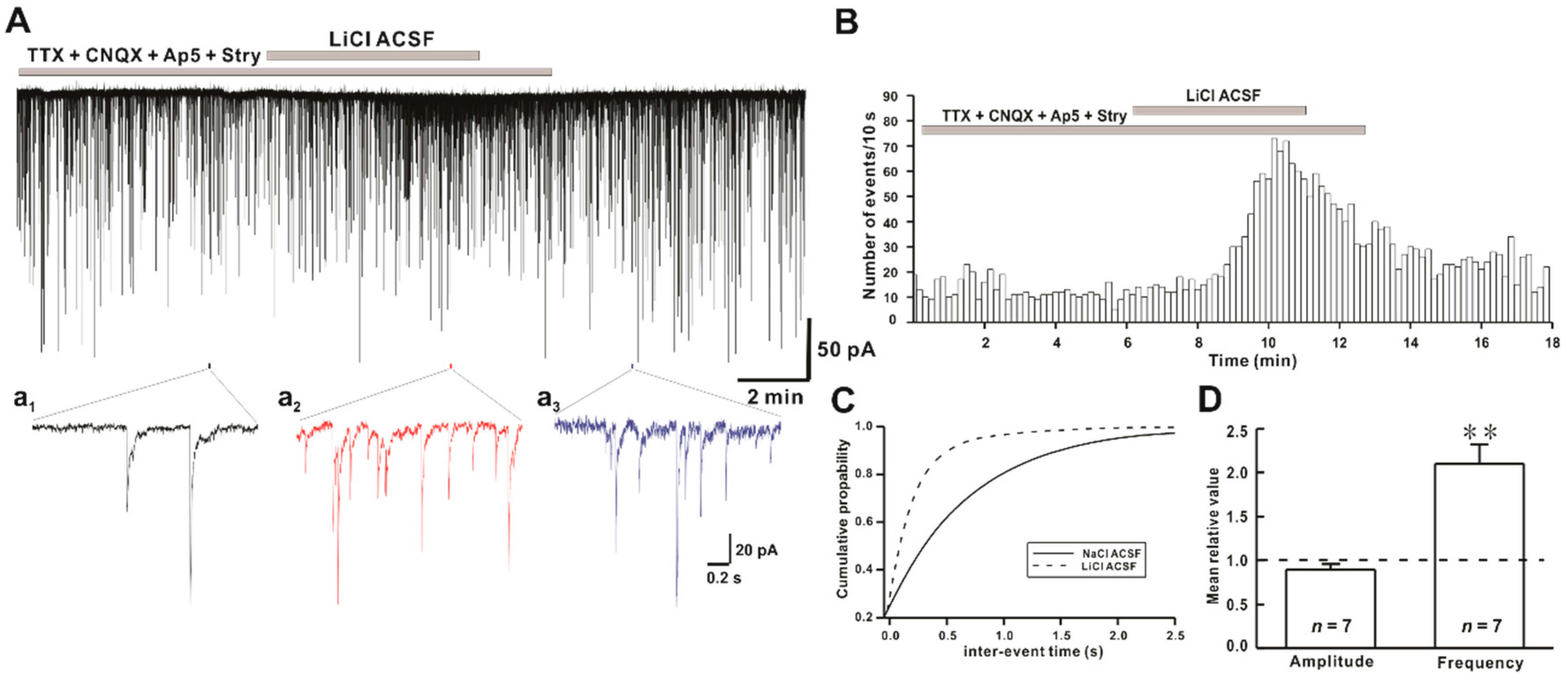
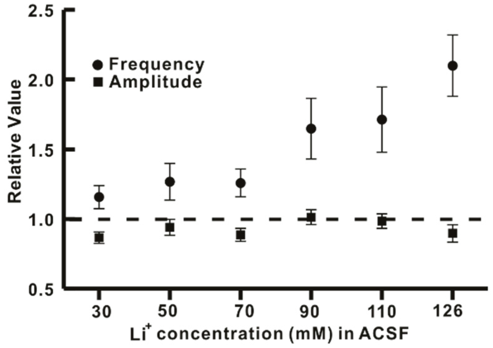
2.3. Li+ Increases the Frequency of GABAergic mIPSCs on hPOA Neurons

In the CNS, GABAergic and glycinergic neurotransmissions are the major inhibitory inputs in neuronal regulation. The pre-optic area contains GABAergic neurons [26] and receives glycinergic intervention from a different region of the brain [7,8]. To determine whether GABA release might be involved in the increase of mIPSCs frequency by Li+, mIPSCs were recorded in the presence of strychnine, a glycine receptor antagonist. Mean frequency and amplitude of GABAergic mIPSCs under normal ACSF condition were 1.90 ± 0.41 Hz (n = 7) and 44.7 ± 7.7 pA (n = 7), respectively. The mean frequency of GABAergic mIPSCs in the presence of strychnine was increased to 3.65 ± 0.61 Hz (n = 7) by Li+. However, the mean amplitude was not changed (38.1 ± 5.03 pA, n = 7), (Figure 3A). The time-frequency histogram showed an immediate and reversible increase in the event of GABAergic mIPSCs in the presence of Li+ ACSF (Figure 3B). In the cumulative probability plot, the IEI of the GABAergic mIPSCs was shifted to the left, as shown in Figure 3A, indicating an increase in the frequency of GABAergic mIPSCs (Figure 3C). The frequency of GABAergic mIPSCs was significantly increased by Li+ application (relative frequency compared to control: 2.1 ± 0.22, n = 7, p < 0.01). However, the amplitude of sPSCs was not affected by Li+ (relative amplitude: 0.9 ± 0.06, n = 7, p > 0.05) (Figure 3D).
Furthermore, the concentration-dependent replacement of Na+ ions with Li+ ions in the ACSF solution augmented the frequency of GABAergic mIPSCs in a concentration-dependent manner without affecting the amplitude, as shown in Figure 4.

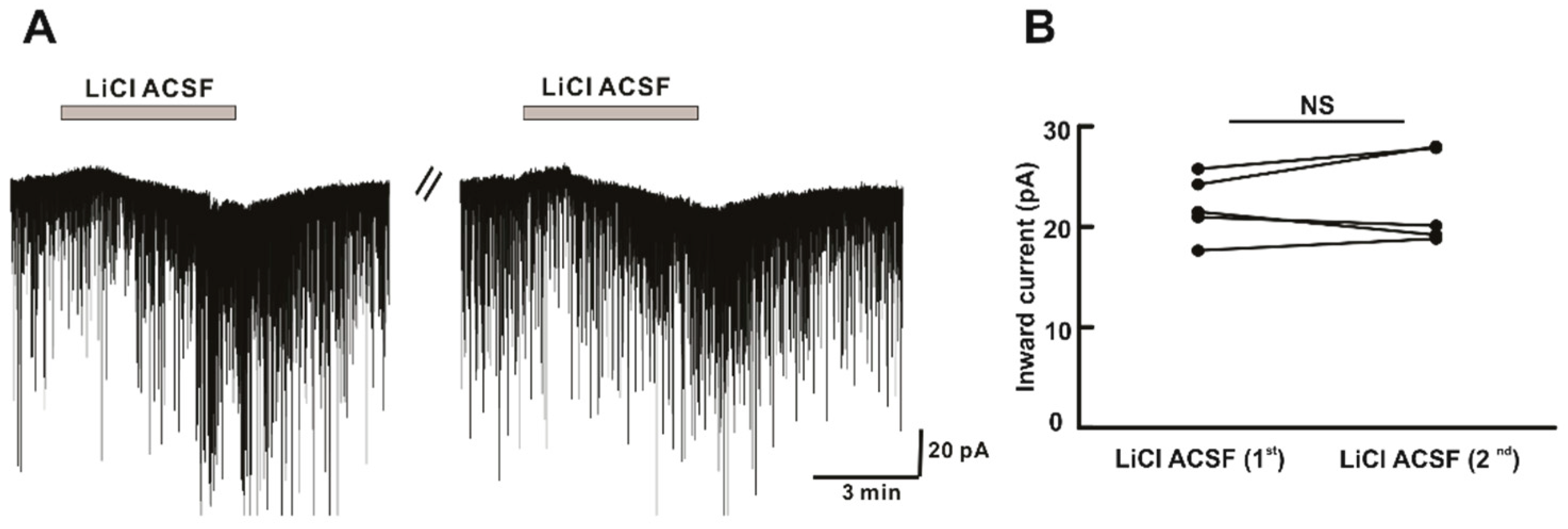
2.4. Li+ Perfusion Induces Repeatable Inward Currents on hPOA Neurons

In addition to the effect of Li+ in postsynaptic currents, we observed a change in holding current on Li+ perfusion. In the whole-cell voltage-clamp experiment, a reversible inward current of magnitude (−21.6 ± 4.6 pA, n = 14) was observed in 14 of 18 neurons tested during Li+ ACSF perfusion. With successive application of LiCl ACSF, there was no significant difference in inward current induced during the first and second applications (Figure 5A). The mean amplitude of inward current (−22.0 ± 1.40 pA, n = 5) induced by the first application of LiCl ACSF was similar to that induced by the second application (−22.8 ± 2.1 pA, n = 5, p > 0.05) (Figure 5B). This indicated that Li+ induced a repeatable and non-desensitized response on hPOA neurons during the successive perfusion.

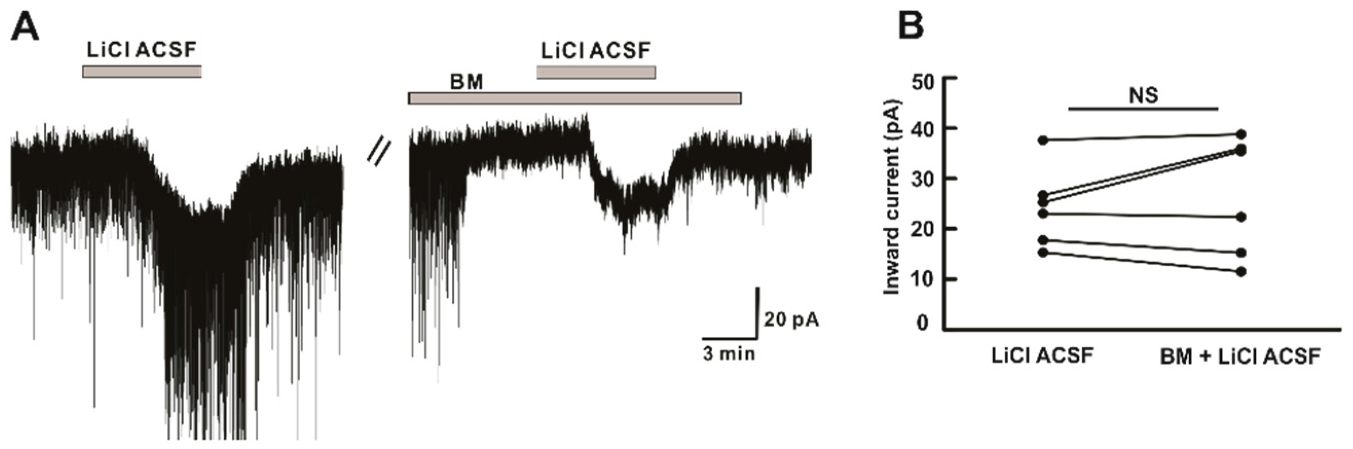
2.5. Li+ Directly Acts on hPOA Neurons and Its Action Is Independent of Amino-Acid Receptors

Next, we attempted to explore the direct action of Li+ ions on hPOA neurons. Neurons in the preoptic area can generate currents due to activation of GABAA, glycine, or ionotropic glutamate receptors [27,28]. Therefore, inward current induced by Li+ was recorded in the presence of blocking mixture (BM) including TTX (0.5 μM), picrotoxin (GABAA receptor antagonist, 50 μM), CNQX (non-NMDA glutamate receptor antagonist, 10 μM), AP5 (NMDA glutamate receptor antagonist, 20 μM), and strychnine (glycine receptor antagonist, 2 μM). In the presence of BM, the inward current induced by LiCl ACSF was maintained (Figure 6A). There was a similarity in the mean amplitude of inward current induced by Li+ alone (−24.3 ± 3.21 pA, n = 6) and Li+ in the presence of BM (−26.6 ± 4.8 pA, n = 6, p > 0.05) (Figure 6B), indicating Li+ directly acts on the post-synaptic hPOA neurons and such responses on hPOA are not due to the activation of GABAA, glycine, or ionotropic glutamate receptors.

3. Discussion

Results of this study showed that Li+ increased GABAergic synaptic activities in hypothalamic preoptic area neurons by action potential independent presynaptic mechanisms as TTX (a voltage-gated Na+ channel blocker) did not affect the frequency of spontaneous inhibitory postsynaptic currents (sIPSCs) increased by Li+. Besides, Li+ perfusion induced inward current for the majority of hPOA neurons. These findings are consistent with previous studies, showing that Li+ administration can alter the resting potential [29,30], and enhance GABAergic activities [15].
In the hypothalamic preoptic area, about 70 neuronal populations have been identified with inhibitory neurons being the most abundant [31]. GABAergic and glutamatergic neurotransmissions are the major inputs for neuronal regulation in the hypothalamus [32,33]. Besides, this region has interconnections to various brain areas [11] and receives glycinergic innervation [7,8]. GABA and glycine are major inhibitory transmitters whereas glutamate is the major excitatory transmitter in CNS. Electrophysiological experiments have revealed both excitatory and inhibitory effects of Li+ on neuronal excitability [15,29,34].
In electrophysiology, complete or partial replacement of Na+ by Li+ in the external medium could modulate neuronal properties and a neurotransmitter system. For example, Li+ induced depolarization and altered the frequency and shape of the action potential in mitral cells [29]. Similarly, both excitatory and inhibitory synaptic transmission were enhanced by Li+ perfusion [15,35,36]. Furthermore, chronic lithium administration resulted in a significant change of the brain neurotransmitter system of selected brain regions [37]. At the neuronal level, Li+ exhibited both pre-synaptic and post-synaptic action to modulate neurotransmission mediated by dopamine, GABA, glutamate, and serotonin [14,38]. Li+ induced a change in the GABAergic system and GABA receptors have been well documented in various areas of CNS, such as corpus striatum [37], prefrontal cortex [39], hypothalamus [37,40], and dentate gyrus [15].
Findings of the present study suggest that the replacement of Na+ by Li+ in ACSF could rapidly increase the frequency of synaptic activities without altering their amplitudes. In addition, there was no significant difference between the increased ratio of sIPSCs and mIPSCs frequency induced by Li+, indicating that Li+ enhanced the action potential-independent presynaptic mechanism mediated GABAergic synaptic events. However, Lee et al. have observed that exposure to Li+ (25 mM) could enhance GABAergic synaptic activities by AP-dependent and AP-independent presynaptic mechanisms in hippocampal slices [15]. Our results also revealed that concentration-dependent replacement of Na+ by Li+ in ACSF increased frequencies of GABAergic mIPSCs without affecting their amplitudes.
In addition to the presynaptic effect, the postsynaptic effect of Li+ on neuronal regulation has been reported in various neuronal groups [29,41,42]. Replacement of Na+ with Li+ can induce depolarization in cortical neurons [29], giant neurons [41], CA1 neurons [43], spinal motoneurons, and olfactory cortex [42]. Similarly, we observed an inward current when LiCl ACSF perfusion was performed. Li+ induced non-desensitizing repeatable inward currents. Such responses were preserved in the presence of tetrodotoxin (TTX, a voltage-gated Na+ channel blocker) and blocker for amino acid receptors. Previous findings have suggested that Li+ can replace Na+ ions and pass through neuronal membranes along Na+ channels [29,41,44]. In addition, Li+ can interact with electrogenic Na+/K+ pumps as three action sites for Li+ interaction has been identified [42]. To induce membrane depolarization, Li+ might suppress the activity of an electrogenic Na+ pump, reduce the intracellular K+ concentration, and increase the release of an excitatory transmitter [42,45,46]. Grafe et.al have demonstrated that Li+ can induce a shift of the K+ equilibrium potential responsible for membrane depolarization of neurons [42]. In the present study, we observed that Li+ directly acted on hPOA neurons independent of voltage-gated Na+ channels, GABAA, glycine, or ionotropic glutamate receptors. However, we could not elucidate the complete mechanism responsible for the induced inward current.
Neurotransmission disturbances have been reported in several neurological disorders [47,48], and lithium salt is widely used as a therapeutic agent for treating neurological and psychiatric disorders [49,50]. The therapeutic mechanism of Li+ involves its neuroprotective effect, neurotropic effect, and neuronal plasticity [13,51]. Lithium exhibits a neuroprotective effect by modulating glutamatergic transmission and inhibiting N-methyl-D-aspartate (NMDA) receptor-mediated calcium influx induced excitotoxicity [52,53]. Besides, Li+ can regulate synaptic plasticity by suppressing α-amino-3-hydroxy-5-methyl-4-isoxazolepropionic acid (AMPA) glutamate receptor trafficking [54]. Despite its therapeutic effects on neurological and psychiatric disorders, it can disturb the neuroendocrine axis, mainly in hypothalamic-pituitary-thyroid, hypothalamic-pituitary-gonadal, and hypothalamic-pituitary-adrenal axis in patients under lithium therapy [55,56]. Besides, oral lithium administration can result in Li+ accumulation in central neuroendocrine tissues, such as the hypothalamus and pituitary gland [57]. Furthermore, our electrophysiological data showed an increase in GABAergic neurotransmission across the hypothalamic preoptic area upon lithium exposure. This indicates that Li+ treatment might directly affect the hypothalamic region of the brain and regulate the release of various neurohormones involved in synchronizing the neuroendocrine axis. However, the mechanism behind the induced inward current on hPOA neurons upon Li+ perfusion warrants further investigation.
It has been reported that lithium and anti-epileptic drugs showed side effects on the endocrine system. Alteration in the endocrine system might lead to endocrine complications and related health problems. Theses complications may arise from altered neurotransmission in the hypothalamus. Using the electrophysiology approach, we showed that GABAergic activity was increased across hypothalamic neurons upon Li+ exposure, explaining the cause for the disruption in the neuroendocrine axis in patients receiving lithium therapy.

4. Materials and Methods

4.1. Animals

Electrophysiological experiments were performed using brain slices prepared from immature mice (postnatal day 10 to 25) housed under 12 h light/12 h dark cycle (lights on at 07:00 AM) with free access to food and water. All animal care conditions and experiments were approved by the Institutional Animal Care and Use Committee of Jeonbuk National University with approval number of CBNU-2018-071 (4 September 2018) and CBNU-2019-089 (14 November 2019).

4.2. Brain Slice Preparation

Mice were decapitated. Their brains were promptly excised [28] and immersed in ice-cold artificial cerebrospinal fluid (ACSF) with the following compositions (in mM): 126 NaCl, 2.5 KCl, 2.4 CaCl2, 1.2 MgCl2, 11 D-glucose, 1.4 NaH2PO4, and 25 NaHCO3 (pH 7.4 when bubbled with 95% O2 and 5% CO2). Coronal brain slices (230 to 300 μm) containing a hypothalamic preoptic area were obtained with a vibratome (VT1200S, Leica biosystems, Wetzlar, Germany) in ice-cold ACSF. These slices were stored in oxygenated ACSF for at least 1 h at room temperature before electrophysiological recording. Various concentrations of LiCl ACSF were prepared by replacing NaCl iso-osmotically with LiCl.

4.3. Whole-Cell Patch-Clamp Recording and Data Analysis

Coronal brain slices were transferred to the recording chamber, entirely submerged, and continuously perfused with oxygenated ACSF at a flow rate of 4 to 5 mL/min. Neurons located in the hypothalamic preoptic area were targeted visually with an upright microscope (BX51W1, Olympus, Tokyo, Japan) equipped with Nomarski differential interference contrast optics. Patch pipettes were prepared from thin-wall borosilicate capillary glass (PG52151–4; WPI, Sarasota, FL, USA) using a Flaming/Brown puller (P-97, Sutter Instruments Co., Novato, CA USA). Patch pipettes were filled with an internal solution containing (in mM): 140 KCl, 1 CaCl2, 1 MgCl2, 10 HEPES, 4 MgATP, and 10 EGTA (pH 7.3 with KOH). Tip resistance of the loaded pipette ranged from 4 to 6 MΩ. Electrode potential was nullified before giga-seal was achieved. Neurons were voltage-clamped at a holding of −60 mV. These signals were sequentially amplified and filtered at 1 kHz with an Axopatch 200B (Molecular Devices, San Jose, CA, USA) and digitized at 1 kHz using a Digidata 1440A interface (Molecular Devices, San Jose, CA, USA). Acquisition and subsequent analysis of acquired data were performed using a Clampex 10.6 software (Molecular Devices) and an Origin 2018 software (OriginLab Corp., Northampton, MA, USA). Neurons displaying a shift in holding current > 5 pA were considered to have responded. All recordings were made at room temperature.
Synaptic events were analyzed and counted using a Mini-Analysis program (version 6.0.7, Synaptosoft, Decatur, GA, USA). Initially, synaptic currents were screened automatically at >10 pA amplitude threshold and then manually accepted or rejected based on the decay time constant (>5 ms) and the rise time constant (<8 ms). The frequency and amplitude of synaptic events in a single neuron were compared using the Kolmogorov-Smirnov test. All values are provided as a mean ± standard error of the mean. Student’s paired t-test was used to compare relative mean amplitude and frequency between two groups. Statistical significance was considered when p-values were less than 0.05. “n” represents the number of neurons recorded.

4.4. Chemicals

Chemicals including lithium chloride (LiCl), picrotoxin, strychnine hydrochloride (strychnine), and chemicals for ACSF were purchased from Sigma-Aldrich (St. Louis, MO, USA). Tetrodotoxin citrate (TTX), DL-AP5 (AP5), and CNQX disodium salt (CNQX) were purchased from Tocris Bioscience (Avonmouth, Bristol, UK). Stocks were diluted (usually by 1000 times) to a working concentration in ACSF before bath application.

Author Contributions

S.R. performed the experiments, analyzed the data, and wrote the draft. S.H.J. and S.J.P. contributed to reviewing and editing the draft. S.K.H. conceptualized and designed the study and completed the manuscript. All authors have read and agreed to the published version of the manuscript.

Funding

This research was supported by the Basic Research Program, through the National Research Foundation of Korea (NRF) funded by the Ministry of Education (2016R1D1A3B03932241). The funders had no role in the design, analysis, or writing of this article.

Institutional Review Board Statement

All animal care conditions and experiments were approved by the Institutional Animal Care and Use Committee of Jeonbuk National University, CBNU-2018-071 (4 September 2018) and CBNU-2019-089 (14 November 2019).

Informed Consent Statement

Not applicable.

Conflicts of Interest

The authors declare no conflict of interest.

References

  1. Watanabe, M.; Maemura, K.; Kanbara, K.; Tamayama, T.; Hayasaki, H. GABA and GABA receptors in the central nervous system and other organs. Int. Rev. Cytol. 2002, 231, 1–47. [Google Scholar]
  2. Kirmse, K.; Holthoff, K. Functions of GABAergic transmission in the immature brain. e-Neuroforum 2017, 23, 27–33. [Google Scholar] [CrossRef]
  3. Ramamoorthi, K.; Lin, Y. The contribution of GABAergic dysfunction to neurodevelopmental disorders. Trends Mol. Med. 2011, 17, 452–462. [Google Scholar] [CrossRef]
  4. Deidda, G.; Bozarth, I.F.; Cancedda, L. Modulation of GABAergic transmission in development and neurodevelopmental disorders: Investigating physiology and pathology to gain therapeutic perspectives. Front. Cell Neurosci. 2014, 8, 119. [Google Scholar] [CrossRef] [Green Version]
  5. Belousov, A.B.; O’Hara, B.F.; Denisova, J.V. Acetylcholine becomes the major excitatory neurotransmitter in the hypothalamus in vitro in the absence of glutamate excitation. J. Neurosci. 2001, 21, 2015–2027. [Google Scholar] [CrossRef] [Green Version]
  6. Vincent, S.R.; Hökfelt, T.; Wu, J.Y. GABA neuron systems in hypothalamus and the pituitary gland. Neuroendocrinology 1982, 34, 117–125. [Google Scholar] [CrossRef]
  7. Rampon, C.; Luppi, P.H.; Fort, P.; Peyron, C.; Jouvet, M. Distribution of glycine-immunoreactive cell bodies and fibers in the rat brain. Neuroscience 1996, 75, 737–755. [Google Scholar] [CrossRef]
  8. Zeilhofer, H.U.; Studler, B.; Arabadzisz, D.; Schweizer, C.; Ahmadi, S.; Layh, B.; Bösl, M.R.; Fritschy, J.M. Glycinergic neurons expressing enhanced green fluorescent protein in bacterial artificial chromosome transgenic mice. J. Comp. Neurol. 2005, 482, 123–141. [Google Scholar] [CrossRef]
  9. Hoffman, N.W.; Wuarin, J.P.; Dudek, F.E. Whole-cell recordings of spontaneous synaptic currents in medial preoptic neurons from rat hypothalamic slices: Mediation by amino acid neurotransmitters. Brain Res. 1994, 660, 349–352. [Google Scholar] [CrossRef]
  10. McCann, S.M.; Vijayan, E.; Negro-Vilar, A.; Mizunuma, H.; Mangat, H. Gamma aminobutyric acid (GABA), a modulator of anterior pituitary hormone secretion by hypothalamic and pituitary action. Psychoneuroendocrinology 1984, 9, 97–106. [Google Scholar] [CrossRef]
  11. Yu, S.; François, M.; Huesing, C.; Münzberg, H. The hypothalamic preoptic area and body weight control. Neuroendocrinology 2017, 106, 187–194. [Google Scholar] [CrossRef] [PubMed] [Green Version]
  12. Mikosha, A.S.; Kovzun, O.I.; Tronko, M.D. Biological effects of lithium–fundamental and medical aspects. Ukr. Biochem. J. 2017, 89, 5–16. [Google Scholar] [CrossRef] [Green Version]
  13. Won, E.; Kim, Y.K. An oldie but goodie: Lithium in the treatment of bipolar disorder through neuroprotective and neurotrophic mechanisms. Int. J. Mol. Sci. 2017, 18, 2679. [Google Scholar] [CrossRef] [Green Version]
  14. Malhi, G.S.; Tanious, M.; Das, P.; Coulston, C.M.; Berk, M. Potential mechanisms of action of lithium in bipolar disorder. CNS Drugs 2013, 27, 135–153. [Google Scholar] [CrossRef] [PubMed]
  15. Lee, S.H.; Sohn, J.W.; Ahn, S.C.; Park, W.S.; Ho, W.K. Li+ enhances GABAergic inputs to granule cells in the rat hippocampal dentate gyrus. Neuropharmacology 2004, 46, 638–646. [Google Scholar] [CrossRef]
  16. Smythe, G.A.; Brandstater, J.F.; Lazarus, L. Acute effects of lithium on central dopamine and serotonin activity reflected by inhibition of prolactin and growth hormone secretion in the rat. Aust. J. Biol. Sci. 1979, 32, 329–334. [Google Scholar] [CrossRef] [Green Version]
  17. Bschor, T.; Ritter, D.; Winkelmann, P.; Erbe, S.; Uhr, M.; Ising, M.; Lewitzka, U. Lithium monotherapy increases ACTH and cortisol response in the DEX/CRH test in unipolar depressed subjects. A study with 30 treatment-naive patients. PLoS ONE 2011, 6, e27613. [Google Scholar] [CrossRef] [Green Version]
  18. Anai, H.; Ueta, Y.; Serino, R.; Nomura, M.; Kabashima, N.; Shibuya, I.; Takasugi, M.; Nakashima, Y.; Yamashita, H. Upregulation of the expression of vasopressin gene in the paraventricular and supraoptic nuclei of the lithium-induced diabetes insipidus rat. Brain Res. 1997, 772, 161–166. [Google Scholar] [CrossRef]
  19. Burns, G.; Herz, A.; Nikolarakis, K. Stimulation of hypothalamic opioid peptide release by lithium is mediated by opioid autoreceptors: Evidence from a combined in vitro, ex vivo study. Neuroscience 1990, 36, 691–697. [Google Scholar] [CrossRef]
  20. Sheikha, S.H.; LeGate, L.S.; Banerji, T.K. Lithium suppresses ovariectomy-induced surges in plasma gonadotropins in rats. Life Sci. 1989, 44, 1363–1369. [Google Scholar] [CrossRef]
  21. Allagui, M.; Hfaiedh, N.; Croute, F.; Guermazi, F.; Vincent, C.; Soleilhavoup, J.; El, A.F. Side effects of low serum lithium concentrations on renal, thyroid, and sexual functions in male and female rats. Comptes Rendus Biol. 2005, 328, 900–911. [Google Scholar] [CrossRef]
  22. Bschor, T.; Adli, M.; Baethge, C.; Eichmann, U.; Ising, M.; Uhr, M.; Modell, S.; Künzel, H.; Müller-Oerlinghausen, B.; Bauer, M. Lithium augmentation increases the ACTH and cortisol response in the combined DEX/CRH test in unipolar major depression. Neuropsychopharmacology 2002, 27, 470–478. [Google Scholar] [CrossRef] [Green Version]
  23. Sugawara, M.; Hashimoto, K.; Hattori, T.; Takao, T.; Suemaru, S.; Ota, Z. Effects of lithium on the hypothalamo-pituitary-adrenal axis. Endocrinol. Jpn. 1988, 35, 655–663. [Google Scholar] [CrossRef] [PubMed] [Green Version]
  24. Lombardi, G.; Panza, N.; Biondi, B.; Di Lorenzo, L.; Lupoli, G.; Muscettola, G.; Carella, C.; Bellastella, A. Effects of lithium treatment on hypothalamic-pituitary-thyroid axis: A longitudinal study. J. Endocrinol. Investig. 1993, 16, 259–263. [Google Scholar] [CrossRef] [PubMed]
  25. Kusalic, M.; Engelsmann, F. Effect of lithium maintenance treatment on hypothalamic pituitary gonadal axis in bipolar men. J. Psychiatry Neurosci. 1996, 21, 181–186. [Google Scholar] [PubMed]
  26. Tappaz, M.L.; Brownstein, M.J.; Kopin, I.J. Glutamate decarboxylase (GAD) and γ-aminobutyric acid (GABA) in discrete nuclei of hypothalamus and substantia nigra. Brain Res. 1977, 125, 109–121. [Google Scholar] [CrossRef]
  27. Bhattarai, J.P.; Park, S.J.; Chun, S.W.; Cho, D.H.; Han, S.K. Activation of synaptic and extrasynaptic glycine receptors by taurine in preoptic hypothalamic neurons. Neurosci. Lett. 2015, 608, 51–56. [Google Scholar] [CrossRef]
  28. Rijal, S.; Cho, D.H.; Park, S.-A.; Jang, S.H.; Ábrahám, I.M.; Han, S.K. Melatonin Suppresses the Kainate Receptor-Mediated Excitation on Gonadotropin-Releasing Hormone Neurons in Female and Male Prepubertal Mice. Int. J. Mol. Sci. 2020, 21, 5991. [Google Scholar] [CrossRef]
  29. Butler-Munro, C.; Coddington, E.J.; Shirley, C.H.; Heyward, P.M. Lithium modulates cortical excitability in vitro. Brain Res. 2010, 1352, 50–60. [Google Scholar] [CrossRef]
  30. Wu, M.; Zaborszky, L.; Hajszan, T.; Van Den Pol, A.N.; Alreja, M. Hypocretin/orexin innervation and excitation of identified septohippocampal cholinergic neurons. J. Neurosci. 2004, 24, 3527–3536. [Google Scholar] [CrossRef] [Green Version]
  31. Moffitt, J.R.; Bambah-Mukku, D.; Eichhorn, S.W.; Vaughn, E.; Shekhar, K.; Perez, J.D.; Rubinstein, N.D.; Hao, J.; Regev, A.; Dulac, C. Molecular, spatial, and functional single-cell profiling of the hypothalamic preoptic region. Science 2018, 362, 641. [Google Scholar] [CrossRef] [PubMed] [Green Version]
  32. Decavel, C.; Van Den Pol, A.N. GABA: A dominant neurotransmitter in the hypothalamus. J. Comp. Neurol. 1990, 302, 1019–1037. [Google Scholar] [CrossRef] [PubMed]
  33. Van Den Pol, A.N.; Trombley, P.Q. Glutamate neurons in hypothalamus regulate excitatory transmission. J. Neurosci. 1993, 13, 2829–2836. [Google Scholar] [CrossRef] [PubMed] [Green Version]
  34. Lacaille, J.C.; Cloutier, S.; Reader, T.A. Lithium reduced synaptic transmisson and increased neuronal excitability without altering endogenous serotonin, norepinephrine and dopamine in rat hippocampal slices in vitro. Prog. Neuropsychopharmacol. Biol. Psychiatry 1992, 3, 397–412. [Google Scholar] [CrossRef]
  35. Higashitani, Y.; Kudo, Y.; Ogura, A.; Kato, H. Acute effects of lithium on synaptic transmission in rat hippocampus studied in vitro. Biol. Psychiatry 1990, 27, 174–182. [Google Scholar] [CrossRef]
  36. Valentín, A.; García-Seoane, J.J.; Colino, A. Lithium enhances synaptic transmission in neonatal rat hippocampus. Neuroscience 1997, 78, 385–391. [Google Scholar] [CrossRef]
  37. Maggi, A.; Enna, S. Regional alterations in rat brain neurotransmitter systems following chronic lithium treatment. J. Neurochem. 1980, 34, 888–892. [Google Scholar] [CrossRef]
  38. Tanimoto, K.; Maeda, K.; Terada, T. Inhibitory effect of lithium on neuroleptic and serotonin receptors in rat brain. Brain Res. 1983, 265, 148–151. [Google Scholar] [CrossRef]
  39. Antonelli, T.; Ferioli, V.; Lo Gallo, G.; Tomasini, M.C.; Fernandez, M.; O’Connor, W.T.; Glennon, J.C.; Tanganelli, S.; Ferraro, L. Differential effects of acute and short-term lithium administration on dialysate glutamate and GABA levels in the frontal cortex of the conscious rat. Synapse 2000, 38, 355–362. [Google Scholar] [CrossRef]
  40. Gottesfeld, Z.; Ebstein, B.S.; Samuel, D. Effect of lithium on concentrations of glutamate and GABA levels in amygdala and hypothalamus of rat. Nat. New Biol. 1971, 234, 124–125. [Google Scholar] [CrossRef]
  41. Akoev, G.; Sizaya, N. Influence of lithium ions on the electrical activity of nerve cells of the leech. Neurophysiology 1970, 2, 484–489. [Google Scholar] [CrossRef]
  42. Grafe, P.; Reddy, M.; Emmert, H.; Ten Bruggencate, G. Effects of lithium on electrical activity and potassium ion distribution in the vertebrate central nervous system. Brain Res. 1983, 279, 65–76. [Google Scholar] [CrossRef] [Green Version]
  43. Liu, X.; Leung, L.S. Sodium-activated potassium conductance participates in the depolarizing afterpotential following a single action potential in rat hippocampal CA1 pyramidal cells. Brain Res. 2004, 1023, 185–192. [Google Scholar] [CrossRef]
  44. Janka, Z.; Jones, D. Lithium entry into neural cells via sodium channels: A morphometric approach. Neuroscience 1982, 7, 2849–2857. [Google Scholar] [CrossRef]
  45. Obara, S.; Grundfest, H. Effects of lithium on different membrane components of crayfish stretch receptor neurons. J. Gen. Physiol. 1968, 51, 635–654. [Google Scholar] [CrossRef] [PubMed] [Green Version]
  46. Giacobini, E.; Hovmark, S.; Stepita-Klauco, M. Studies on the Mechanism of Action of Lithium Ions: II. Potassium Sensitive Influx of Lithium Ions into the Crayfish Stretch Receptor Neuron Determined by Microflamephotometry. Acta Physiol. Scand. 1970, 80, 528–532. [Google Scholar] [CrossRef]
  47. Leite, J.A.; Orellana, A.M.M.; Kinoshita, P.F.; de Mello, N.P.; Scavone, C.; Kawamoto, E.M. Neuroinflammation and Neurotransmission Mechanisms Involved in Neuropsychiatric Disorders. In Mechanisms of Neuroinflammation; IntechOpen: London, UK, 2017. [Google Scholar]
  48. Sheffler, Z.M.; Reddy, V.; Pillarisetty, L.S. Physiology, Neurotransmitters; StatPearls Publishing: Treasure Island, FL, USA, 2020. [Google Scholar]
  49. Forlenza, O.V.; De-Paula, V.J.R.; Diniz, B.S.O. Neuroprotective effects of lithium: Implications for the treatment of Alzheimer’s disease and related neurodegenerative disorders. ACS Chem. Neurosci. 2014, 5, 443–450. [Google Scholar] [CrossRef] [PubMed] [Green Version]
  50. Maletzky, B.M.; Shore, J.H. Lithium treatment for psychiatric disorders. West. J. Med. 1978, 128, 488–498. [Google Scholar]
  51. Segal, J. Lithium-an update on the mechanisms of action. Part two: Neural effects and neuroanatomical substrate. Afr. J. Psychiatry 2004, 7, 18–24. [Google Scholar] [CrossRef] [Green Version]
  52. Shibuya-Tayoshi, S.; Tayoshi, S.Y.; Sumitani, S.; Ueno, S.; Harada, M.; Ohmori, T. Lithium effects on brain glutamatergic and GABAergic systems of healthy volunteers as measured by proton magnetic resonance spectroscopy. Prog. Neuropsychopharmacol. Biol. Psychiatry 2008, 32, 249–256. [Google Scholar] [CrossRef]
  53. Nonaka, S.; Hough, C.J.; Chuang, D.M. Chronic lithium treatment robustly protects neurons in the central nervous system against excitotoxicity by inhibiting N-methyl-D-aspartate receptor-mediated calcium influx. Proc. Natl. Acad. Sci. USA 1998, 95, 2642–2647. [Google Scholar] [CrossRef] [PubMed] [Green Version]
  54. Du, J.; Gray, N.A.; Falke, C.A.; Chen, W.; Yuan, P.; Szabo, S.T.; Einat, H.; Manji, H.K. Modulation of synaptic plasticity by antimanic agents: The role of AMPA glutamate receptor subunit 1 synaptic expression. J. Neurosci. 2004, 24, 6578–6589. [Google Scholar] [CrossRef] [PubMed] [Green Version]
  55. Giusti, C.F.; Amorim, S.R.; Guerra, R.A.; Portes, E.S. Endocrine disturbances related to the use of lithium. Arq. Bras. Endocrinol. Metabol. 2012, 56, 153–158. [Google Scholar] [CrossRef] [PubMed] [Green Version]
  56. Filippa, V.P.; Mohamed, F.H. Lithium Therapy Effects on the Reproductive System. In Psychiatry and Neuroscience Update; Springer: Cham, Switzerland, 2019; pp. 187–200. [Google Scholar]
  57. Pfeifer, W.D.; Davis, L.; Van der Velde, C.D. Lithium accumulation in some endocrine tissues. Acta Biol. Med. Ger. 1976, 35, 1519–1523. [Google Scholar]
Figure 1. Effect of Li+ (126 mM) on spontaneous inhibitory postsynaptic currents (sIPSCs) on hypothalamic preoptic area (hPOA) neurons. (A) A representative current trace of sIPSCs recorded in the presence of Na+ and Li+ artifical cerebrospinal fluid (ACSF) on hPOA neurons at a holding potential of −60 mV. (a1a3), sections of the current trace in Figure 1A show sIPSCs before, during, and after perfusion of Li+ at 2 s intervals, respectively. (B) A spike frequency histogram (bin size 10 s) of current traces in Figure 1A. (C) A cumulative probability plot of sIPSCs inter-event interval (IEI) in the presence of Na+ (solid line) and Li+ (dotted line). Note that the cumulative probability curve was left-shifted by Li+, indicating the increase of sIPSCs frequency (Kolmogorov-Smirnov test, p < 0.05). (D) Mean relative amplitude and frequency of sIPSCs in LiCl ACSF compared to NaCl ACSF (** p < 0.01 by a paired t-test).
Ijms 22 03908 g001
Figure 2. Effect of Li+ on miniature inhibitory postsynaptic currents (mIPSCs) on hPOA neurons. (A) A representative current trace of mIPSCs recorded in the presence of Na+ and Li+ ACSF on hPOA neurons at a holding potential of −60 mV. (a1a3), sections of the current trace in Figure 2A show mIPSCs before, during, and after perfusion of Li+ at 2-s intervals, respectively. (B) A spike frequency histogram of the current trace in Figure 2A. (C) A cumulative probability plot of mIPSCs inter-event interval (IEI) in the presence of Na+ (solid line) and Li+ (dotted line). Note that the cumulative probability curve was left-shifted by Li+, indicating the increase of mIPSCs frequency (Kolmogorov-Smirnov test, p < 0.05). (D) Mean relative amplitude and frequency of mIPSCs in LiCl ACSF compared to NaCl ACSF (** p < 0.01 by paired t-test).
Ijms 22 03908 g002
Figure 3. Effect of Li+ on GABAergic mIPSCs on hPOA neurons. (A) A representative current trace of GABAergic mIPSCs recorded in the presence of Na+ and Li+ ACSF on hPOA neuron. (a1a3), sections of the current trace in Figure 3A show GABAergic mIPSCs before, during, and after perfusion of Li+ at 2-s intervals, respectively. (B) A spike frequency histogram of the trace in Figure 3A. (C) A cumulative probability plot of GABAergic mIPSCs inter-event interval (IEI) in the presence of Na+ (solid line) and Li+ (dotted line). Note that the cumulative probability curve was left-shifted by Li+, indicating the increase of GABAergic mIPSCs frequency (Kolmogorov-Smirnov test, p < 0.05). (D) Mean relative amplitude and frequency of GABAergic mIPSCs in LiCl ACSF compared to NaCl ACSF (** p < 0.01 by paired t-test).
Ijms 22 03908 g003
Figure 4. Concentration-dependent effect of Li+ on GABAergic mIPSCs. The graph shows a concentration-dependent increase in GABAergic mIPSCs frequency by Li+ without affecting the amplitude.
Ijms 22 03908 g004
Figure 5. Li+ induces repeatable inward currents on hPOA neurons. (A) A representative current trace showing repeatable inward currents on hPOA neuron induced by perfusion of LiCl ACSF. (B) Before and after plot shows no significant difference in the mean inward currents between the first and the second perfusion of LiCl ACSF (n = 5, p > 0.05 by paired t-test). NS, not significant.
Ijms 22 03908 g005
Figure 6. Li+ acts on postsynaptic hPOA neurons. (A) A representative current trace showing inward currents induced upon perfusion of LiCl ACSF. Li+ induced inward current persisted in the presence of BM (blocking mixture), including TTX (Na+ channel blocker), picrotoxin (GABAA receptor blocker), strychnine (glycine receptor blocker), CNQX (non-NMDA glutamate receptor antagonist), and AP5 (NMDA receptor antagonist). (B) The before and after plot shows no significant difference in the mean inward current between Li+ alone and Li+ in the presence of blocking mixture (n = 6, p > 0.05 by the paired t-test). NS, not significant.
Ijms 22 03908 g006
Publisher’s Note: MDPI stays neutral with regard to jurisdictional claims in published maps and institutional affiliations.

