Review

Helicobacter pylori Outer Membrane Vesicles and Extracellular Vesicles from Helicobacter pylori-Infected Cells in Gastric Disease Development

by
María Fernanda González
1,2,
Paula Díaz
1,2,
Alejandra Sandoval-Bórquez
1,2,
Daniela Herrera
1,2 and
Andrew F. G. Quest
1,2,3,*
1
Center for studies on Exercise, Metabolism and Cancer (CEMC), Laboratory of Cellular Communication, Program of Cell and Molecular Biology, Faculty of Medicine, Institute of Biomedical Sciences (ICBM), Universidad de Chile, Santiago 8380453, Chile
2
Advanced Center for Chronic Diseases (ACCDiS), Faculty of Medicine, Universidad de Chile, Santiago 8380453, Chile
3
Corporación Centro de Estudios Científicos de las Enfermedades Crónicas (CECEC), Santiago 7680201, Chile
*
Author to whom correspondence should be addressed.
Int. J. Mol. Sci. 2021, 22(9), 4823; https://0-doi-org.brum.beds.ac.uk/10.3390/ijms22094823
Submission received: 27 February 2021 / Revised: 31 March 2021 / Accepted: 2 April 2021 / Published: 1 May 2021
(This article belongs to the Special Issue Helicobacter: Infection, Diagnosis and Treatment)

Abstract

:
Extracellular vesicles (EVs) are cell-derived vesicles important in intercellular communication that play an essential role in host-pathogen interactions, spreading pathogen-derived as well as host-derived molecules during infection. Pathogens can induce changes in the composition of EVs derived from the infected cells and use them to manipulate their microenvironment and, for instance, modulate innate and adaptive inflammatory immune responses, both in a stimulatory or suppressive manner. Gastric cancer is one of the leading causes of cancer-related deaths worldwide and infection with Helicobacter pylori (H. pylori) is considered the main risk factor for developing this disease, which is characterized by a strong inflammatory component. EVs released by host cells infected with H. pylori contribute significantly to inflammation, and in doing so promote the development of disease. Additionally, H. pylori liberates vesicles, called outer membrane vesicles (H. pylori-OMVs), which contribute to atrophia and cell transformation in the gastric epithelium. In this review, the participation of both EVs from cells infected with H. pylori and H. pylori-OMVs associated with the development of gastric cancer will be discussed. By deciphering which functions of these external vesicles during H. pylori infection benefit the host or the pathogen, novel treatment strategies may become available to prevent disease.

1. Helicobacter pylori, Inflammation and Gastric Cancer

Helicobacter pylori (H. pylori) is a Gram-negative, microaerophilic, spiral bacterium, which colonizes the epithelium of the human stomach [1]. H. pylori infection is one of the most common infectious diseases, affecting approximately 50% of the world’s population [2]. Infection rates differ according to geographic region, remaining higher in developing countries (>85%) [3]. The prevalence of H. pylori infection is associated with low socioeconomic status and poor sanitary conditions, which are considered risk factors [4]. The most likely route of transmission is oral-fecal, especially through contaminated food [5]. In the last three decades, the incidence of H. pylori infection has remained constant or even increased due to population growth, re-infection and increasing antibiotic resistance [6].
H. pylori was described for the first time by Warren and Marshall in biopsies from patients with gastritis and peptic ulceration [7]. Numerous studies have identified H. pylori as the principal etiologic agent associated with the development of chronic gastritis, peptic ulcer disease, mucosa-associated lymphoid tissue lymphoma and gastric cancer [8]. Recently, H. pylori has also been associated with diseases outside the gastrointestinal tract, such as, immune thrombocytopenic-purpura, iron-deficiency anemia, vitamin B12 deficiency, neurodegenerative disorders and metabolic syndrome [9].
H. pylori colonization of the human stomach and its role as a bacterial carcinogen is a complex process involving communication between host cells, the microorganism and the host environment [10]. In the initial phase of infection, H. pylori neutralizes the acidic environment of the stomach by liberating urease, which hydrolyzes urea to generate carbon dioxide and ammonia [11] that neutralize the acidic microenvironment surrounding the bacteria [12]. In addition, ammonia produces various alterations in host cells by affecting vesicular membrane transport, protein synthesis and ATP production, among others [11]. Moreover, urease activity regulates H. pylori-macrophage interactions, by modulating the phagosome pH and megasome formation, which are necessary for H. pylori survival [12].
H pylori has 4–8 flagella and chemotaxis receptors which it uses to colonize the stomach lining and reach epithelial cells [13]. Colonization by H. pylori leads to inflammation and neutrophil infiltration [13,14]. The flagellar filament of H. pylori is mainly composed of the structural proteins FlaA, and FlaB [12]. These flagellins are the first targets of antibodies generated by the host adaptive immune response [13]. However, flagellins, particularly FlaA, are not recognized by toll-like receptor 5 (TLR5) [15,16]. This ability of H. pylori to evade the immune system, contributes to the persistence of the bacteria in the gastric epithelium [13].
H. pylori attaches to the gastric epithelium by producing adhesins, which interact with host cell receptors [12]. These specific interactions protect the bacteria from displacement by forces produced during peristalsis, leading to successful and persistent infection [11,12]. After colonization, virulence factors, such as cytotoxin-associated gene A (CagA) and vacuolating cytotoxin A (VacA), cause further damage to the host epithelium and may increase the risk of gastric disease [10]. The cag pathogenicity island (cagPAI) is a region of the bacterial chromosome, coding CagA and the Cag type IV secretion system (T4SS), which are detectable in more virulent H. pylori strains [10]. Accordingly, the presence of cagPAI has been associated with higher levels of inflammation in the stomach [11].
CagA is an oncoprotein produced in the cytoplasm of the bacteria, which is then injected into host cells via the T4SS [12,17]. Once in the cytoplasm, CagA localizes to the inner surface of the plasma membrane where it is phosphorylated on tyrosine residues by c-SRC and c-ABL kinases [10]. CagA then can modify host cell signaling in multiple ways, such as by activating PI3K/AKT [18], Wnt/β-catenin [19] and MAPK/ERK signaling [10]. CagA also modulates cell adhesion and migration by binding to the phosphatase SHP-2 [12,20]. Other effects include the alteration of epithelial cell polarity and the dysregulation of apical-junctional complexes [10,21]. In addition, CagA activates NF-κB-dependent inflammatory signaling and interleukin-8 (IL-8) production in infected gastric cells [10,22]. This leads to recruitment of neutrophils and macrophages, which further increases the release of cytokines, as well as reactive oxygen species (ROS) and growth factors [22,23]. This microenvironment results in chronic inflammation, which promotes tumorigenesis [22].
VacA contains p33 and p55 protein subunits, which combine to generate an oligomeric complex [12] that forms selective anion channels in the host cell membrane and can release bicarbonate and organic anions into the host cytoplasm [12,24]. In this way, VacA aids H. pylori survival by inducing a flow of metabolic substrates from the mucosa to the stomach lumen [25]. This produces changes in the permeability of the plasma membrane and disrupts gastric epithelium cell integrity [12]. VacA also forms channels in the membranes of endosomes and mitochondria [24,26]. Moreover, VacA internalizes into cells via endocytosis, affecting endosomal maturation, among others [12,25]. In late endosomal compartments, these anion selective channels cause an increase in osmotic pressure, resulting in the formation of vacuoles [25,26,27]. In mitochondria, VacA induces changes in the mitochondrial membrane potential and release of cytochrome C leading to apoptosis [25,28]. In addition to its cytotoxic properties, VacA acts as an immunosuppressor by inhibiting the development, activation and proliferation of T cells [10,29], as well as preventing antigen presentation by MHC class II molecules in B cells [25]. Moreover, VacA delays phagocytosis, inhibiting phagosome maturation in macrophages [25,30]. All H. pylori strains contain genes coding for VacA; however, there are multiple alleles that confer variations in vacuolation capacity [10,31]. Studies reveal that VacA in allelic variant s1/m1 and s1/m2 strains is associated with more severe chronic inflammation than for other genotypes [25].
H. pylori is an established group 1 carcinogen and presence of the bacteria is therefore linked to carcinogenesis [32]. Stomach or gastric cancer is a multifactorial and heterogeneous disease, whereby gastric adenocarcinoma (glandular origin) represents the most common histological type (~90%) of all neoplasms originating in the stomach [33,34]. Other less common variants of gastric cancer include lympho-proliferative, mesenchymal and neuroendocrine tumors [33]. Gastric adenocarcinoma is preceded by progressive changes in the gastric mucosa, beginning with non-atrophic gastritis, triggered primarily by H. pylori, followed by multifocal atrophic gastritis, which advances to intestinal metaplasia, dysplasia, and finally invasive carcinoma [35,36]. This cascade of precancerous lesions describes a human model of intestinal-type gastric carcinogenesis, which is the major gastric adenocarcinoma subtype and represents 60% of all cases [34,37]. This intestinal-type gastric cancer is characterized by the presence of tumor cells that are arranged in irregular tubular or glandular structures [38].
The oncogenic effects of H. pylori in epithelial cells are attributed either directly to the toxic action of the virulence factors described above, and/or indirectly to inflammatory processes produced by the infection [39]. Gastritis caused by H. pylori infection is characterized by infiltration of the lamina propria with mononuclear leukocytes (chronic inflammation) and polymorphonuclear cells (acute inflammation) [36]. The immune response triggered by infection is both of the innate and adaptive type [36,39]. T helper-1 (Th1) cells and their signature cytokines, IL-1β, tumor necrosis factor (TNF)-α and particularly interferon (IFN)-γ, are essential for the development of gastritis [36,40]. Recruited neutrophils and macrophages produce large amounts of ROS and reactive nitrogen species (RNS) [39,41]. This results in an increase in oxidative stress, which causes DNA damage [39]. Loss of normal glandular tissue, or atrophy, is the result of chronic inflammation and tends to be multifocal (multifocal atrophic gastritis) [36]. The increased expression of multiple cytokines by the gastric mucosa accelerates the progression of atrophic changes in cells [39]. In advanced stages of atrophy, intestinal metaplasia occurs, where a phenotypic change in the gastric mucosa is observed [36]. Normal epithelial cells are replaced by cells with an intestinal phenotype [42]. Intestinal metaplasia is considered a condition that predisposes to malignancy [36,42]. Dysplasia is characterized by a neoplastic phenotype, both in cell morphology and architectural organization of the epithelium [36]. The progression from superficial gastritis to an atrophic, metaplastic and finally neoplastic mucosa is associated with the virulence of H. pylori, as well as environmental and host factors [43]. Both gastric mucosal inflammation and H. pylori may cause host cell genomic instability, damage of the DNA mismatch repair system, abnormal DNA methylation, dysregulation of noncoding gene expression, which all contribute to an accumulation of mutations and the loss of normal regulation of cell function [43,44].
Besides participation of the aforementioned H. pylori virulence factors to disease development, the bacteria also liberates outer membrane vesicles (H. pylori-OMVs) [45,46] that participate in such events. Biologically active H. pylori compounds can be introduced by H. pylori-OMVs into host cells where they may alter cell signaling pathways, impair cellular function, promote infection, and regulate immune responses [31,46]. In addition, H. pylori colonizes the epithelium of the stomach and causes pleiotropic changes in the infected cells of the stomach lining. All eukaryotic cells release host-derived extracellular vesicles (EVs) [47], and after infection by a pathogen these EVs may change in number, composition, or both. The details of these changes and, as a consequence, the biological effects such EVs may have in recipient cells are just beginning to be unraveled. As for H. pylori infection, little is known in this respect. However, the available evidence suggests they are important in this context. Thus, in the rest of this review we will elaborate on the contribution of H. pylori infected host-derived EVs and H. pylori-OMVs to the genesis and progression of gastric cancer.

2. What Are Extracellular Vesicles?

Extracellular vesicles (EVs) are membranous vesicles derived from cells that are important in local and distant intercellular communication, although initially such vesicles were thought to represent a mechanism by which cells eliminate waste products. All human cells that have been investigated are able to secrete EVs. Furthermore, this is a highly conserved process throughout evolution, from bacteria, protozoa, fungi, and plants to animals [48,49,50]. EVs were discovered in sheep reticulocytes, during their differentiation process. Johnstone’s group reported that immature sheep reticulocytes released most of their transferrin receptor in association with small membrane vesicles, through fusion of multi-vesicular bodies (MVBs) with the cell membrane [51,52]. Subsequently, it was shown that reticulocytes from other species also release EVs, which, in addition to transferrin receptors, also contain other proteins, such as acetylcholinesterase [52,53]. Although in the context of reticulocyte development it is clear why EVs were considered a means by which cells eliminated unwanted molecules, nowadays their role is viewed as being much more complex.
EVs are now widely recognized as vectors of intercellular communication that permit the exchange of many different types of molecules relevant to signaling events important during physiological and developmental, as well as pathological processes, such as infection by pathogens, inflammation or cancer [50,54,55,56,57]. EVs are highly heterogeneous but are generally segregated into two main groups: exosomes and microvesicles. Exosomes are of endosomal origin and their size varies between 30–100 nm diameter. They are generated by inward budding of the membrane of late endosomes destined to form MVBs. These intraluminal vesicles can be released to the extracellular space in the form of exosomes after fusion of the endosomal membrane with the plasma membrane [58]. Microvesicles, on the other hand, are larger in size ranging from 50–1000 nm or more in diameter. This type of vesicle is generated by outward budding and fission from the plasma membrane. However, depending on the cell of origin, these EVs are also known as ectosomes, oncosomes, migrasomes or apoptotic bodies [59,60,61].
The message conveyed by EVs is dictated by the profile of molecules that they transport within, as well as on their surface. The molecular profile of the EVs depends on the cell of origin and its physiological state. A large variety of molecules have been found in EVs, including proteins, such as growth factors, angiogenic factors, IL precursors, and nucleic acids, such as mRNA, miRNA, small interfering RNA, double stranded DNA, among others [56,57,62,63]. However, it seems that the content of RNA and short DNA is more relevant in microvesicles [64]. Furthermore, on their surface, EVs carry a variety of membrane proteins and cell surface molecules, in addition to several extracellular proteins, such as matrix-remodeling-associated protein 5, neural cell adhesion molecule L1-like protein, ADAM metallopeptidase with thrombospodin type 1 motif 6, CYR61 and tenascin C, to mention a few [65,66]. The lipid composition has also been analyzed and includes those commonly found in the plasma membrane, such as phospholipids, sphingomyelin and cholesterol [67].
Of these two groups of EVs, the biogenesis of exosomes is the most studied. There are at least two different mechanisms for the biogenesis of exosomes. The perhaps best-characterized route involves the sorting complex required for transport (ESCRT), a complex that selects and clusters ubiquitylated transmembrane proteins in microdomains via ESCRT-0 and ESCRT-I subunits, and then triggers budding and fission of this microdomain to the MVB interior via recruitment of the ESCRT-II and the ESCRT-III subunits [58,68,69]. Accessory proteins are also involved, such as ALIX and tumor susceptibility gene 101 (tsg101), among others [68]. The second mechanism is ESCRT-independent and involves specific lipids. In this case, accumulation of ceramide and the formation of microdomains in the endosomal membrane are necessary for the genesis of intraluminal vesicles. Important for this process is the activity of neutral sphingomyelinases, which hydrolyze sphingomyelin to generate ceramide that, thanks to its small polar head group, favors the deformation and bending of the endosomal membrane [70]. Furthermore, a second ESCRT-independent mechanism for endosomal EV formation has been identified, involving proteins of the tetraspanin family, such as CD9, CD63, CD81, and CD82 [68,71]. Depletion of ESCRT elements generally decreases EV release but does not prevent it completely. Similar results are obtained upon inhibition of neutral sphingomyelinase activity [69,72], demonstrating that cells generate EVs by different mechanisms. Moreover, secreted exosomes are heterogeneous because they can be derived from different MVB subpopulations that coexist in cells [73]. For example, in polarized cells, exosomes of different composition are released from the apical and basolateral sides of the cell [74]. Beyond these more generic mechanisms mentioned so far, additional mechanisms are available for the recruitment of specific proteins and lipids [75]. For instance, it has been suggested that the inclusion of cytosolic proteins into exosomes involves chaperones, such as Hsp70 and Hsc70 [76,77]. Regarding RNA content, the findings that ESCRT-II is an RNA-binding complex suggests that it may function to select RNA species that are incorporated into intraluminal vesicles [78].
Reportedly, the release of exosomes occurs in response to changes in intracellular calcium in cell lines, such as human erythroleukemia and mast cells [79,80]. Furthermore, potassium-induced depolarization appears to increase exosome release in neurons [81]. The Rab superfamily of small GTPases, known to control many intracellular pathways and the formation of membrane domains, appears also to play an important role in docking and tethering during exosome biogenesis. Specifically, the silencing of Rab27a and Rab27b reduces exosome release and docking of MVBs to the plasma membrane. In addition, when Rab27a is silenced, the size of MVBs increases. Moreover, when Rab27b expression is reduced, MVBs are redistributed toward the perinuclear region. In addition, silencing of the Rab27 effectors Slp4 and Slac2b, reduced exosome release [82]. These results indicate that both Rab27a and Rab27b are important for exosome release but have different specific functions. As to the regulation of MVB fusion with the plasma membrane, less is known. This process is likely to be facilitated by SNARE proteins [58].
After exosomes are released, their function depends on the interaction with the target cell. Exosomes contain adhesion proteins, including integrins, that facilitate exosome adhesion to recipient cells. In this manner, exosomes can induce changes through direct contact with the cell surface, as well as by fusing with the plasma membrane, resulting in the release of their contents into the cytosol. Additionally, upon surface binding exosomes may enter cells through internalization by endocytosis or phagocytosis. The type of interaction appears to be influenced by the origin of the exosomes and the nature of the recipient cell. When exosomes are internalized, they can be degraded, which will lead to the recycling of their content to fuel the metabolism of the recipient cell. Alternatively, they can fuse with the endosomal membrane of the recipient cell and thereby release their content into the cytosol to modulate cell function [58].
A variety of functions have been attributed to EVs. Exosomes released by cardiomyocytes under hypoxic conditions are enriched with angiogenic and pro-survival factors. Cardiac stem cells release exosomes with cardioprotective and regenerative properties, being potentially useful in cell-free cardiotherapy [83]. In cancer cell lines, inhibition of microvesicle release decreases the appearance of metastasis [84,85]. There is also evidence showing that exosomes secreted by tumors contribute to tumor progression by decreasing immune responses, promoting angiogenesis, inducing proliferation of cells, promoting extracellular matrix remodeling, or increasing migration, which favors metastasis [69,86,87]. For a more detailed overview of this topic, please consider the following literature [70,88,89,90].

3. Extracellular Vesicles in Host–Bacteria Interactions

During infection, EVs are released from pathogen-infected host cells (EVs) and also shed directly from pathogens, in which case they are referred to as outer membrane vesicles (OMVs). Both, host-derived EVs and pathogen-derived OMVs contribute to the dissemination of pathogenic components (e.g., pathogen-associated molecular patterns (PAMPs) including, proteins, lipids, nucleic acids and carbohydrates) to the surrounding cells and tissues [91,92,93]. EV transport of pathogenic molecules between cells contributes to host-pathogen cross-talk, the activation and modulation of the host immune system and other cellular signaling pathways.

3.1. Host-Derived Extracellular Vesicles

As mentioned above, EVs (also known as host-derived EVs) play roles in cell-cell communication via the presence of specific proteins and nucleic acids, which may also serve as biomarkers of disease [94]. Moreover, EVs released by infected host cells contribute significantly to promoting an inflammatory response and therefore, to promoting the development of disease.
Much of the information available regarding cellular EV production and function during bacterial infection arises from work on Mycobacterium sp. Particularly, Mycobacterium tuberculosis PAMPs are transported from the phagosome to MVBs during macrophage cell infection. These PAMPs are also found in EVs released by infected macrophages and their content can be detected in neighboring uninfected cells [91,95,96]. In addition, exosomes may serve as antigen-presenting vesicles, which have stimulatory or tolerogenic effects [97,98]. Mycobacterium avium-infected macrophages release EVs that stimulate pro-inflammatory responses in uninfected macrophages [99,100]. EVs released from Mycobacterium tuberculosis-, Mycobacterium bovis- and Mycobacterium smegmatis-infected macrophages were also shown to be pro-inflammatory [91,101]. In particular, the Mycobacterium 19-kDa lipoprotein present on EVs released from Mycobacterium tuberculosis-infected cells was reported to drive this inflammatory response, via pattern recognition receptors, such as the TLR/MyD88 pathway [102]. In addition, EVs from Mycobacterium bovis- and Mycobacterium tuberculosis-infected macrophages can stimulate pro-inflammatory responses in vivo, as intranasal injection of mice induced TNF-α and IL-12 production, as well as recruitment of macrophages and neutrophils to the lung [91]. Macrophages treated with EVs from Mycobacterium tuberculosis secrete chemokines that activate naïve macrophages and induce T-cell migration in vitro [103]. These results suggest that EVs from Mycobacterium-infected cells can promote both recruitment and activation of immune cells in vitro and in vivo and may play a role in promoting the innate immune response upon Mycobacterium infection.
To the contrary, Mycobacterium sp. components present in EVs may also suppress immune responses. Indeed, EVs released from Mycobacterium tuberculosis-infected cells can partially suppress IFN-γ mediated activation of recipient naïve macrophages, a response mediated by TLR2/MyD88 signaling [104]. In addition, EVs from Mycobacterium tuberculosis-infected cells have also been reported to contain a member of the Mycobacterium-PE gene family (Rv1818c) that induced apoptosis in T cells [105]. This implies that, besides their effects in macrophages, these EVs also suppress immune responses in T cells. Available evidence also shows that EVs from Mycobacterium tuberculosis- or Mycobacterium bovis-infected cells activate antigen-specific CD4+ and CD8+ T cells in vivo, as well as promote the activation and maturation of bone marrow-derived dendritic cells [106]. These results suggest that EVs are a rich source of antigens to stimulate immune responses.
Alternatively, EVs from Salmonella-infected macrophages are also pro-inflammatory, by increasing TNF-α production in monocytes [91]. These EVs contain lipopolysaccharide, a known PAMP, in Salmonella and other Gram-negative bacteria. EVs from tumor cells infected with Mycoplasma induce a mixed cytokine response, including production of both IFN-α and IL-10, thereby activating inhibitory B cells [107]. Moreover, the anthrax toxin produced by Bacillus anthracis can be released inside EVs from an infected epithelial cell line [108]. In addition, EVs released from Chlamydia pneumoniae-infected cells contain tissue factor, a protein participating in blood coagulation, which has been associated with cell proliferation, migration and apoptosis [109]. Moreover, several cytotoxic and secreted proteins are associated with host vesicles released from Chlamydia trachomatis, which contribute to the dissemination of bacterial virulence factors [110].

3.2. Outer Membrane Vesicles in Host-Bacteria Interactions

Shedding of outer membrane vesicles (OMVs) was initially observed in the Gram-negative bacteria Vibrio cholera [111] and Neisseria meningitides [112]. OMVs are generated by “blebbing” of the outer membrane of the bacteria and contain proteins, membrane components as well as nucleic acids. Among other functions, the release of OMVs aids in Gram-negative bacterial survival [113,114]. Evidence has also been obtained on the shedding of OMVs by Gram-positive bacteria [115]. However, there is limited knowledge concerning the mechanisms by which OMVs interact with cell surfaces and transfer their cargos. Initially, OMVs dock at the plasma membrane of the recipient cell where they are internalized via endocytosis, or fuse with the plasma membrane [116]. OMV surface proteins interact with specific receptors, which leads to the activation of signaling cascades [58]. PAMPs are present on the surface of pathogens [117] and OMVs where they may interact with pattern-recognition receptors (PRRs) present on recipient cells [118,119,120]. This suggests that molecules on the surface of OMVs may target cells or organs in the host to create a niche for infection and survival.
OMVs from bacteria have been shown to compromise the immune defenses by inducing apoptosis of host cells. For instance, OMVs from Neisseria gonorrhoeae contain a porin protein (PorB), which is transported to mitochondria in host macrophages, where it triggers loss of mitochondria membrane potential and cytochrome C release, leading to caspase activation and apoptosis [121]. Moreover, cytotoxic proteins found in OMVs derived from a highly virulent Escherichia coli strain cause activation of caspase-9 and caspase-3 after OMV uptake by intestinal epithelial cell lines [122]. OMVs released by Bacillus anthracis, a Gram-positive bacterium, also contain cytotoxic proteins [123]. Particularly, the OMV-associated toxins released by this bacterium (anthrolysin and anthrax toxin polypeptide) induced toxicity in macrophages, and also in mice in vivo [123].
Additionally, bacteria-derived toxins transported by OMVs can modulate immune cell functions to promote pathogen survival. For example, cytotoxic necrotizing factor type 1 (CNF1), found in OMVs from Escherichia coli, reduced antimicrobial activity and chemotaxis in polymorphonuclear leukocytes [124]. In addition, the OMV-associated Cif-toxin, derived from Pseudomonas aeruginosa, downregulated the expression of the cystic fibrosis transmembrane conductance regulator in airway epithelial cells, thereby increasing Pseudomonas aeruginosa pathogenicity [125]. Additionally, Moraxella catarrhalis sheds OMVs that reduced TLR2-induced IL-8 production by alveolar epithelial cells [126], leading to increased bacterial survival in the lungs.
Pathogen-derived OMVs also contain different RNA classes and DNA. Depending on the type of nucleic acid and its localization in OMVs, these molecules either regulate host mRNA or activate RNA/DNA sensing receptors [127]. For instance, OMVs from Moraxella catarrhalis induce proliferation of tonsillar B cells and activate TLR9 via OMV-associated DNA containing CpG oligodeoxynucleotide-motifs [127]. Although some studies report on the presence of small RNAs in OMVs [128,129], only a few have provided evidence that OMV-associated RNA is responsible for modulating host cell responses. In particular, OMVs from Pseudomonas aeruginosa contain a specific transfer RNA-derived small RNA (tsRNA; sRNA52320), which could be detected in primary bronchial epithelial cells treated with Pseudomonas aeruginosa OMVs [129]. OMV-mediated transfer of sRNA52320 led to reduced lipopolysaccharide (LPS)-induced IL-8 production, providing indirect evidence that OMV-enclosed tRNAs mediated this effect. Additionally, Pseudomonas aeruginosa-derived OMVs significantly reduced IL-8 production in bronchoalveolar lavage fluid and lowered lung neutrophil infiltration, which was sRNA52320-dependent [129].
Besides the localized cellular effects of OMVs, at present there is growing evidence that OMVs from the host microbiota can enter the systemic circulation [130,131,132,133,134]. Particularly, the gut microbiota is highly dynamic and affected by diverse environmental factors (e.g., diet, exercise, pharmaceutical drugs). Thus, the presence of OMVs in systemic circulation outside of the gastrointestinal tract, or other microbial niches, points towards OMVs as key mediators of bacteria-host communication.

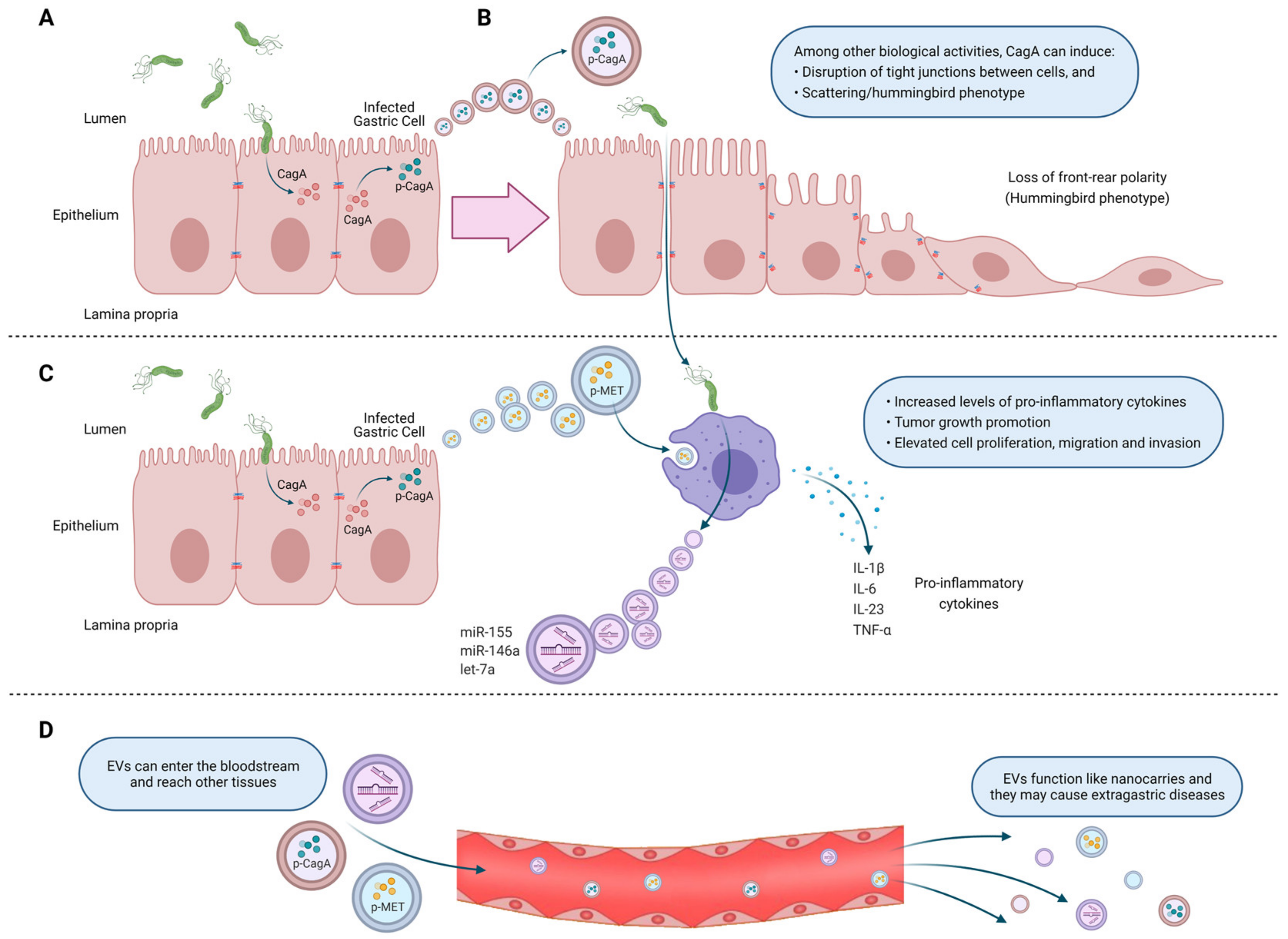
4. Extracellular Vesicles from Helicobacter pylori-Infected Host Cells. Mechanisms of Action Associated Primarily with Gastric Cancer Development

One of the first of the few available studies, in which exosomes were evaluated during infection by H. pylori, reported that CagA, one of the main virulence factors of H. pylori, is present in EVs isolated from the serum of infected patients (Figure 1A) [135]. Furthermore, gastric epithelial cells overexpressing CagA induced the release of EVs containing CagA in its phosphorylated form. Incubation of WT-A10 gastric epithelial cells with such EVs induced an elongated cell shape, known as the hummingbird phenotype (Figure 1B). CagA is considered an oncoprotein, which acts as a pathogenic scaffold protein, thereby altering multiple intracellular signaling pathways in infected cells. As CagA resides inside exosomes, which may transport their content to any part of the body, this mechanism can be invoked to explain how H. pylori triggers both local changes in the stomach and also systemic changes.
Che et al. (2018) studied exosome-mediated communication between H. pylori-infected gastric cancer cells and macrophages. They demonstrated that after infection with H. pylori, gastric cancer cells release exosomes containing phosphorylated mesenchymal epithelial transition factor (MET) (Figure 1C), which after being internalized by macrophages increased mRNA levels of the proinflammatory cytokines IL-1α and IL-6, as well as increased IL-1α secretion, which promoted tumor growth and progression in vivo. in vitro, gastric cancer cells pre-treated with the supernatant of macrophages incubated with exosomes containing MET (MET(+) exosomes) increased cell proliferation, migration and invasion, as well as the number of colonies in a clonogenic assay. Moreover, in mice injected with gastric cells treated with MET(+) exosomes, tumor volume and weight were increased compared to the tumors formed by gastric cancer cells treated with supernatants from macrophages incubated with control buffer or MET(−) exosomes. In conclusion, after infection by H. pylori, gastric cells secrete and transfer exosomes containing active MET to nearby immune cells, which in turn promote gastric cancer progression via IL-1α [136].
Alternatively, Chen et al., (2018) found that stimulation of gastric epithelial cells with exosomes purified from the serum of H. pylori-positive patients, with chronic gastritis, increased IL-1α by activating the soluble IL-6 receptor (sIL-6R). The signaling events triggered by the membrane-bound IL-6R are anti-inflammatory and protective, while to the contrary, sIL-6R signaling is proinflammatory [137]. There are only two known ways to express sIL-6R, namely either by proteolysis of the membrane-bound protein or alternative splicing [138,139]. Since in this study the authors were able to rule out the contribution of the first mechanism, they suggest that the increase in sIL-6R is mediated by alternative mRNA splicing [140].
H. pylori-infected macrophages, release exosomes containing elevated levels of the microRNAs; miR-155, miR-146a and let-7a (Figure 1C). To evaluate the role of microRNA-155, purified exosomes from macrophages were loaded by electroporation with microRNA-155 (Exo+miR155). Such exosomes promoted the expression of the pro-inflammatory cytokines TNF-α, IL-6 and IL-23, and also increased the expression of the proteins CD40, CD63, CD81 and MCH-I in macrophages. Additionally, treatment of macrophages with miR-155 loaded exosomes caused downregulation of MyD88 and NF-κB and inhibited intracellular H. pylori proliferation [141].
Interestingly, a metagenomic analysis of 16S rDNA sequenced in EVs isolated from gastric juice samples of patients with gastric cancer, revealed that H. pylori was more abundant in patients compared to the control group [142]. This finding runs contrary to previous biopsy sequencing studies, where a decrease in H. pylori levels was found in patients with gastric cancer [143]. Since a biopsy sample represents only a small part of the stomach while gastric juice content likely reflects events in most of the stomach, isolating EVs from gastric juice should provide a better method for diagnosing H. pylori than conventional biopsy-based tests. Moreover, the elevated levels of 16S rDNA from H. pylori identified in EVs isolated from gastric juice in patients with gastric cancer compared to healthy controls, is indicative of high levels of H. pylori-derived OMVs, which, as will be discussed subsequently, could promote damage to the mucosa gastric.
Beyond the more local effects in the stomach described so far, a considerable body of evidence links the presence of H. pylori also to the development of extra-gastric diseases, including cardiovascular diseases, such as coronary heart disease, and ischemic stroke. Furthermore, H. pylori presence is associated with an enhanced risk of developing diabetes mellitus, hematological diseases and Parkinson’s disease, among others (reviewed in [144]. Furthermore, epidemiological studies suggest that infection with CagA-positive H. pylori strains increases the risk of colorectal and pancreatic cancer [145,146].
Until recently, specific mechanisms explaining these systemic effects of H. pylori had not been established, although some hypotheses were put forward. For example, as a direct effect in the vascular wall, bacterial infection induces endothelial dysfunction. Alternatively, indirect effects may be attributed to increased systemic inflammation, which enhances the levels of circulating pro-inflammatory cytokines that may accelerate the development of cardiovascular diseases. In addition, recent evidence, like that reviewed here, propose a new mechanism, in which cell to cell communication mediated by EVs during H. pylori infection could play a central role (Figure 1D). While still rather speculative as a working hypothesis, this is certainly an area of research that merits future attention.
In summary, EV-mediated communication between H. pylori-infected and uninfected gastric epithelial cells, in a local context, may promote changes that favor the development and/or progression of gastric cancer. Furthermore, these EVs released by infected gastric epithelial cells modulate the responses of immune cells, like macrophages, to either promote or inhibit inflammation. Finally, these EVs can enter the bloodstream and reach other tissues, where they may cause extra-gastric diseases, since they can function as nanocarriers for molecules derived from pathogens and pathogen-infected cells.

5. Outer Membrane Vesicles from Helicobacter pylori

H. pylori, like many of the Gram-negative bacteria mentioned above, secrete outer membrane vesicles (OMVs) derived from the bacterial outer membrane. These vesicles ranging from 20–450 nm size participate in the communication between bacteria as well as with the environment and, therefore, are important players in bacterial pathogenesis [147,148,149]. It has been reported that H. pylori releases OMVs both in vitro and in vivo [150,151,152,153]. OMVs secreted from H. pylori (H. pylori-OMVs) retain many of the surface molecules of the bacteria, such as LPS, peptidoglycan and outer membrane proteins [147]. Additionally, H. pylori-OMVs contain cytoplasmic molecules, such as proteins associated with translation and virulence factors [154]. Olofsson et al. (2010), determined that H. pylori-OMVs phospholipid composition is similar to that of the bacteria outer membrane and includes phosphatidylglycerol, cardiolipin, lyso-phophatidylethanolamine, phosphatidylethanolamine and cholesterol. However, some components are enriched in the OMVs compared to the bacterial outer membrane, as is the case for the protease HtrA [155]. Mass spectrometry analysis revealed that 77% of the bacterial outer membrane proteins could also be detected in H. pylori-OMVs. Moreover, several proteins associated with H. pylori virulence, such as the urease subunits, VacA, CagA, BabA and SabA adhesins were identified (Figure 2). In addition, γ-glutamyl transpeptidase, the protease HtrA, as well as the cytoplasmic proteins GroEL, catalase, metabolic and ribosomal proteins were also identified in H. pylori-OMVs [155].
Proteomics analysis of OMVs from H. pylori identified a subset of proteins that are not detected in their parent bacterium, suggesting that there is a mechanism for cargo selection [149]. In addition, the size, protein composition and cargo selection for OMVs depended on the stage of H. pylori growth [149]. In this study, H. pylori-OMVs were isolated and purified from bacterial cultures in early, late and stationary growth phase. The results show that H. pylori-OMVs isolated from bacteria in the late and stationary phases of growth are smaller but more abundant than H. pylori-OMVs from an early phase bacteria. Furthermore, OMVs isolated from H. pylori in early growth phase are enriched in metabolic proteins and virulence factors, such as VacA, urease and CagA compared to OMVs from later stage bacteria [149]. On the other hand, the environmental conditions and nutrient availability can also affect the composition of H. pylori-OMVs. When H. pylori was cultured on Brucella broth or blood agar, the derived OMVs showed a different protein pattern [155]. In addition, proteomic analysis revealed that the number of OMV associated proteins may vary between H. pylori strains J99 and NCTC 11637 [154]. However, the mechanisms that produce these differences are unknown.
According to epidemiological studies, there is an association between H. pylori infection and iron metabolism. Therefore, an unbalanced iron metabolism could affect the outcome of H. pylori infection [156,157,158]. Interestingly, iron availability affects the composition of H. pylori-OMVs. In particular, Keenan et al. (2008), determined that H. pylori-OMVs obtained from iron-containing media are LPS enriched, whereas H. pylori-OMVs from bacteria grown under iron-limiting conditions have less and shorter LPS [159]. Furthermore, iron deficiency causes a decrease in bacterial growth, but does not reduce the release of OMVs. However, OMVs released under iron deficient conditions contain less VacA, which is consistent with the absence of vacuolation observed in HEp-2 cells after incubation with these OMVs [160].
OMV formation initiates when protrusions are generated from the outer membrane of Gram-negative bacteria that then pinch off, capturing some of the periplasm content [155]. The ability of H. pylori-OMVs to elicit a response in the host depends on uptake and the entry into epithelial cells [148]. The internalization of OMVs secreted by H. pylori occurs primarily through clathrin-dependent endocytosis and, to a lesser extent, through clathrin-independent mechanisms [161,162]. Concerning cholesterol-dependent mechanisms for internalization, contradictory evidence has been obtained [161,162]. On the other hand, Parker et al. (2010) showed that within 20 min of stimulation of AGS cells with H. pylori-OMVs, all associated OMVs localized within the cells, and that uptake occurred at a higher rate in the presence of VacA. Additionally, the authors observed that H. pylori-OMV uptake is inhibited by addition of LPS [163]. More recently, Turner et al. (2018), demonstrated that the size of OMVs determines the endocytic entry mechanism. A heterogenous sized H. pylori-OMV population results in uptake by micropinocytosis, clathrin- and caveolin-mediated endocytosis. Generally, smaller OMVs (20–100 nm) prefer caveolin-mediated internalization, whereas larger OMVs (90–450 nm) seem to prefer clathrin- and dynamin-mediated endocytosis [148]. Interestingly, larger OMVs contain more and a wider range of proteins than smaller OMVs. For example, large OMVs, but not small OMVs contain BabA and SabA adhesins [148]. However, both populations have virulence factors, such as urease and VacA. Thus, OMV size determines the role these vesicles play in pathogenesis [148].
Accordingly, H. pylori-OMVs transport on their surface or inside, several virulence factors, such as VacA and CagA, and are rapidly internalized by gastric epithelial cells [93,155,163]. Additionally, both the adhesins BabA and SabA were also found on the OMV surface and shown to be biologically active as mediators of adhesion to the human gastric mucosa. There is a high variability between strains in terms of OMV protein profiles. Such variations also affect VacA presence [93,164]. VacA was identified using specific antibodies in H. pylori-OMVs present in gastric biopsies and H. pylori cultures. In addition, VacA was shown to be biologically active in OMVs, since vacuolation was observed after incubation of HEp-2 cells with OMVs from H. pylori 60190 (Figure 2) [151]. Additionally, VacA is internalized and can be observed intracellularly in epithelial cells when these are incubated with either the free form or bound to OMVs. Ricci et al. (2005), showed that approximately 75% of total VacA is released in the free form and the rest is in OMVs. Although both soluble VacA and OMV VacA can induce vacuolization, the OMV VacA is less effective in this respect and may perhaps play a different role [165].
Another important virulence factor identified in OMVs is urease. Proteomic analysis revealed that UreA, a urease catalytic subunit, was present in H. pylori-OMVs isolated from H. pylori strain 26695 and CCUG 17875. Moreover, in AGS cells treated with H. pylori-OMVs from the strain 26695, UreA localized to the cytoplasm and nucleus. Therefore, OMVs can transfer UreA to gastric cells. In addition, the nuclear targeting of UreA lead to morphological changes, suggesting that UreA has functions that are independent of its role as an enzyme [166]. Consistent with this notion, recent studies showed that urease also functions as a ligand for TLR2 to induce stabilization of hypoxia-inducible factor-1α [167].
Catalase (KatA) is a virulence factor that promotes bacterial survival by eliminating hydrogen peroxide and hypochlorite generated by immune cells [168]. Notably, catalase was identified in H. pylori-OMVs at a 7-fold higher concentration than in H. pylori itself. Thus, H. pylori-OMVs, are endowed with a strong anti-oxidant capacity that surrounds the bacteria and protects against oxidative damage produced by the immune system [168].
H. pylori like many bacteria, forms biofilms, which contain extracellular matrix components, including exo-polysaccharides, proteins, lipids and DNA. Moreover, biofilm formation is important for survival and successful infection by many pathogenic bacteria. Interestingly, OMVs are detected in the matrix of H. pylori biofilms and the production of H. pylori-OMVs was found to strongly correlate with biofilm formation [169]. Therefore, another role of OMVs may be to promote H. pylori biofilm formation and thereby enhance bacterial survival as well as favor infection of the stomach epithelium.

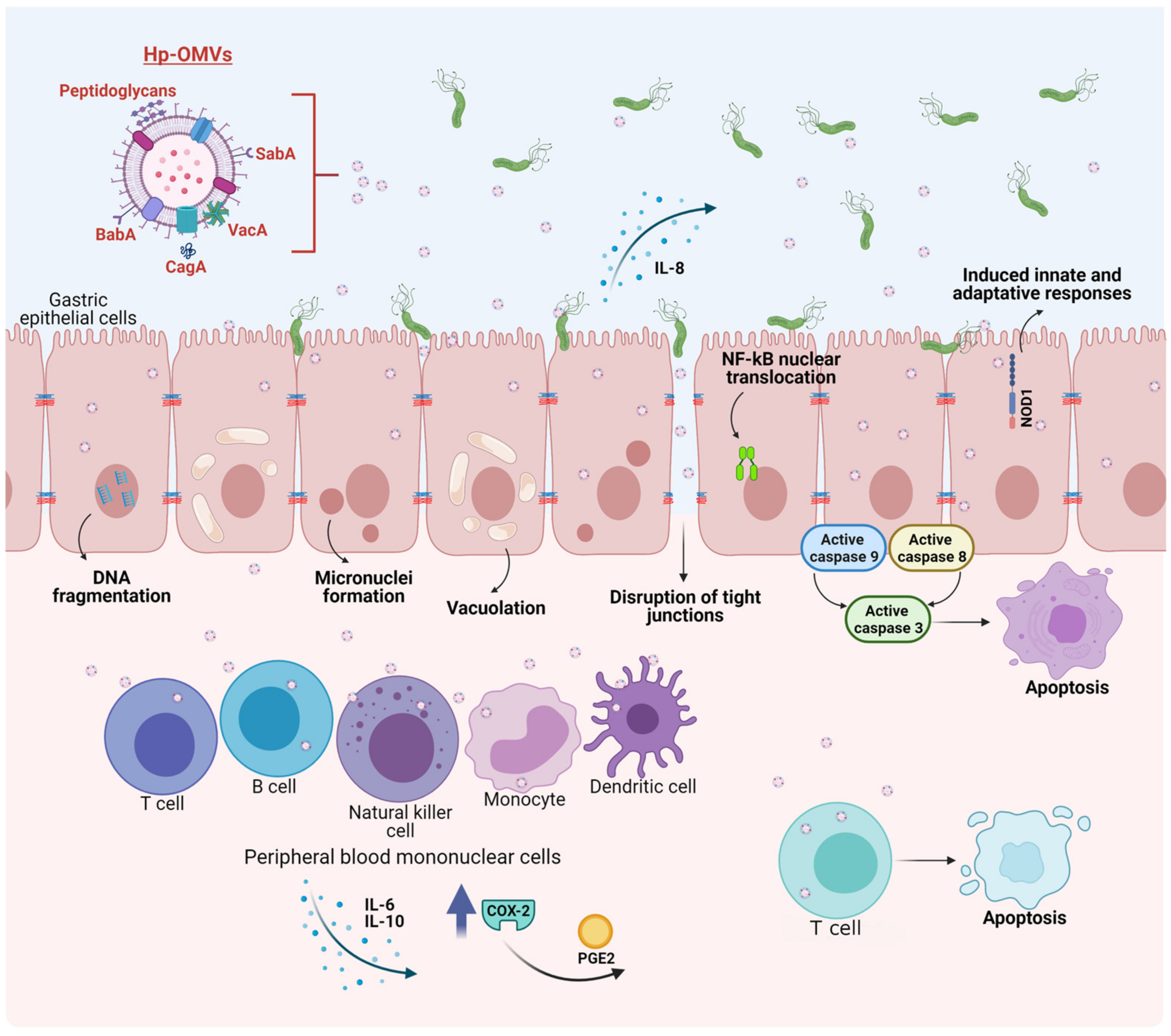
6. Biological Effects on Host Cells of Outer Membrane Vesicles Released by Helicobacter pylori. Mechanisms of Action Associated with Gastric Cancer Development

OMVs from H. pylori have many effects, both in vitro and in vivo [170]. In particular, H. pylori-OMVs have been shown to induce micronuclei formation in gastric AGS cells, indicating genomic damage, apparently mediated by VacA. In the same gastric epithelial cell line, internalization of H. pylori-OMVs lead to the formation of large vacuoles, as early as 4 h after incubation (Figure 2). Additionally, disruption of vacuole integrity and reduction in cellular glutathione were observed and restoring glutathione levels by the addition of glutathione ester in vitro to AGS cells prevented micronuclei formation caused by H. pylori-OMV stimulation, suggesting a role for oxidative stress in the infection process [153]. As mentioned previously, H. pylori-OMVs also contain active CagA, which appears to promote the disruption of tight junctions between epithelial cells (Figure 2), given that CagA from H. pylori-OMVs localizes in close proximity of zona occludens-1 protein junctions, which correlates with a disorganized and diffuse zona occludens-1 pattern after stimulation by H. pylori-OMVs. Moreover, CagA in H. pylori-OMVs enhances histone H1 affinity for ATP, which may modulate gene transcription, although this point was not addressed in the study [171].
Adhesins important for H. pylori adherence to the human gastric epithelium, such as SabA and BabA, are present in H. pylori-OMVs, and are biologically active, as they promote adherence between OMVs isolated in vitro, as well as to the human gastric mucosa in biopsies. Moreover, pre-incubation of H. pylori-OMVs with Leb-receptor or sLex receptor conjugates before adding OMVs to the human gastric mucosa from biopsies, prevented H. pylori-OMV binding, suggesting that SabA and BabA are important for adherence to the gastric mucosa [155]. The effect of H. pylori-OMVs on epithelial cell proliferation appears to be dose dependent. After 24 h of stimulation, low OMV doses increased AGS cell proliferation up to 30%; alternatively, at high doses, approximately 50% growth arrest and a decrease in proliferation is observed. In addition, high doses of H. pylori-OMVs increase toxicity and IL-8 production [163,172]. The decrease in cell viability of gastric epithelial cells after stimuli with H. pylori-OMVs was also corroborated by others [173]. Other effects in AGS cells observed in this study included vacuolation and apoptosis. Specifically, DNA fragmentation and activation of the caspases-8, 9 and 3 were observed but these events occurred in the absence of cytochrome C release (Figure 2) [173].
Less is known about the effects of H. pylori-OMVs on cells of the host immune system; however recent evidence shows that they have opposite effects. For instance, treatment with H. pylori-OMVs stimulate proliferation and the release of IL-6 and 10 from peripheral blood mononuclear cells [93]. Alternatively, H. pylori-OMVs induce apoptosis in Jurkat T cells (immortalized human T cells) and in naïve CD4+ cells [93]. H. pylori-OMVs also induce the expression of cyclo-oxygenase-2 in peripheral blood mononuclear cells, and as a consequence, H. pylori-OMVs can inhibit human T cell proliferation, via prostaglandin-E2. Induction of IL-10 was also observed in peripheral blood mononuclear cells (Figure 2) [174].
H. pylori-OMVs induced, in a dose-dependent manner, IL-8 in gastric AGS cells, and IL-6 as well as TNF-α in mouse macrophages. In vivo, oral and intraperitoneal administration of H. pylori-OMVs in mice induced IFN-α, IL-17 and IL-4 expression, as well as increased immunoglobulin G generation. In vivo, H. pylori-OMVs targeted stomach epithelial cells in mice. Thus, H. pylori-OMVs induce immune responses both in vitro and in vivo. Since, as mentioned before, H. pylori-OMVs are abundant in the stomach of gastric cancer patients, these can infiltrate the gastric mucosa to induce inflammation, and potentially promote gastric cancer [142].
H. pylori-OMVs also contain peptidoglycans, which stimulate the cytosolic nucleotide binding oligomerization domain 1 (NOD1) response in epithelial cells (Figure 2). In addition, H. pylori-OMVs administered intra-gastrically induced innate and adaptive immune responses in mice, via NOD1. Specifically, H. pylori-OMVs induce NF-κB reporter activity and nuclear translocation of the NF-κB p65 subunit, in addition to IL-8 production (Figure 2) [147]. IL-8 and IL-6 are pro-inflammatory cytokines that prolong inflammation, while IL-10 is an anti-inflammatory cytokine, which has a suppressive function on cytokine secretion. Thus, H. pylori-OMVs can elicit opposing effects in the immune system, by increasing or decreasing the proliferation of immune cells, as well as by elevating both, pro- and anti-inflammatory cytokines. Further research is required to determine how H. pylori-OMVs contribute to the development of gastric cancer associated with H. pylori infection.

7. Helicobacter pylori-Outer Membrane Vesicles and Extracellular Vesicles from Helicobacter pylori Infected-Host Cells in Vaccine Development and Biomarker Research

Recent applications of OMV and EV research include vaccine development and biomarker identification [175,176]. Currently, the first line of treatment against H. pylori is antibiotic combination therapy. However, in the last decades resistance to antibiotics has increased [177]. Therefore, new alternative forms of therapy are being developed, and vaccines have emerged as a potential candidate [178].
Vaccines developed against the bacterial outer membrane have been used to induce protection against various pathogens. Hence, the use of OMVs for the same purpose has recently been explored. The first challenge in developing a vaccine targeting H. pylori-OMVs, is the method used for isolating large quantities of OMVs. In this respect, Turner et al. (2015), described that the TolB protein is key to H. pylori membrane integrity and H. pylori-OMVs from a ΔtolB mutant strain increased the production of H. pylori-OMVs that were more immunogenic [179]. Keenan et al. (2000) evaluated immunogenicity of H. pylori-OMVs in a murine model of immune protection and such effects were attributed to increased serum immunoglobulin G antibody against lipoprotein 20 [175]. Liu et al. (2019), tested the immune protective response of H. pylori-OMVs obtained from the gerbil-adapted H. pylori strain 7.13 using a mouse model. The results showed that H. pylori-OMVs generate a strong humoral and mucosal immune response without inflammation that reduces H. pylori load, suggesting a potential use as a treatment [178]. Subsequently, the combination of H. pylori-OMVs with outer membrane proteins or whole cell vaccines, two commonly studied approaches against H. pylori, confirmed that H. pylori-OMVs from H. pylori strain 7.13 were effective as an adjuvant. The analysis demonstrated that vaccines with H. pylori-OMVs induced a Th2 and Th17 biased immunity, increasing cell mediated and humoral immunity, which reduces H. pylori colonization in a mouse model. Consequently, H. pylori-OMVs are effective adjuvants for these types of vaccines [180].
The high death rate from gastric cancer is due to the fact that most patients with the disease are not diagnosed until they exhibit symptoms. This makes it difficult to detect gastric cancer at an early stage [181]. Since the infection with H. pylori is associated with the development of gastric precancerous lesions and gastric cancer, its detection is essential for early diagnosis. Current H. pylori diagnosis involves both invasive and non-invasive techniques. The invasive approach requires obtaining an endoscopic biopsy, which can be analyzed enzymatically using the rapid urease test or histologically with specific antibodies or by PCR. Among the techniques for processing gastric biopsies, PCR appears to be a more sensitive method compared to the others [182]. Progress has been made in obtaining endoscopic images through the development of linked color imaging (LCI) and blue laser imaging (BLI), whereby sensitivity and specificity are on the order of 83.8–85.4% and 79.5–99.5%, respectively [183]. In addition, artificial intelligence has been used to analyze the presence of H. pylori in images obtained by LCI [184]. On the other hand, non-invasive tests include the urea breathing test (UBT), serology and the H. pylori stool antigen test. For the UBT, patients ingest labeled 13C-urea as a substrate for the bacterial urease [185,186]. The 13C-labeled carbon dioxide produced by the reaction is detected in the patient’s breath by mass spectrometry or using an isotope-selective infrared spectroscope [185]. In recent years, the development of portable mass spectrometers has reduced equipment size and the cost of the analysis [187]. Serology is useful mostly for screening and in epidemiological studies [188]. To date, none of the current H. pylori diagnostic options can be considered a gold standard individually. For instance, besides its invasiveness, endoscopic imaging and biopsies require specialized equipment and medical staff. Regarding the non-invasive methods, the sensitivity and specificity still needs to be improved, although values lie between 93–96% and 94–97% for the UBT and the fecal antigen test, respectively [188]. Of note, efforts have been made in the area of next generation sequencing to permit the detection of H. pylori mutations that confer resistance to the most commonly used antibiotics and coincide phenotypically with antibiotic susceptibility [188]. On the other hand, although it is well known that the serological test does not necessarily detect a current infection, an attempt has been made to diagnose an active infection by detecting seropositivity for 13 H. pylori proteins by multiplex. This study concludes that seropositivity to ≥2 of the proteins VacA, GroEl, HcpC and HP1564 is sufficient to identify an ongoing H. pylori infection [189]. The sensitivity limitations and the invasive nature of most diagnostic methods, together with the lack of a gold standard method, make it necessary to develop more sensitive and specific non-invasive tests for the detection of H. pylori infection.
EVs can potentially be used as biomarker carriers, since their content is protected from degradation in the bloodstream by their lipid membrane. These EVs can be isolated from blood or other biological fluids, such as urine, gastric juice or saliva, among others, to identify molecules associated with different diseases. EVs exhibit variations in the molecular profile in healthy subjects compared to patients with diverse diseases, and such information can be exploited for diagnosis [190]. Indeed, as mentioned before, during H. pylori infection, CagA is detected in EVs from patient sera [135]. However, there are not many studies that identify H. pylori-OMVs in circulation, mainly because it is still very difficult to differentiate pathogen-derived OMVs from EVs produced by host cells. However, some reports have revealed the presence of OMVs in circulation [130,191]. Current improvements in methods for the isolation of EVs and pathogen-derived OMVs, along with the identification of molecules that serve as biomarkers, suggest that EVs could be used as a diagnostic tool for the detection of H. pylori infection and preneoplastic gastric lesions in the near future.

8. Conclusions

During H. pylori infection, host cells release EVs carrying an altered profile of host-molecules, and even pathogen-derived proteins. These H. pylori-infected host cell EVs can have a variety of effects on gastric epithelial cells, gastric cancer cells and macrophages, including changes in epithelial cell morphology and increased release of cytokines by immune cells. In addition, during H. pylori infection, the bacteria releases OMVs, bearing pathogen-derived molecules, including the virulence factors CagA, VacA, Urease, LPS, SabA, BabA, among many others, that induce changes in epithelial cells, including vacuolation, micronuclei formation, disruption of tight junctions between epithelial cells, and either cell proliferation or growth arrest, depending on the OMV dose. The effects of H. pylori-OMVs on the immune system have been found to be contradictory, as they promote or inhibit proliferation of certain immune cells and increase pro-inflammatory and anti-inflammatory cytokine release. Overall, H. pylori-infected host cell EVs and H. pylori-OMVs can promote cellular changes that tend to favor the development and/or progression of gastric cancer. In addition, H. pylori-OMVs are being evaluated for use in the development of vaccines against H. pylori development and so far, show promising results. Finally, EVs have proven to be useful for diagnosis, as they can be isolated from various biological fluids, such as blood, urine, saliva and gastric juice, among others. Improvements in the purification of EVs and in the identification of biomarkers for detection of H. pylori infection, as well as preneoplastic gastric lesions, hold considerable promise for use in the diagnosis and prevention of gastric cancer. Future studies on EVs/OMVs in H. pylori infection should focus on the development of vaccines against H. pylori and early diagnosis of H. pylori infection along with preneoplastic gastric lesions, in order to improve the prevention of gastric cancer and the survival of gastric cancer patients.

Author Contributions

Writing—original draft preparation, M.F.G., P.D., A.S.-B. and D.H.; writing—review and editing, M.F.G., P.D., A.S.-B., D.H. and A.F.G.Q.; supervision, A.F.G.Q.; funding acquisition, A.S.-B., A.F.G.Q., M.F.G. and D.H. All authors have read and agreed to the published version of the manuscript.

Funding

This research was funded by FONDECYT, grant number 1170925, 1210644 to A.F.G.Q., FONDECYT, grant number 3180783 to A.S.-B., and FONDAP—Agencia Nacional de Investigación y Desarrollo (ANID), Chile, grant number 15130011 to A.F.G.Q. ANID fellowship 21170292 to M.F.G.; 21191668 to D.H.

Institutional Review Board Statement

Not applicable.

Informed Consent Statement

Not applicable.

Conflicts of Interest

The authors declare no conflict of interest.

References

  1. Hooi, J.K.Y.; Lai, W.Y.; Ng, W.K.; Suen, M.M.Y.; Underwood, F.E.; Tanyingoh, D.; Malfertheiner, P.; Graham, D.Y.; Wong, V.W.S.; Wu, J.C.Y.; et al. Global Prevalence of Helicobacter pylori Infection: Systematic Review and Meta-Analysis. Gastroenterology 2017, 153, 420–429. [Google Scholar] [CrossRef] [Green Version]
  2. Crowe, S.E. Helicobacter pylori Infection. N. Engl. J. Med. 2019, 380, 1158–1165. [Google Scholar] [CrossRef]
  3. Hu, Y.; Wan, J.H.; Li, X.Y.; Zhu, Y.; Graham, D.Y.; Lu, N.H. Systematic review with meta-analysis: The global recurrence rate of Helicobacter pylori. Aliment. Pharmacol. Ther. 2017, 46, 773–779. [Google Scholar] [CrossRef] [Green Version]
  4. Leja, M.; Axon, A.; Brenner, H. Epidemiology of Helicobacter pylori infection. Helicobacter 2016, 21 (Suppl. S1), 3–7. [Google Scholar] [CrossRef]
  5. Zamani, M.; Vahedi, A.; Maghdouri, Z.; Shokri-Shirvani, J. Role of food in environmental transmission of Helicobacter pylori. Caspian J. Intern. Med. 2017, 8, 146–152. [Google Scholar] [CrossRef]
  6. Thung, I.; Aramin, H.; Vavinskaya, V.; Gupta, S.; Park, J.Y.; Crowe, S.E.; Valasek, M.A. Review article: The global emergence of Helicobacter pylori antibiotic resistance. Aliment. Pharmacol. Ther. 2016, 43, 514–533. [Google Scholar] [CrossRef] [Green Version]
  7. Marshall, B.J.; Warren, J.R. Unidentified curved bacilli in the stomach of patients with gastritis and peptic ulceration. Lancet 1984, 1, 1311–1315. [Google Scholar] [CrossRef]
  8. Nagy, P.; Johansson, S.; Molloy-Bland, M. Systematic review of time trends in the prevalence of Helicobacter pylori infection in China and the USA. Gut Pathog. 2016, 8, 8. [Google Scholar] [CrossRef] [Green Version]
  9. Gravina, A.G.; Zagari, R.M.; De Musis, C.; Romano, L.; Loguercio, C.; Romano, M. Helicobacter pylori and extragastric diseases: A review. World J. Gastroenterol. 2018, 24, 3204–3221. [Google Scholar] [CrossRef]
  10. Amieva, M.; Peek, R.M., Jr. Pathobiology of Helicobacter pylori-Induced Gastric Cancer. Gastroenterology 2016, 150, 64–78. [Google Scholar] [CrossRef] [Green Version]
  11. Roesler, B.M.; Rabelo-Goncalves, E.M.; Zeitune, J.M. Virulence Factors of Helicobacter pylori: A Review. Clin. Med. Insights Gastroenterol. 2014, 7, 9–17. [Google Scholar] [CrossRef] [PubMed]
  12. Kao, C.Y.; Sheu, B.S.; Wu, J.J. Helicobacter pylori infection: An overview of bacterial virulence factors and pathogenesis. Biomed. J. 2016, 39, 14–23. [Google Scholar] [CrossRef] [Green Version]
  13. Gu, H. Role of Flagella in the Pathogenesis of Helicobacter pylori. Curr. Microbiol. 2017, 74, 863–869. [Google Scholar] [CrossRef] [PubMed] [Green Version]
  14. Aihara, E.; Closson, C.; Matthis, A.L.; Schumacher, M.A.; Engevik, A.C.; Zavros, Y.; Ottemann, K.M.; Montrose, M.H. Motility and chemotaxis mediate the preferential colonization of gastric injury sites by Helicobacter pylori. PLoS Pathog. 2014, 10, e1004275. [Google Scholar] [CrossRef] [PubMed] [Green Version]
  15. Mori, J.; Vranac, T.; Smrekar, B.; Cernilec, M.; Serbec, V.C.; Horvat, S.; Ihan, A.; Bencina, M.; Jerala, R. Chimeric flagellin as the self-adjuvanting antigen for the activation of immune response against Helicobacter pylori. Vaccine 2012, 30, 5856–5863. [Google Scholar] [CrossRef] [PubMed]
  16. Yang, J.; Yan, H. TLR5: Beyond the recognition of flagellin. Cell Mol. Immunol. 2017, 14, 1017–1019. [Google Scholar] [CrossRef]
  17. Tegtmeyer, N.; Lind, J.; Schmid, B.; Backert, S. Helicobacter pylori CagL Y58/E59 mutation turns-off type IV secretion-dependent delivery of CagA into host cells. PLoS ONE 2014, 9, e97782. [Google Scholar] [CrossRef] [PubMed] [Green Version]
  18. Li, N.; Tang, B.; Jia, Y.P.; Zhu, P.; Zhuang, Y.; Fang, Y.; Li, Q.; Wang, K.; Zhang, W.J.; Guo, G.; et al. Helicobacter pylori CagA Protein Negatively Regulates Autophagy and Promotes Inflammatory Response via c-Met-PI3K/Akt-mTOR Signaling Pathway. Front. Cell. Infect. Microbiol. 2017, 7, 417. [Google Scholar] [CrossRef]
  19. Yong, X.; Tang, B.; Xiao, Y.F.; Xie, R.; Qin, Y.; Luo, G.; Hu, C.J.; Dong, H.; Yang, S.M. Helicobacter pylori upregulates Nanog and Oct4 via Wnt/beta-catenin signaling pathway to promote cancer stem cell-like properties in human gastric cancer. Cancer Lett. 2016, 374, 292–303. [Google Scholar] [CrossRef] [Green Version]
  20. Coulombe, G.; Rivard, N. New and Unexpected Biological Functions for the Src-Homology 2 Domain-Containing Phosphatase SHP-2 in the Gastrointestinal Tract. Cell. Mol. Gastroenterol. Hepatol. 2016, 2, 11–21. [Google Scholar] [CrossRef] [Green Version]
  21. Wroblewski, L.E.; Piazuelo, M.B.; Chaturvedi, R.; Schumacher, M.; Aihara, E.; Feng, R.; Noto, J.M.; Delgado, A.; Israel, D.A.; Zavros, Y.; et al. Helicobacter pylori targets cancer-associated apical-junctional constituents in gastroids and gastric epithelial cells. Gut 2015, 64, 720–730. [Google Scholar] [CrossRef] [Green Version]
  22. Gorrell, R.J.; Guan, J.; Xin, Y.; Tafreshi, M.A.; Hutton, M.L.; McGuckin, M.A.; Ferrero, R.L.; Kwok, T. A novel NOD1- and CagA-independent pathway of interleukin-8 induction mediated by the Helicobacter pylori type IV secretion system. Cell. Microbiol. 2013, 15, 554–570. [Google Scholar] [CrossRef]
  23. Waskito, L.A.; Salama, N.R.; Yamaoka, Y. Pathogenesis of Helicobacter pylori infection. Helicobacter 2018, 23 (Suppl. S1), e12516. [Google Scholar] [CrossRef] [Green Version]
  24. Chambers, M.G.; Pyburn, T.M.; Gonzalez-Rivera, C.; Collier, S.E.; Eli, I.; Yip, C.K.; Takizawa, Y.; Lacy, D.B.; Cover, T.L.; Ohi, M.D. Structural analysis of the oligomeric states of Helicobacter pylori VacA toxin. J. Mol. Biol. 2013, 425, 524–535. [Google Scholar] [CrossRef] [PubMed] [Green Version]
  25. Chauhan, N.; Tay, A.C.Y.; Marshall, B.J.; Jain, U. Helicobacter pylori VacA, a distinct toxin exerts diverse functionalities in numerous cells: An overview. Helicobacter 2019, 24, e12544. [Google Scholar] [CrossRef] [Green Version]
  26. Palframan, S.L.; Kwok, T.; Gabriel, K. Vacuolating cytotoxin A (VacA), a key toxin for Helicobacter pylori pathogenesis. Front. Cell. Infect. Microbiol. 2012, 2, 92. [Google Scholar] [CrossRef] [PubMed] [Green Version]
  27. Terebiznik, M.R.; Vazquez, C.L.; Torbicki, K.; Banks, D.; Wang, T.; Hong, W.; Blanke, S.R.; Colombo, M.I.; Jones, N.L. Helicobacter pylori VacA toxin promotes bacterial intracellular survival in gastric epithelial cells. Infect. Immun. 2006, 74, 6599–6614. [Google Scholar] [CrossRef] [Green Version]
  28. Rassow, J. Helicobacter pylori vacuolating toxin A and apoptosis. Cell. Commun. Signal 2011, 9, 26. [Google Scholar] [CrossRef] [Green Version]
  29. Kern, B.; Jain, U.; Utsch, C.; Otto, A.; Busch, B.; Jimenez-Soto, L.; Becher, D.; Haas, R. Characterization of Helicobacter pylori VacA-containing vacuoles (VCVs), VacA intracellular trafficking and interference with calcium signalling in T lymphocytes. Cell. Microbiol. 2015, 17, 1811–1832. [Google Scholar] [CrossRef] [PubMed] [Green Version]
  30. Djekic, A.; Muller, A. The Immunomodulator VacA Promotes Immune Tolerance and Persistent Helicobacter pylori Infection through Its Activities on T-Cells and Antigen-Presenting Cells. Toxins (Basel) 2016, 8, 187. [Google Scholar] [CrossRef]
  31. Ansari, S.; Yamaoka, Y. Helicobacter pylori Virulence Factors Exploiting Gastric Colonization and its Pathogenicity. Toxins (Basel) 2019, 11, 677. [Google Scholar] [CrossRef] [Green Version]
  32. IARC. Schistosomes, liver flukes and Helicobacter pylori. IARC Working Group on the Evaluation of Carcinogenic Risks to Humans. Lyon, 7–14 June 1994. In IARC Monographs on the Evaluation of Carcinogenic Risks to Humans; IARC: Lyon, France, 1994; Volume 61, pp. 1–241. [Google Scholar]
  33. Ajani, J.A.; Lee, J.; Sano, T.; Janjigian, Y.Y.; Fan, D.; Song, S. Gastric adenocarcinoma. Nat. Rev. Dis. Primers 2017, 3, 17036. [Google Scholar] [CrossRef] [PubMed]
  34. Ansari, S.; Gantuya, B.; Tuan, V.P.; Yamaoka, Y. Diffuse Gastric Cancer: A Summary of Analogous Contributing Factors for Its Molecular Pathogenicity. Int. J. Mol. Sci. 2018, 19, 2424. [Google Scholar] [CrossRef] [PubMed] [Green Version]
  35. Correa, P.; Haenszel, W.; Cuello, C.; Tannenbaum, S.; Archer, M. A model for gastric cancer epidemiology. Lancet 1975, 2, 58–60. [Google Scholar] [CrossRef]
  36. Correa, P.; Piazuelo, M.B. The gastric precancerous cascade. J. Dig. Dis. 2012, 13, 2–9. [Google Scholar] [CrossRef] [Green Version]
  37. Lauren, P. The Two Histological Main Types of Gastric Carcinoma: Diffuse and So-Called Intestinal-Type Carcinoma. An Attempt at a Histo-Clinical Classification. Acta Pathol. Microbiol. Scand. 1965, 64, 31–49. [Google Scholar] [CrossRef]
  38. Ma, J.; Shen, H.; Kapesa, L.; Zeng, S. Lauren classification and individualized chemotherapy in gastric cancer. Oncol. Lett. 2016, 11, 2959–2964. [Google Scholar] [CrossRef] [Green Version]
  39. Ishaq, S.; Nunn, L. Helicobacter pylori and gastric cancer: A state of the art review. Gastroenterol. Hepatol. Bed Bench 2015, 8, S6–S14. [Google Scholar]
  40. Bagheri, N.; Salimzadeh, L.; Shirzad, H. The role of T helper 1-cell response in Helicobacter pylori-infection. Microb. Pathog. 2018, 123, 1–8. [Google Scholar] [CrossRef]
  41. Handa, O.; Naito, Y.; Yoshikawa, T. Redox biology and gastric carcinogenesis: The role of Helicobacter pylori. Redox Rep. 2011, 16, 1–7. [Google Scholar] [CrossRef] [PubMed] [Green Version]
  42. Giroux, V.; Rustgi, A.K. Metaplasia: Tissue injury adaptation and a precursor to the dysplasia-cancer sequence. Nat. Rev. Cancer 2017, 17, 594–604. [Google Scholar] [CrossRef]
  43. Miftahussurur, M.; Yamaoka, Y.; Graham, D.Y. Helicobacter pylori as an oncogenic pathogen, revisited. Expert Rev. Mol. Med. 2017, 19, e4. [Google Scholar] [CrossRef]
  44. Sandoval-Borquez, A.; Saavedra, K.; Carrasco-Avino, G.; Garcia-Bloj, B.; Fry, J.; Wichmann, I.; Corvalan, A.H. Noncoding Genomics in Gastric Cancer and the Gastric Precancerous Cascade: Pathogenesis and Biomarkers. Dis. Markers 2015, 2015, 503762. [Google Scholar] [CrossRef] [Green Version]
  45. Polakovicova, I.; Jerez, S.; Wichmann, I.A.; Sandoval-Borquez, A.; Carrasco-Veliz, N.; Corvalan, A.H. Role of microRNAs and Exosomes in Helicobacter pylori and Epstein-Barr Virus Associated Gastric Cancers. Front. Microbiol. 2018, 9, 636. [Google Scholar] [CrossRef] [PubMed] [Green Version]
  46. Chmiela, M.; Kupcinskas, J. Review: Pathogenesis of Helicobacter pylori infection. Helicobacter 2019, 24 (Suppl. S1), e12638. [Google Scholar] [CrossRef] [PubMed] [Green Version]
  47. Delabranche, X.; Berger, A.; Boisrame-Helms, J.; Meziani, F. Microparticles and infectious diseases. Med. Mal. Infect. 2012, 42, 335–343. [Google Scholar] [CrossRef]
  48. Lawson, C.; Kovacs, D.; Finding, E.; Ulfelder, E.; Luis-Fuentes, V. Extracellular Vesicles: Evolutionarily Conserved Mediators of Intercellular Communication. Yale J. Biol. Med. 2017, 90, 481–491. [Google Scholar]
  49. Brown, L.; Wolf, J.M.; Prados-Rosales, R.; Casadevall, A. Through the wall: Extracellular vesicles in Gram-positive bacteria, mycobacteria and fungi. Nat. Rev. Microbiol. 2015, 13, 620–630. [Google Scholar] [CrossRef] [Green Version]
  50. Schorey, J.S.; Cheng, Y.; Singh, P.P.; Smith, V.L. Exosomes and other extracellular vesicles in host-pathogen interactions. EMBO Rep. 2015, 16, 24–43. [Google Scholar] [CrossRef] [Green Version]
  51. Pan, B.T.; Teng, K.; Wu, C.; Adam, M.; Johnstone, R.M. Electron microscopic evidence for externalization of the transferrin receptor in vesicular form in sheep reticulocytes. J. Cell Biol. 1985, 101, 942–948. [Google Scholar] [CrossRef] [Green Version]
  52. Johnstone, R.M.; Adam, M.; Hammond, J.R.; Orr, L.; Turbide, C. Vesicle formation during reticulocyte maturation. Association of plasma membrane activities with released vesicles (exosomes). J. Biol. Chem. 1987, 262, 9412–9420. [Google Scholar] [CrossRef]
  53. Johnstone, R.M.; Ahn, J. A common mechanism may be involved in the selective loss of plasma membrane functions during reticulocyte maturation. Biomed. Biochim. Acta 1990, 49, S70–S75. [Google Scholar]
  54. Yuana, Y.; Sturk, A.; Nieuwland, R. Extracellular vesicles in physiological and pathological conditions. Blood Rev. 2013, 27, 31–39. [Google Scholar] [CrossRef] [PubMed] [Green Version]
  55. Machtinger, R.; Laurent, L.C.; Baccarelli, A.A. Extracellular vesicles: Roles in gamete maturation, fertilization and embryo implantation. Hum. Reprod. Update 2016, 22, 182–193. [Google Scholar] [CrossRef]
  56. Buzas, E.I.; Gyorgy, B.; Nagy, G.; Falus, A.; Gay, S. Emerging role of extracellular vesicles in inflammatory diseases. Nat. Rev. Rheumatol. 2014, 10, 356–364. [Google Scholar] [CrossRef]
  57. Yan, Y.; Fu, G.; Ye, Y.; Ming, L. Exosomes participate in the carcinogenesis and the malignant behavior of gastric cancer. Scand. J. Gastroenterol. 2017, 52, 499–504. [Google Scholar] [CrossRef]
  58. Van Niel, G.; D’Angelo, G.; Raposo, G. Shedding light on the cell biology of extracellular vesicles. Nat. Rev. Mol. Cell Biol. 2018, 19, 213–228. [Google Scholar] [CrossRef]
  59. Minciacchi, V.R.; Freeman, M.R.; Di Vizio, D. Extracellular vesicles in cancer: Exosomes, microvesicles and the emerging role of large oncosomes. Semin. Cell Dev. Biol. 2015, 40, 41–51. [Google Scholar] [CrossRef] [Green Version]
  60. Tavano, S.; Heisenberg, C.P. Migrasomes take center stage. Nat. Cell Biol. 2019, 21, 918–920. [Google Scholar] [CrossRef]
  61. Akers, J.C.; Gonda, D.; Kim, R.; Carter, B.S.; Chen, C.C. Biogenesis of extracellular vesicles (EV): Exosomes, microvesicles, retrovirus-like vesicles, and apoptotic bodies. J. Neurooncol. 2013, 113, 1–11. [Google Scholar] [CrossRef] [Green Version]
  62. Syn, N.; Wang, L.; Sethi, G.; Thiery, J.P.; Goh, B.C. Exosome-Mediated Metastasis: From Epithelial-Mesenchymal Transition to Escape from Immunosurveillance. Trends Pharmacol. Sci. 2016, 37, 606–617. [Google Scholar] [CrossRef]
  63. O’Brien, K.; Breyne, K.; Ughetto, S.; Laurent, L.C.; Breakefield, X.O. RNA delivery by extracellular vesicles in mammalian cells and its applications. Nat. Rev. Mol. Cell Biol. 2020, 21, 585–606. [Google Scholar] [CrossRef] [PubMed]
  64. Vagner, T.; Spinelli, C.; Minciacchi, V.R.; Balaj, L.; Zandian, M.; Conley, A.; Zijlstra, A.; Freeman, M.R.; Demichelis, F.; De, S.; et al. Large extracellular vesicles carry most of the tumour DNA circulating in prostate cancer patient plasma. J. Extracell. Vesicles 2018, 7, 1505403. [Google Scholar] [CrossRef] [Green Version]
  65. Campos, A.; Salomon, C.; Bustos, R.; Diaz, J.; Martinez, S.; Silva, V.; Reyes, C.; Diaz-Valdivia, N.; Varas-Godoy, M.; Lobos-Gonzalez, L.; et al. Caveolin-1-containing extracellular vesicles transport adhesion proteins and promote malignancy in breast cancer cell lines. Nanomedicine (London) 2018, 13, 2597–2609. [Google Scholar] [CrossRef] [PubMed] [Green Version]
  66. Manek, R.; Moghieb, A.; Yang, Z.; Kumar, D.; Kobessiy, F.; Sarkis, G.A.; Raghavan, V.; Wang, K.K.W. Protein Biomarkers and Neuroproteomics Characterization of Microvesicles/Exosomes from Human Cerebrospinal Fluid Following Traumatic Brain Injury. Mol. Neurobiol. 2018, 55, 6112–6128. [Google Scholar] [CrossRef] [PubMed]
  67. Choi, D.S.; Kim, D.K.; Kim, Y.K.; Gho, Y.S. Proteomics, transcriptomics and lipidomics of exosomes and ectosomes. Proteomics 2013, 13, 1554–1571. [Google Scholar] [CrossRef] [PubMed]
  68. Abels, E.R.; Breakefield, X.O. Introduction to Extracellular Vesicles: Biogenesis, RNA Cargo Selection, Content, Release, and Uptake. Cell. Mol. Neurobiol. 2016, 36, 301–312. [Google Scholar] [CrossRef] [PubMed]
  69. Andaloussi, S.E.L.; Mager, I.; Breakefield, X.O.; Wood, M.J. Extracellular vesicles: Biology and emerging therapeutic opportunities. Nat. Rev. Drug Discov. 2013, 12, 347–357. [Google Scholar] [CrossRef] [PubMed]
  70. Zhang, H.-G. Emerging Concepts of Tumor Exosome-Mediated Cell-Cell Communication; Springer: New York, NY, USA, 2013. [Google Scholar]
  71. Van Niel, G.; Charrin, S.; Simoes, S.; Romao, M.; Rochin, L.; Saftig, P.; Marks, M.S.; Rubinstein, E.; Raposo, G. The tetraspanin CD63 regulates ESCRT-independent and -dependent endosomal sorting during melanogenesis. Dev. Cell 2011, 21, 708–721. [Google Scholar] [CrossRef] [Green Version]
  72. Essandoh, K.; Yang, L.; Wang, X.; Huang, W.; Qin, D.; Hao, J.; Wang, Y.; Zingarelli, B.; Peng, T.; Fan, G.C. Blockade of exosome generation with GW4869 dampens the sepsis-induced inflammation and cardiac dysfunction. Biochim. Biophys. Acta 2015, 1852, 2362–2371. [Google Scholar] [CrossRef] [PubMed] [Green Version]
  73. Van Niel, G.; Raposo, G.; Candalh, C.; Boussac, M.; Hershberg, R.; Cerf-Bensussan, N.; Heyman, M. Intestinal epithelial cells secrete exosome-like vesicles. Gastroenterology 2001, 121, 337–349. [Google Scholar] [CrossRef] [PubMed]
  74. Buschow, S.I.; Nolte-’t Hoen, E.N.; van Niel, G.; Pols, M.S.; ten Broeke, T.; Lauwen, M.; Ossendorp, F.; Melief, C.J.; Raposo, G.; Wubbolts, R.; et al. MHC II in dendritic cells is targeted to lysosomes or T cell-induced exosomes via distinct multivesicular body pathways. Traffic 2009, 10, 1528–1542. [Google Scholar] [CrossRef]
  75. Carayon, K.; Chaoui, K.; Ronzier, E.; Lazar, I.; Bertrand-Michel, J.; Roques, V.; Balor, S.; Terce, F.; Lopez, A.; Salome, L.; et al. Proteolipidic composition of exosomes changes during reticulocyte maturation. J. Biol. Chem. 2011, 286, 34426–34439. [Google Scholar] [CrossRef] [PubMed] [Green Version]
  76. Geminard, C.; De Gassart, A.; Blanc, L.; Vidal, M. Degradation of AP2 during reticulocyte maturation enhances binding of hsc70 and Alix to a common site on TFR for sorting into exosomes. Traffic 2004, 5, 181–193. [Google Scholar] [CrossRef]
  77. Thery, C.; Boussac, M.; Veron, P.; Ricciardi-Castagnoli, P.; Raposo, G.; Garin, J.; Amigorena, S. Proteomic analysis of dendritic cell-derived exosomes: A secreted subcellular compartment distinct from apoptotic vesicles. J. Immunol. 2001, 166, 7309–7318. [Google Scholar] [CrossRef] [Green Version]
  78. Irion, U.; St Johnston, D. bicoid RNA localization requires specific binding of an endosomal sorting complex. Nature 2007, 445, 554–558. [Google Scholar] [CrossRef]
  79. Savina, A.; Fader, C.M.; Damiani, M.T.; Colombo, M.I. Rab11 promotes docking and fusion of multivesicular bodies in a calcium-dependent manner. Traffic 2005, 6, 131–143. [Google Scholar] [CrossRef] [PubMed]
  80. Raposo, G.; Tenza, D.; Mecheri, S.; Peronet, R.; Bonnerot, C.; Desaymard, C. Accumulation of major histocompatibility complex class II molecules in mast cell secretory granules and their release upon degranulation. Mol. Biol. Cell 1997, 8, 2631–2645. [Google Scholar] [CrossRef] [Green Version]
  81. Faure, J.; Lachenal, G.; Court, M.; Hirrlinger, J.; Chatellard-Causse, C.; Blot, B.; Grange, J.; Schoehn, G.; Goldberg, Y.; Boyer, V.; et al. Exosomes are released by cultured cortical neurones. Mol. Cell Neurosci. 2006, 31, 642–648. [Google Scholar] [CrossRef]
  82. Ostrowski, M.; Carmo, N.B.; Krumeich, S.; Fanget, I.; Raposo, G.; Savina, A.; Moita, C.F.; Schauer, K.; Hume, A.N.; Freitas, R.P.; et al. Rab27a and Rab27b control different steps of the exosome secretion pathway. Nat. Cell Biol. 2010, 12, 19–30. [Google Scholar] [CrossRef] [PubMed] [Green Version]
  83. Chistiakov, D.A.; Orekhov, A.N.; Bobryshev, Y.V. Cardiac Extracellular Vesicles in Normal and Infarcted Heart. Int. J. Mol. Sci. 2016, 17, 63. [Google Scholar] [CrossRef]
  84. Janowska-Wieczorek, A.; Wysoczynski, M.; Kijowski, J.; Marquez-Curtis, L.; Machalinski, B.; Ratajczak, J.; Ratajczak, M.Z. Microvesicles derived from activated platelets induce metastasis and angiogenesis in lung cancer. Int. J. Cancer 2005, 113, 752–760. [Google Scholar] [CrossRef] [PubMed]
  85. Lee, T.H.; D’Asti, E.; Magnus, N.; Al-Nedawi, K.; Meehan, B.; Rak, J. Microvesicles as mediators of intercellular communication in cancer--the emerging science of cellular ‘debris’. Semin. Immunopathol. 2011, 33, 455–467. [Google Scholar] [CrossRef] [Green Version]
  86. Rak, J. Microparticles in cancer. Semin. Thromb. Hemost. 2010, 36, 888–906. [Google Scholar] [CrossRef] [PubMed] [Green Version]
  87. Hood, J.L.; San, R.S.; Wickline, S.A. Exosomes released by melanoma cells prepare sentinel lymph nodes for tumor metastasis. Cancer Res. 2011, 71, 3792–3801. [Google Scholar] [CrossRef] [PubMed] [Green Version]
  88. Kalluri, R.; LeBleu, V.S. The biology, function, and biomedical applications of exosomes. Science 2020, 367. [Google Scholar] [CrossRef]
  89. Gurunathan, S.; Kang, M.H.; Jeyaraj, M.; Qasim, M.; Kim, J.H. Review of the Isolation, Characterization, Biological Function, and Multifarious Therapeutic Approaches of Exosomes. Cells 2019, 8, 307. [Google Scholar] [CrossRef] [Green Version]
  90. Zhang, L.; Yu, D. Exosomes in cancer development, metastasis, and immunity. Biochim. Biophys. Acta Rev. Cancer 2019, 1871, 455–468. [Google Scholar] [CrossRef]
  91. Bhatnagar, S.; Shinagawa, K.; Castellino, F.J.; Schorey, J.S. Exosomes released from macrophages infected with intracellular pathogens stimulate a proinflammatory response in vitro and in vivo. Blood 2007, 110, 3234–3244. [Google Scholar] [CrossRef] [Green Version]
  92. Giri, P.K.; Kruh, N.A.; Dobos, K.M.; Schorey, J.S. Proteomic analysis identifies highly antigenic proteins in exosomes from M. tuberculosis-infected and culture filtrate protein-treated macrophages. Proteomics 2010, 10, 3190–3202. [Google Scholar] [CrossRef]
  93. Winter, J.; Letley, D.; Rhead, J.; Atherton, J.; Robinson, K. Helicobacter pylori membrane vesicles stimulate innate pro- and anti-inflammatory responses and induce apoptosis in Jurkat T cells. Infect. Immun. 2014, 82, 1372–1381. [Google Scholar] [CrossRef] [Green Version]
  94. Jutley, J.K.; Kelleher, J.; Whelan, P.; Mikel, J. Cytosolic retinoic acid-binding protein in human prostatic dysplasia and neoplasia. Prostate 1987, 11, 127–132. [Google Scholar] [CrossRef]
  95. Beatty, W.L.; Ullrich, H.J.; Russell, D.G. Mycobacterial surface moieties are released from infected macrophages by a constitutive exocytic event. Eur. J. Cell Biol. 2001, 80, 31–40. [Google Scholar] [CrossRef]
  96. Beatty, W.L.; Russell, D.G. Identification of mycobacterial surface proteins released into subcellular compartments of infected macrophages. Infect. Immun. 2000, 68, 6997–7002. [Google Scholar] [CrossRef] [Green Version]
  97. Thery, C.; Ostrowski, M.; Segura, E. Membrane vesicles as conveyors of immune responses. Nat. Rev. Immunol. 2009, 9, 581–593. [Google Scholar] [CrossRef] [PubMed]
  98. Bobrie, A.; Colombo, M.; Raposo, G.; Thery, C. Exosome secretion: Molecular mechanisms and roles in immune responses. Traffic 2011, 12, 1659–1668. [Google Scholar] [CrossRef] [PubMed]
  99. Bhatnagar, S.; Schorey, J.S. Exosomes released from infected macrophages contain Mycobacterium avium glycopeptidolipids and are proinflammatory. J. Biol. Chem. 2007, 282, 25779–25789. [Google Scholar] [CrossRef] [PubMed] [Green Version]
  100. Wang, J.J.; Chen, C.; Xie, P.F.; Pan, Y.; Tan, Y.H.; Tang, L.J. Proteomic analysis and immune properties of exosomes released by macrophages infected with Mycobacterium avium. Microbes. Infect. 2014, 16, 283–291. [Google Scholar] [CrossRef] [PubMed]
  101. Anand, P.K.; Anand, E.; Bleck, C.K.; Anes, E.; Griffiths, G. Exosomal Hsp70 induces a pro-inflammatory response to foreign particles including mycobacteria. PLoS ONE 2010, 5, e10136. [Google Scholar] [CrossRef]
  102. Schorey, J.S.; Bhatnagar, S. Exosome function: From tumor immunology to pathogen biology. Traffic 2008, 9, 871–881. [Google Scholar] [CrossRef] [Green Version]
  103. Singh, P.P.; Smith, V.L.; Karakousis, P.C.; Schorey, J.S. Exosomes isolated from mycobacteria-infected mice or cultured macrophages can recruit and activate immune cells in vitro and in vivo. J. Immunol. 2012, 189, 777–785. [Google Scholar] [CrossRef] [PubMed] [Green Version]
  104. Singh, P.P.; LeMaire, C.; Tan, J.C.; Zeng, E.; Schorey, J.S. Exosomes released from M. tuberculosis infected cells can suppress IFN-gamma mediated activation of naive macrophages. PLoS ONE 2011, 6, e18564. [Google Scholar] [CrossRef] [PubMed] [Green Version]
  105. Balaji, K.N.; Goyal, G.; Narayana, Y.; Srinivas, M.; Chaturvedi, R.; Mohammad, S. Apoptosis triggered by Rv1818c, a PE family gene from Mycobacterium tuberculosis is regulated by mitochondrial intermediates in T cells. Microbes. Infect. 2007, 9, 271–281. [Google Scholar] [CrossRef] [PubMed]
  106. Giri, P.K.; Schorey, J.S. Exosomes derived from M. Bovis BCG infected macrophages activate antigen-specific CD4+ and CD8+ T cells in vitro and in vivo. PLoS ONE 2008, 3, e2461. [Google Scholar] [CrossRef] [PubMed] [Green Version]
  107. Yang, C.; Chalasani, G.; Ng, Y.H.; Robbins, P.D. Exosomes released from Mycoplasma infected tumor cells activate inhibitory B cells. PLoS ONE 2012, 7, e36138. [Google Scholar] [CrossRef] [PubMed] [Green Version]
  108. Abrami, L.; Brandi, L.; Moayeri, M.; Brown, M.J.; Krantz, B.A.; Leppla, S.H.; van der Goot, F.G. Hijacking multivesicular bodies enables long-term and exosome-mediated long-distance action of anthrax toxin. Cell Rep. 2013, 5, 986–996. [Google Scholar] [CrossRef] [Green Version]
  109. Ettelaie, C.; Collier, M.E.; James, N.J.; Li, C. Induction of tissue factor expression and release as microparticles in ECV304 cell line by Chlamydia pneumoniae infection. Atherosclerosis 2007, 190, 343–351. [Google Scholar] [CrossRef]
  110. Frohlich, K.; Hua, Z.; Wang, J.; Shen, L. Isolation of Chlamydia trachomatis and membrane vesicles derived from host and bacteria. J. Microbiol. Methods 2012, 91, 222–230. [Google Scholar] [CrossRef] [Green Version]
  111. Chatterjee, S.N.; Das, J. Electron microscopic observations on the excretion of cell-wall material by Vibrio cholerae. J. Gen. Microbiol. 1967, 49, 1–11. [Google Scholar] [CrossRef] [Green Version]
  112. Devoe, I.W.; Gilchrist, J.E. Release of endotoxin in the form of cell wall blebs during in vitro growth of Neisseria meningitidis. J. Exp. Med. 1973, 138, 1156–1167. [Google Scholar] [CrossRef] [Green Version]
  113. Schooling, S.R.; Beveridge, T.J. Membrane vesicles: An overlooked component of the matrices of biofilms. J. Bacteriol. 2006, 188, 5945–5957. [Google Scholar] [CrossRef] [Green Version]
  114. Renelli, M.; Matias, V.; Lo, R.Y.; Beveridge, T.J. DNA-containing membrane vesicles of Pseudomonas aeruginosa PAO1 and their genetic transformation potential. Microbiology (Read.) 2004, 150, 2161–2169. [Google Scholar] [CrossRef]
  115. Dorward, D.W.; Garon, C.F. DNA Is Packaged within Membrane-Derived Vesicles of Gram-Negative but Not Gram-Positive Bacteria. Appl. Environ. Microbiol. 1990, 56, 1960–1962. [Google Scholar] [CrossRef] [PubMed] [Green Version]
  116. O’Donoghue, E.J.; Krachler, A.M. Mechanisms of outer membrane vesicle entry into host cells. Cell Microbiol. 2016, 18, 1508–1517. [Google Scholar] [CrossRef] [PubMed]
  117. Broz, P.; Monack, D.M. Newly described pattern recognition receptors team up against intracellular pathogens. Nat. Rev. Immunol. 2013, 13, 551–565. [Google Scholar] [CrossRef] [PubMed]
  118. Pathirana, R.D.; Kaparakis-Liaskos, M. Bacterial membrane vesicles: Biogenesis, immune regulation and pathogenesis. Cell Microbiol. 2016, 18, 1518–1524. [Google Scholar] [CrossRef] [PubMed] [Green Version]
  119. Hashimoto, M.; Matsumoto, T.; Tamura-Nakano, M.; Ozono, M.; Hashiguchi, S.; Suda, Y. Characterization of outer membrane vesicles of Acetobacter pasteurianus NBRC3283. J. Biosci. Bioeng. 2018, 125, 425–431. [Google Scholar] [CrossRef]
  120. Canas, M.A.; Fabrega, M.J.; Gimenez, R.; Badia, J.; Baldoma, L. Outer Membrane Vesicles From Probiotic and Commensal Escherichia coli Activate NOD1-Mediated Immune Responses in Intestinal Epithelial Cells. Front. Microbiol. 2018, 9, 498. [Google Scholar] [CrossRef] [PubMed] [Green Version]
  121. Deo, P.; Chow, S.H.; Hay, I.D.; Kleifeld, O.; Costin, A.; Elgass, K.D.; Jiang, J.H.; Ramm, G.; Gabriel, K.; Dougan, G.; et al. Outer membrane vesicles from Neisseria gonorrhoeae target PorB to mitochondria and induce apoptosis. PLoS Pathog. 2018, 14, e1006945. [Google Scholar] [CrossRef] [Green Version]
  122. Kunsmann, L.; Ruter, C.; Bauwens, A.; Greune, L.; Gluder, M.; Kemper, B.; Fruth, A.; Wai, S.N.; He, X.; Lloubes, R.; et al. Virulence from vesicles: Novel mechanisms of host cell injury by Escherichia coli O104:H4 outbreak strain. Sci. Rep. 2015, 5, 13252. [Google Scholar] [CrossRef] [Green Version]
  123. Rivera, J.; Cordero, R.J.; Nakouzi, A.S.; Frases, S.; Nicola, A.; Casadevall, A. Bacillus anthracis produces membrane-derived vesicles containing biologically active toxins. Proc. Natl. Acad. Sci. USA 2010, 107, 19002–19007. [Google Scholar] [CrossRef] [PubMed] [Green Version]
  124. Davis, J.M.; Carvalho, H.M.; Rasmussen, S.B.; O’Brien, A.D. Cytotoxic necrotizing factor type 1 delivered by outer membrane vesicles of uropathogenic Escherichia coli attenuates polymorphonuclear leukocyte antimicrobial activity and chemotaxis. Infect. Immun. 2006, 74, 4401–4408. [Google Scholar] [CrossRef] [PubMed] [Green Version]
  125. Bomberger, J.M.; Ye, S.; Maceachran, D.P.; Koeppen, K.; Barnaby, R.L.; O’Toole, G.A.; Stanton, B.A. A Pseudomonas aeruginosa toxin that hijacks the host ubiquitin proteolytic system. PLoS Pathog. 2011, 7, e1001325. [Google Scholar] [CrossRef] [Green Version]
  126. Schaar, V.; de Vries, S.P.; Perez Vidakovics, M.L.; Bootsma, H.J.; Larsson, L.; Hermans, P.W.; Bjartell, A.; Morgelin, M.; Riesbeck, K. Multicomponent Moraxella catarrhalis outer membrane vesicles induce an inflammatory response and are internalized by human epithelial cells. Cell Microbiol. 2011, 13, 432–449. [Google Scholar] [CrossRef]
  127. Vidakovics, M.L.; Jendholm, J.; Morgelin, M.; Mansson, A.; Larsson, C.; Cardell, L.O.; Riesbeck, K. B cell activation by outer membrane vesicles--a novel virulence mechanism. PLoS Pathog. 2010, 6, e1000724. [Google Scholar] [CrossRef] [Green Version]
  128. Blenkiron, C.; Simonov, D.; Muthukaruppan, A.; Tsai, P.; Dauros, P.; Green, S.; Hong, J.; Print, C.G.; Swift, S.; Phillips, A.R. Uropathogenic Escherichia coli Releases Extracellular Vesicles That Are Associated with RNA. PLoS ONE 2016, 11, e0160440. [Google Scholar] [CrossRef] [PubMed]
  129. Koeppen, K.; Hampton, T.H.; Jarek, M.; Scharfe, M.; Gerber, S.A.; Mielcarz, D.W.; Demers, E.G.; Dolben, E.L.; Hammond, J.H.; Hogan, D.A.; et al. A Novel Mechanism of Host-Pathogen Interaction through sRNA in Bacterial Outer Membrane Vesicles. PLoS Pathog. 2016, 12, e1005672. [Google Scholar] [CrossRef]
  130. Tulkens, J.; De Wever, O.; Hendrix, A. Analyzing bacterial extracellular vesicles in human body fluids by orthogonal biophysical separation and biochemical characterization. Nat. Protoc. 2020, 15, 40–67. [Google Scholar] [CrossRef]
  131. Tulkens, J.; Vergauwen, G.; Van Deun, J.; Geeurickx, E.; Dhondt, B.; Lippens, L.; De Scheerder, M.A.; Miinalainen, I.; Rappu, P.; De Geest, B.G.; et al. Increased levels of systemic LPS-positive bacterial extracellular vesicles in patients with intestinal barrier dysfunction. Gut 2020, 69, 191–193. [Google Scholar] [CrossRef] [Green Version]
  132. Stentz, R.; Carvalho, A.L.; Jones, E.J.; Carding, S.R. Fantastic voyage: The journey of intestinal microbiota-derived microvesicles through the body. Biochem. Soc. Trans. 2018, 46, 1021–1027. [Google Scholar] [CrossRef]
  133. Park, J.Y.; Choi, J.; Lee, Y.; Lee, J.E.; Lee, E.H.; Kwon, H.J.; Yang, J.; Jeong, B.R.; Kim, Y.K.; Han, P.L. Metagenome Analysis of Bodily Microbiota in a Mouse Model of Alzheimer Disease Using Bacteria-derived Membrane Vesicles in Blood. Exp. Neurobiol. 2017, 26, 369–379. [Google Scholar] [CrossRef]
  134. Poore, G.D.; Kopylova, E.; Zhu, Q.; Carpenter, C.; Fraraccio, S.; Wandro, S.; Kosciolek, T.; Janssen, S.; Metcalf, J.; Song, S.J.; et al. Microbiome analyses of blood and tissues suggest cancer diagnostic approach. Nature 2020, 579, 567–574. [Google Scholar] [CrossRef]
  135. Shimoda, A.; Ueda, K.; Nishiumi, S.; Murata-Kamiya, N.; Mukai, S.A.; Sawada, S.; Azuma, T.; Hatakeyama, M.; Akiyoshi, K. Exosomes as nanocarriers for systemic delivery of the Helicobacter pylori virulence factor CagA. Sci. Rep. 2016, 6, 18346. [Google Scholar] [CrossRef] [Green Version]
  136. Che, Y.; Geng, B.; Xu, Y.; Miao, X.; Chen, L.; Mu, X.; Pan, J.; Zhang, C.; Zhao, T.; Wang, C.; et al. Helicobacter pylori-induced exosomal MET educates tumour-associated macrophages to promote gastric cancer progression. J. Cell Mol. Med. 2018, 22, 5708–5719. [Google Scholar] [CrossRef] [PubMed]
  137. Rose-John, S. The Soluble Interleukin 6 Receptor: Advanced Therapeutic Options in Inflammation. Clin. Pharmacol. Ther. 2017, 102, 591–598. [Google Scholar] [CrossRef]
  138. Lust, J.A.; Donovan, K.A.; Kline, M.P.; Greipp, P.R.; Kyle, R.A.; Maihle, N.J. Isolation of an mRNA encoding a soluble form of the human interleukin-6 receptor. Cytokine 1992, 4, 96–100. [Google Scholar] [CrossRef]
  139. Schumacher, N.; Meyer, D.; Mauermann, A.; von der Heyde, J.; Wolf, J.; Schwarz, J.; Knittler, K.; Murphy, G.; Michalek, M.; Garbers, C.; et al. Shedding of Endogenous Interleukin-6 Receptor (IL-6R) Is Governed by A Disintegrin and Metalloproteinase (ADAM) Proteases while a Full-length IL-6R Isoform Localizes to Circulating Microvesicles. J. Biol. Chem. 2015, 290, 26059–26071. [Google Scholar] [CrossRef] [PubMed] [Green Version]
  140. Chen, Y.; Wang, X.; Yu, Y.; Xiao, Y.; Huang, J.; Yao, Z.; Chen, X.; Zhou, T.; Li, P.; Xu, C. Serum exosomes of chronic gastritis patients infected with Helicobacter pylori mediate IL-1alpha expression via IL-6 trans-signalling in gastric epithelial cells. Clin. Exp. Immunol. 2018, 194, 339–349. [Google Scholar] [CrossRef] [Green Version]
  141. Wang, J.; Deng, Z.; Wang, Z.; Wu, J.; Gu, T.; Jiang, Y.; Li, G. MicroRNA-155 in exosomes secreted from helicobacter pylori infection macrophages immunomodulates inflammatory response. Am. J. Transl. Res. 2016, 8, 3700–3709. [Google Scholar] [PubMed]
  142. Choi, H.I.; Choi, J.P.; Seo, J.; Kim, B.J.; Rho, M.; Han, J.K.; Kim, J.G. Helicobacter pylori-derived extracellular vesicles increased in the gastric juices of gastric adenocarcinoma patients and induced inflammation mainly via specific targeting of gastric epithelial cells. Exp. Mol. Med. 2017, 49, e330. [Google Scholar] [CrossRef] [PubMed]
  143. Khosravi, Y.; Dieye, Y.; Poh, B.H.; Ng, C.G.; Loke, M.F.; Goh, K.L.; Vadivelu, J. Culturable bacterial microbiota of the stomach of Helicobacter pylori positive and negative gastric disease patients. Sci. World J. 2014, 2014, 610421. [Google Scholar] [CrossRef] [PubMed] [Green Version]
  144. Bravo, D.; Hoare, A.; Soto, C.; Valenzuela, M.A.; Quest, A.F. Helicobacter pylori in human health and disease: Mechanisms for local gastric and systemic effects. World J. Gastroenterol. 2018, 24, 3071–3089. [Google Scholar] [CrossRef] [PubMed]
  145. Strofilas, A.; Lagoudianakis, E.E.; Seretis, C.; Pappas, A.; Koronakis, N.; Keramidaris, D.; Koukoutsis, I.; Chrysikos, I.; Manouras, I.; Manouras, A. Association of helicobacter pylori infection and colon cancer. J. Clin. Med. Res. 2012, 4, 172–176. [Google Scholar] [CrossRef] [PubMed] [Green Version]
  146. Risch, H.A.; Lu, L.; Kidd, M.S.; Wang, J.; Zhang, W.; Ni, Q.; Gao, Y.T.; Yu, H. Helicobacter pylori seropositivities and risk of pancreatic carcinoma. Cancer Epidemiol. Biomarkers Prev. 2014, 23, 172–178. [Google Scholar] [CrossRef] [Green Version]
  147. Kaparakis, M.; Turnbull, L.; Carneiro, L.; Firth, S.; Coleman, H.A.; Parkington, H.C.; Le Bourhis, L.; Karrar, A.; Viala, J.; Mak, J.; et al. Bacterial membrane vesicles deliver peptidoglycan to NOD1 in epithelial cells. Cell Microbiol. 2010, 12, 372–385. [Google Scholar] [CrossRef] [Green Version]
  148. Turner, L.; Bitto, N.J.; Steer, D.L.; Lo, C.; D’Costa, K.; Ramm, G.; Shambrook, M.; Hill, A.F.; Ferrero, R.L.; Kaparakis-Liaskos, M. Helicobacter pylori Outer Membrane Vesicle Size Determines Their Mechanisms of Host Cell Entry and Protein Content. Front. Immunol. 2018, 9, 1466. [Google Scholar] [CrossRef] [Green Version]
  149. Zavan, L.; Bitto, N.J.; Johnston, E.L.; Greening, D.W.; Kaparakis-Liaskos, M. Helicobacter pylori Growth Stage Determines the Size, Protein Composition, and Preferential Cargo Packaging of Outer Membrane Vesicles. Proteomics 2019, 19, e1800209. [Google Scholar] [CrossRef] [Green Version]
  150. Fiocca, R.; Necchi, V.; Sommi, P.; Ricci, V.; Telford, J.; Cover, T.L.; Solcia, E. Release of Helicobacter pylori vacuolating cytotoxin by both a specific secretion pathway and budding of outer membrane vesicles. Uptake of released toxin and vesicles by gastric epithelium. J. Pathol. 1999, 188, 220–226. [Google Scholar] [CrossRef]
  151. Keenan, J.; Day, T.; Neal, S.; Cook, B.; Perez-Perez, G.; Allardyce, R.; Bagshaw, P. A role for the bacterial outer membrane in the pathogenesis of Helicobacter pylori infection. FEMS Microbiol. Lett. 2000, 182, 259–264. [Google Scholar] [CrossRef]
  152. Heczko, U.; Smith, V.C.; Mark Meloche, R.; Buchan, A.M.; Finlay, B.B. Characteristics of Helicobacter pylori attachment to human primary antral epithelial cells. Microbes. Infect. 2000, 2, 1669–1676. [Google Scholar] [CrossRef]
  153. Chitcholtan, K.; Hampton, M.B.; Keenan, J.I. Outer membrane vesicles enhance the carcinogenic potential of Helicobacter pylori. Carcinogenesis 2008, 29, 2400–2405. [Google Scholar] [CrossRef] [PubMed] [Green Version]
  154. Mullaney, E.; Brown, P.A.; Smith, S.M.; Botting, C.H.; Yamaoka, Y.Y.; Terres, A.M.; Kelleher, D.P.; Windle, H.J. Proteomic and functional characterization of the outer membrane vesicles from the gastric pathogen Helicobacter pylori. Proteomics Clin. Appl. 2009, 3, 785–796. [Google Scholar] [CrossRef] [PubMed]
  155. Olofsson, A.; Vallstrom, A.; Petzold, K.; Tegtmeyer, N.; Schleucher, J.; Carlsson, S.; Haas, R.; Backert, S.; Wai, S.N.; Grobner, G.; et al. Biochemical and functional characterization of Helicobacter pylori vesicles. Mol. Microbiol. 2010, 77, 1539–1555. [Google Scholar] [CrossRef] [Green Version]
  156. Collett, J.A.; Burt, M.J.; Frampton, C.M.; Yeo, K.H.; Chapman, T.M.; Buttimore, R.C.; Cook, H.B.; Chapman, B.A. Seroprevalence of Helicobacter pylori in the adult population of Christchurch: Risk factors and relationship to dyspeptic symptoms and iron studies. N. Z. Med. J. 1999, 112, 292–295. [Google Scholar] [PubMed]
  157. Yip, R.; Limburg, P.J.; Ahlquist, D.A.; Carpenter, H.A.; O’Neill, A.; Kruse, D.; Stitham, S.; Gold, B.D.; Gunter, E.W.; Looker, A.C.; et al. Pervasive occult gastrointestinal bleeding in an Alaska native population with prevalent iron deficiency. Role of Helicobacter pylori gastritis. JAMA 1997, 277, 1135–1139. [Google Scholar] [CrossRef]
  158. Milman, N.; Rosenstock, S.; Andersen, L.; Jorgensen, T.; Bonnevie, O. Serum ferritin, hemoglobin, and Helicobacter pylori infection: A seroepidemiologic survey comprising 2794 Danish adults. Gastroenterology 1998, 115, 268–274. [Google Scholar] [CrossRef]
  159. Keenan, J.I.; Davis, K.A.; Beaugie, C.R.; McGovern, J.J.; Moran, A.P. Alterations in Helicobacter pylori outer membrane and outer membrane vesicle-associated lipopolysaccharides under iron-limiting growth conditions. Innate Immun. 2008, 14, 279–290. [Google Scholar] [CrossRef]
  160. Keenan, J.I.; Allardyce, R.A. Iron influences the expression of Helicobacter pylori outer membrane vesicle-associated virulence factors. Eur. J. Gastroenterol. Hepatol. 2000, 12, 1267–1273. [Google Scholar] [CrossRef]
  161. Hynes, S.O.; Keenan, J.I.; Ferris, J.A.; Annuk, H.; Moran, A.P. Lewis epitopes on outer membrane vesicles of relevance to Helicobacter pylori pathogenesis. Helicobacter 2005, 10, 146–156. [Google Scholar] [CrossRef]
  162. Olofsson, A.; Nygard Skalman, L.; Obi, I.; Lundmark, R.; Arnqvist, A. Uptake of Helicobacter pylori vesicles is facilitated by clathrin-dependent and clathrin-independent endocytic pathways. mBio 2014, 5, e00979-14. [Google Scholar] [CrossRef] [Green Version]
  163. Parker, H.; Chitcholtan, K.; Hampton, M.B.; Keenan, J.I. Uptake of Helicobacter pylori outer membrane vesicles by gastric epithelial cells. Infect. Immun. 2010, 78, 5054–5061. [Google Scholar] [CrossRef] [Green Version]
  164. Keenan, J.I.; Allardyce, R.A.; Bagshaw, P.F. Dual silver staining to characterise Helicobacter spp. outer membrane components. J. Immunol. Methods 1997, 209, 17–24. [Google Scholar] [CrossRef]
  165. Ricci, V.; Chiozzi, V.; Necchi, V.; Oldani, A.; Romano, M.; Solcia, E.; Ventura, U. Free-soluble and outer membrane vesicle-associated VacA from Helicobacter pylori: Two forms of release, a different activity. Biochem. Biophys. Res. Commun. 2005, 337, 173–178. [Google Scholar] [CrossRef]
  166. Lee, J.H.; Jun, S.H.; Kim, J.M.; Baik, S.C.; Lee, J.C. Morphological changes in human gastric epithelial cells induced by nuclear targeting of Helicobacter pylori urease subunit A. J. Microbiol. 2015, 53, 406–414. [Google Scholar] [CrossRef]
  167. Valenzuela-Valderrama, M.; Cerda-Opazo, P.; Backert, S.; Gonzalez, M.F.; Carrasco-Veliz, N.; Jorquera-Cordero, C.; Wehinger, S.; Canales, J.; Bravo, D.; Quest, A.F.G. The Helicobacter pylori Urease Virulence Factor Is Required for the Induction of Hypoxia-Induced Factor-1alpha in Gastric Cells. Cancers (Basel) 2019, 11, 799. [Google Scholar] [CrossRef] [PubMed] [Green Version]
  168. Lekmeechai, S.; Su, Y.C.; Brant, M.; Alvarado-Kristensson, M.; Vallstrom, A.; Obi, I.; Arnqvist, A.; Riesbeck, K. Helicobacter pylori Outer Membrane Vesicles Protect the Pathogen From Reactive Oxygen Species of the Respiratory Burst. Front. Microbiol. 2018, 9, 1837. [Google Scholar] [CrossRef] [PubMed] [Green Version]
  169. Yonezawa, H.; Osaki, T.; Kurata, S.; Fukuda, M.; Kawakami, H.; Ochiai, K.; Hanawa, T.; Kamiya, S. Outer membrane vesicles of Helicobacter pylori TK1402 are involved in biofilm formation. BMC Microbiol. 2009, 9, 197. [Google Scholar] [CrossRef] [Green Version]
  170. Chmiela, M.; Walczak, N.; Rudnicka, K. Helicobacter pylori outer membrane vesicles involvement in the infection development and Helicobacter pylori-related diseases. J. Biomed. Sci. 2018, 25, 78. [Google Scholar] [CrossRef] [PubMed]
  171. Turkina, M.V.; Olofsson, A.; Magnusson, K.E.; Arnqvist, A.; Vikstrom, E. Helicobacter pylori vesicles carrying CagA localize in the vicinity of cell-cell contacts and induce histone H1 binding to ATP in epithelial cells. FEMS Microbiol. Lett. 2015, 362. [Google Scholar] [CrossRef] [Green Version]
  172. Ismail, S.; Hampton, M.B.; Keenan, J.I. Helicobacter pylori outer membrane vesicles modulate proliferation and interleukin-8 production by gastric epithelial cells. Infect. Immun. 2003, 71, 5670–5675. [Google Scholar] [CrossRef] [Green Version]
  173. Ayala, G.; Torres, L.; Espinosa, M.; Fierros-Zarate, G.; Maldonado, V.; Melendez-Zajgla, J. External membrane vesicles from Helicobacter pylori induce apoptosis in gastric epithelial cells. FEMS Microbiol. Lett. 2006, 260, 178–185. [Google Scholar] [CrossRef] [Green Version]
  174. Hock, B.D.; McKenzie, J.L.; Keenan, J.I. Helicobacter pylori outer membrane vesicles inhibit human T cell responses via induction of monocyte COX-2 expression. Pathog. Dis. 2017, 75. [Google Scholar] [CrossRef] [PubMed] [Green Version]
  175. Keenan, J.; Oliaro, J.; Domigan, N.; Potter, H.; Aitken, G.; Allardyce, R.; Roake, J. Immune response to an 18-kilodalton outer membrane antigen identifies lipoprotein 20 as a Helicobacter pylori vaccine candidate. Infect. Immun. 2000, 68, 3337–3343. [Google Scholar] [CrossRef] [PubMed] [Green Version]
  176. Arhin, F.F.; Moreau, F.; Coulton, J.W.; Mills, E.L. Outer membrane proteins and serosubtyping with outer membrane vesicles from clinical isolates of Neisseria meningitidis. Curr. Microbiol. 1997, 34, 18–22. [Google Scholar] [CrossRef] [PubMed]
  177. Flores-Trevino, S.; Mendoza-Olazaran, S.; Bocanegra-Ibarias, P.; Maldonado-Garza, H.J.; Garza-Gonzalez, E. Helicobacter pylori drug resistance: Therapy changes and challenges. Expert Rev. Gastroenterol. Hepatol. 2018, 12, 819–827. [Google Scholar] [CrossRef]
  178. Liu, Q.; Li, X.; Zhang, Y.; Song, Z.; Li, R.; Ruan, H.; Huang, X. Orally-administered outer-membrane vesicles from Helicobacter pylori reduce H. pylori infection via Th2-biased immune responses in mice. Pathog. Dis. 2019, 77. [Google Scholar] [CrossRef] [Green Version]
  179. Turner, L.; Praszkier, J.; Hutton, M.L.; Steer, D.; Ramm, G.; Kaparakis-Liaskos, M.; Ferrero, R.L. Increased Outer Membrane Vesicle Formation in a Helicobacter pylori tolB Mutant. Helicobacter 2015, 20, 269–283. [Google Scholar] [CrossRef]
  180. Song, Z.; Li, B.; Zhang, Y.; Li, R.; Ruan, H.; Wu, J.; Liu, Q. Outer Membrane Vesicles of Helicobacter pylori 7.13 as Adjuvants Promote Protective Efficacy Against Helicobacter pylori Infection. Front. Microbiol. 2020, 11, 1340. [Google Scholar] [CrossRef] [PubMed]
  181. Axon, A. Symptoms and diagnosis of gastric cancer at early curable stage. Best Pract. Res. Clin. Gastroenterol. 2006, 20, 697–708. [Google Scholar] [CrossRef]
  182. Szymczak, A.; Ferenc, S.; Majewska, J.; Miernikiewicz, P.; Gnus, J.; Witkiewicz, W.; Dąbrowska, K. Application of 16S rRNA gene sequencing in Helicobacter pylori detection. PeerJ 2020, 13, e9099. [Google Scholar] [CrossRef] [PubMed]
  183. Godbole, G.; Mégraud, F.; Bessède, E. Diagnosis of Helicobacter pylori infection. Helicobacter 2020, 25 (Suppl. S1), e12735. [Google Scholar] [CrossRef] [PubMed]
  184. Yasuda, T.; Hiroyasu, T.; Hiwa, S.; Okada, Y.; Hayashi, S.; Nakahata, Y.; Yagi, N. Potential of automatic diagnosis system with linked color imaging for diagnosis of Helicobacter pylori infection. Dig. Endosc. 2020, 32, 373–381. [Google Scholar] [CrossRef] [PubMed] [Green Version]
  185. Lopes, A.I.; Vale, F.F.; Oleastro, M. Helicobacter pylori infection-recent developments in diagnosis. World journal of gastroenterology. World J. Gastroenterol. 2014, 20, 9299–9313. [Google Scholar] [CrossRef] [Green Version]
  186. Mohammadian, T.; Ganji, L. The diagnostic tests for detection of Helicobacter pylori infection. Monoclon. Antib. Immunodiagn. Immunother. 2019, 38, 1–7. [Google Scholar] [CrossRef] [PubMed]
  187. Sreekumar, J.; France, N.; Taylor, S.; Matthews, T.; Turner, P.; Bliss, P.; Watson, A.J.M. Diagnosis of Helicobacter pylori by carbon-13 urea breath test using a portable mass spectrometer. SAGE Open Med. 2015, 10. [Google Scholar] [CrossRef] [PubMed] [Green Version]
  188. Wang, Y.K.; Kuo, F.C.; Liu, C.J.; Wu, M.C.; Shih, H.Y.; Wang, S.S.; Wu, J.Y.; Kuo, C.H.; Huang, Y.K.; Wu, D.C. Diagnosis of Helicobacter pylori infection: Current options and developments. World J. Gastroenterol. 2015, 21, 11221–11235. [Google Scholar] [CrossRef]
  189. Butt, J.; Blot, W.J.; Shrubsole, M.J.; Varga, M.G.; Hendrix, L.H.; Crankshaw, S.; Epplein, M. Performance of multiplex serology in discriminating active vs past Helicobacter pylori infection in a primarily African American population in the southeastern United States. Helicobacter 2020, 25, e12671. [Google Scholar] [CrossRef] [PubMed]
  190. Barile, L.; Vassalli, G. Exosomes: Therapy delivery tools and biomarkers of diseases. Pharmacol. Ther. 2017, 174, 63–78. [Google Scholar] [CrossRef] [Green Version]
  191. Namork, E.; Brandtzaeg, P. Fatal meningococcal septicaemia with "blebbing" meningococcus. Lancet 2002, 360, 1741. [Google Scholar] [CrossRef]
Figure 1. Extracellular vesicles (EVs) from H. pylori infected-host cells are associated with gastric cancer development. (A) During H. pylori infection, the virulence factor CagA is delivered into gastric epithelial cells. (B) EVs released from H. pylori-infected gastric epithelial cells contain CagA in its phosphorylated form (p-CagA), which induces morphological changes in host cells, causing gastric epithelial cells to elongate and spread (“hummingbird” phenotype). (C) Likewise, after H. pylori infection, EVs containing phosphorylated mesenchymal epithelial transition factor (p-MET) are released. These are internalized by macrophages, which release pro-inflammatory cytokines that promote tumor growth and increase cell proliferation, migration, and invasion. On the other hand, H. pylori-infected macrophages release EVs containing high levels of microRNAs, which either inhibit H. pylori proliferation or exert pro-inflammatory effects, depending on the microRNA type. (D) Finally, EVs can enter the bloodstream and reach other tissues, where they can cause extra-gastric diseases.
Ijms 22 04823 g001
Figure 2. Biological effects of outer membrane vesicles released by H. pylori (H. pylori-OMVs) on host cells. SabA and BabA present in H. pylori-OMVs promote adherence to the gastric mucosa. Internalization of H. pylori-OMVs by epithelial cells leads to DNA fragmentation and activation of caspases-8, 9 and 3. H. pylori-OMVs induce micronuclei formation and vacuoles mediated by VacA. CagA promotes the disruption of tight junctions between cells. Moreover, H. pylori-OMVs induce NF-κB nuclear translocation and increase IL-8 production. Peptidoglycans from H. pylori-OMVs, stimulate the cytosolic nucleotide binding oligomerization domain 1 (NOD1) response. In peripheral blood mononuclear cells, H. pylori-OMVs stimulate proliferation and the release of IL-6 and IL-10. H. pylori-OMVs also induce the expression of cyclo-oxygenase-2 (COX-2) and the production of prostaglandin E2 (PGE2). Alternatively, the presence of VacA induces apoptosis in T cells.
Ijms 22 04823 g002
Publisher’s Note: MDPI stays neutral with regard to jurisdictional claims in published maps and institutional affiliations.

