Review

Preventive Applications of Polyphenols in Dentistry—A Review

1
Clinic of Operative Dentistry, Medical Faculty Carl Gustav Carus, Technische Universität Dresden, Fetscherstraße 74, D-01307 Dresden, Germany
2
Special Food Chemistry and Food Production, TU Dresden, Bergstraße 66, D-01069 Dresden, Germany
3
Clinic of Operative Dentistry, Periodontology and Preventive Dentistry, University Hospital, Saarland University, Building 73, D-66421 Homburg, Germany
*
Author to whom correspondence should be addressed.
Int. J. Mol. Sci. 2021, 22(9), 4892; https://0-doi-org.brum.beds.ac.uk/10.3390/ijms22094892
Submission received: 29 March 2021 / Revised: 27 April 2021 / Accepted: 27 April 2021 / Published: 5 May 2021
(This article belongs to the Special Issue Biological Properties of Medicinal Plants)

Abstract

:
Polyphenols are natural substances that have been shown to provide various health benefits. Antioxidant, anti-inflammatory, and anti-carcinogenic effects have been described. At the same time, they inhibit the actions of bacteria, viruses, and fungi. Thus, studies have also examined their effects within the oral cavity. This review provides an overview on the different polyphenols, and their structure and interactions with the tooth surface and the pellicle. In particular, the effects of various tea polyphenols on bioadhesion and erosion have been reviewed. The current research confirms that polyphenols can reduce the growth of cariogenic bacteria. Furthermore, they can decrease the adherence of bacteria to the tooth surface and improve the erosion-protective properties of the acquired enamel pellicle. Tea polyphenols, especially, have the potential to contribute to an oral health-related diet. However, in vitro studies have mainly been conducted. In situ studies and clinical studies need to be extended and supplemented in order to significantly contribute to additive prevention measures in caries prophylaxis.

1. Introduction

Oral health is determined by a balance of commensal and pathogenic bacteria in the oral cavity. Here, commensal microbiota are dominant. Oral diseases are characterized by a breakdown of this homeostatic balance and result in a settlement of pathogenic bacteria [1]. From an economic point of view, oral diseases are a great burden for the health systems of many countries. They affect people over their complete live span. They are chronic and non-communicable diseases, and are often connected with factors such as smoking, bad oral hygiene, and alcohol abuse [2]. It is estimated that 3.5 billion people around the world suffer from oral diseases [3]. Caries is the number one reason for an oral disease in nearly 2.3 billion adults and more than 530 million children [3]. The expenses of caries treatment are not always covered by the governmental health services. Public health expenditure implies costs of around 5% and private health expenditures around 20% of the total costs. Although an early intervention and preventive measures are possible, it must be emphasized that the priority of prevention in general must increase. Through increasing urbanization, the lifestyle of people in low-income and medium-income countries changes. The advertising industry promotes the consumption of food and beverages with a high amount of sugar. In contrast, the awareness for oral health does not equally increase. Clearly, further development of preventive measures and health education must be emphasized.
Modern postindustrial nutrition contains high amounts of fermentable carbohydrates. In addition, a high proportion of acidic and sugar-containing foods and beverages is part of this type of nutrition. The teeth are protected by a physiological barrier provided by saliva under physiological conditions. This barrier is represented by a protective proteinaceous layer on the tooth surface (termed the acquired enamel pellicle). However, protection against demineralization via acids is not completely provided [4]. Preventive measures such as nutrition education, instructions of oral hygiene, and preventive and early therapeutic advances are necessary to put oral health and nutrition much more into the consumer’s focus [5]. The effect of these approaches is limited. In particular, the demographic change with an increase in life expectancy leads to topics such as age-adequate nutrition. Therefore, nutrition promoting oral health becomes central to dental research [6]. One of the main challenges in caries research is the increase in early childhood caries. Another important patient group is the group of multimorbid patients. The objective of food chemical analysis and nutritional science should focus on these specific groups. Main attention should be paid to the optimization of oral health and a prevention of age-dependent functional and physical degeneration. In this context, functional foods are increasingly becoming the focus of consumer attention. Functional foods fulfill basic functions such as providing a healthy diet and good nutrition. In addition, they contain health-related active components. These components are represented by bioactive ingredients such as polyphenols and promote oral and general health. The main goal is maintaining lifelong oral health.
Polyphenols belong to secondary phytochemicals, which are synthesized as protective and defense substances by plants. They contain a broad spectrum of molecules with at least one aromatic ring structure, different substitutes, and one or more hydroxyl group. Biological properties of polyphenols are antioxidative [7], anti-carcinogenic [8] and anti-inflammatory activities [9]. They are detected mainly in higher plants. Consciously or unconsciously, secondary phytochemicals are obtained via diet. The amount of ingested polyphenols is dependent on cultural and individual dietary habits. Plants used medicinally often contain high concentrations of polyphenolic compounds [10]. Based on their positive properties, polyphenols are of high interest for preventive dental science and novel approaches. These natural agents will be discussed in detail in the following sections. Previous reviews focused on the anticaries effects of polyphenolic compounds and their interactions with bacterial metabolic pathways, mainly reviewing in vitro studies [10,11]. This review represents a narrative overview of the current studies, focusing on the effects of polyphenols on the tooth surface, particularly the pellicle layer. Thereby, polyphenolic agents have been included that had been tested under in situ or in vivo conditions with potential clinical relevance for preventive dental medicine. Only a few selected in vitro studies were included to explain the specific mechanisms of action.

2. Polyphenols and Their Dietary Sources

Polyphenols are secondary plant compounds that occur almost ubiquitously in nature, with approximately 100,000 to 200,000 different metabolites known [12,13]. Depending on eating habits, polyphenols are taken in with daily food consumption to varying extents. They are found in vegetables, fruits, nuts, and legumes, and they are ingredients of polyphenolic beverages such as tea, coffee, and red wine [14]. Many reviews provide a good overview of the polyphenol content in the mentioned aliments (Table 1). Foods differ not only in their total polyphenol content, but also in their composition. Citrus fruits, for example, are very rich in flavones [15,16,17], and legumes such as soybeans contain almost exclusively isoflavones [18,19,20]. However, differences can also be found within one type of food. During cultivation, harvesting, and processing, the polyphenol content may vary. Depending on storage, the concentration and composition of the polyphenols can change [21]. During the maturation process, the concentration of anthocyanin increases while the concentration of phenolic acid decreases [21,22]. Changes in polyphenol content can also occur during cooking processes. Onions and tomatoes, for example, lose 75% of their quercetin content during cooking [23].
With a view to the low-sugar, mouth-healthy diet, it is particularly worth considering the various types of vegetables or tea.

3. Polyphenolic Tea Drugs in Dentistry

3.1. Classic Tea Drugs

The botanical name of the tea plant is camellia sinensis, an evergreen shrub belonging to the genus of the camellias, growing in tropical climates. Classic tea drugs are black, white, green, and oolong tea. They all originate from the leaves of camellia sinensis and they differ in their manufacturing process.

3.1.1. Green Tea

Green tea is not fermented at all. Tea leaves are treated with steam or heated after harvesting. No enzymatic reaction can be observed compared to oolong and black tea. The flavor is determined by the selection of specific stems and leaves. The age of the leaves and the harvesting methods are thereby essentially significant.
Catechins are quantitatively dominant in green tea. They represent 15% of the dry mass. These catechins are epigallocatechin, epigallocatechingallate, and epicatechin [47]. Schlesier (2002) presumed that a major proportion of the antioxidative activity could be attributed to epigallocatechin, and the rest of the antioxidative activity come from epicatechin and from epicatechingallate. The amount of catechin decreases in the fermentation process. Consequently, green tea contains more catechins than fully fermented black tea.

3.1.2. Oolong Tea

Oolong tea is a semi-fermented tea. During the manufacturing process, the tea leaves are only broken down at their edges. Consequently, the fermentation process is only partially undergone. Therefore, oolong tea contains less catechin than green tea. The catechins contained in oolong tea are epigallocatechine, epicatechine, epigallocatechin gallate, epicatechin gallate, gallocatachine, catechine, gallocatechin gallate, catechin gallate and epigallocatechin 3-O-(3-O-methyl) gallate [48,49,50,51,52,53,54].

3.1.3. Black Tea

Black tea is completely fermented. After harvesting, the fresh tea leaves wither and become softer. Afterwards, they are rolled, and the cell walls of the leaves are broken down. Conclusively, a leakage of fluid can be observed. The fluids react with oxygen, and the polyphenol oxidase becomes activated. Through this process, a complete fermentation is achieved.
Through the fermentation process, 60% of the green tea catechins remain in black tea [55]. They react with the polyphenoloxidase to produce higher molecular theaflavines and theaflavingallate. Thereby, epigallocatechingallate is the most affected polyphenol during the oxidation process. Therefore, only 70% of the green tea epigallocatechin remains in black tea [47]. Moreover, black tea contains high concentrations of gallic acid. Gallic acid cannot be oxidized by polyphenoloxidase. Nevertheless, gallic acid can be oxidized to epitheaflavin acid by catechin–chinones. Due to the oxidation, the amount of theaflavines and theaflavin acid decreases and higher molecular thearubiginines are formed [56,57,58,59].
Overall, the concentration of catechins such as epigallocatechingallate, epicatechingallate and epicatechin decreases during the aging process of the tea leaves; thus, the concentration of theaflavins, thearubigin, and theabrownin increases [60]. Repeated tea infusion leads to a reduced amount of polyphenols overall; catechins are especially affected. In conclusion, multiple tea infusions reduce the antioxidative capacity of the tea.

3.2. Other Tea Drugs

Besides the classical tea drugs, black, green and oolong tea, the aim of the present study was to review the effect of different tea drugs containing high contents of polyphenols on the acquired enamel pellicle. Therefore, all available in situ and in vivo studies with effects on the pellicle layer were screened. Tea drugs with advantages for oral health were preselected, and single in vitro studies were chosen to explain distinct mechanisms involved in polyphenol–pellicle interactions.

3.2.1. Cistus Incanus Tea

Medical properties of the evergreen shrubs of the genus cistus have been known for hundreds of years in the Mediterranean regions. Plant extracts from cistus incanus have been used to heal skin lesions. The reason is the antibacterial effect of cistus incanus.
Cistus incanus tea contains triple the amount of red wine polyphenols [61,62]. Cistus incanus polyphenols can be divided into three groups: ellagitannins (including gallic acid), flavanols, and flavonoles. Wittpahl et al. (2015) [63] determined the main elagitannins in cistus incanus. Punicalagin, punicalin, punicalagin-gallate and cornusiin B were thereby identified. Catechins and gallocatechines were determined in the flavanol group [64,65,66,67]. The six main flavanoles in cistus incanus tea are myricetin-galactoside, myricetin-pentoside, quercetin-galactoside, quercetin-xyloside, quercetin-rhamnoside, and tiliroside [68].

3.2.2. Inula Viscosa Tea

The genus Inula viscosa (synonyme: dittrichia viscosa) originates from the family of composite plants (asteraceae) and contains more than 100 species [69]. This plant can be found in Africa, Asia and Europe, especially in the Mediterranean region. For hundreds of years, Inula viscosa has been used as a traditional folk remedy [69]. Positive properties are: antioxidative, anti-inflammatory, antiviral, antibacterial, antifungal, anti-carcinogenic, and antiallergic effects [69,70,71,72].
Brahmi-Chendouh et al. (2019) [73] examined the polyphenol profile of Inula viscosa with UHPLC-HR-MS/MS methods. They identified 43 polyphenolic compounds such as caffeic acid, caffeic acid derivates, 3,7-dihydroxycumarin, flavan-3-oles, quercetin glycoside, naringin and kaempferolglycoside [73]. In addition, the aglycones apigenin, naringenin, and luteolin were detected by other study groups [74,75,76,77].

3.2.3. Fragaria Vesca Tea

Fragaria vesca, also known as wild strawberry, belongs to the genus fragaria, family rosaceae [78,79]. This plant can be found in moderate temperate zones in Europe and Asia [79]. Fragaria vesca is famous for its fruits, although the leaves and roots also contain high amounts of polyphenolic compounds [80,81,82].
A leaf extract from fragaria vesca contains 5–10% ellagitannines such as agrimoniin, pedunculagin, oligomere proanthocyanidines, ellagic acid, and flavonoles such as quercetin and kaempferol [83], as well as the anthocyanes anthocyanine and phenolic acids [78].

3.2.4. Hamamelis Virginiana Tea

Hamamelis virginiana (witch-hazel) belongs to the family of hamamelidaceae [79]. These shrubs originate from the eastern part of North America. In addition, it is harvested in Europe. Hamamelis virginiana shrubs can reach a height of 7 m and bloom in autumn.
Bark extracts and leaf extracts of hamamelis virginiana are rich in hydrolysable and condensed tannins [79] and proanthocyanidines [84]. In the hydrolysable tannin fraction, hamamelitannin is dominant [85]. The proanthocyanidine fraction mainly contains catechin monomers and gallocatechin monomers, as well as procyanidin dimers. Hamamelitannin is a main component of the bark.

3.2.5. Tormentil Tea

Tormentil originates from the genus potentilla and belongs to the rosaceae family. This herbaceous plant can be found in Central and North America and Europe [79]. Some smaller plant species were even discovered in tropical mountainous regions of South America [86]. The rhizomes have a bitter astringent taste and contain 20% tannins [86], ellagitannins, procyanidine, agrimoniin, a dimeric ellagitannin [87], 3-O-galloylquinic acid and catechins [88]. Additionally, tormentil has high concentrations of proanthocyanidines [89].

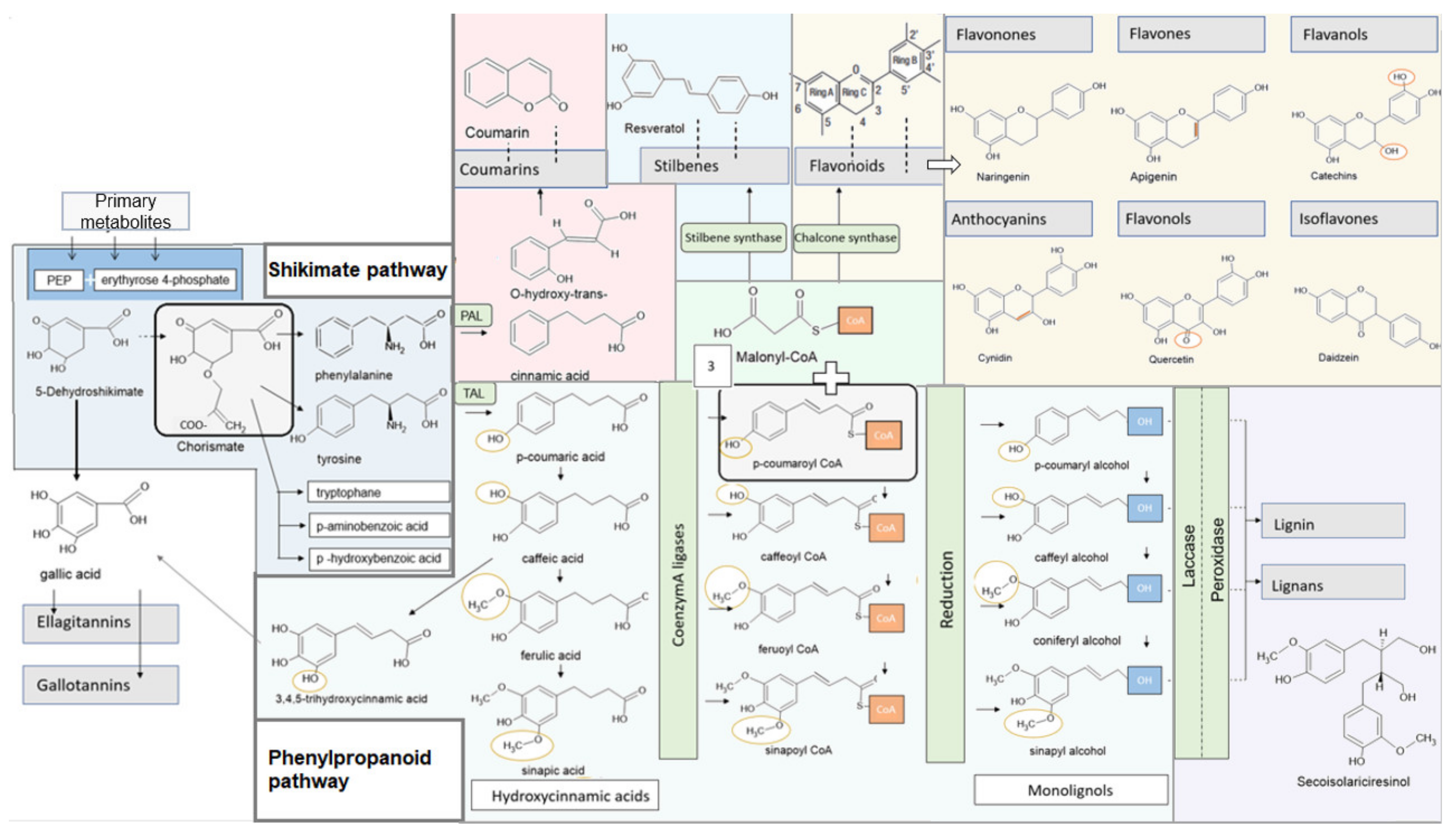
4. Metabolic Pathways in Higher Plants

Phenols consist of an aromatic benzene ring with a hydroxyl group [90]. As soon as aromatic carbon compounds with two or more linked aromatic rings are generated, the substances are called polyphenols [91]. Plants have different metabolic pathways to synthesize different polyphenols. These pathways can be divided into a primary and a secondary metabolism. The primary pathways such as glycolysis, pentose–phosphate pathway, lipid–protein, and nucleic acid biosynthesis generate metabolites that are essential for plant survival [92]. However, plants also have the ability to secure biological benefits through the synthesis of various secondary plant metabolites. The plant synthesizes these components as natural deterrents against pests. The large variety of different secondary metabolites is caused by the phenylpropanoid metabolism [93] (Figure 1). It is named after the amino acid phenylalanine, which is usually the compound with which the biosynthesis begins.
In contrast to animals, plants have mechanisms to synthesize the amino acids phenylalanine and tryptophane themselves. Thus far, two pathways are known: the mevalonic acid pathway, and the shikimate pathway. In higher plants the shikimate pathway plays the major role [94,95].

4.1. Shikimate Pathway

The shikimate pathway is found in bacteria, fungi, and higher plants. It converts the primary metabolites phosphoenolpyruvate and erythyrose 4-phosphate via seven different enzymes to chorismic acid. The condensation reaction between erythrose 4-phosphate and phosphoenol pyruvate produces 3-dexoxy-d-arabino-heptulosonic acid-7-phosphate. It is then oxidized, dephosphorylated, and cyclized by dehydroquinate synthase. The resulting 3-dehydrochinate is dehydrated and then reduced to shikimate. Condensation with another phosphoenol, pyruvate, results in chorismate via the intermediate stage 5-O-enolpyruvyl shikimate-3-phosphate. This provides the precursor for the formation of the three amino acids tyrosine, phenylalanine and tryptophane, as well as p-amino and p-hydroxybenzoic acid (Figure 1) [94,95].

4.2. Phenylpropanoid Synthesis and Structure of the Individual Phenylpropanoids

The synthesis of polyphenols in plants is usually based on the amino acids phenylalanine and tyrosine. An exception is the formation of gallic acid, and therefore also the production of ellagitannins and gallotannins. They originate primarily from the shikimic acid pathway (Figure 1) [96]. Moreover, there are also a few plant phenols that are synthesized via a metabolic pathway called the polyketide pathway [97]
Phenylalanine ammonia lyase (PAL) is an enzyme responsible for the amination of phenylalanine to trans-cinnamic acid. Much less frequently, tyrosine is metabolized for the formation of phenylpropanoids. Tyrosine ammonia lyase (TAL) is responsible for the formation of tyrosine to p-coumaric acid [93]. Cinnamic acid and its derivatives are the basis for the synthesis of all polyphenols.

4.3. Specific Synthesis of Different Polyphenols

4.3.1. Lignans, Lignin

Starting from the trans-cinnamic acid, the formation of different cinnamic acid derivatives can be distinguished. The formation of p-coumaric acid occurs through hydroxylation. By the methylation of p-coumaric, coffee acid can be formed and further methylation results in sinapin acid. These hydroxycinnamic acids can be activated by Coenzym A ligases (CoA ligases). Through reduction, it produces the corresponding alcohols, which are summarized under the term monolignols. This results in p-coumaryl alcohol, caffeyl alcohol, coniferyl alcohol, and sinapyl alcohol (Figure 1).
Monolignols themselves are toxic and unstable; thus, they are hardly detectable in living organisms. Glycosylation of the phenolic hydroxyl groups produces monolignol glycosides. In this state, the monolignols can be stored and transported within the plant. Two different enzymes, laccase and peroxidase, are responsible for the polymerization of the lignin alcohols to lignin and the lignans [98] (Figure 1).

4.3.2. Gallic Acid, Hydrolysable Tannins: Ellagitannins, Gallotannins

For the formation of gallic acid two ways are are of particular importance [99]: Firstly via the phenylpropanoid pathway, where it is formed by the beta-oxidation of the 3,4,5-trihydroxycinnamic acid, which represents a further hydroxylation of the caffeic acid [100]; the second possibility results from the dehydrogenation of shikimic acid, which can proceed more immediately and is therefore considered the main synthesis pathway for gallic acid (Figure 1) [101].
It forms the basis for the synthesis of hydrolysable tannins, which include gallotannins and ellagitannins. They are esters of gallic acid with a polyol, such as 1-O-galloyl-β-d-glucopyranose. An increased substitution of the monoesters leads to 2-, 3-, 4-, 6-Penta-O-Galloyl-β-d-Glucopyranose. From this intermediate, ellagitannins can be formed by oxidative linkage with gallic acid; in gallotannins the galloyl groups are depsidically linked [102,103].

4.3.3. Flavonoids, Stilbenes

The starting point for the formation of flavonoids and stilbenes is p-coumaroyl-CoA. Both pathways are linked with three molecules of malonyl-CoA from fatty acid metabolism [96].
The chalcone synthase forms tetrahydroxychalcone. This is normally converted by the chalcone isomerase directly to naringenin, a flavonoid [96]. Flavonoids consist of two benzene rings connected by a three-carbon linking chain. In addition, a pyran ring is formed centrally. Depending on the degree of oxidation of this ring, six different subclasses of flavonoids can be distinguished: flavanols, flavones, flavanones, flavanonols, anthocyanins, and isoflavones (Figure 1) [104].
The stilbenes are characterized by a skeleton with 1,2-diphenylethylene. They derive from the phenylpropanoid pathway and are formed by the reaction of the CoA esters of the cinnamic acid derivatives and three malonyl-CoA molecules with the stilbene synthase. Glycosylation, methylation, and prenylation lead to the formation of various complex stilbenes [105].

4.3.4. Catechins and Theaflavins

Catechins and theaflavins are essential polyphenols of classical tea drugs. They belong to the flavonoid group. Their structure is characterized by two benzene rings that are linked by a heterocyclic pyran or pyron. At the position of the C3 carbon atom, the binding of gallate groups can occur through esterification [106].
This can result in various subgroups, such as catechin gallate (CG), gallocatechin (GC), epicatechin (EC), epicatechin gallate (ECG), gallocatechin gallate (GCG), epigallocatechin (EGC), and epigallcatechin gallate (EGCG) [107].
Fermentation can lead to the linking of catechins, resulting in the formation of theaflavins [108]. These are dimers of the catechins [109].

4.3.5. Coumarins

The simple coumarins consist of a 1,2-benzopyrene skeleton. The synthesis starts with an ortho-hydroxylation of the cinnamic acid to o-coumaric acid. The product undergoes an isomerization from cis- to trans-formation. With the loss of a water molecule after lactonization, coumarin is formed (Figure 1) [110]. Various metabolites of simple coumarins are formed by hydroxyl, methoxyl, methylenedioxy, or isopentenyl groups. As soon as a furan or pyran ring is generated, the resulting coumarins are called furano- or pyranocoumarins. Dimers or trimers of the coumarins are called dicumarins; isomeric forms create isocoumarins [111].

5. Chemical, Physical and Biological Properties of Polyphenols

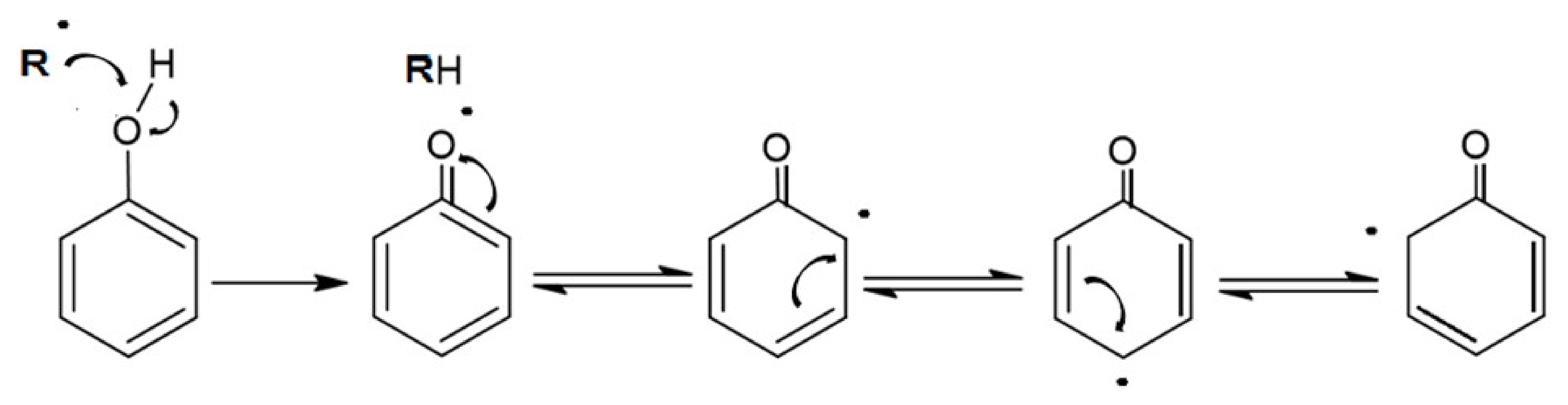
5.1. Chemical Properties

The characteristic structure of polyphenols determines their antioxidant properties [7]. Within the benzene ring, the carbon atoms are single-bonded. The remaining six electrons are free in movement. The hydroxyl group can also cause interactions with the electrons of the benzene ring. Within the conjugated aromatic ring, the unpaired electrons can be delocalized (Figure 2) and thus serve as a radical scavenger [96]. In addition, some phenolic compounds possess dihydroxy groups that can serve as metal chelators [96,97,98,99,100,101,102,103,104,105,106,107,108,109,110,111,112].

5.2. Physical Properties

Purified phenolic compounds are often white solids that have characteristic aromatic odors, such as vanillin, methyl salicylate, and eugenol [96]. There are some phenols that are more hydrophilic, such as the hydroxycinnamic acid glucosides, whereas others are more hydrophobic, such as polymethoxy flavonoids [96,113]. Hydrophobic compounds can be easily absorbed into the cell via diffusion. Various systemic pathways exist for the entry of hydrophilic compounds [96,114,115].

5.3. Biological Properties

These chemical and physical properties help to secure biological advantages for the plant. Lignin, as a complex polymer, is instrumental in three-dimensional structure formation. It helps to ensure mechanical transport and serves as a water barrier and water transport [116]. Particularly, flavones and flavonols play a role in protecting against UV light due to their absorption spectra. At the same time, the antioxidant properties of phenolic compounds help scavenge radicals that would cause damage from increased UV radiation [117,118,119]. Pigmented polyphenols serve as attractants for pollination of the plant [96,120]. The plant has different systems to defend itself from predators and pathogens [96]. Phenolic compounds often have antibacterial and antiviral activity themselves. Gallotannins and condensed tannins react strongly with various proteins and are thus also a decisive factor in the inhibition of enzymes [96]. Tannins, in particular, are also responsible for astringency and the bitter taste [121], which is intended as a protection against pests [96]. Some phenols even have a toxic effect to a certain extent [122].

6. Pellicle Formation and Initial Bacterial Colonization on Solid Surfaces

Soft and solid surfaces in the oral cavity are covered by a proteinaceous film called the pellicle [123,124]. Therefore, soft as well as hard tissues (enamel and dentin surfaces) are substrates for pellicle formation.

6.1. Mucosal Pellicle

Bioadhesion and -adsorption processes are significantly different on soft tissues compared to hard tissues. In contrast to oral hard tissues, oral epithelia represent shedding structures with high turnover rates. Thereby, outer layers of oral epithelia are covered with viable and dead cells. The formation of the mucosal pellicle is influenced by hydrophobic interactions and the cross-linking of proteins and salivary components [125,126,127,128,129]. Microorganisms can directly adhere to the epithelial cell membranes, whereas on the tooth surface, bacteria adhere indirectly through attachment to the acquired enamel pellicle layer [124,130,131,132,133]. The mucosal pellicle’s components are represented by soluble and epithelial mucins (mucin 5B, 7, 1, 4, 16) CA VI, sIgA, cystatin, amylase and PrPs [125,129,134,135,136]. Therefore, the mucosal pellicle represents an interface consisting of salivary and epithelial molecules [136]. As soon as polyphenols are consumed by food intake, they come into contact with the mucosal pellicle and modify the mucosal pellicle’s composition [137]. Nevertheless, a systematic examination of the influence of polyphenolic agents to the mucosal pellicle layer is difficult, due to the shedding character of this interface.

6.2. Acquired Enamel Pellicle

An acquired enamel pellicle is formed after a few seconds through the instantaneous adsorption of proteins, macromolecules (e.g., lipids), carbohydrates, and nucleic acids derived from saliva, gingival crevicular fluid, blood, bacteria, mucosa, and diet [138,139,140,141] (Figure 3). During pellicle formation in hard tissues and dental materials, two stages can be observed with transmission electron microscopy (TEM) [141,142]. The first stage shows a densely structured basal layer, reaching a thickness of about 10–20 nm [141,142,143,144]. The second phase of pellicle formation is characterized by protein–protein interactions leading to an outer pellicle layer with a globular appearance [144,145]. During the pellicle formation process, an increase in the pellicle thickness (100–1000 nm) can be observed after 60 min [141,143,146,147,148] (Figure 3).
There is high variability in pellicle protein profiles of different individuals [140,150,151]. The main functions are: lubrication of the enamel surface [133], mediation of de- and remineralization processes [123,152,153], and antibacterial functions [154,155,156]. The pellicle contains components that favor the adherence of oral bacteria. Bacteria have adapted to the physiological pellicle layer; thus, that the pellicle also promotes biofilm formation [157,158]. Specific receptors or ligands for the adhesion of bacterial species are glycolipids, fibrinogen, or collagen [158]. Pioneer bacteria such as different Streptococcus spp. or Actinomyces spp. are initially attached to the tooth surface via unspecific interactions [158,159,160]. Afterwards, they connect to the tooth surface irreversibly via specific receptors [161,162,163,164]. The number of adherent bacteria increases within the time of biofilm maturation. A cross-linking between the microorganisms of the biofilm can therefore be observed [165,166].
Within the consortium of caries-promoting bacteria, Streptococcus mutans is often utilized in in vitro studies to test specific anti-caries agents. For a long time, S. mutans was considered to be the most significant microorganism in the development of caries [167]. This bacterium is aciduric and has an acidogenic effect. S. mutans produces intracellular and extracellular polysaccharides [165,168]. Intracellular polysaccharides can act as the substrate supply for bacteria inside the three-dimensional biofilm structure. Extracellular polysaccharides are synthesized by bacterial glycosyltransferases (GTFs) [167]. The different isoenzymes of the GTF may differ in their properties. GTFs serve as specific receptors and are able to transform other bacteria into glucan producers [169]. Caries development has long been considered as a finely coordinated multifactorial process, in which the three-dimensional structure of the EPS matrix plays an important role [4].
The EPS matrix modulates the diffusion properties of the biofilm. Consequently, polymicrobial acid production is distributed spatially within the biofilm [170], and the hyperacidity within the biofilm structure is not buffered by saliva because the EPS matrix controls the transfer of salivary components [4]. Hence, dietary sugars can subsequently diffuse into the biofilm and salivary components are blocked [171,172,173]. The expressions of specific genes such as gtfB-genes, which are involved into the intake of carbohydrates, are upregulated if the host consumes a combination of sucrose and starch. This results in a complex conversion of the S. mutans transcriptome and regulates the dynamics of biofilm progression and pathogenicity [4,174,175]. This might be an explanation why combinations of sucrose and starch are more cariogenic than the individual substances [176]. This mechanism emphasizes the modulating effect of nutrition on the biofilm’s virulence. The pellicle is the starting point for biofilm formation on the tooth surface; therefore, anti-adherent and anticariogenic agents must attack this layer in order to reduce bacterial adherence.

6.3. Polyphenols as Anti-Adherent Agents

External agents can modify the mucosa [137] and acquired enamel pellicle layer [68,77,149,177,178,179,180,181] (Figure 4), thereby bacterial adherence to the tooth surface is reduced [63,77,149,182] and the erosion-protective potential of the pellicle layer is enhanced [178,183] (Figure 5). Secondary phytochemicals such as polyphenols can act as such external agents.
Studies have shown that polyphenols already interact with salivary components in the oral cavity [184]. Typical salivary proteins for polyphenol-protein interactions are histatins and prolin rich proteins (PrPs). They also represent typical pellicle proteins [151,185,186,187]. It was shown in a recent proteome analysis that ECGC can increase the abundance of important pellicle proteins e.g., prolin rich proteins (PRPs), calcium-bind proteins and statherin [188]. One of the main mechanisms between PrPs and tannins derived from food intake is the so-called astringency. It describes a mouth puckering tactile feeling [121]. Astringency and bitter taste of food are strongly connected with flavor characteristics of a plant-based nutrition [121]. Often, consumers add milk to tea beverages to reduce astringency [121,189]. As a result, the bioavailability of secondary plant extracts is reduced, due to interactions between the polyphenols and the milk proteins.
Generally, astringency is attributed to the binding process between polyphenols and different classes of salivary components like PrPs, statherin and mucins [121]. PrPs are divided into basic, glycosylated and acidic PrPs. They provide the oral homoeostasis through the regulation of the calcium balance at the tooth surface [190,191]. Mucins are the main components of the mucosal pellicle and act as lubricants in the acquired enamel pellicle layer (e.g., mucin 7) [192]. Statherin represents an acidic salivary protein rich in tyrosin. Its main function is the regulation of the calcium and phosphate homoeostasis and the remineralisation of the enamel [121,193]. All these components are also integral parts of the acquired enamel pellicle. The interaction between these proteins and polyphenols are obtained by molecular forces, hydrophobic interactions and hydrogen bondings [194]. The connection of polyphenol-protein-complexes to the acquired enamel pellicle is probably based on crosslinking reactions leading to a resistant and hard to remove pellicle layer on the tooth surface [179].
Previous studies have also shown that polyphenols can denature pellicle proteins leading to an impaired interaction between microorganisms and bacterial receptors [182] (Figure 6). Thereby, the original structure of the receptor protein is broken down changing the bonding of the secondary, tertiary and quaternary structure. Especially plant polyphenols seem to have a tanning effect on the pellicle layer [63,141,179]. As a result, the pellicle is more electron dense and thicker as visualized by TEM [63,68,77,177,178] (Figure 4). Through the increase of pellicle-bound proteins, the pellicle becomes more protective against erosive attacks [178,183]. Besides, polyphenols have the ability to inhibit proteolytic salivary enzymes [62], matrix metalloproteinase, α-amylase and GTF [195,196]. Conclusively, glucan formation is reduced and less open binding sites for specific bacterial adhesion are available for cariogenic microorganisms [63,182,196,197]. Intentional interaction of plant substances with the acquired enamel pellicle are therefore a promising approach for caries prophylaxis and prevention of erosive loss of tooth structures [198].

7. Influence of Tea Polyphenols onto the Acquired Enamel Pellicle

7.1. Green Tea

The influence of green tea onto the acquired pellicle was examined by Hannig et al. (2009) [182]. They observed a significant reduction of initial bacterial adhesion after 30 and 120 min of intraoral pellicle formation [182] (Table 2). Epigallocatechingallate is one of the main anti-oxidative polyphenolic compounds in green tea. In many studies EGCG is tested representatively as the main polyphenolic substance of green tea. Rehage et al. (2017) [68] demonstrated that rinsing with EGCG leads to an immediate increase in pellicle thickness and density (Table 2). The ultrastructure of the pellicle was therefore distinctly modified as shown via TEM (Figure 4, Figure 7). Further, EGCG-protein-aggregates accumulated at the pellicle surface leading to a fissured sectional image in TEM [68]. They were also detectable via REM [68]. Zimmermann et al. (2019) [181] confirmed these observations and showed that there was no influence of EGCG-rinsings on the negative charge of the in situ pellicle [181] (Table 2). Soares et al. (2020) [121] observed that flavanoles like EGCG and ECG highly interact with oral cells. In contrast, anthocyanes do not show such affinities.

7.2. Black Tea

Joiner et al. (2003) [180] examined the influence of black tea onto the acquired enamel pellicle. They showed that the pellicle structure was modified by adsorption of black tea components (Table 2). The modified pellicle could not be eluted by different buffer solutions. As a consequence, the removal, dissolution and replacement of these adsorbed components was strongly hampered under in vitro conditions [180]. It is assumed that the interactions of the black tea polyphenols lead to a cross-linking between the polyphenolic compounds and the pellicle as well as salivary proteins. In the following, pellicle formation is progressing. Whereby the polyphenol-modified pellicle layers are more resistant against removal than unmodified layers [179] (Table 2). This modification results in a doubling of the total protein amount binding to the hydroxy apatite surface in vitro [180] (Table 2). Further, the polyphenolic fractions of black tea contribute to this effect. Nevertheless, theaflavin, epigallocatechin and epigallocatechingallate showed less strong effects than the whole black tea extract. Pharmakokinetic investigations demonstrated that high concentrations of tea polyphenols, like theaflavin were still detectable in saliva after 1 h [199]. Theaflavin and thearubiginines are the main pigments of black tea. Ellipsometric studies showed that theaflavin is able to discolor the pellicle [179], which also might lead to stained teeth. Hannig et al. (2009) [182] observed the influence of black tea on the initial bacterial adhesion of the tooth surface after 30 and 120 min in situ. The combination of fluorescence methods (DAPI and FISH) clearly showed a significant reduction of initial bacterial adhesion after rinsing with black tea. Interestingly, black tea leads to a significant reduction of immobilized lysozyme activity after 3 and 30 min of oral exposition in situ [200] (Table 2).

7.3. Cistus Incanus Tea

The study of Wittpahl et al. (2015) [63] investigated different types of cistus incanus tea. They detected that especially the types rich in polyphenols had significant antibacterial effects in the in vitro-Live/Dead assay. After fractionation of the samples into seven fractions, only the fraction rich in ellagitannins and the flavonoid group had remaining weak antibacterial effects (Table 3). Simultaneously, the other fractions had no remaining effect at all [63]. Abdollahzadeh et al. (2011) [201] postulated that the in vitro antibacterial effects of cistus incanus against S. mutans are mainly attributable to hydrolyzable tannins and polyphenols with low molecular weight. On the other side, Naz et al. (2007) [202] report that polyphenols like quercetin, myrecetin and gallic acid are responsible for the antibacterial effect, whereas Hamilton-Miller et al. (2001) [203] and Otake et al. (1991) [204] assume that catechins inhibt the bacterial adherence to the tooth surface (Table 3). The study of Wittpahl et al. (2015) [63] shows in the in situ part of their examination that the antiadherent effect of cistus incanus cannot be attributed to single fractions of the polyphenolic plant compound but to the whole extract. Thus the ingredients of the whole extract display synergistic effects leading to a strong antiadherent and antibacterial effect. These findings have also been confirmed by the in situ study of Hannig et al. (2009) [182] (Table 3). Another in situ study showed in this context that cistus incanus tea had no impact on the immobilized pellicle enzymes [62] and detected no effect on the accumulation of lysozyme into the pellicle layer [198]. The underlying mechanisms are still not fully understood.

7.4. Inula Viscosa Tea

A recent in situ study demonstrated that 10 min-rinsing with Inula viscosa tea results in a significant reduction of the initial bacterial colonisation on the tooth surface after 8 h of oral exposition [77] (Table 3). Thereby, Inula viscosa tea exerts a significant antiadherent effect on oral streptococci [77]. Active polyphenolic substances in Inula viscosa tea are the flavonoid-aglycones apigenin, naringenin and luteolin. Their antibacterial effects on oral streptococci was observed in many in vitro studies [205,206,207,208] (Table 3). An animal study detected an inhibitory influence of apigenin on the glycosyltransferase isoenzymes [209]. Besides, different essential oil components of Inula viscosa tea seem to contribute to its antibacterial properties [210]. Though, the dominating antibacterial and antiadherent action is so far attributed to the polyphenolic compounds of Inula viscosa tea [77].

7.5. Fragaria Vesca, Hamamelis and Tormentil Tea

Kirsch et al. (2020) [149] examined the effect of the three polyphenolic tea drugs fragaria vesca, hamamelis and tormentil. They detected a prolonged antiadherent effect on bacterial colonization of the tooth surface as well as a distinct modification of the pellicle ultrastructure via TEM [149] (Figure 7). Thereby a thickening and an enhanced density of the ultrastructure was detected even after 8 h of oral exposition [149] (Table 3).

8. Mechanisms for Provided Effects by Polyphenols

8.1. Effect of Polyphenols on Oral Proteins

Besides their direct effect on bacterial cells, polyphenols exert an impact on the proteins of saliva and pellicle. Thereby, a so-called tanning effect can be observed [63,141,179,180]. The underlying principle is a denaturation of the receptor proteins functional groups resulting in a hampered bacterial adhesion (Figure 6) [182]. Polyphenols influence enzymes of the anabolism and catabolism of bacteria. It was shown that the human enzyme α-amylase and the bacterial isoenzymes glycosyltransferases are inhibited by polyphenols [196,197]. Both enzymes are part of the enamel pellicle and promote bacterial adhesion and biofilm formation.
It has been shown that bacterial glucan formation is reduced in situ after rinsing with hamamelis tea, fragaria vesca tea and tormentill tea [149]. Hence, it is assumed that rinsing with polyphenolic beverages leads to a reduction of binding sites for bacterial adherence on the tooth surface [203,204,212]. Interestingly, polyphenols from cistus incanus demonstrated no inhibition of the immobilized enzymes lysozyme, α-amylase and glycosyltransferase in situ, only peroxidase was affected [62]. The adsorption process of enzymes into the pellicle structure changes the secondary and tertiary structure of the specific enzymes [213]. As soon as the enzymes are integrated, the active site of the enzyme undergoes some chemical transformations and substantial conformational rearrangements of the proteins can be observed. Therefore, proteins that are originally soluble become insoluble through the adsorption to the pellicle layer. Nevertheless, the enzyme activity maintains [214,215,216] and can be enhanced [217] or reduced [218]. Due to these conformational changes and the reduction of free binding sites, polyphenols do not show the same impact on immobilized pellicle enzymes under in situ conditions compared to the in vitro situation. Through the immobilization process an inhibition of the enzymes (except peroxidase) is no longer possible. Consequently, the tanning effect of polyphenols does not affect all proteins of the acquired enamel pellicle [213], especially immobilized enzymes are excluded. Although, enzyme activities are not affected, polyphenols lead to a targeted protein enrichment of the pellicle layer. So far, all tested polyphenolic solutions showed an increase in pellicle thickness and denseness in in-situ-studies via TEM [68,77,149,177,178] or ellipsometric approaches [179,180] (Figure 4, Figure 7 and Figure 8). Consequently, the accumulation of specific proteins derived from saliva, gingival crevicular fluid, blood, epithelia and bacteria on the enamel surface is promoted and the proteins maintain their activity level [62]. This thickening effect has further tooth surface protecting effects (Figure 5). It was shown that tea drugs and plant extracts high in polyphenols such as black tea, green tea, Grape seed extract, Grapefruit seed extract, Origanum and leaves of the Ribes nigrum increase the erosion protective properties of the pellicle [178,183].
Further, typical pellicle components like histatines and PrPs interact with polyphenols, building complexes in saliva and pellicle [185,186]. This is the reason for the astringent character of polyphenols [186]. Thereby the polyphenols can form insoluble aggregates with salivary proteins (Figure 9) [186], as shown by expectorate samples visualized by TEM [149].
At the pellicle layer these precipitates can also be found. The starting point are pellicle proteins with a high affinity to polyphenols, such as proline-rich proteins, amylases, cystatins and histatins. Afterwards interactions with the adsorbed glycoprotein layer can be observed [219]. On the chemical level different stages can be detected during the formation of polyphenol-protein-aggregates. First, soluble complexes are formed between the polyphenol’s aromatic rings and the pyrrolidine ring of the protein’s proline residues [220]. Second, insoluble complexes are built through cross-linking between peptides, and in the third stage, aggregates of complex precipitates separate spontaneously [221,222]. The cross-linking is described by two types of binding: hydrophobic interactions based on van der Waals-London interactions between the most apolar amino acids and the benzene rings of the polyphenols and on the other site hydrogen bonds [187,223,224]. As a consequence, more resistant pellicle layers are formed on the tooth surface, which are difficult to remove as shown for black tea [179]. These modified pellicle layers have higher acid-resistant properties than the physiological pellicle can provide [178,183] (Figure 5). Joiner et al. (2004) [179] assume that the prolin-rich proteins remain their polyphenol-binding site, so direct attachment of further polyphenols into the pellicle layers are possible. Covalent bonds play a subordinate role and are formed between the quinone forms of polyphenolic compounds (usually resulting from the oxidation of the latter) and the amino or thiol groups of proteins [225]. Hydrogen bonds, hydrophobic interactions and entropy are the driving factors of these processes between polyphenols and proteins [194]. External factors that influence the formation and the stability of the polyphenol-protein-aggregates are pH-value [226,227], ionic strength [227] and the presence of polysaccharides which can also disrupt the aggregation or rather modify the interaction processes [228]. Additional determinants for the stability of the aggregates are polyphenol ratio and concentration, temperature and the specific types of polyphenols and proteins [229]. Distinct differences can be observed between pellicle and salivary poteins since salivary proteins adsorb selectively to the pellicle layer [151]. The pellicle contains a wide range of protective proteins and enzymes from non-glandular sources enriched in the pellicle layer [150,155,181,230,231].
The interaction between polyphenols and proteins can lead to astringent sensations. This tactile feeling might be attributed to the degree of cross-linking between the adsorbed salivary proteins, polyphenols and epithelia of the oral cavity through transglutaminase enzyme reactions [129]. Nayak and Carpenter (2008) observed the polyphenol-protein-precipitates via TCA methods. Thereby, they detected mucin 1, 5a, 7, statherin and secretory components as the main protein components in these precipitates. In a proteome analysis statherin amounts were increased after the application of polyphenols (ECGC) [188]. These specific proteins are also components of the salivary and mucosal pellicle [184,232,233]. Consequently, these proteins are possible targets for intentional modulation of the pellicle. Further research, like complex proteome analysis are necessary to give further proof.

8.2. Effect of Polyphenolic Compounds on Bacteria

Most of the assumed reasons for antiadherent and antibacterial effects of polyphenols have been derived from in vitro studies [70,197,203,204,234,235,236,237,238,239,240]. Thereby, it was shown that polyphenols can break trough bacterial cell walls and interact with intracellular membranes [63]. Further, the chemical character of polyphenols results in complex formations with metal ions. Consequently, the essential metal ions are no longer available for microorganisms leading to a reduction of bacteria [11,63,202,241].
The effect of tea polyphenols to cariogenic bacteria was mainly examined under in vitro conditions. Karygianni et al. (2014) [242] detected the antibacterial properties of Inula viscosa tea against nine relevant oral microorganisms. Thereby, Inula viscosa tea showed strong antibacterial effects against S. mutans, S. sobrinus, S. oralis, E. faecalis, Candida albicans, E. coli, Staph. aureus, Porphyromonas gingivalis, Prevotella intermedia, Fusobacterium nucleatum and Parvimonas micra [242]. The study group detected that a methanolic solution of Inula viscosa tea was more effective than an ethyl acetate extract [242]. Besides, Xiao et al. (2000 a, b) [243,244] examined that tea polyphenols can inhibit the adherence of cariogenic bacteria like S. mutans and A. viscosus to hydroxyapatite covered with saliva or collagen in vitro. Other in vitro studies investigated the inhibition of GTF-dependent glucan synthesis by specific polyphenols [197,245,246,247,248,249]. It is assumed that only the polymeric fraction of tea polyphenols is responsible for this inhibiting activity [247,248]. Besides, three studies relate the inhibiting effect of acid production by mutans streptococci with the inhibition of the bacterial enzyme proton translocating F-ATPase [245,246,250]. Thereby, bacterial growth [245,250] and adhesion to hydroxyapatite is inhibited [243,244]. In addition, it is assumed that microorganisms that are exposed to polyphenols up-regulate proteins that modulate the defense mechanisms of the bacterial cell and down-regulate metabolic and biosynthetic proteins that are involved in amino acid synthesis, protein formation and energy metabolism [205,251]. Consequently, glucan formation is also down regulated as shown in the in situ study by Kirsch [149]. A distinct influence on the 3-dimensional structure of the biofilm is thereby possible, in addition available binding sites and the pathogenicity of the biofilm might be influenced.

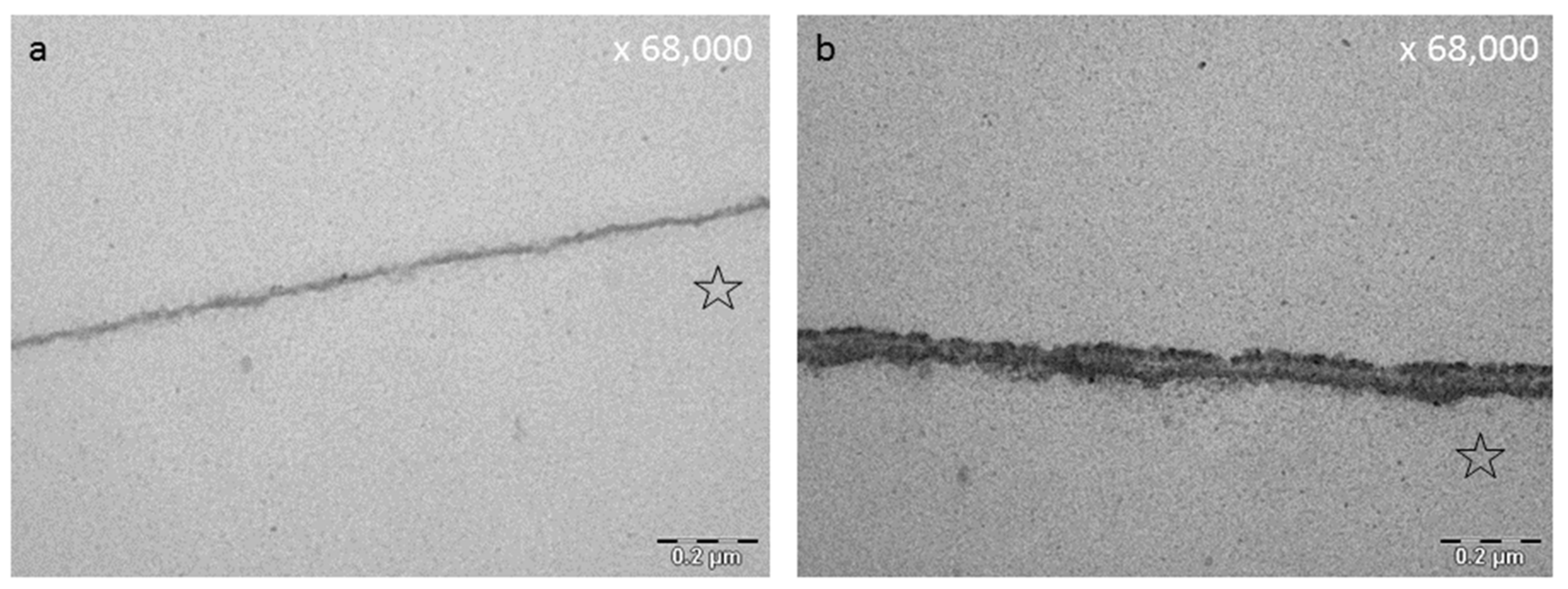
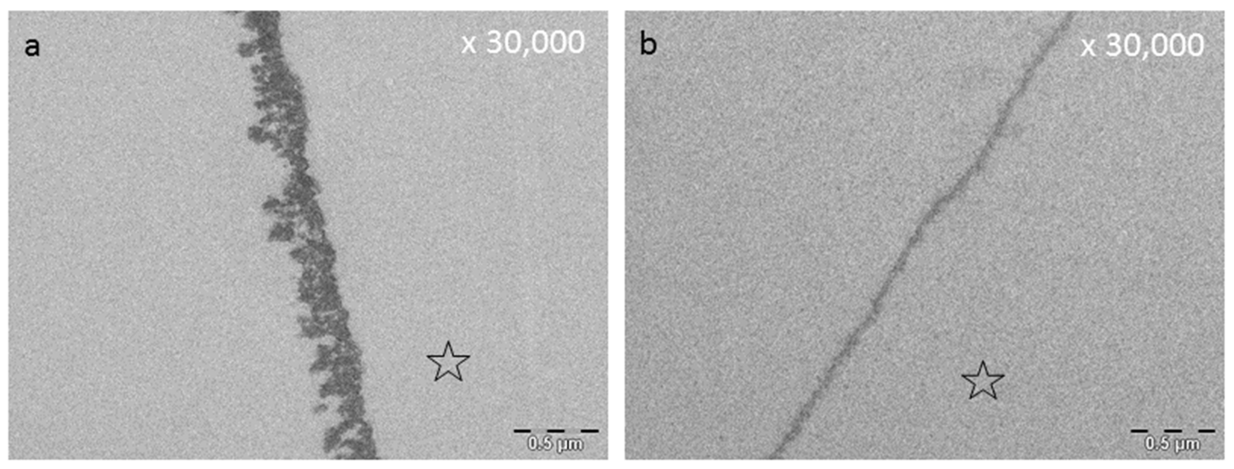
8.3. Effect of Tea Polyphenols on the Ultrastructure of the Pellicle

The pellicle layer represents a 0.1–1 μm thick protein layer on the tooth surface [138]. The defined two-layer structure of the pellicle consists of an electron-dense basal layer and a less electron dense second layer with granular and globular structures as visualized by TEM [124,141] (Figure 3). It was shown that the pellicle ultrastructure can be enhanced by plant extracts high in polyphenols, such as Origanum and leaves of Ribes nigrum (Figure 7) [178].
Tea polyphenols can modify the pellicle layer as well. A pellicle thickening and enhancing effect can be observed [179] (Figure 3, Figure 4, Figure 7 and Figure 8). Rehage et al. (2017) [68] showed that rinsing with EGCG can enhance the pellicle thickness under in vitro conditions. This effect is caused by the integration of protein-polyphenol-aggregates with an average diameter of 70 nm. Consequently, the pellicle layer obtains a fissured appearance in TEM images [68]. Further, rinsing with EGCG under in situ conditions leads to a more homogenous appearance of the pellicle structure. Also under in situ conditions, electron dense, aggregated structures can be detected [68]. These structures consist of ECGC-protein-aggregates. In the in situ study, the pellicle was not as fissured as under in vitro conditions. Consequently, ECGC-molecules and salivary proteins already connect in the salivary film of the oral cavity, leading to the assumption that the effective concentration of the tea polyphenols that connects with the tooth surface is reduced significantly [68].
Similar effects were shown for Inula viscosa tea. After rinsing a thicker more electron dense pellicle layer was observed [77]. Round electron dense complexes mit embedded vacuoles above the pellicles basal layer were detected by TEM [77]. Inula viscosa contains hydrophobic components such as essential oils (nerolidol and linalool) [77,210]. These hydrophobic lipid components may cause the observed vacuoles inside the modified pellicle structure [77].
Kirsch et al. (2020) [149] examined the effect of fragaria vesca, hamamelis and tormentil tea on the ultrastructure of the pellicle. In their study, a modification of the ultrastructure of the pellicle was still detectable after 8 h of oral exposition. Similar effects of these specific polyphenols on the ultrastructure were visible. A significant enhanced and more electron dense pellicle ultrastructure with polyphenol-protein aggregates was visualized by TEM [149] (Figure 7). In their study, the pH of the phenolic tea drugs differed (fragaria vesca: 5.53, tormentil: 5.28, hamamelis: 4.3). Therefore, zones of demineralization were detected after rinsing with hamamelis. These areas were infiltrated and covered with pellicle compounds. The authors thereby assume a repair process of superficial erosive defects filled with proteinaceous structures. On top of these structures a granular surface pellicle has formed, leading to repair processes. These areas are known as subsurface pellicle and they can be identified in the enamel [149,252]. It would be very interesting to examine the antiadherent properties of the polyphenolic tea drugs at a higher pH-value to prevent initial demineralization of the tooth surface.
In summary, polyphenolic tea drugs lead to a distinct modification of the pellicle layer as shown by TEM in multiple studies. The extent to which a modification can be detected depends on the polyphenolic tea drug.

8.4. Influence of Tea Polyphenols on Erosive Attacks of the Enamel Surface

A recent proteomic analysis showed that the treatment with EGCG gel enriches proteins with potential functions to protect against caries and erosion [188]. Thereby the abundance of important structural and functional proteins was increased (e.g., PRPs, calcium-bind proteins and statherin). Additionally, the EGCG-modified pellicle proteins were examined after citric acid attacks [188]. The result was an increase in cystatins and profilin-1 [188]. Hertel et al. (2017) [177] simulated alimentary acid attacks by incubation in HCL and examined whether polyphenols have an erosion-protective effect on the pellicle layer and the enamel surface [77] (Figure 5). Erosive conditions were simulated with pH-values 2.0, 2.3 and 3.0. After rinsing with Inula viscosa tea a loss of the pellicles basal layer and pore volume and density of the enamel surface was detected after 30 min and 120 min oral exposition [77]. Thus, Inula viscosa tea provides no erosion protective potential for the pellicle covered tooth surface [77]. In contrast rinsing with a combination of watery plant extracts of Origanum and Ribes nigrum leaves lead to a more electron dense and thicker pellicle layer with an enhanced erosion protective potential of the pellicle layer and therefore a protection of the enamel surface against erosive attacks [178]. These results were confirmed by another in vitro study that examined the effect of different polyphenol-rich teas and natural extracts on the erosion –protective properties of the pellicle [183]. This study outlines that not all teas high in polyphenols have beneficial effects. Black tea and green tea, as well as grape seed extract and grapefruit seed extract seem to have pellicle-modifying abilities and improved its protecting effect against enamel erosion under in vitro conditions [183].

9. Discussion

For conventional prevention, the use of fluoride is recommended. Studies show that regular fluoride intake can limit but not completely prevent the development of caries [253]. Biofilm removal and oral hygiene measures, as well as the additional influence of fluoride, can only alleviate this problem [254]. The antiseptical solution chlorhexidine is used postoperatively for prophylaxis. It has a very good antibacterial efficacy, but is insufficiently effective against bacteria already bound to the tooth surface [255,256]. The cationic character of the chlorhexidine molecules is responsible for the effect on the negatively charged bacterial surface. At the same time, however, this structure also causes severe discolouration, dry mouth, irritations of flavour and a shift of the bacterial balance in the oral cavity. Therefore, CHX is not suitable for daily use [257].
The formation of antibiotic resistance is also a problem in the treatment of infectious diseases caused by bacteria. There are more than 700 microbial species in the oral flora [258]. If bacteria are already present in a biofilm structure with an extracellular matrix, resistance is 1000 times more likely than among planctonic bacteria [4].
Polyphenols are secondary plant compounds associated with antibacterial, anti-inflammatory and anti-carcinogenic properties [7,8]. The current body of studies shows that polyphenols lead to thickening and strengthening of the pellicle layer [68,77,179], improving the pellicles erosion-protective potential [178,183]. In addition, polyphenols show efficacy in inhibiting bacterial growth [149,242,243,244], adhesion of bacteria to the tooth surface [149,182,259] and inhibition of enzyme activity [196,197]. Consequently, there is a further reduction of receptors that can serve as targets for the adherence of bacteria [203,204,212] and reduced glucan formation in situ [149]. So far, there are only two studies that examined the effect of polyphenolic tea fractions onto the acquired pellicle [63,141]. The comparison of the effect of the total extract versus fractions high in specific polyphenols highlight that complete extracts of specific teas have a stronger antibacterial effect than the specific fractions [63,141]. Besides investigating the total extract or fractions of polyphenol plant extracts, some studies analysed the effect of individual substances like epigallogcatechingallate or tannic acid [177,259,260]. Thereby, tannic acid is a good example to illustrate the mode of action and the long-term effect of polyphenols onto the pellicle layer [177,259,260]. So far, pure polyphenolic substances are not available for customers, due to the high costs for the production of pure substances like tannic acid. A closer look at the effect of tannic acid reveals a good efficacy on short-term pellicles (30 min, 120 min and 8 h). A study by Xi et al. (2020) [259] could additionally observe that tannic acid still exhibits anti-adherent properties after 24 h (Figure 10). Schestakov et al. (2020) [260] even detected anti-adherent efficacy after 48 h, which is similar to the effect of chlorhexidine and therefore of particular clinical relevance. The effects of tannic acid underline the importance to gain further knowledge about polyphenols since they provide long-lasting effects, which could be beneficial for preventive oral health care.
Overall, most of the currently available studies are mainly based on in vitro conditions. Further studies, especially in vivo and in situ studies, are necessary to evaluate possible interactions with long-term medication. Besides, it is necessary to understand the interactions of polyphenols with other surfaces within the mouth (mucosal pellicle, tongue) in order to reduce the total number of oral bacteria and minimise recolonization of the teeth. Bioadhesion is not only limited to the tooth surface (acquired enamel pellicle), these phenomena occur at all surfaces in the oral cavity. However, there are differences in adsorption since oral soft tissues constantly change through desquamation. Therefore the mucosal pellicle is an own entity. Interactions between polyphenols and components of the mucosal pellicle can be observed [137]. Polyphenols (tannins) react with mucins (mucin 5B) to mucin 5B-tannin-precipitates and their size increase with higher tannin concentrations and galloyalation [137]. In presence of basic PrPs, the size and distribution of these polyphenol-mucin-aggregates is altered [137]. In this context, promising results from a clinical trial suggests the use of cistus incanus tea in patients with mucositis or treatment-related radiation-induced oral mucositis [261]. Thereby, cistus incanus had an effect on the occurrence of mucositis regarding latency and incidence [261].

10. Conclusion and Perspectives

Although polyphenolic teas and beverages do not completely prevent bacterial adherence to the tooth surface but some of them reduce it significantly over a period of time [149]. Therefore the authors recommend the use of this tea, high in polyphenolic components, in addition to intensive oral care and hygiene along with the application of fluorides [261]. Especially patients with oral mucositis can benefit from the oral application of polyphenols [261].
Future studies should include further polyphenolic tea beverages to gain more basic knowledge about polyphenolic interactions within the oral cavity. Especially studies regarding the influence of specific polyphenols on the pellicle microbiome and proteom should be subject to further investigations.
Overall, polyphenolic teas and substances are relevant biological approaches for preventive strategies. Nevertheless, the most important part in preventive measures is still mechanical biofilm management where cleaning with a toothbrush and the use of fluorides remain essential parts of oral health. Polyphenols can be considered as important additives to a tooth-friendly and healthy diet.

Funding

The study has been partially supported by the German Research Foundation (DFG HA 2718/27-1; HA 5192/19-1; SP 464/6-1).

Institutional Review Board Statement

Not applicable.

Informed Consent Statement

Not applicable.

Data Availability Statement

Not applicable.

Acknowledgments

Open Access Funding by the Publication Fund of the TU Dresden.

Conflicts of Interest

The authors declare that they have no competing interests.

References

  1. Unachukwu, U.J.; Ahmed, S.; Kavalier, A.; Lyles, J.T.; Kennelly, E.J. White and Green Teas (Camellia sinensis var. sinensis): Variation in Phenolic, Methylxanthine, and Antioxidant Profiles. J. Food Sci. 2010, 75, C541–C548. [Google Scholar] [CrossRef] [Green Version]
  2. Ditano-Vazquez, P.; Torres-Pena, J.D.; Galeano-Valle, F.; Perez-Caballero, A.I.; Demelo-Rodriguez, P.; Lopez-Miranda, J.; Katsiki, N.; Delgado-Lista, J.; Alvarez-Sala-Walther, L.A. The Fluid Aspect of the Mediterranean Diet in the Prevention and Management of Cardiovascular Disease and Diabetes: The Role of Polyphenol Content in Moderate Consumption of Wine and Olive Oil. Nutrients 2019, 11, 2833. [Google Scholar] [CrossRef] [Green Version]
  3. Niemetz, R.; Gross, G.G. Enzymology of gallotannin and ellagitannin biosynthesis. Phytochemistry 2005, 66, 2001–2011. [Google Scholar] [CrossRef]
  4. Hannig, C.; Attin, T.; Hannig, M.; Henze, E.; Brinkmann, K.; Zech, R. Immobilisation and activity of human alpha-amylase in the acquired enamel pellicle. Arch. Oral Biol. 2004, 49, 469–475. [Google Scholar] [CrossRef]
  5. He, J.; Li, Y.; Cao, Y.; Xue, J.; Zhou, X. The oral microbiome diversity and its relation to human diseases. Folia Microbiol. 2015, 60, 69–80. [Google Scholar] [CrossRef] [PubMed]
  6. Sivaramakrishnan, K.; Parker, R.G. The United Nations High Level Meeting on the Prevention and Control of Noncommunicable Diseases: A Missed Opportunity? Am. J. Public Health 2012, 102, 2010–2012. [Google Scholar] [CrossRef]
  7. James, S.L.; Abate, D.; Abate, K.H.; Abay, S.M.; Abbafati, C.; Abbasi, N.; Abbastabar, H.; Abd-Allah, F.; Abdela, J.; Abdelalim, A.; et al. Global, regional, and national incidence, prevalence, and years lived with disability for 354 diseases and injuries for 195 countries and territories, 1990–2017: A systematic analysis for the Global Burden of Disease Study 2017. Lancet 2018, 392, 1789–1858. [Google Scholar] [CrossRef] [Green Version]
  8. Bowen, W.H.; Koo, H. Biology of Streptococcus mutans-Derived Glucosyltransferases: Role in Extracellular Matrix Formation of Cariogenic Biofilms. Caries Res. 2011, 45, 69–86. [Google Scholar] [CrossRef]
  9. Woelber, J.P.; Bremer, K.; Vach, K.; Konig, D.; Hellwig, E.; Ratka-Kruger, P.; Al-Ahmad, A.; Tennert, C. An oral health optimized diet can reduce gingival and periodontal inflammation in humans—A randomized controlled pilot study. BMC Oral Health 2016, 16, 109. [Google Scholar] [CrossRef] [Green Version]
  10. Gondivkar, S.M.; Gadbail, A.R.; Gondivkar, R.S.; Sarode, S.C.; Sarode, G.S.; Patil, S.; Awan, K.H. Nutrition and oral health. Dis. Mon. 2019, 65, 147–154. [Google Scholar] [CrossRef]
  11. Bhattacharya, U.; Mukhopadhyay, S.; Giri, A.K. Comparative Antimutagenic and Anticancer Activity of Three Fractions of Black Tea Polyphenols Thearubigins. Nutr. Cancer 2011, 63, 1122–1132. [Google Scholar] [CrossRef] [PubMed]
  12. Borchardt, J.R.; Wyse, D.L.; Sheaffer, C.C.; Kauppi, K.L.; Fulcher, R.G.; Ehlke, N.J.; Biesboer, D.D.; Bey, R.F. Antioxidant and antimicrobial activity of seed from plants of the Mississippi river basin. J. Med. Plants Res. 2009, 3, 707–718. [Google Scholar]
  13. Bowden, G.H.W. Controlled environment model for accumulation of biofilms of oral bacteria. Method Enzymol. 1999, 310, 216–224. [Google Scholar]
  14. Ferrazzano, G.F.; Amato, I.; Ingenito, A.; Zarrelli, A.; Pinto, G.; Pollio, A. Plant Polyphenols and Their Anti-Cariogenic Properties: A Review. Molecules 2011, 16, 1486–1507. [Google Scholar] [CrossRef] [Green Version]
  15. Jeon, J.G.; Rosalen, P.L.; Falsetta, M.L.; Koo, H. Natural Products in Caries Research: Current (Limited) Knowledge, Challenges and Future Perspective. Caries Res. 2011, 45, 243–263. [Google Scholar] [CrossRef] [PubMed]
  16. Metcalf, R.L.; Kogan, M. Plant volatiles as insect attractants. Crit. Rev. Plant Sci. 1987, 5, 251–301. [Google Scholar] [CrossRef]
  17. Pereira, D.M.; Valentao, P.; Pereira, J.A.; Andrade, P.B. Phenolics: From Chemistry to Biology. Molecules 2009, 14, 2202–2211. [Google Scholar] [CrossRef]
  18. Martens, S.; Mithofer, A. Flavones and flavone synthases. Phytochemistry 2005, 66, 2399–2407. [Google Scholar] [CrossRef]
  19. Erlund, I.; Meririnne, E.; Alfthan, G.; Aro, A. Plasma kinetics and urinary excretion of the flavanones naringenin and hesperetin in humans after ingestion of orange juice and grapefruit juice. J. Nutr. 2001, 131, 235–241. [Google Scholar] [CrossRef]
  20. Kanaze, F.I.; Bounartzi, M.I.; Georgarakis, M.; Niopas, I. Pharmacokinetics of the citrus flavanone aglycones hesperetin and naringenin after single oral administration in human subjects. Eur. J. Clin. Nutr. 2007, 61, 472–477. [Google Scholar] [CrossRef] [Green Version]
  21. Nielsen, I.L.F.; Chee, W.S.S.; Poulsen, L.; Offord-Cavin, E.; Rasmussen, S.E.; Frederiksen, H.; Enslen, M.; Barron, D.; Horcajada, M.N.; Williamson, G. Bioavailability is improved by enzymatic modification of the citrus flavonoid hesperidin in humans: A randomized, double-blind, crossover trial. J. Nutr. 2006, 136, 404–408. [Google Scholar] [CrossRef] [Green Version]
  22. Bloedon, L.T.; Jeffcoat, A.R.; Lopaczynski, W.; Schell, M.J.; Black, T.M.; Dix, K.J.; Thomas, B.F.; Albright, C.; Busby, M.G.; Crowell, J.A.; et al. Safety and pharmacokinetics of purified soy isoflavones: Single-dose administration to postmenopausal women. Am. J. Clin. Nutr. 2002, 76, 1126–1137. [Google Scholar] [CrossRef] [Green Version]
  23. Kano, M.; Takayanagi, T.; Harada, K.; Sawada, S.; Ishikawa, F. Bioavailability of isoflavones after ingestion of soy beverages in healthy adults. J. Nutr. 2006, 136, 2291–2296. [Google Scholar] [CrossRef]
  24. Setchell, K.D.R.; Brown, N.M.; Desai, P.B.; Zimmer-Nechimias, L.; Wolfe, B.; Jakate, A.S.; Creutzinger, V.; Heubi, J.E. Bioavailability, disposition, and dose-response effects of soy isoflavones when consumed by healthy women at physiologically typical dietary intakes. J. Nutr. 2003, 133, 1027–1035. [Google Scholar] [CrossRef] [Green Version]
  25. DuPont, M.S.; Mondin, Z.; Williamson, G.; Price, K.R. Effect of variety, processing, and storage on the flavonoid glycoside content and composition of lettuce and endive. J. Agric. Food Chem. 2000, 48, 3957–3964. [Google Scholar] [CrossRef]
  26. Van der Sluis, A.A.; Dekker, M.; de Jager, A.; Jongen, W.M.F. Activity and concentration of polyphenolic antioxidants in apple: Effect of cultivar, harvest year, and storage conditions. J. Agric. Food Chem. 2001, 49, 3606–3613. [Google Scholar] [CrossRef]
  27. Friedman, M. Chemistry, biochemistry, and dietary role of potato polyphenols. A review. J. Agric. Food Chem. 1997, 45, 1523–1540. [Google Scholar] [CrossRef]
  28. Schlesier, K.; Harwat, M.; Bohm, V.; Bitsch, R. Assessment of antioxidant activity by using different in vitro methods. Free Radic. Res. 2002, 36, 177–187. [Google Scholar] [CrossRef]
  29. Theppakorn, T.; Luthfiyyah, A.; Ploysri, K. Comparison of the Composition and Antioxidant Capacities of Green Teas Produced from the Assam and the Chinese Varieties Cultivated in Thailand. J. Microbiol. Biotechnol. Food. 2014, 3, 364–370. [Google Scholar]
  30. Hosoda, K.; Wang, M.F.; Liao, M.L.; Chuang, C.K.; Iha, M.; Clevidence, B.; Yamamoto, S. Antihyperglycemic effect of oolong tea in type 2 diabetes. Diabetes Care 2003, 26, 1714–1718. [Google Scholar] [CrossRef] [Green Version]
  31. Ng, H.L.H.; Premilovac, D.; Rattigan, S.; Richards, S.M.; Muniyappa, R.; Quon, M.J.; Keske, M.A. Acute vascular and metabolic actions of the green tea polyphenol epigallocatechin 3-gallate in rat skeletal muscle. J. Nutr. Biochem. 2017, 40, 23–31. [Google Scholar] [CrossRef]
  32. Yi, R.; Wang, R.; Sun, P.; Zhao, X. Antioxidant-mediated preventative effect of Dragon-pearl tea crude polyphenol extract on reserpine-induced gastric ulcers. Exp. Ther. Med. 2015, 10, 338–344. [Google Scholar] [CrossRef] [Green Version]
  33. Kurata, S.; Sakurai, T. Simultaneous Analysis of Eight Catechins in Tea Drinks by Gas Chromatography/Mass Spectrometry. Bunseki Kagaku 2012, 61, 63–68. [Google Scholar] [CrossRef] [Green Version]
  34. Fei, X.; Shen, Y.; Li, X.; Guo, H. The association of tea consumption and the risk and progression of prostate cancer: A meta-analysis. Int. J. Clin. Exp. Med. 2014, 7, 3881–3891. [Google Scholar]
  35. Zhang, X.; Wu, Z.F.; Weng, P.F. Antioxidant and Hepatoprotective Effect of (-)-Epigallocatechin 3-O-(3-O-Methyl) gallate (EGCG3” Me) from Chinese Oolong Tea. J. Agric. Food Chem. 2014, 62, 10046–10054. [Google Scholar] [CrossRef]
  36. Astill, C.; Birch, M.R.; Dacombe, C.; Humphrey, P.G.; Martin, P.T. Factors affecting the caffeine and polyphenol contents of black and green tea infusions. J. Agric. Food Chem. 2001, 49, 5340–5347. [Google Scholar] [CrossRef]
  37. Robertson, A. Effects of Catechin Concentration on the Formation of Black Tea Polyphenols during Invitro Oxidation. Phytochemistry 1983, 22, 897–903. [Google Scholar] [CrossRef]
  38. Graham, H.N. Green Tea Composition, Consumption, and Polyphenol Chemistry. Prev. Med. 1992, 21, 334–350. [Google Scholar] [CrossRef]
  39. Opie, S.C.; Clifford, M.N.; Robertson, A. The Formation of Thearubigin-Like Substances by in-Vitro Polyphenol Oxidase-Mediated Fermentation of Individual Flavan-3-Ols. J. Sci. Food Agric. 1995, 67, 501–505. [Google Scholar] [CrossRef]
  40. Kuhr, S.; Herzig, B.; Ehgelhardt, U.H. Studies of Polymer Polyphenols in Tea. Z. Lebensm Unters For. 1994, 199, 13–16. [Google Scholar] [CrossRef]
  41. Dwyer, J.T.; Peterson, J. Tea and flavonoids: Where we are, where to go next. Am. J. Clin. Nutr. 2013, 98 (Suppl. S6), 1611s–1618s. [Google Scholar] [CrossRef] [Green Version]
  42. Pokorny, O. Antioxidativ Wirksame Verbindungen in Brombeerblättern, Cistus Incanus, Himbeerblättern, Pfefferminze und Anderen Kräuter- und Früchtetees; Technische Universität Braunschweig: Braunschweig, Germany, 2005. [Google Scholar]
  43. Hannig, C.; Spitzmuller, B.; Al-Ahmad, A.; Hannig, M. Effects of Cistus-tea on bacterial colonization and enzyme activities of the in situ pellicle. J. Dent. 2008, 36, 540–545. [Google Scholar] [CrossRef] [PubMed]
  44. Wittpahl, G.; Kolling-Speer, I.; Basche, S.; Herrmann, E.; Hannig, M.; Speer, K.; Hannig, C. The Polyphenolic Composition of Cistus incanus Herbal Tea and Its Antibacterial and Anti-adherent Activity against Streptococcus mutans. Planta Med. 2015, 81, 1727–1735. [Google Scholar] [CrossRef] [PubMed] [Green Version]
  45. Petereit, F.; Kolodziej, H.; Nahrstedt, A. Flavan-3-Ols and Proanthocyanidins from Cistus-Incanus. Phytochemistry 1991, 30, 981–985. [Google Scholar] [CrossRef]
  46. Petereit, F.; Kolodziej, H.; Nahrstedt, A. Proanthocyanidins and Biogenetically Related Dihydroflavonols from Cistus-Incanus L. Basic Life Sci. 1992, 59, 729–737. [Google Scholar]
  47. Barrajon-Catalan, E.; Fernandez-Arroyo, S.; Roldan, C.; Guillen, E.; Saura, D.; Segura-Carretero, A.; Micol, V. A Systematic Study of the Polyphenolic Composition of Aqueous Extracts Deriving from Several Cistus Genus Species: Evolutionary Relationship. Phytochem. Anal. 2011, 22, 303–312. [Google Scholar] [CrossRef] [PubMed]
  48. Pomponio, R.; Gotti, R.; Santagati, N.A.; Cavrini, V. Analysis of catechins in extracts of Cistus species by microemulsion electrokinetic chromatography. J. Chromatogr. A 2003, 990, 215–223. [Google Scholar] [CrossRef]
  49. Rehage, M.; Delius, J.; Hofmann, T.; Hannig, M. Oral astringent stimuli alter the enamel pellicle’s ultrastructure as revealed by electron microscopy. J. Dent. 2017, 63, 21–29. [Google Scholar] [CrossRef] [PubMed]
  50. Mahmoudi, H.; Hosni, K.; Zaouali, W.; Amri, I.; Zargouni, H.; Hamida, N.B.; Kaddour, R.; Hamrouni, L.; Nasri, M.B.; Ouerghi, Z. Comprehensive Phytochemical Analysis, Antioxidant and Antifungal Activities of Inula viscosa Aiton Leaves. J. Food Saf. 2016, 36, 77–88. [Google Scholar] [CrossRef]
  51. Gonzali, S.; Perata, P. Anthocyanins from Purple Tomatoes as Novel Antioxidants to Promote Human Health. Antioxidants 2020, 9, 1017. [Google Scholar] [CrossRef]
  52. Talib, W.H.; Abu Zarga, M.H.; Mahasneh, A.M. Antiproliferative, Antimicrobial and Apoptosis Inducing Effects of Compounds Isolated from Inula viscosa. Molecules 2012, 17, 3291–3303. [Google Scholar] [CrossRef] [PubMed]
  53. Park, Y.N.; Lee, Y.J.; Choi, J.H.; Jin, M.; Yang, J.H.; Li, Y.; Lee, J.; Li, X.; Kim, K.J.; Son, J.K.; et al. Alleviation of OVA-Induced Airway Inflammation by Flowers of Inula japonica in a Murine Model of Asthma. Biosci. Biotechnol. Biochem. 2011, 75, 871–876. [Google Scholar] [CrossRef]
  54. Cantrell, C.L.; Pridgeon, J.W.; Fronczek, F.R.; Becnel, J.J. Structure—Activity Relationship Studies on Derivatives of Eudesmanolides from Inula helenium as Toxicants against Aedes aegypti Larvae and Adults. Chem. Biodivers. 2010, 7, 1681–1697. [Google Scholar] [CrossRef]
  55. Brahmi-Chendouh, N.; Piccolella, S.; Crescente, G.; Pacifico, F.; Boulekbache, L.; Hamri-Zeghichi, S.; Akkal, S.; Madani, K.; Pacifico, S. A nutraceutical extract from Inula viscosa leaves: UHPLC-HR-MS/MS based polyphenol profile, and antioxidant and cytotoxic activities. J. Food Drug Anal. 2019, 27, 692–702. [Google Scholar] [CrossRef]
  56. Cushnie, T.P.T.; Lamb, A.J. Antimicrobial activity of flavonoids. Int. J. Antimicrob. Agents 2005, 26, 343–356. [Google Scholar] [CrossRef] [PubMed]
  57. Grande, M.; Piera, F.; Cuenca, A.; Torres, P.; Bellido, I.S. Flavonoids from Inula-Viscosa. Planta Med. 1985, 51, 414–419. [Google Scholar] [CrossRef]
  58. Wollenweber, E.; Mayer, K.; Roitman, J.N. Exudate Flavonoids of Inula-Viscosa. Phytochemistry 1991, 30, 2445–2446. [Google Scholar] [CrossRef]
  59. Hertel, S.; Graffy, L.; Potschke, S.; Basche, S.; Al-Ahmad, A.; Hoth-Hannig, W.; Hannig, M.; Hannig, C. Effect of Inula viscosa on the pellicle’s protective properties and initial bioadhesion in-situ. Arch. Oral Biol. 2016, 71, 87–96. [Google Scholar] [CrossRef]
  60. Najda, A.; Dyduch, J.; Dyduch-Siemińska, M.; Gantner, M. Comparative analysis of secondary metabolites contents in Fragaria vesca L. fruits. Ann. Agric. Environ. Med. 2014, 21, 339–343. [Google Scholar] [CrossRef] [PubMed] [Green Version]
  61. Wichtl, M. Herbal Drugs and Phytopharmaceuticals: A Handbook for Practice on a Scientific Basis, 3rd ed.; Medpharm: Stuttgart, Germany; CRC Press: Boca Raton, FL, USA, 2004; 704p. [Google Scholar]
  62. D’Urso, G.; Maldini, M.; Pintore, G.; d’Aquino, L.; Montoro, P.; Pizza, C. Characterisation of Fragaria vesca fruit from Italy following a metabolomics approach through integrated mass spectrometry techniques. LWT-Food Sci. Technol. 2016, 74, 387–395. [Google Scholar] [CrossRef]
  63. Mudnic, I.; Modun, D.; Brizic, I.; Vukovic, J.; Generalic, I.; Katalinic, V.; Bilusic, T.; Ljubenkov, I.; Boban, M. Cardiovascular effects in vitro of aqueous extract of wild strawberry (Fragaria vesca L.) leaves. Phytomedicine 2009, 16, 462–469. [Google Scholar] [CrossRef]
  64. Dias, M.I.; Barros, L.; Oliveira, M.B.P.P.; Santos-Buelga, C.; Ferreira, I.C.F.R. Phenolic profile and antioxidant properties of commercial and wild Fragaria vesca L. roots: A comparison between hydromethanolic and aqueous extracts. Ind. Crop. Prod. 2015, 63, 125–132. [Google Scholar] [CrossRef] [Green Version]
  65. Liberal, J.; Francisco, V.; Costa, G.; Figueirinha, A.; Amaral, M.T.; Marques, C.; Girao, H.; Lopes, M.C.; Cruz, M.T.; Batista, M.T. Bioactivity of Fragaria vesca leaves through inflammation, proteasome and autophagy modulation. J. Ethnopharmacol. 2014, 158, 113–122. [Google Scholar] [CrossRef] [PubMed]
  66. Vennat, B.; Pourrat, H.; Pouget, M.P.; Gross, D.; Pourrat, A. Tannins from Hamamelis-Virginiana—Identification of Proanthocyanidins and Hamamelitannin Quantification in Leaf, Bark, and Stem Extracts. Planta Med. 1988, 54, 454–457. [Google Scholar] [CrossRef] [PubMed]
  67. Gonzalez, M.J.; Torres, J.L.; Medina, I. Impact of Thermal Processing on the Activity of Gallotannins and Condensed Tannins from Hamamelis virginiana Used as Functional Ingredients in Seafood. J. Agric. Food Chem. 2010, 58, 4274–4283. [Google Scholar] [CrossRef]
  68. Tomczyk, M.; Latté, K.P. Potentilla—A review of its phytochemical and pharmacological profile. J Ethnopharmacol. 2009, 122, 184–204. [Google Scholar] [CrossRef] [PubMed]
  69. Hoffmann, J.; Casetti, F.; Bullerkotte, U.; Haarhaus, B.; Vagedes, J.; Schempp, C.M.; Wolfle, U. Anti-Inflammatory Effects of Agrimoniin-Enriched Fractions of Potentilla erecta. Molecules 2016, 21, 792. [Google Scholar] [CrossRef] [PubMed]
  70. Mazurek, S.; Fecka, I.; Weglinska, M.; Szostak, R. Quantification of active ingredients in Potentilla tormentilla by Raman and infrared spectroscopy. Talanta 2018, 189, 308–314. [Google Scholar] [CrossRef]
  71. Mari, A.; Eletto, D.; Pizza, C.; Montoro, P.; Piacente, S. Integrated mass spectrometry approach to profile proanthocyanidins occurring in food supplements: Analysis of Potentilla erecta L. rhizomes. Food Chem. 2013, 141, 4171–4178. [Google Scholar] [CrossRef]
  72. Bravo, L. Polyphenols: Chemistry, dietary sources, metabolism, and nutritional significance. Nutr. Rev. 1998, 56, 317–333. [Google Scholar] [CrossRef]
  73. Kuhnert, N. Polyphenole: Vielseitige Pflanzeninhaltsstoffe. Chem. Unserer Zeit 2013, 47, 80–91. [Google Scholar] [CrossRef]
  74. Saltveit, M.E. Synthesis and Metabolism of Phenolic Compounds. In Fruit and Vegetable Phytochemicals: Chemistry and Human Health, 2nd ed.; Wiley: Hoboken, NJ, USA, 2018; Volumes I & II, pp. 115–123. [Google Scholar]
  75. Vogt, T. Phenylpropanoid Biosynthesis. Mol. Plant 2010, 3, 2–20. [Google Scholar] [CrossRef] [PubMed] [Green Version]
  76. Herrmann, K.M. The Shikimate Pathway as an Entry to Aromatic Secondary Metabolism. Plant Physiol. 1995, 107, 7–12. [Google Scholar] [CrossRef] [PubMed] [Green Version]
  77. Herrmann, K.M. The Shikimate Pathway—Early Steps in the Biosynthesis of Aromatic-Compounds. Plant Cell 1995, 7, 907–919. [Google Scholar] [CrossRef]
  78. Parr, A.J.; Bolwell, G.P. Phenols in the plant and in man. The potential for possible nutritional enhancement of the diet by modifying the phenols content or profile. J. Sci. Food Agric. 2000, 80, 985–1012. [Google Scholar] [CrossRef]
  79. Kurosaki, F.; Itoh, M.; Nishi, A. Interaction between Cerulenin and 6-Hydroxymellein Synthase in Carrot Cell-Extracts. Phytochemistry 1994, 35, 297–299. [Google Scholar] [CrossRef]
  80. Whetten, R.; Sederoff, R. Lignin Biosynthesis. Plant Cell 1995, 7, 1001–1013. [Google Scholar] [CrossRef]
  81. Dewick, P.M.; Haslam, E. Phenol Biosynthesis in Higher Plants—Gallic Acids. Biochem. J. 1969, 113, 537. [Google Scholar] [CrossRef] [PubMed] [Green Version]
  82. Zenk, M.H. Zur Frage Der Biosynthese Von Gallussaure. Z. Naturforsch. B 1964, 19, 83. [Google Scholar] [CrossRef]
  83. Saijo, R. Pathway of Gallic Acid Biosynthesis and Its Esterification with Catechins in Young Tea Shoots. Agric. Biol. Chem. Tokyo 1983, 47, 455–460. [Google Scholar] [CrossRef]
  84. Hofmann, A.S.; Gross, G.G. Biosynthesis of gallotannins: Formation of polygalloylglucoses by enzymatic acylation of 1,2,3,4,6-penta-O-galloylglucose. Arch. Biochem. Biophys. 1990, 283, 530–532. [Google Scholar] [CrossRef]
  85. D’Archivio, M.; Filesi, C.; Di Benedetto, R.; Gargiulo, R.; Giovannini, C.; Masella, R. Polyphenols, dietary sources and bioavailability. Ann. Ist. Super. Sanita 2007, 43, 348–361. [Google Scholar]
  86. Dubrovina, A.S.; Kiselev, K.V. Regulation of stilbene biosynthesis in plants. Planta 2017, 246, 597–623. [Google Scholar] [CrossRef] [PubMed]
  87. Salah, N.; Miller, N.J.; Paganga, G.; Tijburg, L.; Bolwell, G.P.; Riceevans, C. Polyphenolic Flavanols as Scavengers of Aqueous-Phase Radicals and as Chain-Breaking Antioxidants. Arch. Biochem. Biophys. 1995, 322, 339–346. [Google Scholar] [CrossRef]
  88. Zhao, C.N.; Tang, G.Y.; Cao, S.Y.; Xu, X.Y.; Gan, R.Y.; Liu, Q.; Mao, Q.Q.; Shang, A.; Li, H.B. Phenolic Profiles and Antioxidant Activities of 30 Tea Infusions from Green, Black, Oolong, White, Yellow and Dark Teas. Antioxidants 2019, 8, 215. [Google Scholar] [CrossRef] [Green Version]
  89. Khan, N.; Mukhtar, H. Tea Polyphenols in Promotion of Human Health. Nutrients 2019, 11, 39. [Google Scholar] [CrossRef] [PubMed] [Green Version]
  90. He, H.F. Research progress on theaflavins: Efficacy, formation, and preparation. Food Nutr. Res. 2017, 61, 1344521. [Google Scholar] [CrossRef] [PubMed] [Green Version]
  91. Kosuge, T.; Conn, E.E. The metabolism of aromatic compounds in higher plants. I. Coumarin and o-coumaric acid. J. Biol. Chem. 1959, 234, 2133–2137. [Google Scholar] [CrossRef]
  92. Zhu, J.J.; Jiang, J.G. Pharmacological and Nutritional Effects of Natural Coumarins and Their Structure-Activity Relationships. Mol. Nutr. Food Res. 2018, 62, 1701073. [Google Scholar] [CrossRef] [PubMed]
  93. Morel, I.; Lescoat, G.; Cogrel, P.; Sergent, O.; Pasdeloup, N.; Brissot, P.; Cillard, P.; Cillard, J. Antioxidant and Iron-Chelating Activities of the Flavonoids Catechin, Quercetin and Diosmetin on Iron-Loaded Rat Hepatocyte Cultures. Biochem. Pharmacol. 1993, 45, 13–19. [Google Scholar] [CrossRef]
  94. Foti, M.; Piattelli, M.; Baratta, M.T.; Ruberto, G. Flavonoids, coumarins, and cinnamic acids as antioxidants in a micellar system. Structure-activity relationship. J. Agric. Food Chem. 1996, 44, 497–501. [Google Scholar] [CrossRef]
  95. Hollman, P.C.H.; van Trijp, J.M.P.; Mengelers, M.J.B.; de Vries, J.H.M.; Katan, M.B. Bioavailability of the dietary antioxidant flavonol quercetin in man. Cancer Lett. 1997, 114, 139–140. [Google Scholar] [CrossRef]
  96. Ader, P.; Grenacher, B.; Langguth, P.; Scharrer, E.; Wolffram, S. Cinnamate uptake by rat small intestine: Transport kinetics and transepithelial transfer. Exp. Physiol. 1996, 81, 943–955. [Google Scholar] [CrossRef] [Green Version]
  97. Vanholme, R.; De Meester, B.; Ralph, J.; Boerjan, W. Lignin biosynthesis and its integration into metabolism. Curr. Opin. Biotechnol. 2019, 56, 230–239. [Google Scholar] [CrossRef]
  98. Cuadra, P.; Harborne, J.B.; Waterman, P.G. Increases in surface flavonols and photosynthetic pigments in Gnaphalium luteo-album in response to UV-B radiation. Phytochemistry 1997, 45, 1377–1383. [Google Scholar] [CrossRef]
  99. Olsson, L.C.; Veit, M.; Weissenbock, G.; Bornman, J.F. Differential flavonoid response to enhanced UV-B radiation in Brassica napus. Phytochemistry 1998, 49, 1021–1028. [Google Scholar] [CrossRef]
  100. Majer, P.; Neugart, S.; Krumbein, A.; Schreiner, M.; Hideg, E. Singlet oxygen scavenging by leaf flavonoids contributes to sunlight acclimation in Tilia platyphyllos. Environ. Exp. Bot. 2014, 100, 1–9. [Google Scholar] [CrossRef] [Green Version]
  101. Zito, P.; Rosselli, S.; Bruno, M.; Maggio, A.; Sajeva, M. Floral scent in Iris planifolia (Iridaceae) suggests food reward. Phytochemistry 2019, 158, 86–90. [Google Scholar] [CrossRef]
  102. Soares, S.; Brandao, E.; Guerreiro, C.; Soares, S.; Mateus, N.; de Freitas, V. Tannins in Food: Insights into the Molecular Perception of Astringency and Bitter Taste. Molecules 2020, 25, 2590. [Google Scholar] [CrossRef]
  103. Appendino, G.; Szallasi, A. Euphorbium: Modern research on its active principle, resiniferatoxin, revives an ancient medicine. Life Sci. 1997, 60, 681–696. [Google Scholar] [CrossRef]
  104. Hannig, M.; Hannig, C. The Pellicle and Erosion. Erosive Tooth Wear: From Diagnosis to Therapy, 2nd ed.; Karger: Berlin, Germany, 2014; Volume 25, pp. 206–214. [Google Scholar] [CrossRef]
  105. Hannig, C.; Hannig, M. The oral cavity—a key system to understand substratum-dependent bioadhesion on solid surfaces in man. Clin. Oral Investig. 2009, 13, 123–139. [Google Scholar] [CrossRef]
  106. Gibbins, H.L.; Proctor, G.B.; Yakubov, G.E.; Wilson, S.; Carpenter, G.H. Concentration of salivary protective proteins within the bound oral mucosal pellicle. Oral Dis. 2014, 20, 707–713. [Google Scholar] [CrossRef] [Green Version]
  107. Gibbins, H.L.; Yakubov, G.E.; Proctor, G.B.; Wilson, S.; Carpenter, G.H. What interactions drive the salivary mucosal pellicle formation? Colloid Surf. B 2014, 120, 184–192. [Google Scholar] [CrossRef] [Green Version]
  108. Gibbins, H.L.; Proctor, G.B.; Yakubov, G.E.; Wilson, S.; Carpenter, G.H. SIgA binding to mucosal surfaces is mediated by mucin-mucin interactions. PLoS ONE 2015, 10, e0119677. [Google Scholar] [CrossRef] [PubMed] [Green Version]
  109. Bradway, S.D.; Bergey, E.J.; Jones, P.C.; Levine, M.J. Oral Mucosal Pellicle—Adsorption and Transpeptidation of Salivary Components to Buccal Epithelial-Cells. Biochem. J. 1989, 261, 887–896. [Google Scholar] [CrossRef] [Green Version]
  110. Bradway, S.D.; Bergey, E.J.; Scannapieco, F.A.; Ramasubbu, N.; Zawacki, S.; Levine, M.J. Formation of Salivary-Mucosal Pellicle—the Role of Transglutaminase. Biochem. J. 1992, 284, 557–564. [Google Scholar] [CrossRef] [PubMed] [Green Version]
  111. Vitkov, L.; Krautgartner, W.D.; Hannig, M.; Fuchs, K. Fimbria-mediated bacterial adhesion to human oral epithelium. FEMS Microbiol. Lett. 2001, 202, 25–30. [Google Scholar] [CrossRef]
  112. Vitkov, L.; Krautgartner, W.D.; Hannig, M.; Weitgasser, R.; Stoiber, W. Candida attachment to oral epithelium. Oral Microbiol. Immun. 2002, 17, 60–64. [Google Scholar] [CrossRef]
  113. Vitkov, L.; Hannig, M.; Krautgartner, W.D.; Fuchs, K. Bacterial attachment to gingiva in periodontitis. J. Dent. Res. 2002, 81, B287. [Google Scholar]
  114. Chagnot, C.; Zorgani, M.A.; Astruc, T.; Desvaux, M. Proteinaceous determinants of surface colonization in bacteria: Bacterial adhesion and biofilm formation from a protein secretion perspective. Front. Microbiol. 2013, 4, 303. [Google Scholar] [CrossRef] [PubMed] [Green Version]
  115. Asikainen, P.; Ruotsalainen, T.J.; Mikkonen, J.J.W.; Koistinen, A.; ten Bruggenkate, C.; Kullaa, A.M. The defence architecture of the superficial cells of the oral mucosa. Med. Hypotheses 2012, 78, 790–792. [Google Scholar] [CrossRef] [PubMed]
  116. Morzel, M.; Siying, T.; Brignot, H.; Lherminier, J. Immunocytological detection of salivary mucins (MUC5B) on the mucosal pellicle lining human epithelial buccal cells. Microsc. Res. Tech. 2014, 77, 453–457. [Google Scholar] [CrossRef]
  117. Kullaa, A.M.; Asikainen, P.; Herrala, M.; Ukkonen, H.; Mikkonen, J.J.W. Microstructure of Oral Epithelial Cells as an Underlying Basis for Salivary Mucosal Pellicle. Ultrastruct Pathol. 2014, 38, 382–386. [Google Scholar] [CrossRef]
  118. Ployon, S.; Morzel, M.; Belloir, C.; Bonnotte, A.; Bourillot, E.; Briand, L.; Lesniewska, E.; Lherminier, J.; Aybeke, E.; Canon, F. Mechanisms of astringency: Structural alteration of the oral mucosal pellicle by dietary tannins and protective effect of bPRPs. Food Chem. 2018, 253, 79–87. [Google Scholar] [CrossRef]
  119. Siqueira, W.L.; Custodio, W.; McDonald, E.E. New Insights into the Composition and Functions of the Acquired Enamel Pellicle. J. Dent. Res. 2012, 91, 1110–1118. [Google Scholar] [CrossRef]
  120. Lendenmann, U.; Grogan, J.; Oppenheim, F.G. Saliva and dental pellicle—A review. Adv. Dent. Res. 2000, 14, 22–28. [Google Scholar] [CrossRef]
  121. Trautmann, S.; Barghash, A.; Fecher-Trost, C.; Schalkowsky, P.; Hannig, C.; Kirsch, J.; Rupf, S.; Keller, A.; Helms, V.; Hannig, M. Proteomic Analysis of the Initial Oral Pellicle in Caries-Active and Caries-Free Individuals. Proteom. Clin. Appl. 2019, 13, e1800143. [Google Scholar] [CrossRef] [PubMed]
  122. Hannig, M.; Joiner, A. The structure, function and properties of the acquired pellicle. Monogr. Oral Sci. 2006, 19, 29–64. [Google Scholar] [CrossRef]
  123. Leach, S.A.; Critchley, P.; Kolendo, A.B.; Saxton, C.A. Salivary glycoproteins as components of the enamel integuments. Caries Res. 1967, 1, 104–111. [Google Scholar] [CrossRef] [PubMed]
  124. Guth-Thiel, S.; Kraus-Kuleszka, I.; Mantz, H.; Hoth-Hannig, W.; Hahl, H.; Dudek, J.; Jacobs, K.; Hannig, M. Comprehensive measurements of salivary pellicle thickness formed at different intraoral sites on Si wafers and bovine enamel. Colloids Surf. B Biointerfaces 2019, 174, 246–251. [Google Scholar] [CrossRef]
  125. Hannig, M. Ultrastructural investigation of pellicle morphogenesis at two different intraoral sites during a 24-h period. Clin. Oral Investig. 1999, 3, 88–95. [Google Scholar] [CrossRef]
  126. Adams, D.; Addy, M. Mouthrinses. Adv. Dent. Res. 1994, 8, 291–301. [Google Scholar] [CrossRef]
  127. Amaechi, B.T.; Higham, S.M.; Edgar, W.M.; Milosevic, A. Thickness of acquired salivary pellicle as a determinant of the sites of dental erosion. J. Dent. Res. 1999, 78, 1821–1828. [Google Scholar] [CrossRef] [PubMed]
  128. Bennick, A.; Cannon, M.; Madapallimattam, G. The nature of the hydroxyapatite-binding site in salivary acidic proline-rich proteins. Biochem. J. 1979, 183, 115–126. [Google Scholar] [CrossRef] [PubMed] [Green Version]
  129. Hannig, M.; Balz, M. Influence of in vivo formed salivary pellicle on enamel erosion. Caries Res. 1999, 33, 372–379. [Google Scholar] [CrossRef] [PubMed]
  130. Kirsch, J.; Jung, A.; Hille, K.; König, B.; Hannig, C.; Kölling-Speer, I.; Speer, K.; Hannig, M. Effect of fragaria vesca, hamamelis and tormentil on the initial bacterial colonization in situ. Arch. Oral Biol. 2020, 118, 104853. [Google Scholar] [CrossRef] [PubMed]
  131. Delius, J.; Trautmann, S.; Medard, G.; Kuster, B.; Hannig, M.; Hofmann, T. Label-free quantitative proteome analysis of the surface-bound salivary pellicle. Colloids Surf. B Biointerfaces 2017, 152, 68–76. [Google Scholar] [CrossRef] [PubMed]
  132. Trautmann, S.; Kunzel, N.; Fecher-Trost, C.; Barghash, A.; Schalkowsky, P.; Dudek, J.; Delius, J.; Helms, V.; Hannig, M. Deep Proteomic Insights into the Individual Short-Term Pellicle Formation on Enamel-An In Situ Pilot Study. Proteom. Clin. Appl. 2020, 14, e2070054. [Google Scholar] [CrossRef]
  133. Douglas, W.H.; Reeh, E.S.; Ramasubbu, N.; Raj, P.A.; Bhandary, K.K.; Levine, M.J. Statherin—A Major Boundary Lubricant of Human Saliva. Biochem. Biophys. Res. Commun. 1991, 180, 91–97. [Google Scholar] [CrossRef]
  134. Zahradnik, R.T.; Moreno, E.C.; Burke, E.J. Effect of Salivary Pellicle on Enamel Subsurface Demineralization Invitro. J. Dent. Res. 1976, 55, 664–670. [Google Scholar] [CrossRef]
  135. Laible, N.J.; Germaine, G.R. Bactericidal Activity of Human Lysozyme, Muramidase-Inactive Lysozyme, and Cationic Polypeptides against Streptococcus-Sanguis and Streptococcus-Faecalis—Inhibition by Chitin Oligosaccharides. Infect. Immun. 1985, 48, 720–728. [Google Scholar] [CrossRef] [PubMed] [Green Version]
  136. Siqueira, W.L.; Zhang, W.M.; Helmerhorst, E.J.; Gygi, S.P.; Oppenheim, F.G. Identification of protein components in in vivo human acquired enamel pellicle using LC-ESI-MS/MS. J. Proteome Res. 2007, 6, 2152–2160. [Google Scholar] [CrossRef]
  137. Yao, Y.; Grogan, J.; Zehnder, M.; Lendenmann, U.; Nam, B.; Wu, Z.; Costello, C.E.; Oppenheim, F.G. Compositional analysis of human acquired enamel pellicle by mass spectrometry. Arch. Oral Biol. 2001, 46, 293–303. [Google Scholar] [CrossRef]
  138. Nobbs, A.H.; Jenkinson, H.F.; Jakubovics, N.S. Stick to Your Gums: Mechanisms of Oral Microbial Adherence. J. Dent. Res. 2011, 90, 1271–1278. [Google Scholar] [CrossRef] [PubMed] [Green Version]
  139. Sterzenbach, T.; Helbig, R.; Hannig, C.; Hannig, M. Bioadhesion in the oral cavity and approaches for biofilm management by surface modifications. Clin. Oral Investig. 2020, 24, 4237–4260. [Google Scholar] [CrossRef]
  140. Ruhl, S.; Sandberg, A.L.; Cisar, J.O. Salivary receptors for the proline-rich protein-binding and lectin-like adhesins of oral actinomyces and streptococci. J. Dent. Res. 2004, 83, 505–510. [Google Scholar] [CrossRef]
  141. Cisar, J.O.; Sandberg, A.L.; Reddy, G.P.; Abeygunawardana, C.; Bush, C.A. Structural and antigenic types of cell wall polysaccharides from viridans group streptococci with receptors for oral actinomyces and streptococcal lectins. Infect. Immun. 1997, 65, 5035–5041. [Google Scholar] [CrossRef] [PubMed] [Green Version]
  142. Hannig, C.; Kirsch, J.; Al-Ahmad, A.; Kensche, A.; Hannig, M.; Kummerer, K. Do edible oils reduce bacterial colonization of enamel in situ? Clin. Oral Investig. 2013, 17, 649–658. [Google Scholar] [CrossRef]
  143. Kensche, A.; Reich, M.; Kummerer, K.; Hannig, M.; Hannig, C. Lipids in preventive dentistry. Clin. Oral Investig. 2013, 17, 669–685. [Google Scholar] [CrossRef] [PubMed]
  144. Hori, K.; Matsumoto, S. Bacterial adhesion: From mechanism to control. Biochem. Eng. J. 2010, 48, 424–434. [Google Scholar] [CrossRef]
  145. Liljemark, W.F.; Bloomquist, C. Human oral microbial ecology and dental caries and periodontal diseases. Crit. Rev. Oral Biol. Med. 1996, 7, 180–198. [Google Scholar] [CrossRef]
  146. Marsh, P.D.; Martin, M.V. Orale Mikrobiologie; Georg Thieme Verlag: Stuttgart, Germany; Thieme Publishers: New York, NY, USA, 2003. [Google Scholar]
  147. Quirynen, M.; Bollen, C.M. The influence of surface roughness and surface-free energy on supra- and subgingival plaque formation in man. A review of the literature. J. Clin. Periodontol. 1995, 22, 1–14. [Google Scholar] [CrossRef]
  148. Loesche, W.J. Role of Streptococcus-Mutans in Human Dental Decay. Microbiol. Rev. 1986, 50, 353–380. [Google Scholar] [CrossRef] [PubMed]
  149. Nobbs, A.H.; Lamont, R.J.; Jenkinson, H.F. Streptococcus Adherence and Colonization. Microbiol. Mol. Biol. Rev. 2009, 73, 407–450. [Google Scholar] [CrossRef] [Green Version]
  150. Gregoire, S.; Xiao, J.; Silva, B.B.; Gonzalez, I.; Agidi, P.S.; Klein, M.I.; Ambatipudi, K.S.; Rosalen, P.L.; Bauserman, R.; Waugh, R.E.; et al. Role of glucosyltransferase B in interactions of Candida albicans with Streptococcus mutans and with an experimental pellicle on hydroxyapatite surfaces. Appl. Environ. Microbiol. 2011, 77, 6357–6367. [Google Scholar] [CrossRef] [Green Version]
  151. Flemming, H.C.; Wingender, J.; Szewzyk, U.; Steinberg, P.; Rice, S.A.; Kjelleberg, S. Biofilms: An emergent form of bacterial life. Nat. Rev. Microbiol. 2016, 14, 563–575. [Google Scholar] [CrossRef]
  152. Koo, H.; Falsetta, M.L.; Klein, M.I. The Exopolysaccharide Matrix: A Virulence Determinant of Cariogenic Biofilm. J. Dent. Res. 2013, 92, 1065–1073. [Google Scholar] [CrossRef] [PubMed] [Green Version]
  153. Hwang, B.I.; Bae, J.Y.; An, Y.K.; Jung, D.H.; Seo, D.H.; Jung, D.H. Construction of glucans-polymerized enzyme system (G-PES) and its application to functional sugar biosynthesis. New Biotechnol. 2016, 33, S200. [Google Scholar] [CrossRef]
  154. Guo, L.H.; McLean, J.S.; Lux, R.; He, X.S.; Shi, W.Y. The well-coordinated linkage between acidogenicity and aciduricity via insoluble glucans on the surface of Streptococcus mutans. Sci. Rep. 2015, 5, 18015. [Google Scholar] [CrossRef] [Green Version]
  155. Klein, M.I.; Debaz, L.; Agidi, S.; Lee, H.; Xie, G.; Lin, A.H.M.; Hamaker, B.R.; Lemos, J.A.; Koo, H. Dynamics of Streptococcus mutans Transcriptome in Response to Starch and Sucrose during Biofilm Development. PLoS ONE. 2010, 5, e13478. [Google Scholar] [CrossRef] [PubMed]
  156. Klein, M.I.; Duarte, S.; Xiao, J.; Mitra, S.; Foster, T.H.; Koo, H. Structural and Molecular Basis of the Role of Starch and Sucrose in Streptococcus mutans Biofilm Development. Appl. Environ. Microb. 2009, 75, 837–841. [Google Scholar] [CrossRef] [Green Version]
  157. Ribeiro, C.C.C.; Tabchoury, C.P.M.; Del Bel Cury, A.A.; Tenuta, L.M.A.; Rosalen, P.L.; Cury, J.A. Effect of starch on the cariogenic potential of sucrose. Brit. J. Nutr. 2005, 94, 44–50. [Google Scholar] [CrossRef] [PubMed]
  158. Hertel, S.; Potschke, S.; Basche, S.; Delius, J.; Hoth-Hannig, W.; Hannig, M.; Hannig, C. Effect of Tannic Acid on the Protective Properties of the in situ Formed Pellicle. Caries Res. 2017, 51, 34–45. [Google Scholar] [CrossRef] [PubMed]
  159. Weber, M.T.; Hannig, M.; Potschke, S.; Hohne, F.; Hannig, C. Application of Plant Extracts for the Prevention of Dental Erosion: An in situ/in vitro Study. Caries Res. 2015, 49, 477–487. [Google Scholar] [CrossRef] [PubMed]
  160. Joiner, A.; Muller, D.; Elofsson, U.M.; Arnebrant, T. Ellipsometry analysis of the in vitro adsorption of tea polyphenols onto salivary pellicles. Eur. J. Oral Sci. 2004, 112, 510–515. [Google Scholar] [CrossRef] [PubMed]
  161. Joiner, A.; Muller, D.; Elofsson, U.M.; Malmsten, M.; Arnebrant, T. Adsorption from black tea and red wine onto in vitro salivary pellicles studied by ellipsometry. Eur. J. Oral Sci. 2003, 111, 417–422. [Google Scholar] [CrossRef]
  162. Zimmermann, R.; Delius, J.; Friedrichs, J.; Stehl, S.; Hofmann, T.; Hannig, C.; Rehage, M.; Werner, C.; Hannig, M. Impact of oral astringent stimuli on surface charge and morphology of the protein-rich pellicle at the tooth-saliva interphase. Colloids Surf. B Biointerfaces 2019, 174, 451–458. [Google Scholar] [CrossRef] [PubMed]
  163. Hannig, C.; Sorg, J.; Spitzmuller, B.; Hannig, M.; Al-Ahmad, A. Polyphenolic beverages reduce initial bacterial adherence to enamel in situ. J. Dent. 2009, 37, 560–566. [Google Scholar] [CrossRef]
  164. Niemeyer, S.H.; Baumann, T.; Lussi, A.; Meyer-Lueckel, H.; Scaramucci, T.; Carvalho, T.S. Salivary pellicle modification with polyphenol-rich teas and natural extracts to improve protection against dental erosion. J Dent. 2021, 105, 103567. [Google Scholar] [CrossRef]
  165. Nayak, A.; Carpenter, G.H. A physiological model of tea-induced astringency. Physiol. Behav. 2008, 95, 290–294. [Google Scholar] [CrossRef]
  166. Yan, Q.Y.; Bennick, A. Identification of Histatins as Tannin-Binding Proteins in Human Saliva. Biochem. J. 1995, 311, 341–347. [Google Scholar] [CrossRef] [PubMed] [Green Version]
  167. Baxter, N.J.; Lilley, T.H.; Haslam, E.; Williamson, M.P. Multiple interactions between polyphenols and a salivary proline-rich protein repeat result in complexation and precipitation. Biochemistry 1997, 36, 5566–5577. [Google Scholar] [CrossRef] [PubMed]
  168. Jobstl, E.; O’Connell, J.; Fairclough, J.P.A.; Williamson, M.P. Molecular model for astringency produced by polyphenol/protein interactions. Biomacromolecules 2004, 5, 942–949. [Google Scholar] [CrossRef]
  169. De Souza, E.S.C.M.; da Silva Ventura, T.M.; de Pau, L.; la Silva, C.; de Lima Leite, A.; Buzalaf, M.A.R. Effect of gels containing chlorhexidine or epigallocatechin-3-gallate on the protein composition of the acquired enamel pellicle. Arch. Oral Biol. 2017, 82, 92–98. [Google Scholar] [CrossRef]
  170. Ares, G.; Barreiro, C.; Deliza, R.; Gambaro, A. Alternatives to reduce the bitterness, astringency and characteristic flavour of antioxidant extracts. Food Res. Int. 2009, 42, 871–878. [Google Scholar] [CrossRef]
  171. Schupbach, P.; Oppenheim, F.G.; Lendenmann, U.; Lamkin, M.S.; Yao, Y.; Guggenheim, B. Electron-microscopic demonstration of proline-rich proteins, statherin, and histatins in acquired enamel pellicles in vitro. Eur. J. Oral Sci. 2001, 109, 60–68. [Google Scholar] [CrossRef]
  172. Bennick, A. Salivary proline-rich proteins. Mol. Cell. Biochem. 1982, 45, 83–99. [Google Scholar] [CrossRef]
  173. Frenkel, E.S.; Ribbeck, K. Salivary Mucins Protect Surfaces from Colonization by Cariogenic Bacteria. Appl. Environ. Microb. 2015, 81, 332–338. [Google Scholar] [CrossRef] [PubMed] [Green Version]
  174. Schlesinger, D.H.; Hay, D.I. Complete Covalent Structure of Statherin, a Tyrosine-Rich Acidic Peptide Which Inhibits Calcium-Phosphate Precipitation from Human Parotid Saliva. J. Biol. Chem. 1977, 252, 1689–1695. [Google Scholar] [CrossRef]
  175. Yao, J.W.; Lin, C.J.; Chen, G.Y.; Lin, F.; Tao, T. The interactions of epigallocatechin-3-gallate with human whole saliva and parotid saliva. Arch. Oral Biol. 2010, 55, 470–478. [Google Scholar] [CrossRef] [PubMed]
  176. Jean-Gilles, D.; Li, L.Y.; Vaidyanathan, V.G.; King, R.; Cho, B.; Worthen, D.R.; Chichester, C.O.; Seeram, N.P. Inhibitory effects of polyphenol punicalagin on type-II collagen degradation in vitro and inflammation in vivo. Chem.-Biol. Interact. 2013, 205, 90–99. [Google Scholar] [CrossRef]
  177. Kandra, L.; Gyemant, G.; Zajacz, A.; Batta, G. Inhibitory effects of tannin on human salivary alpha-amylase. Biochem. Biophys. Res. Commun. 2004, 319, 1265–1271. [Google Scholar] [CrossRef]
  178. Yanagida, A.; Kanda, T.; Tanabe, M.; Matsudaira, F.; Cordeiro, J.G.O. Inhibitory effects of apple polyphenols and related compounds on cariogenic factors of mutans streptococci. J. Agric. Food Chem. 2000, 48, 5666–5671. [Google Scholar] [CrossRef] [PubMed]
  179. Hannig, C.; Spitzmuller, B.; Hoth-Hannig, W.; Hannig, M. Targeted immobilisation of lysozyme in the enamel pellicle from different solutions. Clin. Oral Investig. 2011, 15, 65–73. [Google Scholar] [CrossRef] [PubMed]
  180. Lee, M.J.; Lambert, J.D.; Prabhu, S.; Meng, X.; Lu, H.; Maliakal, P.; Ho, C.T.; Yang, C.S. Delivery of tea polyphenols to the oral cavity by green tea leaves and black tea extract. Cancer Epidemiol. Biomark. Prev. 2004, 13, 132–137. [Google Scholar] [CrossRef] [Green Version]
  181. Hannig, C.; Spitzmuller, B.; Hannig, M. Characterisation of lysozyme activity in the in situ pellicle using a fluorimetric assay. Clin. Oral Investig. 2009, 13, 15–21. [Google Scholar] [CrossRef]
  182. Abdollahzadeh, S.; Mashouf, R.; Mortazavi, H.; Moghaddam, M.; Roozbahani, N.; Vahedi, M. Antibacterial and antifungal activities of Punica granatum peel extracts against oral pathogens. J. Dent. 2011, 8, 1–6. [Google Scholar]
  183. Naz, S.; Siddiqi, R.; Ahmad, S.; Rasool, S.; Sayeed, S.A. Antibacterial activity directed isolation of compounds from Punica granatum. J. Food Sci. 2007, 72, M341–M345. [Google Scholar] [CrossRef] [PubMed]
  184. Liu, Y.; Tikunov, Y.; Schouten, R.E.; Marcelis, L.F.M.; Visser, R.G.F.; Bovy, A. Anthocyanin Biosynthesis and Degradation Mechanisms in Solanaceous Vegetables: A Review. Front. Chem. 2018, 6, 52. [Google Scholar] [CrossRef]
  185. Scalbert, A.; Williamson, G. Dietary intake and bioavailability of polyphenols. J. Nutr. 2000, 130, 2073s–2085s. [Google Scholar] [CrossRef]
  186. Difonzo, G.; Troilo, M.; Squeo, G.; Pasqualone, A.; Caponio, F. Functional compounds from olive pomace to obtain high-added value foods—A review. J. Sci. Food Agric. 2021, 101, 15–26. [Google Scholar] [CrossRef]
  187. Ho, K.K.H.Y.; Ferruzzi, M.G.; Wightman, J.D. Potential health benefits of (poly)phenols derived from fruit and 100% fruit juice. Nutr. Rev. 2020, 78, 145–174. [Google Scholar] [CrossRef]
  188. Pereira-Caro, G.; Polyviou, T.; Ludwig, I.A.; Nastase, A.M.; Moreno-Rojas, J.M.; Garcia, A.L.; Malkova, D.; Crozier, A. Bioavailability of orange juice (poly) phenols: The impact of short-term cessation of training by male endurance athletes. Am. J. Clin. Nutr. 2017, 106, 791–800. [Google Scholar] [CrossRef] [PubMed] [Green Version]
  189. Swallah, M.S.; Sun, H.; Affoh, R.; Fu, H.; Yu, H. Antioxidant Potential Overviews of Secondary Metabolites (Polyphenols) in Fruits. Int. J. Food Sci. 2020, 2020, 9081686. [Google Scholar] [CrossRef]
  190. Fourati, M.; Smaoui, S.; Ben Hlima, H.; Elhadef, K.; Ben Braiek, O.; Ennouri, K.; Mtibaa, A.C.; Mellouli, L. Bioactive Compounds and Pharmacological Potential of Pomegranate (Punica granatum) Seeds—A Review. Plant Food Hum. Nutr. 2020, 75, 477–486. [Google Scholar] [CrossRef]
  191. Hamilton-Miller, J.M.T. Anti-cariogenic properties of tea (Camellia sinensis). J. Med. Microbiol. 2001, 50, 299–302. [Google Scholar] [CrossRef] [PubMed]
  192. Otake, S.; Makimura, M.; Kuroki, T.; Nishihara, Y.; Hirasawa, M. Anticaries Effects of Polyphenolic Compounds from Japanese Green Tea. Caries Res. 1991, 25, 438–443. [Google Scholar] [CrossRef]
  193. Petti, S.; Scully, C. Polyphenols, oral health and disease: A review. J. Dent. 2009, 37, 413–423. [Google Scholar] [CrossRef] [PubMed]
  194. Yamamoto, H.; Ogawa, T. Antimicrobial activity of perilla seed polyphenols against oral pathogenic bacteria. Biosci. Biotechnol. Biochem. 2002, 66, 921–924. [Google Scholar] [CrossRef]
  195. Osawa, K.; Yasuda, H.; Maruyama, T.; Morita, H.; Takeya, K.; Itokawa, H. Isoflavanones from the Heartwood of Swartzia-Polyphylla and Their Antibacterial Activity against Cariogenic Bacteria. Chem. Pharm. Bull. 1992, 40, 2970–2974. [Google Scholar] [CrossRef] [PubMed] [Green Version]
  196. Koo, H.; Schobel, B.; Scott-Anne, K.; Watson, G.; Bowen, W.H.; Cury, J.A.; Rosalen, P.L.; Park, Y.K.; Marquis, R.E.; Bowen, W.H. Apigenin and tt-farnesol with fluoride effects on S. mutans biofilms and dental caries. J. Dent. Res. 2005, 84, 1016–1020. [Google Scholar] [CrossRef] [PubMed]
  197. Koo, H.; Pearson, S.K.; Scott-Anne, K.; Abranches, J.; Cury, J.A.; Rosalen, P.L.; Park, Y.K.; Marquis, R.E.; Bowen, W.H. Effects of apigenin and tt-farnesol on glucosyltransferase activity, biofilm viability and caries development in rats. Oral Microbiol. Immun. 2002, 17, 337–343. [Google Scholar] [CrossRef]
  198. Nasser, M.; Housheh, S.; Kourini, A.; Maala, N. Chemical Composition of Essential Oil from Leaves and Flowers of Inula Viscosa (L.) in Al-Qadmous Region, Syria. Int. J. Pharm. Sci. Res. 2014, 5, 5177–5182. [Google Scholar] [CrossRef]
  199. Takahashi, N.; Yamada, T. Acid-induced acidogenicity and acid tolerance of non-mutans streptococci. J. Dent. Res. 1998, 77, 286. [Google Scholar] [CrossRef]
  200. Schilling, K.M.; Bowen, W.H. Glucans synthesized in situ in experimental salivary pellicle function as specific binding sites for Streptococcus mutans. Infect. Immun. 1992, 60, 284–295. [Google Scholar] [CrossRef] [Green Version]
  201. Hannig, C.; Hannig, M.; Attin, T. Enzymes in the acquired enamel pellicle. Eur. J. Oral Sci. 2005, 113, 2–13. [Google Scholar] [CrossRef]
  202. Deimling, D.; Breschi, L.; Hoth-Hannig, W.; Ruggeri, A.; Hannig, C.; Nekrashevych, Y.; Prati, C.; Hannig, M. Electron microscopic detection of salivary alpha-amylase in the pellicle formed in situ. Eur. J. Oral Sci. 2004, 112, 503–509. [Google Scholar] [CrossRef]
  203. Bar-Ya’akov, I.; Tian, L.; Amir, R.; Holland, D. Primary Metabolites, Anthocyanins, and Hydrolyzable Tannins in the Pomegranate Fruit. Front. Plant Sci. 2019, 10, 620. [Google Scholar] [CrossRef] [PubMed] [Green Version]
  204. Maldonado-Celis, M.E.; Yahia, E.M.; Bedoya, R.; Landazuri, P.; Loango, N.; Aguillon, J.; Restrepo, B.; Guerrero Ospina, J.C. Chemical Composition of Mango (Mangifera indica L.) Fruit: Nutritional and Phytochemical Compounds. Front. Plant Sci. 2019, 10, 1073. [Google Scholar] [CrossRef]
  205. Miller, K.; Feucht, W.; Schmid, M. Bioactive Compounds of Strawberry and Blueberry and Their Potential Health Effects Based on Human Intervention Studies: A Brief Overview. Nutrients 2019, 11, 1510. [Google Scholar] [CrossRef] [PubMed] [Green Version]
  206. Skrovankova, S.; Sumczynski, D.; Mlcek, J.; Jurikova, T.; Sochor, J. Bioactive Compounds and Antioxidant Activity in Different Types of Berries. Int. J. Mol. Sci. 2015, 16, 24673–24706. [Google Scholar] [CrossRef] [PubMed] [Green Version]
  207. Aksic, M.F.; Zagorac, D.D.; Sredojevic, M.; Milivojevic, J.; Gasic, U.; Meland, M.; Natic, M. Chemometric Characterization of Strawberries and Blueberries according to Their Phenolic Profile: Combined Effect of Cultivar and Cultivation System. Molecules 2019, 24, 4310. [Google Scholar] [CrossRef] [Green Version]
  208. Lamuel-Raventos, R.M.; Onge, M.S. Prebiotic nut compounds and human microbiota. Crit. Rev. Food Sci. Nutr. 2017, 57, 3154–3163. [Google Scholar] [CrossRef]
  209. Bolling, B.W.; Chen, C.Y.; McKay, D.L.; Blumberg, J.B. Tree nut phytochemicals: Composition, antioxidant capacity, bioactivity, impact factors. A systematic review of almonds, Brazils, cashews, hazelnuts, macadamias, pecans, pine nuts, pistachios and walnuts. Nutr. Res. Rev. 2011, 24, 244–275. [Google Scholar] [CrossRef] [PubMed] [Green Version]
  210. Chavez-Mendoza, C.; Sanchez, E. Bioactive Compounds from Mexican Varieties of the Common Bean (Phaseolus vulgaris): Implications for Health. Molecules 2017, 22, 1360. [Google Scholar] [CrossRef] [Green Version]
  211. Hithamani, G.; Srinivasan, K. Bioaccessibility of polyphenols from selected cereal grains and legumes as influenced by food acidulants. J. Sci. Food Agric. 2017, 97, 621–628. [Google Scholar] [CrossRef] [PubMed]
  212. Bhagat, A.R.; Delgado, A.M.; Issaoui, M.; Chammem, N.; Fiorino, M.; Pellerito, A.; Natalello, S. Review of the Role of Fluid Dairy in Delivery of Polyphenolic Compounds in the Diet: Chocolate Milk, Coffee Beverages, Matcha Green Tea, and Beyond. J. AOAC Int. 2019, 102, 1365–1372. [Google Scholar] [CrossRef] [PubMed]
  213. Balentine, D.A.; Wiseman, S.A.; Bouwens, L.C.M. The chemistry of tea flavonoids. Crit. Rev. Food Sci. 1997, 37, 693–704. [Google Scholar] [CrossRef]
  214. Drynan, J.W.; Clifford, M.N.; Obuchowicz, J.; Kuhnert, N. The chemistry of low molecular weight black tea polyphenols. Nat. Prod. Rep. 2010, 27, 417–462. [Google Scholar] [CrossRef]
  215. Wang, Y.; Ho, C.T. Polyphenolic Chemistry of Tea and Coffee: A Century of Progress. J. Agric. Food Chem. 2009, 57, 8109–8114. [Google Scholar] [CrossRef]
  216. Hannig, C.; Spitzmuller, B.; Knausenberger, S.; Hoth-Hannig, W.; Hellwig, E.; Hannig, M. Detection and activity of peroxidase in the in situ formed enamel pellicle. Arch. Oral Biol. 2008, 53, 849–858. [Google Scholar] [CrossRef] [PubMed]
  217. Schilling, K.M.; Bowen, W.H. The activity of glucosyltransferase adsorbed onto saliva-coated hydroxyapatite. J. Dent. Res. 1988, 67, 2–8. [Google Scholar] [CrossRef]
  218. Vacca Smith, A.M.; Bowen, W.H. In situ studies of pellicle formation on hydroxyapatite discs. Arch. Oral Biol. 2000, 45, 277–291. [Google Scholar] [CrossRef]
  219. Dinnella, C.; Recchia, A.; Fia, G.; Bertuccioli, M.; Monteleone, E. Saliva Characteristics and Individual Sensitivity to Phenolic Astringent Stimuli. Chem. Senses 2009, 34, 295–304. [Google Scholar] [CrossRef] [Green Version]
  220. Charlton, A.J.; Baxter, N.J.; Khan, M.L.; Moir, A.J.G.; Haslam, E.; Davies, A.P.; Williamson, M.P. Polyphenol/peptide binding and precipitation. J. Agric. Food Chem. 2002, 50, 1593–1601. [Google Scholar] [CrossRef]
  221. Siebert, K.J.; Troukhanova, N.V.; Lynn, P.Y. Nature of polyphenol-protein interactions. J. Agric. Food Chem. 1996, 44, 80–85. [Google Scholar] [CrossRef]
  222. Soares, S.; Mateus, N.; de Freitas, V. Carbohydrates inhibit salivary proteins precipitation by condensed tannins. J. Agric. Food Chem. 2012, 60, 3966–3972. [Google Scholar] [CrossRef]
  223. Jobstl, E.; Howse, J.R.; Fairclough, J.P.A.; Williamson, M.P. Noncovalent cross-linking of casein by epigallocatechin gallate characterized by single molecule force microscopy. J. Agric. Food Chem. 2006, 54, 4077–4081. [Google Scholar] [CrossRef] [PubMed]
  224. Oh, H.I.; Hoff, J.E.; Armstrong, G.S.; Haff, L.A. Hydrophobic Interaction in Tannin-Protein Complexes. J. Agric. Food Chem. 1980, 28, 394–398. [Google Scholar] [CrossRef]
  225. Kalyanaraman, B.; Premovic, P.I.; Sealy, R.C. Semiquinone Anion Radicals from Addition of Amino-Acids, Peptides, and Proteins to Quinones Derived from Oxidation of Catechols and Catecholamines—An Electron-Spin-Resonance Spin Stabilization Study. J. Biol. Chem. 1987, 262, 11080–11087. [Google Scholar] [CrossRef]
  226. Fontoin, H.; Saucier, C.; Teissedre, P.L.; Glories, Y. Effect of pH, ethanol and acidity on astringency and bitterness of grape seed tannin oligomers in model wine solution. Food Qual. Prefer. 2008, 19, 286–291. [Google Scholar] [CrossRef]
  227. Kawamoto, H.; Nakatsubo, F. Effects of environmental factors on two-stage tannin-protein co-precipitation. Phytochemistry 1997, 46, 479–483. [Google Scholar] [CrossRef]
  228. Mcrae, J.M.; Kennedy, J.A. Wine and Grape Tannin Interactions with Salivary Proteins and Their Impact on Astringency: A Review of Current Research. Molecules 2011, 16, 2348–2364. [Google Scholar] [CrossRef] [PubMed] [Green Version]
  229. Bajec, M.R.; Pickering, G.J. Astringency: Mechanisms and perception. Crit. Rev. Food Sci. 2008, 48, 858–875. [Google Scholar] [CrossRef] [PubMed]
  230. Lee, Y.H.; Zimmerman, J.N.; Custodio, W.; Xiao, Y.; Basiri, T.; Hatibovic-Kofman, S.; Siqueira, W.L. Proteomic evaluation of acquired enamel pellicle during in vivo formation. PLoS ONE 2013, 8, e67919. [Google Scholar] [CrossRef] [Green Version]
  231. Vitorino, R.; Calheiros-Lobo, M.J.; Duarte, J.A.; Domingues, P.M.; Amado, F.M. Peptide profile of human acquired enamel pellicle using MALDI tandem MS. J. Sep. Sci. 2008, 31, 523–537. [Google Scholar] [CrossRef] [PubMed]
  232. Yao, Y.; Lamkin, M.S.; Oppenheim, F.G. Pellicle precursor protein crosslinking characterization of an adduct between acidic proline-rich protein (PRP-1) and statherin generated by transglutaminase. J. Dent. Res. 2000, 79, 930–938. [Google Scholar] [CrossRef] [PubMed]
  233. Won, S.H.; Kho, H.S.; Kim, Y.K.; Chung, S.C.; Lee, S.W. Analysis of residual saliva and minor salivary gland secretions. Arch. Oral Biol. 2001, 46, 619–624. [Google Scholar] [CrossRef]
  234. Smullen, J.; Koutsou, G.A.; Foster, H.A.; Zumbe, A.; Storey, D.M. The antibacterial activity of plant extracts containing polyphenols against Streptococcus mutans. Caries Res. 2007, 41, 342–349. [Google Scholar] [CrossRef]
  235. Gyawali, R.; Ibrahim, S.A. Natural products as antimicrobial agents. Food Control 2014, 46, 412–429. [Google Scholar] [CrossRef]
  236. Cowan, M.M. Plant products as antimicrobial agents. Clin. Microbiol. Rev. 1999, 12, 564. [Google Scholar] [CrossRef] [PubMed] [Green Version]
  237. Shinada, K.; Tagashira, M.; Watanabe, H.; Sopapornamorn, P.; Kanayama, A.; Kanda, T.; Ikeda, M.; Kawaguchi, Y. Hop bract polyphenols reduced three-day dental plaque regrowth. J. Dent. Res. 2007, 86, 848–851. [Google Scholar] [CrossRef] [Green Version]
  238. Philip, N.; Leishman, S.; Walsh, L. Potential Role for Natural Products in Dental Caries Control. Oral Health Prev. Dent. 2019, 17, 479–485. [Google Scholar] [CrossRef]
  239. Neto, C.C.; Penndorf, K.A.; Feldman, M.; Meron-Sudai, S.; Zakay-Rones, Z.; Steinberg, D.; Fridman, M.; Kashman, Y.; Ginsburg, I.; Ofek, I.; et al. Characterization of non-dialyzable constituents from cranberry juice that inhibit adhesion, co-aggregation and biofilm formation by oral bacteria. Food Funct. 2017, 8, 1955–1965. [Google Scholar] [CrossRef]
  240. Karygianni, L.; Al-Ahmad, A.; Argyropoulou, A.; Hellwig, E.; Anderson, A.C.; Skaltsounis, A.L. Natural Antimicrobials and Oral Microorganisms: A Systematic Review on Herbal Interventions for the Eradication of Multispecies Oral Biofilms. Front. Microbiol. 2016, 6, 1529. [Google Scholar] [CrossRef] [Green Version]
  241. Scalbert, A. Antimicrobial Properties of Tannins. Phytochemistry 1991, 30, 3875–3883. [Google Scholar] [CrossRef]
  242. Karygianni, L.; Cecere, M.; Skaltsounis, A.L.; Argyropoulou, A.; Hellwig, E.; Aligiannis, N.; Wittmer, A.; Al-Ahmad, A. High-level antimicrobial efficacy of representative Mediterranean natural plant extracts against oral microorganisms. Biomed Res. Int. 2014, 2014, 839019. [Google Scholar] [CrossRef]
  243. Xiao, Y.; Liu, T.; Zhan, L.; Zhou, X. The effects of tea polyphenols on the adherence of cariogenic bacterium to the collagen in vitro. Hua Xi Kou Qiang Yi Xue Za Zhi 2000, 18, 340–342. [Google Scholar] [PubMed]
  244. Xiao, Y.; Liu, T.; Zhan, L.; Zhou, X. The effects of tea polyphenols on the adherence of cariogenic bacterium to the salivary acquired pellicle in vitro. Hua Xi Kou Qiang Yi Xue Za Zhi 2000, 18, 336–339. [Google Scholar] [PubMed]
  245. Duarte, S.; Gregoire, S.; Singh, A.P.; Vorsa, N.; Schaich, K.; Bowen, W.H.; Koo, H. Inhibitory effects of cranberry polyphenols on formation and acidogenicity of Streptococcus mutans biofilms. FEMS Microbiol. Lett. 2006, 257, 50–56. [Google Scholar] [CrossRef] [Green Version]
  246. Thimothe, J.; Bonsi, I.A.; Padilla-Zakour, O.I.; Koo, H. Chemical characterization of red wine grape (Vitis vinifera and Vitis interspecific hybrids) and pomace phenolic extracts and their biological activity against Streptococcus mutans. J. Agric. Food Chem. 2007, 55, 10200–10207. [Google Scholar] [CrossRef]
  247. Matsumoto, M.; Hamada, S.; Ooshima, T. Molecular analysis of the inhibitory effects of oolong tea polyphenols on glucan-binding domain of recombinant glucosyltransferases from Streptococcus mutans MT8148. FEMS Microbiol. Lett. 2003, 228, 73–80. [Google Scholar] [CrossRef] [Green Version]
  248. Nakahara, K.; Kawabata, S.; Ono, H.; Ogura, K.; Tanaka, T.; Ooshima, T.; Hamada, S. Inhibitory Effect of Oolong Tea Polyphenols on Glucosyltransferases of Mutans Streptococci. Appl. Environ. Microb. 1993, 59, 968–973. [Google Scholar] [CrossRef] [Green Version]
  249. Ooshima, T.; Minami, T.; Aono, W.; Izumitani, A.; Sobue, S.; Fujiwara, T.; Kawabata, S.; Hamada, S. Oolong Tea Polyphenols Inhibit Experimental Dental-Caries in Spf Rats Infected with Mutans Streptococci. Caries Res. 1993, 27, 124–129. [Google Scholar] [CrossRef]
  250. Percival, R.S.; Devine, D.A.; Duggal, M.S.; Chartron, S.; Marsh, P.D. The effect of cocoa polyphenols on the growth, metabolism, and biofilm formation by Streptococcus mutans and Streptococcus sanguinis. Eur. J. Oral Sci. 2006, 114, 343–348. [Google Scholar] [CrossRef] [PubMed]
  251. Cho, Y.S.; Schiller, N.L.; Kahng, H.Y.; Oh, K.H. Cellular responses and proteomic analysis of Escherichia coli exposed to green tea polyphenols. Curr. Microbiol. 2007, 55, 501–506. [Google Scholar] [CrossRef] [PubMed]
  252. Kensche, A.; Buschbeck, E.; Konig, B.; Koch, M.; Kirsch, J.; Hannig, C.; Hannig, M. Effect of fluoride mouthrinses and stannous ions on the erosion protective properties of the in situ pellicle. Sci. Rep. 2019, 9, 5336. [Google Scholar] [CrossRef]
  253. Bernabe, E.; Vehkalahti, M.M.; Sheiham, A.; Lundqvist, A.; Suominen, A.L. The Shape of the Dose-Response Relationship between Sugars and Caries in Adults. J. Dent. Res. 2016, 95, 167–172. [Google Scholar] [CrossRef] [Green Version]
  254. Van der Mei, H.C.; Engels, E.; de Vries, J.; Busscher, H.J. Effects of amine fluoride on biofilm growth and salivary pellicles. Caries Res. 2008, 42, 19–27. [Google Scholar] [CrossRef] [PubMed]
  255. Crall, J.J. Improving oral health and oral health care delivery for children. J. Calif. Dent. Assoc. 2011, 39, 90–100. [Google Scholar] [PubMed]
  256. Martinez-Hernandez, M.; Reda, B.; Hannig, M. Chlorhexidine rinsing inhibits biofilm formation and causes biofilm disruption on dental enamel in situ. Clin. Oral Investig. 2020, 24, 3843–3853. [Google Scholar] [CrossRef] [PubMed]
  257. Moshrefi, A. Chlorhexidine. J. West. Soc. Periodontol. Periodontal Abstr. 2002, 50, 5–9. [Google Scholar]
  258. Keijser, B.J.; Zaura, E.; Huse, S.M.; van der Vossen, J.M.; Schuren, F.H.; Montijn, R.C.; ten Cate, J.M.; Crielaard, W. Pyrosequencing analysis of the oral microflora of healthy adults. J Dent Res. 2008, 87, 1016–1020. [Google Scholar] [CrossRef] [PubMed]
  259. Xi, Q.P.; Hoth-Hannig, W.; Deng, S.L.; Jin, X.T.; Fu, B.P.; Hannig, M. The effect of polyphenol-containing solutions on in situ biofilm formation on enamel and dentin. J. Dent. 2020, 102, 103482. [Google Scholar] [CrossRef] [PubMed]
  260. Schestakow, A.; Hannig, M. Effects of Experimental Agents Containing Tannic Acid or Chitosan on the Bacterial Biofilm Formation in Situ. Biomolecules 2020, 10, 1315. [Google Scholar] [CrossRef] [PubMed]
  261. Ebert, N.; Kensche, A.; Lock, S.; Hadiwikarta, W.W.; Hansch, A.; Dorr, W.; Krause, M.; Hannig, C.; Baumann, M. Results of a randomized controlled phase III trial: Efficacy of polyphenol-containing cystus (R) tea mouthwash solution for the reduction of mucositis in head and neck cancer patients undergoing external beam radiotherapy. Strahlenther. Onkol. 2021, 197, 63–73. [Google Scholar] [CrossRef]
Figure 1. Illustration of phenylpropanoids synthesis and the synthesis of various polyphenols.
Ijms 22 04892 g001
Figure 2. Illustration of the delocalization of electrons.
Ijms 22 04892 g002
Figure 3. Representative TEM images of the 10 s (a) and the 24 h pellicle layer (b). The formation of a dense basal layer (b, grey arrow) can already be visualized after a few seconds. After 2 h, there are granular and globular structures (b, white arrow) which have absorbed onto this basal layer due to protein–protein interactions. As a result, an increase in pellicle thickness can be observed. (b) The result after 24 h biofilm formation. Please note the segmentation of the bacterial cell, indicating viability of the microorganisms (b, black arrow). Please note that all TEM images (Figure 3, Figure 4, Figure 5, Figure 7, Figure 8 and Figure 10) depict pellicles formed in situ. For this purpose, bovine enamel slabs were fixed to individual splints with silicone impression material for defined exposure to the oral fluids. The enamel was removed during the extensive sample preparation by acid etching; the former enamel site is marked with an asterisk. For details of the methods, please refer to Hertel et al. (2016) [77], Rehage et al. (2017) [68], and Kirsch et al. (2020) [149].
Ijms 22 04892 g003
Figure 4. Effect of polyphenols on the pellicle’s ultrastructure, representative TEM images of the in situ pellicle after rinsing with tannic acid—oral exposure time 30 min: the control (a) shows a thin basal pellicle layer (Rehage et al. (2017) [68]; (b) a distinct increase in pellicle thickness and electron density after ex vivo rinsing with tannic acid (170 mg/mL, pH = 5.0). The former enamel site is marked with an asterisk.
Ijms 22 04892 g004
Figure 5. Both TEM images visualize the 2 h pellicle after a pellicle formation time of 1 min and rinsing with tannic acid for 10 min (a,b). (b) Sample generated after an additional treatment with HCl (pH 2.3) for 1 min in vitro (Hertel et al. (2017) [177]. In comparison, Figure 7a shows a 2 h control pellicle without rinsing. The former enamel site is marked with an asterisk.
Ijms 22 04892 g005
Figure 6. Denaturation of pellicle proteins through polyphenols. (a) Simplified representation of the pellicle ultrastructure on the tooth surface with an attached three-dimensional biofilm. Thereby, some proteins serve as receptors for bacterial cells to adhere to the tooth surface. (b) Hampered bacterial adhesion at the tooth surface. Polyphenols cause denaturation and changes in secondary and tertiary structure of proteins. In the following binding of bacteria to the polyphenol-modified-receptors is inhibited. Polyphenols also lead to a lysis of bacterial cells. In addition, polyphenol-protein aggregates adhere at the tooth surface. Consequently, the pellicle is denser and thicker in its ultrastructure.
Ijms 22 04892 g006
Figure 7. Representative TEM images of the pellicle ultrastructure after initial rinsing with different polyphenolic agents followed by 2 h of intraoral exposure. Rinsing with polyphenolic compounds leads to an increased pellicle thickness and density. The former enamel site was marked with an asterisk. (a) Native pellicle, control, no rinsing, intraoral exposure: 2 h, (b) Control, effect of CHX, intraoral exposure: 2 h, (c) Leaves of R. nigrum, watery plant extract 8 mL, 10 min rinsing, intraoral exposure: 2 h. For details consider Weber et al. (2015) [178], (d) Hamamelis (bark), 0.2 mg/8 mL, 10 min rinsing, intraoral exposure: 2 h. For details consider Kirsch et al. (2020) [149].
Ijms 22 04892 g007
Figure 8. TEM images visualizing the effect of cistus incanus tea on the in situ pellicle layer after rinsing with 8 mL of cistus incanus tea for 10 min (Wittpahl et al. (2015) [63]). (a) shows the results after 30 min, (b) after 120 min. The former enamel site was marked with an asterisk.
Ijms 22 04892 g008
Figure 9. Effect of polyphenols on salivary proteins: Representative TEM-image of a saliva-sample after 30-s in vivo rinsing with 5% tannic acid, pH 2.0. The rinsing of tannic acid leads to aggregation of polyphenolic compounds with salivary proteins visible as electron dense globules.
Ijms 22 04892 g009
Figure 10. Sustainable long-term effects of tannic acid on bioadhesion and biofilm formation in situ, TEM. After 24 h of oral exposure, bacterial biofilm formation was observed (a,b) as also described in Xi et al. (2020) [259]. (a) shows an image of the control without rinsing. The basal pellicle layer is marked with a grey arrow, the globular pellicle layer is marked with a white arrow and the bacterial cell is marked with a black arrow. Tannic acid modified pellicle is covered by bacteria and extracellular biofilm matrix (b). The volunteers rinsed with a solution containing 1% tannic acid and 1% chinese gallnut extract, 2 times a day. The first time 3 min after the splint was inserted, the second time before night sleeping. As also seen in Figure 9, aggregates enriched from the saliva can be observed above the pellicle. The former enamel site was marked with an asterisk.
Ijms 22 04892 g010
Table 1. Food and beverages with high polyphenolic contents.
Food or BeverageReferences
VegetablesDifferent Tomato Varieties[24,25,26]
Pepper[25]
Eggplant[25]
Olive Pomace[27]
Potato[25,26]
Lettuce, Onion[26]
FruitsFruit and Fruit Juices[28,29]
Different Fruits[30]
Pomegranate[31,32]
Mango[33]
Strawberry[34,35,36]
Blueberry[34,35,36]
Raspberry[35]
Blackberry[35]
Cranberry[35]
Cherry[26]
Apple[26]
NutsRaw Nuts[37,38]
LegumesBeans[39]
Chickpea, Green Gram, Pearl Millet, Finger Millet[40]
TeaBlack Tea[41,42,43,44]
Green Tea[41,42,43,44,45]
White Tea[45]
Coffee [41,44]
WineRed and White Wine[46]
Table 2. Comparison of different tea polyphenols and their effect on the tooth surface.
Tea VarietyPolyphenolEffectReferenceStudy Design
Green TeaEGCG- Increase in pellicle thickness and density[68]In situ
- Increase in pellicle thickness and density
- No chance of charge of the tooth surface
[181]In situ
Whole Green Tea- Significant reduction in initial bacterial adhesion after 30 and 120 min of oral exposure[182]In situ
Black TeaWhole BlackTea, ECG, EGCG, Theaflavin- Absorption of ECG, EGCG, theaflavin on the tooth surface, modification of the in-vitro pellicle[179]In vitro
- Modification of pellicle structure due to cross-linking between polyphenolic compounds and the pellicle as well as salivary proteins[180]In vitro
Catechins, Theaflavins- Binding to salivary[199]In vitro,
in vivo
Whole Black Tea- Significant reduction in the initial bacterial adhesion after 30 and 120 min[182]In situ
- Significant reduction in immobilized lysozyme activity after 3 and 30 min[200]In situ
Table 3. Comparison of different tea polyphenols and their effect on the tooth surface.
Tea VarietyPolyphenolEffectReferenceStudy Design
Cistus IncanusEllagitannins, Flavonoid Group- Antibacterial effects In-vitro-Live/Dead assay
- In situ: Reduced initial bacterial colonisation
[63]In vitro,
in situ
Hydrolysable Tannins- Antibacterial effect against S. mutans[201]In vitro
Quercetin, Myrecetin, Gallic Acid [202]In vitro
Catechins- Prevention of bacterial adhesion
- Inhibition of GTF
- Inhibition of amylase
[203], [204]In vitro
Whole Extract- Significant reduction in the initial bacterial adhesion after 30 and 120 min[182]In situ
- No impact of lysozyme and the immobilized enzymes[62], [198]In situ
Inula ViscosaFlavonoids- Antibacterial activity against cariogenic bacteria[211]In vitro
Luteolin- Antimicrobial effect[206]In vitro
Apigenin- Antibacterial activity[208]In vitro
[209]In vivo
Whole Extract- Antiadherent effect
- Significant reduction in adherent bacteria
[63], [77], [182]In situ
Fragaria Vesca, Hamamelis, Tormentil - Prolonged antiadherent effect on bacterial colonization, thickening and an enhanced density of ultrastructure[149]In situ
Publisher’s Note: MDPI stays neutral with regard to jurisdictional claims in published maps and institutional affiliations.

