Article

The Cytotoxic Effect of Isolated Cannabinoid Extracts on Polypoid Colorectal Tissue

1
Department of Gastroenterology, Tel-Aviv Sourasky Medical Center, Tel-Aviv, Israel, Affiliated to Sackler Faculty of Medicine, Tel-Aviv University, Tel Aviv 6997801, Israel
2
CNBX Pharmaceuticals Ltd., Rehovot 7608801, Israel
*
Author to whom correspondence should be addressed.
Int. J. Mol. Sci. 2022, 23(19), 11366; https://0-doi-org.brum.beds.ac.uk/10.3390/ijms231911366
Submission received: 30 July 2022 / Revised: 13 September 2022 / Accepted: 19 September 2022 / Published: 26 September 2022

Abstract

:
Purified cannabinoids have been shown to prevent proliferation and induce apoptosis in colorectal carcinoma cell lines. To assess the cytotoxic effect of cannabinoid extracts and purified cannabinoids on both colorectal polyps and normal colonic cells, as well as their synergistic interaction. Various blends were tested to identify the optimal synergistic effect. Methods: Biopsies from polyps and healthy colonic tissue were obtained from 22 patients undergoing colonic polypectomies. The toxicity of a variety of cannabinoid extracts and purified cannabinoids at different concentrations was evaluated. The synergistic effect of cannabinoids was calculated based on the cells’ survival. Isolated cannabinoids illustrated different toxic effects on the viability of cells derived from colorectal polyps. THC-d8 and THC-d9 were the most toxic and exhibited persistent toxicity in all the polyps tested. CBD was more toxic to polypoid cells in comparison to normal colonic cells at a concentration of 15 µM. The combinations of the cannabinoids CBDV, THCV, CBDVA, CBCA, and CBGA exhibited a synergistic inhibitory effect on the viability of cells derived from colon polyps of patients. Isolated cannabinoid compounds interacted synergistically against colonic polyps, and some also possessed a differential toxic effect on polyp and adjacent colonic tissue, suggesting possible future therapeutic value.

1. Introduction

Colorectal cancer (CRC) is the third most common cancer affecting both males and females in the United States [1] despite a steady decline in incidence and mortality rates, which can be attributed to colonoscopy screening and the preventive resection of adenomatous polyps [2]. CRC is a heterogeneous disease that differs in clinical presentation, molecular characteristics, and prognosis [3]. A series of molecular and histopathological changes leads the normal colonic epithelial cells to form aberrant crypt foci (ACF) and colorectal polyps that can further develop into CRC [4,5]. Endoscopic resection (i.e., polypectomy), the primary therapeutic intervention for colorectal polyps, can abort the processes of CRC development and reduce the prevalence of CRC [5].
Lesions ≥ 20 mm, whether sessile or flat and laterally spreading, are forms of advanced mucosal neoplasia and are considered high-risk precursors of CRC [6,7,8]. Surgery or the use of advanced polypectomy techniques such as endoscopic mucosal resection (EMR), endoscopic submucosal dissection (ESD), or hybrid techniques may be required in these cases [7]. Despite advances in resection techniques, recurrence following the EMR of advanced mucosal neoplasia ranges from 10% to 30% [9,10]. As the natural history of CRC is protracted, clinical trials have concentrated on the prevention of the recurrence of polyps at the polypectomy site in order to prevent their malignant transformation into adenocarcinoma [11]. Decreasing recurrence could be addressed through better resection techniques (e.g., ESD or resection margin ablation), and could be augmented through the creation of a microenvironment unfavorable to cancer growth. Chemoprevention has been shown to decrease polyp appearance and recurrence in various studies [12,13,14].
Cannabinoids are a group of compounds extracted from the cannabis plant. They include endocannabinoids, phytocannabinoids, and synthetic cannabinoids [15]. The pharmacological activity of cannabinoids is mediated by G-protein-coupled receptors (GPCRs): cannabinoid receptor 1 (CB1) and cannabinoid receptor 2 (CB2) [16]. An increasing number of studies have shown that phytocannabinoids can prevent proliferation, and induce apoptosis in a variety of cancer cell types, including breast, lung, prostate, skin, intestine, glioma, and others [17]. The ability to regulate signaling pathways that are critical for cell growth and survival is what makes these actions possible [17].
In a previous study, tetrahydrocannabinol (THC) treatment induced apoptosis in a CB1-dependent pathway in CRC cells by inhibiting various survival signaling cascades, while activating the proapoptotic BCL-2 family member BAD [18]. Additionally, cannabidiol (CBD) reduces cell proliferation in CRC cell lines. In an animal model, it reduced ACF (preneoplastic lesions), polyp, and tumor formation and counteracted colon cancer-induced changes in gene expression [19]. A CBD-rich cannabis extract was also shown to inhibit CRC cell proliferation and to attenuate colon carcinogenesis [20]. This activity involved both CB1- and CB2-receptor activation [20]. Cannabigerol (CBG) was found to promote apoptosis, stimulate reactive oxygen species (ROS) production, and reduce cell growth in CRC cells. In vivo, CBG inhibited the growth of chemically induced colon carcinogenesis and xenograft tumors [21]. It has been proposed that botanical extraction is more effective in achieving the desired therapeutic outcome than the synthetic ingredient, as the combination of compounds may have a synergistic effect [22].
Despite the accumulating literature regarding the anticancer action of cannabinoids, there is little data regarding their cytotoxic activity on colorectal polyps [23], and their potential therapeutic role in local chemoprevention of polyp recurrence at polypectomy sites or in the prevention of malignant transformation. In this study, we aimed to evaluate the cytotoxic effect of cannabinoid extracts on colorectal polyps versus normal colonic cells, as well as their synergistic interaction. Various blends were tested in order to try to find the optimal synergistic effect.

2. Results

P001-P022 designate the various patients’ polyps. Two patients were excluded from the study analysis due to borderline-positive hepatitis A virus (HAV) serology [patients’ polyps’ number 11 and 15 (P011 and P015)]. Cannabinoid extracts, as well as isolated cannabinoids, were tested on patients’ polyps and adjacent colonic mucosa numbered 1 and 2 (P001-P002). Isolated cannabinoids alone were tested on patients’ polyps and adjacent mucosa numbered 3–10 (P003–P010). P003 was used for calibration. Patients’ polyps numbered 12–22 (P012–P014 and P016–P022) were evaluated for synergistic interaction in inhibiting the viability of colon-polyp-derived cells by various isolated cannabinoids. Patient and polyp characteristics are presented in Table 1.

2.1. Evaluation of the Toxicity of Cannabinoids on Colon-Polyp-Derived Cells

The isolated cannabinoids’ effect on the viability of the polyp-derived cells was evaluated and is presented in Table 2. The isolated cannabinoids THC-d8 and THC-d9 (THC predominant isomeric forms) were the most toxic and exhibited persistent toxicity in all the polyps tested. CBCA and CBGA were the least toxic. Furthermore, some cannabinoids exhibited a broad variability in their capacity to elicit toxicity on different polyps.

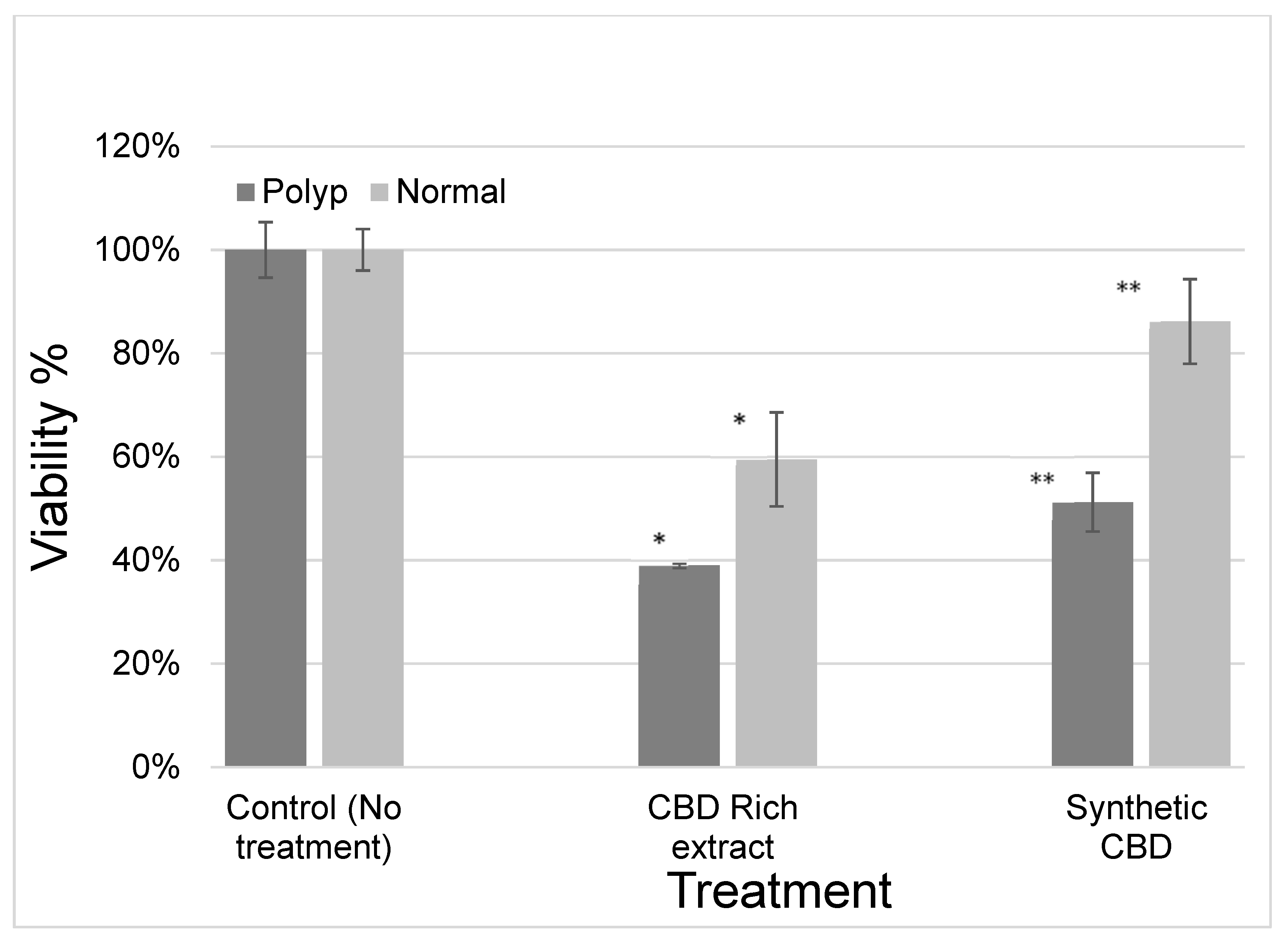
2.2. Evaluation of the Efficacy of Cannabinoids on the Viability of Cells Derived from Patient Colon Polyps and Adjacent Normal Cells

Polyp and adjacent normal colonic tissues were obtained following polyp resection. The differential toxic effect of the various cannabinoids extracts as well as of the isolated cannabinoids was examined. Most of the cannabinoids showed similar toxicity to polyp or adjacent colonic tissue. However, cannabidiol (CBD) exerted a differential toxic effect on polypoid cells and adjacent normal colonic cells as shown in Figure 1 (p < 0.03). CBD-rich extract had a similar effect as synthetic CBD.

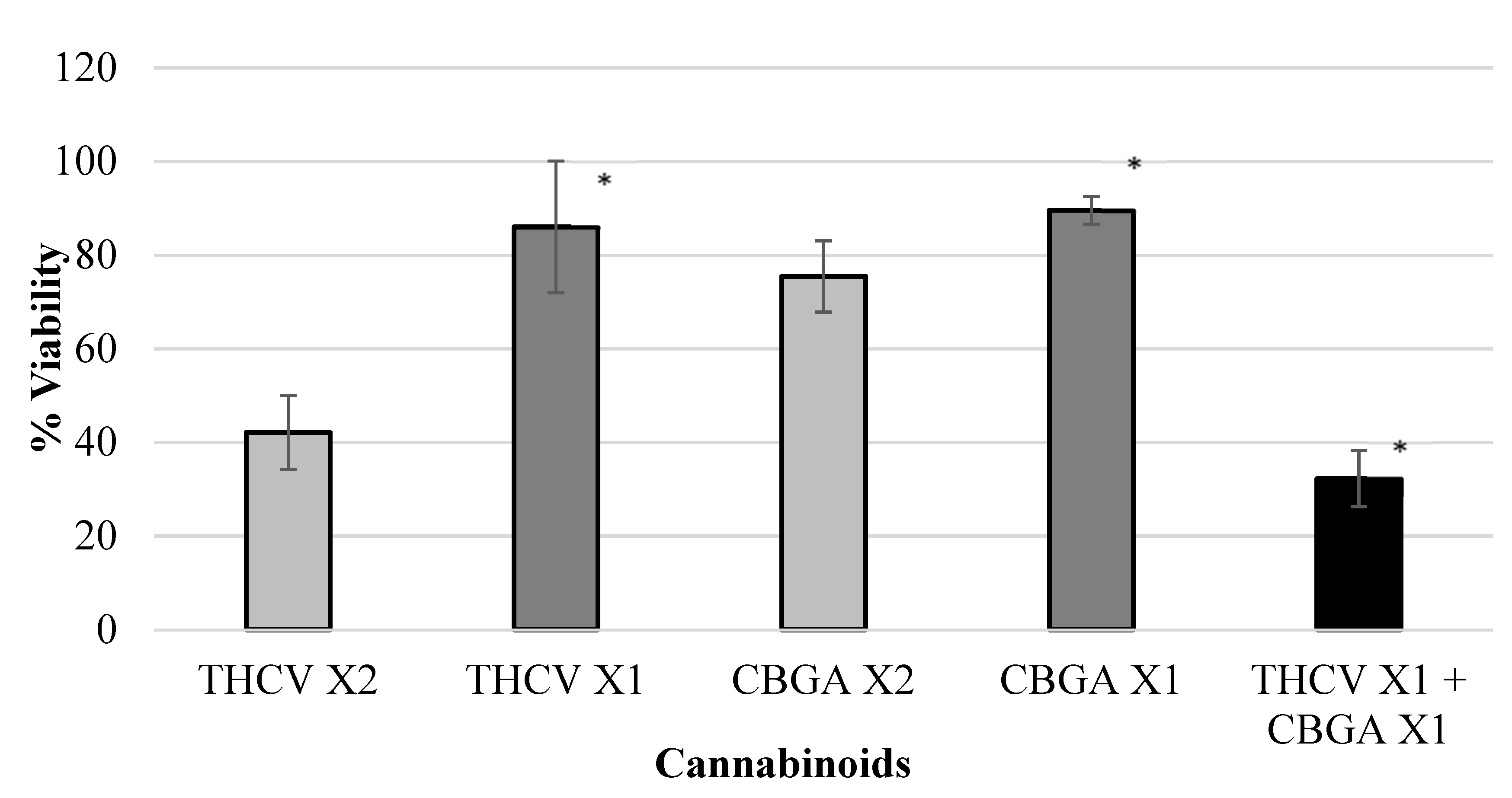
2.3. Evaluation of the Effect of Combinations of Cannabinoids on Colon Cancer Cell Viability

Various isolated cannabinoids were evaluated for synergistic interaction in inhibiting colon-polyp-derived cell viability. As shown in Table 3 and in Figure 2, Figure 3, Figure 4 and Figure 5, combinations between CBCA (at X2 concentration—29 µM or X1 concentration—14.5 µM), CBDV (at X2 concentration—47 µM or X1 concentration—23.5 µM), THCV (at X2 concentration—40/48 µM or X1 concentration—20/24 µM), and CBGA (at X2 concentration—51.2 µM or X1 concentration—25.6 µM), significantly inhibited the viability of the polyp-derived cells. The cannabinoid combinations CBCA + CBDV (p < 0.0001, Bliss equation calculation = 0.6), CBCA + THCV (p < 0.01, Bliss equation calculation = 0.48), CBDV + CBGA (p < 0.0004, Bliss equation calculation = 0.5), and THCV + CBGA (p < 0.0025, Bliss equation calculation = 0.45) presented a synergistic effect. However, the cannabinoid combinations THCV+CBDVA and THCV+ CBDV had no synergistic effect, as presented in Figure 6 and Figure 7 (p > 5, Bliss equation calculation = 0.01 and p > 5, Bliss equation calculation = −0.15, respectively). Notably, the cannabinoids CBCA and CBGA exhibited weak toxicity when administered as a single active compound (Table 1) and became significantly more toxic when combined with other cannabinoids. The nature of the interaction between isolated cannabinoids varied significantly when combined with different cannabinoids. For example, THCV presented a synergistic interaction when combined with CBGA (Figure 5), an additive interaction when combined with CBDVA (Figure 6), and an antagonist interaction when combined with CBDV (Figure 7).

3. Discussion

Previous studies have suggested that cannabinoids have cytotoxic activity in CRC [18,19,20,21]. Colorectal polyps are the primary premalignant precursors of CRC [4,5]. Therefore, to examine the possibility of the therapeutic or preventive potential of both cannabinoid extracts and isolated cannabinoids, their effect on biopsies of colorectal polyps and healthy tissue from patients scheduled for colonic polypectomy were analyzed. Biopsies of polyps and normal colon tissue of the same patient were exposed to a variety of cannabinoids in a range of concentrations.
The present study provides evidence of a cytotoxic activity of botanic cannabinoid extracts and synthetic cannabinoids on colorectal polyps. We were able to demonstrate that isolated cannabinoids presented different effects on the viability of cells derived from colorectal polyps. Some of the cannabinoids (mainly THC-d8 and THC-d9) were the most toxic and exhibited persistent toxicity in all polyps tested. Additionally, several of the cannabinoids exhibited a broad variability in their capacity to elicit toxicity when different polyps were tested.
In view of the differential effect of the various cannabinoids on different polyps, it may be necessary to initially test the effect of the extracts on polyps, prior to using them as therapeutic agents. Cells of a resected polyp could potentially be incubated with various cannabinoids to identify and select the agents that most effectively inhibit the viability of the lesion’s cells. Those toxic agents could be then prescribed for subsequent treatments by applying a pharmaceutical composition composed of one or more of the effective agents. Moreover, such personalized combinations, specifically those with differential activity against polyp-derived cells, could be administered for preventive purposes.
CBD was shown to have a differential cytotoxic effect in the presented study. It appeared to be more toxic to polypoid cells than to adjacent colonic cells at a concentration of 15 µM. Since nearly every carcinoma begins with a polyp [24], CBD could be a potential candidate as a chemopreventive agent to either prevent or suppress the progression of neoplastic polyps. The cytotoxic effect of CBD further suggests its therapeutic anticancer value. The differential effect of CBD could potentially allow local administration into the mucosa and/or submucosa layers during polypectomy to reduce the risk of recurrence without harming the adjacent normal colonic mucosa.
Combinations of the cannabinoids CBDV, THCV, CBDVA, CBCA, and CBGA exhibited a synergistic inhibitory effect on the viability of cells derived from colon polyps of patients. Previous studies have demonstrated a synergistic interaction of cytotoxic activity against colorectal polyps for several combinations [23]. Synergism between plant-produced compounds was previously suggested because in some cases, unrefined content of the flower extract with its different extracted compounds may have an advantage over the activity of an isolated compound [25,26,27]. It is well known that cancerous cells may develop resistance to anti-cancer drugs. Thus, utilizing combinations of cannabinoids may be of particular interest in the treatment of cancer in general, as well as in the inhibition of pre-cancerous lesions and avoidance of recurrence following endoscopic removal. Nevertheless, in vivo experiments of synergism would be necessary.
To the best of our knowledge, in vivo studies assessing the effect of local administration of cannabinoids to polyp resection beds have not been published to date. Two theoretical measures of safety, the use of non-psychotropic purified cannabinoids and synergism, should be provided for optimized general and local safety profiles. Moreover, the concentrations of cannabinoids used in the cell experiments (i.e., 50 µM) were orders of magnitude lower than those used in clinical trials (i.e., up to 3 mM/day), as shown in a recent publication [28]. Certainly, studies with both systemic and local administration of escalating doses of purified cannabinoid combinations will have to be conducted before moving forward to efficacy studies and human trials.
To conclude, our study results support the potential cytotoxic effect of cannabinoid extracts on colorectal polyps, as well as their synergistic and differential interactions. Further studies examining this postulation and the ultimate combination of cannabinoids for inhibiting/decreasing the recurrence rate of neoplastic polyps, and for preventing their malignant transformation into adenocarcinoma, are needed. Furthermore, the combined activity of selected cannabinoids with conventional chemotherapeutic agents, as well as biological selective agents, should be explored.

4. Materials and Methods

4.1. Polyp and Normal Colon Derived Cells

Cells were isolated from polyp specimens and from normal colon specimens. Biopsies from polyps and healthy colonic tissue from the same patient were obtained from twenty-two patients scheduled for polyp resection. Only patients with a large polyp that appeared benign endoscopically, was a granular lateral spreading tumor with 0-Is or 0-IIa morphology as per the Paris endoscopic classification [29], and was over 20 mm in size, which was found suitable for piecemeal endoscopic mucosal resection (EMR) by a gastroenterologist, were included. Exclusion criteria included: positive infectious serology (HAV, HBV, HCV, and HIV serologies were tested), polyp of less than 20 mm in size, malignant appearing polyp, polyp amendable for en bloc resection, a granular lateral spreading tumor with 0-IIb/0-IIc/0-III morphology as per the Paris endoscopic classification, a non-granular lateral spreading tumor pregnancy, breast-feeding, alcohol/drug abuse, minority, and impaired judgment. Overall, following final exclusion, twenty polyps from twenty patients were evaluated.
The study was conducted according to the guidelines of the Declaration of Helsinki and was approved by the Institutional Ethics Committee of Tel Aviv medical center (Helsinki approval no.TLV-0194-20). Informed Consent Statement: Informed consent was obtained from all subjects involved in the study.
Biopsies taken during each colonoscopy were placed in tissue culture media preservation media (MACS, Tissue storage solution, cat.130-100-008, Miltenyi Biotec, Bergisch Gladbach, Germany) and immediately transported to the laboratory. Dissociation of the biopsies’ tissue into single-cell suspensions for subsequent use was performed by using the MACS Technology by Miltenyi Biotec (Human Tumor Dissociation Kit cat. 130-095-929 and gentle MACS octo Dissociator) according to manufacturer’s instructions. After dissociation, cell viability was determined by using cell-permeable dye (Propidium Iodide, PI) and analyzed by flow cytometric analysis.

4.2. Polyp Derived Cells Viability and Counting

1 μL PI was diluted in 1 mL of 0.5%BSA/PBS buffer. A total of 90 μL diluted PI were added to 10 μL cells and was mixed well. Cell viability was analyzed by flow cytometric analysis (Attune NxT Flow Cytometer, Thermo Fisher Scientific, Waltham, MA, USA).

4.3. Extraction of Cannabis Inflorescence

Dry inflorescences of cannabis plants (CBD rich extract, RCK Ltd., Hof Ashkelon, Israel) were used in this experiment for the extraction of cannabis tinctures. The inflorescences were pulverized with liquid nitrogen and then extracted by press at 75 °C for 120 s. The decarboxylation of extracts was performed by heating for 2 h at 140 °C. The heated extracts contained mainly CBD (76%). The tincture was resuspended in dimethyl sulfoxide (DMSO) for a stock solution of 100 mM (control DMSO). For the treatments, the stock solution was diluted accordingly for cell cultures and biopsies in all experiments. Data regarding types of the CBD-rich extract as well as the percentage of cannabinoids as measured by HPLC chromatogram are presented in Table 4.

4.4. Isolated Cannabinoids

Isolated cannabinoids were purchased from RESTEK for analytical purposes as HPLC markers and for research. The cannabinoid purity was > 99%. The IC50, as measured in our laboratory on many cell lines, ranged from 15–100 µM. Based on this measurement, the starting concentrations for polyp experiments were determined. Later, these concentrations were adjusted to obtain the concentrations defined as 1X and 2X for each cannabinoid. Due to the very small number of cells obtained from one polyp, the ability to perform experiments with different concentrations was limited, as well as the variation from polyp to polyp. Therefore, an IC50 test was not performed for these cannabinoids on polyps; hence, we performed toxicity experiments at uniform cannabinoid concentrations and synergy experiments. The cannabinoids were dissolved in DMSO for a stock solution of 20 mM. The tested cannabinoid concentration did not exceed 60 µM and the DMSO concentration did not exceed 0.3%. This concentration of DMSO showed no toxic effect on the cells in these experiments. The cannabinoids and extracts were analyzed by HPLC (SHIMADZU, Nexera-i—LC-2040C Plus).

4.5. Cytotoxicity Assay

CTG (CellTiter-Glo Luminescent Cell Viability Assay, G7573, Promega) was used to check the cytotoxic effect of the cannabinoids and extracts. CTG was added to each well of the treatments and incubated at room temperature for 10 min. The luminescence was then recorded. The percentage of live cells was calculated relative to the nontreated control wells.

4.6. Cannabinoids Extracts and Compounds Cytotoxicity

To evaluate cannabinoids’ toxicity effects, about 4000–5000 polyp-derived cells were plated into each well in a 384-well plate. Cells were incubated for 48 h at 37 °C with various purified cannabinoids or extracts at different concentrations. The experiment sets were calibrated extensively on many cell lines and different types of cancerous and adjacent normal tissue. Time from 12–72 h was evaluated. The time point of 48 h of incubation was defined as the preferred time in terms of assay sensitivity and allowed the cells to perform normal behavior, such as cell division attempts, protein synthesis, etc. Cell viability (toxicity) was performed by CTG assay.

4.7. Cannabinoids Differential Effects

Polyp and adjacent normal colonic tissues were obtained following polyp resection. The differential toxic effect of the extracts on polyp cells as compared to normal colonic tissue cells was evaluated using an assessment of overall cell viability by CTG assay.

4.8. Cannabinoids Combined Effects (Synergy)

To determine whether the interaction of different cannabinoids was synergistic, that is, their combined activity was greater than the sum of their separate activities, the extent of activity in different combined concentrations of cannabinoids was examined. To examine synergy in cytotoxic activity between cannabinoids, a CTG assay was used on biopsy cells as described above. For the synergy experiments, cannabinoids were used at a concentration of 1× (viability above 85%) and 2×. For the synergic assay, we used 1× of A, 1× of B, and 1× of A + B. The synergistic effect was calculated according to cells’ survival experimental results according to the Bliss Equations (1) and (2) as follows:
Survival = S , SAB ( Cal ) = SA × SB
Synergy ( > 0 ) = SAB ( Cal ) SAB ( Exp )
S designates survival, wherein SAB (calculated) is a calculation of survival, e.g., of cells exposed to a combination of cannabinoid A and cannabinoid B, SA is the survival of cells exposed to cannabinoid A only, and SB is the survival of cells exposed to cannabinoid B only. SAB (Exp) designates the observed survival of cells exposed to the combination of cannabinoid A and cannabinoid B.

4.9. Statistical Analyses

Student’s t-test was used for p value assessment. Statistical analysis was performed on the results by performing Student’s t-Test (Excel Analysis ToolPak).

5. Patents

International Patent Application, publication No.: WO/2011/110866 discloses the use of phytocannabinoids, either in an isolated form or in the form of a botanical drug substance (BDS) in the treatment of cancer (https://patents.google.com/patent/WO2021245677A1/en, accessed on 5 June 2020).

Author Contributions

Conceptualization, D.B.-A.S.; Data curation, D.B.-A.S., I.H. and E.S.; Formal analysis, D.B.-A.S., I.H. and E.S.; Funding acquisition, E.S.; Investigation, D.B.-A.S., N.G. and E.S.; Methodology, D.B.-A.S., I.H. and E.S.; Project administration, D.B.-A.S. and E.S.; Supervision, D.B.-A.S.; Validation, D.B.-A.S. and E.S.; Visualization, D.B.-A.S. and E.S.; Writing—original draft, D.B.-A.S.; Writing—review & editing, D.B.-A.S., I.H., N.G., O.S. and E.S. All authors have read and agreed to the published version of the manuscript.

Funding

This research was partially funded by CNBX Pharmaceuticals Ltd. We received no external funding for article processing charges.

Institutional Review Board Statement

The study was conducted according to the guidelines of the Declaration of Helsinki and approved by the Institutional Ethics Committee of Tel Aviv medical center (Helsinki approval no.TLV-0194-20).

Informed Consent Statement

Informed consent was obtained from all subjects involved in the study.

Data Availability Statement

Not applicable.

Conflicts of Interest

I.H. is a member of the scientific research team of CNBX Pharmaceuticals Ltd. He was partially involved in the design, execution, interpretation, and writing of the study.

References

  1. Siegel, R.L.; Miller, K.D.; Jemal, A. Cancer statistics, 2018. CA Cancer J. Clin. 2018, 68, 7–30. [Google Scholar] [CrossRef] [PubMed]
  2. Edwards, B.K.; Ward, E.; Kohler, B.A.; Eheman, C.; Zauber, A.G.; Anderson, R.N.; Jemal, A.; Schymura, M.J.; Lansdorp-Vogelaar, I.; Seeff, L.C.; et al. Annual report to the nation on the status of cancer, 1975–2006, featuring colorectal cancer trends and impact of interventions (risk factors, screening, and treatment) to reduce future rates. Cancer 2010, 116, 544–573. [Google Scholar] [CrossRef] [PubMed]
  3. Linnekamp, J.F.; Wang, X.; Medema, J.P.; Vermeulen, L. Colorectal cancer heterogeneity and targeted therapy: A case for molecular disease subtypes. Cancer Res. 2015, 75, 245–249. [Google Scholar] [CrossRef] [PubMed]
  4. Markowitz, S.D.; Bertagnolli, M.M. Molecular origins of cancer: Molecular basis of colorectal cancer. N. Engl. J. Med. 2009, 361, 2449–2460. [Google Scholar] [CrossRef]
  5. Zauber, A.G.; Winawer, S.J.; O’Brien, M.J.; Lansdorp-Vogelaar, I.; van Ballegooijen, M.; Hankey, B.F.; Shi, W.; Bond, J.H.; Schapiro, M.; Panish, J.F.; et al. Colonoscopic polypectomy and long-term prevention of colorectal-cancer deaths. N. Engl. J. Med. 2012, 366, 687–696. [Google Scholar] [CrossRef]
  6. Rotondano, G.; Bianco, M.A.; Buffoli, F.; Gizzi, G.; Tessari, F.; Cipolletta, L. The Cooperative Italian FLIN Study Group: Prevalence and clinico-pathological features of colorectal laterally spreading tumors. Endoscopy 2011, 43, 856–861. [Google Scholar] [CrossRef]
  7. Klein, A.; Bourke, M.J. Advanced polypectomy and resection techniques. Gastrointest. Endosc. Clin. 2015, 25, 303–333. [Google Scholar] [CrossRef]
  8. Christie, J.P. Colonoscopic excision of large sessile polyps. Am. J. Gastroenterol. 1977, 67, 430–438. [Google Scholar]
  9. Moss, A.; Williams, S.J.; Hourigan, L.F.; Brown, G.; Tam, W.; Singh, R.; Zanati, S.; Burgess, N.G.; Sonson, R.; Byth, K.; et al. Long-term adenoma recurrence following wide-field endoscopic mucosal resection (WF-EMR) for advanced colonic mucosal neoplasia is infrequent: Results and risk factors in 1000 cases from the Australian Colonic EMR (ACE) study. Gut 2015, 64, 57–65. [Google Scholar] [CrossRef]
  10. Moss, A.; Bourke, M.J.; Williams, S.J.; Hourigan, L.F.; Brown, G.; Tam, W.; Singh, R.; Zanati, S.; Chen, R.Y.; Byth, K. Endosc.opic mucosal resection outcomes and prediction of submucosal cancer from advanced colonic mucosal neoplasia. Gastroenterology 2011, 140, 1909–1918. [Google Scholar] [CrossRef]
  11. Klein, A.; Tate, D.J.; Jayasekeran, V.; Hourigan, L.; Singh, R.; Brown, G.; Bahin, F.F.; Burgess, N.; Williams, S.J.; Lee, E.; et al. Thermal Ablation of Mucosal Defect Margins Reduces Adenoma Recurrence After Colonic Endoscopic Mucosal Resection. Gastroenterology 2019, 156, 604–613.e603. [Google Scholar] [CrossRef] [PubMed] [Green Version]
  12. Baron, J.A.; Cole, B.F.; Sandler, R.S.; Haile, R.W.; Ahnen, D.; Bresalier, R.; McKeown-Eyssen, G.; Summers, R.W.; Rothstein, R.; Burke, C.A.; et al. A randomized trial of aspirin to prevent colorectal adenomas. N. Engl. J. Med. 2003, 348, 891–899. [Google Scholar] [CrossRef] [PubMed]
  13. Baron, J.A.; Barry, E.L.; Mott, L.A.; Rees, J.R.; Sandler, R.S.; Snover, D.C.; Bostick, R.M.; Ivanova, A.; Cole, B.F.; Ahnen, D.J.; et al. A Trial of Calcium and Vitamin D for the Prevention of Colorectal Adenomas. N. Engl. J. Med. 2015, 373, 1519–1530. [Google Scholar] [CrossRef]
  14. Higurashi, T.; Hosono, K.; Takahashi, H.; Komiya, Y.; Umezawa, S.; Sakai, E.; Uchiyama, T.; Taniguchi, L.; Hata, Y.; Uchiyama, S.; et al. Metformin for chemoprevention of metachronous colorectal adenoma or polyps in post-polypectomy patients without diabetes: A multicentre double-blind, placebo-controlled, randomised phase 3 trial. Lancet Oncol. 2016, 17, 475–483. [Google Scholar] [CrossRef]
  15. Radwan, M.M.; ElSohly, M.A.; El-Alfy, A.T.; Ahmed, S.A.; Slade, D.; Husni, A.S.; Manly, S.P.; Wilson, L.; Seale, S.; Cutler, S.J.; et al. Isolation and Pharmacol.ogical Evaluation of Minor Cannabinoids from High-Potency Cannabis sativa. J. Nat. Prod. 2015, 78, 1271–1276. [Google Scholar] [CrossRef] [PubMed]
  16. Howlett, A.C.; Abood, M.E. CB1 and CB2 Receptor Pharmacology. Adv. Pharmacol. 2017, 80, 169–206. [Google Scholar] [CrossRef]
  17. Javid, F.A.; Phillips, R.M.; Afshinjavid, S.; Verde, R.; Ligresti, A. Cannabinoid pharmacology in cancer research: A new hope for cancer patients? Eur. J. Pharmacol. 2016, 775, 1–14. [Google Scholar] [CrossRef]
  18. Greenhough, A.; Patsos, H.A.; Williams, A.C.; Paraskeva, C. The cannabinoid delta(9)-tetrahydrocannabinol inhibits RAS-MAPK and PI3K-AKT survival signalling and induces BAD-mediated apoptosis in colorectal cancer cells. Int. J. Cancer 2007, 121, 2172–2180. [Google Scholar] [CrossRef]
  19. Aviello, G.; Romano, B.; Borrelli, F.; Capasso, R.; Gallo, L.; Piscitelli, F.; Di Marzo, V.; Izzo, A.A. Chemopreventive effect of the non-psychotropic phytocannabinoid cannabidiol on experimental colon cancer. J. Mol. Med. 2012, 90, 925–934. [Google Scholar] [CrossRef]
  20. Romano, B.; Borrelli, F.; Pagano, E.; Cascio, M.G.; Pertwee, R.G.; Izzo, A.A. Inhibition of colon carcinogenesis by a standardized Cannabis sativa extract with high content of cannabidiol. Phytomedicine 2014, 21, 631–639. [Google Scholar] [CrossRef]
  21. Borrelli, F.; Pagano, E.; Romano, B.; Panzera, S.; Maiello, F.; Coppola, D.; De Petrocellis, L.; Buono, L.; Orlando, P.; Izzo, A.A. Colon carcinogenesis is inhibited by the TRPM8 antagonist cannabigerol, a Cannabis-derived non-psychotropic cannabinoid. Carcinogenesis 2014, 35, 2787–2797. [Google Scholar] [CrossRef] [PubMed] [Green Version]
  22. Pertwee, R.G. The diverse CB1 and CB2 receptor pharmacology of three plant cannabinoids: Delta9-tetrahydrocannabinol, cannabidiol and delta9-tetrahydrocannabivarin. Br. J. Pharmacol. 2008, 153, 199–215. [Google Scholar] [CrossRef] [PubMed]
  23. Nallathambi, R.; Mazuz, M.; Namdar, D.; Shik, M.; Namintzer, D.; Vinayaka, A.C.; Ion, A.; Faigenboim, A.; Nasser, A.; Laish, I.; et al. Identification of Synergistic Interaction Between Cannabis-Derived Compounds for Cytotoxic Activity in Colorectal Cancer Cell Lines and Colon Polyps That Induces Apoptosis-Related Cell Death and Distinct Gene Expression. Cannabis Cannabinoid. Res. 2018, 3, 120–135. [Google Scholar] [CrossRef] [PubMed]
  24. Winawer, S.; Fletcher, R.; Rex, D.; Bond, J.; Burt, R.; Ferrucci, J.; Ganiats, T.; Levin, T.; Woolf, S.; Johnson, D.; et al. Colorectal cancer screening and surveillance: Clinical guidelines and rationale-Update based on new evidence. Gastroenterology 2003, 124, 544–560. [Google Scholar] [CrossRef]
  25. Ben-Shabat, S.; Fride, E.; Sheskin, T.; Tamiri, T.; Rhee, M.H.; Vogel, Z.; Bisogno, T.; De Petrocellis, L.; Di Marzo, V.; Mechoulam, R. An entourage effect: Inactive endogenous fatty acid glycerol esters enhance 2-arachidonoyl-glycerol cannabinoid activity. Eur. J. Pharmacol. 1998, 353, 23–31. [Google Scholar] [CrossRef]
  26. Russo, E.B. Taming THC: Potential cannabis synergy and phytocannabinoid-terpenoid entourage effects. Br. J. Pharmacol. 2011, 163, 1344–1364. [Google Scholar] [CrossRef]
  27. Blasco-Benito, S.; Seijo-Vila, M.; Caro-Villalobos, M.; Tundidor, I.; Andradas, C.; Garcia-Taboada, E.; Wade, J.; Smith, S.; Guzman, M.; Perez-Gomez, E.; et al. Appraising the “entourage effect”: Antitumor action of a pure cannabinoid versus a botanical drug preparation in preclinical models of breast cancer. Biochem. Pharmacol. 2018, 157, 285–293. [Google Scholar] [CrossRef]
  28. Larsen, C.; Shahinas, J. Dosage, Efficacy and Safety of Cannabidiol Administration in Adults: A Systematic Review of Human Trials. J. Clin. Med. Res. 2020, 12, 129–141. [Google Scholar] [CrossRef]
  29. The Paris endoscopic classification of superficial neoplastic lesions: Esophagus, stomach, and colon: November 30 to December 1, 2002. Gastrointest. Endosc. 2003, 58, S3–S43. [CrossRef]
Figure 1. Influence of cannabidiol (CBD) and CBD-rich extract on the viability of polypoid cells and adjacent normal colonic cells. CBD-rich extract had a similar effect as synthetic CBD. * p < 0.03, ** p < 0.02.
Ijms 23 11366 g001
Figure 2. Viability of polyp cells exposed to CBDV and CBCA. Combinations between CBCA (at X2 concentration—29 µM or X1 concentration—14.5 µM) and CBDV (at X2 concentration—47 µM or X1 concentration—23.5 µM) significantly inhibited the viability of polyp-derived cells. The cannabinoid combination CBCA + CBDV presented a synergistic effect (* p < 0.0001, Bliss equation calculation = 0.6). Legend: cannabichromene acid (CBCA), cannabidivarin (CBDV).
Ijms 23 11366 g002
Figure 3. Viability of polyp cells exposed to CBCA and THCV. Combinations between CBCA (at X2 concentration—29 µM or X1 concentration—14.5 µM) and THCV (at X2 concentration—40/48 µM or X1 concentration—20/24 µM) significantly inhibited the viability of polyp-derived cells. The cannabinoid combination CBCA + THCV presented a synergistic effect (* p < 0.01, Bliss equation calculation = 0.48). Legend: cannabichromene acid (CBCA), tetrahydrocannabivarin (THCV).
Ijms 23 11366 g003
Figure 4. Viability of polyp cells exposed to CBDV and CBGA. Combinations between CBDV (at X2 concentration—47 µM or X1 concentration—23.5 µM) and CBGA (at X2 concentration—51.2 µM or X1 concentration—25.6 µM) significantly inhibited the viability of polyp-derived cells. The cannabinoid combination CBDV + CBGA presented a synergistic effect (* p < 0.0004, Bliss equation calculation = 0.5). Legend: cannabidivarin (CBDV), cannabigerolic acid (CBGA).
Ijms 23 11366 g004
Figure 5. Viability of polyp cells exposed to THCV and CBGA. Combinations between THCV (at X2 concentration—40/48 µM or X1 concentration—20/24 µM) and CBGA (at X2 concentration—51.2 µM or X1 concentration—25.6 µM) significantly inhibited the viability of polyp-derived cells. The cannabinoid combination THCV + CBGA (* p < 0.0025, Bliss equation calculation = 0.45) presented a synergistic effect. Legend: tetrahydrocannabivarin (THCV), cannabigerolic acid (CBGA).
Ijms 23 11366 g005
Figure 6. Viability of polyp cells exposed to THCV and CBDVA. The cannabinoid combination THCV + CBDVA showed no synergistic effect (* p > 5, Bliss equation calculation = 0.01). Legend: tetrahydrocannabivarin (THCV), cannabidivarinic acid (CBDVA).
Ijms 23 11366 g006
Figure 7. Viability of polyp cells exposed to THCV and CBDV. The cannabinoid combination THCV + CBDV had no synergistic effect (* p > 5, Bliss equation calculation = −0.15). Legend: tetrahydrocannabivarin (THCV), cannabidivarin (CBDV).
Ijms 23 11366 g007
Table 1. Patient and polyp characteristics.
Patient No.Sex (F = Female, M = Male)AgeHistologyPolyp SitePolyp Size
1F57VA w LGDCecum25 mm
2M69TVA w LGDCecum20 mm
3M77TVA w HGDDescending
colon
40 mm
4F52TSASigmoid colon70 mm
5F50VA w LGDSigmoid colon50 mm
6M62TVA w LGDAscending
colon
30 mm
7F66SSA w LGDTransverse
colon
25 mm
8F64TVA w LGDAscending
colon
30 mm
9M60TVA w LGDAscending
colon
20 mm
10M62TVA w LGDAscending
colon
40 mm
11 *M80TVA w LGDCecum20 mm
12F55TVA w HGDAscending
colon
30 mm
13F50SSA no dysplasiaAscending
colon
20 mm
14F74SSA no dysplasiaAscending
colon
20 mm
15 *F73VA w LGDAscending
colon
40 mm
16M63TA w HGDCecum50 mm
17F71TA w LGD + SSASplenic
flexure
25 mm
18M60TA w LGDAscending
colon
25 mm
19M80VA w LGDHepatic
flexure
30 mm
20F66VA w LGDCecum70 mm
21M51TVA w LGDRectum30 mm
22M80TA w LGDTransverse
colon
30 mm
* Patient polyp numbers 11 and 15 were excluded from study analysis due to borderline-positive hepatitis A virus (HAV) serology. Legend: tubular adenoma (TA), tubulovillous adenoma (TVA), villous adenoma (VA), traditional serrated adenoma (TSA), sessile serrated adenoma (SSA), low grade dysplasia (LGD), high grade dysplasia (HGD).
Table 2. Toxicity of cannabinoids on colon-polyp-derived cells.
CannabinoidP004P005P006P008P009P010Average Viability
THC-d810%23%44%4%7%5%15%
THC-d920%20%47%13%21%8%21%
CBC21%21%61%15%22%11%25%
CBN24%32%54%18%16%13%26%
CBDV37%63%62%23%39%9%39%
CBD21%41%54%35%62%24%40%
THCV59%74%70%47%40%18%51%
CBL73%55%89%55%63%29%61%
CBG75%69%58%69%65%37%62%
THCA59%91%99%52%60%32%66%
CBDA104%97%66%38%73%26%67%
CBDVA72%90%83%48%69%56%70%
CBNA66%94%76%83%70%60%75%
CBCA63%86%105%91%72%80%83%
CBGA71%96%84%116%70%97%89%
Toxicity of cannabinoids on colon-polyp-derived cells (P004–P010 designate the various patients’ polyps), shown as percentage of survival in increasing order. Average survival is shown on the rightmost column. Legend: tetrahydrocannabinol (THC), cannabichromene (CBC), cannabinol (CBN), cannabidivarin (CBDV), cannabidiol (CBD), tetrahydrocannabivarin (THCV), cannabicyclol (CBL), cannabigerol (CBG), tetrahydrocannabinolic acid (THCA), cannabidiolic acid (CBDA), cannabidivarinic acid (CBDVA), cannabinolic acid (CBNA), cannabichromene acid (CBCA), cannabigerolic acid (CBGA).
Table 3. The effect of combinations of cannabinoids on polyp cells’ viability.
Cannab. ACannab. BCells
Viability at ×2 Conc. of Cannab. A
Cells Viability at ×1 Conc. of Cannab. ACells
Viability at ×2 Conc. of Cannab. B
Cells Viability at ×1 Conc. of Cannab. BCells Viability at ×1 Conc. of Cannab. A + Cannab. B
CBCACBDV67101549839
CBDVCBDVA5498799439
CBDVCBGA5498759038
CBCATHCV67101428639
CBCACBGA67101759061
CBGATHCV7590428632
CBCACBDVA67101799479
CBDVACBGA7994759061
CBDVATHCV7994428672
The cannabinoid combinations: CBCA + CBDV, CBCA + THCV, CBDV + CBGA, and CBGA + THCV presented synergistic effects. Legend: cannabichromene acid (CBCA), cannabidivarin (CBDV), cannabigerolic acid (CBGA), cannabid varinic acid (CBDVA).
Table 4. Cannabinoid percentage in CBD-rich extract tincture.
CBDVTHCVCBDCBGCBDACBGACBNTHC-d9THC-d8CBCTHCACBDVACBCA
0.33.176.01.60.10.10.12.90.05.60.00.05.9
Types and percentage of cannabinoids as measured by HPLC chromatogram. Legend: cannabidivarin (CBDV), tetrahydrocannabivarin (THCV), cannabidiol (CBD), cannabigerol (CBG), cannabidiolic acid (CBDA), cannabigerolic acid (CBGA), cannabinol (CBN), tetrahydrocannabinol (THC), cannabichromene (CBC), tetrahydrocannabinolic acid (THCA), cannabidivarinic acid (CBDVA), cannabichromene acid (CBCA).
Publisher’s Note: MDPI stays neutral with regard to jurisdictional claims in published maps and institutional affiliations.

