Review

Role of Mesenchymal Stem Cells and Their Paracrine Mediators in Macrophage Polarization: An Approach to Reduce Inflammation in Osteoarthritis

1
Department of Biomedical Sciences, Chonnam National University Medical School, Hwasun 58128, Korea
2
Department of Orthopaedics Surgery, Center for Joint Disease of Chonnam National University Hwasun Hospital, 322 Seoyang-ro, Hwasun-eup 519-763, Korea
3
Korea Biomedical Materials and Devices Innovation Research Center, Chonnam National University Hospital, 42 Jebong-ro, Dong-gu, Gwangju 501-757, Korea
*
Author to whom correspondence should be addressed.
Int. J. Mol. Sci. 2022, 23(21), 13016; https://0-doi-org.brum.beds.ac.uk/10.3390/ijms232113016
Submission received: 7 October 2022 / Revised: 21 October 2022 / Accepted: 25 October 2022 / Published: 27 October 2022
(This article belongs to the Special Issue Musculoskeletal Disorders (MSDs) and Osteoimmunology)

Abstract

:
Osteoarthritis (OA) is a low-grade inflammatory disorder of the joints that causes deterioration of the cartilage, bone remodeling, formation of osteophytes, meniscal damage, and synovial inflammation (synovitis). The synovium is the primary site of inflammation in OA and is frequently characterized by hyperplasia of the synovial lining and infiltration of inflammatory cells, primarily macrophages. Macrophages play a crucial role in the early inflammatory response through the production of several inflammatory cytokines, chemokines, growth factors, and proteinases. These pro-inflammatory mediators are activators of numerous signaling pathways that trigger other cytokines to further recruit more macrophages to the joint, ultimately leading to pain and disease progression. Very few therapeutic alternatives are available for treating inflammation in OA due to the condition’s low self-healing capacity and the lack of clear diagnostic biomarkers. In this review, we opted to explore the immunomodulatory properties of mesenchymal stem cells (MSCs) and their paracrine mediators-dependent as a therapeutic intervention for OA, with a primary focus on the practicality of polarizing macrophages as suppression of M1 macrophages and enhancement of M2 macrophages can significantly reduce OA symptoms.

1. Introduction

Osteoarthritis (OA) is the most common chronic musculoskeletal condition that affects weight-bearing joints, such as the spine, hips, knees, and hands [1]. OA affects approximately 300 million people globally [2] and is ranked sixth in Asia and eleventh worldwide in terms of years lived with disability (YLD) [3]. Stiffness, decreased range of motion, joint instability, edema, muscle weakness, weariness, and pain-related psychological distress are the most typical symptoms of OA. The risk factors involved in OA interact in a complex manner; they can be divided into individual-level risk factors (age, gender, obesity, heredity, and diet) [4] and joint-level risk factors (injuries, misalignments, and incorrect joint loading) [5]. OA has significant economic effects due to the growing frequency of joint replacements, increasing medical costs, and an aging population [6]. As the population ages, OA is expected to become the primary cause of disability in elderly people by 2030 [7].
OA was initially thought to be caused by wear and tear of the joint’s cartilage and bony understructure due to constant mechanical stress. However, the pathogenesis of OA is remarkably more complicated. Instead of a simple wear-and-tear condition, OA is now recognized as a complex ailment in which inflammation plays a significant role in joint deterioration [8]. The synovium is the principal site of inflammation in OA. Macrophages have a major role in the inflammation of the synovium in OA. Synovial macrophages promote the progression of OA by initiating an inflammatory response through the production of pro-inflammatory cytokines and mediators that control immune system activity and encourage the secretion of additional pro-inflammatory mediators by cartilage and synovial cells [9]. Numerous researchers have demonstrated that inhibiting inflammation either by reprogramming macrophages or depleting them has proven to be effective [10]. The primary reason for targeting macrophages is their remarkable plasticity and capacity to fulfill several biological roles in response to signals from the tissue microenvironment. Numerous strategies have been developed to target these properties; however, no macrophage-specific treatment is currently available on the market for any condition.
Mesenchymal stem cells (MSCs) play an essential role in the polarization of macrophages [11]. There has been a significant shift in perspective on the role of MSCs in recent years. MSCs were first thought to be therapeutically useful because of their potential to differentiate into several other cell types that might potentially replace cells in damaged or diseased tissues [12]. It is now well accepted that MSCs’ paracrine secretion is responsible for the vast majority of their therapeutic effects. The paracrine secretions consist of soluble proteins, free nucleic acids, lipids, and extracellular vesicles (EVs) collectively known as the secretome. The secretome has been shown to promote remodeling of the extracellular matrix (ECM), manage local inflammation, and enhance macrophage polarization by decreasing the release of pro-inflammatory factors and increasing anti-inflammatory factors [13,14,15].
In this article, we review the current understanding of the role of macrophages in OA inflammation and the existing techniques to polarize macrophages. We will also explore the recent developments in the use of MSC secretome to polarize macrophages in diverse inflammatory models that will help in facilitating the translation of future research for the treatment of inflammation in OA.

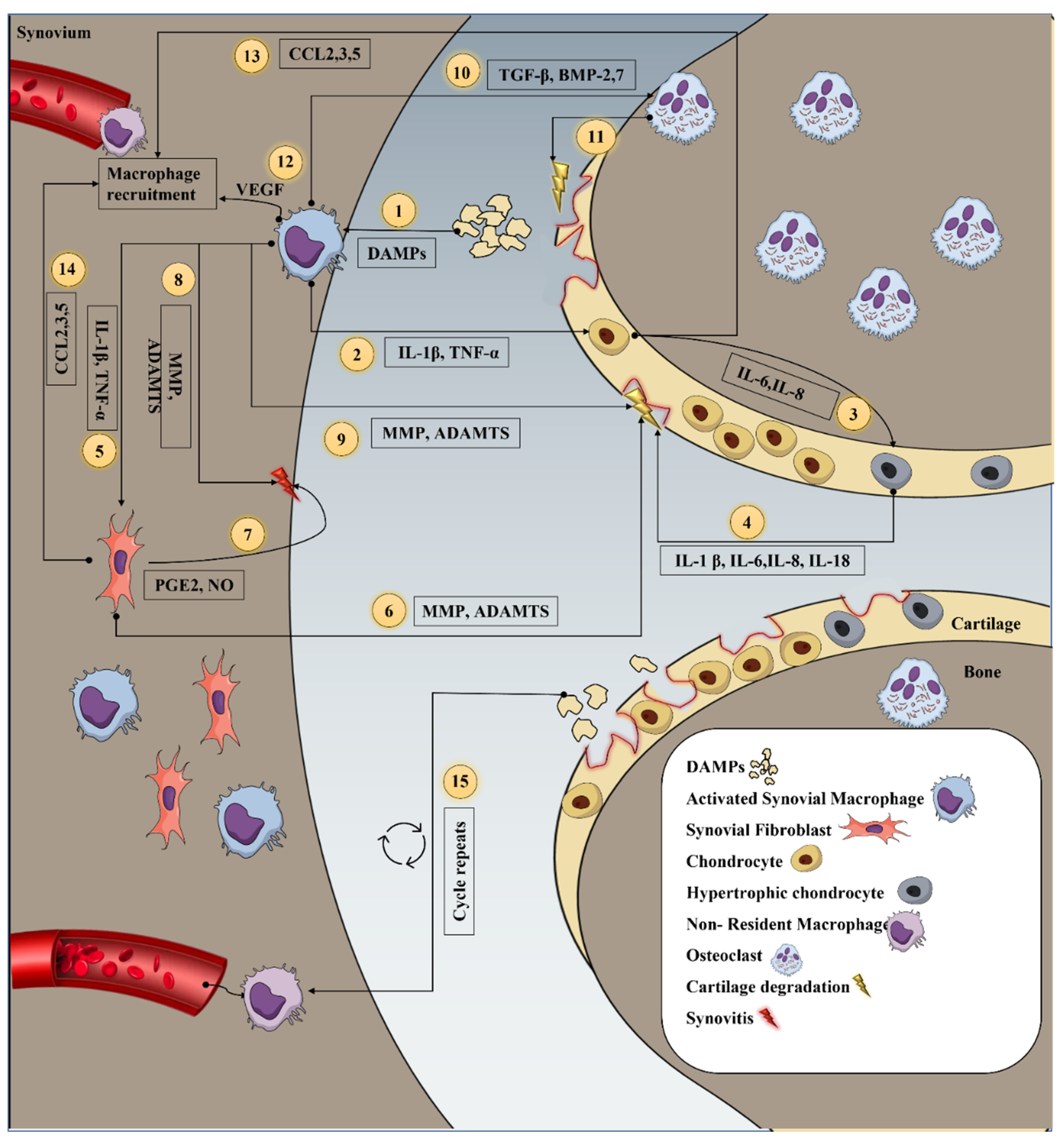
2. Inflammation in Osteoarthritis

OA has always been an uncertain condition in terms of inflammation even though the name itself denotes that it is an inflammatory process. Previously it was believed that OA was caused due to biomechanical causes and was employed as a negative control for inflammation during comparisons to rheumatoid arthritis (RA) [16]. It has now been brought to light that OA is much more than just an injury caused due to overuse of the joint. OA is a complex biological response as a result of its interaction with tissue resident cells and their mediators which amplifies physical stress incapacitating the normal function of ligaments, muscles and menisci [17,18]. Research has identified the process of inflammation as the initial step along the negative chain of events that leads to early OA.
Although the inflammatory response in OA is not as pronounced as RA, several authors have confirmed there is low-grade inflammation in OA [19]. The presence of inflammation in OA has been studied extensively using various techniques, in the early 1980′s Goldenberg et al. exhibited that majority of the inflammation is present in the synovium of the OA patients through histopathological analysis [20]. Additionally, the histological evidence was validated when correlated to the levels of serum C-reactive protein (CRP) and the levels of the inflammatory marker interleukin-6 (IL-6) in the synovial fluid in patients undergoing total hip or knee arthroplasty [21]. A comparative study between synovial tissues of patients with early and late OA revealed increased infiltration of mononuclear cells and inflammatory cytokines in patients with early knee OA to late knee OA [22]. Later, due to sensitive imaging techniques like magnetic resonance imaging (MRI) and direct arthroscopic visualization, it was confirmed that inflammation is visible in the synovium at the early stages of OA even before there is visible articular cartilage damage [23]. A recent study revealed that synovitis is one of the key factors in identifying early OA which was confirmed through the analysis of serum matrix metalloproteinase-3 (MMP-3) concentration, effusion-synovitis volume and synovial score [24]. All these studies validate the significant role of synovitis at any (early or late) stage of OA.
In the past decade, researchers have also been investigating the connection between low-grade synovitis and the manifestation of OA’s clinical symptoms. Synovitis has been linked to more severe symptoms including pain and joint dysfunction and may generate a more rapid deterioration of cartilage [4]. Synovitis has been linked to symptoms such as discomfort in people with knee OA. The correlation between pain and synovitis on MRI, found that changes in pain levels over time corresponded with changes in synovitis, lending credence to the idea that the two are causally connected [25]. Recently, a similar relationship between pain and synovitis was described using contrast-enhanced MRI wherein the likelihood of experiencing painful knee OA was found to rise 9-fold with increasing synovitis severity [26]. Ayral et al., published a study that established a link between synovitis and the progression of cartilage degeneration. The presence or absence of synovitis and the overall health of the cartilage surfaces were easily discerned during the initial arthroscopy. The rate of cartilage degeneration was measured by an arthroscopic examination performed 12 months after the original surgery. The presence of synovitis was related with more severe chondropathy at baseline and was present in around 50% of patients. Moreover, at one year, patients with synovitis were more likely to have advanced cartilage pathology than those without the inflammation [27].
This suggests that synovitis is associated with pain and cartilage erosive mechanisms, making it a possible target for disease- and symptom-modifying therapy. The inactivation of critical inflammatory pathways either by removing macrophages or reprogramming them can reduce joint structural pathology, including cartilage degradation. Although inflammation does not pertain to synovial tissue alone, synovium is the major site of gross and microscopic inflammatory change [28], and targeting inflammation can further reduce the vicious cycle of the disease, thus making it the major focus of this review.

3. Inflammatory Mediators Secreted by Macrophage and Its Interaction with Resident Cells during OA

During OA, the macrophages fail to keep up their stability and are activated through various ways mainly when macrophages are stimulated by damage associated molecular patterns (DAMPs) or pathogen-associated molecular patterns (PAMPs) upon interaction with germline-encoded surface pattern recognition receptors (PRRs) on macrophages; they activate the nuclear factor kappa-light-chain-enhancer of activated B cells (NF-κB) pathway, causing the cells to release an increased amount of inflammatory mediators [29]. Another key signaling channel is the NOD-like receptor family, pyrin domain-containing 3 (NLRP3) inflammasome-mediated pathway. Both pathways can activate the macrophages during OA and trigger the production of two of the most extensively studied pro-inflammatory cytokines, interleukin-1β (IL-1β) and tumor necrosis factor (TNF-α). The cascade of mediators activated by the pro-inflammatory cytokines is shown in Figure 1.
IL-1β released by macrophages stimulates chondrocytes to synthesize MMPs, especially collagenase-1 (MMP-1), stromelysin (MMP-3), collagenase-3 (MMP-13), and ADAMTS-4 and 5, which are known to cause cartilage degradation and synovial damage [30,31]. IL-1β increases the production of other cytokines, such as IL-6, and IL-8 and chemokines, such as CCL2 (monocyte chemoattractant protein-1, MCP-1), macrophage inflammatory protein-1 alpha (MIP-1α/CCL3), and C–C motif chemokine ligand 5 (CCL5) in chondrocytes via a paracrine mechanism [32]. These mediators attract new macrophages to the joint, where they continue to release IL-1β, thereby prolonging the inflammatory cycle [33]. Additionally, IL-1β promotes the release of a variety of pro-inflammatory mediators, such as prostaglandin E2 (PGE2), nitric oxide (NO), and cyclooxygenase-2 (COX-2), which stimulate the extracellular signal-regulated kinases (ERKs) pathway. Activation of the ERK pathway inhibits type II collagen and aggrecan formation, as well as ECM synthesis [34]. Likewise, the c-Jun N-terminal kinases (JNKs) pathway also inhibits collagen II synthesis by inhibiting the SOX-9 gene. The mitogen-activated protein kinase (MAPK) signaling cascades regulates MMP-1, MMP-13, and ADAMTS-4, whereas MMP-3 and ADAMTS-5 are exclusively regulated by the ERK and JNK pathways, respectively [35].
Similarly, TNF-α also exhibits comparable effects on chondrocytes, increases IL-6, IL-8 and IL-18, suppresses the production of proteoglycans and type II collagen, and stimulates chondrocytes to generate MMPs and ADAMTS for ECM degradation [36,37]. Alternate to secreting inflammatory mediators, activated macrophages also produce growth factors. Vascular endothelial growth factor (VEGF) contributes to the severity and inflammation of OA. The articular cartilage, serum, and synovium of patients with late-stage OA show elevated VEGF expression. An increase in angiogenesis and VEGF production is the root cause of synovitis. VEGF has been shown to increase symptoms in patients with OA by stimulating inflammation-promoting macrophages to migrate throughout the inflamed tissue while also delivering nutrition and oxygen [38]. Other growth factors, including bone morphogenetic protein-2 (BMP-2), bone morphogenetic protein-7 (BMP-7), and transforming growth factor beta (TGF-β), contribute to the production of osteophytes and synovial fibrosis. Apart from osteophyte production, macrophages induce the formation of osteoclasts, which disintegrate the underlying bone and further degrade the cartilage and bone beneath by prompting osteoblasts to release a variety of cytokines and MMPs [39]. In addition to chondrocytes, activated macrophages stimulate neighboring fibroblast like synoviocyte (FLS) to produce a variety of inflammatory cytokines and chemokines, as well as pro-inflammatory mediators resulting in synovial hyperplasia, joint swelling, inflammation, and pain. FLS secrete a clear, viscid, lubricating fluid known as synovial fluid [40]. Macrophages also impair the primary role of FLS of maintaining cartilage homeostasis and shielding the cartilage surface from friction and deterioration [41]. The role of macrophages in inflammation is owed to their secretion of inflammatory cytokines. The majority of the cells within the joint interact with the cytokines released by macrophages (as shown in Figure 1), and these interactions influence the production of cytokines, other inflammatory mediators, and enzymes by these cells via intracellular signal transduction pathways, which plays a crucial role in the pathogenesis of OA.

4. Depletion of Macrophage

Macrophages’ role in inflammation emphasizes that they have a critical role in OA immunopathogenesis and are not just a consequence of it. Therefore, depletion of macrophages can be a potential intervention that promote tissue repair and remodeling. Blom et al. demonstrated that removal of macrophages from the synovial lining decreased the expression of MMP-3 and MMP-9 in the synovium but not in the cartilage, and also reduced osteophyte formation in the collagenase induced OA (CIOA) mouse model [42]. Bondeson et al., showed that depleting synovial macrophages with anti-CD14-conjugated magnetic beads reduced inflammatory cytokines [43]. Correspondingly, the pro-inflammatory cytokines produced by FLS cease to secrete these cytokines and halts ECM degradation [44]. Wu et al. sought to determine whether the same is true for obesity-related OA and discovered that short-term macrophage depletion elevated synovitis and T-cell and neutrophil infiltration into the operated joint. These researchers concluded that macrophages are essential regulators of the responses of other immune cells and macrophage depletion cannot be employed to reduce inflammation in obese arthritic patients [45]. However, these results oppose those of Sun et al., who demonstrated that clodronate-liposome-mediated macrophage depletion and resolution of inflammation using a pro-resolving lipid mediator, resolvin D1, reduce pro-inflammatory gene expression and enhance anti-inflammatory gene expression in a similar obesity-associated mouse model of OA [46].
Crucial components to consider include the number of rounds of macrophage depletion and the type of depletion, which explains the reduction in OA severity observed in the clodronate-liposome-mediated depletion model, which was subjected to local depletion and frequent injections [46]. Although macrophage depletion is effective in reducing the severity of OA and inflammation, there are a few disadvantages associated with this approach. Macrophages are not only agents of destruction, but also play critical defensive and reparative roles in the host. Therefore, their depletion may have unintended consequences. When inflammation is localized to a single organ, systemic depletion of macrophages will have a major impact on the ability of macrophages to maintain homeostasis in all healthy tissues, which is clearly not a promising therapeutic approach [47]. Finally, macrophage depletion impairs the host immune system, which should be avoided.

5. Macrophage Phenotype and Polarization

Often, during an inflammatory response, macrophages can exhibit a spectrum of phenotypes; however, the two most frequent phenotypes that define macrophages are classically stimulated M1 and alternatively stimulated M2 macrophages [48]. M1 macrophages (CD80+, CD86+) mainly exert pro-inflammatory effects; they are formed owing to numerous stimuli, such as TNF-α, interferon gamma (IFN-γ), or lipopolysaccharide (LPS), leading to the release of large amounts of pro-inflammatory cytokines (IL-1β, IL-6, IL-8, and IL-18) [49]. M2 macrophage phenotype (CD163+ and CD206+) pertains to tissue repair and downregulation of inflammation and secrete anti-inflammatory cytokines (IL-4, IL-10, and IL-13) [49]. The presence of both M1 and M2 macrophages in the synovium has been described by Liu et al.; they conducted a study which analyzed the ratio of M1/M2 macrophages in human normal vs. OA knee samples and concluded that the increase in M1/M2 ratio positively corresponded to the severity of OA classified through the level of Kellgren Lawrence grade of OA in the knee [50]. Furthermore, the ratio of M1/M2 was also studied in canine [51] and equine models [52] wherein the synovial fluid samples showed higher M1/M2 ratio compared to normal groups. The presence of inflammatory macrophages in equine model was also confirmed through coculture of osteochondral-synovial explant ex vivo OA model wherein the ratio of NO (µM)/urea (µM) increased over time, suggesting that macrophages in the synovium gradually underwent a shift to M1 phenotype [53]. Another study correlated the radiographic OA intensity and symptoms with the quantity of activated macrophages present in the OA knee joints detected utilizing the imaging agent 99mTc-EC20 (etarfolatide), which specifically binds to folate receptor β (FR β) on activated, but not resting, macrophages or other immune cells [54]. Zhang et al. showed that predominantly M1 macrophages accumulated in human and mouse OA synovial tissue and not M2 macrophages. Further confirmation was provided with the help of a transgenic mouse model having enhanced M1 or M2 macrophages, the M1 macrophages in the synovium aggravated CIOA whereas the presence of M2 macrophage downregulated the development of OA [55]. Studies carried out with the help of anterior cruciate ligament transection (ACLT) rodent model and destabilization of medial meniscus (DMM) murine model, two well established animal models of OA also reported higher number of F4/80+ CD86+ and nitric oxide synthase 2 (NOS2+) M1 macrophages in the synovium [55]. However, data collected using the in-silico method CIBERSORT using publicly accessible transcriptome information revealed an abundance of M2 macrophages (30.1%), resting T-cells (23.9%), and activated NK cells (16.2%) in the synovial tissue of OA patients. While these statistics differ somewhat from the immunological profile of normal synovium (26.8%, 24.1%, 15.0%, respectively), the increase in M2 macrophages was statistically significant [56]. However, further research is needed to determine the importance of these alterations and the processes by which they may develop.
Currently, the options for treating OA are very limited. The already existing conventional therapeutic approaches such as physiotherapy, pharmacological drugs and surgery are not adequate as they are not significant enough to modify the prevailing course of the disease or prevent the process of cartilage degeneration. There is a significant need for disease modifying therapeutic intervention for OA. Recently, researchers have focused their attention on targeting macrophages due to their high plasticity and ability to perform distinct biological functions based on the signals received within the tissue microenvironment. In spite of the hypothesized detrimental role of activated macrophages in OA, their systemic depletion was found to be fatal. Instead, reprogramming macrophages may be a future therapeutic strategy [57]. Polarizing macrophages to an anti-inflammatory phenotype holds great promise for the treatment of inflammation in OA. Currently many biomolecules derived from herbal plant extracts, nanoparticles and neutralizing antibodies are gaining a lot of attention for their macrophage polarization ability owing to their anti-inflammatory properties and immunoregulatory activities. Here we summarize the different molecules currently available and the mechanism by which they polarize macrophages (Table 1).

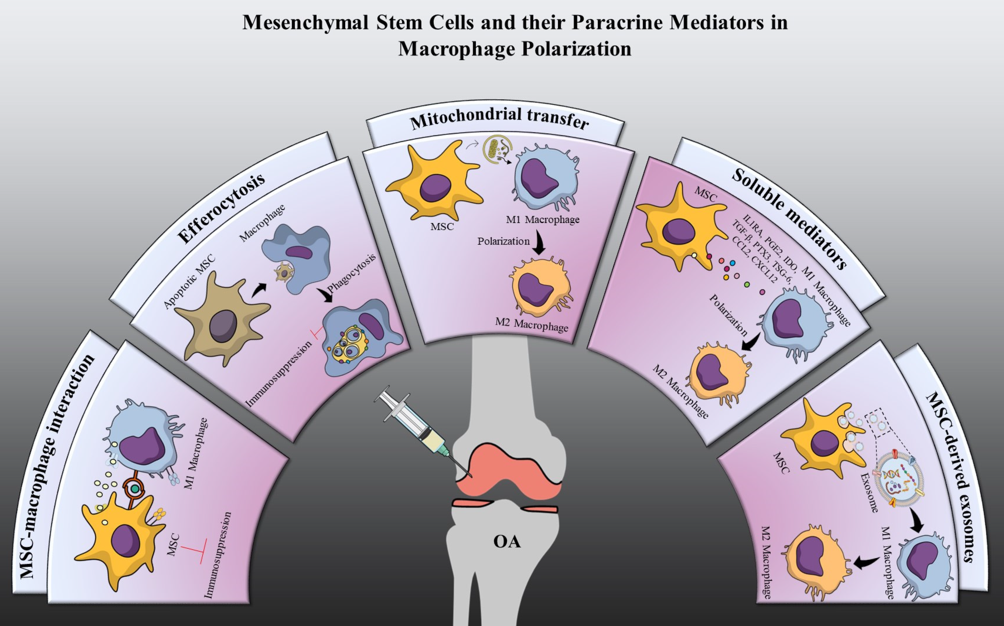
6. Macrophage Polarization by Mesenchymal Stem Cells

All of the aforementioned ways for polarizing macrophages are capable of reducing inflammation and pain, but they cannot repair the cartilage. Further cartilage deterioration requires complete knee arthroplasty [72,73]. OA treatment has the potential to be revolutionized by stem cell treatment. MSCs have piqued the interest of many researchers because of their experimental applicability, and the ability to differentiate into many lineages such as bone, muscle, fat, and cartilage [12]. MSCs can be extracted from bone marrow, synovium, adipose tissue, umbilical cord, blood, dental pulp and endometrium [74]. The International Society for Cellular Therapy (ISCT) has established three baseline requirements that all MSCs, regardless of their origin, must satisfy. Initial attachment to the plastic surface is required for MSCs to proliferate under standard circumstances. Additional requirements for MSCs include the expression of the surface markers CD73, CD90, and CD105. Furthermore, MSCs are required to differentiate into osteoblasts, chondrocytes, and adipocytes under certain in vitro conditions [75]. One of the most compelling arguments for making MSCs a standard treatment for OA is that they can repair cartilage, allowing damaged cartilage to regenerate [76]. MSCs are self-renewing stromal cells that can develop into a variety of cell types [77]. Although Friedenstein was the first to effectively isolate bone-forming cells from a guinea pig, Owen provided this field of research a much-needed boost by extending it to rats [78]. In 1992, it was announced that human bone marrow MSCs (BM-MSCs) had been extracted and cultivated to increase in number; by 1995, they were being pumped into patients [79]. Over the past quarter century, infusion approaches have demonstrated such a high level of safety that the Food and Drug Administration (FDA) now lists more than 950 clinical trials involving MSCs. MSCs have been used to treat a variety of orthopedic disorders, including OA, due to their tissue regeneration and immunomodulatory properties. MSCs have been shown to be effective in treating OA in phase 1 trials over the last few years and a number of unpublished Phase 2 trials, notably ADIPOA2 [80,81].
MSCs have an effect on immune cells, such as macrophages, dendritic cells, T lymphocytes, and natural killer (NK) cells [82]. MSCs play a pivotal role in controlling different functions of macrophages, such as differentiation of naive macrophages, modulation of their phagocytic ability, enhancement of their bactericidal effect, and manipulation of the plasticity and polarity of macrophages. MSCs have been reported to possess the property of immune evasiveness due to their close and reciprocal interaction with immune cells and their immunomodulatory properties [83]. This suggests that MSCs may be immune-protected when injected into an allogenic environment, preventing detection and rejection by the immune system. Nevertheless, MSCs do not need to remain in the body for an extended period of time in order to exert a therapeutic effect. A brief presence can permanently alter tissue cell behavior under certain pathological conditions. Thus, it is necessary to comprehend how the host immune system reacts to allogenic MSCs and how this may influence the therapeutic efficacy of MSCs in various inflammation models. MSC-based therapies have been developed in the context of inflammation observed in numerous disease models, such as graft versus host disease (GVHD) [84], inflammatory bowel disease (IBD) [85], diabetic cardiomyopathy [86], and many others. In this review, we will discuss some of the fundamentals that link inflammation to different disease models, as well as some of the biological properties of MSCs that can help them be used as a treatment for inflammatory diseases like OA. MSCs manifest their immunomodulatory properties and induce polarization of macrophages to an anti-inflammatory M2 phenotype through two primary methods: cellular interaction and paracrine factor-mediated mechanisms involving cytokines and hormones, and exosome-mediated mechanisms involving RNAs, and other molecules as shown in Figure 2.

7. Cellular Interaction

7.1. Immunosuppression through MSCs-Macrophage Interaction

Recent studies have revealed the ability of MSCs to regulate macrophage polarization through direct cellular contact. Abumaree et al. reported human placental MSCs (hp-MSC) mediated differentiation of macrophages from M1 into M2 either through direct cellular contact or through partial interaction of various soluble mediators such as macrophage colony-stimulating factor (M-CSF), IL-10, B7-H4, Leukemia inhibitory factor (LIF), VEGF, PGE2, TGFβ-1 with glucocorticoid receptor and progesterone receptor [87]. The results of Yanhong Li et al. demonstrate that direct cell-to-cell contact between MSCs, and pro-inflammatory macrophages is significant. The crosstalk between MSCs and macrophages was enhanced when they were in direct contact with each other due to the stimulation of macrophages that upregulated the expression of CD200 in MSCs and M1 macrophages expressed the CD200R receptor, which enabled the contact, and this linkage drove the M1 to M2 transition [88]. Audrey Varin et al. observed a similar interaction between M1 macrophages and MSCs, in which the interaction was with the accumulated CD54 marker generated in the interface of MSC-M1 macrophages that induced Ca2+ ion signaling and enhanced MSC’s immunosuppressive ability [89]. In response to inflammation, MSCs upregulate the expression of CD54, one of their most abundant adhesion molecules [90]. In multiple studies, CD54-Leukocyte function associated antigen-1 (LFA-1) interactions have been shown to determine CD54′s ability to polarize macrophages. Takizawa et al. have shown that CD54-LFA1-mediated interaction between MSCs and macrophages in hypoxia which prompted the proliferation of M2-macrophages in mice [91]. Additionally, CD54high MSCs enhance mouse survival in a GVHD model by migrating to secondary lymph nodes to inhibit dendritic cell maturation and Th1 differentiation of CD4+ T-cells [92].

7.2. Immunosuppression through Efferocytosis

Apoptotic cells communicate with immune cells either directly or indirectly via phagocytosis. In the direct method, the immune cells directly interact with the apoptotic cells, creating an immunosuppressive environment by releasing IL-10 and TGF-β to dampen the pro-inflammatory response of LPS-stimulated macrophages that secrete IL-1β and TNF-α [93,94]. Indirect effects reduce LPS reactivity, through phagocytosis of apoptotic cells which reduces the inflammatory phenotype of the immune system. Galleu et al. demonstrated that in vivo naturally occurring MSC apoptosis is instrumental for the delivery of immunosuppression. In a model of GVHD, the release of granules containing the perforin granzyme B by recipient T-cells caused the apoptosis of MSCs. The regulatory T-cells and macrophages ingest apoptotic MSCs and release indoleamine 2,3-dioxygenase (IDO), restoring an anti-immunogenic environment [95]. Similarly, Akiyama et al. found that MSCs can also promote an anti-inflammatory milieu by controlling T-cell apoptosis via the FAS/FASL pathway, causing macrophages to produce large amounts of TGF-β, which then upregulates regulatory T-cells, resulting in immunological tolerance [96]. These findings provide credence to the theory that MSCs can provide therapeutic benefit without engraftment. As stated previously, these results demonstrate that despite their brief post-administration survival, MSCs induce potent immunosuppression.
Although cellular interaction is an essential functional mechanism to regulate macrophage polarization, recent studies indicate that the immunomodulatory properties of MSC are largely dependent on the paracrine mediators secreted by MSCs.

8. Paracrine Mediators

MSCs secrete a plethora of paracrine mediators that includes various cytokines, chemokines, growth factors and enzymes. These paracrine mediators released into the extracellular space are denoted as the secretome, and these mediators have been associated with the majority of immunomodulatory effects exhibited by MSCs. The secretome has been characterized in multiple studies with the help of proteomics profiling, liquid chromatography-mass spectrometry (LC-MS) and enzyme-linked immunosorbent assay (ELISA). The composition and concentration of the components in the secretome can vary drastically depending upon the cellular source and preparation parameters. The components in the secretome can be categorized majorly as: 1. Soluble mediators and 2. EVs.

8.1. Soluble Mediators

8.1.1. IL1 Receptor Antagonist (IL1RA)

A variety of cells, including monocytes, macrophages, and synovial cells, secrete IL-1. Multiple chemokines, cytokines, and inflammatory mediators are induced by IL-1. Numerous studies have documented the role of IL1RA in MSCs’ anti-inflammatory effect. By expressing IL-1RA, MSCs have shown to suppress inflammation in IL1RA knock out mice [97]. MSC-secreted IL1RA has been shown to act on both macrophages and B lymphocytes, preventing the B lymphocytes from differentiating into plasmablasts and encouraging the macrophages to adopt an anti-inflammatory M2 phenotype [98]. Similarly, Phinney et al., showed that MSC secreted IL1RA can protect lung tissue from bleomycin induced inflammation and fibrosis in mice by inhibiting TNF-α and IL-1α, two vital proinflammatory cytokines in the lung produced by activated macrophages [99].

8.1.2. Indoleamine 2,3-Dioxygenase (IDO)

The tryptophan catabolism enzyme IDO is an inducible catalytic rate-limiting enzyme. IDO degrades tryptophan and creates tryptophan-degrading kynurenines, which have an immuno-regulatory impact [100]. IDO is often produced by MSCs in response to inflammatory cytokines, and it has an immunosuppressive impact by polarizing macrophages to the anti-inflammatory M2 phenotype that secretes IL-10 [101]. Song-Guo et al. demonstrated that human gingiva-derived MSCs (GMSCs) reduced inflammatory macrophage activation partly through the IDO/CD73 signal pathways, leading to the conversion of inflammatory macrophages to anti-inflammatory macrophages in an atherosclerosis mice model [102].

8.1.3. Prostaglandin E2 (PGE2)

PGE2 is a complex lipid molecule that is influenced by the cell type and microenvironment in which it is produced. MSC-produced PGE2 helps reduce inflammation and promotes macrophage polarization from M1 to M2 [103]. According to Vasandan et al., Salmonella enterica infected macrophages cocultured with MSCs activated respiratory burst and NO-dependent killing mechanisms, boosting macrophage microbicidal activity. MSCs on the other hand, released increased quantities of PGE2 after being treated with IFN-γ, converting M1 macrophages to M2 macrophages. With the help of COX-2 knocked down MSCs (COX-2KD MSCs), the role of PGE2 in inducing M1 macrophages towards an anti-inflammatory M2 phenotype was confirmed. The inducible enzyme COX-2 is involved in the synthesis of PGE2, which when knocked down prevents PGE2 synthesis and inhibits M1 to M2 macrophage polarization [104]. Similarly, in a diabetic cardiomyopathy mouse model, MSC infusion in the presence of high glucose and LPS resulted in increased PGE2 release, which reduced cardiac inflammation by polarizing macrophages from M1 to M2 and secreting IL-10 [105]. The anti-inflammatory properties of PGE2 releasing MSCs were demonstrated once more in an IBD mice model. The study used a chitosan-based injectable hydrogel with immobilized C domain peptide of insulin-like growth factor-1 on chitosan (CS-IGF-1C) and hP-MSCs to reduced inflammation and participated in M1-M2 bioenergetic shifts.

8.1.4. Tumor Necrosis Factor-Stimulated Gene-6 (TSG-6)

TSG-6 is a protein produced during inflammation that has been related to a number of protective and anti-inflammatory properties, including mediating many of MSCs’ immunomodulatory and therapeutic effects [106]. The MSC-derived TSG-6 that is upregulated when MSCs are in direct contact with M1 macrophages reduces T-cell proliferation and pro-inflammatory responses, and may promote the switch from M1 to M2 phenotypes in LPS-induced or spontaneous abortions in mice [88]. In an inflammatory setting, TSG-6 secretion increases. Hongyu Son et al. demonstrated this phenomenon in rats with severe acute pancreatitis, where chorionic plate-derived MSCs produced significant quantities of TSG-6 in a hyperinflammatory environment, repairing pancreatic injury, reducing inflammation and polarizing macrophages from M1 to M2. Similarly, human umbilical cord (UC)-MSCs secreted TSG-6 decreased severe burns and the associated inflammation by inhibiting P38 and JNK signaling [107].

8.1.5. Transforming Growth Factor Beta (TGF-β)

TGF-β is a cytokine that plays a function in immunoregulation and tissue repair [108]. TGF-β works as an immunosuppressive cytokine in MSCs, regulating activated T-cells and macrophages as well as inhibiting the production of iNOS along the SMAD3 pathway in a dose-dependent manner [109]. TGF-β produced by MSCs amid excessive inflammatory responses can cause LPS-stimulated macrophages to polarize to the M2 phenotype, reducing inflammation through the Akt/FoxO1 pathway [110].

8.1.6. Pentraxin 3 (PTX3)

PTX3 is a characteristic acute-phase protein that plays a crucial role in inhibition of inflammation and apoptosis in cells. It is well established that important activators of the inflammatory and reparative response following tissue damage elicit enhanced PTX3 secretion from many different cell types, including MSCs [111]. By treating LPS-stimulated macrophages with PTX3 released from umbilical cord blood (UCB)-MSCs, Kim et al. showed that PTX3 promoted macrophage polarization, leading to decreased inflammation and better anti-inflammatory effects [112]. To a comparable extent, PTX3 secreted by Adipose derived MSCs (ADSCs) also favored an M2 macrophage phenotype and stimulated IL-10 expression in PBMCs isolated from individuals before the onset of metabolic syndrome [113].

8.1.7. Chemokines

MSCs secrete immunomodulatory chemoattractants including C–X–C motif chemokine 12 (CXCL12) [114] and CCL2 [115,116], which have a role in changing the macrophage phenotype to an anti-inflammatory M2 macrophage while down regulating M1-specific markers. Jacques Galipeau et al. demonstrated a synergistic effect of both CXCL12 and CCL2 production by MSCs into its secretome exhibiting an anti-inflammatory environment where tissue resident macrophages are polarized to the M2 phenotype, reducing severe colitis [116].

8.1.8. Mitochondrial Transfer

Mitochondrial transfer has been viewed as a viable therapeutic method since the ability to transport organelles and selective membrane vesicles through extremely sensitive nanotubular structures [117] and also replace damaged mitochondria with healthy mitochondria from an external source [118]. Mitochondria plays an important role in the metabolic reprogramming of the macrophages during their activation [119]. Mitochondrial transfers to macrophages regulate immunomodulatory effects. Maroun Khoury et al. confirmed the immunomodulatory properties of MSC mitochondria in their study of transferring mitochondria to T-cell populations via artificial and natural methods, promoting differentiation into T-regulatory-cells [120]. This approach shows promise to control inflammatory diseases in a mouse graft versus host disease model. Donation of mitochondria by mesenchymal stromal cells causes macrophages to adopt an anti-inflammatory phenotype. Yanrong Lu et al. investigated the mechanisms underlying MSC’s improved mitochondrial function in macrophages (M2) [121] and found that transferring mitochondria from MSCs to macrophages reduced inflammation and reduced kidney injury in a mouse model of diabetic nephropathy by promoting mitochondrial biogenesis via regulating the transcription of PPARGC1A or PGC-1α and clearing out damaged cells via PGC-1/TFEB mediated autophagy [122].

8.2. Extracellular Vesicles

It has been shown that MSCs have a paracrine function that goes beyond the release of soluble mediators through the release of EVs. EVs had previously been regarded as inert cellular debris, that was generated as a consequence of cell damage or as a result of dynamic plasma membrane turnover. However, the discovery of EVs’ distinct roles as facilitators of cellular interactions, in which EVs may transport functional molecules to recipient cells and modify their biological and pathological activities, represented a significant milestone in the development of this field of study [123]. EVs are made up of a lipid bilayer containing proteins/peptides, lipids, and genetic material including messenger RNA (mRNA), microRNA (miRNA), and DNA. EVs may also contain ribosomes, proteasomes, and mitochondria [124]. Due to overlapping properties with other nano-sized lipids, protein molecules, and nucleic acid complexes, adequate EV purification and characterization procedures are crucial for drawing exact findings. It is difficult to study EVs because, similar to other nanostructures, they are at or below the detection limit of many standard analytical techniques. The minimal information for extracellular vesicle studies (MISEV) has been crucial in setting the framework and completing the purpose of the International Society for Extracellular Vesicles (ISEV), which is to advance EV science internationally. Recommendations were given at MISEV2018 in six categories, including EV terminology, specimen collection and preliminary processing, EV isolation and concentration, EV characterization, functional research, and reporting necessities/deviations [125].
Following the established standards, researchers are exploring the possibility of MSC-EV-based treatments. EVs derived from MSCs possess many of the same therapeutic effects as MSCs. In a model of silicosis, Phinney et al. discovered that the transfer of mitochondria and miRNAs to human macrophages via EVs improved macrophage bioenergetics while inhibiting Toll-like receptor signaling [126]. Morrison et al. demonstrated that the mitochondria-laden EVs can also alter the phenotype of macrophages in acute respiratory distress syndrome to increase oxidative phosphorylation, phagocytic capacity, and CD206 expression, while decreasing proinflammatory cytokine production in macrophages [127]. Subsequently, EVs generated from MSCs are divided into three primary kinds in order to better understand the biogenesis method, size, and surface markers 1. Exosomes are the smallest vesicles (30–100 nm) released when multivesicular formations, such as intraluminal vesicles (ILVs), fuse with the plasma membrane. 2. Microvesicles are vesicular (0.1–1.0 µm) entities that are shed via plasma membrane blebbing. 3. The largest EVs (1–5 µm) are apoptotic bodies, which are formed at the late stages of apoptosis [128]. Due to their nanoscale dimensions and excellent protective effects, exosomes have received the most research interest in recent years.

8.2.1. Exosomes

Exosomes are nanosized vesicles of endocytic origin. Their development is influenced by the process of endocytosis and exocytosis. Exosome development begins when an early endosome absorbs a small amount of intracellular fluid [129]. When an early endosome evolves into a late endosome, an intraluminal body, also known as a multivesicular body (MVB), is produced. Exosome production is dependent on multiple biological pathways [130]. Endosomal sorting complex required for transport (ESCRT) is one of them; it possesses both ESCRT-dependent and ESCRT-independent transport pathways. The ESCRT apparatus includes ESCRT-(0, I, II and III) complexes [131]. As MVBs grow from the early endosome, ESCRT-dependent/-independent machinery forms ILVs. Following transport and fusion with the plasma membrane by MVBs, the ILV containing exosomes are released [129]. Exosomes have been demonstrated to perform numerous functions, including pro-angiogenic, pro-tumorigenic, anti-clotting, anti-inflammatory and immune modulation [132]. MSC exosomes induce immunosuppressive effects by encouraging the formation of anti-inflammatory M2 macrophages and inhibiting the synthesis of pro-inflammatory proteins [133,134]. Exosomes produced from MSCs have been proven in multiple studies to prevent macrophage polarization and recruitment. Stimulated by MSC exosomes, colonic macrophages adopt an immunosuppressive M2 phenotype [135]. By promoting the formation of M2 macrophages, MSC exosomes can reduce inflammation, and ultimately promote wound healing and tendon repair [136]. MSC exosomes can also alleviate alveolar damage and inflammation in the lungs by boosting the M2 macrophages as a proportion of total macrophages [72] ADSCs exosomes produce M2 macrophages through the S1P/SK1/S1PR1 signaling pathway which is protective against cardiac apoptosis and fibrosis [137]. The primary contribution of UC-MSCs derived exosomes to spinal cord injury recovery was inducing macrophages to change from an M1 to an M2 phenotype, where the M2 markers were upregulated and the M1 markers such as TNF-α and iNOS levels decreased [135]. Increased CD163+ regenerative M2 macrophages and decreased CD86+ M1 macrophages were observed in the osteochondral defect and surrounding synovium following treatment with exosomes, and levels of synovial pro-inflammatory cytokines, including IL-1β and TNF-α, were lowered [138]. MSC exosomes transformed monocyte-derived myeloid-derived suppressor cells into M2 macrophages by guiding the expression of programmed death-ligand 1 (PD-L1) in macrophages with the help of TGF-β and semaphorin [139]. MSC exosomes can also increase matrix production and deposition during cartilage regeneration [140,141,142,143]. According to these studies, the matrix-degrading enzymes such as MMP-13 [144,145,146] or ADAMTS-5 [147] were simultaneously inhibited in vivo and increased cartilage regeneration. All of these findings suggest that MSC exosomes may influence the tissue immune cells to adopt a regenerative immune phenotype that is favorable for tissue repair and regeneration.

8.2.2. Exosomal miRNA

MSC exosomes contribute to the immunosuppressive effects on macrophages by way of miRNA. The miRNAs utilized by MSC exosomes to promote the anti-inflammatory phenotype of macrophages in various inflammatory models are listed in Table 2. Ragini et al. conducted a detailed characterization of human amniotic membrane-derived MSCs (hAMSCs)-secreted EVs, and they identified the presence of 200 secreted factors and 754 miRNAs. Of the miRNAs, five miRNAs are involved in M2 (miR-24-3p, miR-146a-5p, miR-222-3p, and miR-34a-5p), and three involved in M1 (miR-125b-5p, miR-145-5p and miR-130a-5p) phenotype regulation [148]. There is mounting evidence that aging affects the physiology of both macrophages and MSCs. Huang et al. demonstrated that young MSC exosomes exhibit high levels of miR-223-5p expression, whereas older MSC exosomes exhibit miR-127-3p and miR-125b-5p expression. Younger MSC exosomes are better able to convert M1 macrophages to M2 than older MSC exosomes [149]. The anti-inflammatory miR-146a derived from MSC exosomes facilitates the transition from M1 to M2-like macrophages [150]. Co-cultivating MSC exosomes with M1 macrophages significantly reduced the production of pro-inflammatory M1-like markers, including IL-6, TNF-α, and CCL5 [151]. MSC exosomes can reduce neural inflammation by upregulation CD206 and Arg1 to regulate the polarization of glial cells and macrophages. In addition, they can block the synthesis of pro-apoptotic proteins, such as Bcl-2 related X protein (BAX) and iNOS, as well as pro-inflammatory cytokines [152]. Overexpression of miRNAs such as miR-140-5p and miR-92a-3p has been associated with anti-OA effects via increased matrix formation and decreased cartilage degradation [145,153]. Other studies have demonstrated that suppression of miR-100-5p or lncRNA KLF3-AS1 can increase the protective effects of MSC exosomes against matrix breakdown during OA [144,146]. In conclusion, MSC exosomes modify the polarity of macrophages and affect factors associated with this process, allowing for improved immunosuppressive regulation and cartilage regeneration. However, macrophage polarization in OA models employing MSC-derived paracrine mediators is poorly understood. The precise mechanism of their impacts on OA treatment, including the role of each miRNA and protein, is not entirely understood. Therefore, more in-depth research is necessary to understand the essential internal components of the secretome and their molecular processes on OA. In this review, we explored the involvement of numerous paracrine mediators in various inflammatory disease models in order to give knowledge that may be used to build a validated mechanism for OA that is comparable to the MSC biology and macrophage interaction reported in other inflammatory models.

9. Priming Enhances Mesenchymal Stem Cell Immunomodulation

The significance of paracrine signaling in MSC therapy, particularly with relation to the immunomodulatory properties of MSCs, is becoming increasingly apparent. This emphasizes the need to modify the secretory profiles of MSCs during in vitro production in order to achieve desired functional properties. This can be accomplished by regulating the secretion of individual immune mediators or by enhancing the secretome’s overall output. Priming MSCs with pro-inflammatory cytokines, pharmacological agents, or small molecules, or by the application of biophysical priming techniques, has increased the release of specific anti-inflammatory and immune regulatory factors [167].

9.1. Proinflammatory Cytokines

Priming MSCs with proinflammatory cytokines such as TNF-α has been demonstrated to upregulate key paracrine mediators such as IDO, PGE2, and hepatocyte growth factor (HGF), but to a lesser extent than IFN-γ. MSCs primed with IFN-γ have been reported to release adhesion proteins VCAM1 and ICAM1, as well as the chemokine ligands CXCL9, CXCL10, and CXCL11 and high levels of HLA-G and IDO. Co-culturing IFN-γ primed MSCs with activated PBMCs increased the frequency of CD4+CD25+CD127dim/- T-cells, IL-10 and IL-6, while decreasing the frequency of Th17 cells, IFN-γ and TNF-α production [168]. MSCs stimulated with IFN-γ enhanced production of programmed cell death-1 ligands (PDL-1) to inhibit T-cell effector function and have also been demonstrated to inhibit NK cell cytotoxicity [169]. Similarly, BM-MSCs preconditioned with IL-17 reduced Th1 secretion of cytokines such as TNF-α, IFN-γ, IL-2 and enhanced iTreg cell formation. Furthermore, genes such as MMP1, MMP13, and CXCL6 that are predominantly correlated with migration and chemostatic responses were identified [170].
In light of the fact that MSCs from varying donors and sources exhibit varying cytokine priming responses [171], it may be necessary to combine cytokine priming in order to maintain a significant and consistent effect. Compared to priming MSCs with a single agent, combined IFN-γ and TNF-α priming significantly reduced donor-specific variability in MSC immunomodulatory potency. Chenyang Liu et al. demonstrated that supernatant from MSCs that have been pretreated with IFN-γ and TNF-α has been shown to switch macrophages to the M2-type, which in turn promotes cutaneous wound recovery with minimal scarring by stimulating the IL-6-dependent signaling pathway [172]. Likewise, in another study MSCs isolated from menstrual blood and stimulated with IFN-γ and TNF-α showed elevated levels of IDO1, EV release, and differential expression of miRNAs related to the immune response and inflammation [173]. Besides IFN-γ and TNF-α, MSCs primed with different combinatorial cytokine cocktail like LPS/ TNF-α also exhibited polarization of macrophages to the M2 phenotype expressing high amounts of PGE2, Arg1, and CD206, and displayed improved alkaline phosphate activity and bone mineralization potential [174]. The miRNA expression profile of foreskin MSCs is drastically altered after treatment with a cytokine cocktail containing IL-1β, TNF-α, IFN-γ, and IFN-α, with 13 miRNAs being downregulated and 3 others being upregulated. These miRNAs with altered expression levels are speculated to target multiple potential signaling pathways that control cellular activity in response to inflammatory cues. Several pro-inflammatory cytokine mixes have been used to alter the expression of immune mediators and miRNAs by MSCs in culture [175]. One of the major drawbacks of this strategy is the cost of recombinant cytokines.

9.2. Chemical Agents

In order to reduce the costs associated with recombinant cytokines, MSCs have been primed with a variety of pharmacological chemicals and small molecules in order to increase their therapeutic efficacy. The use of chemical agents like all-trans retinoic acid (ATRA) has shown to inhibit PBMC production of pro-inflammatory cytokines. Priming MSCs with ATRA improves wound-healing capacities in vivo. The gene expression of COX-2, VEGF, CCR2, HIF-1α, CXCR4, angiopoietin-2 (Ang-2), and angiopoietin-4 (Ang-4) is elevated by preconditioning rat BM-MSCs with ATRA [176]. Matteo Haupt et al. showed that the therapeutic potential of MSC EVs preconditioned with lithium is higher than that of EVs from native MSCs. Increased levels of miR-1906, a new regulator of toll-like receptor 4 (TLR4) signaling, were found in MSC EVs after treatment with lithium, which led to reduced cerebral inflammation and rapid neuroprotection in mice with stroke [177]. The histone deacetylase inhibitor valproic acid (VPA) and the bioactive lipid sphingosine-1-phosphate (S1P) have similar anti-inflammatory and proliferative actions. Priming MSCs with valproic acid and lithium before intranasal infusion improved neuropathological characteristics and function in a mouse model of Huntington’s disease [178]. Priming MSCs with cytokines and chemicals added to MSC culture media facilitates their ex vivo growth for therapeutic applications. This has been examined in relation to the preparation of xeno-free and serum-free media. In a recent study, Jin et al. created a hypoxic, calcium-rich environment for stem cells to grow while preserving them in a xeno-free, chemically defined cryopreservation media. The paracrine factor PTX-3 generated by these stem cells was shown to remodel M1 macrophages into their anti-inflammatory M2 phenotype in a rat OA model [179]. Small molecules are also being investigated as a method of priming MSCs due to their unique qualities such as low cost, minuscule size, robust stability, and non-immunogenicity. Oren Levy et al. showed a decrease in the expression of TNF-α at the site of inflammation after pre-treatment of MSCs with a kinase inhibitor (Ro-31–8425). Similar to Ro-31-8425, priming of MSCs with the small molecule tetrandrine boosted PGE2 synthesis via the NF-κB/COX-2 signaling pathway, which reduced TNF-α production in RAW264.7 during co-culture [180].

9.3. Hypoxia

Under hypoxic growth conditions, when the oxygen level is between 0 and 10%, MSCs can secrete more immunomodulatory molecules. It is well documented that hypoxic preconditioning can stimulate the production of immunomodulatory molecules in MSCs, such as IDO, IL-10 and PGE2 [181]. Hypoxia-exposed MSCs drive bone marrow-derived macrophage polarization to the M2 phenotype via the TGF-1/Smad3 signaling pathway, ameliorating ischemic stroke conditions by reducing apoptotic cells and fibrosis and promoting neovascularization in the infarcted region [182]. According to recent studies, EV density and load might also be modified by employing hypoxic preconditioning. However, hypoxia seemed to have no effect on the mean size, morphology, or surface biomarkers of MSC-derived EVs [183]. In response to hypoxia and serum deprivation, primed MSCs produced more dipeptides, suggesting that hypoxic MSCs augment their pool of free amino acids to meet energy requirements that cannot be properly met by the glycolytic process. Subsequently, it was also established that there are 21 different metabolites in primed MSC derived exosomes that have been linked to immunoregulation. The activation of regulatory T-cells, the polarization of macrophages toward the M2 state, and the regulation of anti-inflammatory responses are all directly influenced by these molecules [184]. Despite evidence indicating that MSCs grown under hypoxic conditions can result in the production of EVs, the real situation is still unclear. The variation may be attributed to the degree of hypoxia, as minute variations in oxygen concentration and exposure time can have a significant impact. In addition, it is important to note that while some studies have shown that hypoxia may promote cellular longevity, others have shown that cells may die [185].

9.4. Biophysical Stimulation

Another strategy that has been investigated is biophysical stimulation of MSCs. Priming approaches, including altering the texture and rigidity of culture surfaces, may influence cytokine release by MSCs [186]; however, this method has limited scalability. In an effort to create an environment that is analogous to that of the MSC niche, researchers have investigated the use of a variety of 3D based cell culture approaches [187]. When maintained in a three-dimensional environment, MSCs tend to produce more immunomodulatory factors. Spheroid creation is the most popular approach for MSC 3D cultivation [188]. Under these conditions, less oxygen may diffuse into the inner layer of cells, creating a hypoxic environment that enhances cell-cell interactions and modifies the release of immunomodulatory molecules. MSCs secreted more TSG6, HGF, and PGE2 when cultured in 3D spheroids; IDO activity and the ability to limit T-cell proliferation were both found to be attenuated when MSCs were cultured in aggregates [189,190]. Hydrogel encapsulation of MSCs is one of the most exciting approaches for producing a 3D-MSC-secretome. Hydrogels permit the change of the mechanical properties such as rigidity and firmness and the inclusion of patterns unique to the natural ECM, both of which increase the secretome’s complexity. Recent attention has been drawn to biopolymer hydrogels due to their capacity to alter the paracrine actions of MSCs [191,192]. The field of cell engineering is expanding fast, making all these methods particularly attractive.

10. Effect of Macrophages on Mesenchymal Stem Cells

We know that the phenotype of macrophages can alter upon interaction with MSCs. Similarly, macrophages also have a feedback impact on MSCs that affects their migration, viability, differentiation, and immunomodulatory capabilities. Guihard et al. demonstrated that conditioned media from human monocytes activated with LPS or TLR ligands promoted bone formation by human BM-MSCs [193]. M1 macrophages promote osteogenesis in MSCs via stimulating the COX-2-PGE pathway [194,195,196]. Regardless of their polarization status (M0, M1, or M2), human ADSCs can be blocked from transforming into adipocytes in vitro by macrophage derived supernatants [197]. According to previous studies, M2-type macrophages enhance MSC proliferation and migration, but M1-type macrophages cause MSC apoptosis [198,199]. According to de Witte et al., the phagocytosis of MSCs by monocytes is essential for the immunological regulation of MSCs [200]. Li et al. discovered that enhanced synthesis of TSG-6 in response to contact with pro-inflammatory macrophages improves MSCs’ inhibitory control of T-cells and macrophages [88]. Mouse BM-MSCs cocultured with macrophages enhanced IL-10 release in response to LPS stimulation via a PGE2-dependent mechanism. MSCs cannot secrete PGE2 under coculture conditions unless activated by TNF-α and iNOS generated by macrophages [201]. In response to pro-inflammatory cytokines produced by macrophages, MSCs produce immune modulators such as PGE2 and IL-1RA [202]. According to the aforementioned research, macrophages produce cytokines that activate MSCs after being activated by pro-inflammatory mediators MSCs respond to the activation of macrophages by modulating the immune response. There is a feedback loop between macrophages and MSCs within the disease microenvironment. MSCs and macrophages work together to keep the inflammatory environment in balance.

11. Challenges and Future Perspectives

Clearly, macrophages are crucial for both homeostasis and disease pathology. In this review, we have focused on the role of macrophages in OA inflammation with a particular emphasis on the role of macrophages in the synovium, as the synovium is the predominant site of gross and microscopic inflammatory change in OA. Synovial macrophages are increasingly believed to contribute to the development and persistence of inflammation in OA, based on data from research including human patients and animal models. The reprogramming of macrophages has therefore been recommended in this review for prevention against synovitis and cartilage destruction in OA. However, a number of unsolved concerns complicate the development of macrophage-targeting therapies for OA. First, macrophage transcriptome research has demonstrated the need for a more in-depth evaluation of macrophage functional characteristics, showing the limitations of the M1/M2 paradigm. Second, the identification and verification of biomarkers that are specific to different macrophage subgroups is essential to the comprehension of macrophage diversity in OA as well as the development of therapeutic alternatives that are connected to the specific immunological profile. Lastly, the engagement of macrophages in the pathogenesis of OA may differ depending on the stage of the condition and the endotype, and the inflammatory mediators and mechanisms vary greatly across individuals. Encouragingly, recent advances in technology, including as single-cell RNA sequencing and mass cytometry, have made it possible to precisely identify the cellular diversity of macrophages. In actuality, a two-phase strategy comprising the characterization of the functional implications of macrophage sub-populations and the development of particular targeted techniques applicable for macrophage remodeling may be essential.
With respect to the second phase of the two-phase strategy, we have highlighted recent efforts to target macrophage activity in OA using small molecules and biologics which have the ability to modulate the inflammation. Despite the fact that all bioactive substances and chemical compounds have been demonstrated to target OA-related signaling pathways and effectively polarize macrophages, further research is necessary. There are various obstacles to overcome before herbal bioactive components may be used in clinical settings. Due to the limited stability and low absorption of bioactive compounds in serum or synovial fluid, the therapy for OA is accompanied by a number of serious side effects. Second, regulated extraction methods cannot compensate for the fact that the quality of raw herbs varies, and it is difficult to standardize the concentration of the principal ingredients. Lastly, to confirm the safety of the compounds for clinical use, rigorous and standardized toxicity studies will be required. In terms of nanoparticle-based approaches, there are still a number of obstacles that must be surmounted before it may be used in human clinical practice. In order to reduce discomfort and improve patient compliance, it is crucial that the nanoparticles dissolve at a set time. Furthermore, the optimal dose, quantity, frequency, and timing of treatment must be established based on the severity and location of the condition. Finally, the inability of bioactive chemicals and nanoparticles to regenerate the cartilage is a fundamental limitation of their usage.
Recently, there has been a lot of interest in the use of MSCs and its secretome for treating OA, in the realms of regenerative medicine and tissue engineering. In light of this, we have emphasized the significance of MSC-derived secretome in polarizing macrophages in diverse models of inflammation, as well as the strategies used to enhance the immunomodulatory potential of MSCs and their paracrine mediators, which give novel insights that may be used for OA research. These insights can be used to develop a gold standard mechanism for OA that closely resembles the MSC biology and macrophage interaction observed in other inflammatory models. MSC’s capacity for natural cartilage repair and regeneration, as well as its ability to modulate macrophage phenotype, is an advantage of employing MSCs to treat OA above all other polarization approaches. Several studies have demonstrated that injection of MSCs stimulates cartilage tissue regeneration, due to the polarization of macrophages [203,204]. There are still certain restrictions to employing MSCs for therapeutic reasons, despite extensive study and remarkable advancements. One of these obstacles is the absence of standardized protocols. Variations in cell source, cell isolation and culture processes, or administration routes are frequently cited as the cause of disparities in reported results [205]. Concerningly low rates of cell retention and survival following implantation are a further factor to consider. It has been demonstrated that less than 1% of MSCs survive more than a week following systemic administration [206]. This therapy window may not be sufficient for the overwhelming majority of individuals [206,207]. Therefore, attention has switched from these difficulties to the MSC-derived secretome since the secretory activities of MSCs are believed to be the underlying rationale for their therapeutic properties [206,207].
Several studies demonstrate that the immunomodulatory actions of MSCs are mostly attributable to their secretome, which has led to a turning point when cell-based therapies could be substituted. In this regard, the immunomodulatory/anti-inflammatory secretome produced from MSCs is the new gold mine, bypassing the limitations of cell-based therapies. This motivates the scientific community to investigate its medicinal properties, and interest in the topic is fast growing. It has been established that employing the secretome as a treatment for OA produces therapeutic outcomes [164,208,209]. However, there is still a long way to go in terms of study, as this therapeutic potential has yet to transcend its constraints. First, it is important to characterize the secretome in depth so that its applications may be studied and reproduced. There are indications of a changing composition of the secretome depending on both external and internal conditioning variables of the source cells. The secretome’s positive effects might also be amplified by priming of MSCs. In this review, we have focused on the methods currently in use to prime MSCs in an effort to boost the secretomes’ immunomodulatory capacity. Priming techniques of MSCs have significant difficulties in clinical translation, including induction of immunogenicity, increased costs, unpredictable results, and a lack of clinically applicable good manufacturing procedures (GMP). In order to do the spadework for different priming approaches in the clinical setting, we need to evaluate (1) Optimal sources for isolating MSCs, (2) Epigenetic modifications, (3) Antigenicity and tumorigenicity of primed and non-primed MSCs, and (4) Appropriate good manufacturing practices (GMP) standards for quality control of MSC products [210]. It is also generally accepted that the functional properties of primary MSCs might vary depending on the age, anatomic origin, and in vitro expansion of donors. Consequently, their secretome profiles may vary substantially [211]. By utilizing standardized MSCs derived from induced pluripotent stem cells, it is possible to circumvent these limitations and expand their therapeutic utility. Similarly, there is no consensus on the optimal approaches to standardize and personalize the secretome content, which is required for the development of medicines with a diverse range of applications. The development of finely tuned procedures for secretome extraction is a further objective that has not yet been reached. According to the available literature, we may propose two methods for modifying for enhancing MSC-derived secretome performance. (1) Standardization of approaches for isolating and purifying the entire secretome with maximum yield and scalability; and (2) Development of secretome delivery protocols and dosages.

12. Conclusions

Current OA treatment outcomes place a significant strain on worldwide healthcare systems. Deterioration of the articular cartilage results in an aberrant immune microenvironment in the joint. This dysfunction of the local microenvironment leads to a wide variety of uncontrolled inflammatory responses. Once inflammatory processes have occurred, OA can be treated by restoring balance to the local immunological environment in the joints. Controlling OA requires polarizing macrophages, a natural component of the joint’s local immunological milieu. Although macrophages are a viable therapeutic target, it is necessary to first comprehend their phenotypes in terms of their characteristics, anatomical location, and origins. The microenvironment influences the phenotype and function of macrophages. Restoring a healthy equilibrium necessitates an in-depth examination of the anatomical setting in which macrophages operate. Modifying the phenotype of macrophages to influence the development, progression, and resolution of inflammation by acting on molecules in signaling pathways and the local microenvironment is a potential field for the treatment of OA.
Despite the fact that MSCs can induce macrophage polarization toward the M2 phenotype, there are a number of clinical risks and complications associated with this cell-based therapy. The MSC-derived secretome, which carry the majority of MSCs’ therapeutic effects, represent a novel therapeutic strategy. This cell-free therapy circumvents the disadvantages of MSCs and has certain advantages. But there is a need for the creation of guidelines to improve experimental conditions for producing MSCs’ secretome, to establish more standardized protocols among the scientific community, and to encourage future collaborative work to bridge the decades-long gap between MSCs’ experimental research and their clinical use.

Author Contributions

S.S.K.: collection of data, manuscript structuring and preparation. J.K.S.: conceptualization and final approval of manuscript; H.K.K.: data curation and manuscript review. J.Y.K. and S.C.L.: supervision and assistance. All authors have read and agreed to the published version of the manuscript.

Funding

This work was supported by the Korea Fund for Regenerative Medicine (KFRM) grant funded by the Korea government (the Ministry of Science and ICT, the Ministry of Health & Welfare) (22C0603L1-11).

Data Availability Statement

Not applicable.

Conflicts of Interest

The authors declare no conflict of interest.

References

  1. Kloppenburg, M.; Berenbaum, F. Osteoarthritis year in review 2019: Epidemiology and therapy. Osteoarthr. Cartil. 2020, 28, 242–248. [Google Scholar] [CrossRef] [PubMed] [Green Version]
  2. Tschon, M.; Contartese, D.; Pagani, S.; Borsari, V.; Fini, M. Gender and sex are key determinants in osteoarthritis not only confounding variables. A systematic review of clinical data. J. Clin. Med. 2021, 10, 3178. [Google Scholar] [CrossRef] [PubMed]
  3. Son, K.M.; Hong, J.I.; Kim, D.; Jang, D.; Crema, M.D.; Kim, H.A. Absence of pain in subjects with advanced radiographic knee osteoarthritis. BMC Musculoskelet. Disord. 2020, 21, 1–9. [Google Scholar] [CrossRef] [PubMed]
  4. Hunter, D.J.; McDougall, J.J.; Keefe, F.J. The Symptoms of Osteoarthritis and the Genesis of Pain. Rheum. Dis. Clin. N. Am. 2008, 34, 623–643. [Google Scholar] [CrossRef] [Green Version]
  5. Krasnokutsky, S.; Attur, M.; Palmer, G.; Samuels, J.; Abramson, S.B. Current concepts in the pathogenesis of osteoarthritis. Osteoarthr. Cartil. 2008, 16, 6–8. [Google Scholar] [CrossRef] [Green Version]
  6. Kim, S. Changes in surgical loads and economic burden of hip and knee replacements in the US: 1997-2004. Arthritis Care Res. 2008, 59, 481–488. [Google Scholar] [CrossRef]
  7. Thomas, E.; Peat, G.; Croft, P. Defining and mapping the person with osteoarthritis for population studies and public health. Rheumatology 2014, 53, 338–345. [Google Scholar] [CrossRef] [Green Version]
  8. Jørgensen, A.E.M.; Kjær, M.; Heinemeier, K.M. The effect of aging and mechanical loading on the metabolism of articular cartilage. J. Rheumatol. 2017, 44, 410–417. [Google Scholar] [CrossRef]
  9. Goldring, S.R.; Goldring, M.B. The role of cytokines in cartilage matrix degeneration in osteoarthritis. Clin. Orthop. Relat. Res. 2004, 427, 27–36. [Google Scholar] [CrossRef]
  10. Shapouri-Moghaddam, A.; Mohammadian, S.; Vazini, H.; Taghadosi, M.; Esmaeili, S.A.; Mardani, F.; Seifi, B.; Mohammadi, A.; Afshari, J.T.; Sahebkar, A. Macrophage plasticity, polarization, and function in health and disease. J. Cell. Physiol. 2018, 233, 6425–6440. [Google Scholar] [CrossRef]
  11. Bohaud, C.; Contreras-Lopez, R.; De La Cruz, J.; Terraza-Aguirre, C.; Wei, M.; Djouad, F.; Jorgensen, C. Pro-regenerative dialogue between macrophages and mesenchymal stem/stromal cells in osteoarthritis. Front. Cell Dev. Biol. 2021, 9, 1–14. [Google Scholar] [CrossRef] [PubMed]
  12. Salem, H.K.; Thiemermann, C. Mesenchymal stromal cells: Current understanding and clinical status. Stem Cells 2010, 28, 585–596. [Google Scholar] [CrossRef] [PubMed] [Green Version]
  13. De Bari, C.; Dell’Accio, F.; Luyten, F.P. Human periosteum-derived cells maintain phenotypic stability and chondrogenic potential throughout expansion regardless of donor age. Arthritis Rheum. 2001, 44, 85–95. [Google Scholar] [CrossRef]
  14. Péault, B.; Rudnicki, M.; Torrente, Y.; Cossu, G.; Tremblay, J.P.; Partridge, T.; Gussoni, E.; Kunkel, L.M.; Huard, J. Stem and progenitor cells in skeletal muscle development, maintenance, and therapy. Mol. Ther. 2007, 15, 867–877. [Google Scholar] [CrossRef] [PubMed]
  15. Harrell, C.R.; Markovic, B.S.; Fellabaum, C.; Arsenijevic, A.; Volarevic, V. Mesenchymal stem cell-based therapy of osteoarthritis: Current knowledge and future perspectives. Biomed. Pharmacother. 2019, 109, 2318–2326. [Google Scholar] [CrossRef] [PubMed]
  16. Farahat, M.N.; Yanni, G.; Poston, R.; Panayi, G.S. Cytokine expression in synovial membranes of patients with rheumatoid arthritis and osteoarthritis. Ann. Rheum. Dis. 1993, 52, 870–875. [Google Scholar] [CrossRef] [Green Version]
  17. van den Bosch, M.H.J. Inflammation in osteoarthritis: Is it time to dampen the alarm(in) in this debilitating disease? Clin. Exp. Immunol. 2019, 195, 153–166. [Google Scholar] [CrossRef] [Green Version]
  18. Berenbaum, F. Osteoarthritis as an inflammatory disease (osteoarthritis is not osteoarthrosis!). Osteoarthr. Cartil. 2013, 21, 16–21. [Google Scholar] [CrossRef] [PubMed] [Green Version]
  19. Robinson, W.H.; Lepus, C.M.; Wang, Q.; Raghu, H.; Mao, R.; Lindstrom, T.M.; Sokolove, J. Low-grade inflammation as a key mediator of the pathogenesis of osteoarthritis. Nat. Rev. Rheumatol. 2016, 12, 580–592. [Google Scholar] [CrossRef]
  20. Goldenberg, D.L.; Egan, M.S.; Cohen, A.S. Inflammatory synovitis in degenerative joint disease. J. Rheumatol. 1982, 9, 204–209. [Google Scholar]
  21. Pearle, A.D.; Scanzello, C.R.; George, S.; Mandl, L.A.; DiCarlo, E.F.; Peterson, M.; Sculco, T.P.; Crow, M.K. Elevated high-sensitivity C-reactive protein levels are associated with local inflammatory findings in patients with osteoarthritis. Osteoarthr. Cartil. 2007, 15, 516–523. [Google Scholar] [CrossRef] [PubMed]
  22. Benito, M.J.; Veale, D.J.; FitzGerald, O.; Van Den Berg, W.B.; Bresnihan, B. Synovial tissue inflammation in early and late osteoarthritis. Ann. Rheum. Dis. 2005, 64, 1263–1267. [Google Scholar] [CrossRef] [PubMed] [Green Version]
  23. Pauli, C.; Grogan, S.P.; Patil, S.; Otsuki, S.; Hasegawa, A.; Koziol, J.; Lotz, M.K.; D’Lima, D.D. Macroscopic and histopathologic analysis of human knee menisci in aging and osteoarthritis. Osteoarthr. Cartil. 2011, 19, 1132–1141. [Google Scholar] [CrossRef] [PubMed] [Green Version]
  24. Ishibashi, K.; Sasaki, E.; Ota, S.; Chiba, D.; Yamamoto, Y.; Tsuda, E.; Yoshikuni, S.; Ihara, K.; Ishibashi, Y. Detection of synovitis in early knee osteoarthritis by MRI and serum biomarkers in Japanese general population. Sci. Rep. 2020, 10, 12310. [Google Scholar] [CrossRef]
  25. Torres, L.; Dunlop, D.D.; Peterfy, C.; Guermazi, A.; Prasad, P.; Hayes, K.W.; Song, J.; Cahue, S.; Chang, A.; Marshall, M.; et al. The relationship between specific tissue lesions and pain severity in persons with knee osteoarthritis. Osteoarthr. Cartil. 2006, 14, 1033–1040. [Google Scholar] [CrossRef] [PubMed] [Green Version]
  26. Baker, K.; Grainger, A.; Niu, J.; Clancy, M.; Guermazi, A.; Crema, M.; Hughes, L.; Buckwalter, J.; Wooley, A.; Nevitt, M.; et al. Relation of synovitis to knee pain using contrast-enhanced MRIs. Ann. Rheum. Dis. 2010, 69, 1779–1783. [Google Scholar] [CrossRef] [PubMed] [Green Version]
  27. Ayral, X.; Pickering, E.H.; Woodworth, T.G.; Mackillop, N.; Dougados, M. Synovitis: A potential predictive factor of structural progression of medial tibiofemoral knee osteoarthritis—Results of a 1 year longitudinal arthroscopic study in 422 patients. Osteoarthr. Cartil. 2005, 13, 361–367. [Google Scholar] [CrossRef] [Green Version]
  28. Davis, J.E.; Ward, R.J.; MacKay, J.W.; Lu, B.; Price, L.L.; McAlindon, T.E.; Eaton, C.B.; Barbe, M.F.; Lo, G.H.; Harkey, M.S.; et al. Effusion-synovitis and infrapatellar fat pad signal intensity alteration differentiate accelerated knee osteoarthritis. Rheumatology 2019, 58, 418–426. [Google Scholar] [CrossRef]
  29. Blom, A.; van der Kraan, P.; van den Berg, W. Cytokine Targeting in Osteoarthritis. Curr. Drug Targets 2007, 8, 283–292. [Google Scholar] [CrossRef]
  30. de Lange-Brokaar, B.J.E.; Ioan-Facsinay, A.; van Osch, G.J.V.M.; Zuurmond, A.M.; Schoones, J.; Toes, R.E.M.; Huizinga, T.W.J.; Kloppenburg, M. Synovial inflammation, immune cells and their cytokines in osteoarthritis: A review. Osteoarthr. Cartil. 2012, 20, 1484–1499. [Google Scholar] [CrossRef] [PubMed] [Green Version]
  31. Mehana, E.S.E.; Khafaga, A.F.; El-Blehi, S.S. The role of matrix metalloproteinases in osteoarthritis pathogenesis: An updated review. Life Sci. 2019, 234, 116786. [Google Scholar] [CrossRef] [PubMed]
  32. Santangelo, K.S.; Nuovo, G.J.; Bertone, A.L. In vivo reduction or blockade of interleukin-1β in primary osteoarthritis influences expression of mediators implicated in pathogenesis. Osteoarthr. Cartil. 2012, 20, 1610–1618. [Google Scholar] [CrossRef] [PubMed] [Green Version]
  33. Jenei-Lanzl, Z.; Meurer, A.; Zaucke, F. Interleukin-1β signaling in osteoarthritis—Chondrocytes in focus. Cell. Signal. 2019, 53, 212–223. [Google Scholar] [CrossRef] [PubMed]
  34. Wang, X.; Li, F.; Fan, C.; Wang, C.; Ruan, H. Analysis of isoform specific ERK signaling on the effects of interleukin-1β on COX-2 expression and PGE2 production in human chondrocytes. Biochem. Biophys. Res. Commun. 2010, 402, 23–29. [Google Scholar] [CrossRef]
  35. Choi, M.-C.; Jo, J.; Park, J.; Kang, H.K.; Park, Y. NF-B Signaling Pathways in Osteoarthritic Cartilage Destruction. Cells 2019, 8, 734. [Google Scholar] [CrossRef] [Green Version]
  36. Zelová, H.; Hošek, J. TNF-α signalling and inflammation: Interactions between old acquaintances. Inflamm. Res. 2013, 62, 641–651. [Google Scholar] [CrossRef]
  37. Xue, J.; Wang, J.; Liu, Q.; Luo, A. Tumor necrosis factor-α induces ADAMTS-4 expression in human osteoarthritis chondrocytes. Mol. Med. Rep. 2013, 8, 1755–1760. [Google Scholar] [CrossRef] [Green Version]
  38. Hamilton, J.L.; Nagao, M.; Levine, B.R.; Chen, D.; Olsen, B.R.; Im, H.J. Targeting VEGF and its receptors for the treatment of osteoarthritis and associated pain. J. Bone Miner. Res. 2017, 31, 911–924. [Google Scholar] [CrossRef] [PubMed] [Green Version]
  39. Yao, Y.; Cai, X.; Ren, F.; Ye, Y.; Wang, F.; Zheng, C.; Qian, Y.; Zhang, M. The macrophage-osteoclast axis in osteoimmunity and osteo-related diseases. Front. Immunol. 2021, 12, 1–17. [Google Scholar] [CrossRef]
  40. Tamer, T.M. Hyaluronan and synovial joint: Function, distribution and healing. Interdiscip. Toxicol. 2013, 6, 111–125. [Google Scholar] [CrossRef]
  41. Blom, A.B.; van den Berg, W.B. The Synovium and Its Role in Osteoarthritis. In Bone and Osteoarthritis; Springer: London, UK, 2007; pp. 65–79. [Google Scholar]
  42. Blom, A.B.; van Lent, P.L.; Libregts, S.; Holthuysen, A.E.; van der Kraan, P.M.; van Rooijen, N.; van den Berg, W.B. Crucial role of macrophages in matrix metalloproteinase–mediated cartilage destruction during experimental osteoarthritis: Involvement of matrix metalloproteinase 3. Arthritis Rheum. 2007, 56, 147–157. [Google Scholar] [CrossRef] [PubMed]
  43. Bondeson, J.; Blom, A.B.; Wainwright, S.; Hughes, C.; Caterson, B.; Van Den Berg, W.B. The role of synovial macrophages and macrophage-produced mediators in driving inflammatory and destructive responses in osteoarthritis. Arthritis Rheum. 2010, 62, 647–657. [Google Scholar] [CrossRef] [PubMed] [Green Version]
  44. Bondeson, J.; Wainwright, S.D.; Lauder, S.; Amos, N.; Hughes, C.E. The role of synovial macrophages and macrophage-produced cytokines in driving aggrecanases, matrix metalloproteinases, and other destructive and inflammatory responses in osteoarthritis. Arthritis Res. Ther. 2006, 8, 1–12. [Google Scholar] [CrossRef] [PubMed] [Green Version]
  45. Wu, C.; McNeill, J.; Goon, K.; Little, D.; Kimmerling, K.; Huebner, J.; Kraus, V.; Guilak, F. Conditional Macrophage Depletion Increases Inflammation and Does Not Inhibit the Development of Osteoarthritis in Obese Macrophage Fas-Induced Apoptosis-Transgenic Mice. Arthritis Rheumatol. 2017, 69, 1772–1783. [Google Scholar] [CrossRef] [Green Version]
  46. Sun, A.R.; Wu, X.; Liu, B.; Chen, Y.; Armitage, C.W.; Kollipara, A.; Crawford, R.; Beagley, K.W.; Mao, X.; Xiao, Y.; et al. Pro-resolving lipid mediator ameliorates obesity induced osteoarthritis by regulating synovial macrophage polarisation. Sci. Rep. 2019, 9, 1–13. [Google Scholar] [CrossRef] [Green Version]
  47. Zhu, X.; Lee, C.W.; Xu, H.; Wang, Y.F.; Yung, P.S.H.; Jiang, Y.; Lee, O.K. Phenotypic alteration of macrophages during osteoarthritis: A systematic review. Arthritis Res. Ther. 2021, 23, 1–13. [Google Scholar] [CrossRef] [PubMed]
  48. Wu, C.-L.; Harasymowicz, N.S.; Klimak, M.A.; Collins, K.H.; Guilak, F. The role of macrophages in osteoarthritis and cartilage repair. Osteoarthr. Cartil. 2020, 28, 544–554. [Google Scholar] [CrossRef]
  49. Kapoor, N.; Niu, J.; Saad, Y.; Kumar, S.; Sirakova, T.; Becerra, E.; Li, X.; Kolattukudy, P.E. Transcription Factors STAT6 and KLF4 Implement Macrophage Polarization via the Dual Catalytic Powers of MCPIP. J. Immunol. 2015, 194, 6011–6023. [Google Scholar] [CrossRef] [Green Version]
  50. Liu, B.; Zhang, M.; Zhao, J.; Zheng, M.; Yang, H. Imbalance of M1/M2 macrophages is linked to severity level of knee osteoarthritis. Exp. Ther. Med. 2018, 16, 5009–5014. [Google Scholar] [CrossRef] [Green Version]
  51. Yarnall, B.W.; Chamberlain, C.S.; Hao, Z.; Muir, P. Proinflammatory polarization of stifle synovial macrophages in dogs with cruciate ligament rupture. Vet. Surg. 2019, 48, 1005–1012. [Google Scholar] [CrossRef]
  52. Menarim, B.C.; Gillis, K.H.; Oliver, A.; Mason, C.; Werre, S.R.; Luo, X.; Byron, C.R.; Kalbfleisch, T.S.; MacLeod, J.N.; Dahlgren, L.A. Inflamed synovial fluid induces a homeostatic response in bone marrow mononuclear cells in vitro: Implications for joint therapy. FASEB J. 2020, 34, 4430–4444. [Google Scholar] [CrossRef] [PubMed]
  53. Wynn, T.A.; Barron, L.; Thompson, R.W.; Madala, S.K.; Wilson, M.S.; Cheever, A.W.; Ramalingam, T. Quantitative Assessment of Macrophage Functions in Repair and Fibrosis. In Current Protocols in Immunology; John Wiley & Sons, Inc.: Hoboken, NJ, USA, 2011; Volume 2011, ISBN 0471142735. [Google Scholar]
  54. Byers Kraus, V.; McDaniel, G.; Huebner, J.L.; Stabler, T.; Pieper, C.; Coleman, R.E.; Petry, N.A.; Low, P.S.; Shen, J.; Mitchell, P. Direct in vivo evidence of activated macrophages in human osteoarthritis. Osteoarthr. Cartil. 2013, 21, S42. [Google Scholar] [CrossRef] [Green Version]
  55. Zhang, H.; Lin, C.; Zeng, C.; Wang, Z.; Wang, H.; Lu, J.; Liu, X.; Shao, Y.; Zhao, C.; Pan, J.; et al. Synovial macrophage M1 polarisation exacerbates experimental osteoarthritis partially through R-spondin-2. Ann. Rheum. Dis. 2018, 77, 1524–1534. [Google Scholar] [CrossRef] [PubMed]
  56. Chen, Z.; Ma, Y.; Li, X.; Deng, Z.; Zheng, M.; Zheng, Q. The Immune Cell Landscape in Different Anatomical Structures of Knee in Osteoarthritis: A Gene Expression-Based Study. Biomed Res. Int. 2020, 2020, 1–21. [Google Scholar] [CrossRef] [Green Version]
  57. Schultze, J.L. Reprogramming of macrophages—New opportunities for therapeutic targeting. Curr. Opin. Pharmacol. 2016, 26, 10–15. [Google Scholar] [CrossRef]
  58. Sun, Z.; Liu, Q.; Lv, Z.; Li, J.; Xu, X.; Sun, H.; Wang, M.; Sun, K.; Shi, T.; Liu, Z.; et al. Targeting macrophagic SHP2 for ameliorating osteoarthritis via TLR signaling. Acta Pharm. Sin. B 2022, 12, 3073–3084. [Google Scholar] [CrossRef] [PubMed]
  59. Zhang, L.; Chen, X.; Cai, P.; Sun, H.; Shen, S.; Guo, B.; Jiang, Q. Reprogramming Mitochondrial Metabolism in Synovial Macrophages of Early Osteoarthritis by a Camouflaged Meta-Defensome. Adv. Mater. 2022, 34, 2202715. [Google Scholar] [CrossRef]
  60. Lu, J.; Zhang, H.; Pan, J.; Hu, Z.; Liu, L.; Liu, Y.; Yu, X.; Bai, X.; Cai, D.; Zhang, H. Fargesin ameliorates osteoarthritis via macrophage reprogramming by downregulating MAPK and NF-κB pathways. Arthritis Res. Ther. 2021, 23, 1–13. [Google Scholar] [CrossRef]
  61. Tian, Z.; Zeng, F.; Zhao, C.; Dong, S. Angelicin Alleviates Post-Trauma Osteoarthritis Progression by Regulating Macrophage Polarization via STAT3 Signaling Pathway. Front. Pharmacol. 2021, 12, 1–11. [Google Scholar] [CrossRef]
  62. Xie, J.W.; Wang, Y.; Xiao, K.; Xu, H.; Luo, Z.Y.; Li, L.; Pei, F.X.; Kraus, V.B.; Huang, Z.Y. Alpha defensin-1 attenuates surgically induced osteoarthritis in association with promoting M1 to M2 macrophage polarization. Osteoarthr. Cartil. 2021, 29, 1048–1059. [Google Scholar] [CrossRef]
  63. Wang, H.; Zhang, H.; Fan, K.; Zhang, D.; Hu, A.; Zeng, X.; Liu, Y.L.; Tan, G.; Wang, H. Frugoside delays osteoarthritis progression via inhibiting miR-155-modulated synovial macrophage M1 polarization. Rheumatology 2021, 60, 4899–4909. [Google Scholar] [CrossRef] [PubMed]
  64. Kivimäki, K.; Leppänen, T.; Hämäläinen, M.; Vuolteenaho, K.; Moilanen, E. Pinosylvin Shifts Macrophage Polarization to Support Resolution of Inflammation. Molecules 2021, 26, 2772. [Google Scholar] [CrossRef]
  65. Lv, Z.; Xu, X.; Sun, Z.; Yang, Y.X.; Guo, H.; Li, J.; Sun, K.; Wu, R.; Xu, J.; Jiang, Q.; et al. TRPV1 alleviates osteoarthritis by inhibiting M1 macrophage polarization via Ca2+/CaMKII/Nrf2 signaling pathway. Cell Death Dis. 2021, 12, 504. [Google Scholar] [CrossRef]
  66. Dravid, A.A.; Dhanabalan, K.M.; Agarwal, S.; Agarwal, R. Resolvin D1-loaded nanoliposomes promote M2 macrophage polarization and are effective in the treatment of osteoarthritis. Bioeng. Transl. Med. 2021, 7, e10281. [Google Scholar] [CrossRef]
  67. Zhou, F.; Mei, J.; Yang, S.; Han, X.; Li, H.; Yu, Z.; Qiao, H.; Tang, T. Modified ZIF-8 Nanoparticles Attenuate Osteoarthritis by Reprogramming the Metabolic Pathway of Synovial Macrophages. ACS Appl. Mater. Interfaces 2020, 12, 2009–2022. [Google Scholar] [CrossRef] [PubMed]
  68. Hu, Y.; Gui, Z.; Zhou, Y.; Xia, L.; Lin, K.; Xu, Y. Quercetin alleviates rat osteoarthritis by inhibiting inflammation and apoptosis of chondrocytes, modulating synovial macrophages polarization to M2 macrophages. Free Radic. Biol. Med. 2019, 145, 146–160. [Google Scholar] [CrossRef] [PubMed]
  69. Zhou, F.; Mei, J.; Han, X.; Li, H.; Yang, S.; Wang, M.; Chu, L.; Qiao, H.; Tang, T. Kinsenoside attenuates osteoarthritis by repolarizing macrophages through inactivating NF-κB/MAPK signaling and protecting chondrocytes. Acta Pharm. Sin. B 2019, 9, 973–985. [Google Scholar] [CrossRef] [PubMed]
  70. Dai, M.; Sui, B.; Xue, Y.; Liu, X.; Sun, J. Cartilage repair in degenerative osteoarthritis mediated by squid type II collagen via immunomodulating activation of M2 macrophages, inhibiting apoptosis and hypertrophy of chondrocytes. Biomaterials 2018, 180, 91–103. [Google Scholar] [CrossRef]
  71. Siebelt, M.; Korthagen, N.; Wei, W.; Groen, H.; Bastiaansen-Jenniskens, Y.; Müller, C.; Waarsing, J.H.; de Jong, M.; Weinans, H. Triamcinolone acetonide activates an anti-inflammatory and folate receptor-positive macrophage that prevents osteophytosis in vivo. Arthritis Res. Ther. 2015, 17, 1–13. [Google Scholar] [CrossRef] [Green Version]
  72. Klinger, J.R.; Pereira, M.; Del Tatto, M.; Brodsky, A.S.; Wu, K.Q.; Dooner, M.S.; Borgovan, T.; Wen, S.; Goldberg, L.R.; Aliotta, J.M.; et al. Mesenchymal Stem Cell Extracellular Vesicles Reverse Sugen/Hypoxia Pulmonary Hypertension in Rats. Am. J. Respir. Cell Mol. Biol. 2020, 62, 577–587. [Google Scholar] [CrossRef]
  73. Jones, I.A.; Togashi, R.; Wilson, M.L.; Heckmann, N.; Vangsness, C.T. Intra-articular treatment options for knee osteoarthritis. Nat. Rev. Rheumatol. 2019, 15, 77–90. [Google Scholar] [CrossRef] [PubMed]
  74. Mushahary, D.; Spittler, A.; Kasper, C.; Weber, V.; Charwat, V. Isolation, cultivation, and characterization of human mesenchymal stem cells. Cytom. Part A 2018, 93, 19–31. [Google Scholar] [CrossRef] [PubMed] [Green Version]
  75. 75. Dominici, M.; Le Blanc, K.; Mueller, I.; Slaper-Cortenbach, I.; Marini, F.; Krause, D.S.; Deans, R.J.; Keating, A.; Prockop, D.J.; Horwitz, E.M. Minimal criteria for defining multipotent mesenchymal stromal cells. The International Society for Cellular Therapy position statement. Cytotherapy 2006, 8, 315–317. [Google Scholar] [CrossRef] [PubMed]
  76. Im, G. Il Tissue Engineering in Osteoarthritis: Current Status and Prospect of Mesenchymal Stem Cell Therapy. BioDrugs 2018, 32, 183–192. [Google Scholar] [CrossRef]
  77. Polak, J.M.; Bishop, A.E. Stem cells and tissue engineering: Past, present, and future. Ann. N. Y. Acad. Sci. 2006, 1068, 352–366. [Google Scholar] [CrossRef] [PubMed]
  78. Friedenstein, A.J.; Chailakhjan, R.K.; Lalykina, K.S. The Development of Fibroblast Colonies in Monolayer Cultures of Guniea-Pig Bone Marrow and Spleen Cells. Cell Prolif. 1970, 3, 393–403. [Google Scholar] [CrossRef]
  79. Owen, M.; Friedenstein, A.J. Stromal stem cells: Marrow-derived osteogenic precursors. Ciba Found. Symp. 1988, 136, 42–60. [Google Scholar] [CrossRef]
  80. Jo, C.H.; Lee, Y.G.; Shin, W.H.; Kim, H.; Chai, J.W.; Jeong, E.C.; Kim, J.E.; Shim, H.; Shin, J.S.; Shin, I.S.; et al. Intra-articular injection of mesenchymal stem cells for the treatment of osteoarthritis of the knee: A proof-of-concept clinical trial. Stem Cells 2014, 32, 1254–1266. [Google Scholar] [CrossRef]
  81. Pers, Y.M.; Rackwitz, L.; Ferreira, R.; Pullig, O.; Delfour, C.; Barry, F.; Sensebe, L.; Casteilla, L.; Fleury, S.; Schrauth, J.; et al. Adipose Mesenchymal Stromal Cell-Based Therapy for Severe Osteoarthritis of the Knee: A Phase I Dose-Escalation Trial. Stemcells Transl. Med. 2016, 5, 847–856. [Google Scholar] [CrossRef] [Green Version]
  82. Song, N.; Scholtemeijer, M.; Shah, K. Msenchymal Stem Cell Immunomodulation: Mechanisms and Therapeutic potential. Physiol. Behav. 2017, 176, 139–148. [Google Scholar] [CrossRef]
  83. Ankrum, J.A.; Ong, J.F.; Karp, J.M. Mesenchymal stem cells: Immune evasive, not immune privileged. Nat. Biotechnol. 2014, 32, 252–260. [Google Scholar] [CrossRef] [PubMed]
  84. Markov, A.; Thangavelu, L.; Aravindhan, S.; Zekiy, A.O.; Jarahian, M.; Chartrand, M.S.; Pathak, Y.; Marofi, F.; Shamlou, S.; Hassanzadeh, A. Mesenchymal stem/stromal cells as a valuable source for the treatment of immune-mediated disorders. Stem Cell Res. Ther. 2021, 12, 192. [Google Scholar] [CrossRef] [PubMed]
  85. Eiro, N.; Fraile, M.; González-Jubete, A.; González, L.O.; Vizoso, F.J. Mesenchymal (Stem) Stromal Cells Based as New Therapeutic Alternative in Inflammatory Bowel Disease: Basic Mechanisms, Experimental and Clinical Evidence, and Challenges. Int. J. Mol. Sci. 2022, 23, 8905. [Google Scholar] [CrossRef] [PubMed]
  86. da Silva, J.S.; Gonçalves, R.G.J.; Vasques, J.F.; Rocha, B.S.; Nascimento-Carlos, B.; Montagnoli, T.L.; Mendez-Otero, R.; de Sá, M.P.L.; Zapata-Sudo, G. Mesenchymal Stem Cell Therapy in Diabetic Cardiomyopathy. Cells 2022, 11, 240. [Google Scholar] [CrossRef] [PubMed]
  87. Abumaree, M.H.; Al Jumah, M.A.; Kalionis, B.; Jawdat, D.; Al Khaldi, A.; Abomaray, F.M.; Fatani, A.S.; Chamley, L.W.; Knawy, B.A. Human Placental Mesenchymal Stem Cells (pMSCs) Play a Role as Immune Suppressive Cells by Shifting Macrophage Differentiation from Inflammatory M1 to Anti-inflammatory M2 Macrophages. Stem Cell Rev. Rep. 2013, 9, 620–641. [Google Scholar] [CrossRef]
  88. Li, Y.; Zhang, D.; Xu, L.; Dong, L.; Zheng, J.; Lin, Y.; Huang, J.; Zhang, Y.; Tao, Y.; Zang, X.; et al. Cell–cell contact with proinflammatory macrophages enhances the immunotherapeutic effect of mesenchymal stem cells in two abortion models. Cell. Mol. Immunol. 2019, 16, 908–920. [Google Scholar] [CrossRef]
  89. Espagnolle, N.; Balguerie, A.; Arnaud, E.; Sensebé, L.; Varin, A. CD54-Mediated Interaction with Pro-inflammatory Macrophages Increases the Immunosuppressive Function of Human Mesenchymal Stromal Cells. Stem Cell Rep. 2017, 8, 961–976. [Google Scholar] [CrossRef] [Green Version]
  90. Ren, G.; Zhao, X.; Zhang, L.; Zhang, J.; L’Huillier, A.; Ling, W.; Roberts, A.I.; Le, A.D.; Shi, S.; Shao, C.; et al. Inflammatory Cytokine-Induced Intercellular Adhesion Molecule-1 and Vascular Cell Adhesion Molecule-1 in Mesenchymal Stem Cells Are Critical for Immunosuppression. J. Immunol. 2010, 184, 2321–2328. [Google Scholar] [CrossRef] [Green Version]
  91. Takizawa, N.; Okubo, N.; Kamo, M.; Chosa, N.; Mikami, T.; Suzuki, K.; Yokota, S.; Ibi, M.; Ohtsuka, M.; Taira, M.; et al. Bone marrow-derived mesenchymal stem cells propagate immunosuppressive/anti-inflammatory macrophages in cell-to-cell contact-independent and -dependent manners under hypoxic culture. Exp. Cell Res. 2017, 358, 411–420. [Google Scholar] [CrossRef]
  92. Tang, B.; Li, X.; Liu, Y.; Chen, X.; Li, X.; Chu, Y.; Zhu, H.; Liu, W.; Xu, F.; Zhou, F.; et al. The Therapeutic Effect of ICAM-1-Overexpressing Mesenchymal Stem Cells on Acute Graft-Versus-Host Disease. Cell. Physiol. Biochem. 2018, 46, 2624–2635. [Google Scholar] [CrossRef]
  93. McDonald, P.P.; Fadok, V.A.; Bratton, D.; Henson, P.M. Transcriptional and translational regulation of inflammatory mediator production by endogenous TGF-beta in macrophages that have ingested apoptotic cells. J. Immunol. 1999, 163, 6164–6172. [Google Scholar] [PubMed]
  94. Korns, D.; Frasch, S.C.; Fernandez-Boyanapalli, R.; Henson, P.M.; Bratton, D.L. Modulation of Macrophage Efferocytosis in Inflammation. Front. Immunol. 2011, 2, 1–10. [Google Scholar] [CrossRef] [PubMed] [Green Version]
  95. Galleu, A.; Riffo-Vasquez, Y.; Trento, C.; Lomas, C.; Dolcetti, L.; Cheung, T.S.; von Bonin, M.; Barbieri, L.; Halai, K.; Ward, S.; et al. Apoptosis in mesenchymal stromal cells induces in vivo recipient-mediated immunomodulation. Sci. Transl. Med. 2017, 9, 1–12. [Google Scholar] [CrossRef] [Green Version]
  96. Akiyama, K.; Chen, C.; Wang, D.; Xu, X.; Qu, C.; Yamaza, T.; Cai, T.; Chen, W.; Sun, L.; Shi, S. Mesenchymal-stem-cell-induced immunoregulation involves FAS-ligand-/FAS-mediated T cell apoptosis. Cell Stem Cell 2012, 10, 544–555. [Google Scholar] [CrossRef] [PubMed] [Green Version]
  97. Lee, K.; Park, N.; Jung, H.; Rim, Y.A.; Nam, Y.; Lee, J.; Park, S.-H.; Ju, J.H. Mesenchymal stem cells ameliorate experimental arthritis via expression of interleukin-1 receptor antagonist. PLoS ONE 2018, 13, e0193086. [Google Scholar] [CrossRef] [Green Version]
  98. Luz-Crawford, P.; Djouad, F.; Toupet, K.; Bony, C.; Franquesa, M.; Hoogduijn, M.J.; Jorgensen, C.; Noël, D. Mesenchymal Stem Cell-Derived Interleukin 1 Receptor Antagonist Promotes Macrophage Polarization and Inhibits B Cell Differentiation. Stem Cells 2016, 34, 483–492. [Google Scholar] [CrossRef] [PubMed] [Green Version]
  99. Ortiz, L.A.; DuTreil, M.; Fattman, C.; Pandey, A.C.; Torres, G.; Go, K.; Phinney, D.G. Interleukin 1 receptor antagonist mediates the antiinflammatory and antifibrotic effect of mesenchymal stem cells during lung injury. Proc. Natl. Acad. Sci. USA 2007, 104, 11002–11007. [Google Scholar] [CrossRef] [PubMed] [Green Version]
  100. Mbongue, J.; Nicholas, D.; Torrez, T.; Kim, N.-S.; Firek, A.; Langridge, W. The Role of Indoleamine 2, 3-Dioxygenase in Immune Suppression and Autoimmunity. Vaccines 2015, 3, 703–729. [Google Scholar] [CrossRef] [Green Version]
  101. Carty, F.; Mahon, B.P.; English, K. The influence of macrophages on mesenchymal stromal cell therapy: Passive or aggressive agents? Clin. Exp. Immunol. 2017, 188, 1–11. [Google Scholar] [CrossRef] [Green Version]
  102. Zhang, X.; Huang, F.; Li, W.; Dang, J.; Yuan, J.; Wang, J.; Zeng, D.-L.; Sun, C.-X.; Liu, Y.-Y.; Ao, Q.; et al. Human Gingiva-Derived Mesenchymal Stem Cells Modulate Monocytes/Macrophages and Alleviate Atherosclerosis. Front. Immunol. 2018, 9, 1–14. [Google Scholar] [CrossRef]
  103. Bouffi, C.; Bony, C.; Courties, G.; Jorgensen, C.; Noël, D. IL-6-Dependent PGE2 Secretion by Mesenchymal Stem Cells Inhibits Local Inflammation in Experimental Arthritis. PLoS ONE 2010, 5, e14247. [Google Scholar] [CrossRef] [PubMed]
  104. Vasandan, A.B.; Jahnavi, S.; Shashank, C.; Prasad, P.; Kumar, A.; Prasanna, S.J. Human Mesenchymal stem cells program macrophage plasticity by altering their metabolic status via a PGE2-dependent mechanism. Sci. Rep. 2016, 6, 38308. [Google Scholar] [CrossRef] [PubMed] [Green Version]
  105. Jin, L.; Deng, Z.; Zhang, J.; Yang, C.; Liu, J.; Han, W.; Ye, P.; Si, Y.; Chen, G. Mesenchymal stem cells promote type 2 macrophage polarization to ameliorate the myocardial injury caused by diabetic cardiomyopathy. J. Transl. Med. 2019, 17, 251. [Google Scholar] [CrossRef] [PubMed] [Green Version]
  106. Day, A.J.; Milner, C.M. TSG-6: A multifunctional protein with anti-inflammatory and tissue-protective properties. Matrix Biol. 2019, 78–79, 60–83. [Google Scholar] [CrossRef] [Green Version]
  107. Liu, L.; Song, H.; Duan, H.; Chai, J.; Yang, J.; Li, X.; Yu, Y.; Zhang, X.; Hu, X.; Xiao, M.; et al. TSG-6 secreted by human umbilical cord-MSCs attenuates severe burn-induced excessive inflammation via inhibiting activations of P38 and JNK signaling. Sci. Rep. 2016, 6, 1–13. [Google Scholar] [CrossRef] [Green Version]
  108. Li, M.O.; Wan, Y.Y.; Sanjabi, S.; Robertson, A.-K.L.; Flavell, R.A. Transforming Growth Factor-β Regulation of Immune Responses. Annu. Rev. Immunol. 2006, 24, 99–146. [Google Scholar] [CrossRef]
  109. Xu, C.; Yu, P.; Han, X.; Du, L.; Gan, J.; Wang, Y.; Shi, Y. TGF-β Promotes Immune Responses in the Presence of Mesenchymal Stem Cells. J. Immunol. 2014, 192, 103–109. [Google Scholar] [CrossRef] [PubMed] [Green Version]
  110. Liu, F.; Qiu, H.; Xue, M.; Zhang, S.; Zhang, X.; Xu, J.; Chen, J.; Yang, Y.; Xie, J. MSC-secreted TGF-β regulates lipopolysaccharide-stimulated macrophage M2-like polarization via the Akt/FoxO1 pathway. Stem Cell Res. Ther. 2019, 10, 345. [Google Scholar] [CrossRef]
  111. Bottazzi, B.; Doni, A.; Garlanda, C.; Mantovani, A. An Integrated View of Humoral Innate Immunity: Pentraxins as a Paradigm. Annu. Rev. Immunol. 2010, 28, 157–183. [Google Scholar] [CrossRef]
  112. Kim, M.; Kwon, J.H.; Bae, Y.K.; Kim, G.-H.; Um, S.; Ha, J.; Choi, S.J.; Oh, W.; Jin, H.J. Soluble PTX3 of Human Umbilical Cord Blood-Derived Mesenchymal Stem Cells Attenuates Hyperoxic Lung Injury by Activating Macrophage Polarization in Neonatal Rat Model. Stem Cells Int. 2020, 2020, 1–18. [Google Scholar] [CrossRef] [Green Version]
  113. Kruger, M.J.; Conradie, M.M.; Conradie, M.; van de Vyver, M. ADSC-conditioned media elicit an ex vivo anti-inflammatory macrophage response. J. Mol. Endocrinol. 2018, 61, 173–184. [Google Scholar] [CrossRef] [PubMed]
  114. Babazadeh, S.; Nassiri, S.M.; Siavashi, V.; Sahlabadi, M.; Hajinasrollah, M.; Zamani-Ahmadmahmudi, M. Macrophage polarization by MSC-derived CXCL12 determines tumor growth. Cell. Mol. Biol. Lett. 2021, 26, 1–15. [Google Scholar] [CrossRef] [PubMed]
  115. Whelan, D.S.; Caplice, N.M.; Clover, A.J.P. Mesenchymal stromal cell derived CCL2 is required for accelerated wound healing. Sci. Rep. 2020, 10, 1–12. [Google Scholar] [CrossRef] [Green Version]
  116. Giri, J.; Das, R.; Nylen, E.; Chinnadurai, R.; Galipeau, J. CCL2 and CXCL12 Derived from Mesenchymal Stromal Cells Cooperatively Polarize IL-10+ Tissue Macrophages to Mitigate Gut Injury. Cell Rep. 2020, 30, 1923–1934.e4. [Google Scholar] [CrossRef] [PubMed] [Green Version]
  117. Rustom, A.; Saffrich, R.; Markovic, I.; Walther, P.; Gerdes, H.H. Nanotubular Highways for Intercellular Organelle Transport. Science 2004, 303, 1007–1010. [Google Scholar] [CrossRef] [PubMed] [Green Version]
  118. Agrawal, A.; Mabalirajan, U. Rejuvenating cellular respiration for optimizing respiratory function: Targeting mitochondria. Am. J. Physiol.-Lung Cell. Mol. Physiol. 2016, 310, L103–L113. [Google Scholar] [CrossRef] [Green Version]
  119. Pang, Y.; Zhang, C.; Gao, J. Macrophages as Emerging Key Players in Mitochondrial Transfers. Front. Cell Dev. Biol. 2021, 9, 1–15. [Google Scholar] [CrossRef]
  120. Court, A.C.; Le-Gatt, A.; Luz-Crawford, P.; Parra, E.; Aliaga-Tobar, V.; Bátiz, L.F.; Contreras, R.A.; Ortúzar, M.I.; Kurte, M.; Elizondo-Vega, R.; et al. Mitochondrial transfer from MSCs to T cells induces Treg differentiation and restricts inflammatory response. EMBO Rep. 2020, 21, 1–17. [Google Scholar] [CrossRef]
  121. Yuan, Y.; Li, L.; Zhu, L.; Liu, F.; Tang, X.; Chen, Y.; Lu, Y. Mesenchymal stem cells elicit macrophages into M2 phenotype via improving transcription factor EB-mediated autophagy to alleviate diabetic nephropathy. Stem Cells 2020, 38, 639–652. [Google Scholar] [CrossRef]
  122. Yuan, Y.; Yuan, L.; Li, L.; Liu, F.; Liu, J.; Chen, Y.; Cheng, J.; Lu, Y. Mitochondrial Transfer from Mesenchymal Stem Cells to Macrophages Restricts Inflammation and Alleviates Kidney Injury in Diabetic Nephropathy Mice via PGC-1α Activation. Stem Cells 2021, 39, 913–928. [Google Scholar] [CrossRef]
  123. Zhang, Y.; Liu, Y.; Liu, H.; Tang, W.H. Exosomes: Biogenesis, biologic function and clinical potential. Cell Biosci. 2019, 9, 19. [Google Scholar] [CrossRef] [PubMed]
  124. Yáñez-Mó, M.; Siljander, P.R.M.; Andreu, Z.; Zavec, A.B.; Borràs, F.E.; Buzas, E.I.; Buzas, K.; Casal, E.; Cappello, F.; Carvalho, J.; et al. Biological properties of extracellular vesicles and their physiological functions. J. Extracell. Vesicles 2015, 4, 27066. [Google Scholar] [CrossRef] [PubMed] [Green Version]
  125. Théry, C.; Witwer, K.W.; Aikawa, E.; Alcaraz, M.J.; Anderson, J.D.; Andriantsitohaina, R.; Antoniou, A.; Arab, T.; Archer, F.; Atkin-Smith, G.K.; et al. Minimal information for studies of extracellular vesicles 2018 (MISEV2018): A position statement of the International Society for Extracellular Vesicles and update of the MISEV2014 guidelines. J. Extracell. Vesicles 2018, 7, 1535750. [Google Scholar] [CrossRef] [Green Version]
  126. Phinney, D.G.; Di Giuseppe, M.; Njah, J.; Sala, E.; Shiva, S.; St Croix, C.M.; Stolz, D.B.; Watkins, S.C.; Di, Y.P.; Leikauf, G.D.; et al. Mesenchymal stem cells use extracellular vesicles to outsource mitophagy and shuttle microRNAs. Nat. Commun. 2015, 6, 8472. [Google Scholar] [CrossRef] [Green Version]
  127. Morrison, T.J.; Jackson, M.V.; Cunningham, E.K.; Kissenpfennig, A.; McAuley, D.F.; O’Kane, C.M.; Krasnodembskaya, A.D. Mesenchymal stromal cells modulate macrophages in clinically relevant lung injury models by extracellular vesicle mitochondrial transfer. Am. J. Respir. Crit. Care Med. 2017, 196, 1275–1286. [Google Scholar] [CrossRef] [Green Version]
  128. Hessvik, N.P.; Llorente, A. Current knowledge on exosome biogenesis and release. Cell. Mol. Life Sci. 2018, 75, 193–208. [Google Scholar] [CrossRef] [Green Version]
  129. Keshtkar, S.; Azarpira, N.; Ghahremani, M.H. Mesenchymal stem cell-derived extracellular vesicles: Novel frontiers in regenerative medicine. Stem Cell Res. Ther. 2018, 9, 63. [Google Scholar] [CrossRef]
  130. McKelvey, K.J.; Powell, K.L.; Ashton, A.W.; Morris, J.M.; McCracken, S.A. Exosomes: Mechanisms of Uptake. J. Circ. Biomarkers 2015, 4, 7. [Google Scholar] [CrossRef] [Green Version]
  131. Gurung, S.; Perocheau, D.; Touramanidou, L.; Baruteau, J. The exosome journey: From biogenesis to uptake and intracellular signalling. Cell Commun. Signal. 2021, 19, 1–19. [Google Scholar] [CrossRef]
  132. Eleuteri, S.; Fierabracci, A. Insights into the Secretome of Mesenchymal Stem Cells and Its Potential Applications. Int. J. Mol. Sci. 2019, 20, 4597. [Google Scholar] [CrossRef] [Green Version]
  133. Lo Sicco, C.; Reverberi, D.; Balbi, C.; Ulivi, V.; Principi, E.; Pascucci, L.; Becherini, P.; Bosco, M.C.; Varesio, L.; Franzin, C.; et al. Mesenchymal Stem Cell-Derived Extracellular Vesicles as Mediators of Anti-Inflammatory Effects: Endorsement of Macrophage Polarization. Stem Cells Transl. Med. 2017, 6, 1018–1028. [Google Scholar] [CrossRef] [PubMed]
  134. Willis, G.; Fernandez-Gonzalez, A.; Reis, M.; Mitsialis, S.; Kourembanas, S. Macrophage Immunomodulation: The Gatekeeper for Mesenchymal Stem Cell Derived-Exosomes in Pulmonary Arterial Hypertension? Int. J. Mol. Sci. 2018, 19, 2534. [Google Scholar] [CrossRef] [PubMed] [Green Version]
  135. Harrell, C.R.; Jovicic, N.; Djonov, V.; Arsenijevic, N.; Volarevic, V. Mesenchymal stem cell-derived exosomes and other extracellular vesicles as new remedies in the therapy of inflammatory diseases. Cells 2019, 8, 1605. [Google Scholar] [CrossRef] [PubMed] [Green Version]
  136. Chamberlain, C.S.; Clements, A.E.B.; Kink, J.A.; Choi, U.; Baer, G.S.; Halanski, M.A.; Hematti, P.; Vanderby, R. Extracellular Vesicle-Educated Macrophages Promote Early Achilles Tendon Healing. Stem Cells 2019, 37, 652–662. [Google Scholar] [CrossRef] [Green Version]
  137. Deng, S.; Zhou, X.; Ge, Z.; Song, Y.; Wang, H.; Liu, X.; Zhang, D. Exosomes from adipose-derived mesenchymal stem cells ameliorate cardiac damage after myocardial infarction by activating S1P/SK1/S1PR1 signaling and promoting macrophage M2 polarization. Int. J. Biochem. Cell Biol. 2019, 114, 105564. [Google Scholar] [CrossRef] [PubMed]
  138. Zhang, S.; Chuah, S.J.; Lai, R.C.; Hui, J.H.P.; Lim, S.K.; Toh, W.S. MSC exosomes mediate cartilage repair by enhancing proliferation, attenuating apoptosis and modulating immune reactivity. Biomaterials 2018, 156, 16–27. [Google Scholar] [CrossRef]
  139. Biswas, S.; Mandal, G.; Roy Chowdhury, S.; Purohit, S.; Payne, K.K.; Anadon, C.; Gupta, A.; Swanson, P.; Yu, X.; Conejo-Garcia, J.R.; et al. Exosomes Produced by Mesenchymal Stem Cells Drive Differentiation of Myeloid Cells into Immunosuppressive M2-Polarized Macrophages in Breast Cancer. J. Immunol. 2019, 203, 3447–3460. [Google Scholar] [CrossRef]
  140. Chen, P.; Zheng, L.; Wang, Y.; Tao, M.; Xie, Z.; Xia, C.; Gu, C.; Chen, J.; Qiu, P.; Mei, S.; et al. Desktop-stereolithography 3D printing of a radially oriented extracellular matrix/mesenchymal stem cell exosome bioink for osteochondral defect regeneration. Theranostics 2019, 9, 2439–2459. [Google Scholar] [CrossRef]
  141. Chen, Y.; Xue, K.; Zhang, X.; Zheng, Z.; Liu, K. Exosomes derived from mature chondrocytes facilitate subcutaneous stable ectopic chondrogenesis of cartilage progenitor cells. Stem Cell Res. Ther. 2018, 9, 318. [Google Scholar] [CrossRef] [Green Version]
  142. Cosenza, S.; Ruiz, M.; Toupet, K.; Jorgensen, C.; Noël, D. Mesenchymal stem cells derived exosomes and microparticles protect cartilage and bone from degradation in osteoarthritis. Sci. Rep. 2017, 7, 1–12. [Google Scholar] [CrossRef] [Green Version]
  143. Zhang, S.; Chu, W.C.; Lai, R.C.; Lim, S.K.; Hui, J.H.P.; Toh, W.S. Exosomes derived from human embryonic mesenchymal stem cells promote osteochondral regeneration. Osteoarthr. Cartil. 2016, 24, 2135–2140. [Google Scholar] [CrossRef] [PubMed]
  144. Liu, Y.; Zou, R.; Wang, Z.; Wen, C.; Zhang, F.; Lin, F. Exosomal KLF3-AS1 from hMSCs promoted cartilage repair and chondrocyte proliferation in osteoarthritis. Biochem. J. 2018, 475, 3629–3638. [Google Scholar] [CrossRef] [PubMed]
  145. Mao, G.; Zhang, Z.; Hu, S.; Zhang, Z.; Chang, Z.; Huang, Z.; Liao, W.; Kang, Y. Exosomes derived from miR-92a-3p-overexpressing human mesenchymal stem cells enhance chondrogenesis and suppress cartilage degradation via targeting WNT5A. Stem Cell Res. Ther. 2018, 9, 247. [Google Scholar] [CrossRef] [PubMed] [Green Version]
  146. Wu, J.; Kuang, L.; Chen, C.; Yang, J.; Zeng, W.-N.; Li, T.; Chen, H.; Huang, S.; Fu, Z.; Li, J.; et al. miR-100-5p-abundant exosomes derived from infrapatellar fat pad MSCs protect articular cartilage and ameliorate gait abnormalities via inhibition of mTOR in osteoarthritis. Biomaterials 2019, 206, 87–100. [Google Scholar] [CrossRef] [PubMed]
  147. Wang, Y.; Yu, D.; Liu, Z.; Zhou, F.; Dai, J.; Wu, B.; Zhou, J.; Heng, B.C.; Zou, X.H.; Ouyang, H.; et al. Exosomes from embryonic mesenchymal stem cells alleviate osteoarthritis through balancing synthesis and degradation of cartilage extracellular matrix. Stem Cell Res. Ther. 2017, 8, 189. [Google Scholar] [CrossRef] [Green Version]
  148. Ragni, E.; Papait, A.; Perucca Orfei, C.; Silini, A.R.; Colombini, A.; Viganò, M.; Libonati, F.; Parolini, O.; de Girolamo, L. Amniotic membrane-mesenchymal stromal cells secreted factors and extracellular vesicle-miRNAs: Anti-inflammatory and regenerative features for musculoskeletal tissues. Stem Cells Transl. Med. 2021, 10, 1044–1062. [Google Scholar] [CrossRef]
  149. Huang, R.; Qin, C.; Wang, J.; Hu, Y.; Zheng, G.; Qiu, G.; Ge, M.; Tao, H.; Shu, Q.; Xu, J. Differential effects of extracellular vesicles from aging and young mesenchymal stem cells in acute lung injury. Aging 2019, 11, 7996–8014. [Google Scholar] [CrossRef]
  150. Song, Y.; Dou, H.; Li, X.; Zhao, X.; Li, Y.; Liu, D.; Ji, J.; Liu, F.; Ding, L.; Ni, Y.; et al. Exosomal miR-146a Contributes to the Enhanced Therapeutic Efficacy of Interleukin-1β-Primed Mesenchymal Stem Cells Against Sepsis. Stem Cells 2017, 35, 1208–1221. [Google Scholar] [CrossRef] [Green Version]
  151. Willis, G.R.; Fernandez-Gonzalez, A.; Anastas, J.; Vitali, S.H.; Liu, X.; Ericsson, M.; Kwong, A.; Mitsialis, S.A.; Kourembanas, S. Mesenchymal Stromal Cell Exosomes Ameliorate Experimental Bronchopulmonary Dysplasia and Restore Lung Function through Macrophage Immunomodulation. Am. J. Respir. Crit. Care Med. 2018, 197, 104–116. [Google Scholar] [CrossRef]
  152. Ni, H.; Yang, S.; Siaw-Debrah, F.; Hu, J.; Wu, K.; He, Z.; Yang, J.; Pan, S.; Lin, X.; Ye, H.; et al. Exosomes Derived From Bone Mesenchymal Stem Cells Ameliorate Early Inflammatory Responses Following Traumatic Brain Injury. Front. Neurosci. 2019, 13, 14. [Google Scholar] [CrossRef] [Green Version]
  153. Tao, S.-C.; Yuan, T.; Zhang, Y.-L.; Yin, W.-J.; Guo, S.-C.; Zhang, C.-Q. Exosomes derived from miR-140-5p-overexpressing human synovial mesenchymal stem cells enhance cartilage tissue regeneration and prevent osteoarthritis of the knee in a rat model. Theranostics 2017, 7, 180–195. [Google Scholar] [CrossRef] [PubMed]
  154. Zhang, Y.; Le, X.; Zheng, S.; Zhang, K.; He, J.; Liu, M.; Tu, C.; Rao, W.; Du, H.; Ouyang, Y.; et al. MicroRNA-146a-5p-modified human umbilical cord mesenchymal stem cells enhance protection against diabetic nephropathy in rats through facilitating M2 macrophage polarization. Stem Cell Res. Ther. 2022, 13, 171. [Google Scholar] [CrossRef] [PubMed]
  155. Li, R.; Li, D.; Wang, H.; Chen, K.; Wang, S.; Xu, J.; Ji, P. Exosomes from adipose-derived stem cells regulate M1/M2 macrophage phenotypic polarization to promote bone healing via miR-451a/MIF. Stem Cell Res. Ther. 2022, 13, 149. [Google Scholar] [CrossRef]
  156. Ma, J.; Chen, L.; Zhu, X.; Li, Q.; Hu, L.; Li, H. Mesenchymal stem cell-derived exosomal miR-21a-5p promotes M2 macrophage polarization and reduces macrophage infiltration to attenuate atherosclerosis. Acta Biochim. Biophys. Sin. 2021, 53, 1227–1236. [Google Scholar] [CrossRef]
  157. Shen, D.; He, Z. Mesenchymal stem cell-derived exosomes regulate the polarization and inflammatory response of macrophages via miR-21-5p to promote repair after myocardial reperfusion injury. Ann. Transl. Med. 2021, 9, 1323. [Google Scholar] [CrossRef] [PubMed]
  158. Dou, R.; Zhang, X.; Xu, X.; Wang, P.; Yan, B. Mesenchymal stem cell exosomal tsRNA-21109 alleviate systemic lupus erythematosus by inhibiting macrophage M1 polarization. Mol. Immunol. 2021, 139, 106–114. [Google Scholar] [CrossRef]
  159. He, X.; Dong, Z.; Cao, Y.; Wang, H.; Liu, S.; Liao, L.; Jin, Y.; Yuan, L.; Li, B. MSC-Derived Exosome Promotes M2 Polarization and Enhances Cutaneous Wound Healing. Stem Cells Int. 2019, 2019, 7132708. [Google Scholar] [CrossRef] [Green Version]
  160. Nakao, Y.; Fukuda, T.; Zhang, Q.; Sanui, T.; Shinjo, T.; Kou, X.; Chen, C.; Liu, D.; Watanabe, Y.; Hayashi, C.; et al. Exosomes from TNF-α-treated human gingiva-derived MSCs enhance M2 macrophage polarization and inhibit periodontal bone loss. Acta Biomater. 2021, 122, 306–324. [Google Scholar] [CrossRef]
  161. Zhao, J.; Li, X.; Hu, J.; Chen, F.; Qiao, S.; Sun, X.; Gao, L.; Xie, J.; Xu, B. Mesenchymal stromal cell-derived exosomes attenuate myocardial ischaemia-reperfusion injury through miR-182-regulated macrophage polarization. Cardiovasc. Res. 2019, 115, 1205–1216. [Google Scholar] [CrossRef] [Green Version]
  162. Liu, W.; Rong, Y.; Wang, J.; Zhou, Z.; Ge, X.; Ji, C.; Jiang, D.; Gong, F.; Li, L.; Chen, J.; et al. Exosome-shuttled miR-216a-5p from hypoxic preconditioned mesenchymal stem cells repair traumatic spinal cord injury by shifting microglial M1/M2 polarization. J. Neuroinflammation 2020, 17, 47. [Google Scholar] [CrossRef] [Green Version]
  163. Li, J.; Xue, H.; Li, T.; Chu, X.; Xin, D.; Xiong, Y. Exosomes derived from mesenchymal stem cells attenuate the progression of atherosclerosis in ApoE À /–mice via miR-let7 mediated in fi ltration and polarization of M2 macrophage. Biochem. Biophys. Res. Commun. 2019, 510, 565–572. [Google Scholar] [CrossRef] [PubMed]
  164. Wang, R.; Xu, B. TGF-β1-modified MSC-derived exosomal miR-135b attenuates cartilage injury via promoting M2 synovial macrophage polarization by targeting MAPK6. Cell Tissue Res. 2021, 384, 113–127. [Google Scholar] [CrossRef] [PubMed]
  165. Zhang, J.; Rong, Y.; Luo, C.; Cui, W. Bone marrow mesenchymal stem cell-derived exosomes prevent osteoarthritis by regulating synovial macrophage polarization. Aging 2020, 12, 25138–25152. [Google Scholar] [CrossRef] [PubMed]
  166. Li, K.; Yan, G.; Huang, H.; Zheng, M.; Ma, K.; Cui, X.; Lu, D.; Zheng, L.; Zhu, B.; Cheng, J.; et al. Anti-inflammatory and immunomodulatory effects of the extracellular vesicles derived from human umbilical cord mesenchymal stem cells on osteoarthritis via M2 macrophages. J. Nanobiotechnology 2022, 20, 38. [Google Scholar] [CrossRef]
  167. Najar, M.; Krayem, M.; Merimi, M.; Burny, A.; Meuleman, N.; Bron, D.; Raicevic, G.; Lagneaux, L. Insights into inflammatory priming of mesenchymal stromal cells: Functional biological impacts. Inflamm. Res. 2018, 67, 467–477. [Google Scholar] [CrossRef]
  168. Wang, Q.; Yang, Q.; Wang, Z.; Tong, H.; Ma, L.; Zhang, Y.; Shan, F.; Meng, Y.; Yuan, Z. Comparative analysis of human mesenchymal stem cells from fetal-bone marrow, adipose tissue, and Warton’s jelly as sources of cell immunomodulatory therapy. Hum. Vaccin. Immunother. 2016, 12, 85–96. [Google Scholar] [CrossRef]
  169. Guan, Q.; Ezzati, P.; Spicer, V.; Krokhin, O.; Wall, D.; Wilkins, J.A. Interferon γ induced compositional changes in human bone marrow derived mesenchymal stem/stromal cells. Clin. Proteomics 2017, 14, 26. [Google Scholar] [CrossRef] [Green Version]
  170. Sivanathan, K.N.; Rojas-Canales, D.; Grey, S.T.; Gronthos, S.; Coates, P.T. Transcriptome Profiling of IL-17A Preactivated Mesenchymal Stem Cells: A Comparative Study to Unmodified and IFN-γ Modified Mesenchymal Stem Cells. Stem Cells Int. 2017, 2017, 1–16. [Google Scholar] [CrossRef] [Green Version]
  171. Huang, C.; Dai, J.; Zhang, X.A. Environmental physical cues determine the lineage specification of mesenchymal stem cells. Biochim. Biophys. Acta–Gen. Subj. 2015, 1850, 1261–1266. [Google Scholar] [CrossRef] [Green Version]
  172. Liu, C.; Xu, Y.; Lu, Y.; Du, P.; Li, X.; Wang, C.; Guo, P.; Diao, L.; Lu, G. Mesenchymal stromal cells pretreated with proinflammatory cytokines enhance skin wound healing via IL-6-dependent M2 polarization. Stem Cell Res. Ther. 2022, 13, 1–17. [Google Scholar] [CrossRef]
  173. de Pedro, M.Á.; Gómez-Serrano, M.; Marinaro, F.; López, E.; Pulido, M.; Preußer, C.; Pogge von Strandmann, E.; Sánchez-Margallo, F.M.; Álvarez, V.; Casado, J.G. IFN-Gamma and TNF-Alpha as a Priming Strategy to Enhance the Immunomodulatory Capacity of Secretomes from Menstrual Blood-Derived Stromal Cells. Int. J. Mol. Sci. 2021, 22, 12177. [Google Scholar] [CrossRef] [PubMed]
  174. Lin, T.; Pajarinen, J.; Nabeshima, A.; Lu, L.; Nathan, K.; Jämsen, E.; Yao, Z.; Goodman, S.B. Preconditioning of murine mesenchymal stem cells synergistically enhanced immunomodulation and osteogenesis. Stem Cell Res. Ther. 2017, 8, 277. [Google Scholar] [CrossRef] [Green Version]
  175. Fayyad-Kazan, H.; Fayyad-Kazan, M.; Badran, B.; Bron, D.; Lagneaux, L.; Najar, M. Study of the microRNA expression profile of foreskin derived mesenchymal stromal cells following inflammation priming. J. Transl. Med. 2017, 15, 10. [Google Scholar] [CrossRef] [PubMed] [Green Version]
  176. Pourjafar, M.; Saidijam, M.; Mansouri, K.; Ghasemibasir, H.; Karimi Dermani, F.; Najafi, R. All-trans retinoic acid preconditioning enhances proliferation, angiogenesis and migration of mesenchymal stem cell in vitro and enhances wound repair in vivo. Cell Prolif. 2016, 50, e12315. [Google Scholar] [CrossRef] [PubMed]
  177. Haupt, M.; Zheng, X.; Kuang, Y.; Lieschke, S.; Janssen, L.; Bosche, B.; Jin, F.; Hein, K.; Kilic, E.; Venkataramani, V.; et al. Lithium modulates miR-1906 levels of mesenchymal stem cell-derived extracellular vesicles contributing to poststroke neuroprotection by toll-like receptor 4 regulation. Stem Cells Transl. Med. 2021, 10, 357–373. [Google Scholar] [CrossRef]
  178. Linares, G.R.; Chiu, C.-T.; Scheuing, L.; Leng, Y.; Liao, H.-M.; Maric, D.; Chuang, D.-M. Preconditioning mesenchymal stem cells with the mood stabilizers lithium and valproic acid enhances therapeutic efficacy in a mouse model of Huntington’s disease. Exp. Neurol. 2016, 281, 81–92. [Google Scholar] [CrossRef]
  179. Lee, M.; Kim, G.-H.; Kim, M.; Seo, J.M.; Kim, Y.M.; Seon, M.R.; Um, S.; Choi, S.J.; Oh, W.; Song, B.R.; et al. PTX-3 Secreted by Intra-Articular-Injected SMUP-Cells Reduces Pain in an Osteoarthritis Rat Model. Cells 2021, 10, 2420. [Google Scholar] [CrossRef]
  180. Yang, Z.; Concannon, J.; Ng, K.S.; Seyb, K.; Mortensen, L.J.; Ranganath, S.; Gu, F.; Levy, O.; Tong, Z.; Martyn, K.; et al. Tetrandrine identified in a small molecule screen to activate mesenchymal stem cells for enhanced immunomodulation. Sci. Rep. 2016, 6, 30263. [Google Scholar] [CrossRef] [Green Version]
  181. Kadle, R.L.; Abdou, S.A.; Villarreal-Ponce, A.P.; Soares, M.A.; Sultan, D.L.; David, J.A.; Massie, J.; Rifkin, W.J.; Rabbani, P.; Ceradini, D.J. Microenvironmental cues enhance mesenchymal stem cell-mediated immunomodulation and regulatory T-cell expansion. PLoS ONE 2018, 13, e0193178. [Google Scholar] [CrossRef]
  182. Kim, R.; Song, B.-W.; Kim, M.; Kim, W.J.; Lee, H.W.; Lee, M.Y.; Kim, J.; Chang, W. Regulation of alternative macrophage activation by MSCs derived hypoxic conditioned medium, via the TGF-β1/Smad3 pathway. BMB Rep. 2020, 53, 600–604. [Google Scholar] [CrossRef]
  183. Műzes, G.; Sipos, F. Mesenchymal Stem Cell-Derived Secretome: A Potential Therapeutic Option for Autoimmune and Immune-Mediated Inflammatory Diseases. Cells 2022, 11, 2300. [Google Scholar] [CrossRef] [PubMed]
  184. Showalter, M.R.; Wancewicz, B.; Fiehn, O.; Archard, J.A.; Clayton, S.; Wagner, J.; Deng, P.; Halmai, J.; Fink, K.D.; Bauer, G.; et al. Primed mesenchymal stem cells package exosomes with metabolites associated with immunomodulation. Biochem. Biophys. Res. Commun. 2019, 512, 729–735. [Google Scholar] [CrossRef] [PubMed]
  185. Yu, H.; Xu, Z.; Qu, G.; Wang, H.; Lin, L.; Li, X.; Xie, X.; Lei, Y.; He, X.; Chen, Y.; et al. Hypoxic Preconditioning Enhances the Efficacy of Mesenchymal Stem Cells-Derived Conditioned Medium in Switching Microglia toward Anti-inflammatory Polarization in Ischemia/Reperfusion. Cell. Mol. Neurobiol. 2021, 41, 505–524. [Google Scholar] [CrossRef] [PubMed]
  186. Wu, Y.-N.; Law, J.B.K.; He, A.Y.; Low, H.Y.; Hui, J.H.P.; Lim, C.T.; Yang, Z.; Lee, E.H. Substrate topography determines the fate of chondrogenesis from human mesenchymal stem cells resulting in specific cartilage phenotype formation. Nanomed. Nanotechnol. Biol. Med. 2014, 10, 1507–1516. [Google Scholar] [CrossRef]
  187. Zhou, Y.; Tsai, T.-L.; Li, W.-J. Strategies to retain properties of bone marrow-derived mesenchymal stem cells ex vivo. Ann. N. Y. Acad. Sci. 2017, 1409, 3–17. [Google Scholar] [CrossRef]
  188. Cesarz, Z.; Tamama, K. Spheroid Culture of Mesenchymal Stem Cells. Stem Cells Int. 2016, 2016, 9176357. [Google Scholar] [CrossRef] [Green Version]
  189. Bartosh, T.J.; Ylöstalo, J.H.; Mohammadipoor, A.; Bazhanov, N.; Coble, K.; Claypool, K.; Lee, R.H.; Choi, H.; Prockop, D.J. Aggregation of human mesenchymal stromal cells (MSCs) into 3D spheroids enhances their antiinflammatory properties. Proc. Natl. Acad. Sci. USA 2010, 107, 13724–13729. [Google Scholar] [CrossRef] [Green Version]
  190. Bogers, S.H.; Barrett, J.G. Three-dimensional culture of equine bone marrow-derived mesenchymal stem cells enhances anti-inflammatory properties in a donor-dependent manner. Stem Cells Dev. 2022, 1, 1–29. [Google Scholar] [CrossRef]
  191. Cao, X.; Duan, L.; Hou, H.; Liu, Y.; Chen, S.; Zhang, S.; Liu, Y.; Wang, C.; Qi, X.; Liu, N.; et al. IGF-1C hydrogel improves the therapeutic effects of MSCs on colitis in mice through PGE 2 -mediated M2 macrophage polarization. Theranostics 2020, 10, 7697–7709. [Google Scholar] [CrossRef]
  192. Saldaña, L.; Bensiamar, F.; Vallés, G.; Mancebo, F.J.; García-Rey, E.; Vilaboa, N. Immunoregulatory potential of mesenchymal stem cells following activation by macrophage-derived soluble factors. Stem Cell Res. Ther. 2019, 10, 58. [Google Scholar] [CrossRef]
  193. Guihard, P.; Danger, Y.; Brounais, B.; David, E.; Brion, R.; Delecrin, J.; Richards, C.D.; Chevalier, S.; Rédini, F.; Heymann, D.; et al. Induction of osteogenesis in mesenchymal stem cells by activated monocytes/macrophages depends on oncostatin M signaling. Stem Cells 2012, 30, 762–772. [Google Scholar] [CrossRef] [PubMed]
  194. Tang, H.; Husch, J.F.A.; Zhang, Y.; Jansen, J.A.; Yang, F.; van den Beucken, J.J.J.P. Coculture with monocytes/macrophages modulates osteogenic differentiation of adipose-derived mesenchymal stromal cells on poly(lactic-co-glycolic) acid/polycaprolactone scaffolds. J. Tissue Eng. Regen. Med. 2019, 13, 785–798. [Google Scholar] [CrossRef] [PubMed] [Green Version]
  195. Nathan, K.; Lu, L.Y.; Lin, T.; Pajarinen, J.; Jämsen, E.; Huang, J.F.; Romero-Lopez, M.; Maruyama, M.; Kohno, Y.; Yao, Z.; et al. Precise immunomodulation of the M1 to M2 macrophage transition enhances mesenchymal stem cell osteogenesis and differs by sex. Bone Jt. Res. 2019, 8, 481–488. [Google Scholar] [CrossRef]
  196. Lu, L.Y.; Loi, F.; Nathan, K.; Lin, T.H.; Pajarinen, J.; Gibon, E.; Nabeshima, A.; Cordova, L.; Jämsen, E.; Yao, Z.; et al. Pro-inflammatory M1 macrophages promote Osteogenesis by mesenchymal stem cells via the COX-2-prostaglandin E2 pathway. J. Orthop. Res. 2017, 35, 2378–2385. [Google Scholar] [CrossRef] [PubMed] [Green Version]
  197. Ma, H.; Li, Y.N.; Song, L.; Liu, R.; Li, X.; Shang, Q.; Wang, Y.; Shao, C.; Shi, Y. Macrophages inhibit adipogenic differentiation of adipose tissue derived mesenchymal stem/stromal cells by producing pro-inflammatory cytokines. Cell Biosci. 2020, 10, 1–12. [Google Scholar] [CrossRef]
  198. Yu, B.; Sondag, G.R.; Malcuit, C.; Kim, M.H.; Safadi, F.F. Macrophage-Associated Osteoactivin/GPNMB Mediates Mesenchymal Stem Cell Survival, Proliferation, and Migration Via a CD44-Dependent Mechanism. J. Cell. Biochem. 2016, 117, 1511–1521. [Google Scholar] [CrossRef] [PubMed]
  199. Xia, Y.; He, X.T.; Xu, X.Y.; Tian, B.M.; An, Y.; Chen, F.M. Exosomes derived from M0, M1 and M2 macrophages exert distinct influences on the proliferation and differentiation of mesenchymal stem cells. PeerJ 2020, 8, e8970. [Google Scholar] [CrossRef] [PubMed]
  200. de Witte, S.F.H.; Luk, F.; Sierra Parraga, J.M.; Gargesha, M.; Merino, A.; Korevaar, S.S.; Shankar, A.S.; O’Flynn, L.; Elliman, S.J.; Roy, D.; et al. Immunomodulation By Therapeutic Mesenchymal Stromal Cells (MSC) Is Triggered Through Phagocytosis of MSC By Monocytic Cells. Stem Cells 2018, 36, 602–615. [Google Scholar] [CrossRef] [Green Version]
  201. Németh, K.; Leelahavanichkul, A.; Yuen, P.S.T.; Mayer, B.; Parmelee, A.; Doi, K.; Robey, P.G.; Leelahavanichkul, K.; Koller, B.H.; Brown, J.M.; et al. Bone marrow stromal cells attenuate sepsis via prostaglandin E 2-dependent reprogramming of host macrophages to increase their interleukin-10 production. Nat. Med. 2009, 15, 42–49. [Google Scholar] [CrossRef] [Green Version]
  202. Reading, J.L.; Vaes, B.; Hull, C.; Sabbah, S.; Hayday, T.; Wang, N.S.; Dipiero, A.; Lehman, N.A.; Taggart, J.M.; Carty, F.; et al. Suppression of IL-7-dependent Effector T-cell expansion by multipotent adult progenitor cells and PGE2. Mol. Ther. 2015, 23, 1783–1793. [Google Scholar] [CrossRef] [Green Version]
  203. Satué, M.; Schüler, C.; Ginner, N.; Erben, R.G. Intra-articularly injected mesenchymal stem cells promote cartilage regeneration, but do not permanently engraft in distant organs. Sci. Rep. 2019, 9, 10153. [Google Scholar] [CrossRef]
  204. Ji, X.; Lei, Z.; Yuan, M.; Zhu, H.; Yuan, X.; Liu, W.; Pu, H.; Jiang, J.; Zhang, Y.; Jiang, X.; et al. Cartilage repair mediated by thermosensitive photocrosslinkable TGFβ1-loaded GM-HPCH via immunomodulating macrophages, recruiting MSCs and promoting chondrogenesis. Theranostics 2020, 10, 2872–2887. [Google Scholar] [CrossRef] [PubMed]
  205. Galipeau, J.; Sensébé, L. Mesenchymal Stromal Cells: Clinical Challenges and Therapeutic Opportunities. Cell Stem Cell 2018, 22, 824–833. [Google Scholar] [CrossRef] [PubMed] [Green Version]
  206. Vizoso, F.J.; Eiro, N.; Cid, S.; Schneider, J.; Perez-Fernandez, R. Mesenchymal stem cell secretome: Toward cell-free therapeutic strategies in regenerative medicine. Int. J. Mol. Sci. 2017, 18, 1852. [Google Scholar] [CrossRef] [Green Version]
  207. Maacha, S.; Sidahmed, H.; Jacob, S.; Gentilcore, G.; Calzone, R.; Grivel, J.-C.; Cugno, C. Paracrine Mechanisms of Mesenchymal Stromal Cells in Angiogenesis. Stem Cells Int. 2020, 2020, 1–12. [Google Scholar] [CrossRef]
  208. Tang, S.; Chen, P.; Zhang, H.; Weng, H.; Fang, Z.; Chen, C.; Peng, G.; Gao, H.; Hu, K.; Chen, J.; et al. Comparison of Curative Effect of Human Umbilical Cord-Derived Mesenchymal Stem Cells and Their Small Extracellular Vesicles in Treating Osteoarthritis. Int. J. Nanomed. 2021, 16, 8185–8202. [Google Scholar] [CrossRef]
  209. Woo, C.H.; Kim, H.K.; Jung, G.Y.; Jung, Y.J.; Lee, K.S.; Yun, Y.E.; Han, J.; Lee, J.; Kim, W.S.; Choi, J.S.; et al. Small extracellular vesicles from human adipose-derived stem cells attenuate cartilage degeneration. J. Extracell. Vesicles 2020, 9, 1735249. [Google Scholar] [CrossRef] [PubMed] [Green Version]
  210. Zhou, T.; Yuan, Z.; Weng, J.; Pei, D.; Du, X.; He, C.; Lai, P. Challenges and advances in clinical applications of mesenchymal stromal cells. J. Hematol. Oncol. 2021, 14, 24. [Google Scholar] [CrossRef]
  211. Assoni, A.; Coatti, G.; Valadares, M.C.; Beccari, M.; Gomes, J.; Pelatti, M.; Mitne-Neto, M.; Carvalho, V.M.; Zatz, M. Different Donors Mesenchymal Stromal Cells Secretomes Reveal Heterogeneous Profile of Relevance for Therapeutic Use. Stem Cells Dev. 2017, 26, 206–214. [Google Scholar] [CrossRef]
Figure 1. Role of macrophages in OA inflammation. Macrophages respond to DAMPs, such as necrotic cell proteins and cartilage fragments, by releasing a variety of pro-inflammatory mediators (1). Macrophage IL-1β and TNF-α encourage chondrocytes to secrete more IL-6 and IL-8 (2,3), leading to a hypertrophic state and the release of more pro-inflammatory mediators that damage the cartilage (4). IL-1β and TNF-α also stimulate synoviocytes to release pro-inflammatory mediators (5), such as MMPs, ADAMTS, PGE2, and NO, that cause cartilage degradation (6) and synovitis (7). Macrophages also release various MMPs and ADAMTS that cause synovitis (8) and cartilage breakdown (9). Macrophages release TGF-β and BMP-2,7 that promote osteoclast development (10), which further degrades the cartilage and bone (11). VEGF produced by macrophages (12) and CCL2,3,5 produced by chondrocytes (13) and synoviocytes (14) help recruit more macrophages to the joint, repeating steps 1–14 (15).
Ijms 23 13016 g001
Figure 2. Schematic representation of mesenchymal stem cells (MSCs) and their paracrine mediators in macrophage polarization. MSCs exert immunosuppressive effects by switching pro-inflammatory M1 macrophages to anti-inflammatory M2 macrophages through (1) MSC-macrophage interaction: Immunosuppression is triggered by adhesion receptors and calcium signaling following a functional contact between MSC and an inflammatory M1 macrophage. (2) Efferocytosis: Apoptosis and efferocytosis of MSCs induces metabolic and inflammatory alterations in macrophages, resulting in immunosuppression. (3) Mitochondrial transfer: Induces functional alterations in macrophages and improves the immune-regulatory activity by M2 macrophage activation. (4) Soluble mediators: Enhances macrophage immunomodulation by reducing pro-inflammatory M1 state and maintaining anti-inflammatory M2 phenotype. (5) Exosomes: The exosomal cargo and molecular contents have the capacity to influence the macrophage phenotypes.
Ijms 23 13016 g002
Table 1. Therapeutic strategies for macrophage polarization.
No.Compound/TargetExperimental ModelMode of Administration and DosageMajor FindingsSignaling PathwayReference
1.SHP099DMM mouse modelIntra-articular
10 μL of 20 μmol/L SHP099
Allosteric src-homology 2-containing protein tyrosine phosphatase 2 (SHP2) inhibitor SHP099, decreased joint synovitis, reduced M1 macrophage polarization, increased COL2, and decreased COL10 and MMP3 in SHP2 knockout mice.NF-κB and PI3K pathway[58]
2.The meta defensomeCIOA mouse modelIntravenous
100 μL of 1 mg/mL meta defensomes
Reprogrammed the mitochondrial (mt) metabolism of M1 macrophages by scavenging mtROS, inhibiting mtNOS and polarized M1 to M2 phenotype via regulating the expression of mitochondrial transcription factor A (TFAM).Reprogramming metabolic pathway of M1 macrophages[59]
3.FargesinCIOA mouse modelIntra-articular
5, 10 or 20 mg/kg body weight (BW)
Increased macrophage polarization and decreased apoptotic chondrocyte and activated macrophage crosstalk in the early stages of OA.p38/MAPK/NF-κB signaling pathways[60]
4.AngelicinDMM mouse modelIntraperitoneal
20 mg/kg BW
Polarizes M1 macrophages to M2 phenotype in the synovial tissues and protective of maintaining the M2 phenotype. Protects the cartilage from damage.CD9/gp130/STAT3 pathway[61]
5.α-defensin-1Meniscal/ligamentous injury (MLI) rat modelIntra-articular
250 µL (10 ng/mL)
α-defensin-1 reprograms macrophages from M1 to M2 phenotype, the polarized M2 macrophage mediates the reprogramming of pro-catabolic chondrocyte to anabolic chondrocyte.Insulin signaling and Toll-like receptor (TLR) pathway[62]
6.FrugosideCIOA mouse modelIntra-articular
0.2 mg/kg BW
Prevents polarization of synovial macrophages to M1 macrophages by downregulating miR-155 levels. Helps to delay cartilage degradation and reduces chondrocyte hypertrophy and ECM degradation.Regulation of miR-155[63]
7.PinosylvinIn vitro murine J774 macrophages and human U937 monocytesIn vitro
10 µM, 30 µM, 60 µM
Suppressed M1 related markers (NO, IL-6, MCP-1, p65 and JNK) and polarized the macrophages to produce M2 markers (Arg-1, Ym1, MRC1, PPARγ and STAT6).NF-κB and JNK pathway[64]
8.Transient receptor potential vanilloid 1 (TRPV1)Radial transection of the medial meniscus rat OA modelIntra-articular
50 μL of 50 μM capsaicin (CPS)
Blocking TRPV1, a potential therapeutic target for macrophage polarization using CPS (agonist of TRPV1) attenuated joint swelling, improved the synovitis score, reduced M1 macrophage levels, decreased cartilage degeneration and osteophyte formation.Ca2+/calmodulin-dependent protein kinase II (CaMKII)/ nuclear factor erythroid 2–related factor 2 (Nrf2) pathway[65]
9.Resolvin D1-loaded nano liposomeDestabilization of the medial meniscus (DMM) mouse modelIntra-articular
1 mg/10 µl
Promoted the resolution of inflammation by increasing the proportion of M2 macrophages in the synovium. The controlled release of resolvin D1 alleviated OA symptoms such as osteophyte formation, cartilage damage and OA associated pain.Acts on formyl peptide receptor 2 (ALX/FPR2)[66]
10.Zeolitic imidazolate framework-8 (ZIF-8) nanoparticles (NPs)ACLT mouse modelIntra-articular
20 µL of 1 mg/ml
ZIF-8 NPs modified with anti-CD16/32 to target M1 macrophages and the encapsulated S-methylisothiourea hemisulfate salt and catalase inhibited NO and H2O2 production and induced O2 production which improved the mitochondrial function. Hypoxia-inducible factors-1α (HIF-1α) was inhibited and prevented chondrocyte hypertrophy in vitro and cartilage degeneration in vivo.MAPK and NF-κB pathway[67]
11.QuercetinRemoval of medial meniscus and the anterior meniscotibial ligamentIntra-articular
8 µM (100 µL/joint cavity)
Induces the M2 phenotype in synovial macrophages, hence reducing inflammation and apoptosis and stimulating chondrocyte glycosaminoglycan synthesis to aid in the repair of destroyed cartilage.Akt/NF-κB signaling pathway[68]
12.KinsenosideACLT mouse modelIntraperitoneal
2.5, 5, 10 mg/kg BW
Plays a multifunctional role by attenuating the infiltration of M1 macrophage, promote polarization of M1 macrophage to M2 phenotype, reduce macrophage conditioned medium and IL-1β induced articular cartilage degeneration and chondrocyte apoptosis.NF-κB/MAPK pathway[69]
13.Marine squid type II collagen (SCII)ACLT mouse model and meniscectomy (pMMx) rat OA modelIntra-articular
10 mg/mL (100 µL/joint cavity)
Mediated phenotypic shift from M0 to M2 in macrophages. Suppressed apoptosis and hypertrophy in chondrocytes and increased the pro-chondrogenic and ECM related markers.STAT6 pathway[70]
14.R-spondin 2 (Rspo2)CIOA and DMM mouse modelIntra-articularAnti-Rspo2 antibody was used to effectively reduce the cartilage degeneration incurred by M1 macrophages that secrete high amounts of Rspo2 and increased the expression of cartilage matrix components (SOX-9, COL2A1, aggrecan).mTORC1 pathway[55]
15.Triamcinolone acetonide (TA)Rat model of severe OAIntra-articular
100 µg TA/70 µl
TA enhanced the expression of folate receptor beta (FRβ+) in macrophages and fully prevented osteophyte development in vivo. Also induced differentiation of monocytes towards anti-inflammatory M2 phenotype resulting in the increase in expression of IL-10 in vitro.Regulates FRβ expression[71]
Table 2. MSC-derived exosomes polarize macrophages to decrease inflammation in various inflammation models.
No.MSC SourceExosome InclusionMode of Administration and DosageMajor FindingsSignaling PathwayDisease ModelReference
1.Human UC-MSCsmiR-146a-5pIntravenous
2 × 10⁶/500 μL UC-MSCs
miR-146a-5p targeted the TRAF6-STAT1 pathway to suppress kidney inflammation and restore renal function by increasing M2 macrophage polarization.TRAF6-STAT1 pathwayStreptozotocin-induced diabetic nephropathy rat model[154]
2.Human ADSCsmiR-451aImplanted
0.8 mg exosomes/1 mL PBS
Targeting macrophage migration inhibitory factor, mir-451a can suppress inflammation and induce the polarization of M1 macrophages to M2 macrophages. Exosomes encapsulated in gelatin nanoparticles hydrogel can precisely reach their targets and exert their effects.Macrophage migration inhibitory (MIF) downregulationSkull defect rat model[155]
3.Mouse BM-MSCsmiR-21a-5pIntravenous
200 μL (0.5 mg/mL) MSC exosomes
miR-21a-5p inhibits the KLF6 and ERK1/2 pathways, preventing macrophage invasion and promoting macrophage polarization to M2.MAPK and Akt pathwayAtherosclerosis mouse model[156]
4.Mouse MSCsmiR-21-5pIntramyocardial 50 μg/25 μL MSCs exosomesmiR-21-5p promotes macrophage polarization to the M2 phenotype, which reduces inflammation and facilitates cardiac repair.5p/TLR4/PI3K/Akt signaling pathway (yet to be confirmed)Myocardial ischemic injury mouse model[157]
5.Human MSCstsRNA-21109In vitrotRNA-derived fragments (tRFs) polarize macrophages toward the M2 phenotype.Rap1, Ras, Hippo, Wnt, MAPK, and TGF-β signaling pathwaysIn vitro[158]
6.Human BM-MSCs, Jaw JM-MSCsmiR-223Intravenous
2 × 10⁶ cells/mL (BMMSC group); 2 × 10⁶ cells/mL (JMMSC group); 200 μg/200 μL BMSCs exosomes
Blocks the pknox1 gene, which is implicated in the activation of M1 proinflammatory macrophages and causes polarization from M1 to M2, resulting in cutaneous wound healing and tissue restoration.pknox1 downregulationSkin excised mouse model (cutaneous wound)[159]
7.TNF-α preconditioned human GMSCsmiR-1260bIntravenous
200 μg/200 μL GMSCs exosomes
TNF-α increased M2 macrophage polarization via boosting CD73 expression on exosomes, hence reducing inflammation and halting bone loss in periodontal tissue. miR-1260b was necessary for osteoclastogenesis inhibition.Wnt5a-mediated RANKL pathwayLigature-induced periodontitis mouse model[160]
8.Mouse BM-MSCsmiR-182Intramyocardial
50 μg/25 μL BM-MSCs exosomes
miR-182 polarizes macrophages to M2 phenotype within the heart through activating the PI3K/Akt pathway and reduces inflammation by negatively regulating the TLR4 mediated NF-κB pathway.TLR4/NF-κB and PI3K/Akt signaling pathwayMyocardial ischemia-reperfusion mouse model[161]
9.Mouse BM-MSCsmiR-216a-5pIntravenous
200 μg/200 μL hypoxia induced exosomes
miR-216a-5p extracted from hypoxic MSCs can decrease microglial-induced neuroinflammation by increasing microglial polarization from M1 to M2 through activating the PI3K/Akt pathway and by blocking the TLR4 signaling pathway.TLR4/NF-κB/PI3K/AktSpinal cord injury mouse model[162]
10.Mouse ADSCsmiR-let7Intravenous
100 μg/200 μL
ADSCs exosomes
miR-let7 inhibits the high mobility group A protein 2 (HMGA2), which promotes the release of pro-inflammatory cytokines via the NF-κB pathway while simultaneously suppressing macrophage infiltration via the IGF2BP1/PTEN pathway.miR-let7/HMGA2/NF-κB pathway and miR-let7/IGF2BP1/PTEN pathwayAtherosclerosis mouse model[163]
11.TGF-β1 treated rat BM-MSCsmiR-135bIntra-articular
1 × 10¹¹ exosome particles/ml
miR-135b inhibited the degradation of cartilage tissues by increasing the polarization of macrophages to the M2 state and inhibiting MAPK6 expression.MAPK6 downregulationOA rat model[164]
12.Rat BM-MSCN/A *Intra-articular
1010 exosomes particles/ml
Exosomes increased the differentiation of synovial macrophages from M1 to M2, reduced chondrocyte hypertrophy and the damage to articular cartilage, delayed the progression of OA, and enhanced joint function.N/AOA rat modified Hulth model[165]
13.Human UC-MSCshas-miR-122-5p, has-miR-148a-3p, has-miR-486-5p, has-miR-let-7a-5p, and has-miR-100-5pIntra-articular
80 μg/ml
Reduced OA progression by transferring important miRNAs to control the PI3K-Akt pathway and polarize M2 macrophages, which affects inflammatory and immunological reactivity.PI3K-Akt pathwayACLT OA rat model [166]
* N/A-Information not available.
Publisher’s Note: MDPI stays neutral with regard to jurisdictional claims in published maps and institutional affiliations.

