Review

Interactions between miRNAs and Double-Strand Breaks DNA Repair Genes, Pursuing a Fine-Tuning of Repair

Departamento de Medicina Genómica y Toxicología Ambiental, Instituto de Investigaciones Biomédicas, Universidad Nacional Autónoma de México, Ciudad Universitaria, Ciudad de Mexico 04510, Mexico
*
Author to whom correspondence should be addressed.
Int. J. Mol. Sci. 2022, 23(6), 3231; https://0-doi-org.brum.beds.ac.uk/10.3390/ijms23063231
Submission received: 14 February 2022 / Revised: 6 March 2022 / Accepted: 9 March 2022 / Published: 17 March 2022
(This article belongs to the Special Issue MicroRNA as Biomarkers in Cancer Diagnostics and Therapeutics (III))

Abstract

:
The repair of DNA damage is a crucial process for the correct maintenance of genetic information, thus, allowing the proper functioning of cells. Among the different types of lesions occurring in DNA, double-strand breaks (DSBs) are considered the most harmful type of lesion, which can result in significant loss of genetic information, leading to diseases, such as cancer. DSB repair occurs through two main mechanisms, called non-homologous end joining (NHEJ) and homologous recombination repair (HRR). There is evidence showing that miRNAs play an important role in the regulation of genes acting in NHEJ and HRR mechanisms, either through direct complementary binding to mRNA targets, thus, repressing translation, or by targeting other genes involved in the transcription and activity of DSB repair genes. Therefore, alteration of miRNA expression has an impact on the ability of cells to repair DSBs, which, in turn, affects cancer therapy sensitivity. This latter gives account of the importance of miRNAs as regulators of NHEJ and HRR and places them as a promising target to improve cancer therapy. Here, we review recent reports demonstrating an association between miRNAs and genes involved in NHEJ and HRR. We employed the Web of Science search query TS (“gene official symbol/gene aliases*” AND “miRNA/microRNA/miR-”) and focused on articles published in the last decade, between 2010 and 2021. We also performed a data analysis to represent miRNA–mRNA validated interactions from TarBase v.8, in order to offer an updated overview about the role of miRNAs as regulators of DSB repair.

1. Introduction

DNA is continuously exposed to a series of physical and chemical agents that can alter its structure, generate mutations, promote genomic instability and, eventually, cause cell death, which favors the development of diseases, such as cancer. Among the various types of lesions arising in DNA, double-strand breaks (DSBs) are considered highly cytotoxic DNA lesions, since they can lead to an increased rate of mutations, thus, promoting chromosomal aberrations and substantial loss of genetic information, which, ultimately, leads to premature aging, neurodegeneration, immunodeficiency, cancer and cell death, [1,2] There are an estimated ten DSBs per day per dividing mammalian cell [3,4,5,6]. DSBs can be produced by exposure to ionizing radiation (IR), replication errors, and inadvertent cleavage by nuclear enzymes and chemical compounds (i.e., bleomycin, doxorubicin), some of which are employed for cancer treatment.
DSBs are repaired through two main DNA repair mechanisms, called non-homologous end joining (NHEJ) and homologous recombination repair (HRR). The intervention of one mechanism or another depends largely on the phase of the cell cycle in which the lesion has occurred; NHEJ is mainly active in G0/G1 and can be active throughout the cell cycle [5], while HRR predominates in S/G2, when homologous templates are available [6,7]. The NHEJ is considered an error-prone mechanism [3], while HRR is considered an error-free process [8].
The repair of DSBs involves complex signaling events, with a large variety of proteins acting as sensors, transducers and effectors [9]. Accurate regulation of the expression of genes whose proteins are involved in such mechanisms is, therefore, critical to achieve an efficient repair of DNA lesions. In this way, miRNAs [10] could act as a genetic switch by fully down-regulating their targets [11]. However, the accepted notion is that a single miRNA can regulate in a mild degree, hundreds of mRNAs can cooperatively function as micromanagers of genetic expression [12,13]. In this sense, miRNAs act to maintain the average expression level of a gene and to reduce the variation in its expression [11,14].
Modifications in the expression levels of these miRNAs interfere with the activity of the DNA repair mechanism. Interestingly, DNA damage has been shown to promote preferential biogenesis of certain miRNAs [15]. However, the directionality of this regulatory process requires additional analysis.
For the preparation of this review, we employed the following Web of Science search query TS = w(gene name/aliases and (“miRNA” or “miR-” or “microRNA”)). A manual inspection of the search results was performed on recent published papers (2010 to 2020) in which a direct or indirect association between miRNAs and NHEJ/HRR genes had been demonstrated, thus, suggesting a miRNA-mediated modulation of DSBs.
According to a data analysis, performed to obtain validated human miRNA–mRNA interactions, involving the genes reviewed in this work, there are 210 miRNAs that interact with HRR genes and 275 miRNAs interacting with NHEJ genes, thus, forming 362 and 358 interactions in HRR and NHEJ, respectively (Figure 1 and Figure 2). Interactions were obtained from TarBase v.8 database (14 February 2021) [16], which contains experimentally validated miRNA–mRNA interactions, through techniques including microarray, reporter gene assay, qPCR, western blot and RNA-Seq. However, most of these interactions remain to be studied within the context of DNA repair.

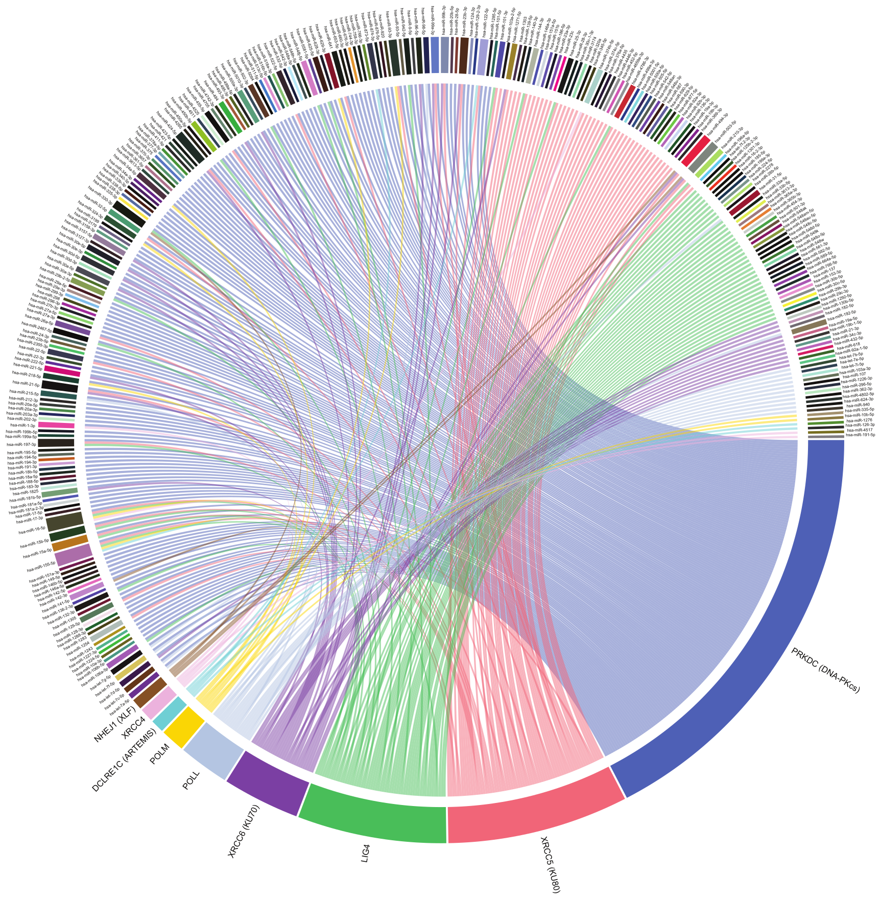
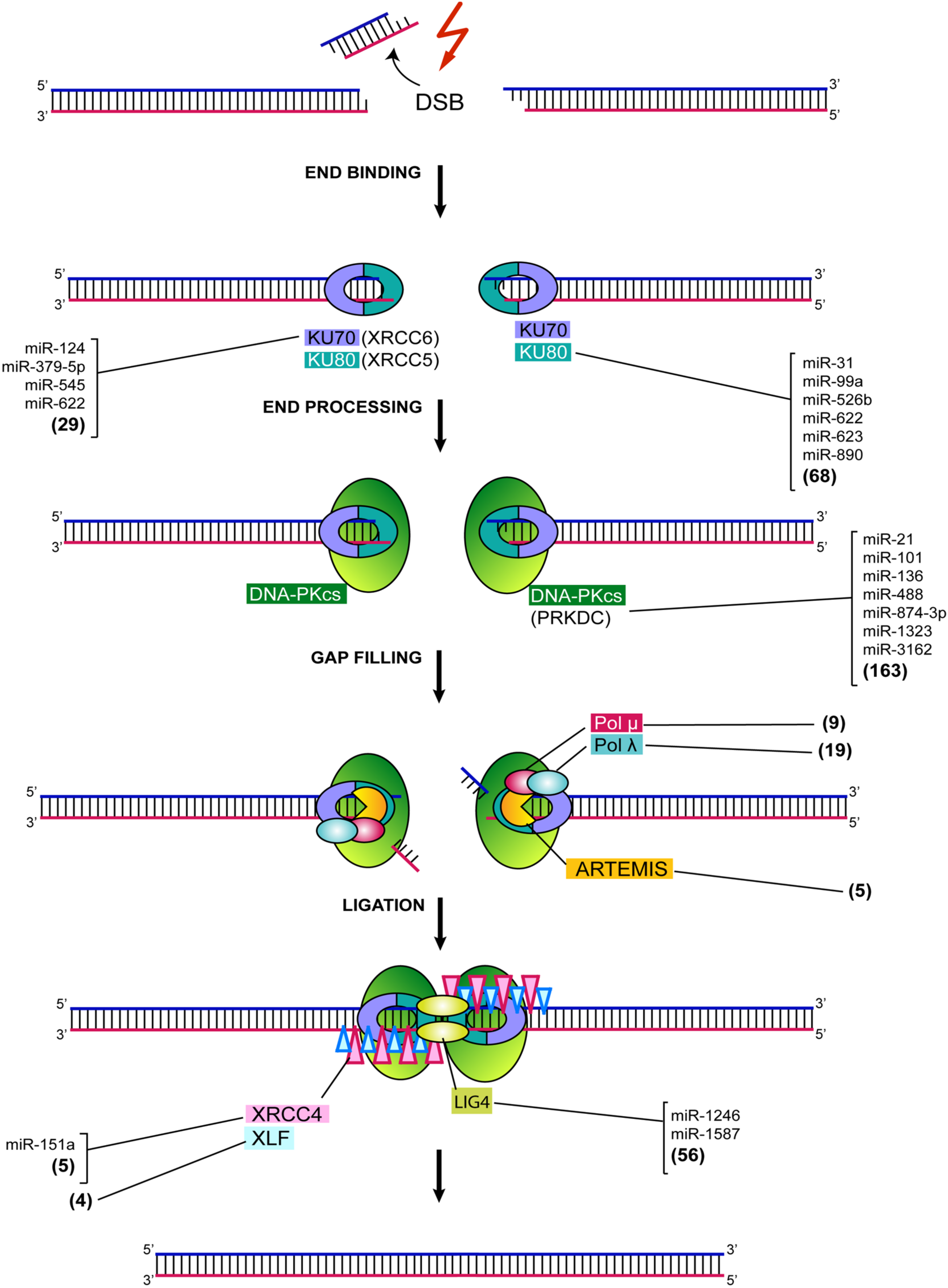
2. MiRNA-Mediated Regulation of NHEJ

DSBs, occurring throughout the cell cycle, are repaired predominantly by the NHEJ pathway [5]. In human cells, NHEJ repairs nearly all DSBs outside of S and G2 cell cycle phases and even about 80% of DSBs within S and G2 that are not proximal to the replication fork [18,19]. More detailed reviews about DNA repair by NHEJ can be found elsewhere [5,19,20]. There is an increasing number of miRNAs reported to act as regulators of the various genes involved in the NHEJ repair. Disturbances in the expression of such genes could have important consequences for the fate of cells, due to inadequate repair of DSBs, resulting in proliferation perturbation and apoptosis [21]. Below, we describe the relationship between the main genes involved in the DSB repair mechanism and some microRNAs.

3. Ku70/80

When a DSB arises in the DNA of mammalian cells, it is first recognized by a heterodimer, consisting of Ku70 and Ku80 (Ku), considered as a Ku:DNA complex, which serves as a node at which the nuclease, polymerases, and ligase of NHEJ can dock later (Figure 3) [3,18].
There are several miRNAs reported to modulate Ku70 (XRCC6) and Ku80 (XRCC5) expression (Figure 3). It has been observed that miR-545 increases radiosensitivity in lung carcinoma cells via inhibiting Ku70 expression [22]. Similarly, up-regulation of miR-379-5p inhibited proliferation and sensitized granulosa cells to DNA damage by repressing Ku70 [23]. MiR-124 has been shown to be up-regulated in brain tissues of ischemia stroke mice and rats. The targeting of Ku70 by miR-124 under these conditions, has been associated with altered cell proliferation and apoptosis [24]. Transfection with miR-890 caused a reduced expression of Ku80, which resulted in a significantly delayed IR-induced DNA damage repair in a prostate cancer model [25]. It has been identified that miR-623 binds to Ku80, thus, significantly decreasing its expression, causing a reduction in cell proliferation, clonogenicity, migration and invasion in lung adenocarcinoma cells [26]. Low constitutive Ku80 mRNA expression, together with radiation-induced high miR-99a expression in peripheral blood lymphocytes, were associated with late rectal bleeding in prostate cancer patients, receiving intensity-moderated radiation therapy [27]. Further, miR-31 was shown to be up-regulated in squamous cell carcinoma and it has been identified to target Ku80, promoting its down-regulation and causing impairment of DNA repair activity [28]. It has been identified that miR-526b binds to Ku80 mRNA, thus, modulating Ku80 expression in non-small cell lung carcinoma cells. miR-526b overexpression was shown to induce S-phase cell cycle arrest and apoptosis and enhanced the expression levels of the p53 and p21, thus, inhibiting lung tumor growth [29]. It has been observed that miR-622 regulates the expression of the Ku complex through direct interaction with Ku70 and Ku80 transcripts, and specifically suppresses NHEJ during the S cell cycle phase and enhances initiation of HR-mediated DSB repair in the S phase, by facilitating the recruitment of MRE11. This latter suggests a role for miR-622 in regulating the balance between HR and NHEJ in the cell cycle [30]. It was demonstrated that miR-502 directly targets Ku70 and XLF in pancreatic cell lines, which might modulate both the assembly of the Ku70/80 complex and final steps of ligation during NHEJ; overexpression of this miRNA makes cells susceptible to DNA damage [31].

4. DNA-PKcs

Ku functions as a specific cofactor for stable recruitment of the DNA-protein kinase catalytic subunit DNA-PKcs (encoded by PRKDC/XRCC7) to DSB sites (Figure 3). DNA-PKcs, thus, acts as a sensor for DSBs, and its major role is to promote NHEJ [32]. The kinase activity of DNA-PKcs regulates end processing and NHEJ through autophosphorylation and also facilitates recruitment of downstream effectors. Given the role of DNA-PKcs in DSB repair, it becomes evident that alterations in its expression might compromise the activity of NHEJ downstream effectors, thereby affecting the ability of cells to deal with DNA damage. There are a number miRNAs identified to target DNA-PKcs (Figure 3). It has been reported that miR-101 directly targets PRKDC. Up-regulation of the miRNA efficiently reduced the protein levels of DNA-PKcs in human lung cancer cells and sensitized them to radiation, both in vitro and in vivo [33,34]. Sensitization to gemcitabine was also observed in pancreatic cancer cells [35] and there is an augmented salinomycin cytotoxicity against osteosarcoma cells [36]. As expected, down-regulation of miR-101 has been associated with DNA-PKcs overexpression and mediated oncogenic actions in renal and hepatocellular carcinoma cells [36,37]. Likewise, ectopic expression of miR-488 resulted in markedly increased cisplatin sensitivity of malignant melanoma cells, due to DNA-PKcs silencing, in vitro and in vivo [38,39] (Figure 3). miR-136 overexpression down-regulated DNA-PK in ovarian cancer cells [40]. It has been shown that overexpression of miR-874-3p has a great impact on the survival of non-small cells lung cancer (NSCLC) A549 cells after irradiation, by targeting DNA-PKcs and, therefore, leading to a 20% radiosensitivity increase [41]. miR-1323 was demonstrated to bind to DNA-PKcs in A549 cancer cells. Ectopic expression of this miR-1323 significantly promoted DNA-PKcs expression and increased the survival of irradiated cells. This latter suggests a plausible mechanism of resistance to radiation via enhanced DNA repair [38].
It has been observed that miR-3162 forms a miRNA/Ago2/YY1/PcG group protein/DNMT complex that can bind to the promoter region of DNA-PKcs, contributing to histone and DNA methylation in bortezomib-treated leukemic cells, thus, mediating the epigenetic gene silencing of DNA-PKcs [42].

5. DCLRE1C, DNA Pol M and Pol L

Some DSBs that are repaired by NHEJ require additional accessory factors, such as nucleases for adequate repair. One such factor is the endonuclease DCLRE1C (Artemis), which interacts with DNA-PKcs to promote the repair of a subset of DSBs, which appear to represent those that incur some level of resection, possibly owing to their increased complexity [32,43] (Figure 3). DNA POLM (Pol µ) and POLL (Pol λ) are two low-fidelity polymerases, involved in NHEJ in human cells [44,45]. Both polymerases can incorporate nucleotides in a template-dependent or template-independent manner [46]. The activity of these polymerases further explains the high level of diversity that can occur at NHEJ junctions and demonstrated that, although resection is one way of generating short stretches of homology between broken DNA ends, the template-independent nucleotide addition of one or both broken DNA ends is another [18]. At the time of writing this review, there are still no published papers about a miRNA-mediated regulation Artemis, Pol μ and Pol λ.

6. XRCC4: DNA Ligase IV Complex and XLF

DNA ligase IV (LIG4) is the most flexible known ligase, since it has the ability to ligate across gaps and ligate incompatible DNA ends [3,47] (Figure 3). LIG4 functions exclusively in NHEJ, making it a central component for the DNA repair process. The protein encoded by XRCC4 combines with LIG4 in the repair of DSBs through NHEJ. Coupling with XRCC4 stimulates LIG4 enzyme activity and loss of either LIG4 or XRCC4, and severely compromises NHEJ [18]. XRCC4 is a direct target of miR-151a, and it has been observed that stable overexpression of miR-151a recovered the temozolomide (TMZ) sensitivity of TMZ-resistant glioblastoma (GBM) cells in vitro, thus, demonstrating the importance of miR-151a/XRCC4 signaling in modulating NHEJ repair and TMZ resistance in GBM [48]. It was demonstrated that miR-1246 packaged in exosomes secreted from irradiated bronchial epithelial BEP2D cells down-regulates LIG4 expression and inhibits proliferation of non-irradiated cells [49]. LIG4 expression was significantly inhibited by overexpression of miR-1587, thus, leading to increased radiosensitivity of CRC cells through DSBs accumulation, cell cycle arrest and apoptosis [50]. XLF is a protein that permits efficient ends ligation by interacting with XRCC4, allowing XLF to complex with XRCC: LIG4 [18]. It remains to be established if there is an XLF regulation mediated by miRNAs.
Vast evidence shows that miRNAs play an important role in the regulation of NHEJ (Figure 3). As we have seen, alterations in the expression levels of NHEJ gene-targeting miRNAs can have an important impact on the DNA damage repair ability of cells, thus, affecting processes, such as radiation and drug sensitivity. However, it remains to be studied in more detail, the processes triggering such alterations, as well as their possible consequences for the cell.

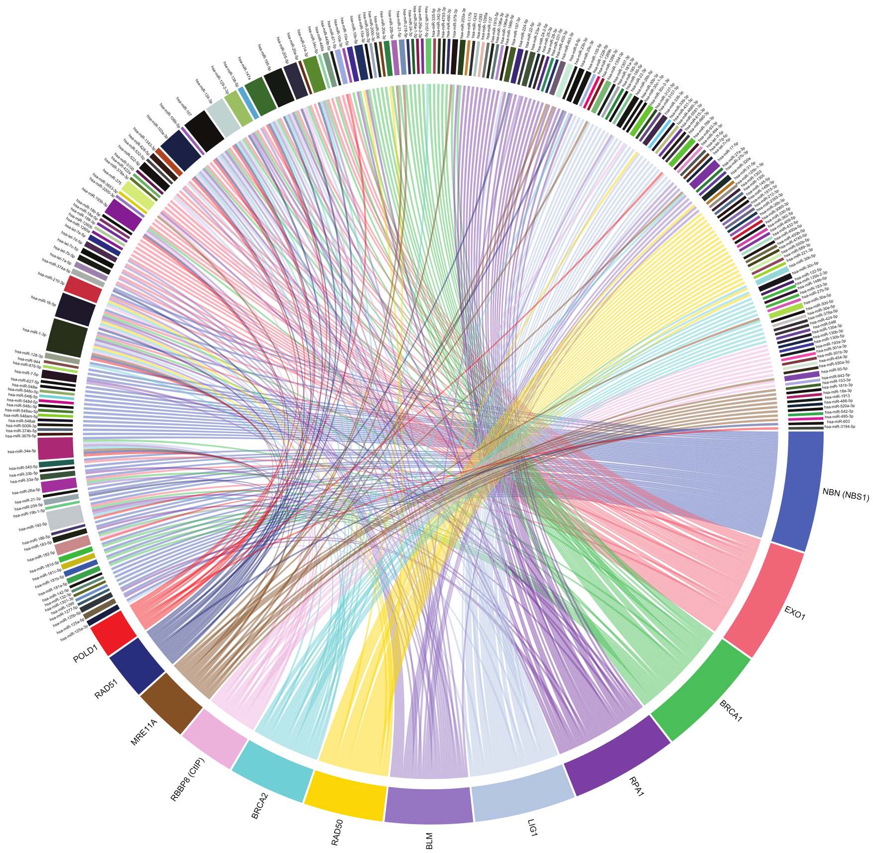
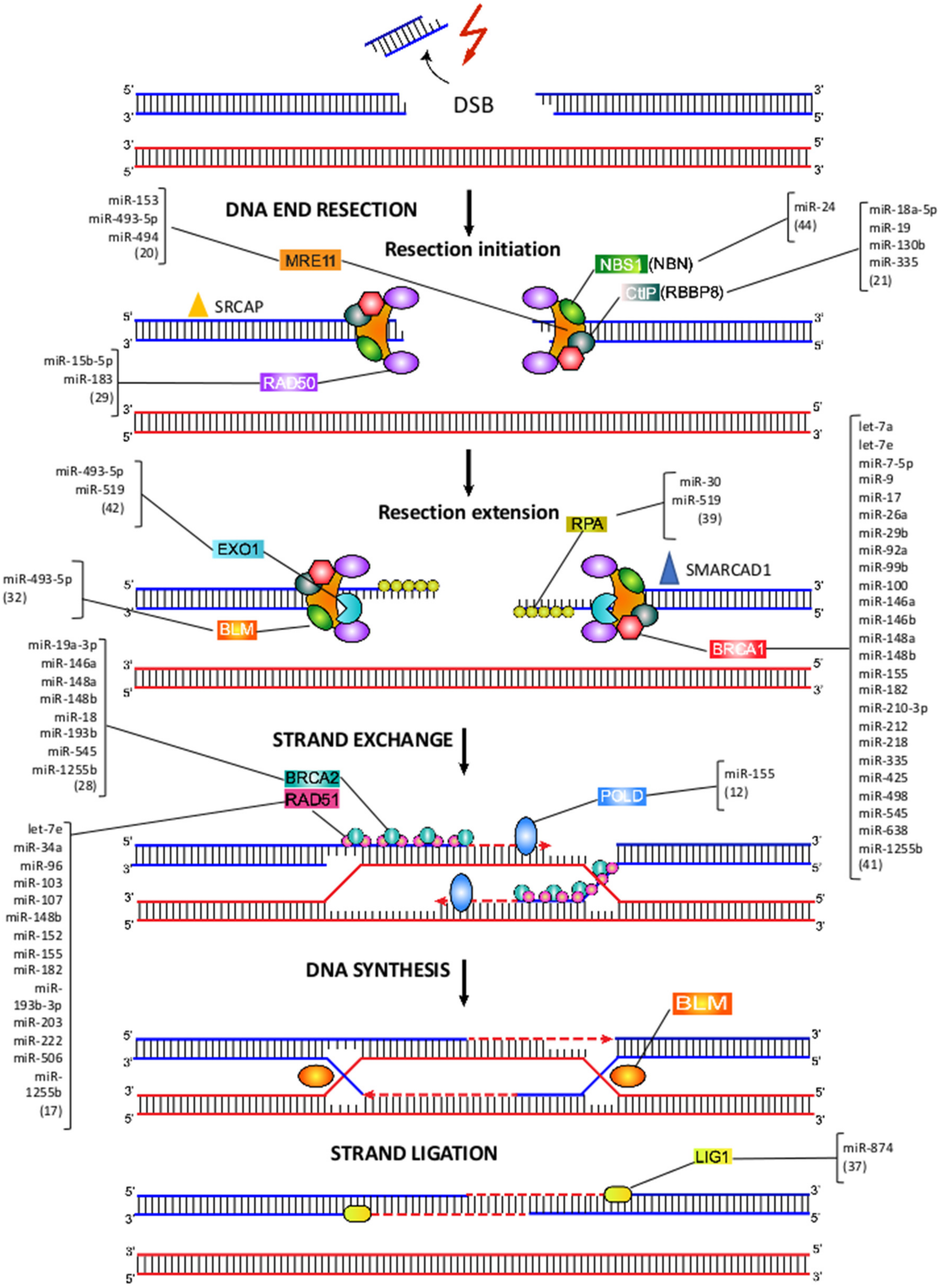
7. MiRNA-Mediated Regulation of HRR

HRR requires the invasion of homologous or sister chromatid strands to repair DNA end DSBs, and is considered a typically error-free DNA repair mechanism [51] (Figure 4). A critical initial step in establishing this repair mechanism is DNA end resection, which generates a long 3′ single-stranded DNA that can invade the homologous DNA strand. This step, in addition to blocking the entry of repair Ku proteins by NHEJ, promotes ATM and ATR activation and is, therefore, restricted to the late S phase and G2 phases of the cell cycle [51] (Figure 4). DNA end resection initiation begins with MRN complex (MRE11-RAD50-NBS1), SRCAP, and CtIP. It is important to mention that the dual activity of MRE11, functioning as both endo and exonuclease, is essential to carry out resection initiation. After that, resection extension continues, in which SMARCAD1 cooperates with EXO1 and BLM/DNA2. Subsequently, the process continues when the ssDNA-ends are covered with RPA. In a third step, RPA is replaced by RAD51 in a BRCA1/2-dependent process, to ultimately perform the recombinase reaction, using a homologous DNA template [52]. For a detailed description of HRR, there are more extensive reviews in this regard [8,52,53,54,55].

8. MRE11-RAD50-NBS1 Complex

The complex of proteins MRE11-RAD50-NBS1 (NBN) (MRN complex) acts as DSB sensor, co-activator of DSB-induced cell cycle checkpoint signaling, and as a DSB repair effector in the HRR pathway [56,57] (Figure 4). A seven-miRNA signature (miR-103, miR-494, miR-99b, miR-21, miR-224, miR-92a, and let-7a) has been reported to be up-regulated in endothelial cells after exposure to radiation, hydrogen peroxide, and cisplatin [58]. Among these miRNAs, miR-494 has been demonstrated to target the MRN complex, which, in turn, leads to senescence and decreased angiogenesis [59].
MRE11 acts as an exonuclease, generating 3′ overhangs that become a substrate for HRR [60]. MRE11 overexpression, commonly observed among cancer patients, has been postulated as a mechanism responsible for increasing cancer risk [61]. miR-493-5p has been demonstrated to downregulate MRE11, along with 14 other genes involved in genomic stability in BRCA2 mutant cells, thus, suggesting a possible role for miR-493-5p in regulating DSB repair, through HRR and resistance to PARP inhibitors and platinum [62]. It has been observed that miRNAs whose expression levels decrease during aging, when transfected into aged mouse livers, resulted in a significant increase in MRE11 expression [63]. Additionally, polymorphisms in predicted miRNA target sites of MRE11, NBS1, RAD51 and RAD52 have been associated with different cancer risks, susceptibility, and survival [64,65]. It has been reported that miR-153 can bind to MRE11, thus, inhibiting its expression in muscle-invasive bladder tumor cells, suggesting a post-transcriptional down-regulation of MRE11A due to miR-153 up-regulation [66].
MRE11 and RAD50 have been observed to form a large ATP-controlled molecular clamp, suited to recognize even blocked DSBs [67]. An inverse correlation between miR-183 and RAD50 expression levels has been observed after pomegranate extract treatment in MCF-7 cells, accompanied by an increase in DSBs, as revealed by an elevated frequency of γ-H2AX foci, followed by cell cycle arrest in G2/M and the induction of apoptosis [68].
It was found that Staphylococcus aureus induces host miR-15b-5p, in both acute wounds and diabetic foot ulcers (DFUs). S. aureus-mediated overexpression of miR-15b-5p resulted in suppression of DNA damage repair mechanisms and inflammatory response in DFUs. Putative target genes of miR-15b included RAD50, which was down-regulated in DFUs. Because of the inhibition of DNA repair mechanisms, DSBs were accumulated in DFU tissue, as indicated by the presence of γH2AX, indicating a mechanism of S. aureus-mediated induction of miR-15b that results in deregulation of DNA repair mechanisms and inflammatory response, contributing to the inhibition of healing in DFUs [69].
Further, miR-24 caused a decrease in the expression of NBS1 by targeting c-Myc, which is known to positively regulate the expression of NBS1. Down-regulation of NBS1 by miR-24 was thought to improve myoblasts’ permissiveness to adeno-associated virus (AAV) transduction, possibly by avoiding MRN sensing and binding to AAV genome [70].

9. CtIP, EXO1 and RPA

In mammals, DNA-end resection is catalyzed by the MRN complex, together with CtIP (CtBP-interacting protein/RBBP8) and EXO1 [71] (Figure 4). CtIP (RBBP8) physically interacts with MRN and, along with EXO1, promotes DSB resection [72]. The targeting of CtIP by aberrantly expressed miR-19 impairs CtIP-mediated DNA-end resection, which resulted in reduced HRR levels and DNA damage hypersensitivity [73]. IR-induced DSBs activate DSB signaling kinase ATM, which leads to the downregulation of miR-335. Overexpression of miR-335 in HeLa cells resulted in reduced CtIP levels and low post-IR colony survival and BRCA1 foci formation. Further, in two patient-derived lymphoblastoid cell lines, with decreased post-IR colony survival, a radiosensitive phenotype, it was demonstrated that elevated miR-335 expression reduced CtIP levels and reduced BRCA1 foci formation [74]. It was demonstrated that miR-18a-5p directly binds to CtIP and inhibits its expression in nasopharyngeal carcinoma cells, thus, affecting cell proliferation and apoptosis. However, miR-18a-5p activity on CtIP is modulated by the lncRNA CASC2 via sponging miR-18a-5p and modulating CtIP expression in vivo [75]. It has been observed that CtIP is a miR-19 target and aberrant expression of the miRNA suppresses DNA-end resection and faithful repair of DSBs by HRR in HEK293T cells, thus, promoting genomic instability [75]. Overexpression of miR-130b resulted in increased DNA damage levels by promoting DSBs through CtIP targeting, as evidenced by elevated phosphorylation of H2AX and increased DNA damage, observed through comet assay in Hela and Siha cells, which was accompanied by accelerated cell apoptosis in, combination with PARP inhibitors [76].
RPA is a heterotrimeric complex (RPA1/2/3), which binds to single-stranded DNA, forming a nucleoprotein complex, protecting from nucleases, preventing formation of secondary structures that would interfere with repair [77,78]. It was reported that miR-519 repressed the expression of RPA and EXO1, thus, augmenting DNA damage, as revealed by the increase in γH2AX foci in miR-519-transfected HeLa cells [79]. Lastly, it was observed that miR-30 binds directly to RPA, overexpression of the miRNA increased DNA damage, cell cycle arrest and inhibited cell proliferation in gastric cancer cells [80]. In another study, miR-493-5p has been demonstrated to downregulate RPA, along with 14 other genes involved in genomic stability in BRCA2 mutant cells, thus, suggesting a possible role for miR-493-5p in regulating DSB repair, through HRR and resistance acquisition of BRCA2 mutant cells to PARP inhibitors and platinum [62].

10. BRCA1 and BRCA2

BRCA1 and BRCA2 are important proteins for the repair of DSBs by HRR. There are numerous reports demonstrating that BRCA1 and BRCA2 expression can be modulated, either directly or indirectly by miRNAs. It has been established that miR-1255b, miR-148b and miR-193b directly target the transcripts of BRCA1, BRCA2 and RAD51, therefore, suppressing the HRR pathway during the G1 phase. Inhibition of these miRNAs increases BRCA1, BRCA2 and RAD51 expression in the G1 phase, thus, leading to impaired DSB repair, which, in turn, can lead to a loss of heterozygosity [81]. BRCA1/BRCA2 mutations and aberrant expression are common as the hallmarks of ovarian and breast cancer [82,83]. It has been reported that miR-182 overexpression contributes to aggressive ovarian cancer, largely by its negative regulation of multiple tumor suppressor genes, involved in tumor growth, invasion, metastasis, and DNA instability [84]. It was reported that expression of BRCA1 and BRCA2, both let-7a targets, was down-regulated after delivery of magnetic core–shell nanoparticle (MCNP)/let-7a constructs to breast cancer cells. Co-delivery of let-7a with doxorubicin induced a synergistic effect, which resulted in a significant diminution of chemoresistance, DNA repair and cell viability [85]. Up-regulation of miR-146a, -148a and -545 were associated with overall survival and progression-free survival in patients with wild-type BRCA1/2. These miRNAs were predicted to target BRCA1/2 and, thus, inferred to alter the DNA damage repair [86]. In a screen to evaluate tumor suppressor genes, oncogenes and miRNA expression in samples of colorectal cancer, it was found that miR-17, miR-425 and miR-92 overexpression were significantly associated with up-regulated expression of BRCA1, suggesting a possible role of these miRNAs in the development of the disease [87].
BRCA1 forms a complex with CtIP and the MRN complex and, thus, plays a crucial role in promoting DSB end resection, which generates a long 3′ single-stranded DNA tail that can invade the homologous DNA strand [88,89]. MiR-182, a BRCA1 negative regulator, is significantly overexpressed in high-grade serous ovarian carcinoma, which results in a significant reduction in BRCA1 expression. As a consequence, this can cause impairment of the repair of DSBs, increased tumor transformation, invasiveness and metastasis [90,91]. Additionally, it has been observed that overexpression of miR-182 impairs HRR activity and renders HL60 cells hypersensitive to IR [92]. Furthermore, it has been proposed that TGFβ acts as stringent regulator of the DNA damage response via suppression of miR-182, which directly targets BRCA1 [93]. In breast tumors cell lines, high levels of miR-146a and miR-146b were inversely correlated with BRCA1 expression. Such direct downregulation was associated with an increased proliferation and reduced HRR rate [94,95].
In a screen to determine miRNAs modulating cell response to DNA damage, it was found that the expression of members of the miR-99 family were up-regulated, following DNA damage, and miR-99 expression correlated with radiation sensitivity. The downregulation of the remodeling complex SNF2H, a miR-99 family target, mediated IR sensitivity through its role in facilitating DNA repair. The up-regulation of the miR-99 family, following IR, decreased the efficiency of BRCA1 recruitment and the rate of DNA repair after exposure to IR [96]. In a different study, it was demonstrated that miR-210-3p attenuates the G2/M cell cycle checkpoint by inactivating the BRCA1 complex function, in response to DNA damage under hypoxia, via targeting the 3′ UTR region of BARD1 mRNA in endometrial stromal cells [97].
The overexpression of miR-7 caused indirect down-regulation of BRCA1 in lymphoblasts, accompanied by an increase in γH2AX, reflecting augmented DNA damage, apoptosis and proliferation inhibition [98]. MiR-212 was identified to contribute to radio-resistance of U251 glioma cells and was shown to attenuate radiation-induced apoptosis by BRCA1 direct targeting [99]. It was confirmed that miR-9 binds directly to BRCA1, thus, decreasing gene expression. Such low BRCA1 expression and high expression of miRNA-9 was associated with platinum sensitivity and longer progression-free survival in ovarian cancer cells [100]. Down-regulation of miR-218 was observed in cisplatin-resistant breast cancer cell lines and it was identified that BRCA1 was the cellular target of miR-218. Restoring miR-218 expression in the MCF-7/DDP cell line could sensitize cells against cisplatin, thereby increasing cisplatin-mediated tumor cell apoptosis and reducing DNA repair [101]. In another study, miR-638 overexpression increased sensitivity to DNA-damaging agents, UV and cisplatin, reduced proliferation rate, as well as decreased invasive ability in TNBC cells. Reduced proliferation, invasive ability, and DNA repair capabilities were associated with down-regulated BRCA1 expression [102].
Overexpression of miR-335 in MCF7 cells resulted in an up-regulation of BRCA1 expression, through inhibition of BRCA1 negative regulator, ID4, which, in turn, impacts on cellular functions, such as proliferation and apoptosis [103]. Expression of miRNA-26a, -29b, -100, and -148a increased in BRCA1 wild type cells (MDB-MB-231), after exposure to gemcitabine, alone and in combination with the PARP1 inhibitor [104]. miR-498 has been found highly expressed in triple-negative breast cancer cells, and its expression was negatively correlated with the levels of BRCA1. It was demonstrated that miR-498 inhibits BRCA1 expression, thus, leading to promoted cell proliferation [105].
BRCA1 was also reported to be targeted by miR-7-5p in non-small cell lung cancer cells. It was observed that such BRCA1 targeting is regulated through lncRNA MEG3 by competitive binding to miR-7-5p [106]. miR-7-5p overexpression was also associated with suppression of cell proliferation and promotion of apoptosis by targeting BRCA1 in TK6 cells [98]. It was reported that BRCA1 can also act as a transcriptional repressor of miR-155 [107]. Up-regulation of miR-155 in breast cancer is likely related to tumor initiation. In contrast to BRCA1, FOXP3 is a transcriptional inducer of miR-155 in breast cancer cell lines. miR-155 is associated with transcriptional regulation by FOXP3, through BRCA1 transcriptional inhibition, subsequently controlling miR-155 and its targets, such as RAD51 [107]. MiR223-3p is a negative regulator of the NHEJ DNA repair and represents a therapeutic pathway for BRCA1- or BAP1-deficient cancers [108].
BRCA2 is targeted by miR-19a-3p in NSCLC, A549; the overexpression of this miRNA affects cell survival after irradiation [41]. BRCA2 is the main mediator of RAD51 nucleofilament formation and strand exchange in mammalian cells [51]. Mutations affecting the proper function of these proteins are associated with increased genomic instability. High genomic instability is thought to be responsible for the significantly increased cancer risk in patients with familial or germline BRCA mutations [109].

11. RAD51

RAD51 forms nucleoprotein filaments with DNA catalyzing the transfer of the new DNA strand between a broken sequence and its homolog, to resynthesize the damaged region [55]. Transfection with let-7e sensitized epithelial ovarian cancer cells to cisplatin, down-regulated BRCA1 and RAD51 expression, and repressed the repair of cisplatin-induced DSBs [110]. It has been reported that, miR-182 targets RAD51 and over-expression of the miRNA decreases HRR, thus, sensitizing acute myelogenous leukemia cells to capecitabine [111]. Overexpression of miR-155 in human breast cancer cells reduces the levels of RAD51 and affects the cellular response to IR, decreasing the efficiency of HRR and enhancing sensitivity to IR [112]. Additionally, HRR decreased efficiency by miR-155 overexpression drives a concurrent NHEJ activity increase and, thus, a higher mutation frequency [113]. Over-expression of miR-203 also increased IR sensitivity of human malignant glioma cell lines and prolonged radiation-induced γH2AX foci formation. It was demonstrated that miR-203 down-regulated RAD51 among other DNA damage response genes, which resulted in inhibited invasion and migration potentials [114]. It was observed that miR-34a directly binds and modulates RAD51 expression, therefore, inhibiting IR-induced DSBs repair in non-small cell lung cancer. Liposomal formulation, containing miR-34a, MRX34 plus radiotherapy, effectively inhibits tumor growth in lung cancer mouse models [115]. Down-regulation of RAD51 by miR-506 affects the HRR pathway and, consequently, increases sensitivity to cisplatin and the PARP inhibitor olaparib in serous ovarian cancer cells [116,117]. In a similar fashion, miR-103 and miR-107, when overexpressed, reduced HRR and sensitized osteosarcoma cells to various DNA-damaging agents, including cisplatin and a PARP inhibitor by targeting RAD51 [118]. Likewise, miR-107 and miR-222 regulate the DNA damage repair and sensitize tumor cells to olaparib by repressing expression of RAD51, thus, impairing DSB repair by HRR [119]. Another study showed that miR-96 directly targets the coding region of RAD51. Overexpression of miR-96 decreased the efficiency of HRR and enhanced the sensitivity of osteosarcoma cells to the PARP inhibitor, AZD2281, and cisplatin, indicating that miR-96 regulates DNA repair and chemosensitivity by repressing RAD51 [120]. Ectopic expression of miR-152 reversed cisplatin resistance, both in vitro and in vivo, by targeting RAD51. On the other hand, it was demonstrated that miR-98-5p could directly target Dicer, causing global miRNA down-regulation. However, miR-152 was identified as the main downstream target of miR-98-5p. Importantly, miR-98-5p expression was associated with poor outcome of epithelial ovarian cancer patients, presumably by indirectly regulating the biogenesis of miR-152 [121]. It has been shown that miR-193a-3p binds to RAD51 and modulates its expression. Such interaction is, in turn, modulated by the long RNA lnc-RI, which is also recognized by miR-193a-3p and acts as a competitive endogenous RNA (ceRNA) to stabilize RAD51 mRNA via competitive binding with miR-193a-3p [122].

12. Pol δ

Extensive evidence shows that POL δ (POLD) is central to HRR-associated DNA synthesis. POLD possesses both high fidelity polymerase and 3′ to 5′ exonuclease activity. This polymerase can efficiently extend up to 80% of the RAD51-mediated invading DNA strands [123]. It has been reported that miR-155 could modulate POLD1 expression through the targeting of FOXO3a, a transcription factor with putative binding sites in the promoter of each of the four polymerase delta subunits [113].

13. BLM and LIG1

BLM is a member of the RecQ family of DNA helicases, and its role in HRR is related to the dissolution of Holliday junctions, generated during the strand invasion step. Mutations in BLM are present in Bloom syndrome patients, and this syndrome is characterized by chromosomal instability and a 10-fold elevation in the frequency of sister chromatid exchanges [124]. BLM expression has been demonstrated to be regulated by miR-493-5p, with this miRNA also regulating the expression of 14 other genes involved in genomic stability in BRCA2 mutant cells, thus, suggesting a possible role for miR-493-5p in regulating DSB repair through HRR and resistance acquisition of BRCA2 mutant cells to PARP inhibitors and platinum [62].
The DNA synthesis from the 3′-end of the invading strand is followed by successive ligation by DNA ligase I (LIG1). A study indicates that LIG1 targeting by miR-874 promotes a radiosensitizing effect in lung cancer cells [41]. So far, no other miRNAs regulating the expression of LIG1 and its consequences on DNA repair have been reported.

14. Other Validated MiRNA–MRNA Interactions

There are a large number of miRNA–mRNA interactions that remain to be studied, to better understand the role of miRNAs in the repair of DSBs. According to a data analysis performed to obtain validated human miRNA–mRNA interactions, involving the genes reviewed in this work, there are 210 miRNAs that interact with HRR genes and 275 miRNAs interacting with NHEJ genes, thus, forming 362 and 358 interactions in HRR and NHEJ, respectively (Figure 1 and Figure 2). Interactions were obtained from TarBase v.8 database [16], which contains experimentally validated miRNA–mRNA interactions, through techniques including microarray, reporter gene assay, qPCR, western blot and RNA-Seq. The vast majority of these interactions remain to be studied within the context of DNA repair. Interestingly, PRKDC mRNA (DNA-PKcs) interacts with 163 different miRNAs, which places it as the gene with the highest number of validated interactions in NHEJ (Table S1). The functioning of NHEJ depends largely on the activity of DNA-PKcs; therefore, its expression needs to be highly regulated so there is an adequate repair of DSBs. Such a degree of regulation could be mediated by the large number of miRNAs targeting PRKDC. Regarding the HRR genes, NBN, EXO1, and BRCA1 top the list of genes with the most miRNA interactions, with 44, 42 and 41 interactions, respectively (Figure 2; Table S2). Despite the large number of miRNAs targeting these genes, with the exception of BRCA1, there are very few published studies that explore its regulation mediated by miRNAs.
The miRNA–mRNA interaction data analysis also revealed that miR-155-5p and miR-16-5p are the miRNAs with the most NHEJ gene interactions, by targeting five genes each (PRKDC, XRCC5, LIG4, POLM and DCLRE1C for miR-155-5p and XRCC4 for miR-16-5p) (Figure 1; Table S2). Interestingly, miR-1-3p showed a strikingly large number of interactions with HRR genes. It was observed that miR-1-3p interacts with 11 HRR genes (BRCA1, BRCA2, EXO1, LIG1, MRE11A, NBN, POLD1, RAD50, RAD51, RBBP8 and RPA1) (Figure 2; Table S2). These data suggest a prominent role for these miRNAs in the post-transcriptional regulation of NHEJ and HRR genes; however, their participation in this aspect has not yet been sufficiently studied.

15. Concluding Remarks

In recent years, interest and reports about the regulatory role of miRNAs in DNA repair have been increasing. Here we reviewed several examples of miRNAs regulating expression of genes involved in DSBs repair. Since DNA repair mechanisms are of great interest in cancer research, it is not surprising that some of the most studied genes for cancer treatment, such as BRCA1 and RAD51, have an equally high number of reports showing miRNA-mediated regulation of these genes. A deeper study on the functioning of other genes involved in HRR and NHEJ is needed to establish their usefulness as new cancer therapy targets. In this sense, knowing the regulation that miRNAs exert on their expression and activity will be of great importance.
DNA repair mechanisms are a promising target for therapeutic intervention. Many of the anti-cancer therapies are aimed at generating DNA damage, specifically DSBs, thus, reducing cell proliferation and inducing apoptosis. However, development of resistance to radiotherapy, chemotherapy or other targeted therapies can occur during the course of treatment due to deregulation of gene expression. For example, more than one hundred genes have been classified into seven categories of mechanisms of chemoresistance (MOC1-7) in hepatocellular carcinoma (HCC), affecting the response to pharmacological strategies employed for HCC treatment [125]. Deregulation of the genes involved in DSB repair, such as XRCC4, XLF and ATM, have been classified as key players in DNA repair–MOC for HRCC (MOC4) [125]. As has been discussed throughout this review, miRNA-mediated modulation of DNA repair genes can prevent or reverse resistance to chemotherapeutics. Therefore, a better understanding about the regulation of DSB repair by miRNAs will allow us to discover new targets and to develop better strategies to overcome tumor resistance. While it is true that much remains to be learned about the regulation of gene expression through miRNAs, in recent years, there has been a growing interest to study the role of miRNAs in DNA repair and as targets for cancer therapy.

Supplementary Materials

The following supporting information can be downloaded at: https://0-www-mdpi-com.brum.beds.ac.uk/article/10.3390/ijms23063231/s1.

Author Contributions

Conceptualization, E.R., M.V. and R.I.P.-V.; methodology, E.R., M.V. and R.I.P.-V.; software, R.I.P.-V.; investigation, E.R., M.V. and R.I.P.-V.; writing—original draft preparation, E.R., M.V. and R.I.P.-V. All authors have read and agreed to the published version of the manuscript.

Funding

We thank PAPIIT (IN211120) for partial financial support.

Institutional Review Board Statement

Experimentally validated human miRNA–mRNA interactions involving NHEJ genes were extracted from TarBase v.8 interactions bulk file. Experimentally validated human miRNA–mRNA interactions involving HRR genes were extracted from TarBase v.8 interactions bulk file.

Informed Consent Statement

Not applicable.

Data Availability Statement

All data used are reflected in the supplemental tables.

Conflicts of Interest

The authors declare no conflict of interest.

Abbreviations

AAV (Adeno-associated virus); Ago2 (Argonaute RISC Catalytic Component 2), ATP (Adenosine triphosphate), BARD1 (BRCA1-associated RING Domain 1), BRCA1 (Breast Cancer Type 1 Susceptibility Protein), BRCA2 (Breast Cancer Type 2 Susceptibility Protein), CASC2 (Cancer Susceptibility 2), CtIP (CtBP-interacting protein/RBBP8), DCLRE1C (DNA Cross-Link Repair 1C / ARTEMIS), DFU (Diabetic foot ulcers), DNA-PKcs (PRKDC, Protein Kinase, DNA-Activated, Catalytic Subunit), DSB (double-strand break), EXO1 (Exonuclease 1), FOXP3 (Forkhead Box P3), GBM (Glioblastoma), H2AX (H2A.X Variant Histone), HCC (Hepatocellular carcinoma); HRR (homologous recombination repair), ID4 (Inhibitor Of DNA Binding 4, HLH Protein), IR (ionizing radiation), Ku70 (XRCC6, X-Ray Repair Cross Complementing 6), Ku80 (XRCC5, X-Ray Repair Cross Complementing 5), LIG1 (DNA Ligase 1); LIG4 (DNA Ligase 4); MCNP (Magnetic core–shell nanoparticles); MEG3 (Maternally Expressed 3); MOC (Mechanism of chemoresistance); MRE11 (MRE11 Homolog, Double-strand Break Repair Nuclease); MRN (MRE11-RAD50-NBS1); NBS1 (NBN, Nibrin); NHEJ (non-homologous end joining); NSCLC (Non-squamous cell lung cancer); PARP1 (Poly(ADP-Ribose) Polymerase 1); PcG (Polycomb group); POLD1 (DNA Polymerase Delta 1, Catalytic Subunit); POLL (DNA Polymerase Lambda); POLM (DNA Polymerase Mu); qPCR (quantitative polymerase chain reaction); RAD50 (RAD50 Double-strand Break Repair Protein); RAD51 (RAD51 Recombinase); RAD52 (RAD52 Homolog, DNA Repair Protein); RBBP8 (RB Binding Protein 8, Endonuclease); RNA-Seq (RNA sequencing); RPA (Replication Protein A1); RPA (RPA1, RPA Interacting Protein); SNF2H (SMCARCA5, SWI/SNF Related, Matrix-Associated, Actin-Dependent Regulator Of Chromatin); SRCAP (SNF2 related CREBBP activator protein; TMZ (Temozolomide); XLF (NHEJ1, Non-Homologous End Joining Factor 1); XRCC4 (X-Ray Repair Cross Complementing 4); YY1 (YY1 Transcription Factor).

References

  1. Goldstein, M.; Kastan, M.B. The DNA damage response: Implications for tumor responses to radiation and chemotherapy. Annu. Rev. Med. 2015, 66, 129–143. [Google Scholar] [CrossRef] [PubMed] [Green Version]
  2. Jackson, S.P.; Bartek, J. The DNA-damage response in human biology and disease. Nature 2009, 461, 1071–1078. [Google Scholar] [CrossRef] [PubMed] [Green Version]
  3. Lieber, M.R.; Wilson, T.E. Snap Shot: Nonhomologous DNA end joining (NHEJ). Cell 2010, 142, 496. [Google Scholar] [CrossRef] [PubMed] [Green Version]
  4. Lieber, M.R. The mechanism of double-strand DNA break repair by the nonhomologous DNA end-joining pathway. Annu. Rev. Biochem. 2010, 79, 181–211. [Google Scholar] [CrossRef] [Green Version]
  5. Chang, H.H.Y.; Pannunzio, N.R.; Adachi, N.; Lieber, M.R. Non-homologous DNA end joining and alternative pathways to double-strand break repair. Nat. Rev. Mol. Cell Biol. 2017, 18, 495–506. [Google Scholar] [CrossRef]
  6. Chiruvella, K.K.; Liang, Z.; Wilson, T.E. Repair of double-strand breaks by end joining. Cold Spring Harb. Perspect. Biol. 2013, 5, a012757. [Google Scholar] [CrossRef]
  7. Karanam, K.; Kafri, R.; Loewer, A.; Lahav, G. Quantitative live cell imaging reveals a gradual shift between DNA repair mechanisms and a maximal use of HR in mid S phase. Mol. Cell 2012, 47, 320–329. [Google Scholar] [CrossRef] [Green Version]
  8. Heyer, W.D.; Ehmsen, K.T.; Liu, J. Regulation of homologous recombination in eukaryotes. Annu. Rev. Genet. 2010, 44, 113–139. [Google Scholar] [CrossRef] [Green Version]
  9. Harper, J.W.; Elledge, S.J. The DNA damage response: Ten years after. Mol. Cell 2007, 28, 739–745. [Google Scholar] [CrossRef]
  10. Bartel, D.P. Metazoan MicroRNAs. Cell 2018, 173, 20–51. [Google Scholar] [CrossRef] [Green Version]
  11. Berezikov, E. Evolution of microRNA diversity and regulation in animals. Nat. Rev. Genet. 2011, 12, 846–860. [Google Scholar] [CrossRef] [PubMed]
  12. Lewis, B.P.; Shih, I.H.; Jones-Rhoades, M.W.; Bartel, D.P.; Burge, C.B. Prediction of mammalian microRNA targets. Cell 2003, 115, 787–798. [Google Scholar] [CrossRef] [Green Version]
  13. Bartel, D.P.; Chen, C.Z. Micromanagers of gene expression: The potentially widespread influence of metazoan microRNAs. Nat. Rev. Genet. 2004, 5, 396–400. [Google Scholar] [CrossRef] [PubMed]
  14. Hornstein, E.; Shomron, N. Canalization of development by microRNAs. Nat. Genet. 2006, 38, S20–S24. [Google Scholar] [CrossRef] [PubMed]
  15. Zhang, X.; Wan, G.; Berger, F.G.; He, X.; Lu, X. The ATM kinase induces microRNA biogenesis in the DNA damage response. Mol. Cell 2011, 41, 371–383. [Google Scholar] [CrossRef] [Green Version]
  16. Karagkouni, D.; Paraskevopoulou, M.D.; Chatzopoulos, S.; Vlachos, I.S.; Tastsoglou, S.; Kanellos, I.; Papadimitriou, D.; Kavakiotis, I.; Maniou, S.; Skoufos, G.; et al. DIANA-TarBase v8: A decade-long collection of experimentally supported miRNA-gene interactions. Nucleic Acids Res 2018, 46, D239–D245. [Google Scholar] [CrossRef] [Green Version]
  17. Gu, Z.; Gu, L.; Eils, R.; Schlesner, M.; Brors, B. Circlize Implements and enhances circular visualization in R. Bioinformatics 2014, 30, 2811–2812. [Google Scholar] [CrossRef] [Green Version]
  18. Pannunzio, N.R.; Watanabe, G.; Lieber, M.R. Nonhomologous DNA end-joining for repair of DNA double-strand breaks. J. Biol. Chem. 2018, 293, 10512–10523. [Google Scholar] [CrossRef] [Green Version]
  19. Beucher, A.; Birraux, J.; Tchouandong, L.; Barton, O.; Shibata, A.; Conrad, S.; Goodarzi, A.A.; Krempler, A.; Jeggo, P.A.; Löbrich, M. ATM and Artemis promote homologous recombination of radiation-induced DNA double-strand breaks in G2. EMBO J. 2009, 28, 3413–3427. [Google Scholar] [CrossRef] [Green Version]
  20. Lieber, M.R.; Ma, Y.; Pannicke, U.; Schwarz, K. Mechanism and regulation of human non-homologous DNA end-joining. Nat. Rev. Mol. Cell Biol. 2003, 4, 712–720. [Google Scholar] [CrossRef]
  21. Vannini, I.; Fanini, F.; Fabbri, M. Emerging roles of microRNAs in cancer. Curr. Opin. Genet. Dev. 2018, 48, 128–133. [Google Scholar] [CrossRef] [PubMed]
  22. Liao, C.; Xiao, W.; Zhu, N.; Liu, Z.; Yang, J.; Wang, Y.; Hong, M. MicroR-545 enhanced radiosensitivity via suppressing Ku70 expression in Lewis lung carcinoma xenograft model. Cancer Cell Int. 2015, 15, 56. [Google Scholar] [CrossRef] [PubMed] [Green Version]
  23. Dang, Y.; Wang, X.; Hao, Y.; Zhang, X.; Zhao, S.; Ma, J.; Qin, Y.; Chen, Z.J. MicroRNA-379-5p is associate with biochemical premature ovarian insufficiency through PARP1 and XRCC6. Cell Death Dis. 2018, 9, 106. [Google Scholar] [CrossRef] [PubMed] [Green Version]
  24. Zhu, F.; Liu, J.L.; Li, J.P.; Xiao, F.; Zhang, Z.X.; Zhang, L. MicroRNA-124 (miR-124) regulates Ku70 expression and is correlated with neuronal death induced by ischemia/reperfusion. J. Mol. Neurosci. 2014, 52, 148–155. [Google Scholar] [CrossRef]
  25. Hatano, K.; Kumar, B.; Zhang, Y.; Coulter, J.B.; Hedayati, M.; Mears, B.; Ni, X.; Kudrolli, T.A.; Chowdhury, W.H.; Rodriguez, R.; et al. A functional screen identifies miRNAs that inhibit DNA repair and sensitize prostate cancer cells to ionizing radiation. Nucleic Acids Res. 2015, 43, 4075–4086. [Google Scholar] [CrossRef] [Green Version]
  26. Wei, S.; Zhang, Z.Y.; Fu, S.L.; Xie, J.G.; Liu, X.S.; Xu, Y.J.; Zhao, J.P.; Xiong, W.N. Hsa-miR-623 suppresses tumor progression in human lung adenocarcinoma. Cell Death Dis. 2016, 7, e2388. [Google Scholar] [CrossRef] [Green Version]
  27. Someya, M.; Yamamoto, H.; Nojima, M.; Hori, M.; Tateoka, K.; Nakata, K.; Takagi, M.; Saito, M.; Hirokawa, N.; Tokino, T.; et al. Relation between Ku80 and microRNA-99a expression and late rectal bleeding after radiotherapy for prostate cancer. Radiother. Oncol. 2015, 115, 235–239. [Google Scholar] [CrossRef]
  28. Tseng, S.H.; Yang, C.C.; Yu, E.H.; Chang, C.; Lee, Y.S.; Liu, C.J.; Chang, K.W.; Lin, S.C. K14-EGFP-miR-31 transgenic mice have high susceptibility to chemical-induced squamous cell tumorigenesis that is associating with Ku80 repression. Int. J. Cancer 2015, 136, 1263–1275. [Google Scholar] [CrossRef]
  29. Zhang, Z.Y.; Fu, S.L.; Xu, S.Q.; Zhou, X.; Liu, X.S.; Xu, Y.J.; Zhao, J.P.; Wei, S. By downregulating Ku80, hsa-miR-526b suppresses non-small cell lung cancer. Oncotarget 2015, 6, 1462–1477. [Google Scholar] [CrossRef] [Green Version]
  30. Choi, Y.E.; Meghani, K.; Brault, M.E.; Leclerc, L.; He, Y.J.; Day, T.A.; Elias, K.M.; Drapkin, R.; Weinstock, D.M.; Dao, F.; et al. Platinum and PARP Inhibitor Resistance Due to Overexpression of MicroRNA-622 in BRCA1-Mutant Ovarian Cancer. Cell Rep. 2016, 14, 429–439. [Google Scholar] [CrossRef] [Green Version]
  31. Smolinska, A.; Swoboda, J.; Fendler, W.; Lerch, M.M.; Sendler, M.; Moskwa, P. MiR-502 is the first reported miRNA simultaneously targeting two components of the classical non-homologous end joining (C-NHEJ) in pancreatic cell lines. Heliyon 2020, 6, e03187. [Google Scholar] [CrossRef] [PubMed] [Green Version]
  32. Blackford, A.N.; Jackson, S.P. ATM, ATR, and DNA-PK: The Trinity at the Heart of the DNA Damage Response. Mol. Cell 2017, 66, 801–817. [Google Scholar] [CrossRef] [PubMed] [Green Version]
  33. Yan, D.; Ng, W.L.; Zhang, X.; Wang, P.; Zhang, Z.; Mo, Y.Y.; Mao, H.; Hao, C.; Olson, J.J.; Curran, W.J.; et al. Targeting DNA-PKcs and ATM with miR-101 sensitizes tumors to radiation. PLoS ONE 2010, 5, e11397. [Google Scholar] [CrossRef] [PubMed]
  34. Chen, S.; Wang, H.; Ng, W.L.; Curran, W.J.; Wang, Y. Radiosensitizing effects of ectopic miR-101 on non-small-cell lung cancer cells depend on the endogenous miR-101 level. Int. J. Radiat. Oncol. Biol. Phys. 2011, 81, 1524–1529. [Google Scholar] [CrossRef] [PubMed]
  35. Hu, H.; He, Y.; Wang, Y.; Chen, W.; Hu, B.; Gu, Y. micorRNA-101 silences DNA-PKcs and sensitizes pancreatic cancer cells to gemcitabine. Biochem. Biophys. Res. Commun. 2017, 483, 725–731. [Google Scholar] [CrossRef] [PubMed]
  36. Zhen, Y.F.; Li, S.T.; Zhu, Y.R.; Wang, X.D.; Zhou, X.Z.; Zhu, L.Q. Identification of DNA-PKcs as a primary resistance factor of salinomycin in osteosarcoma cells. Oncotarget 2016, 7, 79417–79427. [Google Scholar] [CrossRef] [Green Version]
  37. Chen, M.B.; Zhou, Z.T.; Yang, L.; Wei, M.X.; Tang, M.; Ruan, T.Y.; Xu, J.Y.; Zhou, X.Z.; Chen, G.; Lu, P.H. KU-0060648 inhibits hepatocellular carcinoma cells through DNA-PKcs-dependent and DNA-PKcs-independent mechanisms. Oncotarget 2016, 7, 17047–17059. [Google Scholar] [CrossRef]
  38. Li, Y.; Han, W.; Ni, T.T.; Lu, L.; Huang, M.; Zhang, Y.; Cao, H.; Zhang, H.Q.; Luo, W.; Li, H. Knockdown of microRNA-1323 restores sensitivity to radiation by suppression of PRKDC activity in radiation-resistant lung cancer cells. Oncol. Rep. 2015, 33, 2821–2828. [Google Scholar] [CrossRef] [Green Version]
  39. Li, N.; Ma, Y.; Ma, L.; Guan, Y.; Yang, D. MicroRNA-488-3p sensitizes malignant melanoma cells to cisplatin by targeting PRKDC. Cell Biol. Int. 2017, 41, 622–629. [Google Scholar] [CrossRef]
  40. Jeong, J.Y.; Kang, H.; Kim, T.H.; Kim, G.; Heo, J.H.; Kwon, A.Y.; Kim, S.; Jung, S.G.; An, H.J. MicroRNA-136 inhibits cancer stem cell activity and enhances the anti-tumor effect of paclitaxel against chemoresistant ovarian cancer cells by targeting Notch3. Cancer Lett. 2017, 386, 168–178. [Google Scholar] [CrossRef]
  41. Piotto, C.; Biscontin, A.; Millino, C.; Mognato, M. Functional validation of miRNAs targeting genes of DNA double-strand break repair to radiosensitize non-small lung cancer cells. Biochim. Biophys. Acta Gene Regul. Mech. 2018, 1861, 1102–1118. [Google Scholar] [CrossRef] [PubMed]
  42. Chu, Y.Y.; Ko, C.Y.; Wang, S.M.; Lin, P.I.; Wang, H.Y.; Lin, W.C.; Wu, D.Y.; Wang, L.H.; Wang, J.M. Bortezomib-induced miRNAs direct epigenetic silencing of locus genes and trigger apoptosis in leukemia. Cell Death Dis. 2017, 8, e3167. [Google Scholar] [CrossRef] [PubMed] [Green Version]
  43. Riballo, E.; Kühne, M.; Rief, N.; Doherty, A.; Smith, G.C.; Recio, M.J.; Reis, C.; Dahm, K.; Fricke, A.; Krempler, A.; et al. A pathway of double-strand break rejoining dependent upon ATM, Artemis, and proteins locating to gamma-H2AX foci. Mol. Cell 2004, 16, 715–724. [Google Scholar] [CrossRef] [PubMed]
  44. Bebenek, K.; Pedersen, L.C.; Kunkel, T.A. Structure-function studies of DNA polymerase λ. Biochemistry 2014, 53, 2781–2792. [Google Scholar] [CrossRef]
  45. Moon, A.F.; Pryor, J.M.; Ramsden, D.A.; Kunkel, T.A.; Bebenek, K.; Pedersen, L.C. Sustained active site rigidity during synthesis by human DNA polymerase μ. Nat. Struct. Mol. Biol. 2014, 21, 253–260. [Google Scholar] [CrossRef] [Green Version]
  46. Bertocci, B.; De Smet, A.; Weill, J.C.; Reynaud, C.A. Nonoverlapping functions of DNA polymerases mu, lambda, and terminal deoxynucleotidyltransferase during immunoglobulin V(D)J recombination in vivo. Immunity 2006, 25, 31–41. [Google Scholar] [CrossRef]
  47. Gu, J.; Lu, H.; Tippin, B.; Shimazaki, N.; Goodman, M.F.; Lieber, M.R. XRCC4:DNA ligase IV can ligate incompatible DNA ends and can ligate across gaps. EMBO J. 2007, 26, 1010–1023. [Google Scholar] [CrossRef] [Green Version]
  48. Zeng, A.; Wei, Z.; Yan, W.; Yin, J.; Huang, X.; Zhou, X.; Li, R.; Shen, F.; Wu, W.; Wang, X.; et al. Exosomal transfer of miR-151a enhances chemosensitivity to temozolomide in drug-resistant glioblastoma. Cancer Lett. 2018, 436, 10–21. [Google Scholar] [CrossRef]
  49. Mo, L.J.; Song, M.; Huang, Q.H.; Guan, H.; Liu, X.D.; Xie, D.F.; Huang, B.; Huang, R.X.; Zhou, P.K. Exosome-packaged miR-1246 contributes to bystander DNA damage by targeting LIG4. Br. J. Cancer 2018, 119, 492–502. [Google Scholar] [CrossRef] [Green Version]
  50. Liu, R.; Shen, L.; Lin, C.; He, J.; Wang, Q.; Qi, Z.; Zhang, Q.; Zhou, M.; Wang, Z. MiR-1587 Regulates DNA Damage Repair and the Radiosensitivity of CRC Cells via Targeting LIG4. Dose Response 2020, 18, 1559325820936906. [Google Scholar] [CrossRef]
  51. Ceccaldi, R.; Rondinelli, B.; D’Andrea, A.D. Repair Pathway Choices and Consequences at the Double-Strand Break. Trends Cell Biol. 2016, 26, 52–64. [Google Scholar] [CrossRef] [PubMed] [Green Version]
  52. Krajewska, M.; Fehrmann, R.S.; de Vries, E.G.; van Vugt, M.A. Regulators of homologous recombination repair as novel targets for cancer treatment. Front. Genet. 2015, 6, 96. [Google Scholar] [CrossRef] [PubMed] [Green Version]
  53. Li, X.; Heyer, W.D. Homologous recombination in DNA repair and DNA damage tolerance. Cell Res. 2008, 18, 99–113. [Google Scholar] [CrossRef] [PubMed] [Green Version]
  54. Kowalczykowski, S.C. An Overview of the Molecular Mechanisms of Recombinational DNA Repair. Cold Spring Harb. Perspect. Biol. 2015, 7, a016410. [Google Scholar] [CrossRef] [Green Version]
  55. Jasin, M.; Rothstein, R. Repair of strand breaks by homologous recombination. Cold Spring Harb. Perspect. Biol. 2013, 5, a012740. [Google Scholar] [CrossRef]
  56. Xie, A.; Kwok, A.; Scully, R. Role of mammalian MRE11 in classical and alternative nonhomologous end joining. Nat. Struct. Mol. Biol. 2009, 16, 814–818. [Google Scholar] [CrossRef] [Green Version]
  57. Zha, S.; Boboila, C.; Alt, F.W. MRE11: Roles in DNA repair beyond homologous recombination. Nat. Struct. Mol. Biol. 2009, 16, 798–800. [Google Scholar] [CrossRef]
  58. Wilson, R.; Espinosa-Diez, C.; Kanner, N.; Chatterjee, N.; Ruhl, R.; Hipfinger, C.; Advani, S.J.; Li, J.; Khan, O.F.; Franovic, A.; et al. MicroRNA regulation of endothelial TREX1 reprograms the tumour microenvironment. Nat. Commun. 2016, 7, 13597. [Google Scholar] [CrossRef] [Green Version]
  59. Espinosa-Diez, C.; Wilson, R.; Chatterjee, N.; Hudson, C.; Ruhl, R.; Hipfinger, C.; Helms, E.; Khan, O.F.; Anderson, D.G.; Anand, S. MicroRNA regulation of the MRN complex impacts DNA damage, cellular senescence, and angiogenic signaling. Cell Death Dis. 2018, 9, 632. [Google Scholar] [CrossRef]
  60. Van den Bosch, M.; Bree, R.T.; Lowndes, N.F. The MRN complex: Coordinating and mediating the response to broken chromosomes. EMBO Rep. 2003, 4, 844–849. [Google Scholar] [CrossRef] [Green Version]
  61. Cancer Genome Atlas Network. Comprehensive molecular characterization of human colon and rectal cancer. Nature 2012, 487, 330–337. [Google Scholar] [CrossRef] [PubMed] [Green Version]
  62. Meghani, K.; Fuchs, W.; Detappe, A.; Drané, P.; Gogola, E.; Rottenberg, S.; Jonkers, J.; Matulonis, U.; Swisher, E.M.; Konstantinopoulos, P.A.; et al. Multifaceted impact of microRNA 493-5p on genome-stabilizing pathways induces platinum and PARP inhibitor resistance in BRCA2-mutated carcinomas. Cell Rep. 2018, 23, 100–111. [Google Scholar] [CrossRef] [PubMed] [Green Version]
  63. Kim, J.H.; Lee, B.R.; Choi, E.S.; Lee, K.M.; Choi, S.K.; Cho, J.H.; Jeon, W.B.; Kim, E. Reverse Expression of Aging-Associated Molecules through Transfection of miRNAs to Aged Mice. Mol. Ther. Nucleic Acids 2017, 6, 106–115. [Google Scholar] [CrossRef] [PubMed] [Green Version]
  64. Naccarati, A.; Rosa, F.; Vymetalkova, V.; Barone, E.; Jiraskova, K.; Di Gaetano, C.; Novotny, J.; Levy, M.; Vodickova, L.; Gemignani, F.; et al. Double-strand break repair and colorectal cancer: Gene variants within 3’ UTRs and microRNAs binding as modulators of cancer risk and clinical outcome. Oncotarget 2016, 7, 23156–23169. [Google Scholar] [CrossRef] [Green Version]
  65. Wu, Z.; Wang, P.; Song, C.; Wang, K.; Yan, R.; Li, J.; Dai, L. Evaluation of miRNA-binding-site SNPs of MRE11A, NBS1, RAD51 and RAD52 involved in HRR pathway genes and risk of breast cancer in China. Mol. Genet. Genom. 2015, 290, 1141–1153. [Google Scholar] [CrossRef] [PubMed]
  66. Martin, R.M.; Kerr, M.; Teo, M.T.; Jevons, S.J.; Koritzinsky, M.; Wouters, B.G.; Bhattarai, S.; Kiltie, A.E. Post-transcriptional regulation of MRE11 expression in muscle-invasive bladder tumours. Oncotarget 2014, 5, 993–1003. [Google Scholar] [CrossRef] [Green Version]
  67. Lammens, K.; Bemeleit, D.J.; Möckel, C.; Clausing, E.; Schele, A.; Hartung, S.; Schiller, C.B.; Lucas, M.; Angermüller, C.; Söding, J.; et al. The MRE11:RAD50 structure shows an ATP-dependent molecular clamp in DNA double-strand break repair. Cell 2011, 145, 54–66. [Google Scholar] [CrossRef] [Green Version]
  68. Shirode, A.B.; Kovvuru, P.; Chittur, S.V.; Henning, S.M.; Heber, D.; Reliene, R. Antiproliferative effects of pomegranate extract in MCF-7 breast cancer cells are associated with reduced DNA repair gene expression and induction of double-strand breaks. Mol. Carcinog. 2014, 53, 458–470. [Google Scholar] [CrossRef]
  69. Ramirez, H.A.; Pastar, I.; Jozic, I.; Stojadinovic, O.; Stone, R.C.; Ojeh, N.; Gil, J.; Davis, S.C.; Kirsner, R.S.; Tomic-Canic, M. Staphylococcus aureus Triggers Induction of miR-15B-5P to Diminish DNA Repair and Deregulate Inflammatory Response in Diabetic Foot Ulcers. J. Investig. Dermatol. 2018, 138, 1187–1196. [Google Scholar] [CrossRef] [Green Version]
  70. Lovric, J.; Mano, M.; Zentilin, L.; Eulalio, A.; Zacchigna, S.; Giacca, M. Terminal differentiation of cardiac and skeletal myocytes induces permissivity to AAV transduction by relieving inhibition imposed by DNA damage response proteins. Mol. Ther. 2012, 20, 2087–2097. [Google Scholar] [CrossRef] [Green Version]
  71. Kakarougkas, A.; Jeggo, P.A. DNA DSB repair pathway choice: An orchestrated handover mechanism. Br. J. Radiol. 2014, 87, 20130685. [Google Scholar] [CrossRef] [PubMed]
  72. You, Z.; Bailis, J.M. DNA damage and decisions: CtIP coordinates DNA repair and cell cycle checkpoints. Trends Cell Biol. 2010, 20, 402–409. [Google Scholar] [CrossRef] [PubMed] [Green Version]
  73. Hühn, D.; Kousholt, A.N.; Sørensen, C.S.; Sartori, A.A. miR-19, a component of the oncogenic miR-17∼92 cluster, targets the DNA-end resection factor CtIP. Oncogene 2015, 34, 3977–3984. [Google Scholar] [CrossRef] [PubMed] [Green Version]
  74. Martin, N.T.; Nakamura, K.; Davies, R.; Nahas, S.A.; Brown, C.; Tunuguntla, R.; Gatti, R.A.; Hu, H. ATM-dependent MiR-335 targets CtIP and modulates the DNA damage response. PLoS Genet. 2013, 9, e1003505. [Google Scholar] [CrossRef] [PubMed] [Green Version]
  75. Miao, W.J.; Yuan, D.J.; Zhang, G.Z.; Liu, Q.; Ma, H.M.; Jin, Q.Q. lncRNA CASC2/miR-18a-5p axis regulates the malignant potential of nasopharyngeal carcinoma by targeting RBBP8. Oncol. Rep. 2019, 41, 1797–1806. [Google Scholar] [CrossRef] [PubMed] [Green Version]
  76. Yang, L.; Yang, B.; Wang, Y.; Liu, T.; He, Z.; Zhao, H.; Xie, L.; Mu, H. The CTIP-mediated repair of TNF-α-induced DNA double-strand break was impaired by miR-130b in cervical cancer cell. Cell Biochem. Funct. 2020, 37, 534–544. [Google Scholar] [CrossRef] [PubMed] [Green Version]
  77. Maréchal, A.; Zou, L. RPA-coated single-stranded DNA as a platform for post-translational modifications in the DNA damage response. Cell Res. 2015, 25, 9–23. [Google Scholar] [CrossRef] [Green Version]
  78. Zou, Y.; Liu, Y.; Wu, X.; Shell, S.M. Functions of human replication protein A (RPA): From DNA replication to DNA damage and stress responses. J. Cell. Physiol. 2006, 208, 267–273. [Google Scholar] [CrossRef] [Green Version]
  79. Abdelmohsen, K.; Srikantan, S.; Tominaga, K.; Kang, M.J.; Yaniv, Y.; Martindale, J.L.; Yang, X.; Park, S.S.; Becker, K.G.; Subramanian, M.; et al. Growth inhibition by miR-519 via multiple p21-inducing pathways. Mol. Cell. Biol. 2012, 32, 2530–2548. [Google Scholar] [CrossRef] [Green Version]
  80. Zou, Z.; Ni, M.; Zhang, J.; Chen, Y.; Ma, H.; Qian, S.; Tang, L.; Tang, J.; Yao, H.; Zhao, C.; et al. miR-30a can inhibit DNA replication by targeting RPA1 thus slowing cancer cell proliferation. Biochem. J. 2016, 473, 2131–2139. [Google Scholar] [CrossRef]
  81. Choi, Y.E.; Pan, Y.; Park, E.; Konstantinopoulos, P.; De, S.; D’Andrea, A.; Chowdhury, D. MicroRNAs down-regulate homologous recombination in the G1 phase of cycling cells to maintain genomic stability. Elife 2014, 3, e02445. [Google Scholar] [CrossRef]
  82. Roy, R.; Chun, J.; Powell, S.N. BRCA1 and BRCA2: Different roles in a common pathway of genome protection. Nat. Rev. Cancer 2011, 12, 68–78. [Google Scholar] [CrossRef] [PubMed] [Green Version]
  83. Paul, A.; Paul, S. The breast cancer susceptibility genes (BRCA) in breast and ovarian cancers. Front. Biosci. 2014, 19, 605–618. [Google Scholar] [CrossRef] [PubMed] [Green Version]
  84. Xu, X.; Ayub, B.; Liu, Z.; Serna, V.A.; Qiang, W.; Liu, Y.; Hernando, E.; Zabludoff, S.; Kurita, T.; Kong, B.; et al. Anti-miR182 reduces ovarian cancer burden, invasion, and metastasis: An in vivo study in orthotopic xenografts of nude mice. Mol. Cancer Ther. 2014, 13, 1729–1739. [Google Scholar] [CrossRef] [PubMed] [Green Version]
  85. Yin, P.T.; Pongkulapa, T.; Cho, H.Y.; Han, J.; Pasquale, N.J.; Rabie, H.; Kim, J.H.; Choi, J.W.; Lee, K.B. Overcoming Chemoresistance in Cancer via Combined MicroRNA Therapeutics with Anticancer Drugs Using Multifunctional Magnetic Core-Shell Nanoparticles. ACS Appl. Mater. Interfaces 2018, 10, 26954–26963. [Google Scholar] [CrossRef] [PubMed]
  86. Gu, Y.; Zhang, M.; Peng, F.; Fang, L.; Zhang, Y.; Liang, H.; Zhou, W.; Ao, L.; Guo, Z. The BRCA1/2-directed miRNA signature predicts a good prognosis in ovarian cancer patients with wild-type BRCA1/2. Oncotarget 2015, 6, 2397–2406. [Google Scholar] [CrossRef] [Green Version]
  87. Slattery, M.L.; Herrick, J.S.; Mullany, L.E.; Samowitz, W.S.; Sevens, J.R.; Sakoda, L.; Wolff, R.K. The co-regulatory networks of tumor suppressor genes, oncogenes, and miRNAs in colorectal cancer. Genes Chromosomes Cancer 2017, 56, 769–787. [Google Scholar] [CrossRef] [Green Version]
  88. Liu, T.; Huang, J. DNA End Resection: Facts and Mechanisms. Genom. Proteom. Bioinform. 2016, 14, 126–130. [Google Scholar] [CrossRef] [Green Version]
  89. Schlegel, B.P.; Jodelka, F.M.; Nunez, R. BRCA1 promotes induction of ssDNA by ionizing radiation. Cancer Res. 2006, 66, 5181–5189. [Google Scholar] [CrossRef] [Green Version]
  90. McMillen, B.D.; Aponte, M.M.; Liu, Z.; Helenowski, I.B.; Scholtens, D.M.; Buttin, B.M.; Wei, J.J. Expression analysis of MIR182 and its associated target genes in advanced ovarian carcinoma. Mod. Pathol. 2012, 25, 1644–1653. [Google Scholar] [CrossRef] [Green Version]
  91. Liu, Z.; Liu, J.; Segura, M.F.; Shao, C.; Lee, P.; Gong, Y.; Hernando, E.; Wei, J.J. MiR-182 overexpression in tumourigenesis of high-grade serous ovarian carcinoma. J. Pathol. 2012, 228, 204–215. [Google Scholar] [CrossRef] [PubMed]
  92. Moskwa, P.; Buffa, F.M.; Pan, Y.; Panchakshari, R.; Gottipati, P.; Muschel, R.J.; Beech, J.; Kulshrestha, R.; Abdelmohsen, K.; Weinstock, D.M.; et al. miR-182-mediated downregulation of BRCA1 impacts DNA repair and sensitivity to PARP inhibitors. Mol. Cell 2011, 41, 210–220. [Google Scholar] [CrossRef] [PubMed]
  93. Liu, Q.; Martinez-Ruiz, H.; Murnane, J.; Powell, S.N.; Barcellos-Hoff, M.H. Abstract 831: TGFβ controls the DNA damage response via miR-182 regulation of BRCA1 and ATM. Cancer Res. 2017, 77, 831. [Google Scholar]
  94. Garcia, A.I.; Buisson, M.; Bertrand, P.; Rimokh, R.; Rouleau, E.; Lopez, B.S.; Lidereau, R.; Mikaélian, I.; Mazoyer, S. Down-regulation of BRCA1 expression by miR-146a and miR-146b-5p in triple negative sporadic breast cancers. EMBO Mol. Med. 2011, 3, 279–290. [Google Scholar] [CrossRef] [PubMed]
  95. Gao, W.; Hua, J.; Jia, Z.; Ding, J.; Han, Z.; Dong, Y.; Lin, Q.; Yao, Y. Expression of miR-146a-5p in breast cancer and its role in proliferation of breast cancer cells. Oncol. Lett. 2018, 15, 9884–9888. [Google Scholar] [PubMed] [Green Version]
  96. Mueller, A.C.; Sun, D.; Dutta, A. The miR-99 family regulates the DNA damage response through its target SNF2H. Oncogene 2013, 32, 1164–1172. [Google Scholar] [CrossRef] [PubMed] [Green Version]
  97. Dai, Y.; Lin, X.; Xu, W.; Huang, Q.; Shi, L.; Pan, Y.; Zhang, Y.; Zhu, Y.; Li, C.; Liu, L.; et al. MiR-210-3p protects endometriotic cells from oxidative stress-induced cell cycle arrest by targeting BARD1. Cell Death Dis. 2019, 10, 144. [Google Scholar] [CrossRef] [Green Version]
  98. Luo, H.; Liang, H.; Chen, Y.; Chen, S.; Xu, Y.; Xu, L.; Liu, J.; Zhou, K.; Peng, J.; Guo, G.; et al. miR-7-5p overexpression suppresses cell proliferation and promotes apoptosis through inhibiting the ability of DNA damage repair of PARP-1 and BRCA1 in TK6 cells exposed to hydroquinone. Chem. Biol. Interact. 2018, 283, 84–90. [Google Scholar] [CrossRef]
  99. He, X.; Fan, S. hsa-miR-212 modulates the radiosensitivity of glioma cells by targeting BRCA1. Oncol. Rep. 2018, 39, 977–984. [Google Scholar] [CrossRef] [Green Version]
  100. Sun, C.; Li, N.; Yang, Z.; Zhou, B.; He, Y.; Weng, D.; Fang, Y.; Wu, P.; Chen, P.; Yang, X.; et al. miR-9 regulation of BRCA1 and ovarian cancer sensitivity to cisplatin and PARP inhibition. J. Natl. Cancer Inst. 2013, 105, 1750–1758. [Google Scholar] [CrossRef] [Green Version]
  101. He, X.; Xiao, X.; Dong, L.; Wan, N.; Zhou, Z.; Deng, H.; Zhang, X. MiR-218 regulates cisplatin chemosensitivity in breast cancer by targeting BRCA1. Tumour Biol. 2015, 36, 2065–2075. [Google Scholar] [CrossRef] [PubMed]
  102. Tan, X.; Peng, J.; Fu, Y.; An, S.; Rezaei, K.; Tabbara, S.; Teal, C.B.; Man, Y.G.; Brem, R.F.; Fu, S.W. miR-638 mediated regulation of BRCA1 affects DNA repair and sensitivity to UV and cisplatin in triple-negative breast cancer. Breast Cancer Res. 2014, 16, 435. [Google Scholar] [CrossRef] [PubMed] [Green Version]
  103. Heyn, H.; Engelmann, M.; Schreek, S.; Ahrens, P.; Lehmann, U.; Kreipe, H.; Schlegelberger, B.; Beger, C. MicroRNA miR-335 is crucial for the BRCA1 regulatory cascade in breast cancer development. Int. J. Cancer 2011, 129, 2797–2806. [Google Scholar] [CrossRef] [PubMed]
  104. Sasaki, A.; Tsunoda, Y.; Tsuji, M.; Udaka, Y.; Oyamada, H.; Tsuchiya, H.; Oguchi, K. Decreased miR-206 expression in BRCA1 wild-type triple-negative breast cancer cells after concomitant treatment with gemcitabine and a Poly(ADP-ribose) polymerase-1 inhibitor. Anticancer Res. 2014, 34, 4893–4897. [Google Scholar] [PubMed]
  105. Matamala, N.; Vargas, M.T.; González-Cámpora, R.; Arias, J.I.; Menéndez, P.; Andrés-León, E.; Yanowsky, K.; Llaneza-Folgueras, A.; Miñambres, R.; Martínez-Delgado, B.; et al. MicroRNA deregulation in triple negative breast cancer reveals a role of miR-498 in regulating BRCA1 expression. Oncotarget 2016, 7, 20068–20079. [Google Scholar] [CrossRef] [PubMed] [Green Version]
  106. Wu, J.L.; Meng, F.M.; Li, H.J. High expression of lncRNA MEG3 participates in non-small cell lung cancer by regulating microRNA-7-5p. Eur. Rev. Med. Pharmacol. Sci. 2018, 22, 5938–5945. [Google Scholar]
  107. Gao, S.; Wang, Y.; Wang, M.; Li, Z.; Zhao, Z.; Wang, R.X.; Wu, R.; Yuan, Z.; Cui, R.; Jiao, K.; et al. MicroRNA-155, induced by FOXP3 through transcriptional repression of BRCA1, is associated with tumor initiation in human breast cancer. Oncotarget 2017, 8, 41451–41464. [Google Scholar] [CrossRef]
  108. Srinivasan, G.; Williamson, E.A.; Kong, K.; Jaiswal, A.S.; Huang, G.; Kim, H.S.; Schärer, O.; Zhao, W.; Burma, S.; Sung, P.; et al. MiR223-3p promotes synthetic lethality in BRCA1-deficient cancers. Proc. Natl. Acad. Sci. USA 2019, 116, 17438–17443. [Google Scholar] [CrossRef] [Green Version]
  109. O’Connor, M.J. Targeting the DNA Damage Response in Cancer. Mol. Cell 2015, 60, 547–560. [Google Scholar] [CrossRef] [Green Version]
  110. Xiao, M.; Cai, J.; Cai, L.; Jia, J.; Xie, L.; Zhu, Y.; Huang, B.; Jin, D.; Wang, Z. Let-7e sensitizes epithelial ovarian cancer to cisplatin through repressing DNA double-strand break repair. J. Ovarian Res. 2017, 10, 24. [Google Scholar] [CrossRef] [Green Version]
  111. Lai, T.H.; Ewald, B.; Zecevic, A.; Liu, C.; Sulda, M.; Papaioannou, D.; Garzon, R.; Blachly, J.S.; Plunkett, W.; Sampath, D. HDAC Inhibition Induces MicroRNA-182, which Targets RAD51 and Impairs HR Repair to Sensitize Cells to Sapacitabine in Acute Myelogenous Leukemia. Clin. Cancer Res. 2016, 22, 3537–3549. [Google Scholar] [CrossRef] [PubMed] [Green Version]
  112. Gasparini, P.; Lovat, F.; Fassan, M.; Casadei, L.; Cascione, L.; Jacob, N.K.; Carasi, S.; Palmieri, D.; Costinean, S.; Shapiro, C.L.; et al. Protective role of miR-155 in breast cancer through RAD51 targeting impairs homologous recombination after irradiation. Proc. Natl. Acad. Sci. USA 2014, 111, 4536–4541. [Google Scholar] [CrossRef] [PubMed] [Green Version]
  113. Czochor, J.R.; Sulkowski, P.; Glazer, P.M. miR-155 Overexpression Promotes Genomic Instability by Reducing High-fidelity Polymerase Delta Expression and Activating Error-Prone DSB Repair. Mol. Cancer Res. 2016, 14, 363–373. [Google Scholar] [CrossRef] [PubMed] [Green Version]
  114. Chang, J.H.; Hwang, Y.H.; Lee, D.J.; Kim, D.H.; Park, J.M.; Wu, H.-G.; Kim, I.A. MicroRNA-203 Modulates the Radiation Sensitivity of Human Malignant Glioma Cells. Int. J. Radiat. Oncol. 2016, 94, 412–420. [Google Scholar] [CrossRef]
  115. Cortez, M.A.; Valdecanas, D.; Niknam, S.; Peltier, H.J.; Diao, L.; Giri, U.; Komaki, R.; Calin, G.A.; Gomez, D.R.; Chang, J.Y.; et al. In Vivo Delivery of miR-34a Sensitizes Lung Tumors to Radiation Through RAD51 Regulation. Mol. Ther. Nucleic Acids 2015, 4, e270. [Google Scholar] [CrossRef]
  116. Wiemer, E.A.; Berns, E.M. MicroRNA regulation of RAD51 in serous ovarian cancer modulates chemotherapy response. J. Natl. Cancer Inst. 2015, 107, djv161. [Google Scholar] [CrossRef] [Green Version]
  117. Liu, G.; Yang, D.; Rupaimoole, R.; Pecot, C.V.; Sun, Y.; Mangala, L.S.; Li, X.; Ji, P.; Cogdell, D.; Hu, L.; et al. Augmentation of response to chemotherapy by microRNA-506 through regulation of RAD51 in serous ovarian cancers. J. Natl. Cancer Inst. 2015, 107, djv108. [Google Scholar] [CrossRef] [Green Version]
  118. Huang, J.W.; Wang, Y.; Dhillon, K.K.; Calses, P.; Villegas, E.; Mitchell, P.S.; Tewari, M.; Kemp, C.J.; Taniguchi, T. Systematic screen identifies miRNAs that target RAD51 and RAD51D to enhance chemosensitivity. Mol. Cancer Res. 2013, 11, 1564–1573. [Google Scholar] [CrossRef] [Green Version]
  119. Neijenhuis, S.; Bajrami, I.; Miller, R.; Lord, C.J.; Ashworth, A. Identification of miRNA modulators to PARP inhibitor response. DNA Repair. 2013, 12, 394–402. [Google Scholar] [CrossRef]
  120. Wang, Y.; Huang, J.W.; Calses, P.; Kemp, C.J.; Taniguchi, T. MiR-96 downregulates REV1 and RAD51 to promote cellular sensitivity to cisplatin and PARP inhibition. Cancer Res. 2012, 72, 4037–4046. [Google Scholar] [CrossRef] [Green Version]
  121. Wang, Y.; Bao, W.; Liu, Y.; Wang, S.; Xu, S.; Li, X.; Li, Y.; Wu, S. miR-98-5p contributes to cisplatin resistance in epithelial ovarian cancer by suppressing miR-152 biogenesis via targeting Dicer1. Cell Death Dis. 2018, 9, 447. [Google Scholar] [CrossRef] [PubMed]
  122. Shen, L.; Wang, Q.; Liu, R.; Chen, Z.; Zhang, X.; Zhou, P.; Wang, Z. LncRNA lnc-RI regulates homologous recombination repair of DNA double-strand breaks by stabilizing RAD51 mRNA as a competitive endogenous RNA. Nucleic Acids Res. 2018, 46, 717–729. [Google Scholar] [CrossRef] [PubMed] [Green Version]
  123. McVey, M.; Khodaverdian, V.Y.; Meyer, D.; Cerqueira, P.G.; Heyer, W.D. Eukaryotic DNA Polymerases in Homologous Recombination. Annu. Rev. Genet. 2016, 50, 393–421. [Google Scholar] [CrossRef] [Green Version]
  124. Kaur, E.; Agrawal, R.; Sengupta, S. Functions of BLM Helicase in Cells: Is It Acting Like a Double-Edged Sword? Front. Genet. 2021, 12, 277. [Google Scholar] [CrossRef]
  125. Marin, J.J.G.; Macias, R.I.R.; Monte, M.J.; Romero, M.R.; Asensio, M.; Sanchez-Martin, A.; Cives-Losada, C.; Temprano, A.G.; Espinosa-Escudero, R.; Reviejo, M.; et al. Molecular Bases of Drug Resistance in Hepatocellular Carcinoma. Cancers 2020, 12, 1663. [Google Scholar] [CrossRef]
Figure 1. Human miRNA–mRNA-validated interactions involving NHEJ genes according to a data analysis from TarBase v.8 interactions database. There are 275 miRNAs and 358 interactions represented. Graphic created using circlize R package [17].
Ijms 23 03231 g001
Figure 2. Human miRNA–mRNA interactions involving HRR genes according to a data analysis from TarBase v.8 interactions database. There are 210 miRNAs and 362 interactions represented. Graphic created using circlize R package [17].
Ijms 23 03231 g002
Figure 3. General mechanism for repairing DSBs through NHEJ and miRNAs reported to directly or indirectly modulate gene expression of various NHEJ genes. The NHEJ repair mechanism is presented in stages. DNA complex formation stage, of each Ku protein, value in parentheses refers to the number of miRNA–mRNA interactions validated according to the analysis of the TarBase v.8 interactions database, in the same way for the entire figure. The recruitment of DNA-PKcs and the DSB signaling step show the interactions of PRKDC and miRNAs. In the End resection and generation of microhomology regions stage, the number of validated interactions for Pol μ, Pol λ and Artemis is shown. Finally, for the End ligation stage, the specific interactions between XRCC4, XLF and LIG4 are shown through the lines.
Ijms 23 03231 g003
Figure 4. Mechanism of DSB repair by HRR and the miRNAs involved in direct or indirect regulation of some HRR gene expression. The HRR mechanism is represented by stages, the value in parenthesis refers to the number of miRNA–mRNA interactions according to analysis from TarBase v.8 interaction database. In the DSB detection, resection initiation and signaling stage show interactions for MRE11, RAD50, NBS1, and CtIP. Resection extension stage shows interactions of RPA, BRCA1, BLM and EXO1. In homolog strand exchange and DNA synthesis stage is shown miRNA interactions with RAD51, BRCA2 and POLD. Finally for the Strand ligation stage is shown the interaction between LIG1 and miR-874.
Ijms 23 03231 g004
Publisher’s Note: MDPI stays neutral with regard to jurisdictional claims in published maps and institutional affiliations.

