Article

Neurotrophins and Trk Neurotrophin Receptors in the Retina of Adult Killifish (Nothobranchius guentheri)

Zebrafish Neuromorphology Lab, Department of Veterinary Sciences, University of Messina, 98168 Messina, Italy
*
Author to whom correspondence should be addressed.
These authors contributed equally to this work.
Int. J. Mol. Sci. 2024, 25(5), 2732; https://0-doi-org.brum.beds.ac.uk/10.3390/ijms25052732
Submission received: 15 January 2024 / Revised: 19 February 2024 / Accepted: 23 February 2024 / Published: 27 February 2024
(This article belongs to the Collection Feature Papers in Molecular Neurobiology)

Abstract

:
Specific subpopulations of neurons in nerve and sensory systems must be developed and maintained, and this is accomplished in significant part by neurotrophins (NTs) and the signaling receptors on which they act, called tyrosine protein kinase receptors (Trks). The neurotrophins–tyrosine protein kinase receptors (NTs/Trks) system is involved in sensory organ regulation, including the visual system. An NTs/Trks system alteration is associated with neurodegeneration related to aging and diseases, including retinal pathologies. An emergent model in the field of translational medicine, for instance, in aging study, is the annual killifish belonging to the Nothobranchius genus, thanks to its short lifespan. Members of this genus, such as Nothobranchius guentheri, and humans share a similar retinal stratigraphy. Nevertheless, according to the authors’ knowledge, the occurrence and distribution of the NTs/Trks system in the retina of N. guentheri has never been investigated before. Therefore, the present study aimed to localize neurotrophin BDNF, NGF, and NT-3 and TrkA, TrkB, and TrkC receptors in the N. guentheri retina using the immunofluorescence method. The present investigation demonstrates, for the first time, the occurrence of the NTs/Trks system in N. guentheri retina and, consequently, the potential key role of these proteins in the biology and survival of the retinal cells.

1. Introduction

The visual system of vertebrates supports interaction with the environment and is crucial for vital processes such as reproduction, migration, food search, and physical activity [1]. In both mammals and fish, visual system performance relies on specialized cells [2] under the control of neurotrophins (NTs) and their specific receptors. Neurotrophins are growth factors involved in the development, maintenance, and neuronal plasticity of different neuronal subpopulations of the central and peripheral nervous system [3,4,5,6,7].
The limitation of neurotrophin amounts during development controls the number of surviving neurons to ensure a match between neurons and the need for the adequate innervation density of the target. Neurotrophins also regulate decisions about cell fate, axon growth, dendrite growth and pruning, and protein expression, such as ion channels, biosynthetic transmitter enzymes, and neuropeptide transmitters which are essential for normal neuronal function [8]. The availability of neurotrophins is also required in adulthood, where it controls synaptic function and plasticity and supports neuronal cell survival, morphology, and differentiation [9].
Moreover, their role has been observed in the regenerative events of sensory epithelia of teleosts, including zebrafish [10,11]. There are two signal transduction systems that support the biological functions of neurotrophins determined by interactions with two types of receptors: the high affinity Trk receptors (tyrosine kinase receptors) and the low affinity neurotrophin P75 receptor (p75NTR). Three main types are known as transmembrane tyrosine kinase proteins, TrkA (tyrosine protein kinase receptors type A), TrkB (tyrosine protein kinase receptors type B), and TrkC (tyrosine protein kinase receptors type C) [5,12]. The recognition and interaction with the substrate occur in a specific but not exclusive way. TrkA is a receptor for the nerve growth factor (NGF), TrkB binds both brain-derived neurotrophic factor (BDNF) and neurotrophin-4 (NT-4), and TrkC recognizes neurotrophin-3 (NT-3). TrkA and TrkB can also interact, with a lower affinity, with NT-3. Furthermore, the p75 receptor can bind to unprocessed or mature neurotrophin and act as a Trks coreceptor [13]. By interacting with their receptors, neurotrophins play a regulatory role in neuronal proliferation, development, survival, growth, differentiation, and synaptic plasticity [4,5,6]. As a highly differentiated neuroectodermal tissue, retina maintenance is governed by neurotrophin-receptor systems. NGF, BDNF, and NT-3 play distinct and crucial roles in the generation of retinal neurons. For instance, NGF and its receptor are expressed in rat retinal ganglion cells where they play a role during development [14,15].
Specifically, it determines the provisional quantity of newly generated neurons in the retina inducing cell death in the developing retina by activating the neurotrophin receptor p75 [16,17].
Neurotrophins, particularly BDNF, play a crucial role in the structural and functional development of retinal ganglion cells, guiding morphological differentiation and controlling the functional adaptability of visual circuits [18].
In vitro studies have shown that BDNF significantly enhances neurite regeneration in the human retina [19]. Furthermore, the TrkB/BDNF signaling pathway regulates the commitment to and/or differentiation of photoreceptor cells from retinal progenitor cells, guiding and controlling cell fate decisions [20]. NT-3 supports neuron differentiation and sustains the survival of differentiated retinal ganglion and amacrine cells during a distinct post-differentiation period [21].
Microscopic examinations showed that the retina consists of distinct layers of nerve cell bodies and two layers of synapses in all vertebrates, from fish to mammals. Although less complex, fish share many anatomical and physiological characteristics with mammals, including humans [22,23,24,25].
Hence, they are an important additional element to consider in mammalian model research [26]. As a matter of fact, the teleost retina shares a lot of anatomical similarities with the mammalian one. For instance, the retina of the teleost Nothobranchius guentheri shows a clear-layered structure with distinct cells layers: retinal pigment epithelium (RPE), inner segment/outer segment of the photoreceptor layer (PRL), outer nuclear layer (ONL), outer plexiform layer (OPL), inner nuclear layer (INL), inner plexiform layer (IPL), and ganglion cell layer (GCL). The ONL shows differentiated photoreceptors with well-developed outer segments suggesting the high quality of the vision of this species of fish [1].
N. guentheri belongs to the Nothobranchiidae family, a large group of fish, typical of North Africa where they mainly inhabit shallow ephemeral pools and seasonal swamps. The adaptation to this kind of habitat influenced visual system development. For instance, the embryogenesis study of Nothobranchius revealed that the duration of its visual system development can be influenced by diapause that is fundamental in ephemeral ponds. However, the crucial steps of this process are similar in other teleosts [27]. Moreover, the adaptation to the natural environment has meant that the visual system of N. guentheri is already functional at the hatching moment [28]. Nevertheless, the differentiation of retinal cells does not stop after hatching but continues throughout life [29,30]. The development of new cells in the fish retina occurs from the proliferation of multipotent progenitor cells located in the ciliary marginal zone (CMZ) [31,32] and from the division of Müller glial cells [32]. Finally, the ecological adaptability of Nothobranchius to life in shallow waters influenced the thickness of retinal layers [33]. Indeed, the thickness of the temporal, nasal, and cranial areas of the retina are possibly associated with peripheral vision, which plays a fundamental role in various behavioral acts. Some differences between young and old Nothobranchius occur. For instance, aged fish show a thinning of the retina layers and a decrease in the pigment epithelium layer [1]. More specifically, some authors [1,34] demonstrated that in older fish, GCLIPL and INL become thinner because of the significant decrease in the number of GCL and INL cells [1]. The species of the genus Nothobranchius have a short life expectancy, both in the wild and in captivity [35], and they hold the record for the fastest maturing vertebrate with a short life circle [35]. Indeed, maximum life expectancies range from 3 to 28 months depending on the species. Nothobranchius embryo storage is inexpensive, commercially available, and easily reared in captivity. Moreover, the isolation of vertebrate aging-related genes is easy by homology cloning, so it is suitable for test manipulation on aging [36,37,38,39]. The short lifespan allows to perform long-life experiments that are unthinkable in other vertebrates, and the fish of this genus offers the possibility to track the process of tissue aging thanks their different age-related biomarkers [40,41]. In addition, the Nothobranchius retina undergoes neurogenesis and regeneration phenomena, even in postnatal life [24], thanks to the persistence of stem cells. For all these reasons, the Nothobranchius is being established as a model organism in aging research [42], which has been impaired for a long time in vertebrates due to the lack of short-lived models [36]. N. guentheri is not only a suitable object of study in the fields of evolution and development, but has recently also been proposed as a model for studying the structure, formation, and stages of the onset of age-related changes in the visual system [1].
Aging is a complex phenomenon that depends on the interaction of numerous genes, cell pathways, and environmental risk factors. Among the feared complications of aging, there is the age-related degeneration of the retina. Retinal neurons are sensitive to age-related neurodegenerative events. Indeed, it has been demonstrated that alterations and the loss of neuronal cells occur in the aging retina of different experimental models (zebrafish, mice, rats) and humans [43,44,45,46,47,48,49,50,51,52,53,54,55]. The retina is an integral part of the CNS, so it is an intriguing model for studying neurodegenerative phenomena [56,57,58]. However, the aging mechanisms of the killifish retinal system are still poorly understood, and it’s unclear whether changes to the neurotrophin-receptor system could play a role in this process. To shed some light on this aspect, it is necessary to start from the investigation of the occurrence of neurotrophins and their specific receptors in the retina of a model organism for aging studies.
Hence, the present study aims to localize Neurotrophins and tyrosine neurotrophin receptors in the different retinal layers of adult N. guentheri.

2. Results

In order to analyze the localization of neurotrophin/tyrosine kinase receptors (NTs/Trks) system (brain-derived neurotrophic factor/tyrosine protein kinase receptors type B (BDNF/TrkB), nerve growth factor/tyrosine protein kinase receptors type A (NGF/TrkA), and neurotrophin-3/tyrosine protein kinase receptors type C (NT-3/TrkC), an immunohistochemistry study was conducted. The cells that are immunoreactive to the neurotrophin/receptors system have been identified using a topographic approach and using anti-Opsin, anti-Chat, Parvalbumin, and s100p antibodies as specific markers. The observed immunoreaction showed no differences between samples of different sexes. In this perspective, male retinal images will be shown in the present work.

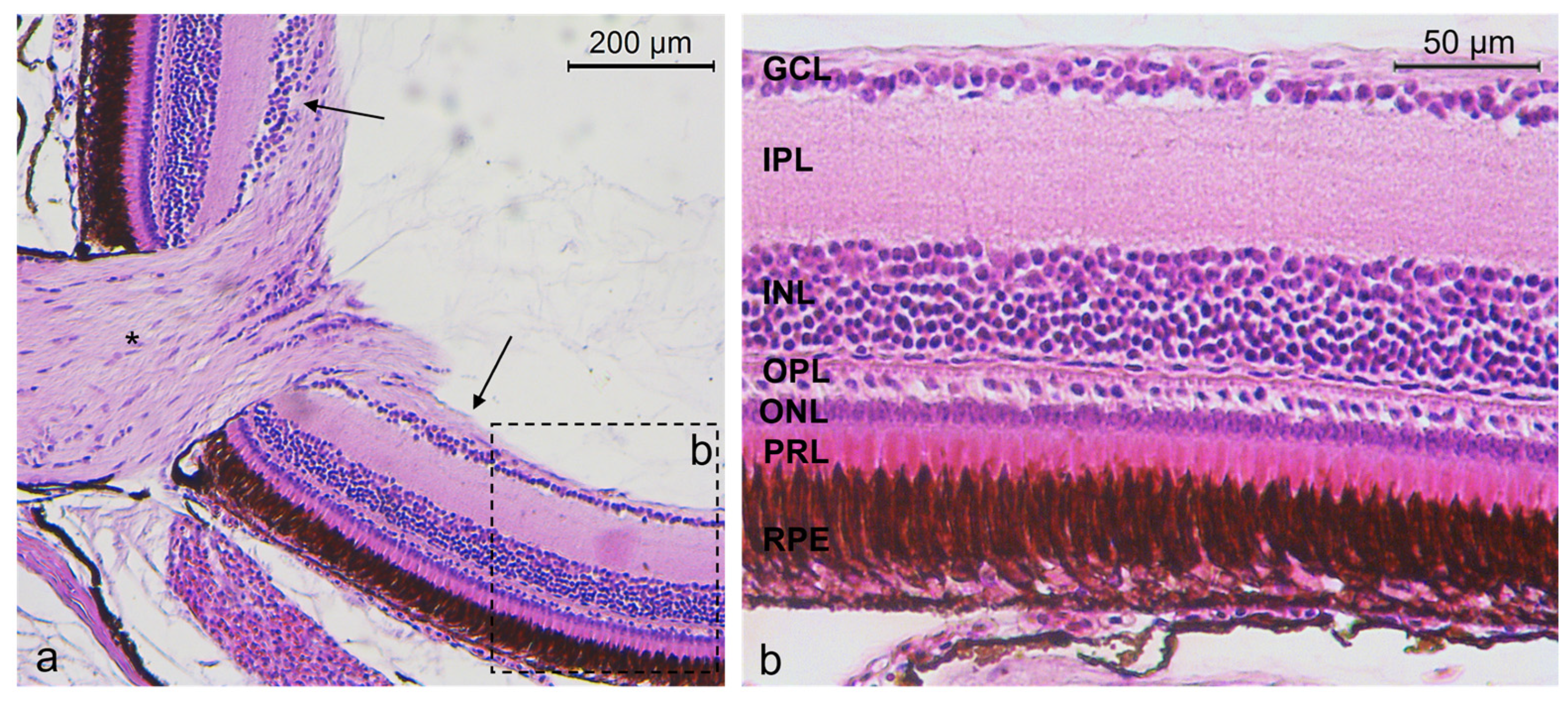
2.1. Histology of N. guentheri Retina

According to the morphological investigation, the retina of Nothobranchius guentheri had a similar stratigraphy to other vertebrates. The retina of N. guentheri was made up of seven layers: retinal pigment epithelium (RPE), photoreceptor layer (PRL) containing inner and outer segment of rods and cones, outer nuclear layer (ONL) containing cells body of rods and cones, inner nuclear layer (INL) containing different subpopulation of amacrine cells (ACs), bipolar cells (BCs), and horizontal cells (HCs), the GCL containing ganglion cells, the outer plexiform layer (OPL) containing the cellular prolongation between ONL and INL, and the inner plexiform layer (IPL) with the cellular prolongation between INL and ganglion cell layer (GCL) (Figure 1).

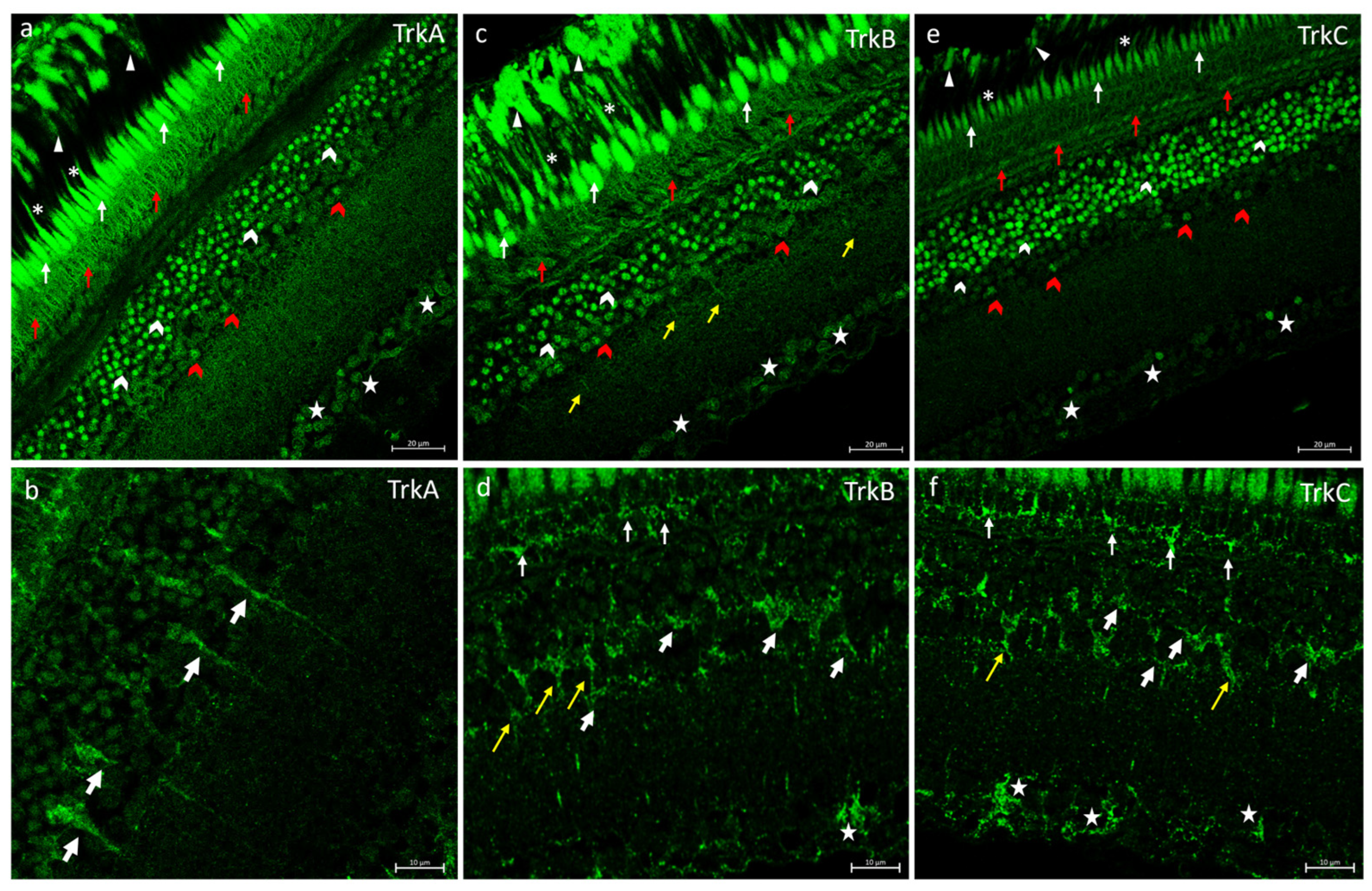
2.2. Trks Immunofluorescences in N. guentheri Retina

In the retina of N. guentheri, the neurotrophins receptors TrkA, TrkB, and TrkC were always localized in the extensions of RPE and in the inner and outer segments of the PRL (Figure 2a–d,f). In the OPL TrkA, TrkB, and TrkC were found (Figure 2a–c). In the INL, several subpopulations of ACs and some BCs were immunopositive to Trks (tyrosine protein kinase receptors) (Figure 2). Also, the neurotrophin receptor TrkB and TrkC were localized in HCs (Figure 2d,f). Different subpopulations of GCs immunostained to Trk receptors (Figure 2) were also observed.

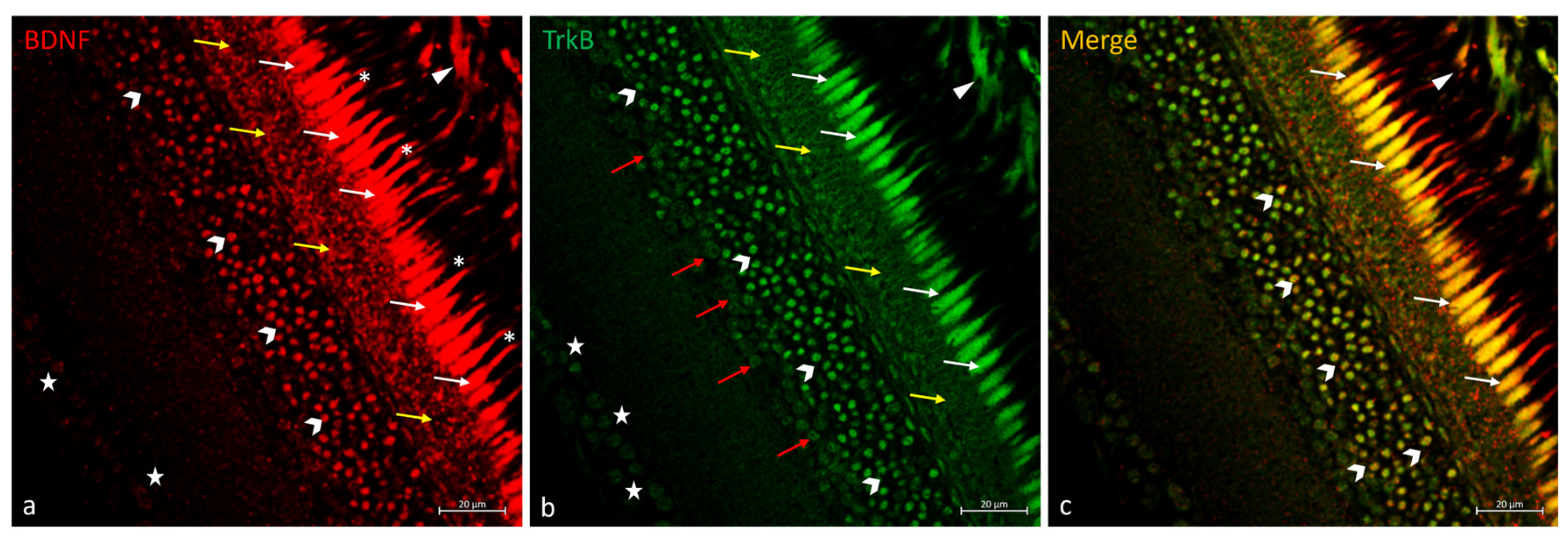
2.3. Double Immunofluorescences of BDNF and TrkB in N. guentheri Retina

In the retina of N. guentheri, the BDNF/TrkB system was immunolocalized in RPE prolongations, and in the inner segment of the PRL (Figure 3). In the PRL, the outer segment was exclusively BDNF immunostained (Figure 3a), and the inner segment of the photoreceptors is BDNF and TrkB was double-marked (Figure 3c). BDNF and TrkB were observed in the OPL, but they did not overlap (Figure 3). Distinct subpopulations of ACs exhibited immunoreactivity to BDNF and TrkB separately (Figure 3a,b), just some of them showed double staining (Figure 3c). In the GCL, the soma of the GCs (ganglion cells) was BDNF and TrkB immunostained (Figure 3a,b).

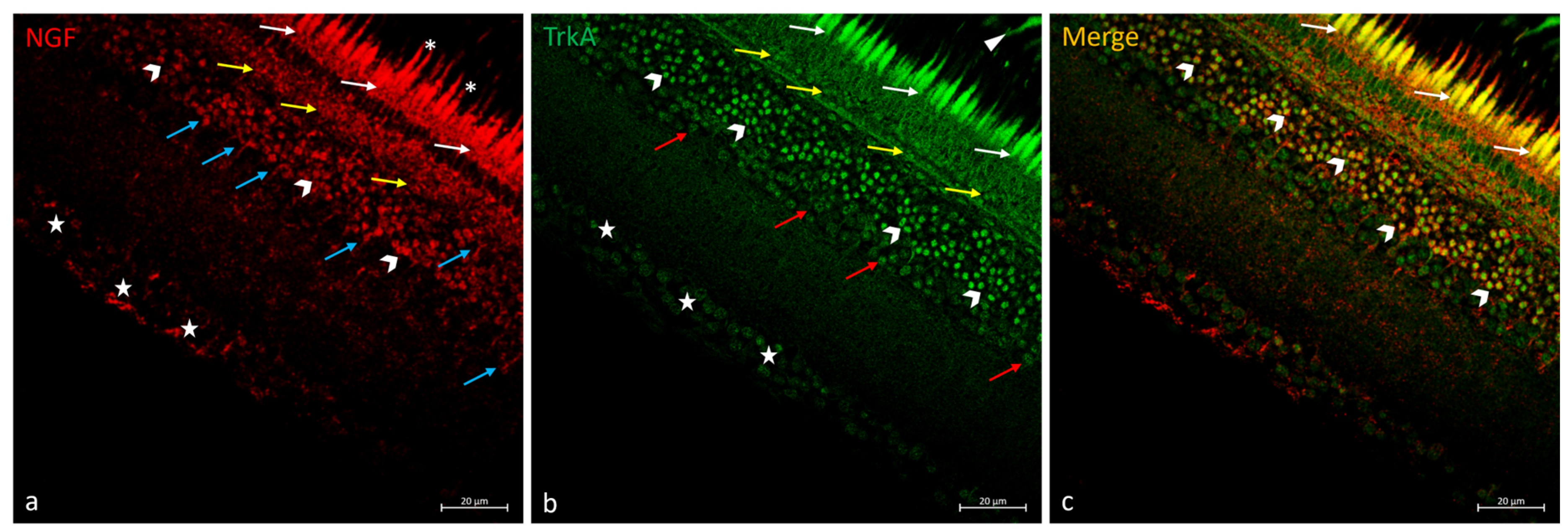
2.4. Double Immunofluorescences of NGF and TrkA in N. guentheri Retina

Research on the NGF/TrkA system in the retina N. guentheri showed RPE extensions are immunopositive to the neurotropin receptor TrkA but not to NGF (Figure 4). In the photoreceptor layer, the outer segment labeled NGF and the double stained inner segment NGF e TrkA were observed (Figure 4). NGF and TrkA were immunolocalized in the OPL (Figure 4b), but merging has not been observed (Figure 4c). In the INL, several subpopulations of ACs were immunopositive to NGF and TrkA (Figure 4a,b), some of these immunopositive exclusively to TrkA (Figure 4b) and some of these colocalized (Figure 4c). BCs immunoreactive to NGF were found (Figure 4a). Moreover, different subpopulations of GCs were NGF and TrkA immunostained (Figure 4a,b) in GCL, and no colocalization views were observed (Figure 4c).

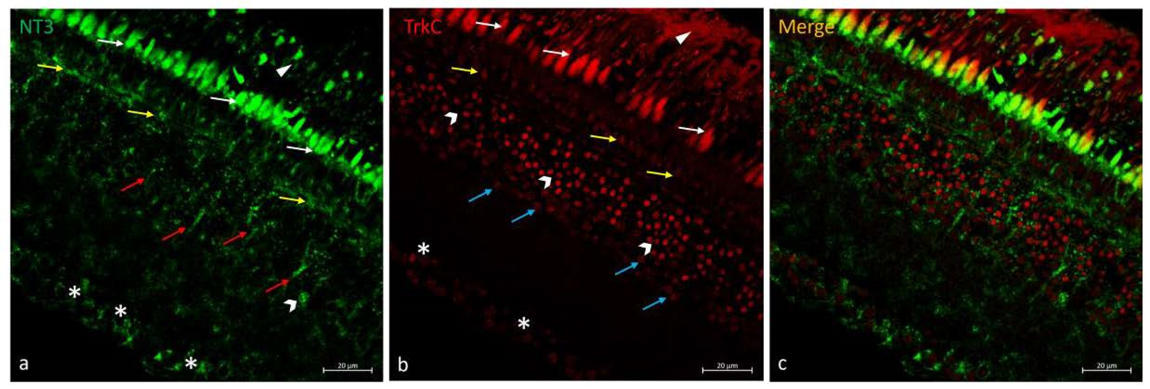
2.5. Double Immunofluorescences of NT-3 and TrkC in N. guentheri Retina

Regarding the NT-3/TrkC system in the N. guentheri retina, RPE cell prolongation immunostain was found. In the PRL, the inner segment immunopositive to NT-3 and the outer segment immunoreactive to TrkC have been observed. The OPL was NT-3 and TrkC immunoreactive (Figure 5). In the INL, BCs immunoreactive with NT-3 (Figure 5a) and a subpopulation of ACs immunopositive to TrkC (Figure 5b) were seen. In GCL, several subpopulations of GCs were NT-3 and TrkC immunostained, respectively (Figure 5a,b). Colocalization views were never observed (Figure 5c).

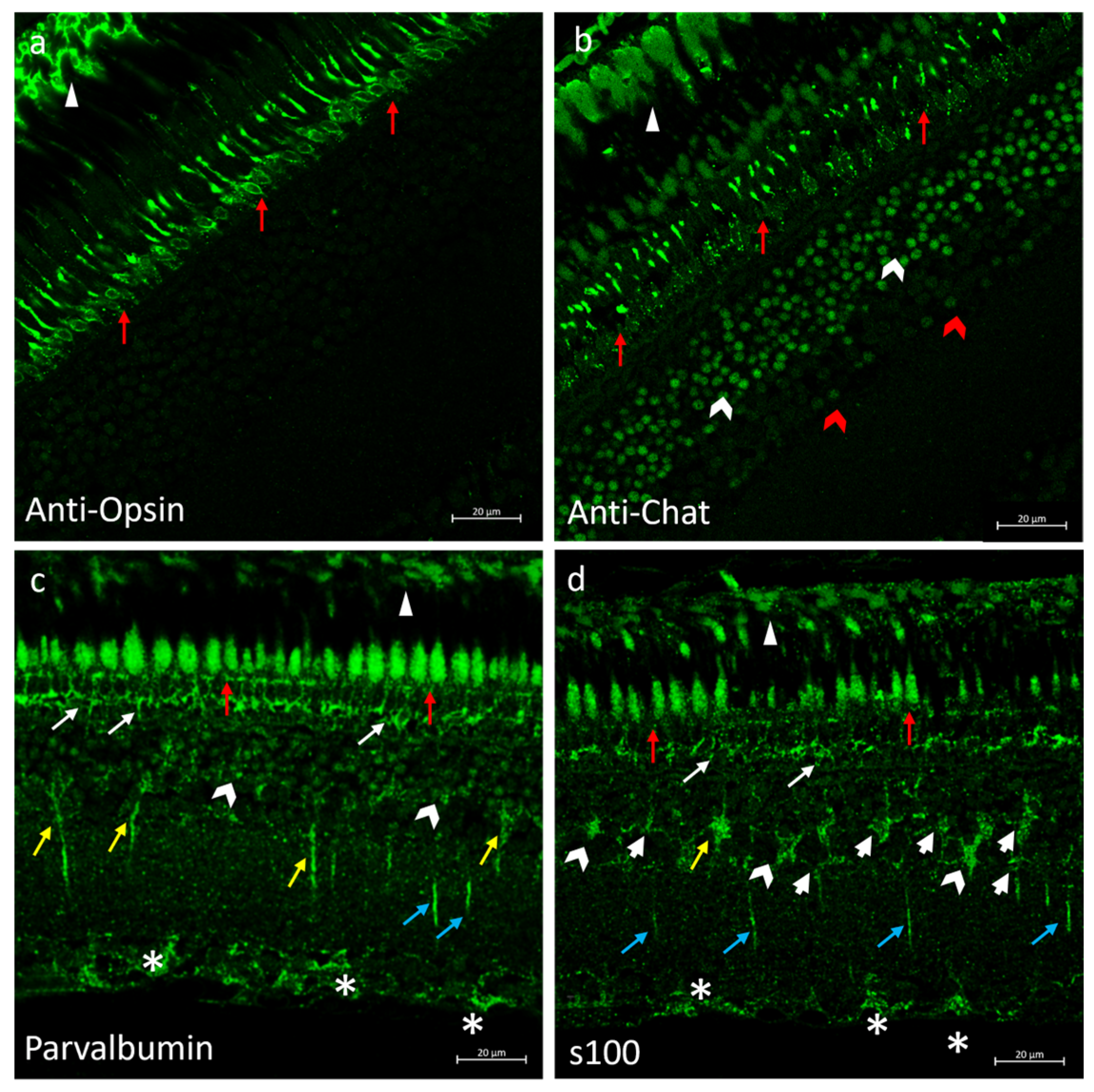
2.6. Immunofluorescences of Anti-Opsin, Anti-Chat, Parvalbumin and s100p in N. guentheri Retina

To ascertain the cellular identity of the immunopositive cells shown, anti-Opsin (specific for rods), anti-Chat (specific for ACs), parvalbumin (specific for BCs), and s100p (specific for HCs and GCs) antibody immunoreactions were investigated. The observed immunostaining perfectly overlapped the specificity of the antibodies used as markers (Figure 6). In addition, the RPE was immunoreactive to anti-chat, anti-opsin, s100, and parvalbumin; the outer segment of the PRL was anti-chat, parvalbumin, and s100 immunostained, and the inner segment of the PRL was parvalbumin and s100 immunoreactive. In the INL, ACs were parvalbumin and s100 immunolabeled; finally, GCs were also parvalbumin immunopositive (Figure 6).

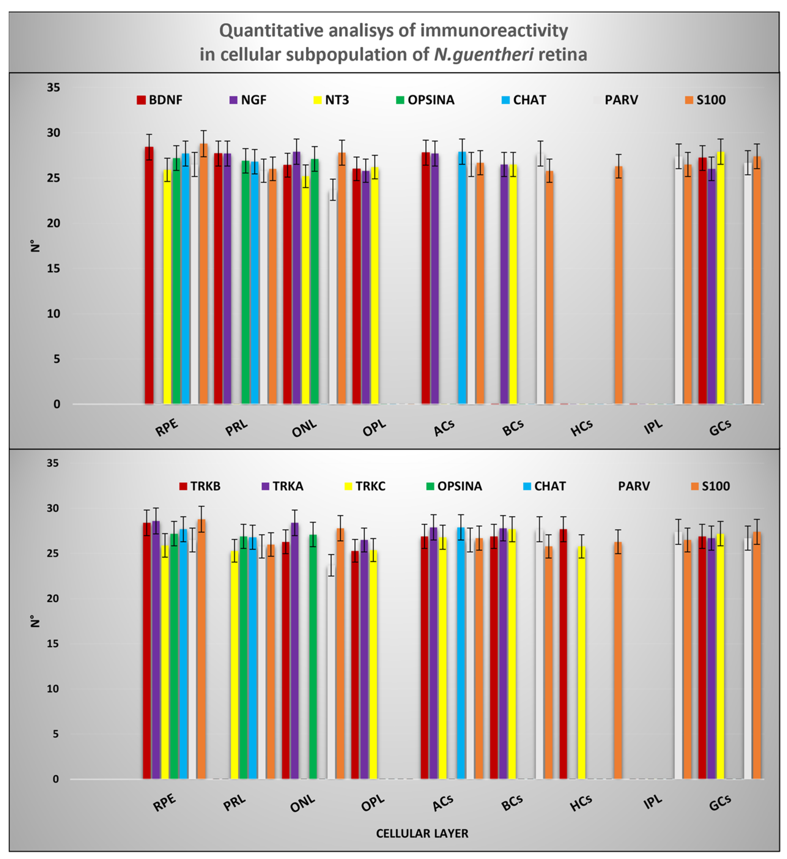
2.7. Quantitative Analysis

According to the results of the quantitative analysis, the cellular prolongations of the RPE were immunoreactive to neurotrophin BDNF and NT-3 and to neurotrophin receptors type A, B, and C. The inner segment of the PRL was always immunopositive to BDNF/TrkB, NGF/TrkA, and the NT-3/TrkC systems; however, the outer segment of the PRL was BDNF and NGF immunoreactive. In the INL, several subpopulations of ACs immunopositive to neurotrophin BDNF and NGF and to Trks (type A, B, and C) were observed. BCs were immunoreactive to Trks (A, B, and C) and to neurotrophins NGF and NT-3, whereas HCs were exclusively immunostained by TrkB and TrkC. Finally, the GCs were immunoreactive to the neurotrophin systems BDNF/TrkB, NGF/TrkA, and NT-3/TrkC, but not always colocalized. The distributions of cellular markers: anti-Chat, anti-Opsin, Parvalbumin, and s100p were perfectly overlapping concerning their specificity. Comparison between the neurotrophin/tyrosine kinase receptor system and specific marker distribution patterns is shown in Figure 7 and Table 1.

3. Discussion

According to the latest WHO estimates, 285 million people worldwide suffer visual impairment, and most of the major eye diseases are age-related. As life expectancy rises worldwide, the prevalence of age-related visual impairments has also seen a notable increase. The retina is one of the eye structures that undergoes various structural and functional changes with advancing age. Aging is frequently accompanied by retinal degeneration phenomena commonly associated with conditions such as age-related macular degeneration (AMD) and the progressive degeneration of photoreceptor cells, impaired retinal pigment epithelium function, and alterations in the vascular network [59]. Hence, visual impairment due to retinal degeneration constitutes one of the significant concerns in the aging population, affecting its quality of life and the healthcare system [60,61]. Aging impacts not only the function of the visual system but also its ability to protect and repair damaged and/or degenerating neurons [34,59,62]. Unfortunately, there is no treatment to curb the neurodegenerative disease of the eye, and the lifespan length of most traditional experimental models has impaired the study of the aging process. Among killifish, Nothobranchius spp. has a relatively short life cycle compared to other vertebrate models and has several aging features already described for humans [63], which makes it an excellent aging model to fill this gap [30]. In addition, it has been observed that Nothobranchius’ central nervous system, including the visual system, shows typical aging features [29,64,65,66,67,68]. Therefore, Nothobranchius spp. seems to be the ideal model for studying cellular and molecular age-dependent changes to understand and deal with neurodegenerative events effectively. Moreover, the visual system is considered an important tool to investigate the brain overall, both in mammals and fish, since the retina is an integral part of the central nervous system [69]. It has been shown that the pathological processes occurring in the retina indicate similar processes occurring in the central nervous system and vice versa [56,57,58,70]. For instance, alterations in neurotrophin balance are involved in both AMD and retinopathy and in Alzheimer’s disease pathogenesis [71,72,73,74]. The research on the aging of the eyes, particularly of the retina, represents a promising strategy for studying neurodegenerative diseases [75], as it is considered a window to the brain. As a sensory system, retina development [76,77] and maintenance [78] are controlled by neurotrophins, in particular, brain-derived neurotrophic factor (BDNF) [7]. Neurotrophins and their receptors have been shown to be evolutionarily conserved and they have been detected in different vertebrates from fish to mammals, including humans [4,79,80,81,82].
They play a key role in guiding the maintenance, regulation, and neurogenesis of teleosts and the human nervous system, the sensory organs, and, among them, the retina.
Being that the retina is an integral part of the nervous system and given the established link between neurotrophins and the overall well-being of the nervous system, it is plausible to hypothesize a relationship between neurotrophin and retinal neurodegeneration during aging. As aging progresses, the decline in neurotrophin support could contribute to the degenerative changes observed in the retina, such as those associated with age-related macular degeneration and retinal degeneration due to the loss of retinal cells [27,28,29,30,31,32,57,58,59,60,61]. Consequently, scientific research focused on the potential neuroprotective and therapeutic role of neurotrophins and their receptors in treatment of retinal diseases and, neurodegeneration [10]. Indeed, studies have reported that intraocular administration of NTs promotes the survival of GCs and axons after injuries [83,84,85,86,87,88,89,90] and, in particular, the intraocular administration of BDNF has shown a protective action on photoreceptors in retinal degeneration and retinal detachment [91]. Hence, the relevance of this field of investigation is intuitive, and choosing appropriate models that mimic human physiological conditions is crucial for the translational relevance of findings. In this context, N. guentheri represents a suitable experimental model because of the functional and morphological similarities of it and the human retina and its emergent role in aging studies. Moreover, the N. guentheri retina has demonstrated properties of neurogenesis and regeneration [1].
However, evidence of NTs-Trks system expression in the retina of this experimental model is still scarce. According to the authors’ knowledge, only TrkB has been localized in the retina of one species of the Nothobranchius genus, N. furzeri [26,91,92,93].
Therefore, this work aimed at studying the localization of the neurotrophins BDNF, NGF (nerve growth factor), NT-3 (neurotrophin-3), and tyrosine protein kinase receptors type A (TrkA), TrkB, and tyrosine protein kinase receptors type C (TrkC) neurotrophin receptors, in the retina of adult N. guentheri as a first step in assessing the suitability of this model organism for translational medicine studies.
This study shows, for the first time, that NTs/Trks systems are widely detectable in the adult N. guentheri retina, mainly in the RPE, PRL (photoreceptor layer inner and outer segments), OPL (outer plexiform layer), and several populations of ACs (amacrine cells), BCs (bipolar cells), and HCs (horizontal cells) in the INL and in the GCL (ganglion cell layer). The present study’s data agree with the localization of NTs/Trks in the retina of other species, including humans [83]. In the present study, NGF neurotrophin and TrkA receptors were observed in RPE, PRL, and OPL, in several populations of ACs, in BCs, and in several GCs populations. This data overlaps with the location of NGF and TrkA retina of mice [94].
In the retina of N. guentheri Trks receptor types A, B, and C were immunolocalized in the external and internal segments of the photoreceptors. In addition, several populations of ACs identified by morpho-topographic approach showed immunoreactivity to neurotrophin receptors type A, B, and C. BCs were always immunopositive to Trks and HCs TrkB and TrkC immunoreactive. Finally, TrkB and TrkC are immunolocalised in different GC populations recognizable by using a morpho-topographic approach and have not shown double staining with neurotrophins. The above-mentioned results partly overlap with the data concerning the mouse retina [95,96,97] and other teleosts, including zebrafish.
In this study, the authors found that the location of Trks receptors in N. guentheri’s retina overlaps with the distribution of BDNF in other teleosts [60,82,85,86].
In addition, the results of this study show the presence of TrkA, TrkB, and TrkC receptors in GCs, where BDNF had already been detected by Gatta et al. [71]. The present results support Gatta’s hypothesis [71] on the mode of autocrine action of BDNF on GCs. in addition, BDNF was observed in the N. guentheri retina in the RPE, in the external and internal segment of photoreceptors layer, and in ACs in the INL. Localizing the BDNF is intriguing because it has been demonstrated that its decrease is associated with age-dependent macular degeneration and retinopathy [10,71,84,85,86,87,88,89,90,91,92,93,94,98,99,100,101].
Based on our knowledge, no data are known in regard to the localization of neurotrophin NT-3 in the retina of N. guentheri. In this study, the authors show, for the first time, that the prolongations of the RPE, the external and internal segments of the photoreceptors, BCs, some ACs, and GCs are immunopositive. These data are similar to what is recognized in other species such as the pigeon (Columba livia [96], lizard (Gallotia galloti) [97], frog [102], chick [17,103], and mice [104]. In addition, Das and colleagues [105] investigated BDNF/TrkB and NT-3/TrkC systems and observed localization in the external segment of the PRL, OPL, INL, IPL, and GCL, as shown in this work in the N. guentheri retina.
Finally, in the retina di N. guentheri BDNF and NT-3 neurotrophins and neurotrophin receptors Trks (A, B, C) were found immunolocalized in the pigmented epithelium, a retinal layer relevant to the study of age-induced damage. Indeed, research demonstrates that RPE changes are a typical characteristic of the aging retina [49]. The thinning of the retinal layers and a decrease in the pigment epithelium layer typical in old specimens of Oryzias latipes and D. rerio [94,106] occur in a short time before the end of the N. guentheri life cycle. This evidence makes the retina of the annual killifish an excellent model in the biomedical research of age-dependent pathologies [1].
To compare the localization of neurotrophins and neurotrophin receptors A, B, and C in different retinal cell layers of the different models with humans, see Table 2.
The data of the present investigation, together with the known evidence on N. furzeri [26,91,93,120,121] and other species [99,102,122] confirm the fundamental role of the neurotrophins/receptors system in the maintenance and modulation of excitatory input in retinal neurons. Consequently, it might also suggest a mode of autocrine action of the NT/Trks system in the retina of the adult N. guentheri.

4. Materials and Methods

4.1. Fish and Tissue Treatment

In this study, paraffin embedded tissue of Nothobranchius guentheri from previous studies were used [123]. Adult specimens of N. guentheri, (discovered dead of unknown causes) 1-year-old, 1 male, and 2 females, from ornamental aquariums (freshwater, 22 °C, pH 6.8–7.0) were used. The heads were quickly removed, fixed in 4% paraformaldehyde (Sigma-Aldrich, Inc., St. Louis, MO, USA # 158127) in phosphate-buffered saline (PBS, Sigma-Aldrich, Inc., St. Louis, MO, USA # P4417) 0.1 m (pH = 7.4) for 12–18 h, dehydrated through graded ethanol series, clarified in xylene, for paraffin wax (Bio-Optica Milano S.p.a Milano, Italy # 08-7910) embedding.

4.2. Optical Microscopy

Included tissues of N. guentheri were cut into 7 μm thick serial sections and collected on gelatin-coated microscope slides [123,124].
Then, serial sections were deparaffinized and rehydrated, washed in distilled water, and stained with Hematoxylin-Eosin (Hematoxylin nuclear staining Bio-Optica Milano S.p.a Italy cat. # 05-M06012. Eosin Y cytoplasmic staining Bio-Optica Milano S.p.a Italy cat. # 05-M10002). At the end, stained sections were examined under a Leica DMRB light microscope equipped with Leica MC 120 HD camera (Leica Application Suite LAS V4.7).

4.3. Immunohistochemistry

To analyze the localization of neurotrophins (NTs) and tyrosine protein kinase receptors (Trks) in N. guentheri retina, some serial slides were deparaffinized and rehydrated, finally washed in PBS. The sections were incubated in 0.1% Triton X100 (Sigma-Aldrich, Inc., St. Louis, MO, USA cat. #X100) PBS solution to permeate the membranes, after incubated in a 0.3% hydrogen peroxide solution (H2O2 Sigma-Aldrich, Inc., St. Louis, MO, USA cat. # 1085971000) to prevent the activity of endogenous peroxidase. The 25% fetal bovine serum solution (Sigma-Aldrich, Inc., St. Louis, MO, USA cat. #F7524) was then added to the rinsed sections. Sections were incubated overnight at 4 °C in a humid chamber with primary antibodies (steps below). Representative sections were incubated with appropriately preabsorbed antisera as mentioned above to provide negative controls. In these circumstances, there was no evidence of positive immunostaining.

4.3.1. TrkA, TrkB, TrkC, Anti-Opsin, Anti-Chat, Parvalbumin and s100p Immunofluorescences

To identify anti-neurotrophin receptors in retinal cells of N. guentheri some serial sections were incubated with tyrosine protein kinase receptors type A (TrkA), tyrosine protein kinase receptors type B (TrkB), tyrosine protein kinase receptors type C (TrkC) (for details see Table 1). Moreover, some representative sections have been incubated with anti-Opsin, anti-Chat, Parvalbumin, and S100p antibodies, recognized as specific markers for retinal cells (for details see Table 1). After rinsing in PBS solution, the sections were incubated for 1 h with a fluorescent secondary antibody Anti-mouse IgG (H+L) Alexa Fluor 488 and Anti-rabbit IgG (H + L) Alexa Fluor 488 (for details see Table 1) at room temperature in a dark humid chamber. Washing, and mounting using Fluoromount Aqueous Mounting Medium (Sigma-Aldrich, Inc., St. Louis, MO, USA cat. #F4680 manufacturer’s notice) were the final steps.

4.3.2. NTs/Trks System Double Immunofluorescences

To investigate the immunolocalization of the NTs/Trks system in n guentheri retina polyclonal BDNF brain-derived neurotrophic factor and NGF (nerve growth factor) were used in double-labelled experiment with monoclonal TrkB and TrkA, respectively, and monoclonal NT-3 (neurotrophin-3) was used in double-labelled experiment with monoclonal TrkC (for details see Table 1). After rinsing in PBS solution, the sections were incubated for 1 h with a fluorescent secondary antibody anti-rabbit alexa fluor 594 and anti-mouse alexa fluor 488 (for details see Table 3) at room temperature in a dark humid chamber. Washing, and mounting using Fluoromount Aqueous Mounting Medium (Sigma-Aldrich, Inc., St. Louis, MO, USA cat. #F4680 manufacturer’s notice) were the final steps.

4.3.3. Confocal Laser Scanning Microscope

A Zeiss LSMDUO confocal laser scanning microscope with META module (Carl Zeiss MicroImaging GmbH, München, Germany) was used to detect the immunofluorescence, and Zen 2011 (LSM 700 Zeiss software ZEN 3.7) was employed to process the images [125,126,127]. Each image was rapidly acquired to minimize photodegradation.

4.4. Statistical Analysis

ImageJ software was used to evaluate microscope fields collected randomly. One-way ANOVA was used to examine the statistical significance of the quantity of retinal pigment epithelium (RPE), PRL (photoreceptor layer inner and outer segments), OPL (outer plexiform layer), Acs (amacrine cells), IPL (inner plexiform layer), BCs (bipolar cells), HCs (horizontal cells) and GCs (ganglion cells) detected by BDNF, NGF, NT-3, TrkA, TrkB, TrkC, anti-Opsin, anti-Chat, Parvalbumin and, s100p. SigmaPlot version 14.0 (Systat Software, San Jose, CA, USA) was used to conduct the statistical analysis. An unpaired Z test was also performed. The information was given as mean values with standard deviations (Δσ). Values of p below 0.05 were considered statistically significant in the following order *** p < 0.001, ** p < 0.01, * p < 0.05.

5. Conclusions

In conclusion, our study provides new insights into the localization of neurotrophins and their specific receptors in the retina of Nothobranchius guentheri, showcasing its potential as an experimental model for investigating retinal aging. Furthermore, the conservation of neurotrophin signaling pathways in N. guentheri suggests its relevance as a translational model for studying retinal aging in humans. However, while our findings offer valuable insights, they just represent a starting point in comprehensively characterizing retinal aging in N. guentheri, future studies are needed to better understand the expression patterns of the NTs/Trks system during development, the aging process, and/or in transgenic N. guentheri models for neurodegenerative diseases. It is still necessary to elucidate the functional consequences of NT-Trk system alteration in the aged retinas of N. guentheri and explore potential therapeutic interventions to mitigate age-related retinal degeneration.

Author Contributions

Conceptualization, M.A., M.C.G. and A.G; methodology, M.A., K.M. and M.C.; software, M.A. and K.M.; validation, M.C.G. and A.G.; formal analysis, M.A., K.M. and C.P.; investigation, M.C.G., K.M. and M.A.; resources, A.G. and M.C.G.; data curation, M.C.G. and M.A.; writing—original draft preparation, M.A. and M.B.; writing—review and editing, M.A., M.B., M.C.G., A.G., F.A., G.M., M.L., R.L., P.G.G., K.M. and C.P.; visualization, M.C.G., F.A., G.M., M.L., R.L., P.G.G. and A.G.; supervision, M.C.G., F.A., G.M., M.L., R.L., P.G.G. and A.G.; project administration, M.A., M.C.G. and A.G.; funding acquisition, M.C.G., G.M. and A.G. All authors have read and agreed to the published version of the manuscript.

Funding

This research received no external funding.

Institutional Review Board Statement

This study has been conducted on samples from previous studies [127].

Informed Consent Statement

Not applicable.

Data Availability Statement

All data presented this study are available from the corresponding author, upon responsible request.

Acknowledgments

The authors are thankful to Leyanis Herrera Ledesma. Department of Morphophysiology, Faculty of Veterinary Medicine, Agricultural University of Havana “Fructuoso Rodríguez Pérez” (UNAH), Mayabeque, Cuba for technical support and collaboration.

Conflicts of Interest

The authors declare no conflicts of interest.

Abbreviations

neurotrophins (NTs), tyrosine protein kinase receptors (Trks), brain-derived neurotrophic factor (BDNF), nerve growth factor (NGF), neurotrophin-3 (NT-3), tyrosine protein kinase receptors type A (TrkA), tyrosine protein kinase receptors type B (TrkB), tyrosine protein kinase receptors type C (TrkC), retinal pigment epithelium (RPE), photoreceptor layer (PRL), outer nuclear layer (ONL), outer plexiform layer (OPL), inner nuclear layer (INL),amacrine cells (ACs), bipolar cells (BCs), horizontal cells (HCs), inner plexiform layer (IPL), ganglion cell layer (GCL), ganglion cells (GCs).

References

  1. Nikiforov-Nikishin, D.L.; Irkha, V.A.; Kochetkov, N.I.; Kalita, T.L.; Nikiforov-Nikishin, A.L.; Blokhin, E.E.; Antipov, S.S.; Makarenkov, D.A.; Zhavnerov, A.N.; Glebova, I.A.; et al. Some Aspects of Development and Histological Structure of the Visual System of Nothobranchius guentheri. Animals 2021, 11, 2755. [Google Scholar] [CrossRef]
  2. Ostrander, G.K.; Hopkins, J. The Laboratory Fish; Elsevier: Amsterdam, The Netherlands, 2000. [Google Scholar]
  3. Catania, S.; Germana, A.; Cabo, R.; Ochoa-Erena, F.; Guerrera, M.; Hannestad, J.; Represa, J.; Vega, J. Neurotrophin and Trk neurotrophin receptors in the inner ear of Salmo salar and Salmo trutta. J. Anat. 2007, 210, 78–88. [Google Scholar] [CrossRef] [PubMed]
  4. García-Suárez, O.; Germanà, A.; Hannestad, J.; Pérez-Pérez, M.; Esteban, I.; Naves, F.J.; Vega, J.A. Changes in the expression of the nerve growth factor receptors TrkA and p75LNGR in the rat thymus with ageing and increased nerve growth factor plasma levels. Cell Tissue Res. 2000, 301, 225–234. [Google Scholar] [CrossRef]
  5. García-Suárez, O.; Pérez-Pérez, M.; Germanà, A.; Esteban, I.; Germanà, G. Involvement of growth factors in thymic involution. Microsc. Res. Tech. 2003, 62, 514–523. [Google Scholar] [CrossRef]
  6. Germanà, A.; Marino, F.; Guerrera, M.C.; Campo, S.; de Girolamo, P.; Montalbano, G.; Germanà, G.P.; Ochoa-Erena, F.J.; Ciriaco, E.; Vega, J.A. Expression and distribution of S100 protein in the nervous system of the adult zebrafish (Danio rerio). Microsc. Res. Tech. 2008, 71, 248–255. [Google Scholar] [CrossRef] [PubMed]
  7. Germana, A.; Catania, S.; Cavallaro, M.; González-Martínez, T.; Ciriaco, E.; Hannestad, J.; Vega, J. Immunohistochemical localization of BDNF-, TrkB-and TrkA-like proteins in the teleost lateral line system. J. Anat. 2002, 200, 477–485. [Google Scholar] [CrossRef] [PubMed]
  8. Reichardt, L.F. Neurotrophin-regulated signalling pathways. Philos. Trans. R. Soc. B Biol. Sci. 2006, 361, 1545–1564. [Google Scholar] [CrossRef] [PubMed]
  9. Skaper, S.D. Neurotrophic Factors: An Overview. In Neurotrophic Factors: Methods and Protocols; Skaper, S.D., Ed.; Springer: New York, NY, USA, 2018; pp. 1–17. [Google Scholar] [CrossRef]
  10. Aragona, M.; Porcino, C.; Guerrera, M.C.; Montalbano, G.; Laurà, R.; Cometa, M.; Levanti, M.; Abbate, F.; Cobo, T.; Capitelli, G.; et al. The BDNF/TrkB Neurotrophin System in the Sensory Organs of Zebrafish. Int. J. Mol. Sci. 2022, 23, 2621. [Google Scholar] [CrossRef] [PubMed]
  11. Aragona, M.; Porcino, C.; Guerrera, M.C.; Montalbano, G.; Laurà, R.; Levanti, M.; Abbate, F.; Cobo, T.; Capitelli, G.; Calapai, F.; et al. Localization of BDNF and Calretinin in Olfactory Epithelium and Taste Buds of Zebrafish (Danio rerio). Int. J. Mol. Sci. 2022, 23, 4696. [Google Scholar] [CrossRef] [PubMed]
  12. Hannestad, J.; Marino, F.; Germanà, A.; Catania, S.; Abbate, F.; Ciriaco, E.; Vega, J. Trk neurotrophin receptor-like proteins in the teleost Dicentrarchus labrax. Cell Tissue Res. 2000, 300, 1–9. [Google Scholar] [CrossRef]
  13. Chao, M.V. Neurotrophins and their receptors: A convergence point for many signalling pathways. Nat. Rev. Neurosci. 2003, 4, 299–309. [Google Scholar] [CrossRef] [PubMed]
  14. Carmignoto, G.; Comelli, M.C.; Candeo, P.; Cavicchioli, L.; Yan, Q.; Merighi, A.; Maffei, L. Expression of NGF receptor and NGF receptor mRNA in the developing and adult rat retina. Exp. Neurol. 1991, 111, 302–311. [Google Scholar] [CrossRef] [PubMed]
  15. Bai, Y.; Dergham, P.; Nedev, H.; Xu, J.; Galan, A.; Rivera, J.C.; ZhiHua, S.; Mehta, H.M.; Woo, S.B.; Sarunic, M.V.; et al. Chronic and Acute Models of Retinal Neurodegeneration TrkA Activity Are Neuroprotective whereas p75NTR Activity Is Neurotoxic through a Paracrine Mechanism. J. Biol. Chem. 2010, 285, 39392–39400. [Google Scholar] [CrossRef] [PubMed]
  16. Frade, J.M.; Barde, Y.-A. Microglia-Derived Nerve Growth Factor Causes Cell Death in the Developing Retina. Neuron 1998, 20, 35–41. [Google Scholar] [CrossRef] [PubMed]
  17. Frade, J.M.; Bovolenta, P.; Rodríguez-Tébar, A. Neurotrophins and other growth factors in the generation of retinal neurons. Microsc. Res. Tech. 1999, 45, 243–251. [Google Scholar] [CrossRef]
  18. Cohen-Cory, S.; Lom, B. Neurotrophic regulation of retinal ganglion cell synaptic connectivity: From axons and dendrites to synapses. Int. J. Dev. Biol. 2004, 48, 947–956. [Google Scholar] [CrossRef] [PubMed]
  19. Takano, M.; Horie, H.; Iijima, Y.; Dezawa, M.; Sawada, H.; Ishikawa, Y. Brain-derived Neurotrophic Factor Enhances Neurite Regeneration from Retinal Ganglion Cells in Aged Human Retina in vitro. Exp. Eye Res. 2002, 74, 319–323. [Google Scholar] [CrossRef] [PubMed]
  20. Turner, B.A.; Sparrow, J.; Cai, B.; Monroe, J.; Mikawa, T.; Hempstead, B.L. TrkB/BDNF signaling regulates photoreceptor progenitor cell fate decisions. Dev. Biol. 2006, 299, 455–465. [Google Scholar] [CrossRef]
  21. de la Rosa, E.J.; Arribas, A.; Frade, J.M.; Rodri’guez-Te’bar, A. Role of neurotrophins in the control of neural development: Neurotrophin-3 promotes both neuron differentiation and survival of cultured chick retinal cells. Neuroscience 1994, 58, 347–352. [Google Scholar] [CrossRef]
  22. Gleeson, M.; Connaughton, V.; Arneson, L.S. Induction of hyperglycaemia in zebrafish (Danio rerio) leads to morphological changes in the retina. Acta Diabetol. 2007, 44, 157–163. [Google Scholar] [CrossRef]
  23. Gestri, G.; Link, B.A.; Neuhauss, S.C. The visual system of zebrafish and its use to model human ocular diseases. Dev. Neurobiol. 2012, 72, 302–327. [Google Scholar] [CrossRef]
  24. Seritrakul, P.; Gross, J.M. Genetic and epigenetic control of retinal development in zebrafish. Curr. Opin. Neurobiol. 2019, 59, 120–127. [Google Scholar] [CrossRef]
  25. Tarboush, R.; Chapman, G.B.; Connaughton, V.P. Ultrastructure of the distal retina of the adult zebrafish, Danio rerio. Tissue Cell 2012, 44, 264–279. [Google Scholar] [CrossRef]
  26. D’Angelo, L.; Avallone, L.; Cellerino, A.; de Girolamo, P.; Paolucci, M.; Varricchio, E.; Lucini, C. Neurotrophin-4 in the brain of adult Nothobranchius furzeri. Ann. Anat.-Anat. Anz. 2016, 207, 47–54. [Google Scholar] [CrossRef] [PubMed]
  27. Braunbeck, T.; Lammer, E. Fish embryo toxicity assays. Ger. Fed. Environ. Agency 2006, 298, 7–119. [Google Scholar]
  28. Kroll, W. Morphological and behavioral embryology and spontaneous diapause in the African killifish, Aphyosemion gardneri. Environ. Biol. Fishes 1984, 11, 21–28. [Google Scholar] [CrossRef]
  29. Tozzini, E.T.; Baumgart, M.; Battistoni, G.; Cellerino, A. Adult neurogenesis in the short-lived teleost Nothobranchius furzeri: Localization of neurogenic niches, molecular characterization and effects of aging. Aging Cell 2012, 11, 241–251. [Google Scholar] [CrossRef]
  30. Vanhunsel, S.; Beckers, A.; Moons, L. Designing neuroreparative strategies using aged regenerating animal models. Ageing Res. Rev. 2020, 62, 101086. [Google Scholar] [CrossRef]
  31. Johns, P.R. Growth of the adult goldfish eye. III. Source of the new retinal cells. J. Comp. Neurol. 1977, 176, 343–357. [Google Scholar] [CrossRef]
  32. Raymond, P.A.; Barthel, L.K.; Bernardos, R.L.; Perkowski, J.J. Molecular characterization of retinal stem cells and their niches in adult zebrafish. BMC Dev. Biol. 2006, 6, 36. [Google Scholar] [CrossRef]
  33. Hrbek, T.; Larson, A. The Evolution of Diapause in the Killifish Family Rivulidae (Atherinomorpha, Cyprinodontiformes): A Molecular Phylogenetic and Biogeographic Perspective. Evolution 1999, 53, 1200–1216. [Google Scholar] [CrossRef]
  34. Vanhunsel, S.; Bergmans, S.; Beckers, A.; Etienne, I.; Van Houcke, J.; Seuntjens, E.; Arckens, L.; De Groef, L.; Moons, L. The killifish visual system as an in vivo model to study brain aging and rejuvenation. npj Aging Mech. Dis. 2021, 7, 22. [Google Scholar] [CrossRef]
  35. Cellerino, A.; Valenzano, D.R.; Reichard, M. From the bush to the bench: The annual Nothobranchius fishes as a new model system in biology. Biol. Rev. 2016, 91, 511–533. [Google Scholar] [CrossRef]
  36. Lucas-Sánchez, A.; Almaida-Pagán, P.F.; Mendiola, P.; de Costa, J. Nothobranchius as a model for aging studies. A review. Aging Dis 2013, 5, 281–291. [Google Scholar] [CrossRef]
  37. Valenzano, D.R.; Sharp, S.; Brunet, A. Transposon-mediated transgenesis in the short-lived African killifish Nothobranchius furzeri, a vertebrate model for aging. G3 Genes Genomes Genet. 2011, 1, 531–538. [Google Scholar] [CrossRef]
  38. Hartmann, N.; Englert, C. A microinjection protocol for the generation of transgenic killifish (Species: Nothobranchius furzeri). Dev. Dyn. 2012, 241, 1133–1141. [Google Scholar] [CrossRef] [PubMed]
  39. Tozzini, E.T.; Cellerino, A. Nothobranchius annual killifishes. EvoDevo 2020, 11, 25. [Google Scholar] [CrossRef] [PubMed]
  40. Liu, C.; Wang, X.; Feng, W.; Li, G.; Su, F.; Zhang, S. Differential expression of aging biomarkers at different life stages of the annual fish Nothobranchius guentheri. Biogerontology 2012, 13, 501–510. [Google Scholar] [CrossRef]
  41. Wang, X.; Shang, X.; Luan, J.; Zhang, S. Identification, expression and function of apolipoprotein E in annual fish Nothobranchius guentheri: Implication for an aging marker. Biogerontology 2014, 15, 233–243. [Google Scholar] [CrossRef]
  42. Mhalhel, K.; Briglia, M.; Aragona, M.; Porcino, C.; Abbate, F.; Guerrera, M.C.; Laurà, R.; Krichen, Y.; Guerbej, H.; Germanà, A.; et al. Nothobranchius as a model for anorexia of aging research: An evolutionary, anatomical, histological, immunohistochemical, and molecular study. Ann. Anat.-Anat. Anz. 2023, 250, 152116. [Google Scholar] [CrossRef]
  43. Jackson, G.R.; De Leon Ortega, J.; Girkin, C.; Rosenstiel, C.E.; Owsley, C. Aging-related changes in the multifocal electroretinogram. J. Opt. Soc. Am. A 2002, 19, 185–189. [Google Scholar] [CrossRef]
  44. Owsley, C.; Jackson, G.R.; Cideciyan, A.V.; Huang, Y.; Fine, S.L.; Ho, A.C.; Maguire, M.G.; Lolley, V.; Jacobson, S.G. Psychophysical Evidence for Rod Vulnerability in Age-Related Macular Degeneration. Investig. Ophthalmol. Vis. Sci. 2000, 41, 267–273. [Google Scholar]
  45. Owsley, C.; Jackson, G.R.; White, M.; Feist, R.; Edwards, D. Delays in rod-mediated dark adaptation in early age-related maculopathy. Ophthalmology 2001, 108, 1196–1202. [Google Scholar] [CrossRef]
  46. Curcio, C.A.; Drucker, D.N. Retinal ganglion cells in Alzheimer’s disease and aging. Ann. Neurol. 1993, 33, 248–257. [Google Scholar] [CrossRef] [PubMed]
  47. Gao, H.; Hollyfield, J.G. Aging of the human retina. Differential loss of neurons and retinal pigment epithelial cells. Investig. Ophthalmol. Vis. Sci. 1992, 33, 1–17. [Google Scholar]
  48. Berkowitz, B.A.; Grady, E.M.; Roberts, R. Confirming a prediction of the calcium hypothesis of photoreceptor aging in mice. Neurobiol. Aging 2014, 35, 1883–1891. [Google Scholar] [CrossRef] [PubMed]
  49. Bonnel, S.; Mohand-Said, S.; Sahel, J.-A. The aging of the retina. Exp. Gerontol. 2003, 38, 825–831. [Google Scholar] [CrossRef] [PubMed]
  50. Cunea, A.; Powner, M.B.; Jeffery, G. Death by color: Differential cone loss in the aging mouse retina. Neurobiol. Aging 2014, 35, 2584–2591. [Google Scholar] [CrossRef] [PubMed]
  51. Fabre, M.; Mateo, L.; Lamaa, D.; Baillif, S.; Pagès, G.; Demange, L.; Ronco, C.; Benhida, R. Recent Advances in Age-Related Macular Degeneration Therapies. Molecules 2022, 27, 5089. [Google Scholar] [CrossRef] [PubMed]
  52. Goyal, M.; Bordt, A.S.; Neitz, J.; Marshak, D.W. Trogocytosis of neurons and glial cells by microglia in a healthy adult macaque retina. Sci. Rep. 2023, 13, 633. [Google Scholar] [CrossRef]
  53. Nadal-Nicolás, F.M.; Vidal-Sanz, M.; Agudo-Barriuso, M. The aging rat retina: From function to anatomy. Neurobiol. Aging 2018, 61, 146–168. [Google Scholar] [CrossRef] [PubMed]
  54. Nag, T.C. Müller cell vulnerability in aging human retina: Implications on photoreceptor cell survival. Exp. Eye Res. 2023, 235, 109645. [Google Scholar] [CrossRef]
  55. Sachdeva, M.M. Retinal Neurodegeneration in Diabetes: An Emerging Concept in Diabetic Retinopathy. Curr. Diabetes Rep. 2021, 21, 65. [Google Scholar] [CrossRef]
  56. Mirzaei, N.; Shi, H.; Oviatt, M.; Doustar, J.; Rentsendorj, A.; Fuchs, D.-T.; Sheyn, J.; Black, K.L.; Koronyo, Y.; Koronyo-Hamaoui, M. Alzheimer’s retinopathy: Seeing disease in the eyes. Front. Neurosci. 2020, 14, 921. [Google Scholar] [CrossRef]
  57. Vandenabeele, M.; Veys, L.; Lemmens, S.; Hadoux, X.; Gelders, G.; Masin, L.; Serneels, L.; Theunis, J.; Saito, T.; Saido, T.C.; et al. The AppNL-G-F mouse retina is a site for preclinical Alzheimer’s disease diagnosis and research. Acta Neuropathol. Commun. 2021, 9, 6. [Google Scholar] [CrossRef]
  58. Veys, L.; Vandenabeele, M.; Ortuño-Lizarán, I.; Baekelandt, V.; Cuenca, N.; Moons, L.; De Groef, L. Retinal α-synuclein deposits in Parkinson’s disease patients and animal models. Acta Neuropathol. 2019, 137, 379–395. [Google Scholar] [CrossRef]
  59. Chader, G.J.; Taylor, A. Preface: The Aging Eye: Normal Changes, Age-Related Diseases, and Sight-Saving Approaches. Investig. Ophthalmol. Vis. Sci. 2013, 54, ORSF1–ORSF4. [Google Scholar] [CrossRef]
  60. Cao, W.; Li, T. COVID-19: Towards understanding of pathogenesis. Cell Res. 2020, 30, 367–369. [Google Scholar] [CrossRef] [PubMed]
  61. Nations, U. World Population Ageing 2019 Division; United Nations Department of Economic and Social Affairs: New York, NY, USA, 2020; p. 64. [Google Scholar]
  62. Salvi, S.M.; Akhtar, S.; Currie, Z. Ageing changes in the eye. Postgrad. Med. J. 2006, 82, 581. [Google Scholar] [CrossRef] [PubMed]
  63. López-Otín, C.; Blasco, M.A.; Partridge, L.; Serrano, M.; Kroemer, G. The Hallmarks of Aging. Cell 2013, 153, 1194–1217. [Google Scholar] [CrossRef]
  64. Genade, T.; Benedetti, M.; Terzibasi, E.; Roncaglia, P.; Valenzano, D.R.; Cattaneo, A.; Cellerino, A. Annual fishes of the genus Nothobranchius as a model system for aging research. Aging Cell 2005, 4, 223–233. [Google Scholar] [CrossRef]
  65. Terzibasi, E.; Valenzano, D.R.; Cellerino, A. The short-lived fish Nothobranchius furzeri as a new model system for aging studies. Exp. Gerontol. 2007, 42, 81–89. [Google Scholar] [CrossRef]
  66. Valenzano, D.R.; Cellerino, A. Resveratrol and the Pharmacology of Aging: A New Vertebrate Model to Validate an Old Molecule. Cell Cycle 2006, 5, 1027–1032. [Google Scholar] [CrossRef]
  67. Baumgart, M.; Groth, M.; Priebe, S.; Savino, A.; Testa, G.; Dix, A.; Ripa, R.; Spallotta, F.; Gaetano, C.; Ori, M.; et al. RNA-seq of the aging brain in the short-lived fish N. furzeri—Conserved pathways and novel genes associated with neurogenesis. Aging Cell 2014, 13, 965–974. [Google Scholar] [CrossRef]
  68. Hartmann, N.; Reichwald, K.; Wittig, I.; Dröse, S.; Schmeisser, S.; Lück, C.; Hahn, C.; Graf, M.; Gausmann, U.; Terzibasi, E.; et al. Mitochondrial DNA copy number and function decrease with age in the short-lived fish Nothobranchius furzeri. Aging Cell 2011, 10, 824–831. [Google Scholar] [CrossRef] [PubMed]
  69. Bollaerts, I.; Veys, L.; Geeraerts, E.; Andries, L.; De Groef, L.; Buyens, T.; Salinas-Navarro, M.; Moons, L.; Van Hove, I. Complementary research models and methods to study axonal regeneration in the vertebrate retinofugal system. Brain Struct. Funct. 2018, 223, 545–567. [Google Scholar] [CrossRef] [PubMed]
  70. Guo, L.; Normando, E.M.; Shah, P.A.; De Groef, L.; Cordeiro, M.F. Oculo-visual abnormalities in Parkinson’s disease: Possible value as biomarkers. Mov. Disord. 2018, 33, 1390–1406. [Google Scholar] [CrossRef] [PubMed]
  71. Telegina, D.V.; Kolosova, N.G.; Kozhevnikova, O.S. Immunohistochemical localization of NGF, BDNF, and their receptors in a normal and AMD-like rat retina. BMC Med. Genom. 2019, 12, 48. [Google Scholar] [CrossRef] [PubMed]
  72. Pezet, S.; McMahon, S.B. Neurotrophins: Mediators and Modulators of Pain. Annu. Rev. Neurosci. 2006, 29, 507–538. [Google Scholar] [CrossRef] [PubMed]
  73. Liu, D.; Zhang, L.; Li, Z.; Zhang, X.; Wu, Y.; Yang, H.; Min, B.; Zhang, X.; Ma, D.; Lu, Y. Thinner changes of the retinal nerve fiber layer in patients with mild cognitive impairment and Alzheimer’s disease. BMC Neurol. 2015, 15, 14. [Google Scholar] [CrossRef] [PubMed]
  74. Michalski, D.; Preißler, H.; Hofmann, S.; Kacza, J.; Härtig, W. Decline of microtubule-associated protein tau after experimental stroke in differently aged wild-type and 3xTg mice with Alzheimer-like alterations. Neuroscience 2016, 330, 1–11. [Google Scholar] [CrossRef]
  75. Parnell, M.; Guo, L.; Abdi, M.; Cordeiro, M.F. Ocular Manifestations of Alzheimer’s Disease in Animal Models. Int. J. Alzheimer’s Dis. 2012, 2012, 786494. [Google Scholar] [CrossRef]
  76. Cellerino, A.; Kohler, K. Brain-derived neurotrophic factor/neurotrophin-4 receptor TrkB is localized on ganglion cells and dopaminergic amacrine cells in the vertebrate retina. J. Comp. Neurol. 1997, 386, 149–160. [Google Scholar] [CrossRef]
  77. Furukawa, S.; Fujita, T.; Shimabukuro, M.; Iwaki, M.; Yamada, Y.; Nakajima, Y.; Nakayama, O.; Makishima, M.; Matsuda, M.; Shimomura, I. Increased oxidative stress in obesity and its impact on metabolic syndrome. J. Clin. Investig. 2017, 114, 1752–1761. [Google Scholar] [CrossRef]
  78. Lein, E.S.; Hohn, A.; Shatz, C.J. Dynamic regulation of BDNF and NT-3 expression during visual system development. J. Comp. Neurol. 2000, 420, 1–18. [Google Scholar] [CrossRef]
  79. Hallböök, F. Evolution of the vertebrate neurotrophin and Trk receptor gene families. Curr. Opin. Neurobiol. 1999, 9, 616–621. [Google Scholar] [CrossRef]
  80. Heinrich, G.; Lum, T. Fish neurotrophins and Trk receptors. Int. J. Dev. Neurosci. 2000, 18, 1–27. [Google Scholar] [CrossRef] [PubMed]
  81. Martin, S.C.; Marazzi, G.; Sandell, J.H.; Heinrich, G. Five Trk Receptors in the Zebrafish. Dev. Biol. 1995, 169, 745–758. [Google Scholar] [CrossRef] [PubMed]
  82. Caminos, E.; Becker, E.; Martín-Zanca, D.; Vecino, E. Neurotrophins and their receptors in the tench retina during optic nerve regeneration. J. Comp. Neurol. 1999, 404, 321–331. [Google Scholar] [CrossRef]
  83. Abed, E.; Corbo, G.; Falsini, B. Neurotrophin Family Members as Neuroprotectants in Retinal Degenerations. BioDrugs 2015, 29, 1–13. [Google Scholar] [CrossRef] [PubMed]
  84. Inanc Tekin, M.; Sekeroglu, M.A.; Demirtas, C.; Tekin, K.; Doguizi, S.; Bayraktar, S.; Yilmazbas, P. Brain-Derived Neurotrophic Factor in Patients With Age-Related Macular Degeneration and Its Correlation With Retinal Layer Thicknesses. Investig. Ophthalmol. Vis. Sci. 2018, 59, 2833–2840. [Google Scholar] [CrossRef]
  85. Taslipinar Uzel, A.G.; Ugurlu, N.; Toklu, Y.; ÇIçek, M.; Boral, B.; Sener, B.; ÇaGil, N. Relationship between stages of diabetic retinopathy and levels of Brain-Derived Neurotrophic Factor in aqueous humor and serum. Retina 2020, 40, 121–125. [Google Scholar] [CrossRef]
  86. Kimura, Y.; Sumiyoshi, M. Olive leaf extract and its main component oleuropein prevent chronic ultraviolet B radiation-induced skin damage and carcinogenesis in hairless mice. J. Nutr. 2009, 139, 2079–2086. [Google Scholar] [CrossRef] [PubMed]
  87. Weber, A.J.; Harman, C.D.; Viswanathan, S. Effects of optic nerve injury, glaucoma, and neuroprotection on the survival, structure, and function of ganglion cells in the mammalian retina. J. Physiol. 2008, 586, 4393–4400. [Google Scholar] [CrossRef]
  88. Bessero, A.-C.; Clarke, P.G.H. Neuroprotection for optic nerve disorders. Curr. Opin. Neurol. 2010, 23, 10–15. [Google Scholar] [CrossRef]
  89. Zhang, X.; Bao, S.; Hambly, B.D.; Gillies, M.C. Vascular endothelial growth factor-A: A multifunctional molecular player in diabetic retinopathy. Int. J. Biochem. Cell Biol. 2009, 41, 2368–2371. [Google Scholar] [CrossRef] [PubMed]
  90. Fang, H.; Liu, C.; Yang, M.; Li, H.; Zhang, F.; Zhang, W.; Zhang, J. Neurotrophic factor and Trk signaling mechanisms underlying the promotion of motor recovery after acute spinal cord injury in rats. Exp. Ther. Med. 2017, 14, 652–656. [Google Scholar] [CrossRef]
  91. Gatta, C.; Castaldo, L.; Cellerino, A.; de Girolamo, P.; Lucini, C.; D’Angelo, L. Brain derived neurotrophic factor in the retina of the teleost N. furzeri. Ann. Anat.-Anat. Anz. 2014, 196, 192–196. [Google Scholar] [CrossRef]
  92. D’Angelo, L.; Castaldo, L.; Cellerino, A.; de Girolamo, P.; Lucini, C. Nerve growth factor in the adult brain of a teleostean model for aging research: Nothobranchius furzeri. Ann. Anat.-Anat. Anz. 2014, 196, 183–191. [Google Scholar] [CrossRef]
  93. de Girolamo, P.; Leggieri, A.; Palladino, A.; Lucini, C.; Attanasio, C.; D’Angelo, L. Cholinergic System and NGF Receptors: Insights from the Brain of the Short-Lived Fish Nothobranchius furzeri. Brain Sci. 2020, 10, 394. [Google Scholar] [CrossRef]
  94. Garcia, T.B.; Hollborn, M.; Bringmann, A. Expression and signaling of NGF in the healthy and injured retina. Cytokine Growth Factor Rev. 2017, 34, 43–57. [Google Scholar] [CrossRef]
  95. Theiss, C.; Güntürkün, O. Distribution of BDNF, NT-3, trkB and trkC in the developing retino-tectal system of the pigeon (Columba livia). Anat. Embryol. 2001, 204, 27–37. [Google Scholar] [CrossRef] [PubMed]
  96. Santos, E.; Monzón-Mayor, M.; Romero-Alemán, M.M.; Yanes, C. Distribution of neurotrophin-3 during the ontogeny and regeneration of the lizard (Gallotia galloti) visual system. Dev. Neurobiol. 2008, 68, 31–44. [Google Scholar] [CrossRef]
  97. Duprey-Díaz, M.V.; Blagburn, J.M.; Blanco, R.E. Neurotrophin-3 and TrkC in the frog visual system: Changes after axotomy. Brain Res. 2003, 982, 54–63. [Google Scholar] [CrossRef]
  98. Llamosas, M.M.; Cernuda-Cernuda, R.; Huerta, J.J.; Vega, J.A.; García-Fernández, J.M. Neurotrophin receptors expression in the developing mouse retina: An immunohistochemical study. Anat. Embryol. 1997, 195, 337–344. [Google Scholar] [CrossRef]
  99. Grishanin, R.N.; Yang, H.; Liu, X.; Donohue-Rolfe, K.; Nune, G.C.; Zang, K.; Xu, B.; Duncan, J.L.; LaVail, M.M.; Copenhagen, D.R.; et al. Retinal TrkB receptors regulate neural development in the inner, but not outer, retina. Mol. Cell. Neurosci. 2008, 38, 431–443. [Google Scholar] [CrossRef] [PubMed]
  100. Nag, T.C.; Wadhwa, S. Developmental expression of calretinin immunoreactivity in the human retina and a comparison with two other EF-hand calcium-binding proteins. Neuroscience 1999, 91, 41–50. [Google Scholar] [CrossRef] [PubMed]
  101. Germanà, A.; Sánchez-Ramos, C.; Guerrera, M.C.; Calavia, M.; Navarro, M.; Zichichi, R.; García-Suárez, O.; Pérez-Piñera, P.; Vega, J.A. Expression and cell localization of brain-derived neurotrophic factor and TrkB during zebrafish retinal development. J. Anat. 2010, 217, 214–222. [Google Scholar] [CrossRef]
  102. Peinado-Ramon, P.; Salvador, M.; Villegas-Perez, M.P.; Vidal-Sanz, M. Effects of axotomy and intraocular administration of NT-4, NT-3, and brain-derived neurotrophic factor on the survival of adult rat retinal ganglion cells. A quantitative in vivo study. Investig. Ophthalmol. Vis. Sci. 1996, 37, 489–500. [Google Scholar]
  103. Bennett, J.L.; Zeiler, S.R.; Jones, K.R. Patterned Expression of BDNF and NT-3 in the Retina and Anterior Segment of the Developing Mammalian Eye. Investig. Ophthalmol. Vis. Sci. 1999, 40, 2996–3005. [Google Scholar]
  104. Das, I.; Hempstead, B.L.; Macleish, P.R.; Sparrow, J.R. Immunohistochemical analysis of the neurotrophins BDNF and NT-3 and their receptors trk B, trk C, and p75 in the developing chick retina. Vis. Neurosci. 1997, 14, 835–842. [Google Scholar] [CrossRef]
  105. Colucci-D’Amato, L.; Speranza, L.; Volpicelli, F. Neurotrophic Factor BDNF, Physiological Functions and Therapeutic Potential in Depression, Neurodegeneration and Brain Cancer. Int. J. Mol. Sci. 2020, 21, 7777. [Google Scholar] [CrossRef]
  106. Kishi, S.; Slack, B.E.; Uchiyama, J.; Zhdanova, I.V. Zebrafish as a Genetic Model in Biological and Behavioral Gerontology: Where Development Meets Aging in Vertebrates—A Mini-Review. Gerontology 2009, 55, 430–441. [Google Scholar] [CrossRef] [PubMed]
  107. Sánchez-Ramos, C.; Bonnin-Arias, C.; Guerrera, M.C.; Calavia, M.; Chamorro, E.; Montalbano, G.; López-Velasco, S.; López-Muñiz, A.; Germanà, A.; Vega, J.A. Light regulates the expression of the BDNF/TrkB system in the adult zebrafish retina. Microsc. Res. Tech. 2013, 76, 42–49. [Google Scholar] [CrossRef] [PubMed]
  108. Colucci, P.; Giannaccini, M.; Baggiani, M.; Kennedy, B.N.; Dente, L.; Raffa, V.; Gabellini, C. Neuroprotective Nanoparticles Targeting the Retina: A Polymeric Platform for Ocular Drug Delivery Applications. Pharmaceutics 2023, 15, 1096. [Google Scholar] [CrossRef]
  109. Mattern, L.; Otten, K.; Miskey, C.; Fuest, M.; Izsvák, Z.; Ivics, Z.; Walter, P.; Thumann, G.; Johnen, S. Molecular and Functional Characterization of BDNF-Overexpressing Human Retinal Pigment Epithelial Cells Established by Sleeping Beauty Transposon-Mediated Gene Transfer. Int. J. Mol. Sci. 2022, 23, 12982. [Google Scholar] [CrossRef] [PubMed]
  110. Rocco, M.L.; Balzamino, B.O.; Petrocchi Passeri, P.; Micera, A.; Aloe, L. Effect of purified murine NGF on isolated photoreceptors of a rodent developing retinitis pigmentosa. PLoS ONE 2015, 10, e0124810. [Google Scholar] [CrossRef]
  111. Harper, M.M.; Boese, E.A.; Kardon, R.H.; Ledolter, J.; Kuehn, M.H. High Correlation between Glaucoma Treatment with Topical Prostaglandin Analogs and BDNF Immunoreactivity in Human Retina. Curr. Eye Res. 2021, 46, 739–745. [Google Scholar] [CrossRef]
  112. Balzamino, B.O.; Biamonte, F.; Esposito, G.; Marino, R.; Fanelli, F.; Keller, F.; Micera, A. Characterization of NGF, trkANGFR, and p75NTR in Retina of Mice Lacking Reelin Glycoprotein. Int. J. Cell Biol. 2014, 2014, 725928. [Google Scholar] [CrossRef]
  113. Martin, K.L.; Lighton, J.R. Aerial CO2 and O2 exchange during terrestrial activity in an amphibious fish, Alticus kirki (Blenniidae). Copeia 1989, 1989, 723–727. [Google Scholar] [CrossRef]
  114. Cellerino, A.; Arango-González, B.A.; Kohler, K. Effects of brain-derived neurotrophic factor on the development of NADPH-diaphorase/nitric oxide synthase-positive amacrine cells in the rodent retina. Eur. J. Neurosci. 1999, 11, 2824–2834. [Google Scholar] [CrossRef]
  115. Yu, J.Y.; Jeong, D.E.; Joo, J.Y.; Kim, S.T. Brain-derived neurotrophic factor levels and macular ganglion cell-inner plexiform layer thickness in macular telangiectasia type 2. Int. Ophthalmol. 2023, 43, 1927–1933. [Google Scholar] [CrossRef] [PubMed]
  116. Zanellato, A.; Comelli, M.C.; Dal Toso, R.; Carmignoto, G. Developing Rat Retinal Ganglion Cells Express the Functional NGF Receptor p140trkA. Dev. Biol. 1993, 159, 105–113. [Google Scholar] [CrossRef]
  117. Park, M.; Kim, H.-M.; Shin, H.-A.; Lee, S.-H.; Hwang, D.-Y.; Lew, H. Human Pluripotent Stem Cell-Derived Neural Progenitor Cells Promote Retinal Ganglion Cell Survival and Axon Recovery in an Optic Nerve Compression Animal Model. Int. J. Mol. Sci. 2021, 22, 12529. [Google Scholar] [CrossRef]
  118. Roberti, G.; Mantelli, F.; Macchi, I.; Massaro-Giordano, M.; Centofanti, M. Nerve Growth Factor Modulation of Retinal Ganglion Cell Physiology. J. Cell. Physiol. 2014, 229, 1130–1133. [Google Scholar] [CrossRef]
  119. Lambert, W.S.; Clark, A.F.; Wordinger, R.J. Neurotrophin and Trk expression by cells of the human lamina cribrosa following oxygen-glucose deprivation. BMC Neurosci. 2004, 5, 51. [Google Scholar] [CrossRef]
  120. D’angelo, L. Brain Atlas of an Emerging Teleostean Model: Nothobranchius furzeri. Anat. Rec. 2013, 296, 681–691. [Google Scholar] [CrossRef]
  121. D’Angelo, L.; De Girolamo, P.; Lucini, C.; Terzibasi, E.T.; Baumgart, M.; Castaldo, L.; Cellerino, A. Brain-derived neurotrophic factor: mRNA expression and protein distribution in the brain of the teleost Nothobranchius furzeri. J. Comp. Neurol. 2014, 522, 1004–1030. [Google Scholar] [CrossRef]
  122. Daly, C.; Shine, L.; Heffernan, T.; Deeti, S.; Reynolds, A.L.; O’Connor, J.J.; Dillon, E.T.; Duffy, D.J.; Kolch, W.; Cagney, G.; et al. A Brain-Derived Neurotrophic Factor Mimetic Is Sufficient to Restore Cone Photoreceptor Visual Function in an Inherited Blindness Model. Sci. Rep. 2017, 7, 11320. [Google Scholar] [CrossRef]
  123. Aragona, M.; Porcino, C.; Guerrera, M.C.; Montalbano, G.; Levanti, M.; Abbate, F.; Laurà, R.; Germanà, A. Localization of Neurotrophin Specific Trk Receptors in Mechanosensory Systems of Killifish (Nothobranchius guentheri). Int. J. Mol. Sci. 2021, 22, 10411. [Google Scholar] [CrossRef]
  124. Abbate, F.; Guerrera, M.C.; Montalbano, G.; Ciriaco, E.; Germanà, A. Morphology of the tongue dorsal surface of gilthead seabream (Sparus aurata). Microsc. Res. Tech. 2012, 75, 1666–1671. [Google Scholar] [CrossRef] [PubMed]
  125. Lauriano, E.; Guerrera, M.; Laurà, R.; Capillo, G.; Pergolizzi, S.; Aragona, M.; Abbate, F.; Germanà, A. Effect of light on the calretinin and calbindin expression in skin club cells of adult zebrafish. Histochem. Cell Biol. 2020, 154, 495–505. [Google Scholar] [CrossRef]
  126. Lauriano, E.R.; Capillo, G.; Icardo, J.M.; Fernandes, J.M.O.; Kiron, V.; Kuciel, M.; Zuwala, K.; Guerrera, M.C.; Aragona, M.; Germana’, A.; et al. Neuroepithelial cells (NECs) and mucous cells express a variety of neurotransmitters and neurotransmitter receptors in the gill and respiratory air-sac of the catfish Heteropneustes fossilis (Siluriformes, Heteropneustidae): A possible role in local immune defence. Zoology 2021, 148, 125958. [Google Scholar] [CrossRef]
  127. Capillo, G.; Zaccone, G.; Cupello, C.; Fernandes, J.M.O.; Viswanath, K.; Kuciel, M.; Zuwala, K.; Guerrera, M.C.; Aragona, M.; Icardo, J.M.; et al. Expression of acetylcholine, its contribution to regulation of immune function and O2 sensing and phylogenetic interpretations of the African butterfly fish Pantodon buchholzi (Osteoglossiformes, Pantodontidae). Fish Shellfish. Immunol. 2021, 111, 189–200. [Google Scholar] [CrossRef] [PubMed]
Figure 1. (a) N. guentheri retina (arrows), optic nerve (asterisk). (b) Stratigraphy of N. guentheri retina: RPE, PRL, ONL, OPL, INL, IPL, GCL. Hematoxylin-Eosin. Magnification (a) 20× (b) 40×.
Ijms 25 02732 g001
Figure 2. Trks immunostaining in N. guentheri retina. (a) TrkA-immunoreactivity in the cytoplasmic prolongations of the cells of the RPE (arrowheads); in the inner segment (arrows) and outer segment (asterisk) of the photoreceptors layer; in the OPL (red arrows); in different subpopulation of ACs (white and red gallon-arrows); in the soma of GCs (stars). (b) BCs TrkA immunopositive (white bold arrows). (c) TrkB immunoreactivity in the cytoplasmic prolongations of the cells of the RPE (arrowheads); in the inner segment (arrows) and outer segment (asterisks) of the photoreceptors layer; in the OPL (red arrows); in several subpopulation of ACs (white and red gallon-arrows); in cellular prolongation in IPL (yellow arrows); in the soma of GCs (stars). (d) TrkB immunostaining in the inner segment of PRL (arrows indicate the soma of rods and cones); in BCs (yellow arrows); in the HCs (white bold cells), and in GCs (stars). (e) TrkC immunoreactivity in the cytoplasmic prolongations of the cells of the RPE (arrowheads); in the inner segment (arrows) and outer segment (asterisk) of the photoreceptors layer; in the OPL (red arrows); in the INL several subpopulations of ACs (white and red gallon-arrows) and, in the soma of GCs (stars). (f) TrkC immunostaining in the inner segment of PRL (arrows indicate the soma of rods and cones); in the BCs (yellow arrows); in the HCs (white bold cells), and in the GCs (stars). (ac) Magnification 40×; Scale bar 20 µm. (df) Magnification 63×; scale bar 10 µm.
Ijms 25 02732 g002
Figure 3. BDNF/TrkB immunostaining in N. guentheri retina. (a) BDNF-immunoreactivity in the cytoplasmic prolongations of the cells of the RPE (arrowheads); in the inner segment (white-arrows) and outer segment of PRL (asterisk); in the OPL (yellow-arrows); in a subpopulation of ACs (gallon-arrows); weakly stained in the soma of GCs (stars). (b) TrkB-immunoreactivity in the cytoplasmic prolongations of the cells of the RPE (arrowheads); in the inner segment of PRL (white-arrows); in the OPL (yellow-arrows); in different subpopulations of ACs (gallon-arrows and red-arrows); in the soma of GCs (stars) poorly stained. (c) Colocalization view of BDNF and TrkB in the cytoplasmic prolongations of the cells of the RPE (arrowheads); in the inner segment of PRL (white-arrows); in a subpopulation of ACs (gallon-arrows). Some ACs and GCs TrkB immunopositive did not show double staining with BDNF. A similar condition was observed for the OPL: BDNF and TrkB immunostained but non-overlapping. Magnification 40×. Scale bar 20 µm.
Ijms 25 02732 g003
Figure 4. NGF/TrkA immunostaining in N. guentheri retina. (a) NGF-immunoreactivity in the inner segment (white-arrows) and outer segment (asterisk) of the photoreceptors layer; in the OPL (yellow arrows); in a subpopulation of ACs (gallon-arrows); in the BCs (blue arrows); in the soma of GCs (stars). (b) TrkA-immunoreactivity in the cytoplasmic prolongations of the RPE (arrowheads); in the inner segment of PRL (white-arrows); in the OPL (yellow-arrows); in different subpopulation of ACs (gallon-arrows and red-arrows); in the soma of GCs (stars) poorly stained. (c) Colocalization view of NGF and TrkA in the inner segment of PRL (white-arrows) and in a subpopulation of ACs (gallon-arrows). Some ACs and GCs TrkA immunopositive did not show double staining with NGF. A similar condition was observed for the OPL: NGF and TrkA immunostained but non-overlapping. Magnification 40×. Scale bar 20 µm.
Ijms 25 02732 g004
Figure 5. NT-3/TrkC immunostaining in N. guentheri retina. (a) NT-3-immunoreactivity in the cytoplasmic prolongations of the cells of the RPE (arrowheads); in the inner segment of PRL (white arrows); in OPL (yellow-arrows); in the BCs (red arrows); in the soma of GCs (asterisks). (b) TrkC-immunoreactivity in the cytoplasmic prolongations of the cells of the RPE (arrowheads); in the outer segment of PRL (white-arrows), in the OPL (yellow-arrows); in different subpopulations of ACs (gallon-arrows and blue-arrows); in the soma of GCs (asterisks) poorly stained. (c) No colocalization view was observed. Magnification 40×. Scale bar 20 µm.
Ijms 25 02732 g005
Figure 6. Anti-opsin, anti-chat, parvalbumin, and s100 as specific markers of subpopulation cells in N. guentheri retina. (a) Anti-Opsin immunoreactivity in the cytoplasmatic prolongation of RPE (arrowheads) and in the rods (red arrows) of PRL. (b) Anti-chat immunoreactivity in the cytoplasmatic prolongation of RPE (arrowheads); in the outer segment of the photoreceptor layer (red arrows); in different subpopulations of ACs (white and red gallon-arrows). (c) Parvalbumin immunoreactivity in the cytoplasmatic prolongation of RPE (arrowheads); in the outer segment (red arrows) and inner segment (white arrows) of PRL; in the ACs (gallon-arrows); in the BCs (yellow arrows) of INL; in the cellular prolongation in IPL (blue arrows); in the soma of GCs (asterisk). (d) s100p immunoreactivity in the cytoplasmatic prolongation of RPE (arrowheads); in the outer segment (red arrows) and inner segment (white arrows) in PRL; in the BCs (yellow arrows), HCs (white bold arrows), ACs (gallon arrows) in the INL; in cellular prolongation of the IPL (blue arrows); in the soma of GCs (asterisk). Magnification 40×. Scale bar 20 µm.
Ijms 25 02732 g006
Figure 7. Graphical representation of immunoreactivity quantitative analysis in: RPE, PRL (inner and outer segments), OPL, ACs, BCs, HCs, IPL, and GCs detected by BDNF, NGF, NT-3, TrkA, TrkB, TrkC, anti-Opsin, anti-Chat, Parvalbumin and, s100p. The statistical analysis shows a different distribution pattern of the antibodies used in this study in cellular layer of N. guentheri retina. N°: mean of retinal layer cells immunopositive to BDNF, NGF, NT-3, TrkA, TrkB, TrkC, anti-Opsin, anti-Chat, Parvalbumin and, s100p.
Ijms 25 02732 g007
Table 1. Mean data ± standard deviation (∆σ) of immunopositivity of: RPE, PRL (inner and outer segments), OPL, ACs, BCs, HCs, IPL, and GCs detected by BDNF, NGF, NT-3, TrkA, TrkB, TrkC, and compared with anti-Opsin, anti-Chat, Parvalbumin and, s100p. The statistical analysis shows a different distribution pattern of the antibodies used in this study in cellular layer of N. guentheri retina. Mean values ± standard deviation (Δσ). (ꟷ) No value. Statistical significance: *** p < 0.001, ** p < 0.01, * p < 0.05.
Antibodies InvestigatedSpecific Antibodies for Retinal Cells
BDNFTrkBNGFTrkANT-3TrkCOpsinChatParvS100p
Mean ± ∆σ
in RPE
28.4 ± 2.15
***
27.8 ± 1.95
*
28.6 ± 2.45
***
25.9 ± 2.46
***
25.9 ± 2.85
***
27.2 ± 1.24
**
26.3 ± 1. 83
***
26.5 ± 2.57
***
28.8 ± 5.11
***
Mean ± ∆σ in PRL
(outer segment)
27.7 ± 2.49
*
27.7 ± 2.49
*
25.3 ± 3.13
***
26.9 ± 3. 82
*
26.8 ± 2. 40
**
25.8 ± 2.35
***
26 ± 3.66
***
Mean ± ∆σ in PRL
(inner segment)
26.4 ± 2.9
**
26.3 ± 2.86
**
27.9 ± 2.80
*
28.4 ± 2.74
***
25.2 ± 3.6
**
27.1 ± 4. 19
**
23.7 ± 3.95
***
27.8 ± 5.97
***
Mean ± ∆σ in OPL26 ± 3.66
***
25.3 ± 3.13
***
25.8 ± 2.35
***
26.5 ± 2.57
***
26.2 ± 2.08
***
25.4 ± 2.15
**
Mean ± ∆σ of ACs27.8 ± 2.31
*
26.9 ± 2.58
***
27.7 ± 2.49
*
27.9 ± 2.79 *26.8 ± 2. 52
***
27.9 ± 3. 04
*
26.5 ± 5.48
***
26.7 ± 4.64
***
Mean ± ∆σ of BCs26.9 ± 2.36
***
26.5 ± 2.57
***
27.8 ± 3.65
*
26.5 ± 2.57
***
27.7 ± 2.14
***
27.7 ± 5.36
***
25.8 ± 4.6
***
Mean ± ∆σ of HCs27.7 ± 2.49
*
25.8 ± 2.67
***
26.3 ± 4
**
Mean ± ∆σ in IPL27.4 ± 4.8
**
26.5 ± 5.12
***
Mean ± ∆σ in GCs27.2 ± 1.24
**
26.9 ± 1. 42
**
26 ± 2.89
*
26.7 ± 3. 40
***
27.9 ± 2.38
*
27.2 ± 2.62
**
26.7 ± 4.42
***
27.4 ± 4.84
***
Table 2. Comparison of different species’ neurotrophin (BDNF, NGF, and NT-3), and tyrosine kinase receptor (TrkA, TrkB, TrkC) localization and expression in the retina layers of N. guentheri.
SpeciesN. guentheri *ZebrafishMouseHuman
AntibodiesBDNFNGFNT-3TrksBDNFNGFNT-3TrksBDNFNGFNT-3TrksBDNFNGFNT-3Trks
ABC ABC ABC ABC
Cell layers RPE+n/a+++++++++++n/a+++n/an/a++
R. [10,81,105,107,108,109,110,111,112][17,96,101][97,113]
PRL++++++++++++++++++n/an/a+++
R. [10,81,110,111,112][95,101,108,114][97,115]
OPL++++++n/a++++n/an/a+++
R. [10,81,110,111,112][95,108][97,115]
INL++++++++++++++n/an/a+++
R. [10,81,110,111,112][95,108,116][97,115]
HCs+++++++n/a+++n/an/an/a+++
R. [10,81,110,111,112][95,108][97]
BCs+++++++++++n/an/an/an/an/an/an/a
R. [10,81,110,111,112][95,101,108,114,116]
ACs++++++++++++n/a+++n/an/a+++
R. [10,110,111,112,117][95,108] [97,118]
IPL++++++n/an/a++++n/an/a+++
R. [10,81,102,110,111,112][95,108][97,119]
GCL+++++++++++++++++++++++
R. [10,81,102,110,111,112][17,95,96,107,120][97,121,122,123]
(*) these data refer to the sample of the present study. (+) positive for the considered antibody; (−) negative for the considered antibody; (n/a) references data not known, to the best of the authors’ knowledge. References (R.), Brain-derived neurotrophic factor (BDNF), nerve growth factor (NGF), neurotrophin-3 (NT-3), tyrosine protein kinase receptors type A (TrkA), tyrosine protein kinase receptors type B (TrkB), tyrosine protein kinase receptors type C (TrkC), retinal pigment epithelium (RPE), photoreceptor layer (PRL), outer nuclear layer (ONL), outer plexiform layer (OPL), inner nuclear layer (INL), amacrine cells (ACs), bipolar cells (BCs), horizontal cells (HCs), inner plexiform layer (IPL), ganglion cell layer (GCL).
Table 3. Details of antibodies.
Primary AntibodiesSupplierCatalogue NumberSourceDilution Antibody ID
BDNFSigma-Aldrich, Inc., St. Louis, MO, USAAB1534SPrabbit1:100AB_90748
NGFSigma-Aldrich, Inc., St. Louis, MO, USAAB1526rabbit1:100AB_90733
NT-3 (A4)Santa Cruz Biotechnology, Inc., Dallas, TX, USAsc-518099mouse1:100
TrkA (Y32Ex)Santa Cruz Biotechnology, Inc., Dallas, TX, USAsc-80398 mouse 1:100 AB_1130726
TrkB (F-1)Santa Cruz Biotechnology, Inc., Dallas, TX, USAsc-377218 mouse 1:100 AB_2801499
TrkC (798)Santa Cruz Biotechnology, Inc., Dallas, TX, USAsc-117 rabbit 1:100 AB_632560
Anti-Opsin Clone RET-P1Sigma-Aldrich, Inc., St. Louis, MO, USAO4886mouse1:100AB_260838
Anti-ChatSigma-Aldrich, Inc., St. Louis, MO, USAAMAB91130mouse1:100AB_2665812
Parvalbumin clone PA235Sigma-Aldrich, Inc., St. Louis, MO, USAP-3171mouse 1:1000 AB_2313693
S100pDako Agilent, Santa Clara, CA, USA Z0311 rabbit 1:100 AB_10013383
Secondary AntibodySupplierCatalogue NumberSourceDilutionAntibody ID
Anti-rabbit IgG (H + L)
Alexa Fluor 594
Molecular Probes, Invitrogen, Waltham, MA, USAA32754 Donkey 1:300 AB_2762827
Anti-rabbit IgG (H + L)
Alexa Fluor 488
Molecular Probes, Invitrogen, Waltham, MA, USAA-11008goat1:300AB_143165
Anti-mouse IgG (H + L)
Alexa Fluor 488
Molecular Probes, Invitrogen, Waltham, MA, USAA-11001 goat 1:300 AB_2534069
Disclaimer/Publisher’s Note: The statements, opinions and data contained in all publications are solely those of the individual author(s) and contributor(s) and not of MDPI and/or the editor(s). MDPI and/or the editor(s) disclaim responsibility for any injury to people or property resulting from any ideas, methods, instructions or products referred to in the content.

