
Synthesis of New 1,2,3,4-Tetrahydroquinoline Hybrid of Ibuprofen and Its Biological Evaluation


Department of Organic Chemistry, Faculty of Chemistry, University of Plovdiv, 24 Tzar Assen str., 4000 Plovdiv, Bulgaria
*
Author to whom correspondence should be addressed.
Molbank 2022, 2022(1), M1350; https://0-doi-org.brum.beds.ac.uk/10.3390/M1350
Submission received: 4 February 2022
/
Revised: 28 February 2022
/
Accepted: 3 March 2022
/
Published: 7 March 2022
(This article belongs to the Special Issue Quinoline, Derivatives and Applications)
Abstract
:Herein we report the obtaining of 1-(3,4-dihydroquinolin-1(2H)-yl)-2- (4-isobutylphenyl)propan-1-one and its characterization. The newly obtained hybrid and its derivatives (hybrids of ibuprofen with 1,2,3,4-tetrahydroisoquinoline, and piperidine) were screened for their in vitro antioxidant, antitryptic, and inhibition of albumin denaturation activity. The lipophilicity was established using both reversed-phase thin layer chromatography and in silico calculations.
1. Introduction
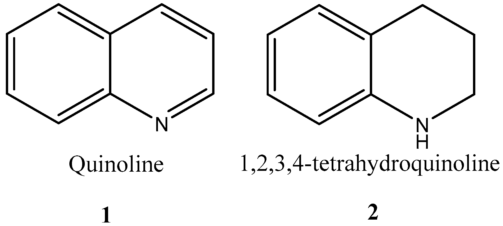
Benzo[b]pyridine or quinoline 1 in Figure 1 is one of the most prominent organic skeletons in medicinal chemistry practice and remains its interest until nowadays due to its pharmacological properties.
The quinoline fragment is part of the vast array of natural and synthetic medicines used in medical practice with diverse biological activities such as anti-inflammatory [1,2], anticancer [3], antimalarial [4,5], etc. The most outstanding performance for the quinoline fragment in drug development reveals its antimalarial and anticancer action, according to a large share of all the bioactive quinoline compounds known.
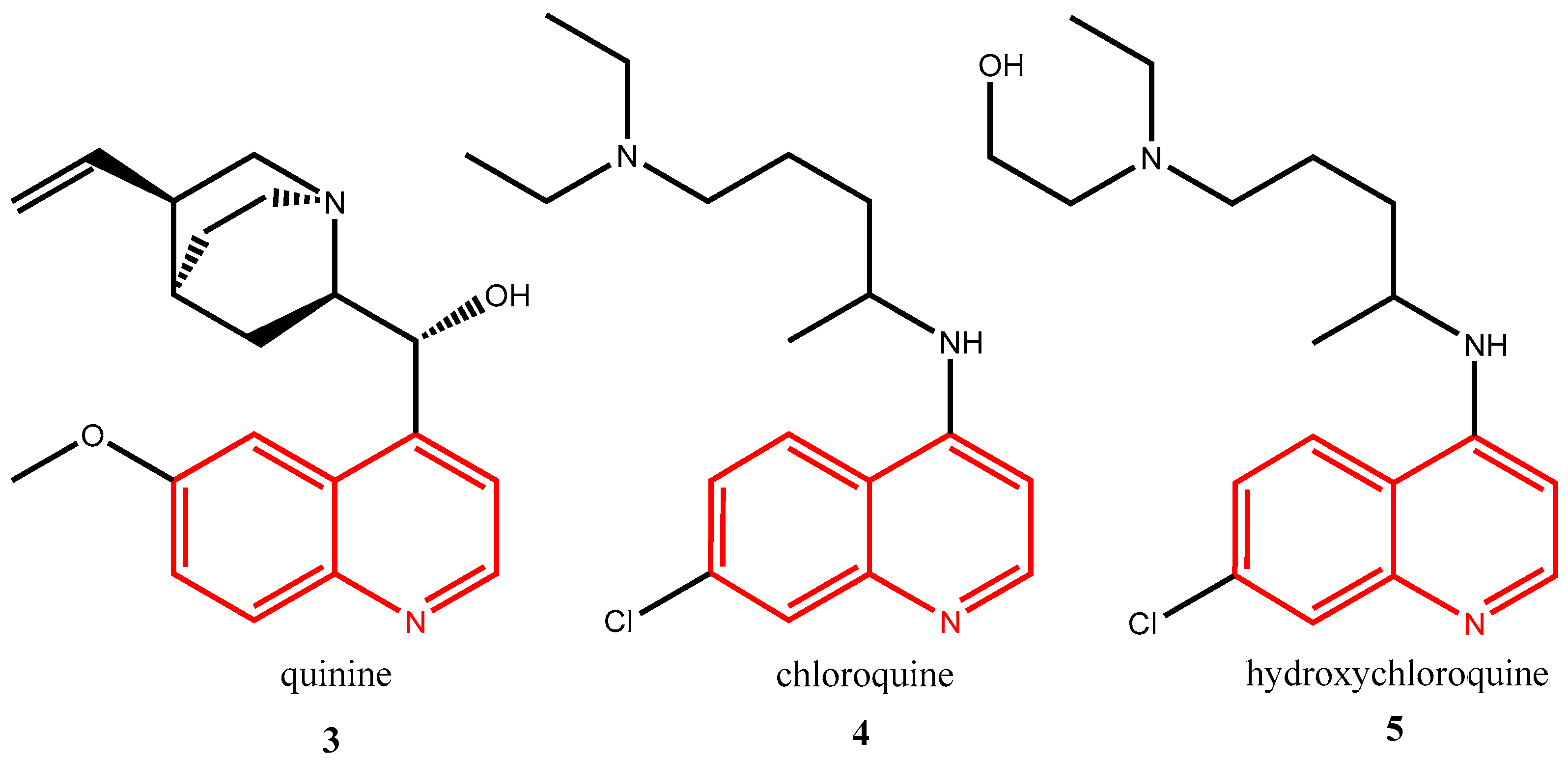
Recently, few drugs become even more popular in connection with the treatment of SARS-CoV-2 (COVID 19). In between those medicines are quinine 3, chloroquine 4, hydroxychloroquine 5, and others are also part of the family of drugs containing a quinoline (given in red) fragment in its structure (Figure 2).
Discovered in 1934 by H. Andersag chloroquine is a drug primarily used to treat malaria. Together with its derivatives chloroquine were used in the treatment of many other conditions such as HIV, rheumatoid arthritis, and systemic lupus erythematosus [6]. Chloroquine 4 blocks up the production of hemozoin by the parasite inside red blood cells, releasing free heme. Then, the complexed heme with chloroquine exerts a toxic effect on the plasma membrane of the Plasmodium species in order to treat malaria [7,8].
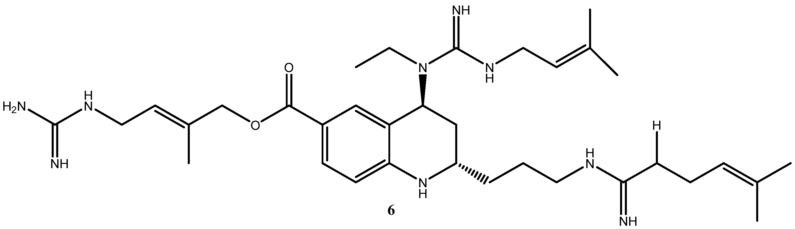
Likewise, the 1,2,3,4-tetrahydroquinoline skeleton is prevalent in many pharmacologically active synthetic and natural products [9]. Another interesting example is the 2,4,6-trisubstituted tetrahydroquinoline 6 (Figure 3) isolated from Martinella iquitosensis. (E)-4-guanidino-2-methylbut-2-en-1-yl(2S,4S)-4-(1-ethyl-3-(3-methylbut-2-en-1-yl)guanidino-2-(3-(5-methylhex-4-enimidamido)propyl)-1,2,3,4-tetrahydroquinoline-6-carboxylate exhibits activity as bradykinin antagonist and with α-adrenergic, histaminergic, and muscarinic receptors.
Many tetrahydroquinolines owing various simple or complex substituents have interesting biochemical activity; some are potential pharmaceutical agents. Thus, a very simple derivative, 2-methyl-5-hydroxy-1,2,3,4-tetrahydroquinoline, exhibits analgesic activity one-eighth as potent as morphine. There are many examples that could be given for the rich and varied biological activity of 1,2,3,4-tetrahydroquinoline derivatives [10].
Ibuprofen, on the other hand, is maybe the most popular non-steroidal anti-inflammatory drug, used in the treatment of many musculoskeletal disorders [11], and is the safest traditional choice for utilization during chronic neuroinflammation such as in Alzheimer’s [12], Parkinson’s [13], and Machado–Joseph disease [14]. Hybrids of ibuprofen with quinoline are interesting [11] in order to study their potential biological activity, and to answer the question of whether they will combine the properties of both parent molecules or not, as we know the carboxylic functional group is not required for the anti-inflammatory properties of profens [15] but for causing gastric toxicity.
Due to the importance of the creation of new hybrid molecules in the medicinal chemistry industry, we are searching for easy, fast, and low-cost ways for the preparation of new drug candidates. The obtaining of exactly this hybrid molecule can lead to the biological properties of the constituent fragments (ibuprofen and 1,2,3,4-tetrahydroquinoline) to be combined in the new one.
2. Results
2.1. Synthesis
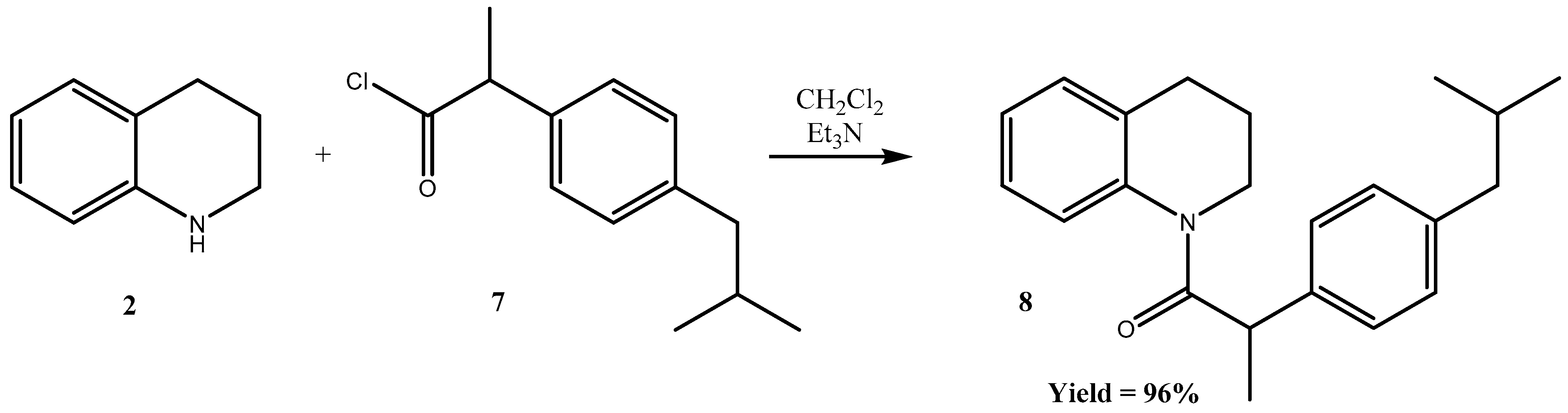
Herein we report the successfully synthesized 1-(3,4-dihydroquinolin-1(2H)-yl)-2-(4-isobutylphenyl)propan-1-one 8, as shown in Scheme 1.
In order to obtain the new hybrid molecule 8 we have started using coupling agents such as DCC and EDC to activate the carboxylic group of ibuprofen using various conditions, but unsuccessfully. We obtained the target compound 8 successfully by acylation of 1,2,3,4-tetrahydroquinoline 2 with 2-(4-isobutylphenyl)propanoyl chloride 7. The reaction generally works in a high yield.
The resultant compound is characterized by UV, 1H- and 13C-NMR, HPLC and HRMS spectra.
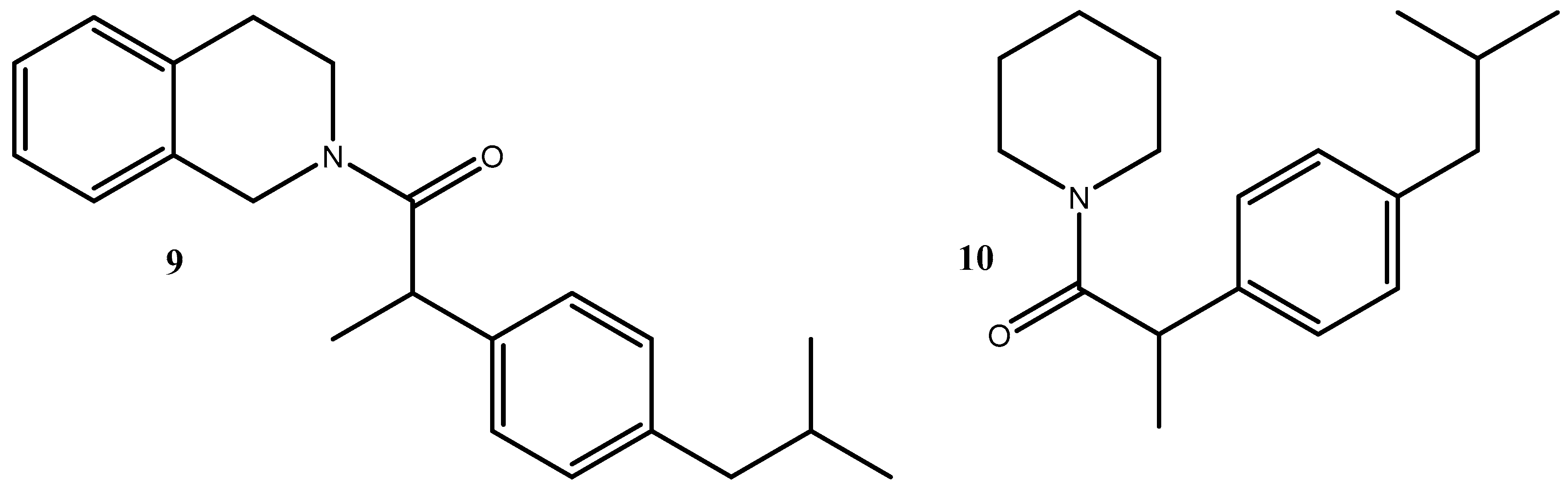
Using the same procedure, we have synthesized two other ibuprofen hybrids already described in the literature in order to evaluate and compare their potential biological properties with the newly obtained ibuprofen hybrid 8. We obtained the 1-(3,4-dihydroisoquinolin-2(1H)-yl)-2-(4-isobutylphenyl)propan-1-one 9 [16], and 2-(4-isobutylphenyl)-1-(piperidin-1-yl)propan-1-one 10 [17,18] (Figure 4).
2.2. Biological Evaluation
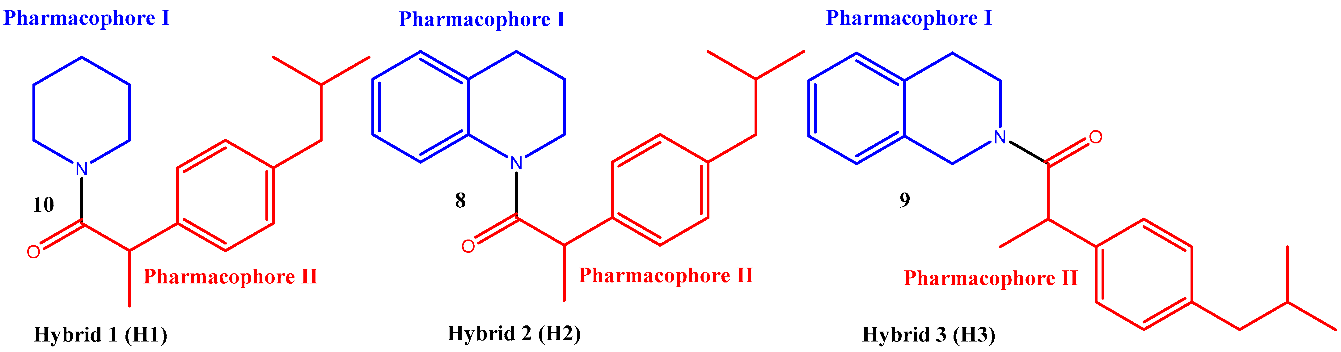
All three compounds 8, 9, and 10 were tested for their in vitro inhibition albumin denaturation (IAD), antioxidant, and antitryptic activity (ATA). The obtained in vitro results were compared to the predicted in silico ones. The results are presented in Table 1. In this study, we have investigated the effect of substituents in the structure of pharmacophore I, while pharmacophore II did not change structurally.
For the purpose of the biological evaluation study, we have named the three compounds hybrid 1 (H1), hybrid 2 (H2), and hybrid 3 (H3) as shown in Figure 5. Pharmacophore II in all molecules is ibuprofen residue while pharmacophore I varies. Pharmacophore I at hybrid 1 is piperidine, at hybrid 2 is 1,2,3,4-tetrahydoquinoline, and 1,2,3,4-tetrahydoisoquinoline is pharmacophore I at the third hybrid.
2.3. Hydrogen Peroxide Scavenging Activity (HPSA)
Reactive oxygen systems (ROS) are chemically reactive oxygen-containing radicals and molecules [super oxide (O2•−), hydroxylene (•OH), peroxyl (ROO•) and alkoxyl (RO•), HOCl, ozone (O3), peroxynitrite ONOO−), singlet oxygen (1O2) and H2O2]. They are naturally formed as a by-product of cellular metabolism. Under physiological conditions, enzyme systems regulate ROS levels.
They are known for damaging vital molecules of biological importance such as phospholipids, proteins, and DNA. The damage caused by them has been shown to be involved in the development of a number of diseases (cancer, cardiovascular disease, atherosclerosis, and Alzheimer’s disease) [19]. Even in a state of physiological health, the toxic effects of oxygen and its derivatives, accumulating in the body, lead to a reduction in life expectancy [20].
In the present work, research has focused on the deactivation of hydrogen peroxide. Hydrogen peroxide is an oxidant that is continuously formed in living tissues as a result of several metabolic processes. However, its detoxification is very important in preventing it from getting into harmful reactions, such as the Fenton reaction [21].
The inflammatory process also causes and accelerates the formation of ROS. Mostly the formation of a superoxide anionic radical at the site of inflammation and this is related to the formation of other ROS species, such as H2O2.
It is also involved in reductive decomposition reactions of hydrogen peroxide (so-called Haber–Weiss reaction) and organic hydroperoxides ROOH, assuming that at least some of the oxygen produced in these reactions is in the singlet state [22,23]. For this reason, the removal of H2O2 is very important to prevent the generation of •OH and the protection of biological systems.
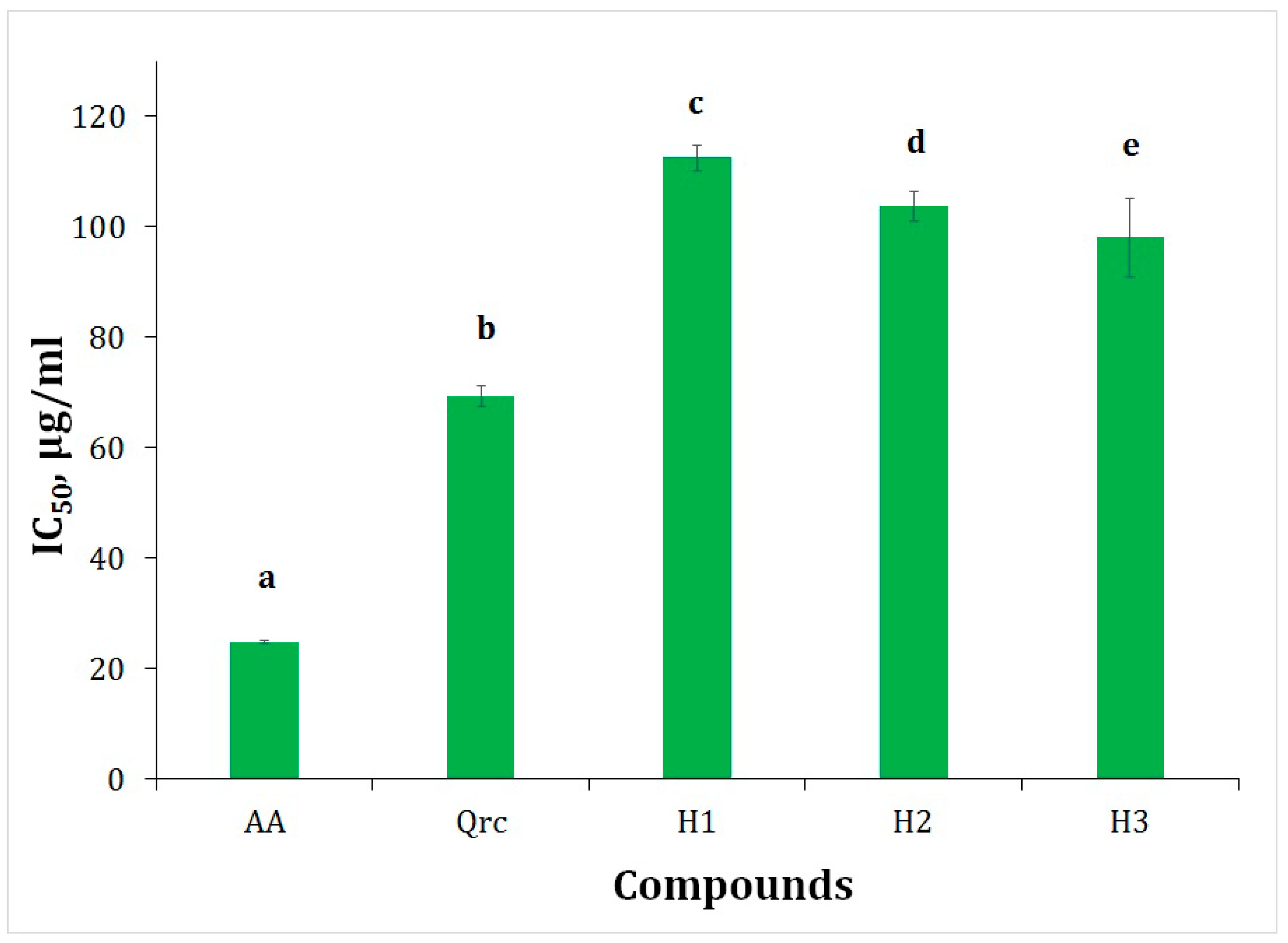
The obtained results of antioxidant activity of the synthetic analogs of ibuprofen were compared to the standards of ascorbic acid and quercetin, as they are natural compounds with proven antioxidant properties.
Compared to ascorbic acid (24.84 µg/mL) and quercetin (69.25 µg/mL) the obtained ibuprofen derivatives demonstrated lower in vitro antioxidant activity. Compound H3 (98.06 µg/mL) demonstrates higher antioxidant activity, compared to the rest of the synthesized compound (H1, H2) (Table 1, Figure 6). In the structure of the evaluated compounds, the presence of benzene nuclei increases the antioxidant effect. Therefore, compounds H2, and H3 show better activity compared to H1.
The presence of OH groups in the structure of the compounds affects their antioxidant activity. That is the reason why ascorbic acid and quercetin are characterized by very good antioxidant activity. In addition, there are free ortho and para positions in both phenolic nuclei in the quercetin structure, while in the structure of the tetrahydroquinoline derivative next to the nitrogen atom there is only one para and ortho position. That is why the analyzed hybrid molecules are showing lower HPSA compared to the quercetin standard.
Sroka reported that the substitution of hydroxyls is the most important factor in increasing the antioxidant and hydrogen peroxide scavenging activities of compounds with two OH groups substituted to the phenolic ring [24].
2.4. Inhibition of Albumin Denaturation (IAD)
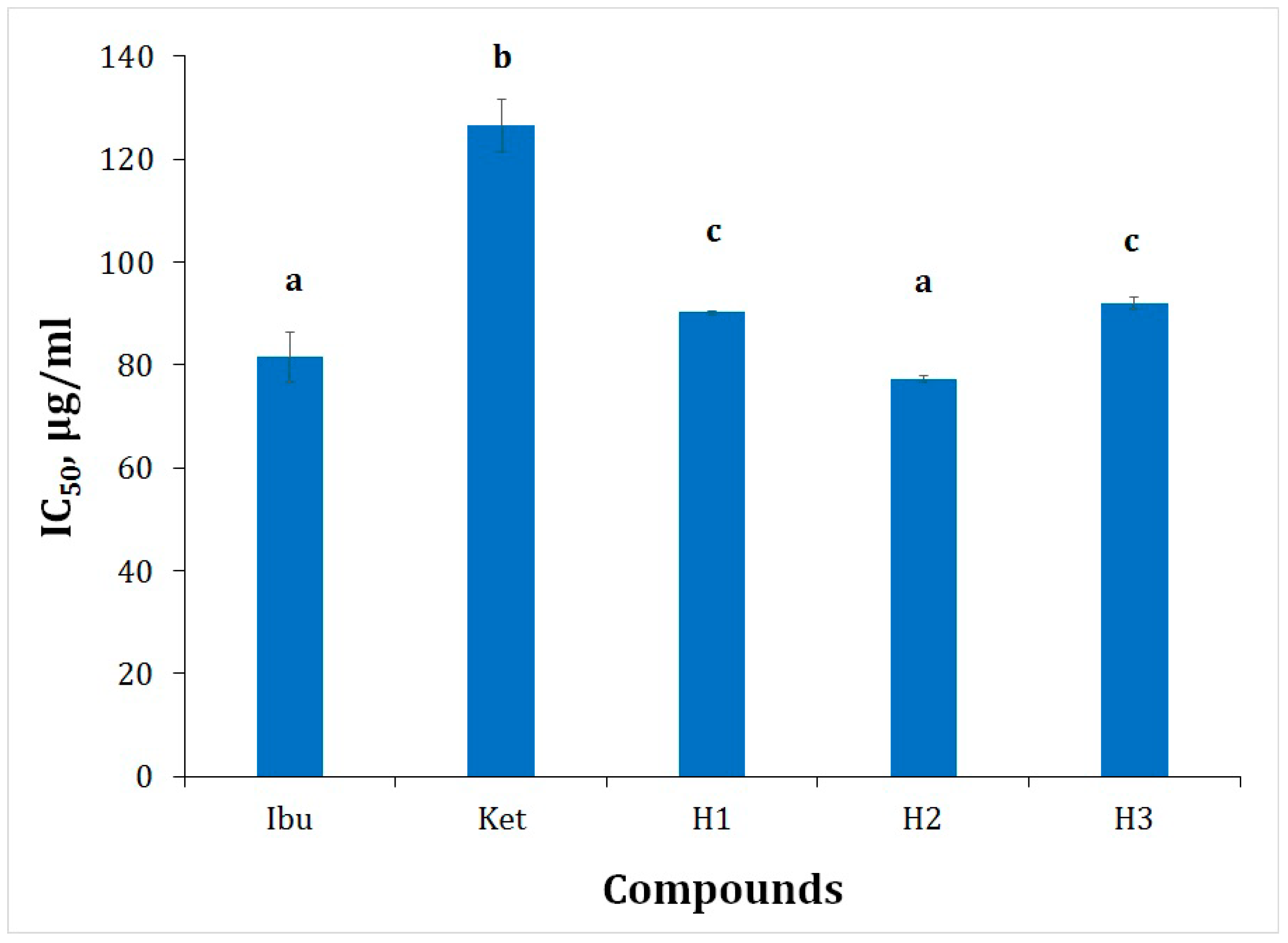
Inflammation is the response of living tissues to injury. It involves a complex array of enzyme activation, mediator release, fluid extravasation, cell migration, tissue breakdown, and repair [25]. The denaturation of proteins is a well-documented cause of inflammation in rheumatoid arthritis. Several anti-inflammatory drugs have shown a dose-dependent ability to inhibit thermally induced protein denaturation [26]. The obtained ibuprofen derivatives were screened for the inhibition of albumin denaturation. This method provides the extent information on to which albumin is protected from denaturation when heated. For this purpose, we have used human albumin. The percentages of inhibition of synthesized ibuprofen derivatives are presented in Figure 7. The results of the study are presented as IC50. As ibuprofen and ketoprofen have proven properties, we have decided to use them as a benchmark to compare the activities of newly synthesized ibuprofen derivatives. The IC50 values of ibuprofen and ketoprofen estimated as IAD is 81.50 μg/mL and 126.58 μg/mL, respectively, (Table 1, Figure 7). All obtained results show the IC50 values of ibuprofen derivatives are in the range from 77.38 to 92.08 μg/mL (Table 1, Figure 7). The IAD of ibuprofen derivatives was assessed using the Duncan test. This test allows a statistical assessment to be made between the mean IADs of the compounds (H1, H2, and H3), and the standards (ibuprofen and ketoprofen). The observed data show that ibuprofen derivatives are more active than ketoprofen. The analysis of variance using Duncan test (with a significance level p ˂ 0.05) revealed that there were not significant differences of the IAD values between ibuprofen and H2, and between H1 and H3.
In general, the resulting compounds (H1-3) exhibit high IAD activity as ibuprofen. The calculated values for anti-inflammatory activity (cAnti-A) show that the standards (ibuprofen and ketoprofen) are characterized by higher activity than the synthesized derivatives (H1-3), i.e., an inverse dependence is observed between in vitro and in silico (Table 1). There is a well-defined relationship between RM and cLogP (Table 1).
Furthermore, IAD analysis reveals that lipophilicity is a major physicochemical parameter. The studied synthetic ibuprofen derivatives show lipophilicity (RM) in the range between 1.04 and 1.33, which to some extent affects albumin protection. RM value of H2 (1.33) shows a greater effect on the stabilization of the albumin molecule (Table 1).
2.5. Antitryptic Activity (ATA)
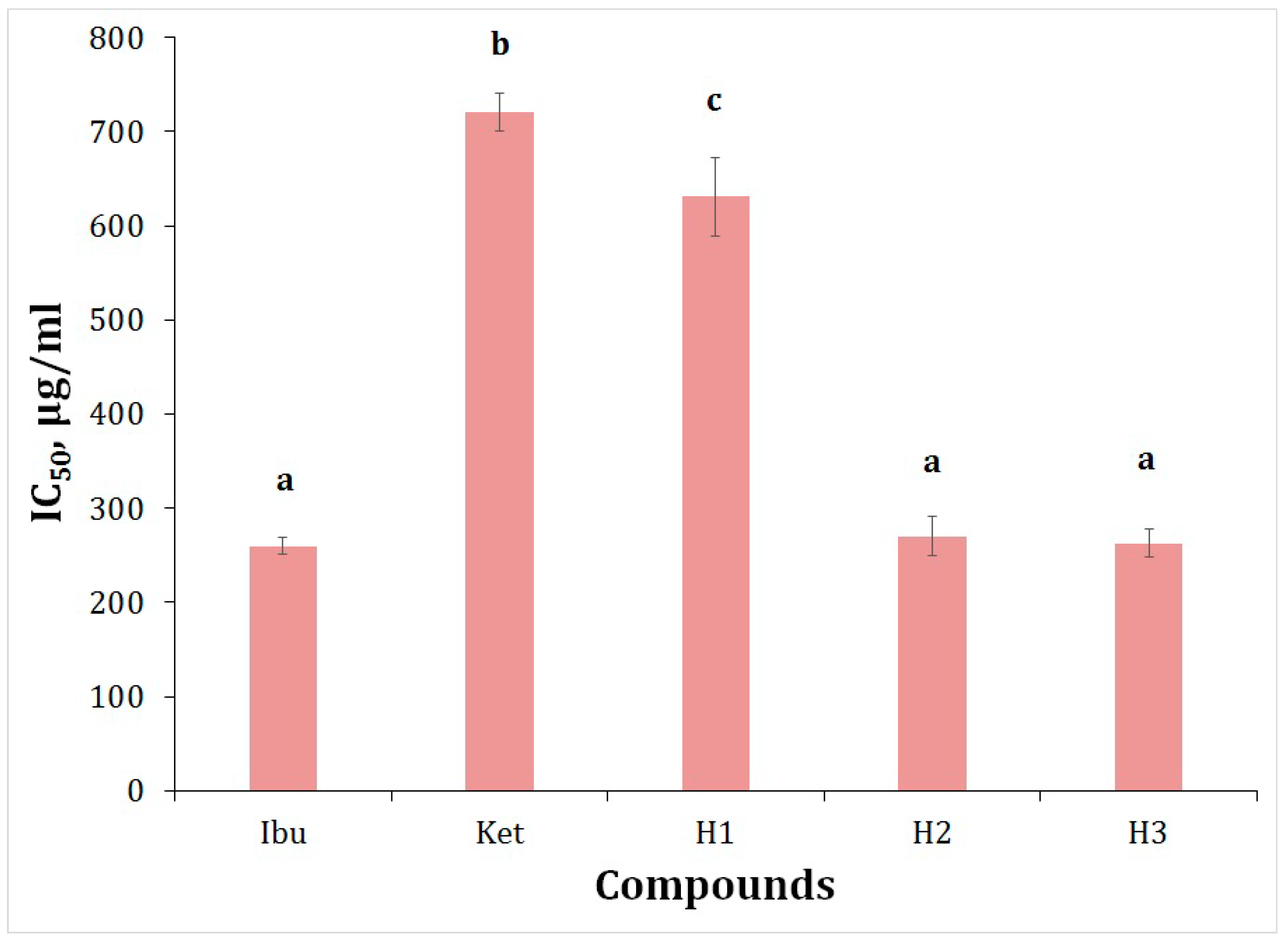
Proteinases have been implicated in arthritic reactions. Neutrophils are known to be a rich source of proteinase which carries many serine proteinases in their lysosomal granules. It was previously reported that leukocyte proteinase plays an important role in the development of tissue damage during inflammatory reactions and that a significant level of protection was provided by proteinase inhibitors [26,27]. In vitro anti-arthritic activity was assessed as antitryptic activity [27]. The IC50 results for the ATA range from 263 to 631.03 μg/mL.
The analysis of variance using Duncan test (with a significance level p ˂ 0.05) revealed that there were no significant statistical differences in the ATA values between ibuprofen and H2 and H3. The results present that the ibuprofen derivatives (H2 and H3) show better antitryptic activity compared to ketoprofen and H1 (Table 1, Figure 8).
2.6. Lipophilicity
Lipophilicity is the most regularly applied parameter used in SAR drug discovery studies. It can be experimentally determined or calculated. Lipophilicity has been correlated with permeability, solubility, and increases in target potency and toxicity. We determined the lipophilicity by reverse phase thin layer chromatography (RPTLC) method as RM values. This is considered to be a reliable, fast, and convenient method for expressing lipophilicity [28]. Aside from the essential role of lipophilicity for the kinetics of biologically active compounds, antioxidants of hydrophilic or lipophilic character are both needed to act as radical scavengers in the aqueous phase or as chain-breaking antioxidants in biological membranes [29].
In the present work, we have investigated the antioxidant and in vitro biological activity of the newly synthesized ibuprofen derivatives. Lipophilicity proved to be an important factor in their activity. The results have shown that compounds (H1-3) are lipophilic with low antioxidant activity. However, it should not be taken as a criterion for whether the compounds will exhibit biological activity. However, the use of lipophilic antioxidants is necessary to neutralize harmful radicals in cell membranes [29].
In general, the in vitro studies results show that the ibuprofen hybrid molecules exhibit IAD and ATA. From both experiments, we derive important information about the properties of potential new drugs, and both experiments are related to preserving the integrity of the albumin molecule. HSA is known to have two major binding sites for drugs: Sudlow sites I and II. According to Sudlow’s nomenclature, bulky heterocyclic molecules bind to Sudlow’s site I (located in subdomain IIA), whereas Sudlow’s site II (located in subdomain IIIA) is preferred by aromatic carboxylates with an extended conformation [30].
Ibuprofen has been shown to have good anti-inflammatory activity. However, it is also known that N-containing 6-membered alkaloids have biological effects such as anti-HIV and anti-tumor [31,32,33]. Therefore, we combined the two biologically active compounds in a new hybrid molecule. From all designed and carried experiments, we found out that the new hybrid molecules exhibit significant IAD and ATA activity.
3. Materials and Methods
3.1. Synthesis
All reagents and chemicals were purchased from commercial sources (Sigma-Aldrich S.A. and Riedel-de Haën, Sofia, Bulgaria) and used as received. Melting points were determined on a Boetius hot stage apparatus and are uncorrected. The NMR spectral data were recorded on a Bruker Avance II+600 spectrometer (BAS-IOCCP—Sofia, Bruker, Billerica, MA, USA). 1H-NMR and 13C-NMR spectra for compound 8 were taken in CDCl3 at 600 MHz and 151 MHz, respectively. Chemical shifts are given in relative ppm and were referenced to tetramethylsilane (TMS) (δ = 0.00 ppm) as an internal standard; the coupling constants are indicated in Hz. The NMR spectra were recorded at room temperature (ac. 295 K). Mass analyses were carried out on a Q Exactive Plus mass spectrometer (Thermo Fisher Scientific, Waltham, MA, USA). HPLC analysis consisted of quaternary mixer Smartline Manager 5000, pump Smartline 1000, and PDA 2800 detector (Knauer, Germany). Chromatographic conditions used: column Purosphere C18, 25 cm × 4.6 mm i.d., 5 µm particle size (Merck, Germany); mobile phase flow rate was set by 1.0 mL/min; sample volume was 20 µL. TLC was carried out on precoated 0.2 mm Fluka silica gel 60 plates (Merck KGaA, Darmstadt, Germany).
3.2. Synthesis of 1-(3,4-Dihydroquinolin-1(2H)-yl)-2-(4-isobutylphenyl)propan-1-one 8
To a solution of amine 2 (1 mmol, 0.133 g) in dichloromethane (15 mL), an equal amount of 2-(4-isobutylphenyl)propanoyl chloride (1 mmol, 0.224 g) 7 was added. After 10 min a little excess of triethylamine was added. After 30 min the solution was washed with diluted hydrochloric acid (HCl:H2O = 1:4, v/v), saturated solution of Na2CO3 and water. The organic layer was dried (Na2SO4), concentrated and filtered on short column with neutral Al2O3.
1-(3,4-dihydroquinolin-1(2H)-yl)-2-(4-isobutylphenyl)propan-1-one 8: bright yellow oil, yield 96% (0.308 g), 1H-NMR (600 MHz, CDCl3) δ 7.20–6.77 (m, 8H), 4.13 (s, 1H), 3.78 (d, J = 68.9 Hz, 1H), 3.52 (d, J = 5.1 Hz, 1H), 2.42 (d, J = 24.5 Hz, 1H), 2.33 (d, J = 7.2 Hz, 2H), 1.82–1.61 (m, 3H), 1.39 (d, J = 6.8 Hz, 3H), 0.79 (dd, J = 6.6, 1.7 Hz, 6H). 13C-NMR (151 MHz, CDCl3) δ 140.02 (C=O), 138.90 (C, Ar), 129.34 (C, Ar), 129.22 (C, Ar), 128.33 (C, Ar), 128.31 (C, Ar), 127.05 (C, Ar), 126.03 (C, Ar), 125.99 (C, Ar), 125.95 (C, Ar), 125.14 (C, Ar), 44.99 (N-CH2), 30.23 (–CH), 26.22 (–CH2), 23.92 (–CH2), 23.90 (–CH), 22.36 (–CH3), 22.31 (–CH2), 20.34 (–CH3). UV λmax, MeOH: 250 (ε = 4.9 × 10⁵) nm. HRMS Electrospray ionization (ESI) m/z calcd for C22H28NO+ = 322.2172, found 322.2175 (mass error Δm = 0.93 ppm).
Copies of all spectra, HPLC chromatogram, and ESI-HRMS (Figures S1–S5) are provided in the Supplementary Materials file.
3.3. Biological Evaluation
Chemicals and Reagents
Chromatographic grade methanol for HPLC analyses was used (VWR, Vienna, Austria). Water for HPLC was prepared with a Millipore purifier (Millipore, USA). Ibuprofen, ketoprofen, potassium dihydrogen phosphate, dipotassium hydrogen phosphate, sodium chloride, potassium chloride, hydrogen peroxide, ascorbic acid, trypsin, Tris-HCl buffer, and perchloric acid were purchased from Sigma-Aldrich, Taufkirchen, Germany. Human albumin 20%-BB, 200 g/L was purchased from BB-NCIPD Ltd., Sofia, Bulgaria. Chromatographic plates Kieselgel 60 F254 were purchased from Merck (Darmstadt, Germany).
3.4. Biological Experiments
Hydrogen Peroxide Scavenging Activity (HPSA)
The ability of ibuprofen derivatives to scavenge hydrogen peroxide was assessed according to the method reported by Ruch [34] with minor modification as described by Manolov et al. [35]. The solution of hydrogen peroxide (43 mM) was prepared in potassium phosphate-buffered solution (0.2 M, pH 7.4). Sample analysis was performed as follows: in test tubes were mixed 0.6 mL hydrogen peroxide (43 mM), 1 mL sample/standard with different concentrations (20–1000 μg/mL) and 2.4 mL potassium phosphate-buffered solution. The mixture was stirred and incubated in dark for 10 min at 37 °C. Absorbance was measured at 230 nm with a spectrophotometer (Camspec M508, Leeds, UK) against a blank solution containing phosphate buffer and hydrogen peroxide without the sample. Ascorbic acid and quercetin were used as standards. The percentage HPSA of the samples was evaluated by comparing with a blank sample and calculated using the following formula:
where Ablank is the absorbance of the blank sample (phosphate buffer and hydrogen peroxide), ACS is the absorbance of the control sample (test sample + phosphate buffer) and ATS is the absorbance of the test sample (test sample + phosphate buffer + hydrogen peroxide). The mean IC50 value was estimated based on three replicates by means of interpolating the graphical dependence of scavenging hydrogen peroxide on concentration.
3.5. Inhibition of Albumin Denaturation (IAD)
In vitro analysis of anti-inflammatory activity was assessed as inhibition of albumin denaturation (IAD). The analysis was performed according Sakat method [36] with minor modification [35]. The experiment was performed with human albumin. The solution of albumin (1%) was prepared in distilled water (pH 7.4). The tested compounds/standard were dissolved firstly in 1.2 mL DMF and PBS up to 25 mL so the final concentration of the stock solution is 1000 μg/mL. Then, a series of working solutions with different concentrations (20–500 μg/mL) in PBS were prepared. The reaction mixture was containing 2 mL test sample/standard of different concentrations and 1 mL albumin (1%). The mixture was incubated at 37 °C for 15 min and then heated at 70 °C for 15 min in water bath. After cooling the turbidity was measured at 660 nm with a spectrophotometer (Camspec M508, Leeds, UK). The experiment was performed three times. Percentage inhibition of albumin denaturation (IAD) was calculated against control. The control sample is albumin with the same concentration dissolved in distilled water.
3.6. Antitryptic Activity (ATA)
This method is known also as an in vitro anti-arthritic activity. The analysis was performed according to the method of Oyedapo and Femurewa [27] with minor modification as described by Manolov et al. [35]. The reaction mixture was containing 2 mL 0.06 mg/mL trypsin, 1 mL Tris–HCl buffer (20 mM, pH 7.4) and 1 mL test sample/standard (in methanol) of different concentrations (20–1000 μg/mL). The mixture was incubated at 37 °C for 5 min. Then, 1 mL of human albumin (4% v/v) was added. The mixture was incubated for an additional 20 min. To the mixture 2 mL of 70% perchloric acid was added for termination of the reaction. The cloudy suspension was cooled and centrifuged at 5000 rpm for 20 min. The absorbance of the supernatant was measured at 280 nm with a spectrophotometer (Camspec M508, Leeds, UK) against the control solution. The control solution was sample/standard in methanol with different concentrations. Ibuprofen was used as standard. The analysis was performed three times. The percentage of antitryptic activity (ATA) of the samples was evaluated by comparing with a blank sample. The blank sample is prepared as the test sample but with a small exception—perchloric acid is added before albumin.
where Ablank is the absorbance of the blank sample, ACS is the absorbance of the control solution (test sample in different concentrations) and ATS is the absorbance of the test samples. The mean IC50 values were estimated by means of interpolating the graphical dependence of ATA on concentration.
3.7. Physicochemical Characterization
3.7.1. Determination of Lipophilicity as RM Values
Determination of lipophilicity of ibuprofen derivatives was estimated according to the method reported by Pontiki and Hadjipavlou-Litina [29].
3.7.2. Prediction of Anti-Inflammatory and Anti-Arthritic Activity
3.8. Statistical Analysis
All the analyses were made in triplicates. Data were expressed as mean ± SD. The level of significance was set at p < 0.05. Statistical program SPSS 19.0 software was used for data analysis by one-way ANOVA followed by Duncan’s post hoc test to evaluate differences between mean values of activities (SPSS Inc., Chicago, IL, USA).
4. Conclusions
In conclusion, we have successfully synthesized and characterized the new hybrid molecule 1-(3,4-dihydroquinolin-1(2H)-yl)-2-(4-isobutylphenyl)propan-1-one, which containing two pharmacologically active fragments—1,2,3,4-tetrahydroquinoline and ibuprofen. The newly obtained hybrid H2 was biologically assessed for its in vitro antioxidant, antitryptic, and inhibition of albumin denaturation activity and compared to its structurally similar derivatives H1 and H3. The lipophilicity was also determined experimentally using reversed-phase thin layer chromatography and in silico calculations. The analyzed hybrid molecules showed significant results, which makes them interesting for further biological assessments.
Supplementary Materials
The following supporting information can be downloaded online. Figure S1: 1H-NMR spectrum of compound 8, Figure S2: 13C-NMR spectrum of compound 8, Figure S3: UV spectrum of compound 8, Figure S4: ESI-HRMS of compound 8, Figure S5: HPLC chromatogram of compound 8.
Author Contributions
Conceptualization, I.I.; methodology, S.M. and D.B.; software, S.M.; validation, D.B., S.M. and I.I.; formal analysis, S.M. and D.B.; investigation, S.M.; resources, I.I.; data curation, I.I.; writing—original draft preparation, S.M. and D.B.; writing—review and editing, S.M. and I.I.; visualization, I.I.; supervision, I.I.; project administration, S.M.; funding acquisition, I.I. All authors have read and agreed to the published version of the manuscript.
Funding
This research was funded by the National Science Fund of the Bulgarian Ministry of Education and Science, grant number KΠ 06 M29/1.
Institutional Review Board Statement
Not applicable.
Informed Consent Statement
Not applicable.
Data Availability Statement
The data presented in this study are available in this article and supporting Supplementary Material.
Acknowledgments
Dimitar Bojilov is thankful to the National Program of Ministry of Education and Science "Young Scientists and Postdoctoral Students" (PMC 577/2018) for financial support.
Conflicts of Interest
The authors declare no conflict of interest.
References
- Ambatkar, M.B.; Khedekar, P.B. Quinoline as TRPV1 antagonists: A new approach against inflammation. J. Drug Deliv. Therapeut. 2019, 9, 782–788. [Google Scholar] [CrossRef]
- Mukherjee, S.; Pal, M. Quinolines: A new hope against inflammation. Drug Discov. Today Off. 2013, 18, 389–398. [Google Scholar] [CrossRef] [PubMed]
- Musiol, R. An overview of quinoline as a privileged scaffold in cancer drug discovery. Expet Opin. Drug Discov. 2017, 12, 583–597. [Google Scholar] [CrossRef] [PubMed]
- Narula, A.K.; Azad, C.S.; Nainwal, L.M. New dimensions in the field of anti-malarial research against malaria resurgence. Eur. J. Med. Chem. 2019, 181, 111353. [Google Scholar] [CrossRef]
- Vandekerckhove, S.; D’hooghe, M. Quinoline-based antimalarial hybrid compounds. Bioorg. Med. Chem. 2015, 23, 5098–5119. [Google Scholar] [CrossRef]
- Plantone, D.; Koudriavtseva, T. Current and future use of chloroquine and hydroxychloroquine in infections, immune, neoplastic, and neurological diseases: A mini review. Clin. Drug Investig. 2018, 38, 653–671. [Google Scholar] [CrossRef]
- Orjih, A.U.; Banyal, H.S.; Chevli, R.; Fitch, S.D. Hemin lyses malaria parasites. Science 1981, 214, 667–669. [Google Scholar] [CrossRef]
- Fitch, S.D.; Chevli, R.; Banyal, H.S.; Phillips, G.; Pfaller, M.A.; Krogstad, D.J. Lysis of Plasmodium falciparum by ferriprotoporphyrin IX and a chloroquine-ferriprotoporphyrin IX complex. Antimicrob. Agents Chemother. 1982, 21, 819–822. [Google Scholar] [CrossRef] [Green Version]
- Alonso, C.; Martin-Encinas, E.; Rubiales, G.; Palacios, F. Reliable synthesis of phosphino- and phosphine sulfide-1,2,3,4-tetrahydroquinolines and phosphine sulfide quinolines. Eur. J. Org. Chem. 2017, 2017, 2916–2924. [Google Scholar] [CrossRef]
- Katritzky, A.; Rachwal, S.; Rachwal, B. Recent progress in the synthesis of 1,2,3,4-tetrahydroquinolines. Tetrahedron 1996, 52, 15031–15070. [Google Scholar] [CrossRef]
- Ghanim, A.; Girgis, A.; Kariuki, B.; Samir, N.; Said, M.; Abdelnaser, A.; Nasr, S.; Bekheit, M.; Abdelhameed, M.; Almalki, A.; et al. Design and synthesis of ibuprofen-quinoline conjugates as potential anti-inflammatory and analgesic drug candidates. Bioorg. Chem. 2022, 119, 105557. [Google Scholar] [CrossRef] [PubMed]
- Behl, T.; Kaur, I.; Fratila, O.; Brata, R.; Bungau, S. Exploring the potential of therapeutic agents targeted towards mitigating the events associated with amyloid β cascade in Alzheimer’s disease. Int. J. Mol. Sci. 2020, 21, 7443. [Google Scholar] [CrossRef] [PubMed]
- Costa, T.; Fernandez-Villalba, E.; Izura, V.; Lucas-Ochoa, A.M.; Menezes-Filho, N.J.; Santana, R.C.; de Oliveira, M.D.; Araújo, F.M.; Estrada, C.; Silva, V.; et al. Combined 1-deoxynojirimycin and ibuprofen treatment decreases microglial activation, phagocytosis and dopaminergic degeneration in MPTP treated mice. J. Neuroimmune Pharmacol. 2021, 16, 390–402. [Google Scholar] [CrossRef] [PubMed]
- Mendonça, L.S.; Nobrega, C.; Tavino, S.; Brinkhaus, M.; Matos, C.; Tom´e, S.; Moreira, R.; Henriques, D.; Kaspar, B.K.; De Almeida, L.P. Ibuprofen enhances synaptic function and neural progenitors’ proliferation markers and improves neuropathology and motor coordination in Machado—Joseph disease models. Hum. Mol. Genet. 2019, 28, 3691–3703. [Google Scholar] [CrossRef]
- Ullah, N.; Huang, Z.; Sanaee, F.; Rodrigues-Dimitrescu, A.; Aldawsari, F.; Jamali, F.; Bhardwaj, A.; Islam, N.; Velazques-Martinez, C. NSAIDs do not require the presence of a carboxylic acid to exert their anti-inflammatory effect—Why do we keep using them? J. Enzyme. Inhib. Med. Chem. 2015, 31, 1018–1028. [Google Scholar] [CrossRef]
- Geng, S.; Xiong, B.; Zhang, Y.; Zhang, J.; He, Y.; Feng, Z. Thiyl radical promoted iron-catalyzed-selective oxidation of benzylic sp3 C-H bonds with molecular oxygen. Chem. Commun. 2019, 55, 12699. [Google Scholar] [CrossRef]
- Backes, B.; Ellman, J. Carbon-carbon bond-forming methods on solid support. Utilization of Kenner’s “Safety-Catch” linker. J. Am. Chem. Soc. 1994, 116, 11171–11172. [Google Scholar] [CrossRef]
- Allegretti, M.; Bertini, R.; Cesta, M.; Bizzarri, C.; Bitondo, R.; Di Cioccio, V.; Galliera, E.; Berdini, V.; Topai, A.; Zampella, G.; et al. 2-Arylpropionic CXC chemokine receptor 1 (CXCR1) ligands as novel noncompetative CXCL8 inhibitors. J. Med. Chem. 2005, 48, 4312–4331. [Google Scholar] [CrossRef]
- Galano, A.; Macías-Ruvalcaba, N.A.; Campos, O.N.M.; Pedraza-Chaverri, J. Mechanism of the OH radical scavenging activity of nordihydroguaiaretic acid: A combined theoretical and experimental study. J. Phys. Chem. B 2010, 114, 6625–6635. [Google Scholar] [CrossRef]
- Halliwell, B.; Gutteridge, J.M.C. Free Radicals in Biology and Medicine; Clarendon Press: Oxford, UK, 1985; Volume 12, p. 346. [Google Scholar]
- Mansouri, A.; Makris, D.P.; Kefalas, P. Determination of hydrogen peroxide scavenging activity of cinnamic and benzoic acids employing a highly sensitive peroxyoxalate chemiluminescence-based assay: Structure–activity relationships. J. Pharm. Biomed. 2005, 39, 22–26. [Google Scholar] [CrossRef]
- Khan, A.U. Singlet molecular oxygen from superoxide anion and sensitized fluorescence of organic molecules. Science 1970, 168, 467–477. [Google Scholar] [CrossRef] [PubMed]
- Kellog, E.W.; Fridovich, I. Superoxide, hydrogen peroxide, and singlet oxygen in lipid peroxidation by a xanthine oxidase system. J. Biol. Chem. 1975, 250, 8812–8817. [Google Scholar] [CrossRef]
- Sroka, Z.; Cisowski, W. Hydrogen peroxide scavenging, antioxidant and anti-radical activity of some phenolic acids. Food Chem. Toxicol. 2003, 41, 753–758. [Google Scholar] [CrossRef]
- Vane, J.R.; Botting, R.M. New insights into the mode of action of anti-inflammatory drugs. Inflamm. Res. 1995, 44, 1–10. [Google Scholar] [CrossRef] [PubMed]
- Jayashree, V.; Bagyalakshmi, S.; Manjula Devi, K.; Richard Daniel, D. In vitro anti-inflammatory activity of 4-benzylpiperidine. Asian J. Pharm.Clin. Res. 2016, 9, 108–110. [Google Scholar] [CrossRef]
- Oyedapo, O.; Famurewa, A. Antiprotease and membrane stabilizing activities of extracts of fagara zanthoxyloides, olax subscorpioides and tetrapleura tetraptera. Int. J. Pharmacogn. 1995, 33, 65–69. [Google Scholar] [CrossRef]
- Hansch, C.; Leo, A.; Hoekman, D. Exploring QSAR: Hydrophobic, Electronic, and Steric Constants; American Chemical Society: Washington, DC, USA, 1995. [Google Scholar]
- Pontiki, E.; Hadjipavlou-Litina, D. Synthesis and pharmacochemical evaluation of novel aryl-acetic acid inhibitors of lipoxygenase, antioxidants, and anti-inflammatory agents. Bioorg. Med. Chem. 2007, 15, 5819–5827. [Google Scholar] [CrossRef]
- Fasano, M.; Curry, S.; Terreno, E.; Galliano, M.; Fanali, G.; Narciso, P.; Ascenzi, P. The extraordinary ligand binding properties of human serum albumin. IUBMB Life 2005, 57, 787–796. [Google Scholar] [CrossRef]
- Zhang, H.; Zembower, D.; Chen, Z. Structural analogues of the michellamine anti-HIV agents. Importance of the tetrahydroisoquinoline rings for biological activity. Bioorg. Med. Chem. Lett. 1997, 7, 2687–2690. [Google Scholar] [CrossRef]
- Scott, J.; Williams, R. Chemistry and Biology of the Tetrahydroisoquinoline Antitumor Antibiotics. Chem. Rev. 2002, 102, 1669–1730. [Google Scholar] [CrossRef]
- Lane, J.; Estevez, A.; Mortara, K.; Callan, O.; Spencer, J.; Williams, R. Antitumor activity of tetrahydroisoquinoline analogues 3-epi-jorumycin and 3-epi-renieramycin G. Bioorg. Med. Chem. Lett. 2006, 16, 3180–3183. [Google Scholar] [CrossRef] [PubMed] [Green Version]
- Ruch, R.; Cheng, S.; Klaunig, J. Prevention of cytotoxicity and inhibition of intercellular communication by antioxidant catechins isolated from Chineese green tea. Carcinogenesis 1989, 10, 1003–1008. [Google Scholar] [CrossRef] [PubMed]
- Manolov, S.; Ivanov, I.; Bojilov, D. Microwave-assisted synthesis of 1,2,3,4-tetrahydroisoquinoline sulfonamide derivatives and their biological evaluation. J. Serbian Chem. Soc. 2021, 86, 139–151. [Google Scholar] [CrossRef]
- Sakat, S.; Juvekar, A.; Gambhire, M. In-Vitro antioxidant and anti-inflammatory activity of methanol extract of Oxalis corniculata linn. Int. J. Pharm. Pharm. Sci. 2010, 2, 146–155. [Google Scholar]
- Sadym, A.; Lagunin, A.; Filimonov, D.; Poroikov, V. Prediction of biological activity spectra via the Internet. SAR QSAR Environ. Res. 2003, 14, 339–347. [Google Scholar] [CrossRef]
- Filimonov, D.; Lagunin, A.; Gloriozova, T.; Rudik, A.; Druzhilovskii, D.; Pogodin, P.; Poroikov, V. Prediction of the Biological Activity Spectra of Organic Compounds Using the Pass Online Web Resource. Chem. Heterocycl. Compd. 2014, 50, 444–457. [Google Scholar] [CrossRef]
Figure 1.
Structural formula of quinoline and 1,2,3,4-tetrahydroquinoline.

Figure 2.
Structural formulas of quinine 3, chloroquine 4, and hydroxychloroquine 5.

Figure 3.
Structural formula of (E)-4-guanidino-2-methylbut-2-en-1-yl(2S,4S)-4-(1-ethyl-3- (3-methylbut-2-en-1-yl)guanidino)-2-(3-(5-methylhex-4-enimidamido)propyl)-1,2,3,4-tetrahydroquinoline-6-carboxylate.
Figure 3.
Structural formula of (E)-4-guanidino-2-methylbut-2-en-1-yl(2S,4S)-4-(1-ethyl-3- (3-methylbut-2-en-1-yl)guanidino)-2-(3-(5-methylhex-4-enimidamido)propyl)-1,2,3,4-tetrahydroquinoline-6-carboxylate.

Scheme 1.
Synthesis of hybrid molecule 8.

Figure 4.
Structural formulas of hybrid molecules of ibuprofen with 1,2,3,4-tetrahidroisoquinoline 9 and piperidine 10.
Figure 4.
Structural formulas of hybrid molecules of ibuprofen with 1,2,3,4-tetrahidroisoquinoline 9 and piperidine 10.

Figure 5.
Structure of ibuprofen hybrids formed by two pharmacophores. Pharmacophore I (blue)– as the structures of piperidine, 1,2,3,4-tetrahydroquinoline and 1,2,3,4-tetrahydroisoquinoline, Pharmacophore II (red)-the structure of ibuprofen.
Figure 5.
Structure of ibuprofen hybrids formed by two pharmacophores. Pharmacophore I (blue)– as the structures of piperidine, 1,2,3,4-tetrahydroquinoline and 1,2,3,4-tetrahydroisoquinoline, Pharmacophore II (red)-the structure of ibuprofen.

Figure 6.
HPSA of ibuprofen derivatives. Ascorbic acid (AA) and quercetin (Qrc) were used as standards. The results of antioxidant activity were expressed as IC50. Different letters for the same method indicate significant difference at p < 0.05 levels by Duncan’s test.
Figure 6.
HPSA of ibuprofen derivatives. Ascorbic acid (AA) and quercetin (Qrc) were used as standards. The results of antioxidant activity were expressed as IC50. Different letters for the same method indicate significant difference at p < 0.05 levels by Duncan’s test.

Figure 7.
IAD of ibuprofen derivatives. Ibuprofen (Ibu) and ketoprofen (Ket) were used as standards. The results of IAD were expressed as IC50. Different letters for the same method indicate significant difference at p < 0.05 levels by Duncan’s test.
Figure 7.
IAD of ibuprofen derivatives. Ibuprofen (Ibu) and ketoprofen (Ket) were used as standards. The results of IAD were expressed as IC50. Different letters for the same method indicate significant difference at p < 0.05 levels by Duncan’s test.

Figure 8.
ATA of ibuprofen derivatives. Ibuprofen (Ibu) and ketoprofen (Ket) were used as standards. The results of ATA were expressed as IC50. Different letters for the same method indicate significant difference at p < 0.05 levels by Duncan’s test.
Figure 8.
ATA of ibuprofen derivatives. Ibuprofen (Ibu) and ketoprofen (Ket) were used as standards. The results of ATA were expressed as IC50. Different letters for the same method indicate significant difference at p < 0.05 levels by Duncan’s test.


Table 1.
In vitro and in silico biological activity results. Hydrogen peroxide scavenging activity (HPSA), inhibition of albumin denaturation (IAD), and antitryptic activity (ATA) were expressed as IC50. Ascorbic acid (AA), quercetin (Qrc), ibuprofen (Ibu) and ketoprofen (Ket) were used as standards. RM is a non-dimensional quantity because it is a function of Rf and is determined by thin-layer chromatography. cAnti-I and cAnti-A are expressed as Pa (probability “to be active”) estimates the chance that the studied compound is belonging to the sub-class of active compounds (resembles the structures of molecules, which are the most typical in a sub-set of “actives” in PASS training set). The value of the most active compound is 1; RM–lipophilicity; cAnti-I – calculated anti-inflammatory activity; cAnti-A–calculated anti-arthritic activity.
| Compounds | IC50 ± SD, μg/mL | RM ± SD | cLogP | Pa | |||
|---|---|---|---|---|---|---|---|
| HPSA | IAD | ATA | cAnti-I | cAnti-A | |||
| AA | 24.84 ± 0.35 | - | - | - | - | - | - |
| Qrc | 69.25 ± 1.82 | - | - | - | - | - | - |
| Ibu | - | 81.50 ± 4.95 | 259.82 ± 9.14 | 1.11 ± 0.010 | 3.72 | 0.903 | 0.573 |
| Ket | - | 126.58 ± 5.00 | 720.57 ± 19.78 | 1.54 ± 0.015 | 3.59 | 0.925 | 0.469 |
| H1 | 112.55 ± 2.32 | 90.23 ± 0.32 | 631.03 ± 41.88 | 1.04 ± 0.015 | 4.47 | 0.565 | - |
| H2 | 103.76 ± 2.61 | 77.38 ± 0.55 | 270.36 ± 20.85 | 1.33 ± 0.015 | 5.78 | 0.526 | - |
| H3 | 98.06 ± 7.17 | 92.08 ± 1.21 | 263.00 ± 14.48 | 1.14 ± 0.015 | 4.96 | 0.505 | - |
Publisher’s Note: MDPI stays neutral with regard to jurisdictional claims in published maps and institutional affiliations. |
© 2022 by the authors. Licensee MDPI, Basel, Switzerland. This article is an open access article distributed under the terms and conditions of the Creative Commons Attribution (CC BY) license (https://creativecommons.org/licenses/by/4.0/).
Share and Cite
MDPI and ACS Style
Manolov, S.; Ivanov, I.; Bojilov, D. Synthesis of New 1,2,3,4-Tetrahydroquinoline Hybrid of Ibuprofen and Its Biological Evaluation. Molbank 2022, 2022, M1350. https://0-doi-org.brum.beds.ac.uk/10.3390/M1350
AMA Style
Manolov S, Ivanov I, Bojilov D. Synthesis of New 1,2,3,4-Tetrahydroquinoline Hybrid of Ibuprofen and Its Biological Evaluation. Molbank. 2022; 2022(1):M1350. https://0-doi-org.brum.beds.ac.uk/10.3390/M1350
Chicago/Turabian StyleManolov, Stanimir, Iliyan Ivanov, and Dimitar Bojilov. 2022. "Synthesis of New 1,2,3,4-Tetrahydroquinoline Hybrid of Ibuprofen and Its Biological Evaluation" Molbank 2022, no. 1: M1350. https://0-doi-org.brum.beds.ac.uk/10.3390/M1350
Note that from the first issue of 2016, this journal uses article numbers instead of page numbers. See further details here.