Article

Novelties in Fuscosporellaceae (Fuscosporellales): Two New Parafuscosporella from Thailand Revealed by Morphology and Phylogenetic Analyses

by
Nattawut Boonyuen
1,*,
Charuwan Chuaseeharonnachai
1,
Salilaporn Nuankaew
1,
Papichaya Kwantong
1,
Natapol Pornputtapong
2,
Nakarin Suwannarach
3,*,
E. B. Gareth Jones
4 and
Sayanh Somrithipol
1
1
Plant-Microbe Interaction Research Team (APMT), Integrative Crop Biotechnology and Management Research Group (ACBG), National Center for Genetic Engineering and Biotechnology (BIOTEC), Pathum Thani 12120, Thailand
2
Department of Biochemistry and Microbiology, Faculty of Pharmaceutical Sciences and Research Unit of DNA Barcoding of Thai Medicinal Plants, Chulalongkorn University, Bangkok 10330, Thailand
3
Research Center of Microbial Diversity and Sustainable Utilization, Faculty of Science, Chiang Mai University, Chiang Mai 50200, Thailand
4
Department of Botany and Microbiology, College of Science, King Saud University, Riyadh 11451, Saudi Arabia
*
Authors to whom correspondence should be addressed.
Submission received: 4 October 2021 / Revised: 19 October 2021 / Accepted: 20 October 2021 / Published: 22 October 2021
(This article belongs to the Special Issue The Hidden Fungal Diversity in Asia)

Abstract

:
Asexual morphs of freshwater fungi have been mostly reported from tropical and subtropical regions. From our ongoing investigation of the diversity and taxonomy of freshwater microfungi in Thailand, a country with rich natural resources and diverse ecosystems, Parafuscosporella ellipsoconidiogena sp. nov. and P. obovata sp. nov., collected from decaying submerged twigs at Phalad Waterfall in a conserved forest in Chiang Mai Zoo, Chiang Mai Province, northern Thailand, are proposed. DNA phylogenies based on a combination of ITS and LSU datasets support the placement of these species in Parafuscosporella (Fuscosporellaceae, Fuscosporellales, Sordariomycetes), and these two novel species differ from known species in terms of morphology. Detailed descriptions, illustrations and a key to Parafuscosporella species are provided, as well as comparisons with other accepted Parafuscosporella species.

1. Introduction

Parafuscosporella belongs to Fuscosporellaceae (Fuscosporellales, Hypocreomycetidae, Sordariomycetes) [1]. The genus is characterized by sporodochial, black colonies; partly immersed, partly superficial, septate, hyaline to pale brown mycelium; semimacronematous, mononematous, simple or branched, mostly moniliform, smooth-walled, hyaline conidiophores; monoblastic, discrete or integrated, globose, subglobose, ellipsoidal or clavate, smooth-walled, hyaline conidiogenous cells; and conidia that are ellipsoidal to broadly obpyriform, transversely septate, smooth, dark brown to black and pale brown at the basal cell [1]. Based on morphological and molecular data, the type species without sexual morph, P. moniliformis Jing Yang, Bhat & K.D. Hyde, was described on dead and decaying submerged wood in Thailand. To date, five accepted species, namely, P. aquatica H. Yang & H. Zhang, P. garethii Boonyuen, Chuaseehar. & Somrith., P. moniliformis, P. mucosa Jing Yang, Bhat & K.D. Hyde, and P. pyriformis H. Yang, W. Dong & H. Zhang, have only been reported from decaying submerged wood in Thailand and China (http://www.speciesfungorum.org; accessed on 12 September 2021) [1,2,3,4]. In this study, we describe P. ellipsoconidiogena and P. obovata as the sixth and seventh species in the genus, respectively, collected from a waterfall in Chiang Mai Zoo, Chiang Mai Province, Thailand. Morphological descriptions and illustrations of P. ellipsoconidiogena sp. nov. and P. obovata sp. nov., a key to the species and an updated combined gene phylogenetic tree (the internal transcribed spacer (ITS) region of ribosomal DNA and large subunit (LSU) of nuclear ribosomal DNA) are provided to reveal their taxonomic position among taxa in the Fuscosporellaceae (Fuscosporellales).

2. Materials and Methods

2.1. Sample Collection, Isolation and Morphological Data

Submerged woody material was randomly collected from Phalad Waterfall located in Chiang Mai Zoo (18°48′32.40′′ N; 98°56′49.20′′ E), Muang District, Chiang Mai Province, northern Thailand (http://www.chiangmai.zoothailand.org/en/accessed on 12 September 2021). The zoo is located on a 200-acre (81 ha) woody area at the foot of Doi Suthep-Pui National Park. Phalad Waterfall is within the Plant Genetic Conservation Project under the Royal Initiation of Her Royal Highness Princess Maha Chakri Sirindhorn (RSPG). Woody samples were placed into plastic bags and transferred to the mycological laboratory at the National Center for Genetic Engineering and Biotechnology (BIOTEC), National Science and Technology Development Agency (NSTDA, Pathum Thani, Thailand), for observation. Decaying wood specimens were incubated in plastic containers with sterile tissue paper soaked with sterile distilled water at room temperature (20–25 °C) for 7–14 days, according to the methods described by Boonyuen et al. [2]. The specimens were observed using a stereomicroscope (Olympus SZ61; Olympus Corporation, Tokyo, Japan) for the presence of freshwater microfungi, and permanent slides were prepared by adding lactoglycerol and sealing with clear nail polish. Morphological characteristics such as conidiophores, conidiogenous cells and conidial dimension were examined. Cultural characteristics such as colony appearance and colour over the plate were also studied. Axenic cultures were obtained by single spore isolation method, following the protocol in Chuaseeharonnachai et al. [5]. Germinated spores were transferred to a potato dextrose agar (PDA, DifcoTM, Sparks, MD, USA) plate and incubated at room temperature (20–25 °C). The type specimens were deposited at the FUNGARIUM BIOTEC Bangkok Herbarium (BBH; https://www.nbt-microbe.org accessed on 22 September 2021), as Parafuscosporella ellipsoconidiogena BBH 49158 (holotype) and P. obovata BBH 49160 (holotype). Pure cultures are maintained in the Thailand Bioresource Research Center (TBRC; https://www.tbrcnetwork.org accessed on 22 September 2021) as TBRC 15503 and TBRC 15505. The Index Fungorum numbers were registered as P. ellipsoconidiogena IF 555786 and P. obovata IF 555787, respectively [6].

2.2. DNA Extraction, PCR Amplification and Sequencing

Genomic DNA was extracted from pure fungal mycelium grown on PDA for 14 days at room temperature using cetyltrimethylammonium bromide (CTAB) lysis buffer as outlined by Sri-indrasutdhi et al. [7]. The ITS region of ribosomal DNA, LSU of nuclear ribosomal DNA, small subunit (SSU) of nuclear ribosomal DNA and RNA polymerase II second largest subunit (RPB2) were amplified via polymerase chain reaction (PCR) using the following primers: ITS1/ITS5/ITS4 [8] for the ITS, LR0R/LR5/LR7 [9] for the LSU, NS1/NS4 for the SSU [8] and fRPB2-5F2/fRPB2-7cR for RPB2 [10].
PCR amplification was performed in a 50 μL reaction volume containing 25 μL of One PCRTM Ultra (Bio-Helix, New Taipei City, Taiwan; a premix and ready-to-use solution, including Taq DNA polymerase, PCR buffer, dNTPs, gel loading dyes, enhancer, and fluorescence dye), 1 μL of each primer (10 μM), 1 μL of genomic DNA extract and 22 μL of sterile deionized water. The PCR thermal cycle programs of the ITS and LSU were as follows: 94 °C for 2 min, followed by 35 cycles of denaturation at 94 °C for 1 min, annealing at 55 °C for 1 min, elongation at 72 °C for 2 min and a final extension at 72 °C for 10 min. The PCR thermal cycle program of the SSU was as follows: 95 °C for 5 min, followed by 34 cycles of denaturation at 95 °C for 1 min, annealing at 55 °C for 1 min, elongation at 72 °C for 1.5 min and a final extension at 72 °C for 10 min. The PCR thermal cycle program of RPB2 was as follows: 95 °C for 5 min, followed by 34 cycles of denaturation at 95 °C for 1 min, annealing at 58 °C for 1 min, elongation at 72 °C for 1.5 min and a final extension at 72 °C for 10 min. The amplicons of the ITS and LSU were purified and sequenced by Macrogen Inc. (Seoul, South Korea) with the same PCR primer used for DNA amplification. The PCR products of RPB2 were purified using a NucleoSpin® Gel and PCR Clean-up Kit (Macherey-Nagel, Düren, Germany) and sequenced at Macrogen Inc. (Seoul, South Korea).

2.3. Sequence Alignment and Phylogenetic Analyses

The SSU, ITS, LSU and RPB2 sequences of our isolates are provided in this study. Based on previous phylogenetic studies on Fuscosporellaceae (Fuscosporellales) by Yang et al. [3], two combined analyses of the ITS and LSU sequences provided resolution at the species level. In addition, there are only a few SSU and RPB2 sequences of Fuscosporellales available in GenBank. Thus, the ITS and LSU datasets were used only for the combined sequence data analyses in this study.
A maximum likelihood (ML) tree was constructed by RAxML-NG v. 1.0.3 using the GTR+G model and the all-in-one analysis option [11]. The best ML tree was identified using the two-step L-BFGS-B method [12], to optimize the parameters of the LG4X model [13]. ML branch support was obtained using nonparametric bootstrapping with 1000 replications.
A Bayesian inference (BI) phylogenetic tree was constructed with the GTR+G model using the Metropolis-coupled Markov chain Monte Carlo (MCMCMC) method in MrBayes 3.2.7a [14]. The MCMCMC searches were run for 1,000,000 generations with sampling every 100 generations. BI posterior probabilities (BIPPs) were summarized and mapped on the best ML tree using the SumTrees program in DendroPy version 4.5.2 [15]. The first 100 trees were excluded as burn-in. The newly obtained sequences taxa used in phylogenetic analyses were deposited in the GenBank database and are provided in Table 1.

3. Results

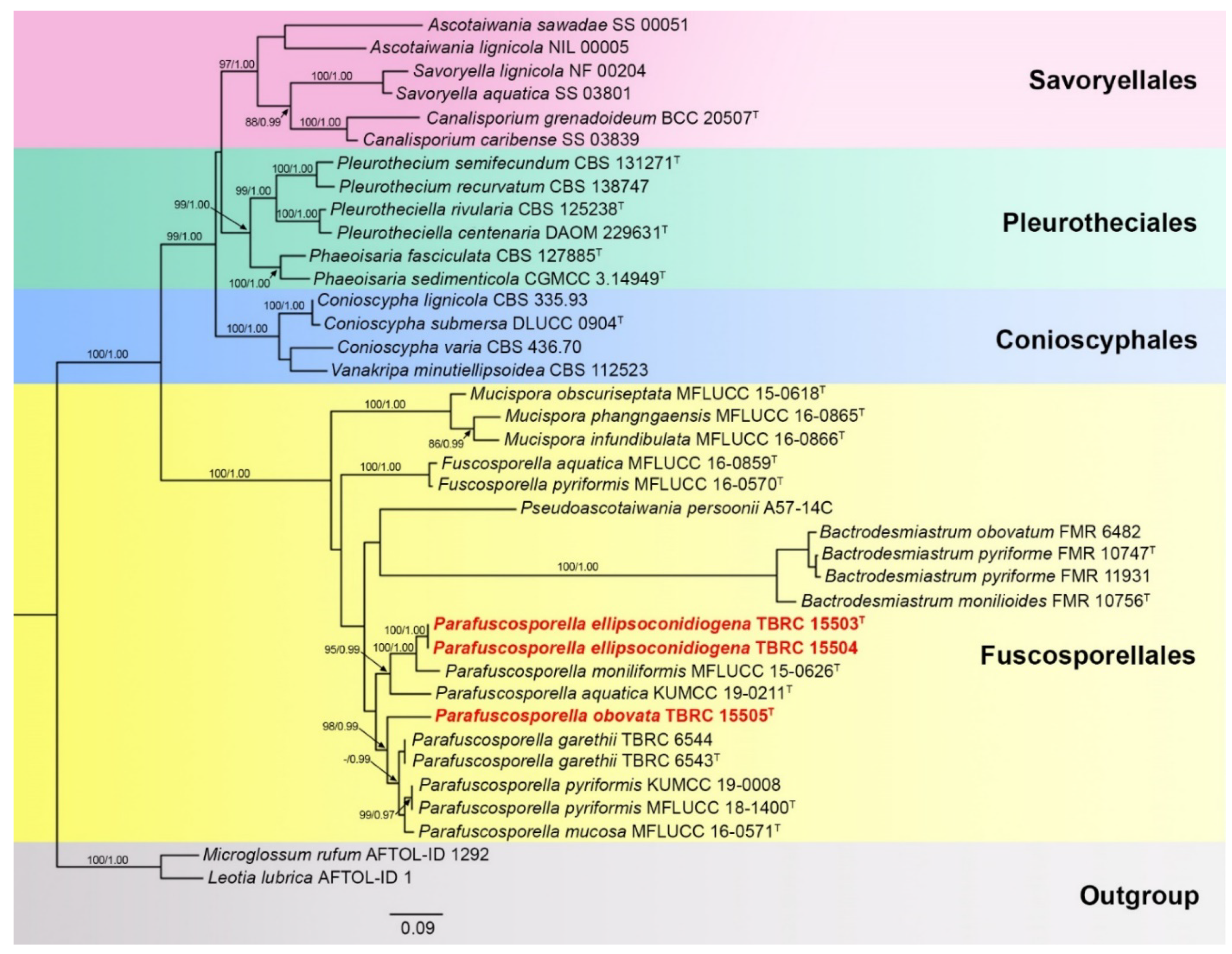
3.1. Molecular Phylogeny
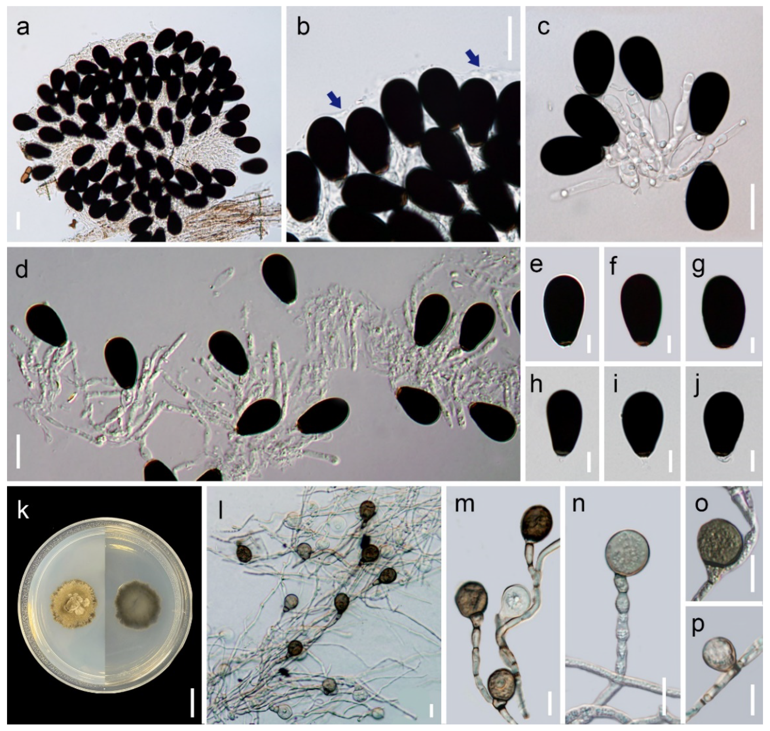
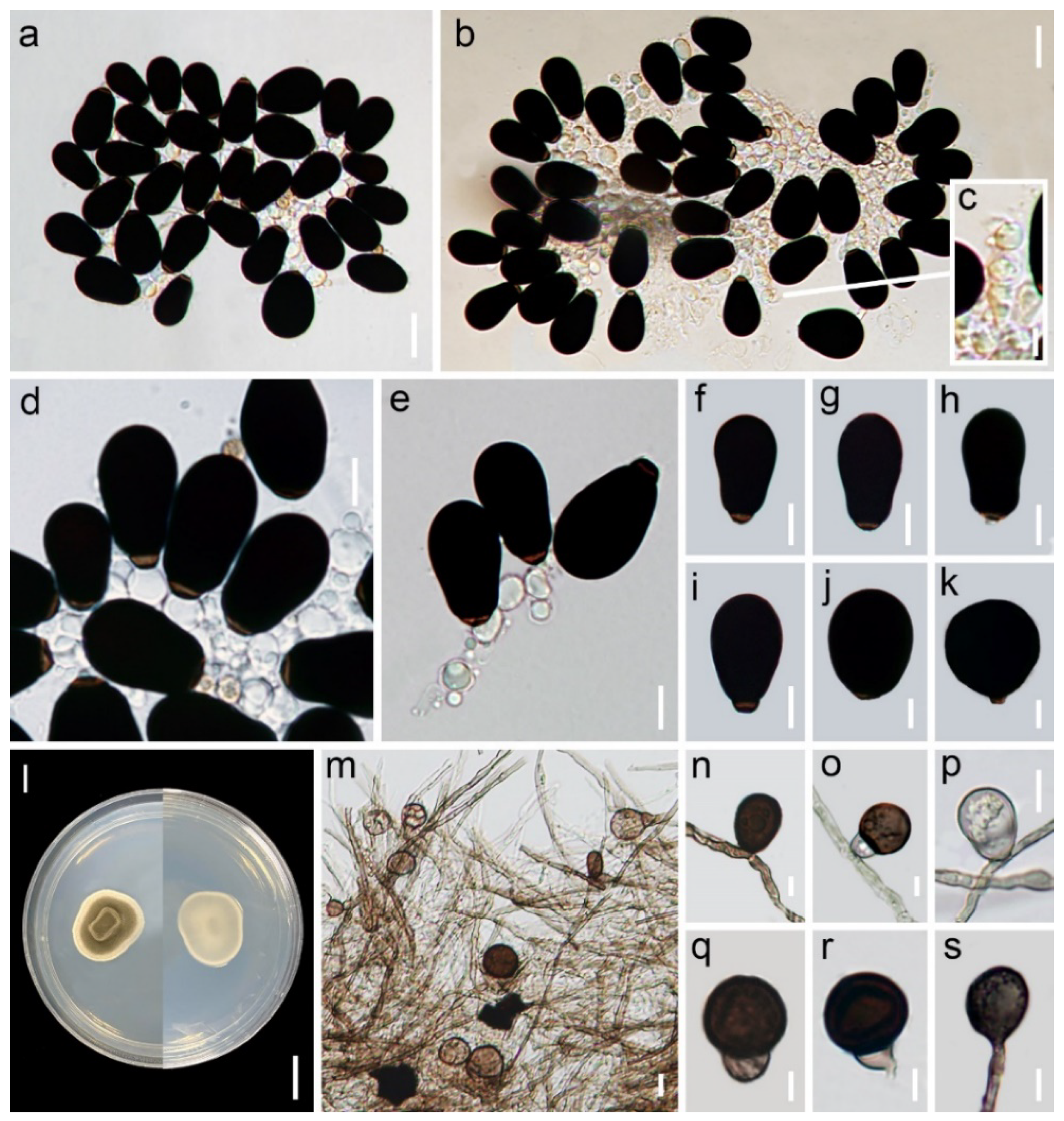
The dataset for the phylogenetic analysis comprised 33 representative strains from Fuscosporellales, Savoryellales, Pleurotheciales and Conioscyphales. Leotia lubrica (AFTOL-ID 1) and Microglossum rufum (AFTOL-ID 1292) were used as outgroups. Based on the combined ITS and LSU sequence data, the phylogram (Figure 1) shows that Parafuscosporella is a monophyletic genus in Fuscosporellaceae (Fuscosporellales, Hypocreomycetidae, Sordariomycetes), with P. ellipsoconidiogena sp. nov. (TBRC 15503 and TBRC 15504) clustering with the closely related P. moniliformis MFLUCC 15-0626, with strong statistical support (ML-BS 100% and BYPP 1.00). The tree generated from ITS sequence data and combined ITS, LSU and RPB2 sequence analyses had a somewhat similar topology (Figures S1 and S2). Parafuscosporella obovata sp. nov. (TBRC 15505) forms a sister clade, in relationship with the clade of P. garethii, P. pyriformis (MFLUCC 18-1400 and KUMCC 19-0008) and P. mucosa (MFLUCC 16-0571; ML-BS 98% and BIPP 0.99), with high statistical support.

3.2. Taxonomy

Parafuscosporella ellipsoconidiogena Chuaseehar., Somrith. & Boonyuen, sp. nov. (Figure 2).
Index Fungorum: IF 555786.
Etymology: Referring to the ellipsoidal shape of the conidiogenous cells.
Description: Asexual morph. Colonies on the natural substratum sporodochial, granular, scattered, black with a jelly-like covering. Mycelium mostly superficial, partially immersed, composed of branched, smooth-walled, septate, hyaline hyphae. Conidiophores semi- to macronematous, mononematous, compact, erect or flexuous, branched, 2–3-septate, mostly moniliform, smooth-walled, septate, hyaline, 25.2–68.8 × 3.6–7.9 μm (avg. 41.3 × 5.6 μm, n = 15), with each cell doliiform, ellipsoidal, fusiform, 7.5–17.5 × 3.6–7.9 μm. Conidiogenous cells holoblastic, monoblastic, integrated, terminal, smooth-walled, hyaline, doliiform, ellipsoidal, fusiform, 7.9–24.3 × 5.1–9.6 μm (avg. 15.3 × 7 μm, n = 20). Conidial secession rhexolytic. Conidia acrogenous, solitary, ellipsoidal to obovoid, smooth-walled, 2-celled with a transverse septum near the base, dark brown to black, 27.5–33 × 15–20 μm (avg. 30.5 × 18 μm, n = 50), with a light brown, short and narrow, truncate basal cell and distinct, hyaline, 1.3−3.8 × 2.5−3 μm basal frills. Sexual morph unknown.
Culture characteristics: On PDA, colonies growing on PDA at 20−25 °C for 30 days, dry, flat, circular, velvety, spreading, brown with beige-brown patches, with a prominent dark brown outer zone of submerged growth and serrate margin, reverse dark brown. Vegetative hyphae partly superficial and partially immersed, branched, smooth-walled, septate, subhyaline to light brown, 2–3.8 μm wide. Conidiophores micronematous, reduced to a single conidiogenous cell. Conidiogenous cells holoblastic, monoblastic, integrated, cylindrical or ellipsoidal, hyaline to pale brown, 3.75–12.5 × 3.75–6.4 μm (avg. 6.3 × 5.5 μm, n = 15). Conidial secession rhexolytic. Conidia acrogenous or pleurogenous, broadly obpyriform, ellipsoidal, obovoid, smooth-walled, 1- or 2-celled with a transverse septum near the base, medium brown to dark brown when mature, 15–27.5 × 10.5–17.5 μm (avg. 20.3 × 14.4 μm, n = 50), with a light brown, triangular basal cell; chlamydospores absent. Sexual morph absent.
Habitat and geographical distribution: Saprobe on submerged twigs, known from Thailand.
Type: Thailand, Chiang Mai Province, Mueang Chiang Mai District, Phalad Waterfall in Chiang Mai Zoo, 18°48’37” N, 98°56’51” E, on submerged twigs of an unidentified plant, 18 August 2018, N. Boonyuen, BBH 49158 holotype, TBRC 15503 ex-holotype living culture; BBH 49159 isotype, TBRC 15504 ex-isotype living culture.
Additional gene sequences: OK054346 (SSU), OK043808 (RPB2), OK054347 (SSU) and OK043809 (RPB2).
SSU: Based on BLAST analysis of the SSU sequences of TBRC 15503 (OK054346) and TBRC 15504 (OK054347), the data revealed that closely related strains with % identities were Parafuscosporella moniliformis MFLUCC 15-0626T (100%), P. mucosa MFLUCC 16-0571T (99.8%) and P. garethii TBRC 6544 (99.6%).
RPB2: BLAST analysis of the RPB2 sequences of TBRC 15503 (OK043808) and TBRC 15504 (OK043809) revealed that the closely related strains with % identities were Parafuscosporella garethii TBRC 6543T (92.7%), P. garethii TBRC 6544 (92.5–92.6%) and P. pyriformis KUMCC 19-0008 (92.0–92.1%).
LSU: BLAST analysis of the LSU sequences of TBRC 15503 (OK044741) and TBRC 15504 (OK044742) showed the most closely related strains with % identities were Parafuscospora garethii TBRC 6543T (97.4–97.6%), P. garethii TBRC 6544 (97.4–97.6%) and P. moniliformis MFLUCC 15-0626T (98.9–99%).
ITS: BLAST analysis of the ITS sequences of TBRC 15503 (OK044749) and TBRC 15504 (OK044750) revealed the most closely related strains with % identities were Parafuscosporella moniliformis MFLUCC 15-0626T (87.6–88.2%) and Parafuscosporella aquatica KUMCC 19–0211T (87.8%).
Note: Parafuscosporella ellipsoconidiogena resembles species of Vanakripa Bhat, W.B. Kendr. & Nag Raj [29] in possessing a sporodochium; large and dark-pigmented conidia; and a narrow-long, hyaline conidiogenous cell resembling the separating cell of Vanakripa.
Parafuscosporella ellipsoconidiogena differs from Vanakripa species due to an absence of vermiform to obpyriform separating cells. Morphologically, P. ellipsoconidiogena is most similar to P. mucosa in having natural substratum colonies with a jelly-like covering, conidiophores arranging only one form in cylindrical or moniliform, ellipsoidal conidiogenous cells, and uniseptate, dark-pigmented conidia [1]. However, they mainly differ in the shape of conidiogenous cells and conidiophores. Parafuscosporella ellipsoconidiogena has doliiform or fusiform conidiogenous cells and moniliform conidiophores, while P. mucosa possesses globose, subglobose or clavate conidiogenous cells and cylindrical conidiophores. Conidiogenous cells of the new species are also longer (7.9–24.3 × 5.1–9.9 μm) than those of P. mucosa. P. mucosa produces cylindrical conidiophores and globose, subglobose, clavate and shorter (7–17 × 4–12 μm) conidiogenous cells that differ from those of P. ellipsoconidiogena [1].
In PDA culture, the sizes of the conidiogenous cells and conidia of both species somewhat overlap, and these two species mainly differ in the shape of the conidiogenous cells as well as the shape and colour of the conidia. Parafuscosporella ellipsoconidiogena has cylindrical or ellipsoidal conidiogenous cells and broadly obpyriform, ellipsoidal, obovoid, medium brown to dark brown conidia, while P. mucosa has doliiform or obovoid conidiogenous cells and globose to subglobose, olivaceous to pale brown conidia [1].
In the phylogenetic tree inferred from the two combined ITS and LSU sequences (Figure 1), P. ellipsoconidiogena is closely related to P. moniliformis. Morphologically, P. ellipsoconidiogena and P. moniliformis share a similar morphology of the sporodochial conidiomata and conidiophores that are mostly moniliform with ellipsoidal moniliform conidiogenous cells [1]. However, P. ellipsoconidiogena differs from P. moniliformis in having natural substratum colonies with a jelly-like covering, doliiform or fusiform, shorter and narrower (7.9–24.3 × 5.1–9.6 μm) conidiogenous cells with obovoid conidia, while in P. moniliformis, conidiomatal colonies without a jelly-like covering, globose, subglobose, clavate, longer and wider (5.5–36 × 5–21 μm) conidiogenous cells with broadly obpyriform conidia [1]. In PDA culture, the differences between P. ellipsoconidiogena and P. moniliformis are in the shape of conidiogenous cells and conidia. In P. ellipsoconidiogena, it has ellipsoidal conidiogenous cells with broadly obpyriform, ellipsoidal or obovoid conidia, while P. moniliformis has subglobose or dumbbell-shaped conidiogenous cells with globose to subglobose conidia [1]. The comparison of Parafuscosporella species on natural substrates and on PDA culture are presented in Table 2 and Table 3.
Parafuscosporella obovata Chuaseehar., Somrith. & Boonyuen, sp. nov. (Figure 3).
Index Fungorum: IF 555787.
Etymology: Referring to the presence of obovoid conidia.
Description: Asexual morph. Colonies on the natural substratum sporodochial, granular, scattered, black. Mycelium mostly superficial, partially immersed, composed of branched, smooth-walled, septate, hyaline hyphae. Conidiophores semi- to macronematous, mononematous, compact, erect or flexuous, branched, 3–4-septate, mostly moniliform, smooth-walled, septate, hyaline, 25.2–42.1 × 5.8–9.7 μm (avg. 31 × 8.9 μm, n = 10), with each cell globose to subglobose, 7.1–9.5 μm diam., or ellipsoidal, 7.1–11.2 × 5.8–9.7 μm. Conidiogenous cells holoblastic, monoblastic, integrated, terminal, smooth-walled, hyaline, globose to subglobose, 9.5–11.2 μm diam. (avg. 8.6 μm diam, n = 20), or obovoid, 9.6–10.1 × 7.1–7.8 μm (avg. 9.9 × 7.2 μm, n = 20). Conidial secession rhexolytic. Conidia acrogenous, solitary, obovoid, broadly obovoid to subglobose, often slightly bent, smooth-walled, 2-celled with a transverse septum near the base, dark brown to black, 22.5–36.3 × 13–32.5 μm (avg. 28.5 × 18.3 μm, n = 50), with a light brown, short and narrow, truncate basal cell and distinct, hyaline, 0.5−5 × 2.5−5 μm basal frills. Sexual morph unknown.
Culture characteristics: On PDA, colonies after 30 days at 20−25 °C, dry, raised, circular, lanose, floccose, spreading, with an olivaceous brown center, a beige-brown outer zone and entire margin, reverse light olivaceous brown center and beige-brown outer zone. Vegetative hyphae partly superficial and partially immersed, branched, smooth-walled, septate, subhyaline to light brown, becoming dark brown with age, 2–5 μm wide.
Conidiophores micronematous, reduced to a single conidiogenous cell. Conidiogenous cells holoblastic, monoblastic, integrated or cylindrical, hyaline to pale brown. Conidial secession rhexolytic. Conidia acrogenous or pleurogenous, ellipsoidal, obovoid to broadly obovoid, obpyriform, smooth-walled, 1- or 2-celled with a transverse septum near the base, brown to dark brown when mature, 11–17.5 × 7.5–13.8 μm (avg. 14.5 × 11.5 μm, n = 50); with a light brown, triangular basal cell; chlamydospores absent. Sexual morph absent.
Habitat and geographical distribution: Saprobe on submerged twigs, known from Thailand.
Type: Thailand, Chiang Mai Province, Mueang Chiang Mai District, Phalad Waterfall in Chiang Mai Zoo, 18°48’37” N, 98°56’51” E, on submerged twigs of an unidentified plant, 30 August 2019, N. Boonyuen, (BBH 49160 holotype, TBRC 15505 ex-holotype living culture).
Additional gene sequences: OK054348 (SSU) and OK043810 (RPB2).
SSU: BLAST analysis of the SSU sequence of TBRC 15505 (OK054348) revealed the most closely related strains were Parafuscosporella mucosa MFLUCC 16-0571 (99.7% identity), P. garethii TBRC 6544 (99.6%) and TBRC 6543T (99.6%).
RPB2: BLAST analysis of the RPB2 sequence of TBRC 15505 (OK043810) revealed the mostly closely related strains with % identities were Parafuscospora garethii TBRC 6544 (94.6%), P. pyriformis KUMCC 19–0008 (94.5%) and P. garethii TBRC 6543 (94.3%).
LSU: BLAST analysis of the LSU sequence of TBRC 15505 showed the most closely related strains with % identities were Parafuscosporella mucosa MFLUCC 16-0571 (97.9%) and P. garethii TBRC 6543T (97.7%).
ITS: BLAST analysis of the ITS sequence of TBRC 15505 showed the most closely related strains with % identities were Parafuscosporella mucosa MFLUCC 16-0571 (89.7%) and Parafuscosporella sp. MAW-2020a (89.7%).
Note: Parafuscosporella obovata is clearly distinct from other members of the genus based on molecular data. Morphologically, P. obovata is most similar to P. aquatica and P. moniliformis in natural substratum colonies without a jelly-like covering, moniliform conidiophores, globose to subglobose conidiogenous cells and dark-pigmented conidia with a transverse septum [1,4]. However, P. moniliformis differs from P. obovata in having larger (5.5–36 × 5–21 μm), ellipsoidal or clavate conidiogenous cells [1]. Moreover, P. moniliformis has ellipsoidal to broadly obpyriform and narrower (28–37 × 14–21 μm) conidia [1], while P. obovata produces obovoid, broadly obovoid to subglobose and wider (22.5–36.3 × 13–32.5 μm) conidia. Parafuscosporella obovata differs from P. aquatica in having broadly obovoid to subglobose and larger conidia, while the smaller conidia of P. aquatica (20–29 × 13–19 μm) are ellipsoidal to obovoid [4].
The comparison among these three species in PDA culture, P. obovata has ellipsoidal, obovoid to broadly obovoid, or obpyriform conidia, whereas P. moniliformis has larger globose to subglobose conidia, and P. aquatica has obovoid to obpyriform uniseptate conidia [1,4]. The conidia of P. obovata are smaller (11–17.5 × 7.5–13.8 μm) than those of P. moniliformis (15.5–24.5 × 13–18.5 μm) and narrower than those of P. aquatica (16–24 × 9–16 μm) [1,4]. In addition, P. moniliformis has subglobose or dumbbell-shaped conidiogenous cells [1], whereas P. obovata and P. aquatica do not present such forms of conidiogenous cells [4]. A key to Parafuscosporella species, including P. ellipsoconidiogena and P. obovata, is provided based on morphological characters on natural substrates and in PDA culture observations.

3.3. Key to the species of Parafuscosporella

1a. Colonies on natural substrate with jelly-like covering……………….………………...2
1b. Colonies on natural substrate without jelly-like covering……………………….…….3
2a. Conidiophores composed of two forms: (a) cylindrical and (b) moniliform……….….4
2b. Conidiophores composed of one form…………………………………………….…….5
3a. Conidia from PDA culture globose to subglobose, 15.5–24.5 × 13–18.5 μm …………………………………………………………………………….. P. moniliformis [1]
3b. Conidia from PDA culture ellipsoidal, obovoid to broadly obovoid, obpyriform ………………………………………………………………………………………………..6
4a. Conidia obpyramidal, coronate apex, 37.5–47.5 × 25–42.5 µm………...… P. garethii [2]
4b. Conidia obovoid to obpyriform, 23–30 × 16–26 μm……………..…….. P. pyriformis [4]
5a. Conidiophores cylindrical……………………………………..………….…. P. mucosa [1]
5b. Conidiophores mostly moniliform…………………..…….. P. ellipsoconidiogena sp. nov.
6a. Conidia from PDA culture mostly 1-septate, 16‒24 × 9‒16 μm……….... P. aquatica [4]
6b. Conidia from PDA culture 0–1-septate, 11–17.5 × 7.5–13.8 μm …….. P. obovata sp. nov.

4. Discussion

In this study, phylogenetic analyses based on the combined ITS and LSU coupled with morphology placed Parafuscosporella species, together with two novel taxa of P. ellipsoconidiogena [1,2,3], and P. obovata within Fuscosporellaceae (Fuscosporellales), in agreement with a previous study [4]. In addition, both novel species described here are clearly separate from the known species in terms of phylogeny and morphology. Thus, two species, P. ellipsoconidiogena and P. obovata, found in Thailand, are newly introduced.
The morphological characters of Parafuscosporella in culture are different from natural material. The culture characteristic of these taxa on PDA is characterized by the absence of conidiomatal colonies; conidiophores reduced to a single conidiogenous cell; integrated or often cylindrical, ellipsoidal, subglobose or dumbbell-shaped conidiogenous cells; and 0–2-septate, pigmented, Humicola-like or Trichocladium-like conidia, as described in Table 3 [1,2,4].
Based on conidial characters, the significant distinctiveness of Parafuscosporella spp. in species identification is mainly on natural material and synthetic media, such as shape, size, septation and conidial formation. To identify Parafuscosporella spp., both morphological description and DNA sequences analyses are needed (i.e., ITS data or the combined analyses of ITS and LSU sequences), so that they can be resolved at the species level [3].
The geographical distribution of Parafuscosporella species show they are only known from Thailand and potentially in China. All Parafuscosporella species are freshwater fungi living on decaying woody material [1,2,3,4]. Parafuscosporella ellipsoconidiogena and P. obovata, introduced here with morphological descriptions and molecular phylogenetic analyses of a multigene DNA sequence dataset, were discovered in Chiang Mai Province in northern Thailand, where previous studies (i.e., from Chiang Dao District, Mae Teang District and Doi Suthep-Pui National Park) have also discovered novel microfungi and new freshwater fungi (i.e., [2,30,31,32,33]). Compared to other provinces and parts of Thailand, Chiang Mai Province has a tropical savanna climate with low latitudes and moderate elevations and is characterized by days that range between warm and hot year-round and nights that are cool with tolerable temperatures. Furthermore, Chiang Mai has three major seasons, including the cool (November to February), dry-hot (March to May) and rainy (June to October) seasons. In this study, two species, P. ellipsoconidiogena and P. obovata, were collected during the rainy season in August 2018 and August 2019, respectively. This season is characterized by a high level of flowing water and abundant decaying submerged wood at Phalad Waterfall located in Chiang Mai Zoo. Located in a conserved and undisturbed forest in Chiang Mai Zoo, the aquatic environment of Phalad Waterfall is undisturbed by humans; as a consequence, it is probably conducive to the discovery of novel fungal species. In addition, regarding fungal distribution, our results are in accordance with earlier studies [2,4,31], showing that Parafuscosporella species are freshwater hyphomycetes on woody substrates. The main advantage of these fungi on submerged woods is that they have the ability to maintain activity at low temperatures and degrade submerged organic matter under various climatic conditions. These new freshwater asexual fungi add to the increasing number of microfungi known from Thailand, and suggests that numerous new species await discovery in other conserved and undisturbed forests of Thailand. As most Parafuscosporella species are documented from Thailand, wider sampling from other global locations is required.

Supplementary Materials

The following are available online at www.mdpi.com/article/10.3390/d13110517/s1, Figure S1: ITS sequence data and, Figure S2: combined ITS, LSU and RPB2 sequence analyses.

Author Contributions

Conceptualization, N.B. and C.C.; methodology, C.C., N.P. and P.K.; visualization, S.N.; morphological analysis, C.C. and S.S.; phylogenetic analysis, N.P. and N.B.; writing—original draft manuscript, N.B., C.C., S.N., P.K. and N.P.; review and editing, N.B., C.C., E.B.G.J., N.S. and S.S.; supervision, N.B.; project administration, N.B. All authors have read and agreed to the published version of the manuscript.

Funding

This research was funded by a research grant from the National Center for Genetic Engineering and Biotechnology (BIOTEC) and partially supported by The Integrated Technology Platform, NSTDA (grant number P1952580), and Chiang Mai University.

Institutional Review Board Statement

Not applicable.

Informed Consent Statement

Not applicable.

Data Availability Statement

Publicly available datasets were analysed in this study. These data can be found here: https://0-www-ncbi-nlm-nih-gov.brum.beds.ac.uk/ and http://www.indexfungorum.org/names/names.asp.

Acknowledgments

The authors extend their appreciation to the Plant Genetic Conservation Project Under the Royal Initiation of Her Royal Highness Princess Maha Chakri Sirindhorn (RSPG). We wish to thank Thitiya Boonpratuang for supporting the collection trip in Chiang Mai Zoo. The last author also thanks Wuthichai Muangman, a director of Chiang Mai Zoo, and the staff team, namely, Keetawat Meepol, Wittaya Singkham, Urarikha Kongprom, Tinnakorn Kulwachai, Wassana Pudchakarn, Songchai Saetang and Adilok Utama, who helped facilitate and participated in fungal sample collection at Chiang Mai Zoo. We are also grateful to Wonnop Visessanguan, Theerayut Toojinda and Jennifer Luangsa-ard for their current support in fungal taxonomy at BIOTEC. Two anonymous reviewers are also acknowledged for their useful comments.

Conflicts of Interest

The authors declare no conflict of interest. The funders had no role in the design of the study; in the collection, analyses, or interpretation of data; in the writing of the manuscript, or in the decision to publish the results.

References

  1. Yang, J.; Maharachchikumbura, S.S.N.; Bhat, D.J.; Hyde, K.D.; McKenzie, E.H.C.; Jones, E.B.G.; Al-Sadi, A.M.; Lumyong, S.J. Fuscosporellales, a new order of aquatic and terrestrial Hypocreomycetidae (Sordariomycetes). Cryptogam. Mycol. 2016, 37, 449–475. [Google Scholar] [CrossRef]
  2. Boonyuen, N.; Chuaseeharonnachai, C.; Suetrong, S.; Sujinda, S.; Somrithipol, S. Parafuscosporella garethii sp. nov. (Fuscosporellales) from a rivulet in a community-based northern forest, in Thailand. Mycosphere 2016, 7, 1265–1272. [Google Scholar] [CrossRef]
  3. Yang, J.; Liu, J.K.; Hyde, K.D.; Jones, E.B.G.; Liu, Z.Y. Two new species in Fuscosporellaceae from freshwater habitats in Thailand. Mycosphere 2017, 8, 1893–1903. [Google Scholar] [CrossRef]
  4. Yang, H.; Dong, W.; Yu, X.D.; Bhat, D.J.; Boonmee, S.; Zhang, H. Four freshwater dematiaceous hyphomycetes in Sordariomycetes with two new species of Parafuscosporella. Phytotaxa 2020, 441, 19–34. [Google Scholar] [CrossRef]
  5. Chuaseeharonnachai, C.; Somrithipol, S.; Boonyuen, N. A new species of Fusticeps from Thailand. Mycosphere 2014, 5, 313–317. [Google Scholar] [CrossRef]
  6. Jayasiri, S.C.; Hyde, K.D.; Ariyawansa, H.A.; Bhat, D.J.; Buyck, B.; Cai, L.; Dai, Y.C.; Abd-Elsalam, K.A.; Ertz, D.; Hidayat, I.; et al. The faces of fungi database: Fungal names linked with morphology, phylogeny and human impacts. Fungal Divers. 2015, 74, 3–18. [Google Scholar] [CrossRef]
  7. Sri-indrasutdhi, V.; Boonyuen, N.; Suetrong, S.; Chuaseeharonnachai, C.; Sivichai, S.; Gareth Jones, E.B. Wood-inhabiting freshwater fungi from Thailand: Ascothailandia grenadoidia gen. et sp. nov., Canalisporium grenadoidia sp. nov. with a key to Canalisporium species (Sordariomycetes, Ascomycota). Mycoscience 2010, 51, 411–420. [Google Scholar] [CrossRef]
  8. White, T.J.; Bruns, T.; Lee, S.; Taylor, J. Amplification and Direct Sequencing of Fungal Ribosomal RNA Genes for Phylogenetics. In PCR Protocols: A Guide to Methods and Applications; Elsevier: Amsterdam, The Netherlands, 1990; pp. 315–322. [Google Scholar] [CrossRef]
  9. Vilgalys, R.; Hester, M. Rapid genetic identification and mapping of enzymatically amplified ribosomal DNA from several Cryptococcus species. J. Bacteriol. 1990, 172, 4238–4246. [Google Scholar] [CrossRef] [Green Version]
  10. Liu, Y.J.; Whelen, S.; Hall, B.D. Phylogenetic relationships among ascomycetes: Evidence from an RNA polymerase II subunit. Mol. Biol. Evol. 1999, 16, 1799–1808. [Google Scholar] [CrossRef]
  11. Kozlov, A.M.; Darriba, D.; Flouri, T.; Morel, B.; Stamatakis, A. RAxML-NG: A fast, scalable and user-friendly tool for maximum likelihood phylogenetic inference. Bioinformatics 2019, 35, 4453–4455. [Google Scholar] [CrossRef] [Green Version]
  12. Fletcher, R. Practical Methods of Optimization; John Wiley & Sons: Hoboken, NJ, USA, 2013. [Google Scholar]
  13. Le, S.Q.; Dang, C.C.; Gascuel, O. Modeling protein evolution with several amino acid replacement matrices depending on site rates. Mol. Biol. Evol. 2012, 29, 2921–2936. [Google Scholar] [CrossRef] [Green Version]
  14. Ronquist, F.; Teslenko, M.; Mark, P.; Ayres, D.L.; Höhna, A.D.S.; Larget, B.; Liu, L.; Suchard, M.A.; Huelsenbeck, J.P. MrBayes 3.2: Efficient bayesian phylogenetic inference and model choice across a large model space. Syst. Biol. 2012, 61, 539–542. [Google Scholar] [CrossRef] [Green Version]
  15. Sukumaran, J.; Holder, M.T. DendroPy: A Python library for phylogenetic computing. Bioinformatics 2010, 26, 1569–1571. [Google Scholar] [CrossRef] [PubMed] [Green Version]
  16. Boonyuen, N.; Chuaseeharonnachai, C.; Suetrong, S.; Sri-indrasutdhi, V.; Sivichai, S.; Gareth Jones, E.B.; Pang, K.L. Savoryellales (Hypocreomycetidae, Sordariomycetes): A novel lineage of aquatic ascomycetes inferred from multiple-gene phylogenies of the genera Ascotaiwania, Ascothailandia, and Savoryella. Mycologia 2011, 103, 1351–1371. [Google Scholar] [CrossRef] [PubMed] [Green Version]
  17. Hernández-Restrepo, M.; Gené, J.; Castañeda-Ruiz, R.F.; Mena-Portales, J.; Guarro, J. Emendation of the genus Bactrodesmiastrum (Sordariomycetes) and description of Bactrodesmiastrum monilioides sp. nov. from plant debris in Spain. Mycol. Prog. 2015, 14, 48. [Google Scholar] [CrossRef]
  18. Hernández-Restrepo, M.; Mena-Portales, J.; Gené, J.; Cano, J.; Guarro, J. New Bactrodesmiastrum and Bactrodesmium from decaying wood in Spain. Mycologia 2013, 105, 172–180. [Google Scholar] [CrossRef]
  19. Réblová, M.; Seifert, K. Conioscyphascus, a new ascomycetous genus for holomorphs with Conioscypha anamorphs. Stud. Mycol. 2004, 50, 95–108. [Google Scholar]
  20. Luo, Z.L.; Hyde, K.D.; Liu, J.K.; Maharachchikumbura, S.S.N.; Jeewon, R.; Bao, D.F.; Bhat, D.J.; Lin, C.G.; Li, W.L.; Jing, Y. Freshwater Sordariomycetes. Fungal Divers. 2019, 99, 451–660. [Google Scholar] [CrossRef] [Green Version]
  21. Vu, D.; Groenewald, M.; de Vries, M.; Gehrmann, T.; Stielow, B.; Eberhardt, U.; Al-Hatmi, A.; Groenewald, J.Z.; Cardinali, G.; Houbraken, D.; et al. Large-scale generation and analysis of filamentous fungal DNA barcodes boosts coverage for kingdom fungi and reveals thresholds for fungal species and higher taxon delimitation. Stud. Mycol. 2019, 92, 135–154. [Google Scholar] [CrossRef]
  22. Lutzoni, F.; Kauff, F.; Cox, C.J.; McLaughlin, D.; Celio, G.; Dentinger, B.; Padamsee, M.; Hibbett, D.; James, T.J.; Baloch, E.; et al. Assembling the fungal tree of life: Progress, classification, and evolution of subcellular traits. Am. J. Bot. 2004, 91, 1446–1480. [Google Scholar] [CrossRef]
  23. Spatafora, J.W.; Sung, G.H.; Johnson, D.; Hesse, C.; O’Rourke, B.; Serdani, M.; Spotts, R.; Lutzoni, F.; Hofstetter, V.; Miadlikowska, J.; et al. A five-gene phylogeny of Pezizomycotina. Mycologia 2006, 98, 1018–1028. [Google Scholar] [CrossRef] [PubMed]
  24. Hyde, K.D.; Norphanphoun, C.; Maharachchikumbura, S.S.N.; Bhat, D.J.; Jones, E.B.G.; Bundhun, D.; Chen, Y.J.; Bao, D.F.; Boonmee, S.; Calabon, M.S.; et al. Refined families of Sordariomycetes. Mycosphere 2020, 11, 305–1059. [Google Scholar] [CrossRef]
  25. Réblová, M.; Seifert, K.A.; Fournier, J.; Štepánek, V. Newly recognised lineages of perithecial ascomycetes: The new orders Conioscyphales and Pleurotheciales. Pers. Mol. Phylogeny Evol. Fungi 2016, 37, 57–81. [Google Scholar] [CrossRef] [Green Version]
  26. Cheng, X.L.; Li, W.; Zhang, T.Y. A new species of Phaeoisaria from intertidal marine sediment collected in Weihai, China. Mycotaxon 2014, 127, 17–24. [Google Scholar] [CrossRef]
  27. Réblová, M.; Seifert, K.A.; Fournier, J.; Štěpánek, V. Phylogenetic classification of Pleurothecium and Pleurotheciella gen. nov. and its dactylaria-like anamorph (Sordariomycetes) based on nuclear ribosomal and protein-coding genes. Mycologia 2012, 104, 1299–1314. [Google Scholar] [CrossRef]
  28. Campbell, J.; Shearer, C.A. Annulusmagnus and Ascitendus, two new genera in the Annulatascaceae. Mycologia 2004, 96, 822–833. [Google Scholar] [CrossRef]
  29. Bhat, D.J.; Kendrick, B. Twenty-five new conidial fungi from the Western Ghats and the Andaman Islands (India). Mycotaxon 1993, 49, 19–90. [Google Scholar]
  30. Bussaban, B.; Lumyong, S.; Lumyong, P.; Hyde, K.D.; McKenzie, E.H.C. Three new species of Pyricularia are isolated as zingiberaceous endophytes from Thailand. Mycologia 2003, 95, 519–524. [Google Scholar] [CrossRef] [PubMed] [Green Version]
  31. Zhang, H.; Jones, G.E.B.; Zhou, D.; Bahkali, A.H.; Hyde, K.D. Checklist of freshwater fungi in Thailand. Cryptogam. Mycol. 2011, 32, 199–217. [Google Scholar] [CrossRef]
  32. Perera, R. New species of Thozetella and Chaetosphaeria and new records of Chaetosphaeria and Tainosphaeria from Thailand. Mycosphere 2016, 7, 1201–1321. [Google Scholar] [CrossRef]
  33. Calabon, M.S.; Jones, E.B.G.; Boonmee, S.; Doilom, M.; Lumyong, S.; Hyde, K.D. Five novel freshwater Ascomycetes indicate high undiscovered diversity in lotic habitats in Thailand. J. Fungi. 2021, 7, 117. [Google Scholar] [CrossRef] [PubMed]
Figure 1. Maximum likelihood (ML) analysis based on combined ITS and LSU sequence data of P. ellipsoconidiogena sp. nov. and P. obovata sp. nov., together with representative species in Fuscosporellales (Hypocreomycetidae, Sordariomycetes) and other closely related orders in Hypocreomycetidae, Sordariomycetes. Bootstrap support values for ML (ML-BS) equal to or greater than 80% (left) and Bayesian posterior probabilities (BIPPs) equal to or greater than 0.95 (right) are given at the nodes. The new strains are in bold red, and the ex-type strains are indicated by T. Leotia lubrica (AFTOL-ID 1) and Microglossum rufum (AFTOL-ID 1292) in Leotiaceae (Leotiales) were used as outgroups.
Diversity 13 00517 g001
Figure 2. Parafuscosporella ellipsoconidiogena (BBH 49158, holotype). (a) Squash mount of a sporodochium. (b) Conidia with a jelly-like covering (arrows indicate a thin hyaline wall of jelly-like covering). (c,d) Conidiophores, conidiogenous cells and conidia. (ej) Conidia. (k) Obverse (left) and reverse (right) views of a colony on PDA after 30 days. (l) Hyphae and conidia from culture. (mp) Conidiogenous cells and conidia. Scale bars: a–d = 20 μm; e–j and l–p = 10 μm; and k = 1 cm.
Diversity 13 00517 g002
Figure 3. Parafuscosporella obovata (BBH 49160, holotype). (a,b) Squash mount of the sporodochia. (c) Conidiophore. (d,e) Conidiogenous cells and conidia. (fk) Conidia. (l) Obverse (left) and reverse (right) views of a colony on PDA after 30 days. (m) Hyphae and conidia from culture. (ns) Conidiogenous cells and conidia. Scale bars: a,b = 20 μm; c–k and m = 10 μm; l = 1 cm; and n–s = 5 μm.
Diversity 13 00517 g003
Table 1. Isolates used in this study with GenBank accession numbers.
Taxon NameStrain NumberGenBank Accession NumberReferences
ITSLSU
Ascotaiwania lignicolaNIL 00005HQ446341HQ446364[16]
Ascotaiwania sawadaeSS 00051HQ446340HQ446363[16]
Bactrodesmiastrum monilioidesFMR 10756 TNR_152539KF771879[17]
Bactrodesmiastrum obovatumFMR 6482NR_152537FR870266[18]
Bactrodesmiastrum pyriformeFMR 10747 TNR_152536FR870265[18]
Bactrodesmiastrum pyriformeFMR 11931HE646636HE646637[18]
Canalisporium caribenseSS 03839GQ390283GQ390268[7]
Canalisporium grenadoideumBCC 20507 TNR_111442GQ390267[7]
Conioscypha lignicolaCBS 335.93-AY484513[19]
Conioscypha submersaDLUCC 0904 TNR_168820MK835856[20]
Conioscypha variaCBS 436.70MH859785MH871548[21]
Fuscosporella aquaticaMFLUCC 16-0859 TNR_156398NG_059853[3]
Fuscosporella pyriformisMFLUCC 16-0570 TNR_152555NG_059711[1]
Leotia lubricaAFTOL-ID 1DQ491484AY544644[22]
Microglossum rufumAFTOL-ID 1292-DQ470981[23]
Mucispora infundibulataMFLUCC 16-0866 TNR_171733NG_073625[24]
Mucispora obscuriseptataMFLUCC 15-0618 TNR_152556NG_059709[1]
Mucispora phangngaensisMFLUCC 16-0865 TNR_156399NG_059854[3]
Parafuscosporella aquaticaKUMCC 19-0211 TMN513034MN512343[4]
Parafuscosporella ellipsoconidiogenaTBRC 15503 TOK044749OK044741This study
Parafuscosporella ellipsoconidiogenaTBRC 15504OK044750OK044742This study
Parafuscosporella garethiiTBRC 6543 TOK135602KX958430[2]
Parafuscosporella garethiiTBRC 6544OK135603KX958431[2]
Parafuscosporella moniliformisMFLUCC 15-0626 TNR_152557NG_059710[1]
Parafuscosporella mucosaMFLUCC 16-0571 TNR_152554NG_059855[1]
Parafuscosporella obovataTBRC 15505 TOK044751OK044743This study
Parafuscosporella pyriformisMFLUCC 18-1400 TMN513031MN512340[4]
Parafuscosporella pyriformisKUMCC 19-0008MN513030MN512339[4]
Phaeoisaria fasciculataCBS 127885 TNR_145395NG_064241[25]
Phaeoisaria sedimenticolaCGMCC 3.14949 TJQ074237JQ031561[26]
Pleurotheciella centenariaDAOM 229631 TNR_111709NG_060098[27]
Pleurotheciella rivulariaCBS 125238 TJQ429160JQ429232[27]
Pleurothecium recurvatumCBS 138747KT278728KT278714[25]
Pleurothecium semifecundumCBS 131271 TJQ429159JQ429240[27]
Pseudoascotaiwania persooniiA57-14C-AY590295[28]
Savoryella aquaticaSS 03801-HQ446372[16]
Savoryella lignicolaNF 00204-HQ446378[16]
Vanakripa minutiellipsoideaCBS 112523MH862895MH874467[21]
Note: The superscript T = ex-type isolates. “-” = sequence is unavailable. New sequences are listed in bold. Abbreviations. AFTOL-ID: Assembling the Fungal Tree of Life; BCC: BIOTEC Culture Collection, Pathum Thani, Thailand; CBS: Centraalbureau voor Schimmelcultures, Utrecht, Netherlands; CGMCC: China General Microbiological Culture Collection Center; DLUCC: Dali University Culture Collection, Yunnan, China; FMR: mycology laboratory at the Faculty of Medicine in Reus, University Rovira i Virgili, Tarragona, Spain; KUMCC, Culture collection of Kunming Institute of Botany, Kunming, China; MFLUCC: Mae Fah Luang University Culture Collection, Chiang Rai, Thailand; TBRC: Thailand Bioresource Research Center, Pathum Thani, Thailand.
Table 2. Descriptions of Parafuscosporella species on natural substrate. The new taxa described in this study are indicated in bold.
SpeciesConidiomaConidiophoreConidiogenous CellConidiumHabitat and Geographical DistributionReference
P. aquaticaSporodochial without jelly-like coveringMostly globose to subglobose in moniliformGlobose to subglobose, 7–14 × 8–11 μmEllipsoidal to obovoid, 1-septate, apical cell dark brown to black, basal cell paler, 20–29 × 13–19 μmDecaying submerged wood, Mukdahan, Thailand[4]
P. ellipsoconidiogenaSporodochial with jelly-like coveringMostly doliiform, ellipsoidal, fusiform in moniliform, with eachDoliiform, ellipsoidal, fusiform, 7.924.3 × 5.19.6 μmEllipsoidal to obovoid, 1-septate, apical cell dark brown to black, basal cell light brown, 27.533 × 1520 μmSubmerged twigs, Chiang Mai, ThailandThis study
P. garethiiSporodochial with jelly-like coveringCylindrical in single or mostly globose to subglobose in moniliformCylindrical, 1.25–2.5 μm wide, mostly globose to subglobose, 8–12.5 μm diam., or ellipsoidal, 10–15 × 7.5–8 μmObpyramidal, coronate apex with 4–9 conical projections, 5 –7.5 × 5 μm, 1–2-septate, distal cell black, lower cells light brown, 37.5–47.5 × 25–42.5 µmDecaying submerged wood, Chiang Mai, Thailand[2]
P. moniliformisSporodochial without jelly-like coveringMostly globose to subglobose, ellipsoidal or clavate in moniliformGlobose, subglobose, ellipsoidal or clavate, 5.5–36 × 5–21 μmEllipsoidal to broadly obpyriform, 1-septate, dark brown to black, basal cell pale brown, 28–37 × 14–21 μmDecaying submerged wood, Prachuap Khiri Khan, Thailand[1]
P. mucosaSporodochial with jelly-like coveringCylindrical in singleGlobose, subglobose, ellipsoidal or clavate, 7–17 × 4–12 μmObovoid to obpyriform, 1-septate, brown to dark brown, basal cell paler, 26.5–36 × 12–26 μmDecaying submerged wood, Prachuap Khiri Khan, Thailand[1]
P. obovataSporodochial without jelly-like coveringMostly globose to subglobose or ellipsoidal in moniliformGlobose to subglobose, 9.5–11.2 μm diam., or obovoid, 9.6–10.1 × 7.1–7.8 μmObovoid, broadly obovoid to subglobose, 1-septate, apical cell dark brown to black, basal cell light brown, 22.536.3 × 1332.5 μmSubmerged twigs, Chiang Mai, ThailandThis study
P. pyriformisSporodochial with jelly-like coveringCylindrical in single or globose to subglobose or ellipsoidal in moniliformCylindrical to clavate, 2–3 × 0.5–1 μm, or globose to subglobose, 8–13 μm diam.Obovoid to obpyriform, 1–2 septate, dark brown to black, basal cells brown, 23–30 × 16–26 μmDecaying submerged wood, Nakhon Si Thammarat, Thailand (Holotype); Yunnan, China (Paratype)[4]
Table 3. Descriptions of Parafuscosporella species in the PDA culture. The new taxa described in this study are indicated in bold.
SpeciesColonyConidiogenous CellConidiumReference
Shape and ColourSize
P.aquaticaBrown, dense and tight mycelia, sparse marginIntegratedObovoid to obpyriform, mostly 1-septate, brown to dark brown16–24 × 9–16 μm[4]
P. ellipsoconidiogenaBrown with beige-brown patches, with a dark brown outer zone, flat, circular, velvety, serrate marginIntegrated, cylindrical, ellipsoidal, 3.75–12.5 × 3.75–6.4 μmBroadly obpyriform, ellipsoidal, obovoid, 01-septate, medium brown to dark brown15–27.5 × 10.5–17.5 μmThis study
P. garethiiRounded, floccose, grey to dark greyIntegrated, cylindricalObovoid to obpyriform, 1‒2-septate, upper cell(s) brown to dark brown, basal cell light brown22.5–30 × 15–25 μm[2]
P. moniliformisDark brown, irregularly layered (on MEA)Integrated or cylindrical, subglobose or dumbbell-shaped, 5–15 × 2–10 μmGlobose to subglobose, 0–1-septate, medium brown to dark brown15.5–24.5 × 13–18.5 μm [1]
P. mucosaDark brown, irregular, sparse aerial hyphae, undulate margin, producing chlamydospores Integrated, doliiform or obovoid, 4–9.5 × 2–5 μmGlobose to subglobose, 0–1-septate, olivaceous to pale brown16.5–29 × 13–19 μm[1]
P. obovataOlivaceous brown with a beige-brown outer zone, raised, circular, lanose, floccose, entire marginIntegrated, cylindricalEllipsoidal, obovoid to broadly obovoid, obpyriform, 01-septate, brown to dark brown11–17.5 × 7.5–13.8 μmThis study
P. pyriformisGrey to dark grey, rounded, floccose, undulate marginIntegrated or cylindrical, 1.5–3 μm wideGlobose to subglobose, sometimes moniliform, aseptate, light brown to brown8–12 × 7–12 μm[4]
Publisher’s Note: MDPI stays neutral with regard to jurisdictional claims in published maps and institutional affiliations.

