
The Fabrication of Cesium Lead Bromide-Coated Cellulose Nanocomposites and Their Effect on the Detection of Nitrogen Gas
 , , and
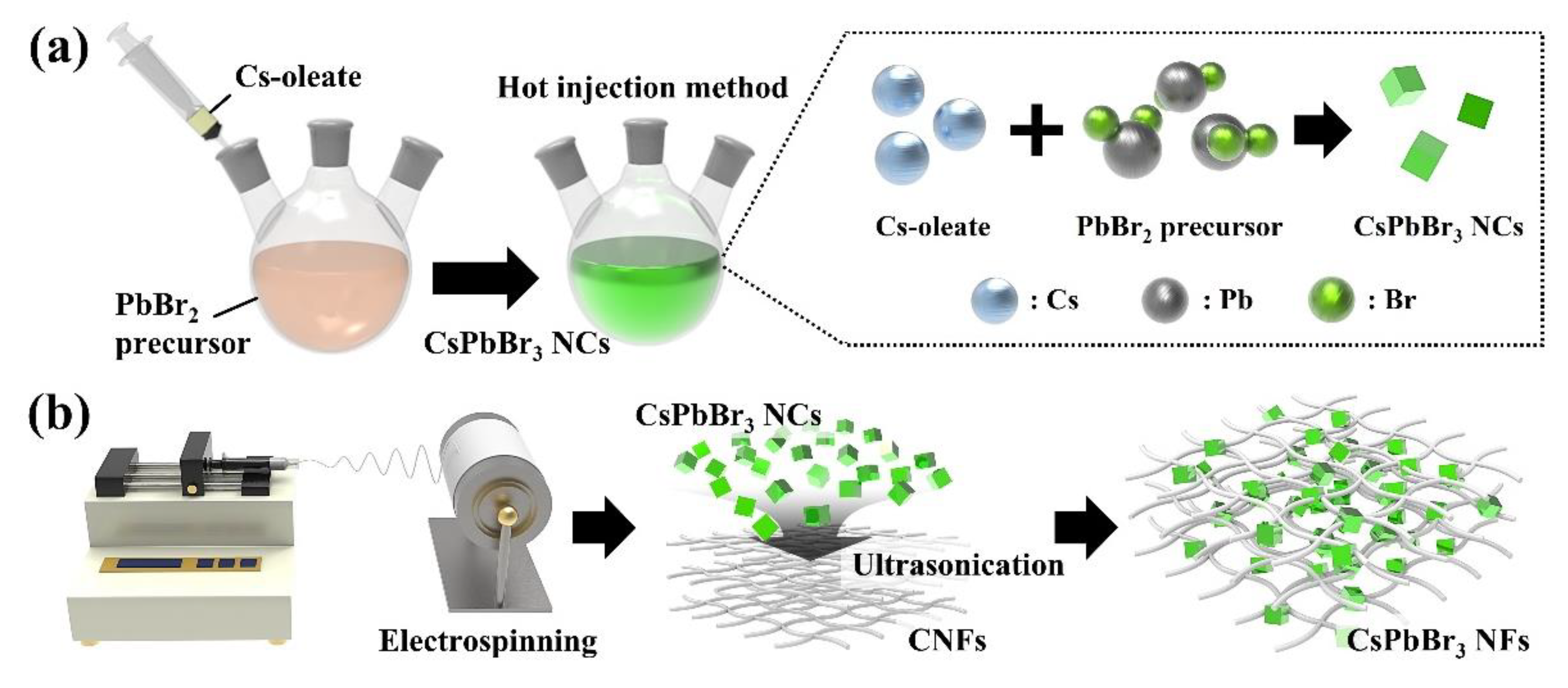
Abstract
:1. Introduction
2. Materials and Methods
2.1. Materials
2.2. Synthesis of Cesium Lead Bromide Nanocrystals through Hot-Injection Method
2.3. Fabrication of Cesium Lead Bromide Nanofibers
2.4. Detection of Nitrogen Gas through Fabricated Cesium Lead Bromide Nanofibers
2.5. Characterization
3. Results
3.1. Formation of Cesium Lead Bromide Nanocrystals-Coated Cellulose Nanofibers
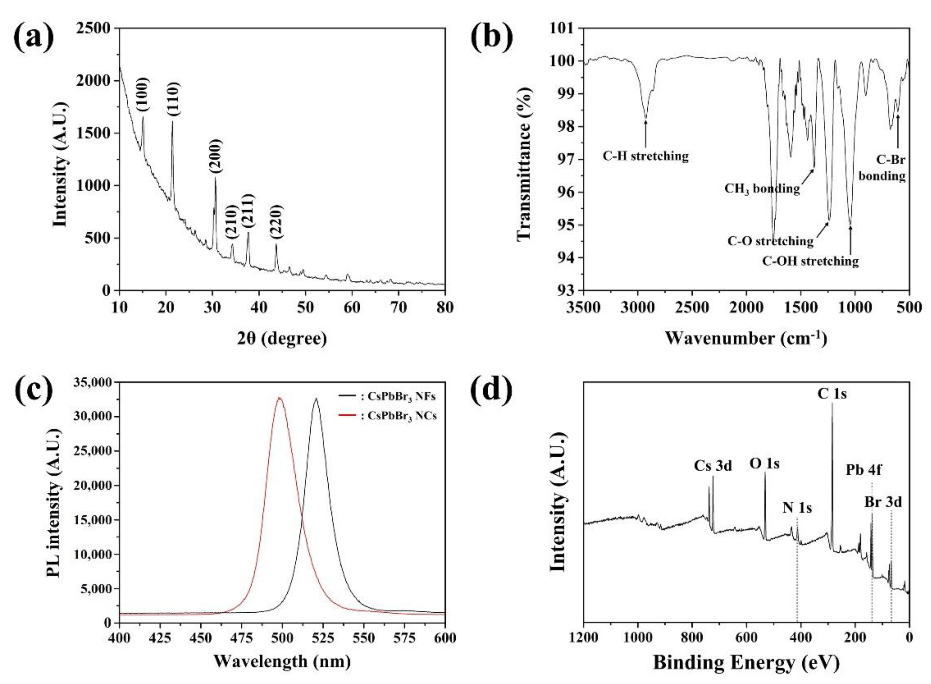
3.2. Characterization of Cesium Lead Bromide Nanocrystals-Coated Cellulose Nanofibers
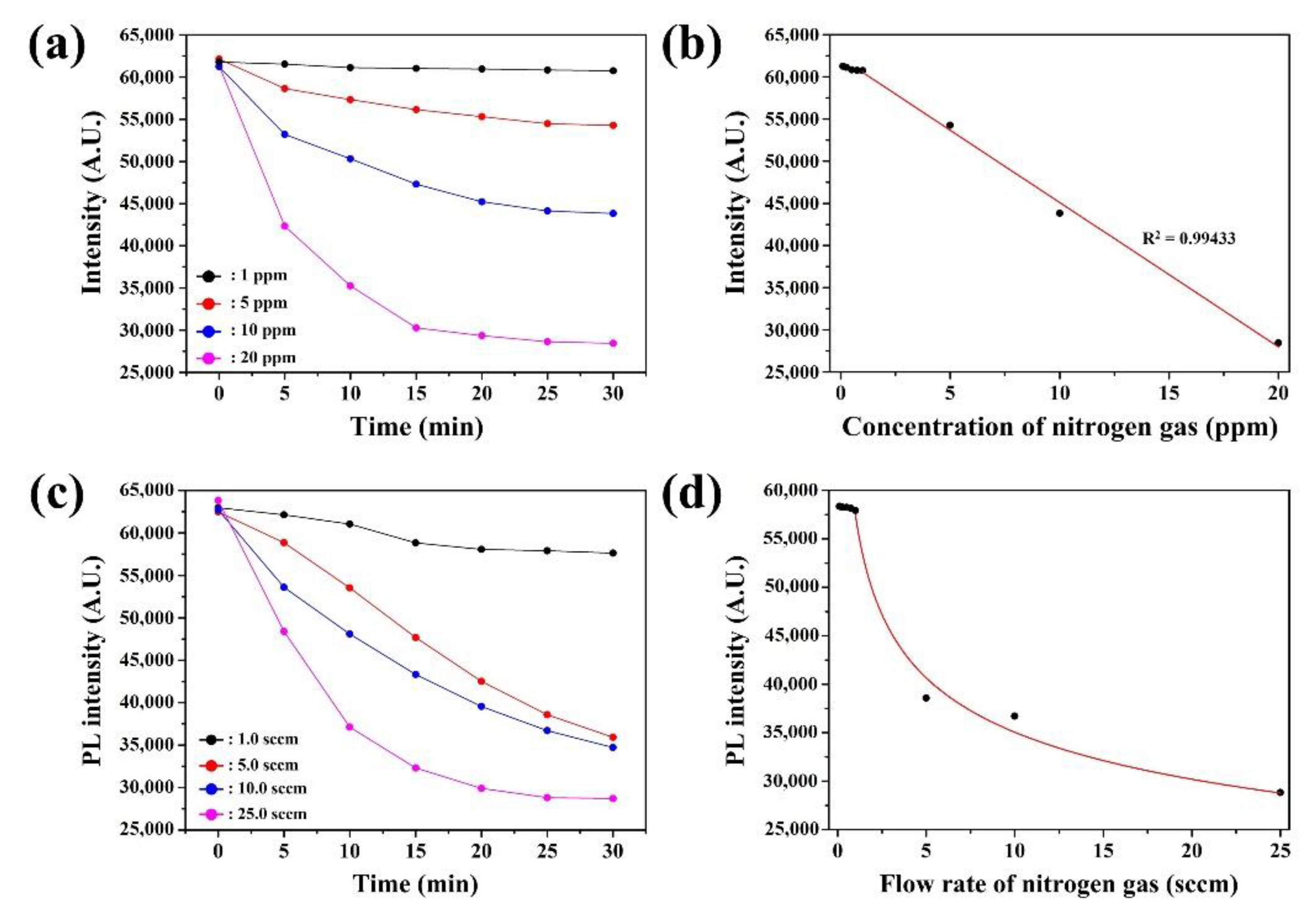
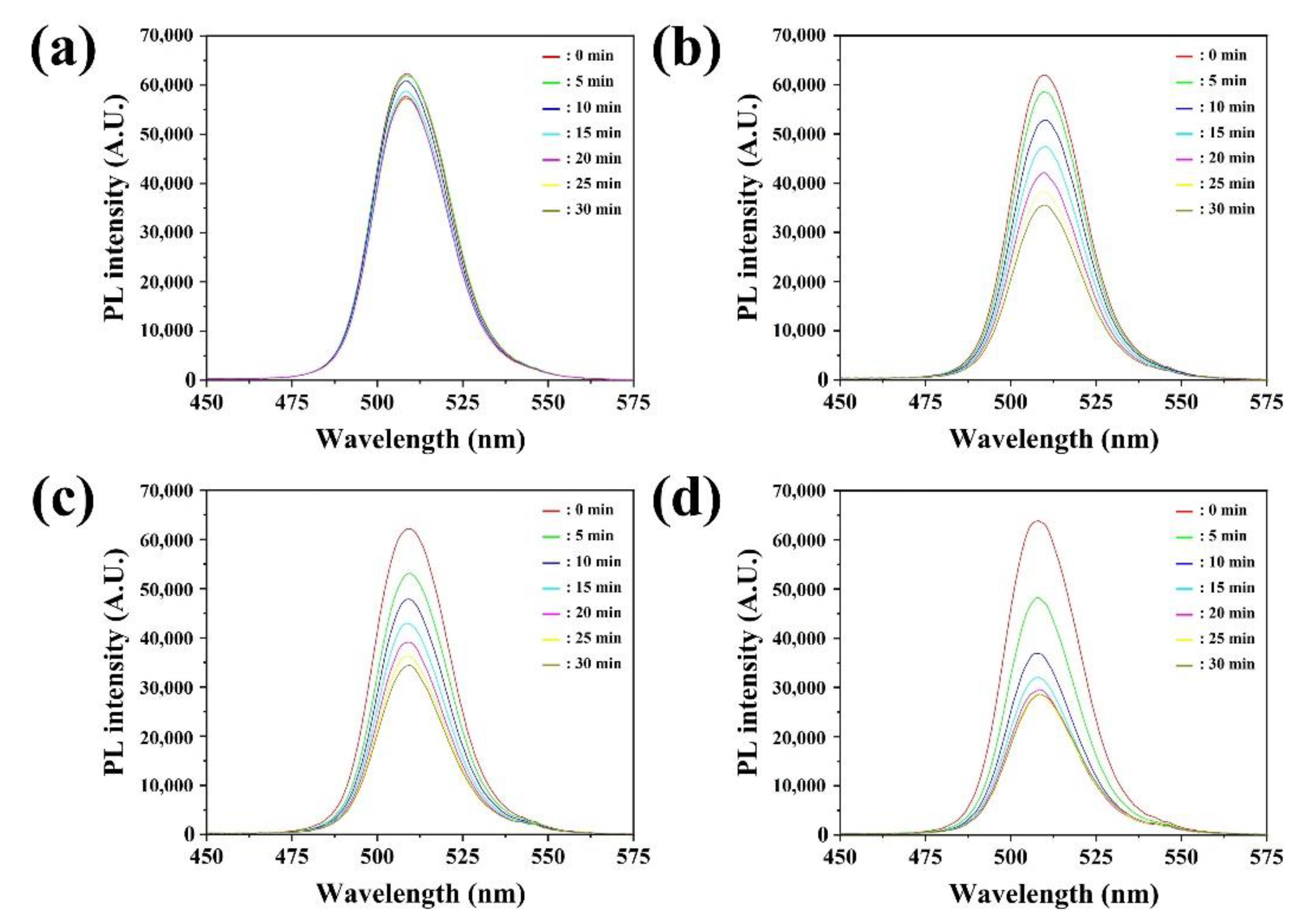
3.3. Detection of Nitrogen Gas through Ceisum Lead Bromide-Coated Cellulose Nanofibers
4. Conclusions
Supplementary Materials
Author Contributions
Funding
Institutional Review Board Statement
Informed Consent Statement
Data Availability Statement
Conflicts of Interest
References
- Aneja, V.P.; Roelle, P.A.; Murray, G.C.; Southerland, J.; Erisman, J.W.; Fowler, D.; Asman, W.A.; Patni, N. Atmospheric nitrogen compounds II: Emissions, transport, transformation, deposition and assessment. Atmos. Environ. 2001, 35, 1903–1911. [Google Scholar] [CrossRef]
- Green, P.A.; Vörösmarty, C.J.; Meybeck, M.; Galloway, J.N.; Peterson, B.J.; Boyer, E.W. Pre-industrial and contemporary fluxes of nitrogen through rivers: A global assessment based on typology. Biogeochemistry 2004, 68, 71–105. [Google Scholar] [CrossRef]
- Ladha, J.K.; Pathak, H.; Krupnik, T.J.; Six, J.; van Kessel, C. Efficiency of fertilizer nitrogen in cereal production: Retrospects and prospects. Adv. Agron. 2005, 87, 85–156. [Google Scholar]
- Yang, X.; Xu, M.; Zou, R.; Angelidaki, I.; Zhang, Y. Microbial protein production from CO2, H2, and recycled nitrogen: Focusing on ammonia toxicity and nitrogen sources. J. Clean. Prod. 2021, 291, 125921. [Google Scholar] [CrossRef]
- Bertness, M.D.; Ewanchuk, P.J.; Silliman, B.R. Anthropogenic modification of New England salt marsh landscapes. Proc. Natl. Acad. Sci. USA 2002, 99, 1395–1398. [Google Scholar] [CrossRef] [Green Version]
- Willett, M. Oxygen sensing for industrial safety—Evolution and new approaches. Sensors 2014, 14, 6084–6103. [Google Scholar] [CrossRef] [Green Version]
- Salem, A.A.; Soliman, A.A.; El-Haty, I.A. Determination of nitrogen dioxide, sulfur dioxide, ozone, and ammonia in ambient air using the passive sampling method associated with ion chromatographic and potentiometric analyses. Air Qual. Atmos. Health 2009, 2, 133–145. [Google Scholar] [CrossRef] [Green Version]
- Beutel, S.; Henkel, S. In situ sensor techniques in modern bioprocess monitoring. Appl. Microbiol. Biotechnol. 2011, 91, 1493–1505. [Google Scholar] [CrossRef]
- Padilla, F.M.; Gallardo, M.; Peña-Fleitas, M.T.; De Souza, R.; Thompson, R.B. Proximal optical sensors for nitrogen management of vegetable crops: A review. Sensors 2018, 18, 2083. [Google Scholar] [CrossRef] [Green Version]
- Thomason, W.; Phillips, S.; Davis, P.; Warren, J.; Alley, M.; Reiter, M. Variable nitrogen rate determination from plant spectral reflectance in soft red winter wheat. Precis. Agric. 2011, 12, 666–681. [Google Scholar] [CrossRef]
- Tremblay, N.; Wang, Z.; Cerovic, Z.G. Sensing crop nitrogen status with fluorescence indicators. A review. Agron. Sustain. Dev. 2012, 32, 451–464. [Google Scholar] [CrossRef]
- Samborski, S.M.; Tremblay, N.; Fallon, E. Strategies to make use of plant sensors-based diagnostic information for nitrogen recommendations. Agron. J. 2009, 101, 800–816. [Google Scholar] [CrossRef]
- Zhang, J.; Hodes, G.; Jin, Z.; Liu, S. All-inorganic CsPbX3 perovskite solar cells: Progress and prospects. Angew. Chem. Int. Ed. 2019, 58, 15596–15618. [Google Scholar] [CrossRef]
- Xue, J.; Zhu, Z.; Xu, X.; Gu, Y.; Wang, S.; Xu, L.; Zou, Y.; Song, J.; Zeng, H.; Chen, Q. Narrowband perovskite photodetector-based image array for potential application in artificial vision. Nano Lett. 2018, 18, 7628–7634. [Google Scholar] [CrossRef]
- Hai, J.; Li, H.; Zhao, Y.; Chen, F.; Peng, Y.; Wang, B. Designing of blue, green, and red CsPbX3 perovskite-codoped flexible films with water resistant property and elimination of anion-exchange for tunable white light emission. Chem. Commun. 2017, 53, 5400–5403. [Google Scholar] [CrossRef]
- Huang, S.; Guo, M.; Tan, J.; Geng, Y.; Wu, J.; Tang, Y.; Su, C.; Lin, C.C.; Liang, Y. Novel fluorescence sensor based on all-inorganic perovskite quantum dots coated with molecularly imprinted polymers for highly selective and sensitive detection of omethoate. ACS Appl. Mater. Interfaces 2018, 10, 39056–39063. [Google Scholar] [CrossRef]
- Seth, S.; Ahmed, T.; De, A.; Samanta, A. Tackling the defects, stability, and photoluminescence of CsPbX3 perovskite nanocrystals. ACS Energy Lett. 2019, 4, 1610–1618. [Google Scholar] [CrossRef]
- Huang, H.; Hao, M.; Song, Y.; Dang, S.; Liu, X.; Dong, Q. Dynamic passivation in perovskite quantum dots for specific ammonia detection at room temperature. Small 2020, 16, 1904462. [Google Scholar] [CrossRef]
- de Mello Donegá, C.; Liljeroth, P.; Vanmaekelbergh, D. Physicochemical evaluation of the hot-injection method, a synthesis route for monodisperse nanocrystals. Small 2005, 1, 1152–1162. [Google Scholar] [CrossRef] [Green Version]
- Protesescu, L.; Yakunin, S.; Bodnarchuk, M.I.; Krieg, F.; Caputo, R.; Hendon, C.H.; Yang, R.X.; Walsh, A.; Kovalenko, M.V. Nanocrystals of cesium lead halide perovskites (CsPbX3, X=Cl, Br, and I): Novel optoelectronic materials showing bright emission with wide color gamut. Nano Lett. 2015, 15, 3692–3696. [Google Scholar] [CrossRef] [Green Version]
- Bhardwaj, N.; Kundu, S.C. Electrospinning: A fascinating fiber fabrication technique. Biotechnol. Adv. 2010, 28, 325–347. [Google Scholar] [CrossRef]
- Teo, W.E.; Ramakrishna, S. A review on electrospinning design and nanofibre assemblies. Nanotechnology 2006, 17, R89. [Google Scholar] [CrossRef]
- Brennan, M.C.; Kuno, M.; Rouvimov, S. Crystal structure of individual CsPbBr3 perovskite nanocubes. Inorg. Chem. 2018, 58, 1555–1560. [Google Scholar] [CrossRef]
- Zhao, M.; Shi, Y.; Dai, J.; Lian, J. Ellipsometric study of the complex optical constants of a CsPbBr3 perovskite thin film. J. Mater. Chem. C 2018, 6, 10450–10455. [Google Scholar] [CrossRef]
- Galkina, O.; Ivanov, V.; Agafonov, A.; Seisenbaeva, G.; Kessler, V. Cellulose nanofiber–titania nanocomposites as potential drug delivery systems for dermal applications. J. Mater. Chem. B 2015, 3, 1688–1698. [Google Scholar] [CrossRef] [Green Version]
- Soni, B.; Mahmoud, B. Chemical isolation and characterization of different cellulose nanofibers from cotton stalks. Carbohydr. Polym. 2015, 134, 581–589. [Google Scholar] [CrossRef]
- Yoon, H.C.; Lee, S.; Song, J.K.; Yang, H.; Do, Y.R. Efficient and stable CsPbBr3 quantum-dot powders passivated and encapsulated with a mixed silicon nitride and silicon oxide inorganic polymer matrix. ACS Appl. Mater. Interfaces 2018, 10, 11756–11767. [Google Scholar] [CrossRef]
- Kuzmenko, V.; Wang, N.; Haque, M.; Naboka, O.; Flygare, M.; Svensson, K.; Gatenholm, P.; Liu, J.; Enoksson, P. Cellulose-derived carbon nanofibers/graphene composite electrodes for powerful compact supercapacitors. RSC Adv. 2017, 7, 45968–45977. [Google Scholar] [CrossRef] [Green Version]
- Dalmis, R.; Köktaş, S.; Seki, Y.; Kılınç, A.Ç. Characterization of a new natural cellulose based fiber from Hierochloe Odarata. Cellulose 2020, 27, 127–139. [Google Scholar] [CrossRef]
- Arcot, L.R.; Lundahl, M.; Rojas, O.J.; Laine, J. Asymmetric cellulose nanocrystals: Thiolation of reducing end groups via NHS–EDC coupling. Cellulose 2014, 21, 4209–4218. [Google Scholar] [CrossRef]
- Yuan, B.; Li, N.; Liu, J.; Xu, F.; Li, C.; Juan, F.; Yu, H.; Li, C.; Cao, B. Improving the performances of CsPbBr3 solar cells fabricated in ambient condition. J. Mater. Sci. Mater. Electron. 2020, 31, 21154–21167. [Google Scholar] [CrossRef]
- Liu, M.; Zhong, G.; Yin, Y.; Miao, J.; Li, K.; Wang, C.; Xu, X.; Shen, C.; Meng, H. Aluminum-doped cesium lead bromide perovskite nanocrystals with stable blue photoluminescence used for display backlight. Adv. Sci. 2017, 4, 1700335. [Google Scholar] [CrossRef] [PubMed]
- Liu, J.; Yu, Y.; Liu, Z.; Zuo, S.; Li, B. AgBr-coupled TiO2: A visible heterostructured photocatalyst for degrading dye pollutants. Int. J. Photoenergy 2012, 254201. [Google Scholar] [CrossRef] [Green Version]
- Hu, Z.; Liu, Z.; Ono, L.K.; Jiang, M.; He, S.; Son, D.-Y.; Qi, Y. The impact of atmosphere on energetics of lead halide perovskites. Adv. Energy Mater. 2020, 10, 2000908. [Google Scholar] [CrossRef]
- Guo, R.; Han, D.; Chen, W.; Dai, L.; Ji, K.; Xiong, Q.; Li, S.; Reb., L.K.; Scheel, M.A.; Pratap, S.; et al. Degradation mechanisms of perovskite solar cells under vacuum and one atmosphere of nitrogen. Nat. Energy 2021, 6, 977–986. [Google Scholar] [CrossRef]
- Park, B.; Kim, S.; Kwak, C.H.; Shanmugam, K.R.; Han, Y.-K.; Cho, Y.; Huh, Y.S. Visual colorimetric detection of ammonia under gaseous and aqueous state: Approach on cesium lead bromide perovskite-loaded porous electrospun nanofibers. J. Ind. Eng. Chem. 2021, 97, 515–522. [Google Scholar] [CrossRef]
- Jin, Z.; Su, Y.; Duan, Y. Development of a polyaniline-based optical ammonia sensor. Sensor. Actuator B. Chem. 2001, 72, 75–79. [Google Scholar] [CrossRef]
- Liu, S.; Volkmer, D.; Kurth, D.G. Smart polyoxometalate-based nitrogen monoxide sensors. Anal Chem. 2004, 76, 4579–4582. [Google Scholar] [CrossRef]
- Das, A.; Dost, R.; Richardson, T.; Grell, M.; Morrison, J.J.; Turner, M.L. A nitrogen dioxide sensor based on an organic transistor constructed from amorphous semiconducting polymers. Adv. Mater. 2007, 19, 4018–4023. [Google Scholar] [CrossRef]


Publisher’s Note: MDPI stays neutral with regard to jurisdictional claims in published maps and institutional affiliations. |
© 2022 by the authors. Licensee MDPI, Basel, Switzerland. This article is an open access article distributed under the terms and conditions of the Creative Commons Attribution (CC BY) license (https://creativecommons.org/licenses/by/4.0/).
Share and Cite
Park, B.; Kang, H.; Han, S.; Kim, H.-U.; Cho, Y.; Huh, Y.S.; Kang, S.-M. The Fabrication of Cesium Lead Bromide-Coated Cellulose Nanocomposites and Their Effect on the Detection of Nitrogen Gas. Sensors 2022, 22, 7737. https://0-doi-org.brum.beds.ac.uk/10.3390/s22207737
Park B, Kang H, Han S, Kim H-U, Cho Y, Huh YS, Kang S-M. The Fabrication of Cesium Lead Bromide-Coated Cellulose Nanocomposites and Their Effect on the Detection of Nitrogen Gas. Sensors. 2022; 22(20):7737. https://0-doi-org.brum.beds.ac.uk/10.3390/s22207737
Chicago/Turabian StylePark, Bumjun, Haneul Kang, Soobin Han, Hyeong-U Kim, Youngjin Cho, Yun Suk Huh, and Sung-Min Kang. 2022. "The Fabrication of Cesium Lead Bromide-Coated Cellulose Nanocomposites and Their Effect on the Detection of Nitrogen Gas" Sensors 22, no. 20: 7737. https://0-doi-org.brum.beds.ac.uk/10.3390/s22207737