
Self-Associated 1,8-Naphthalimide as a Selective Fluorescent Chemosensor for Detection of High pH in Aqueous Solutions and Their Hg2+ Contamination


Abstract
:1. Introduction
2. Materials and Methods
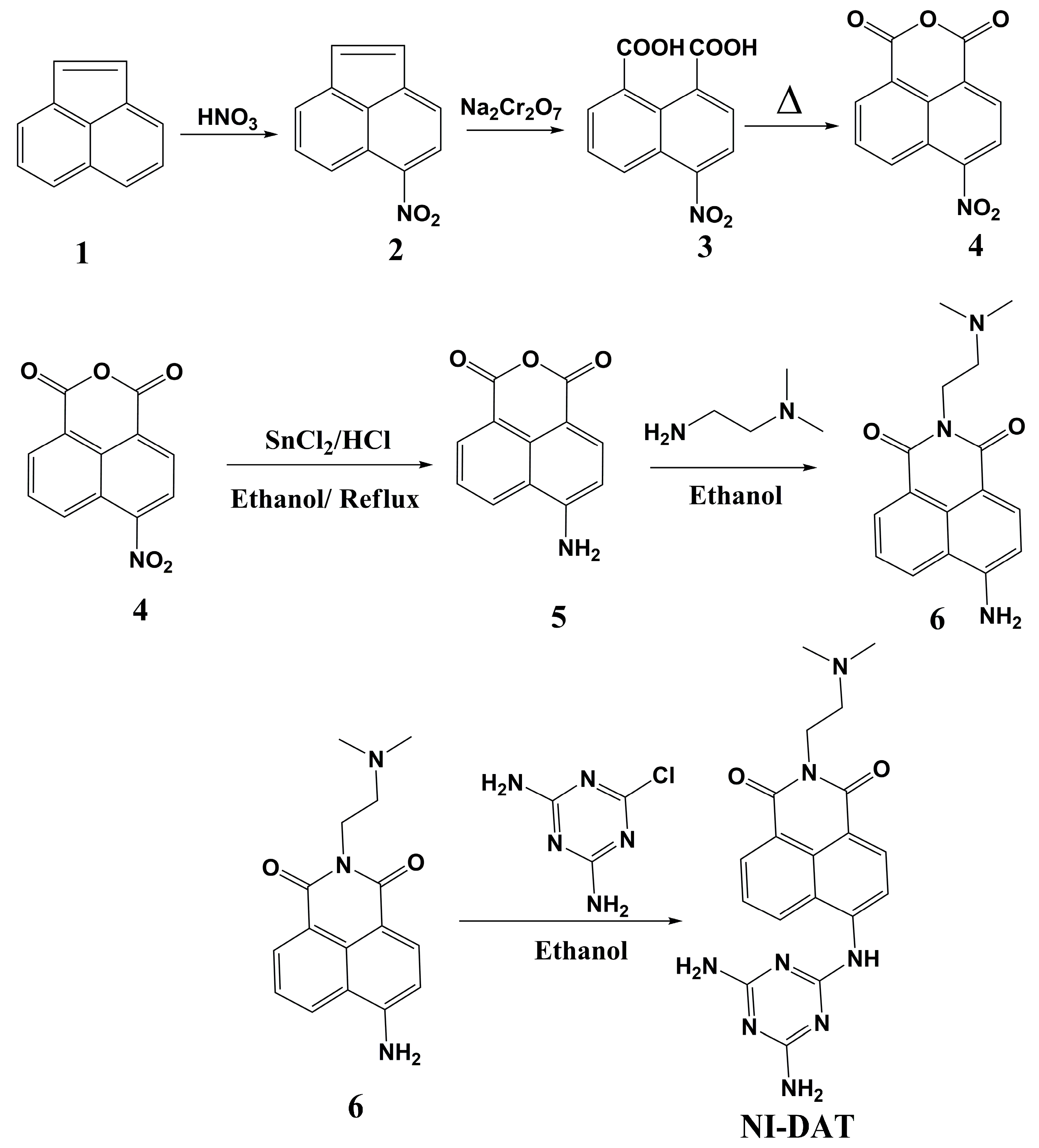
2.1. Synthesis of 4-Nitroacenaphthene (2)
2.2. Synthesis of 4-Nitro-1,8-naphthalic anhydride (4)
2.3. Synthesis of 4-Amino-1,8-naphthalic anhydride (5)
2.4. Synthesis of 4-Amino-N-(2-dimethylaminoethyl)-1,8-naphthalimide (6)
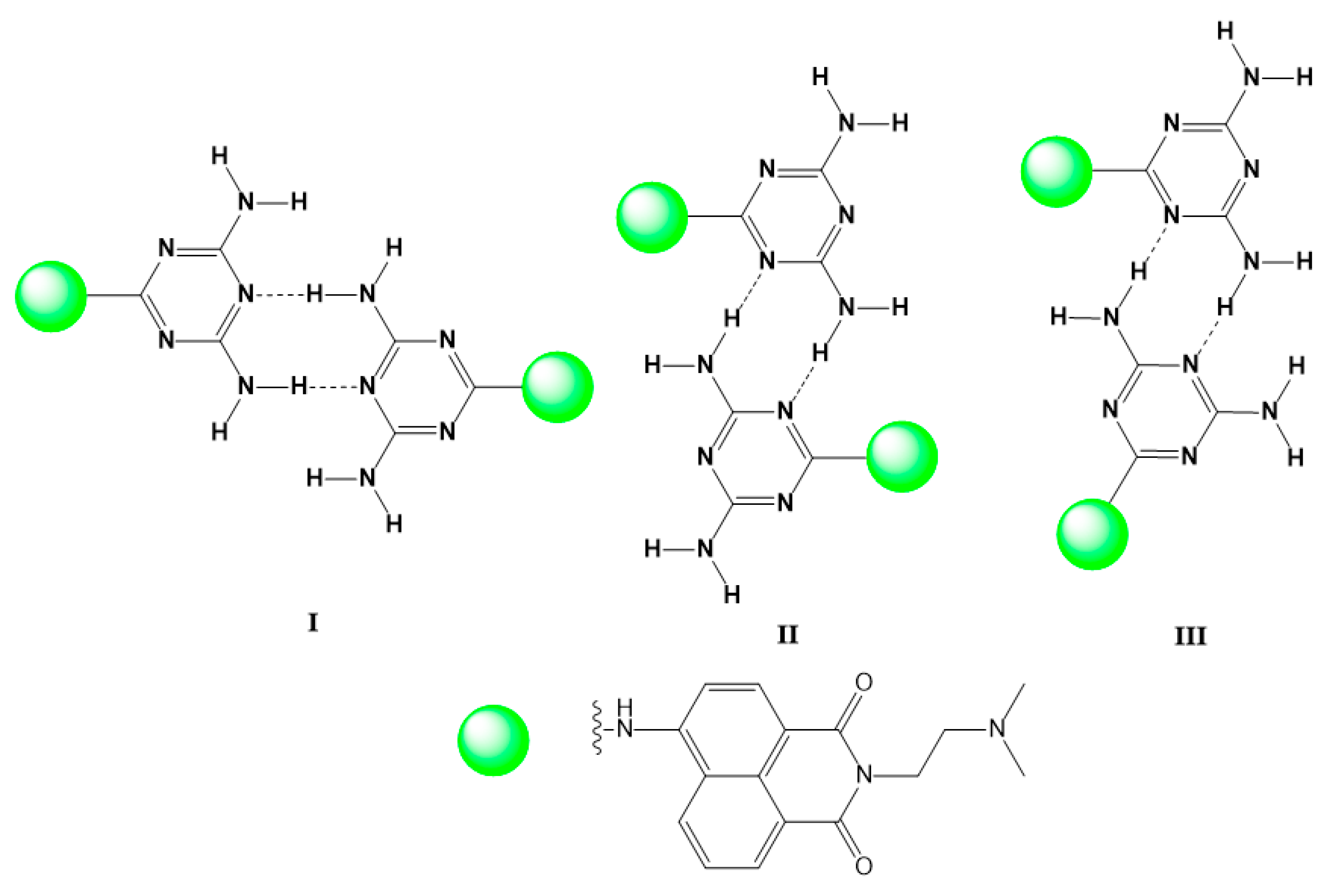
2.5. Synthesis of 4-(4,6-Diamino-1,3,5-triazin-2′-ylamino)-N-(2-dimethylaminoethyl)-1,8-naphthalimide NI-DAT
3. Results and Discussion
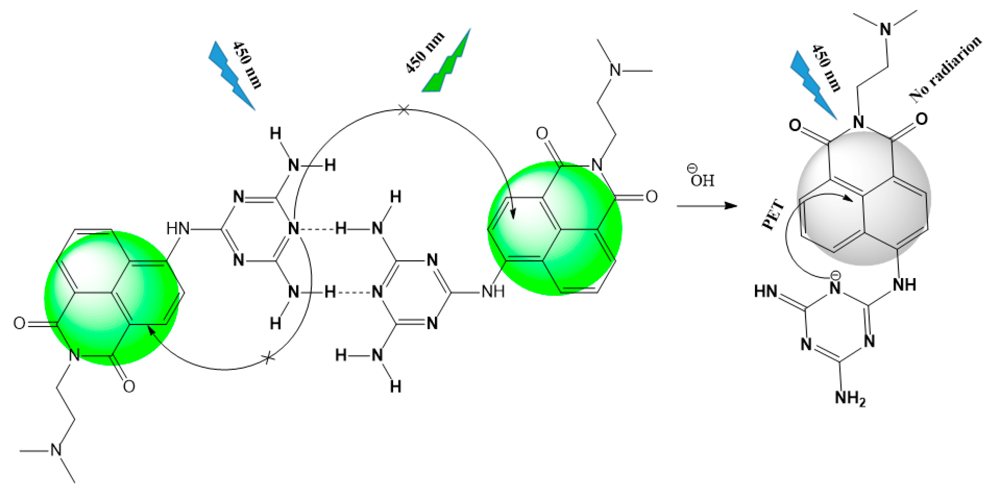
3.1. Design and Synthesis of NI-DAT
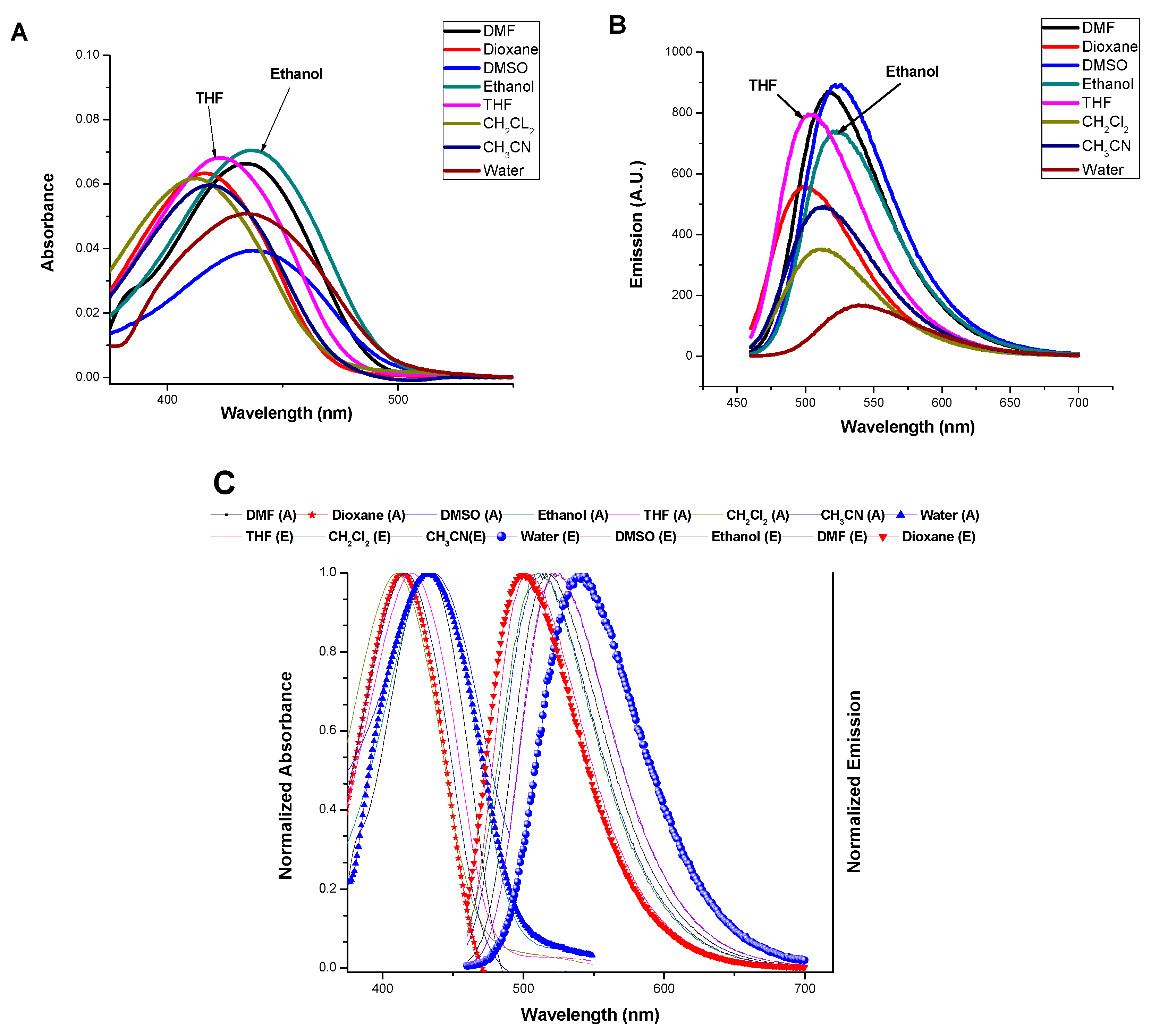
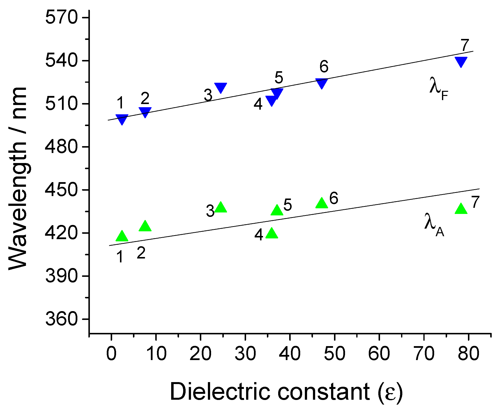
3.2. Photophysical Characteristics
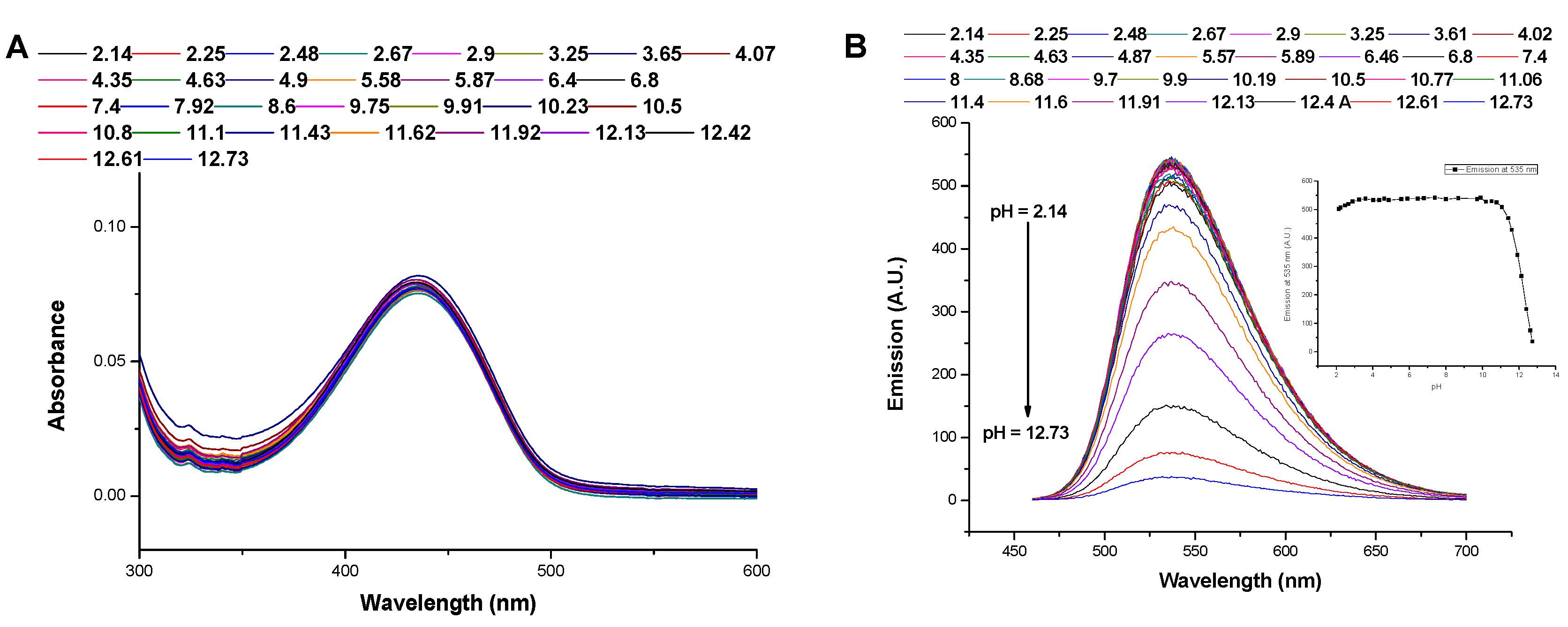
3.3. Impact of the Medium pH
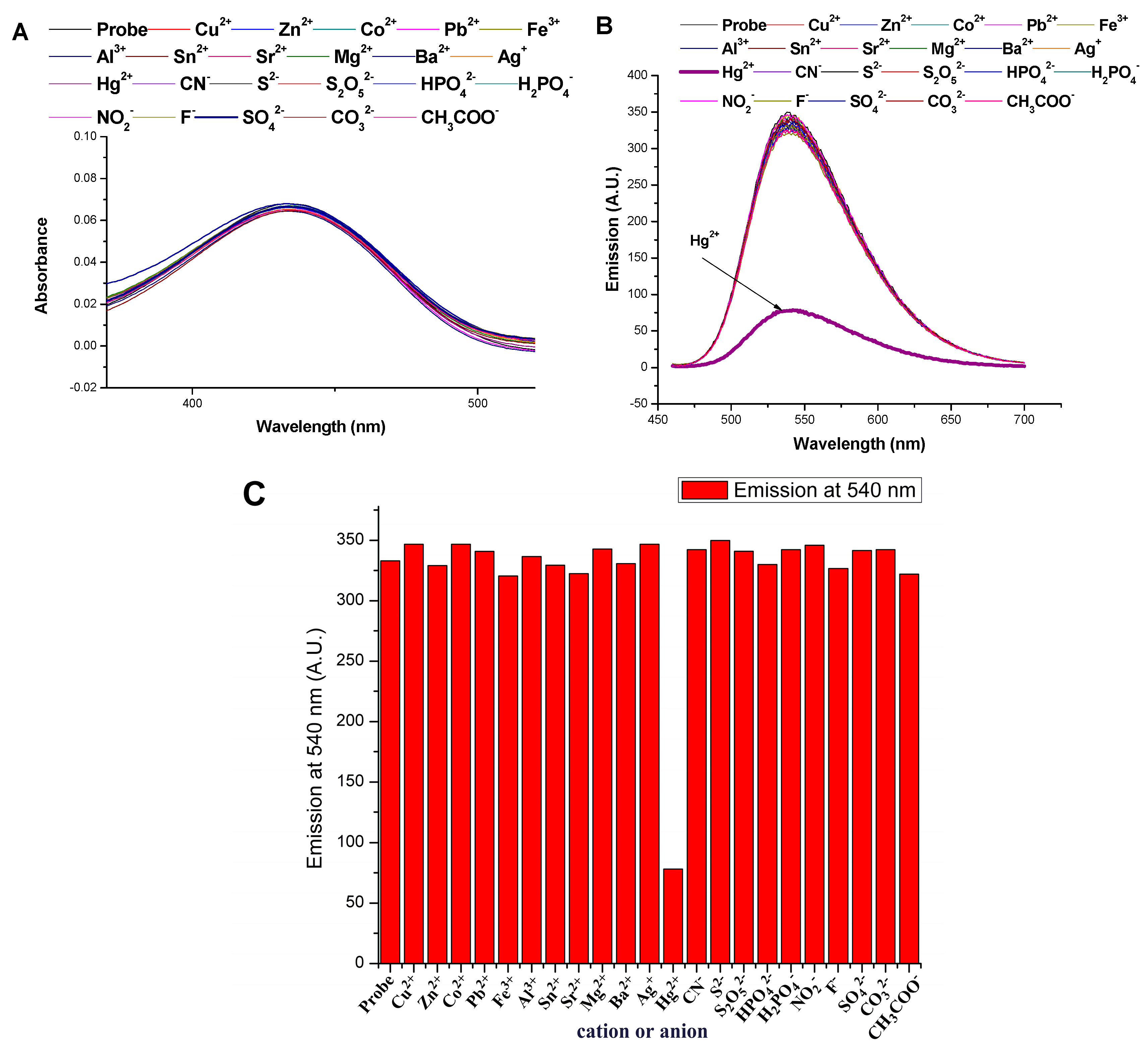
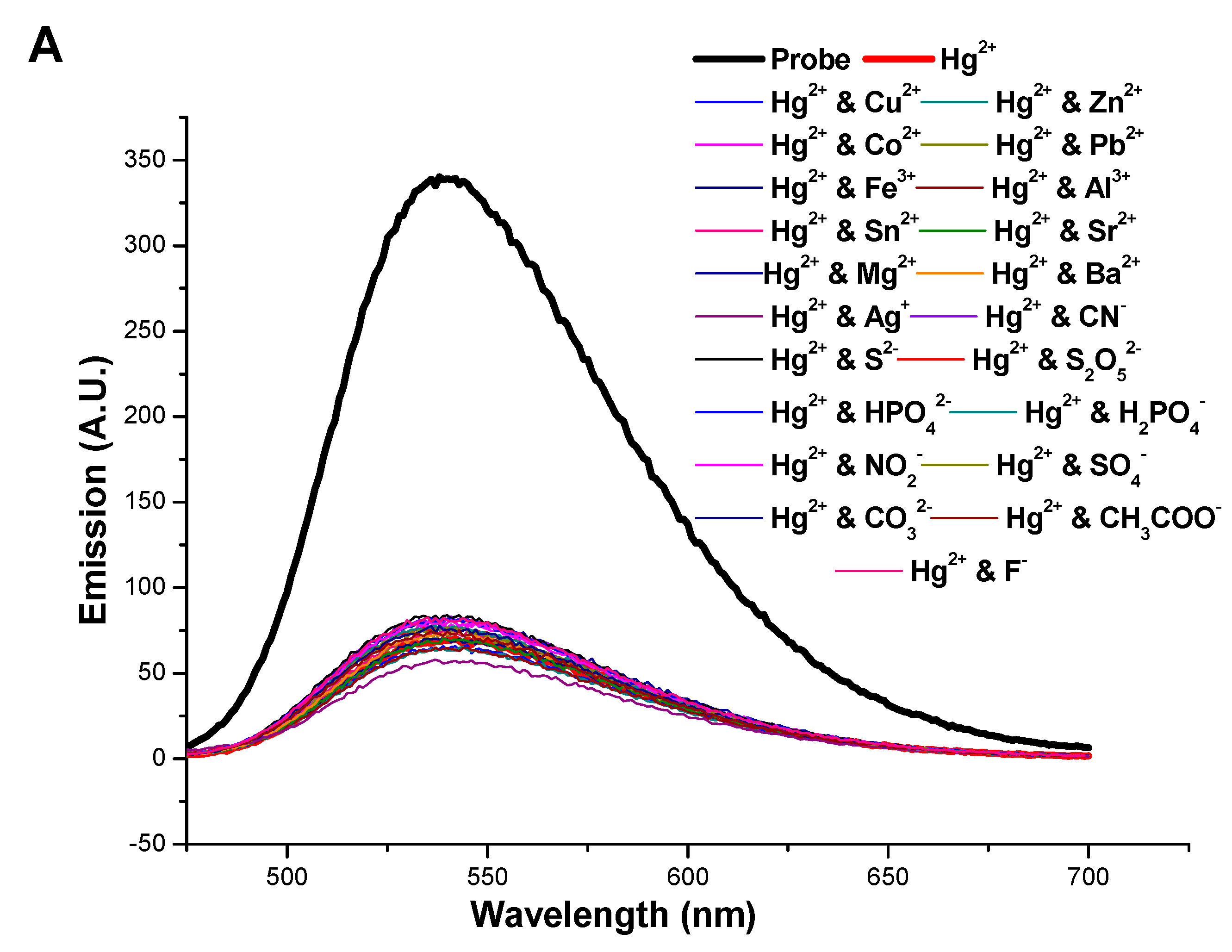
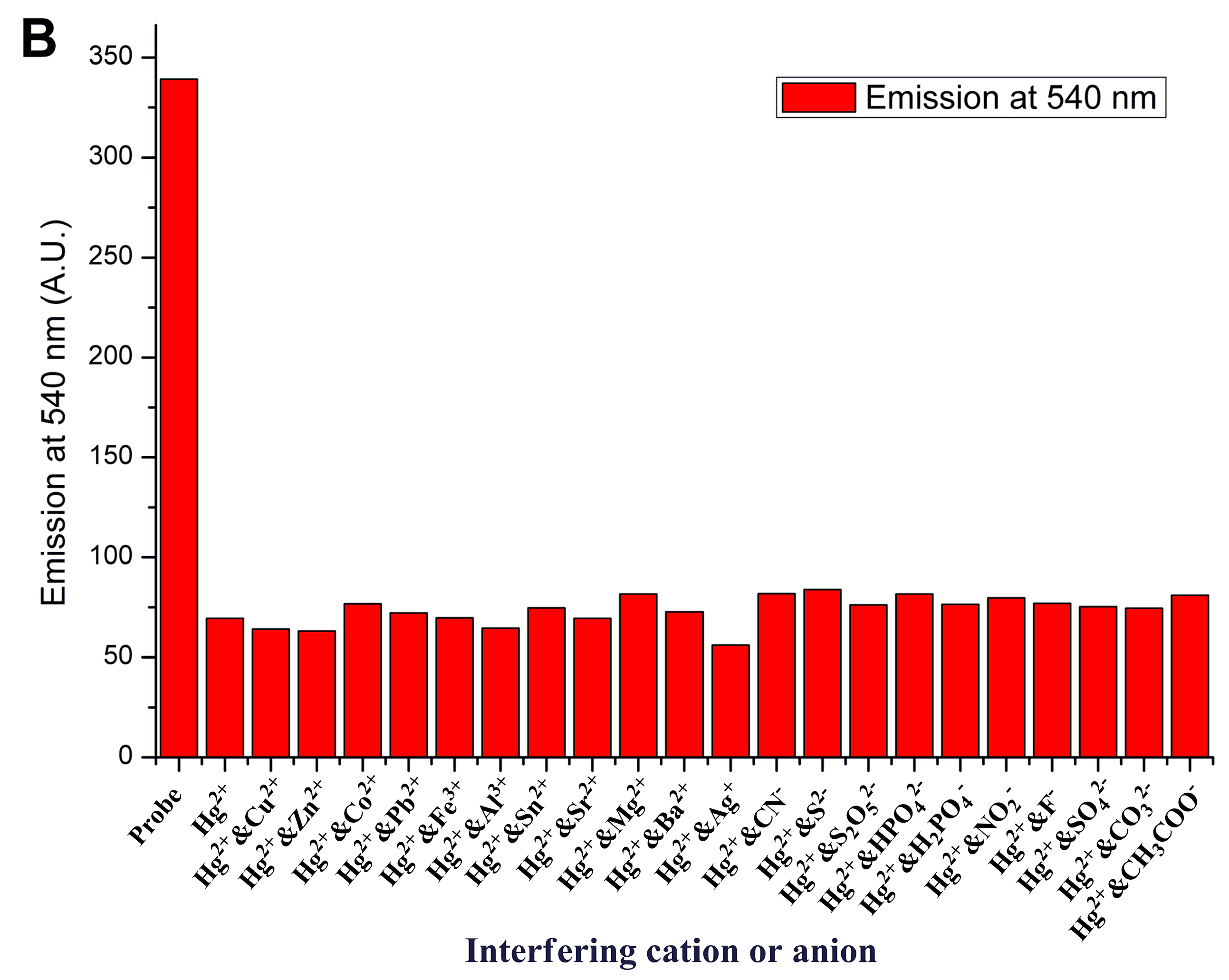
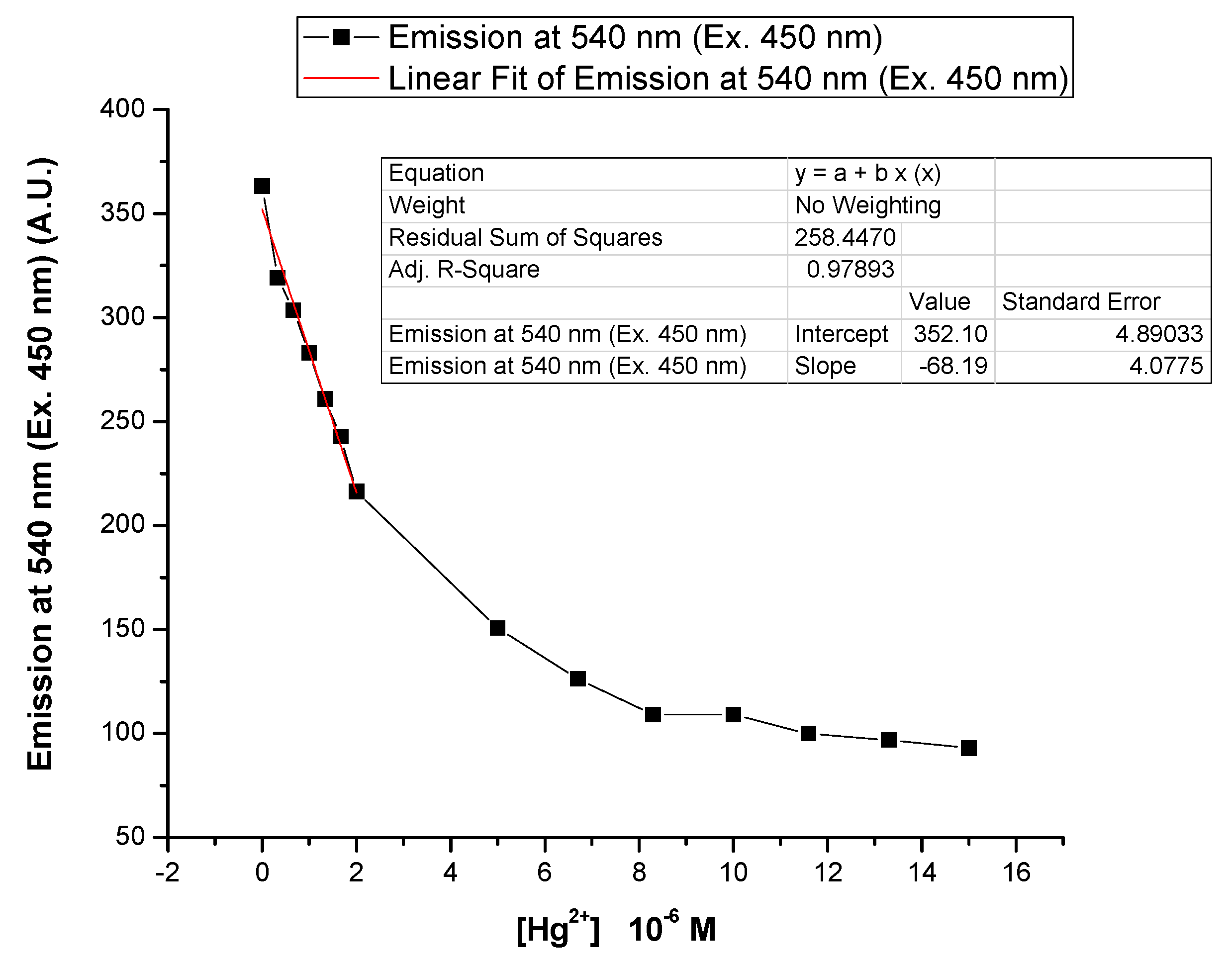
3.4. Sensory Applications towards Cations and Anions
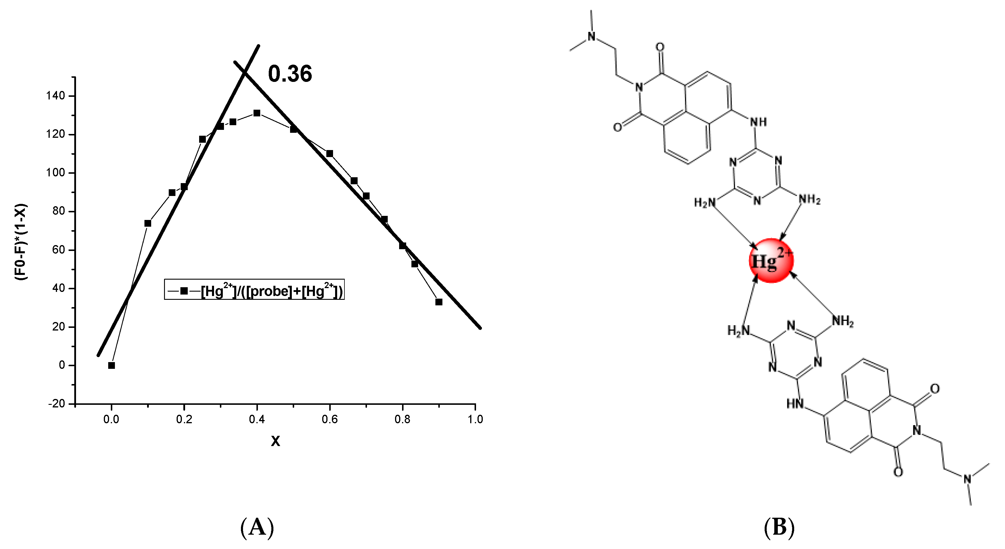
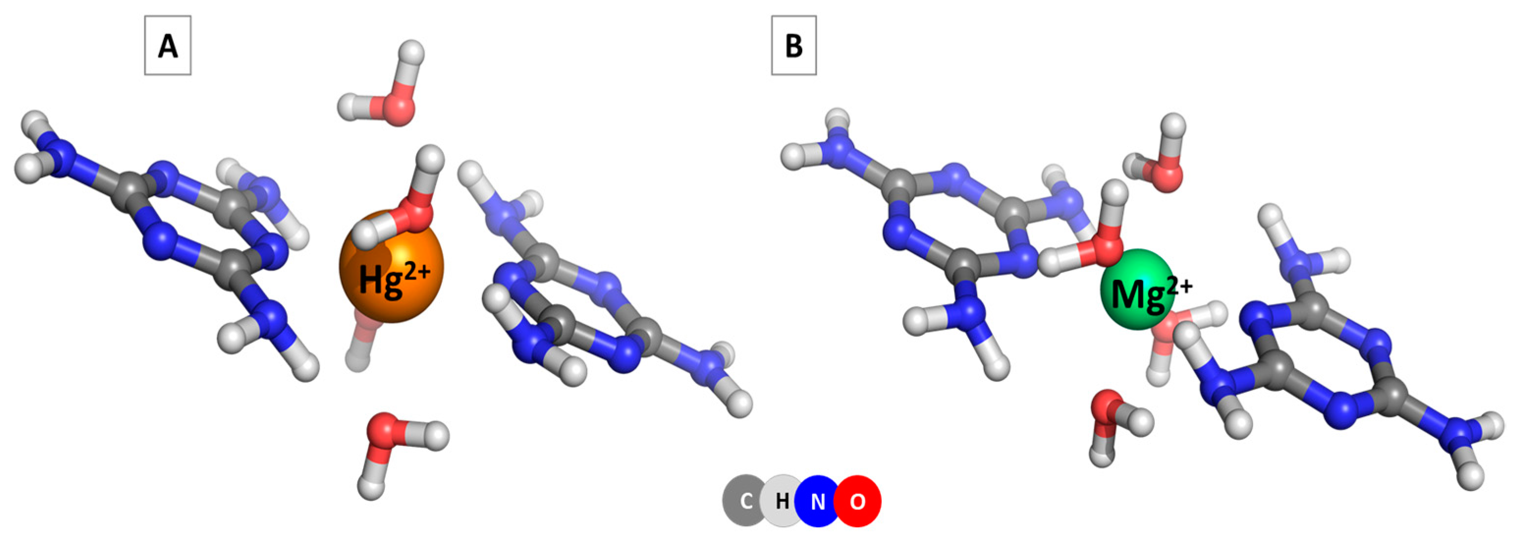
3.5. Computational Studies on the Structure of NI-DAT and Its Hg2+ and Mg2+ Complexes
3.6. Comparison between NI-DAT and Some of the Reported Sensors for Detecting Hg2+
4. Conclusions
Supplementary Materials
Author Contributions
Funding
Data Availability Statement
Acknowledgments
Conflicts of Interest
References
- Jasinski, S.M. The Materials Flow of Mercury in the United States; U.S. Bureau of Mines Information Circular 9412; U.S. Department of the Interior: Washington, DC, USA, 1994. Available online: https://pubs.usgs.gov/usbmic/ic-9412/mercury.pdf (accessed on 5 June 2022).
- Dock, L.; Vather, M. Metal toxicology. In General and Applied Toxicology; Ballantyne, B., Mars, C.T., Syversen, T., Eds.; Macmillan: London, UK, 2000; pp. 2049–2078. [Google Scholar]
- Stacchiotti, A.; Morandini, F.; Bettoni, F.; Schena, I.; Lavazza, A.; Grigolato, P.G.; Apostoli, P.; Rezzani, R.; Aleo, M.F. Stress proteins and oxidative damage in a renal derived cell line exposed to inorganic mercury and lead. Toxicology 2009, 264, 215–224. [Google Scholar] [CrossRef] [PubMed]
- Kim, B.M.; Choi, A.L.; Ha, E.H.; Pedersen, L.; Nielsen, F.; Weihe, P.; Hong, Y.C.; Budtz-Jørgensen, E.; Grandjean, P. Effect of hemoglobin adjustment on the precision of mercury concentrations in maternal and cord blood. Environ. Res. 2014, 132, 407–412. [Google Scholar] [CrossRef] [PubMed] [Green Version]
- Azevedo, B.F.; Furieri, L.B.; Peçanha, F.M.; Wiggers, G.A.; Vassallo, P.F.; Simões, M.R.; Fiorim, J.; de Batista, P.R.; Fioresi, M.; Rossoni, L.; et al. Toxic effects of mercury on the cardiovascular and central nervous systems. J. Biomed. Biotechnol. 2012, 2012, 949048. [Google Scholar] [CrossRef] [Green Version]
- Rice, K.M.; Walker, E.M., Jr.; Wu, M.; Gillette, C.; Blough, E.R. Environmental mercury and its toxic effects. J. Prev. Med. Public Health 2014, 47, 74–83. [Google Scholar] [CrossRef]
- Hui, L.L.; Chan, M.H.M.; Lam, H.S.; Chan, P.H.Y.; Kwok, K.M.; Chan, I.H.S.; Li, A.M.; Fok, T.F. Impact of fetal and childhood mercury exposure on immune status in children. Environ. Res. 2016, 144, 66–72. [Google Scholar] [CrossRef]
- Vallant, B.; Kadnar, R.; Goessler, W. Development of a new HPLC method for the determination of inorganic and methylmercury in biological samples with ICP-MS detection. J. Anal. At. Spectrom. 2007, 22, 322–325. [Google Scholar] [CrossRef]
- Tseng, C.M.; de Diego, A.; Martin, F.M.; Amouroux, D.; Donard, O.F.X. Rapid determination of inorganic mercury and methylmercury in biological reference materials by hydride generation, cryofocusing, atomic absorption spectrometry after open focused microwave-assisted alkaline digestion. J. Anal. At. Spectrom. 1997, 12, 743–750. [Google Scholar] [CrossRef]
- Li, Y.; Chen, C.; Li, B.; Sun, J.; Wang, J.; Gao, Y.; Zhao, Y.; Chai, Z. Elimination efficiency of different reagents for the memory effect of mercury using ICP-MS. J. Anal. At. Spectrom. 2006, 21, 94–96. [Google Scholar] [CrossRef]
- Tan, J.; Yan, X.P. 2,1,3-Benzoxadiazole-based selective chromogenic chemosensor for rapid naked-eye detection of Hg2+ and Cu2+. Talanta 2008, 76, 9–14. [Google Scholar] [CrossRef]
- Sarfo, D.K.; Sivanesan, A.; Emad, L.I.; Ayoko, G.A. Rapid detection of mercury contamination in water by surface enhanced Raman spectroscopy. RSC Adv. 2017, 7, 21567–21575. [Google Scholar] [CrossRef]
- Prakashan, V.P.; George, G.; Sanu, M.S.; Sajna, M.S.; Saritha, A.C.; Sudarsanakumar, C.; Biju, P.R.; Joseph, C.; Unnikrishnan, N.V. Investigations on SPR induced Cu@Ag core shell doped SiO2-TiO2-ZrO2 fiber optic sensor for mercury detection. Appl. Surf. Sci. 2020, 507, 144957. [Google Scholar] [CrossRef]
- Pérez-Marín, L.; Otazo-Sánchez, E.; Macedo-Miranda, G.; Avila-Pérez, P.; Alonso Chamaro, J.; López-Valdivia, H. Mercury(II) ion-selective electrode. Study of 1,3-diphenylthiourea as ionophore. Analyst 2000, 125, 1787–1790. [Google Scholar] [CrossRef]
- Yantasee, W.; Lin, Y.; Zemanian, T.S.; Fryxell, G.E. Voltammetric detection of lead(II) and mercury(II) using a carbon paste electrode modified with thiol self-assembled monolayer on mesoporous silica (SAMMS). Analyst 2003, 128, 467–472. [Google Scholar] [CrossRef] [PubMed]
- Caballero, A.; Lloveras, V.; Curiel, D.; Tárrage, A.; Espinosa, A.; Garcia, R.; Vidal-Gancedo, J.; Rovira, C.; Wurst, K.; Molina, P.; et al. Electroactive thiazole derivatives capped with ferrocenyl units showing charge-transfer transition and selective ion-sensing properties: A combined experimental and theoretical study. Inorg. Chem. 2007, 46, 825–838. [Google Scholar] [CrossRef]
- Grabchev, I.; Chovelon, J.-M.; Nedelcheva, A. Green fluorescence poly(amidoamine) dendrimer functionalized with 1,8-naphthalimide units as potential sensor for metal cations. J. Photochem. Photobiol. A Chem. 2006, 183, 9–14. [Google Scholar] [CrossRef]
- Grabchev, I.; Bosch, P.; McKenna, M.; Nedelcheva, A. Synthesis and spectral properties of new green fluorescent poly(propyleneimine) dendrimers modified with 1,8-naphthalimide as sensors for metal cations. Polymer 2007, 48, 6755–6762. [Google Scholar] [CrossRef]
- Grabchev, I.; Bosch, P.; McKenna, M.; Staneva, D. A new colorimetric and fluorimetric sensor for metal cations based on poly(propylene amine) dendrimer modified with 1,8-naphthalimide. J. Photochem. Photobiol. A Chem. 2008, 201, 75–80. [Google Scholar] [CrossRef]
- Said, A.; Georgiev, N.; Bojinov, V. Sensor activity and logic behavior of dihydroxyphenylhydrazone derivative as a chemosensor for Cu2+ determination in alkaline aqueous solutions. J. Photochem. Photobiol. A Chem. 2015, 311, 16–24. [Google Scholar] [CrossRef]
- Said, A.; Georgiev, N.; Bojinov, V. Synthesis of a single 1,8-naphthalimide fluorophore as a molecular logic lab for simultaneously detecting of Fe3+, Hg2+ and Cu2+. Spectrochim. Acta Part A 2018, 196, 76–82. [Google Scholar] [CrossRef]
- Said, A.; Georgiev, N.; Bojinov, V. Low Molecular Weight Probe for Selective Sensing of PH and Cu2+ Working as Three INHIBIT Based Digital Comparator. J. Fluoresc. 2022, 32, 405–417. [Google Scholar] [CrossRef]
- Said, A.I.; Georgiev, N.I.; Hamdan, S.A.; Bojinov, V.B. A chemosensoring molecular lab for various analytes and its ability to execute a molecular logical digital comparator. J. Fluoresc. 2019, 29, 1431–1443. [Google Scholar] [CrossRef] [PubMed]
- Said, A.; Georgiev, N.; Bojinov, V. The simplest molecular chemosensor for detecting higher pHs, Cu2+ and S2- in aqueous environment and executing various logic gates. J. Photochem. Photobiol. A Chem. 2019, 371, 395–406. [Google Scholar] [CrossRef]
- Li, J.; Zhou, C.; Zhang, H.; Hou, Y.; Pan, Q.; Sun, J.; Li, X. A novel colorimetric and “turn-on” fluorescent sensor for selective detection of Cu2+. Arab. J. Chem. 2022, 15, 104176. [Google Scholar] [CrossRef]
- Ding, H.; Li, C.; Zhang, H.; Lin, N.; Ren, W.; Li, S.; Liu, W.; Xiong, Z.; Xia, B.; Wang, C. A simple fluorescent sensor for highly sensitive detection of UO22+. Chin. Chem. Lett. 2022; in press. [Google Scholar] [CrossRef]
- Zhang, D.; Zhu, L.; Jiang, Q.; Ge, X.; Fang, Y.; Peng, J.; Liu, Y. Real-time and rapid prediction of TVB-N of livestock and poultry meat at three depths for freshness evaluation using a portable fluorescent film sensor. Food Chem. 2023, 400, 134041. [Google Scholar] [CrossRef]
- Kim, D.; Kim, J.; Park, Y.I.; Lee, N.; Hyeon, T. Recent development of inorganic nanoparticles for biomedical imaging. ACS Cent. Sci. 2018, 4, 324–336. [Google Scholar] [CrossRef] [PubMed] [Green Version]
- Georgiev, N.; Said, A.; Toshkova, R.; Tzoneva, R.; Bojinov, V. A novel water-soluble perylenetetracarboxylic diimide as a fluorescent pH probe: Chemosensing, biocompatibility and cell imaging. Dyes Pigm. 2019, 160, 28–36. [Google Scholar] [CrossRef]
- McClure, D.S. Spin-orbit interaction in aromatic molecules. J. Chem. Phys. 1952, 20, 682–686. [Google Scholar] [CrossRef]
- Dong, M.; Tang, J.; Lv, Y.; Liu, Y.; Wang, J.; Wang, T.; Bian, J.; Li, C. A dual-function fluorescent probe for Hg (II) and Cu (II) ions with two mutually independent sensing pathways and its logic gate behavior. Spectrochim. Acta Part A 2020, 226, 117645. [Google Scholar] [CrossRef]
- Gharami, S.; Aich, K.; Ghosh, P.; Patra, L.; Murmu, N.; Mondal, T.K. A fluorescent “ON-OFF-ON” switch for the selective and sequential detection of Hg2+ and I− with applications in imaging using human AGS gastric cancer cells. Dalton Trans. 2020, 49, 187–195. [Google Scholar] [CrossRef]
- Lee, S.W.; Lee, S.Y.; Lee, S.H. Self-assembly of pyrene boronic acid-based chemodosimeters for highly efficient mercury(II) ion detection. Tetrahedron Lett. 2019, 60, 151048. [Google Scholar] [CrossRef]
- Grabchev, I.; Qian, X.; Bojinov, V.; Xiao, Y.; Zhang, W. Synthesis and photophysical properties of 1,8-naphthalimide-labelled PAMAM as PET sensors of protons and of transition metal ions. Polymer 2002, 43, 5731–5736. [Google Scholar] [CrossRef]
- Grabchev, I.; Soumillion, J.-P.; Muls, B.; Ivanova, G. Poly(amidoamine) dendrimer peripherally modified with 4-N,N-dimethylaminoethyleneamino-1,8-naphthalimide as a sensor of metal cations and protons. Photochem. Photobiol. Sci. 2004, 3, 1032–1037. [Google Scholar] [CrossRef]
- Veale, E.B.; Gunnlaugsson, T. Fluorescent sensors for ions based on organic structures. Annu. Rep. Prog. Chem. Sect. B Org. Chem. 2010, 106, 376–406. [Google Scholar] [CrossRef] [Green Version]
- Raveendran, A.V.; Sankeerthana, P.A.; Jayaraj, A.; Swamy, P.C.A. Recent developments on BODIPY based chemosensors for the detection of group IIB metal ions. Results Chem. 2022, 4, 100297. [Google Scholar] [CrossRef]
- Mukherjee, S.; Thilagar, P. Molecular flexibility tuned emission in “V” shaped naphthalimides: Hg(ii) detection and aggregation-induced emission enhancement (AIEE). Chem. Commun. 2013, 49, 7292–7294. [Google Scholar] [CrossRef]
- Sali, S.; Grabchev, I.; Chovelov, J.-M.; Ivanova, G. Selective sensors for Zn2+ cations based on new green fluorescent poly(amidoamine) dendrimers peripherally modified with 1,8-naphthalimides. Spectrochim. Acta Part A 2006, 65, 591–597. [Google Scholar] [CrossRef] [PubMed]
- Meher, N.; Panda, S.; Kumar, S.; Iyer, P.K. Aldehyde group driven aggregation-induced enhanced emission in naphthalimides and its application for ultradetection of hydrazine on multiple platforms. Chem. Sci. 2018, 9, 3978–3985. [Google Scholar] [CrossRef] [PubMed] [Green Version]
- Said, A.I.; Georgiev, N.I.; Bojinov, V.B. A novel dual naked eye colorimetric and fluorescent pH chemosensor and its ability to execute three INHIBIT based digital comparator. Dyes Pigm. 2022, 205, 110489. [Google Scholar] [CrossRef]
- Duke, R.M.; Veale, E.B.; Pfeffer, F.M.; Kruger, P.E.; Gunnlaugsson, T. Colorimetric and fluorescent anion sensors: An overview of recent developments in the use of 1,8-naphthalimide based chemosensors. Chem. Soc. Rev. 2010, 39, 3936–3953. [Google Scholar] [CrossRef]
- Grabchev, I.; Dumas, S.; Chovelon, J.-M.; Nedelcheva, A. First generation poly(propyleneimine) dendrimers functionalised with 1,8-naphthalimide units as fluorescence sensors for metal cations and protons. Tetrahedron 2008, 64, 2113–2119. [Google Scholar] [CrossRef]
- Zhang, S.; Wang, Y.; Xu, H. A new naphthalimide-picolinohydrazide derived fluorescent “turn-on” probe for hypersensitive detection of Al3+ ions and applications of real water analysis and bio-imaging. Spectrochim. Acta Part A 2022, 275, 121193. [Google Scholar] [CrossRef] [PubMed]
- Said, A.; Georgiev, N.; Bojinov, V. A fluorescent bichromophoric “off-on-off” pH probe as a molecular logic device (half-subtractor and digital comparator) operating by controlled PET and ICT processes. Dyes Pigm. 2019, 162, 377–384. [Google Scholar] [CrossRef]
- Staneva, D.; Manov, H.; Yordanova, S.; Stoyanov, S.; Grabchev, I. Synthesis, spectral properties and antimicrobial activity of a new cationic water-soluble pH-dependent poly(propylene imine) dendrimer modified with 1,8-naphthalimides. J. Lumin. 2020, 35, 947–954. [Google Scholar] [CrossRef] [PubMed]
- Staneva, D.; Said, A.I.; Vasileva-Tonkova, E.; Grabchev, I. Enhanced photodynamic efficacy using 1,8-naphthalimides: Potential application in antibacterial photodynamic therapy. Molecules 2022, 27, 5743. [Google Scholar] [CrossRef]
- Geraghty, C.; Wynne, C.; Elmes, R.B.P. 1,8-Naphthalimide based fluorescent sensors for enzymes. Coord. Chem. Rev. 2021, 437, 213713. [Google Scholar] [CrossRef]
- Nie, W.; Yang, J.; Wu, J.; Hu, L. Synthesis and photophysical properties of vice-like 1,8-naphthalimide fluorescent sensor for sensitive detection of Mn2+ and Zn2+. J. Photochem. Photobiol. A 2022, 430, 113951. [Google Scholar] [CrossRef]
- Wu, X.-F.; Ma, Q.-J.; Wei, X.-J.; Hou, Y.-M. Xin Zhu, A selective fluorescent sensor for Hg2+ based on covalently immobilized naphthalimide derivative. Sens. Actuators B Chem. 2013, 183, 565–573. [Google Scholar] [CrossRef]
- Jia, Y.M.; Fang, Y.Y.; Li, Y.; He, L.T.; Fan, W.H.; Fen, W.; Yang, Y.Y.; Liao, J.L.; Liu, N.; Yuan, L.H. Pillar[5]arenes bearing phosphine oxide pendents as Hg2+ selective receptors. Talanta 2014, 125, 322–328. [Google Scholar] [CrossRef]
- Chen, Z.Q.; Wang, X.H.; Chen, X.; Yang, W.J.; Wu, Y.J.; Fu, F.F. Specifically and visually detect methyl-mercury and ethyl-mercury in fish sample based on DNA-templated alloy Ag-Au Nanoparticles. Anal. Chem. 2018, 90, 5489–5495. [Google Scholar] [CrossRef]
- Lee, C.G.; Kang, S.; Oh, J.Y.; Eom, M.S.; Oh, J.S.; Kim, M.G.; Lee, W.S.; Hong, S.K.; Han, M.S. A colorimetric and fluorescent chemosensor for detection of Hg2+ using counterion exchange of cationic polydiacetylene. Tetrahedron Lett. 2017, 58, 4340–4343. [Google Scholar] [CrossRef]
- Ratner, N.; Mandler, D. Electrochemical detection of low concentrations of mercury in water using gold nanoparticles. Anal. Chem. 2015, 7, 5148–5155. [Google Scholar] [CrossRef] [PubMed]
- Li, M.; Sun, Y.; Dong, L.; Feng, Q.C.; Xu, H.; Zang, S.Q. Colorimetric recognition of Cu2+ and fluorescent detection of Hg2+ in aqueous media by a dual chemosensor derived from rhodamine B dye with a NS2 receptor. Sens. Actuators B Chem. 2016, 226, 332–341. [Google Scholar] [CrossRef]
- Ziarani, G.M.; Roshankar, S.; Mohajer, F.; Badiei, A.; Sillanpää, M. The synthesis of SBA-Pr-N-Is-Bu-SO3H as a new Hg2+ Fluorescent sensor. Inorg. Chem. Commun. 2022, 146, 110100. [Google Scholar] [CrossRef]
- Liang, J.-L.; Chen, Q.-N.; Zhang, J.-X.; Lian, W.-Q.; Qiu, Y.-X.; Xie, H.-Y.; Liu, W.-T.; Xie, W.-T.; Xu, W.-Q. A novel triazene-based cadmium metal–organic framework as a selective fluorescent sensor for Hg2+. Polyhedron 2022, 224, 116014. [Google Scholar] [CrossRef]
- Ziarani, G.M.; Roshankar, S.; Mohajer, F.; Badiei, A.; Karimi-Maleh, H.; Gaikwad, S.V. Molecular docking and optical sensor studies based on 2,4-diamino pyrimidine-5-carbonitriles for detection of Hg2+. Environ. Res. 2022, 212, 113245. [Google Scholar] [CrossRef]
- Dhanapal Jothi, D.; Sathiyanarayanan Kulathu Iyer, S.K. Recognition of Hg2+ ion in an organic semi-aqueous medium by a new napthalimide based fluorescent probe and its bioimaging applications. Inorg. Chem. Commun. 2022, 143, 109735. [Google Scholar] [CrossRef]
- Qi, Y.; Li, Y.; Nan, T.; Li, H.; Tang, J.; Liu, S.; Wang, Y. A novel fluorescent probe with large Stokes shift for the detection of Ag+ and Hg2+. Opt. Mater. 2022, 123, 111929. [Google Scholar] [CrossRef]
- Li, X.-H.; Han, X.-F.; Wu, W.-N.; Wang, Y.; Fan, Y.-C.; Zhao, X.-L.; Xu, Z.-H. Simple thiosemicarbazone “switch” sensing of Hg2+ and biothiols in pure aqueous solutions and application to imaging in lysosomes. J. Mol. Struct. 2022, 1250, 131811. [Google Scholar] [CrossRef]
- Hu, Z.; Ma, T.; Chen, Z.; Ye, Z.Q.; Zhang, G.L.; Lou, Y.J.; Yu, Y.P. Solid-Phase Synthesis and Antitumor Evaluation of 2,4-Diamino-6-aryl-1,3,5-triazines. J. Comb. Chem. 2009, 11, 267. [Google Scholar] [CrossRef]
- Sączewski, F.; Bułakowska, A.; Bednarski, P.; Grunert, R. Synthesis, structure and anticancer activity of novel 2,4-diamino-1,3,5-triazine derivatives. Eur. J. Med. Chem. 2006, 41, 219. [Google Scholar] [CrossRef]
- Sączewski, F.; Bułakowska, A. Synthesis, structure and anticancer activity of novel alkenyl-1,3,5-triazine derivatives. Eur. J. Med. Chem. 2006, 41, 611. [Google Scholar] [CrossRef] [PubMed]
- Busto, N.; Valladolid, J.; Aliende, C.; Jalon, F.A.; Manzano, B.R.; Rodriguez, A.M.; Gaspar, J.F.; Martins, C.; Biver, T.; Espino, G.; et al. Preparation of organometallic ruthenium-arene-diaminotriazine complexes as binding agents to DNA. Chem. Asian J. 2012, 7, 788. [Google Scholar] [CrossRef]
- Beijer, F.H.; Sijbesma, R.P.; Vekemans, J.A.J.M.; Meijer, E.W.; Kooijman, H.; Spek, A.L. Hydrogen-Bonded Complexes of Diaminopyridines and Diaminotriazines: Opposite Effect of Acylation on Complex Stabilities. J. Org. Chem. 1996, 61, 6371–6380. [Google Scholar] [CrossRef] [PubMed]
- Laliberte´, D.; Maris, T.; Wuest, J.D. Molecular Tectonics. Porous Hydrogen-Bonded Networks Built from Derivatives of Pentaerythrityl Tetraphenyl Ether. J. Org. Chem. 2004, 69, 1776–1787. [Google Scholar] [CrossRef] [PubMed]
- Maly, K.E.; Dauphin, C.; Wuest, J.D. Self-assembly of columnar mesophases from diaminotriazines. J. Mater. Chem. 2006, 16, 4695–4700. [Google Scholar] [CrossRef]
- Khosravi, A.; Moradian, S.; Gharanjig, K.; Afshar, T.F. Synthesis and spectroscopic studies of some naphthalimide based disperse azo dyestuffs for the dyeing of polyester fibres. Dyes Pigm. 2006, 69, 79–92. [Google Scholar] [CrossRef]
- Okazaki, M.; Suhara, Y.; Fujiyama, M. Studies on Derivatives of 4-Aminonaphthalimide (II). Yuki Gosei Kagaku Kyokaishi J. Synth. Org. Chem. Jpn. 1956, 14, 394–398. [Google Scholar] [CrossRef]
- Dong, M.; Wang, Y.W.; Peng, Y. Highly Selective Ratiometric Fluorescent Sensing for Hg2+ and Au3+, Respectively, in Aqueous Media. Org. Lett. 2010, 12, 5310–5313. [Google Scholar] [CrossRef]
- Chen, L.; Sun, W.; Li, J.; Liu, Z.; Ma, Z.; Zhang, W.; Du, L.; Xu, W.; Fang, H.; Li, M. The first ratiometric fluorescent probes for aminopeptidase N cell imaging. Org. Biomol. Chem. 2013, 11, 378–382. [Google Scholar] [CrossRef]
- Middleton, R.; Parrick, J.; Clarke, E.D.; Wardman, P. Synthesis and fluorescence of N-substituted-1,8-naphthalimides. J. Heterocycl. Chem. 1986, 23, 849–855. [Google Scholar] [CrossRef]
- Jolley, E.A.; Hardebeck, L.K.E.; Ren, Y.; Adams, M.S.; Lewis, M.; Znosko, B.M. The effects of varying the substituent and DNA sequence on the stability of 4-substituted DNA-naphthalimide complexes. Biophys. Chem. 2018, 239, 29–37. [Google Scholar] [CrossRef] [PubMed]
- Duke, R.M.; Gunnlaugsson, T. 3-Urea-1,8-naphthalimides are good chemosensors: A highly selective dual colorimetric and fluorescent ICT based anion sensor for fluoride. Tetrahedron Lett. 2011, 52, 1503–1505. [Google Scholar] [CrossRef]
- Panchenko, P.A.; Fedorov, Y.V.; Fedorova, O.A.; Jonusauskas, G. Comparative analysis of the PET and ICT sensor properties of 1,8-naphthalimides containing aza-15-crown-5 ether moiety. Dyes Pigm. 2013, 98, 347–357. [Google Scholar] [CrossRef] [Green Version]
- Murphy, S.A.; Phelan, C.A.; Veale, E.B.; Kotova, O.; Comby, S.; Gunnlaugsson, T. Fluorescent 4-amino-1,8-naphthalimide Tröger’s bases (TBNaps) possessing (orthogonal) ‘α-amino acids’, esters and di-peptides and their solvent dependent photophysical properties. Org. Biomol. Chem. 2021, 19, 6817–6833. [Google Scholar] [CrossRef] [PubMed]
- Mohan, B.; Noushija, M.K.; Shanmugaraju, S. Amino-1,8-naphthalimide-based fluorescent chemosensors for Zn(II) ion. Tetrahedron Lett. 2022, 109, 154155. [Google Scholar] [CrossRef]
- Li, Z.; Niu, C.; Zeng, G.; Liu, Y.; Gao, P.; Huang, G.; Mao, Y. A novel fluorescence ratiometric pH sensor based on covalently immobilized piperazinyl-1,8-napthalimide and benzothioxanthene. Sens. Actuators B 2006, 114, 308–315. [Google Scholar] [CrossRef]
- Xie, J.; Chen, Y.; Yang, W.; Xu, D.; Zhang, K. Water soluble 1,8-naphthalimide fluorescent pH probes and their application to bioimagings. J. Photochem. Photobiol. A 2011, 223, 111–118. [Google Scholar] [CrossRef]
- Zhou, L.; Jin, Z.; Fan, X.; Yao, Y.; Chen, Z.; Zhang, W.; Qian, J. Synthesis of 1,8-naphthalimide-based fluorescent nano-probes and their application in pH detection. Chin. Chem. Lett. 2018, 29, 1500–1502. [Google Scholar] [CrossRef]
- Sobolev, O.; Cuello, G.J.; Román-Ross, G.; Skipper, N.T.; Charlet, L. Hydration of Hg2+ in aqueous solution studied by neutron diffraction with isotopic substitution. J. Phys. Chem. A 2007, 111, 5123–5125. [Google Scholar] [CrossRef]
- Frisch, M.J.; Trucks, G.W.; Schlegel, H.B.; Scuseria, G.E.; Robb, M.A.; Cheeseman, J.R.; Scalmani, G.; Barone, V.; Mennucci, B.; Petersson, G.A.; et al. Gaussian 09; Gaussian, Inc.: Wallingford, CT, USA, 2013. [Google Scholar]
- Becke, A.D. Density-functional Thermochemistry. III. The Role of Exact Exchange. J. Chem. Phys. 1993, 98, 5648–5652. [Google Scholar] [CrossRef] [Green Version]
- Lee, C.; Yang, W.; Parr, R.G. Development of the Colle-Salvetti Correlation-Energy Formula into a Functional of the Electron Density. Phys. Rev. B 1988, 37, 785–789. [Google Scholar] [CrossRef] [PubMed] [Green Version]
- Hehre, W.J.; Lathan, W.A. Self-Consistent Molecular Orbital Methods. XIV. An Extended Gaussian-Type Basis for Molecular Orbital Studies of Organic Molecules. Inclusion of Second Row Elements. J. Chem. Phys. 1972, 56, 5255–5257. [Google Scholar] [CrossRef]
- Clark, T.; Chandrasekhar, J.; Spitznagel, G.W.; Schleyer, P.V.R. Efficient Diffuse Function-Augmented Basis Sets for Anion Calculations. III. The 3-21+G Basis Set for First-Row Elements, Li–F. J. Comput. Chem. 1983, 4, 294–301. [Google Scholar] [CrossRef]
- Cancès, E.; Mennucci, B.; Tomasi, J. A New Integral Equation Formalism for the Polarizable Continuum Model: Theoretical Background and Applications to Isotropic and Anisotropic Dielectrics. J. Chem. Phys. 1997, 107, 3032–3041. [Google Scholar] [CrossRef]
- Schrödinger, L.; DeLano, W. PyMOL. 2020. Available online: http://www.pymol.org/pymol (accessed on 5 June 2022).


| Dielectric Constant (25 °C) | λA nm | ε (mol−1 L cm−1) | λF nm | νA − νF cm−1 | ΦF | |
|---|---|---|---|---|---|---|
| Water | 78.35 | 436 | 5224 | 540 | 4417 | 0.359 |
| DMSO | 47.1 | 440 | 4993 | 525 | 3680 | 0.783 |
| N,N-dimethylformamide | 37.5 | 435 | 5879 | 518 | 3683 | 0.828 |
| Acetonitrile | 36.71 | 419 | 5869 | 513 | 4373 | 0.717 |
| Ethanol | 24.5 | 437 | 7304 | 522 | 3726 | 0.604 |
| Tetrahydrofuran | 7.58 | 424 | 6918 | 505 | 3783 | 0.874 |
| Dioxane | 2.25 | 417 | 5845 | 500 | 3981 | 0.542 |
| Reaction | ΔG1 | ΔG78 |
|---|---|---|
| 2TAT + [Hg(H2O)6]2+ → TAT@ [Hg(H2O)4]2+@TAT + 2H2O | −44.1 | −13.6 |
| 2TAT + [Mg(H2O)6]2+ → TAT@ [Mg(H2O)4]2+@TAT + 2H2O | −22.9 | 11.9 |
| Ref. | Sensor | Solvent | LOD 10−6 M |
|---|---|---|---|
| [50] |  | water | 2.0 |
| [51] |  | C2H5OH/H2O (3/2) | 2.4 |
| [52] | DNA-templated alloy Ag-Au nanoparticles | - | 5 |
| [53] | Cationic polydiacetylene | - | 8.3 |
| [54] | Gold Nanoparticles | 1 | |
| [55] |  | Ethanol/water (4:1) | 2.4 |
| [56] |  | EtOH/H2O (5:95) | 2.5 |
| [57] | porous triazene-based Cd-MOF |
DMF Unstable in water | 0.22 |
| [58] |  | Ethanol | 149 |
| [59] |  | ACN: H2O (8:2) | 0.003 |
| [60] |  | EtOH | 6 |
| [61] |  | water | 0.5 |
| Current work |  | water/ethanol 5:1 | 0.2 |
Disclaimer/Publisher’s Note: The statements, opinions and data contained in all publications are solely those of the individual author(s) and contributor(s) and not of MDPI and/or the editor(s). MDPI and/or the editor(s) disclaim responsibility for any injury to people or property resulting from any ideas, methods, instructions or products referred to in the content. |
© 2022 by the authors. Licensee MDPI, Basel, Switzerland. This article is an open access article distributed under the terms and conditions of the Creative Commons Attribution (CC BY) license (https://creativecommons.org/licenses/by/4.0/).
Share and Cite
Said, A.I.; Staneva, D.; Angelova, S.; Grabchev, I. Self-Associated 1,8-Naphthalimide as a Selective Fluorescent Chemosensor for Detection of High pH in Aqueous Solutions and Their Hg2+ Contamination. Sensors 2023, 23, 399. https://0-doi-org.brum.beds.ac.uk/10.3390/s23010399
Said AI, Staneva D, Angelova S, Grabchev I. Self-Associated 1,8-Naphthalimide as a Selective Fluorescent Chemosensor for Detection of High pH in Aqueous Solutions and Their Hg2+ Contamination. Sensors. 2023; 23(1):399. https://0-doi-org.brum.beds.ac.uk/10.3390/s23010399
Chicago/Turabian StyleSaid, Awad I., Desislava Staneva, Silvia Angelova, and Ivo Grabchev. 2023. "Self-Associated 1,8-Naphthalimide as a Selective Fluorescent Chemosensor for Detection of High pH in Aqueous Solutions and Their Hg2+ Contamination" Sensors 23, no. 1: 399. https://0-doi-org.brum.beds.ac.uk/10.3390/s23010399