
Assessment of the Foot’s Longitudinal Arch by Different Indicators and Their Correlation with the Foot Loading Paradigm in School-Aged Children: A Cross Sectional Study


Abstract
:1. Introduction
2. Methods
2.1. The Study Subjects
- -
- an informed written consent to participate in the study protocol
- -
- complete study documentation
- -
- absence of any musculoskeletal pathologies
- -
- lack of consent to the study
- -
- incomplete documentation of physical exams
- -
- a history of musculoskeletal pathologies (e.g., juvenile idiopathic arthritis, spondyloarthropathies, arthritis associated with infections, generalized lupus erythematosus, dermatomyositis, scleroderma, vasculitis)
2.2. Study Design
2.3. Statistical Methods
3. Results
4. Discussion
5. Conclusions
- Flatfootedness was not found a common deformity among children and adolescents.
- Statistically significant linkage, as well as a strong correlation, was established for the Staheli, Chippau–-Smirak, and Sztriter–Godunow indices.
- Consequently, the Staheli, Chippaux–Smirak, and Sztriter–Godunow indices were deemed the most reliable indicators in assessing the foot’s longitudinal arches.
Author Contributions
Funding
Institutional Review Board Statement
Informed Consent Statement
Data Availability Statement
Acknowledgments
Conflicts of Interest
References
- Digiovanni, C.W.; Greisberg, J. Foot and Ankle: Core Knowledge in Orthopaedics; Elsevier Mosby: Wrocław, Poland, 2010. [Google Scholar]
- García-Pinillos, F.; Jaén-Carrillo, D.; Latorre-Román, P.; Escalona-Marfil, C.; Soto-Hermoso, V.; Lago-Fuentes, C.; Pueyo-Villa, S.; Domínguez-Azpíroz, I.; Roche-Seruendo, L. Does Arch Stiffness Influence Running Spatiotemporal Parameters? An Analysis of the Relationship between Influencing Factors on Running Performance. Int. J. Environ. Res. Public Health 2021, 18, 2437. [Google Scholar] [CrossRef]
- Zuil-Escobar, J.C.; Martínez-Cepa, C.B.; Martín-Urrialde, J.A.; Gómez-Conesa, A. Reliability and Accuracy of Static Parameters Obtained from Ink and Pressure Platform Footprints. J. Manip. Physiol. Ther. 2016, 39, 510–517. [Google Scholar] [CrossRef]
- Woźniacka, R.; Bac, A.; Matusik, S.; Szczygieł, E.; Ciszek, E. Body weight and the medial longitudinal foot arch: High-arched foot, a hidden problem? Eur. J. Pediatr. 2013, 172, 683–691. [Google Scholar] [CrossRef] [PubMed] [Green Version]
- Žukauskas, S.; Barauskas, V.; Čekanauskas, E. Comparison of multiple flatfoot indicators in 5–8-year-old children. Open Med. 2021, 16, 246–256. [Google Scholar] [CrossRef] [PubMed]
- Banwell, H.A.; Paris, M.E.; Mackintosh, S.; Williams, C.M. Paediatric flexible flat foot: How are we measuring it and are we getting it right? A systematic review. J. Foot Ankle Res. 2018, 11, 21. [Google Scholar] [CrossRef] [PubMed] [Green Version]
- Szczepanowska-Wolowiec, B.; Sztandera, P.; Kotela, I.; Zak, M. Feet deformities and their close association with postural stability deficits in children aged 10–15 years. BMC Musculoskelet. Disord. 2019, 20, 537. [Google Scholar] [CrossRef] [PubMed] [Green Version]
- Szczepanowska-Wolowiec, B.; Sztandera, P.; Kotela, I.; Zak, M. Body weight-dependent foot loads, assessed in terms of BMI and adiposity, in school-aged children: A cross sectional study. Sci. Rep. 2020, 10, 12360. [Google Scholar] [CrossRef]
- Dzięcioł, Z.; Kuryliszyn-Moskal, A.; Dzięcioł, J. Application of plantography examination to the assessment of foot deformity in patients with rheumatoid arthritis. Arch. Med. Sci. 2015, 11, 1015–1020. [Google Scholar] [PubMed]
- Głowacka-Mrotek, I.; Sowa, M.; Nowikiewicz, T.; Siedlecki, Z.; Hagner, W.; Zegarski, W. Foot posture in female patients 5 years after breast-conserving surgery: A case—Control study. Breast Cancer 2018, 25, 325–333. [Google Scholar] [CrossRef] [PubMed] [Green Version]
- Brzeziński, M.; Czubek, Z.; Niedzielska, A.; Jankowski, M.; Kobus, T.; Ossowski, Z. Relationship between lower-extremity defects and body mass among polish children: A cross-sectional study. BMC Musculoskelet. Disord. 2019, 20, 84. [Google Scholar] [CrossRef] [Green Version]
- Chen, K.-C.; Chen, Y.-C.; Yeh, C.-J.; Hsieh, C.-L.; Wang, C.-H. The effect of insoles on symptomatic flatfoot in preschool-aged children: A prospective 1-year follow-up study. Medicine 2019, 98, e17074. [Google Scholar] [CrossRef] [PubMed]
- Chang, C.H.; Chen, Y.C.; Yang, W.T.; Ho, P.C.; Hwang, A.W.; Chen, C.H.; Chang, J.H.; Chang, L.W. Flatfoot diagno-sis by a unique bimodal distribution of footprint index in children. PLoS ONE 2014, 9, e115808. [Google Scholar] [CrossRef]
- Tashiro, Y.; Fukumoto, T.; Uritani, D.; Matsumoto, D.; Nishiguchi, S.; Fukutani, N.; Adachi, D.; Hotta, T.; Morino, S.; Shirooka, H.; et al. Children with flat feet have weaker toe grip strength than those having a normal arch. J. Phys. Ther. Sci. 2015, 27, 3533–3536. [Google Scholar] [CrossRef] [PubMed]
- Woźniacka, R.; Oleksy, Ł.; Jankowicz-Szymańska, A.; Mika, A.; Kielnar, R.; Stolarczyk, A. The association between high-arched feet, plantar pressure distribution and body posture in young women. Sci. Rep. 2019, 9, 17187. [Google Scholar] [CrossRef] [PubMed]
- López-López, D.; Vilar-Fernández, J.M.; Barros-García, G.; Losa-Iglesias, M.E.; Palomo-López, P.; Becerro-De-Bengoa-Vallejo, R.; Calvo-Lobo, C. Foot Arch Height and Quality of Life in Adults: A Strobe Observational Study. Int. J. Environ. Res. Public Health 2018, 15, 1555. [Google Scholar] [CrossRef] [PubMed] [Green Version]
- Cațan, L.; Cerbu, S.; Amaricai, E.; Suciu, O.; Horhat, D.I.; Popoiu, C.M.; Adam, O.; Boia, E. Assessment of Static Plantar Pressure, Stabilometry, Vitamin D and Bone Mineral Density in Female Adolescents with Moderate Idiopathic Scoliosis. Int. J. Environ. Res. Public Health 2020, 17, 2167. [Google Scholar] [CrossRef] [PubMed] [Green Version]
- Buldt, A.K.; Forghany, S.; Landorf, K.B.; Levinger, P.; Murley, G.S.; Menz, H.B. Foot posture is associated with plantar pressure during gait: A comparison of normal, planus and cavus feet. Gait Posture 2018, 62, 235–240. [Google Scholar] [CrossRef]
- Szczepanowska-Wolowiec, B.; Sztandera, P.; Kotela, I.; Zak, M. Vulnerability of the foot’s morphological structure to deformities caused by foot loading paradigm in school-aged children: A cross-sectional study. Sci. Rep. 2021, 11, 2749. [Google Scholar] [CrossRef]
- Lee, J.S.; Kim, K.B.; Jeong, J.O.; Kwon, N.Y.; Jeong, S.M. Correlation of Foot Posture Index with Plantar Pressure and Radiographic Measurements in Pediatric Flatfoot. Ann. Rehabil. Med. 2015, 39, 10–17. [Google Scholar] [CrossRef] [PubMed] [Green Version]
- Kirmizi, M.; Sengul, Y.S.; Angin, S. The effects of gait speed on plantar pressure variables in individuals with normal foot posture and flatfoot. Acta Bioeng. Biomech. 2020, 22, 161–168. [Google Scholar] [CrossRef]
- Abich, Y.; Mihiret, T.; Akalu, T.Y.; Gashaw, M.; Janakiraman, B. Flatfoot and associated factors among Ethiopian school children aged 11 to 15 years: A school-based study. PLoS ONE 2020, 15, e0238001. [Google Scholar] [CrossRef]
- Sadeghi-Demneh, E.; Jafarian, F.; Melvin, J.M.A.; Azadinia, F.; Shamsi, F.; Jafarpishe, M. Flatfoot in School-Age Children: Prevalence and associated factors. Foot Ankle Speéc. 2015, 8, 186–193. [Google Scholar] [CrossRef]
- Chang, J.-H.; Wang, S.-H.; Kuo, C.-L.; Shen, H.C.; Hong, Y.-W.; Lin, L.-C. Prevalence of flexible flatfoot in Taiwanese school-aged children in relation to obesity, gender, and age. Eur. J. Pediatr. 2009, 169, 447–452. [Google Scholar] [CrossRef] [PubMed]
- Bogut, I.; Popović, Ž.; Tomac, Z.; Matijević, V.; Radmilović, G. Prevalence of Foot Deformities in Young Schoolchildren in Slavonia. Acta Clin. Croat. 2019, 58, 288–294. [Google Scholar] [CrossRef] [PubMed]
- Cen, X.; Lu, Z.; Baker, J.; István, B.; Gu, Y. A Comparative Biomechanical Analysis during Planned and Unplanned Gait Termination in Individuals with Different Arch Stiffnesses. Appl. Sci. 2021, 11, 1871. [Google Scholar] [CrossRef]
- Zhang, B.; Lu, Q. A Current Review of Foot Disorder and Plantar Pressure Alternation in the Elderly. Phys. Act. Health 2020, 4, 95–106. [Google Scholar] [CrossRef]
- Puszczałowska-Lizis, E. The relevance choice of indexes to foot structure evaluation in the light of factors analysis. Ortop. Traumatol. Rehabil. 2012, 14, 61–70. [Google Scholar] [CrossRef] [PubMed]
- Demirbüken, I.; Özgül, B.; Timurtaş, E.; Yurdalan, S.U.; Çekin, M.D.; Polat, M.G. Gender and age impact on plantar pressure distribution in early adolescence. Acta Orthop. Traumatol. Turc. 2019, 53, 215–220. [Google Scholar] [CrossRef]


| Variable | Girls (n = 165) Mean ± SD | Min–Max | Boys (n = 171) Mean ± SD | Min–Max | Z | p |
|---|---|---|---|---|---|---|
| Body mass [kg] | 42.91 ± 11.17 | 25–77 | 45.21 ± 12.08 | 25–85 | −1.568 | 0.117 |
| Height [cm] | 150 ± 10 | 128–172 | 151 ± 12 | 130–183 | −0.251 | 0.802 |
| BMI | 18.68 ± 3.25 | 13.3–28.6 | 19.46 ± 3.33 | 13.2–29.1 | −2.122 | 0.034 |
| Age | 11.47 ± 1.53 | 10–15 | 11.42 ± 1.45 | 10–15 | 0.150 | 0.881 |
| Foot | Sztriter–Godunow Index | Chippaux–Smirak Index | Staheli Index | |||
|---|---|---|---|---|---|---|
| Mean | SD | Mean | SD | Mean | SD | |
| Right | 0.290 | 0.155 | 21.693 | 12.034 | 0.349 | 0.192 |
| Left | 0.272 | 0.157 | 20.547 | 11.356 | 0.332 | 0.186 |
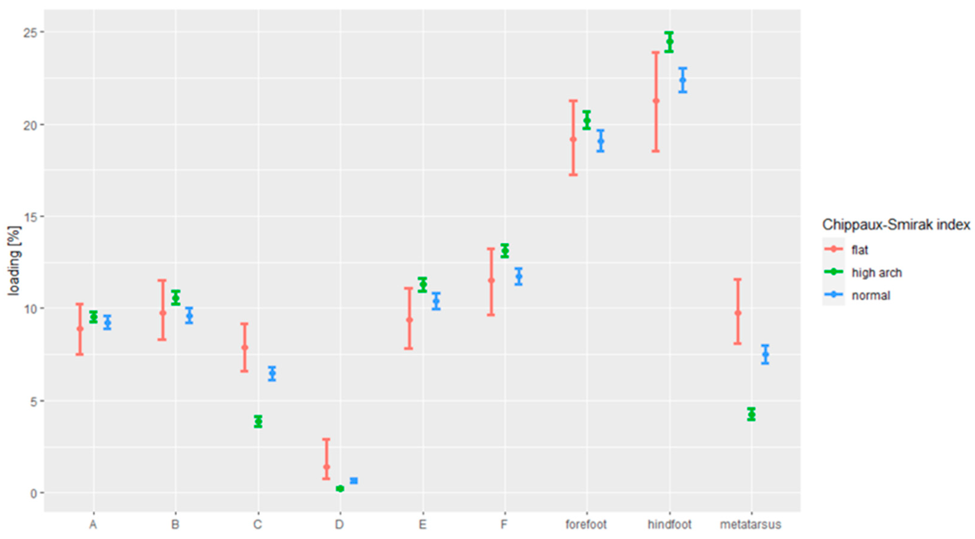
| Loading of Respective Foot Zones | Foot | |||
|---|---|---|---|---|
| Right | Left | |||
| Mean | SD | Mean | SD | |
| Loading of the forefoot % | 20.377 | 3.922 | 19.695 | 4.244 |
| Loading of the metatarsal % | 5.868 | 3.517 | 5.919 | 3.549 |
| Loading of the hindfoot % | 22.201 | 4.898 | 24.820 | 4.812 |
| Loading of foot zone A % | 9.901 | 2.003 | 8.605 | 3.006 |
| Loading of foot zone B % | 10.476 | 2.681 | 11.090 | 4.571 |
| Loading of foot zone C % | 5.296 | 2.893 | 4.805 | 2.605 |
| Loading of foot zone D % | 0.572 | 1.079 | 1.114 | 1.355 |
| Loading of foot zone E % | 10.114 | 2.846 | 12.404 | 3.676 |
| Loading of foot zone F % | 12.087 | 2.803 | 12.416 | 4.320 |
| Foot | Index KY | Index SI | Index CSI | |||||||||
|---|---|---|---|---|---|---|---|---|---|---|---|---|
| Left Foot | Right Foot | Left Foot | Right Foot | Left Foot | Right Foot | |||||||
| N | % | N | % | N | % | N | % | N | % | N | % | |
| Normal | 116 | 34.5 | 134 | 39.9 | 86 | 25.6 | 103 | 30.7 | 115 | 34.2 | 130 | 38.7 |
| Flatfoot | 1 | 0.3 | 0 | 0 | 1 | 0.3 | 1 | 0.3 | 4 | 1.2 | 9 | 2.7 |
| Low arch Foot | 12 | 3.6 | 17 | 5.1 | - | - | - | - | - | - | - | - |
| High arch foot | 207 | 61.6 | 185 | 55.1 | 249 | 74.1 | 232 | 69 | 217 | 64.6 | 197 | 58.6 |
Publisher’s Note: MDPI stays neutral with regard to jurisdictional claims in published maps and institutional affiliations. |
© 2021 by the authors. Licensee MDPI, Basel, Switzerland. This article is an open access article distributed under the terms and conditions of the Creative Commons Attribution (CC BY) license (https://creativecommons.org/licenses/by/4.0/).
Share and Cite
Szczepanowska-Wołowiec, B.; Sztandera, P.; Kotela, I.; Zak, M. Assessment of the Foot’s Longitudinal Arch by Different Indicators and Their Correlation with the Foot Loading Paradigm in School-Aged Children: A Cross Sectional Study. Int. J. Environ. Res. Public Health 2021, 18, 5196. https://0-doi-org.brum.beds.ac.uk/10.3390/ijerph18105196
Szczepanowska-Wołowiec B, Sztandera P, Kotela I, Zak M. Assessment of the Foot’s Longitudinal Arch by Different Indicators and Their Correlation with the Foot Loading Paradigm in School-Aged Children: A Cross Sectional Study. International Journal of Environmental Research and Public Health. 2021; 18(10):5196. https://0-doi-org.brum.beds.ac.uk/10.3390/ijerph18105196
Chicago/Turabian StyleSzczepanowska-Wołowiec, Beata, Paulina Sztandera, Ireneusz Kotela, and Marek Zak. 2021. "Assessment of the Foot’s Longitudinal Arch by Different Indicators and Their Correlation with the Foot Loading Paradigm in School-Aged Children: A Cross Sectional Study" International Journal of Environmental Research and Public Health 18, no. 10: 5196. https://0-doi-org.brum.beds.ac.uk/10.3390/ijerph18105196