
Rodent-Related Zoonotic Pathogens at the Human–Animal–Environment Interface in Qatar: A Systematic Review and Meta-Analysis

 ,
, 

 ,
,
Abstract
:1. Introduction
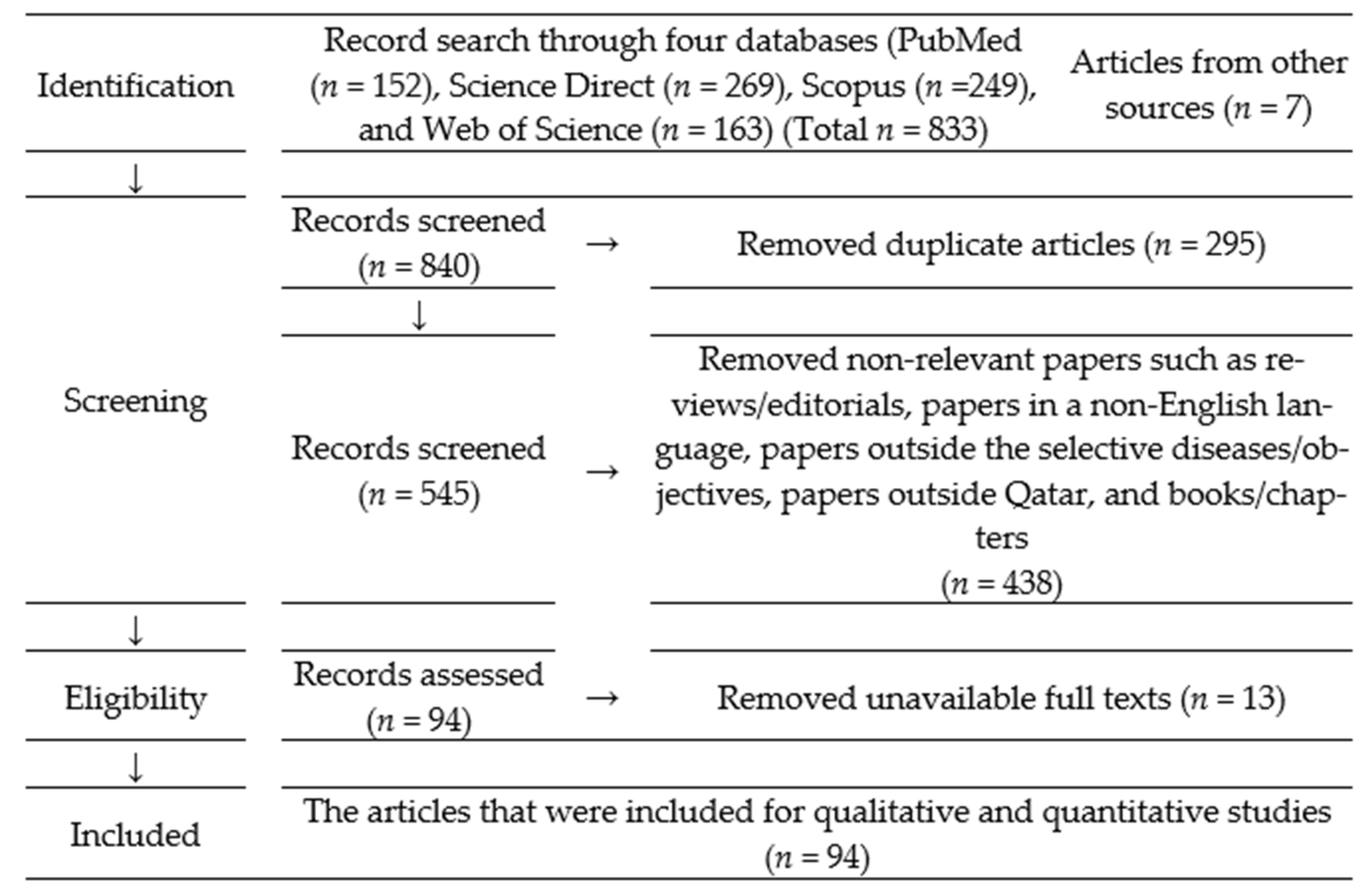
2. Materials and Methods
2.1. Data Search
2.2. Assessing the Searched Articles
2.3. Data Extraction
2.4. Data Analysis
3. Results
3.1. Characteristics of the Studied Articles
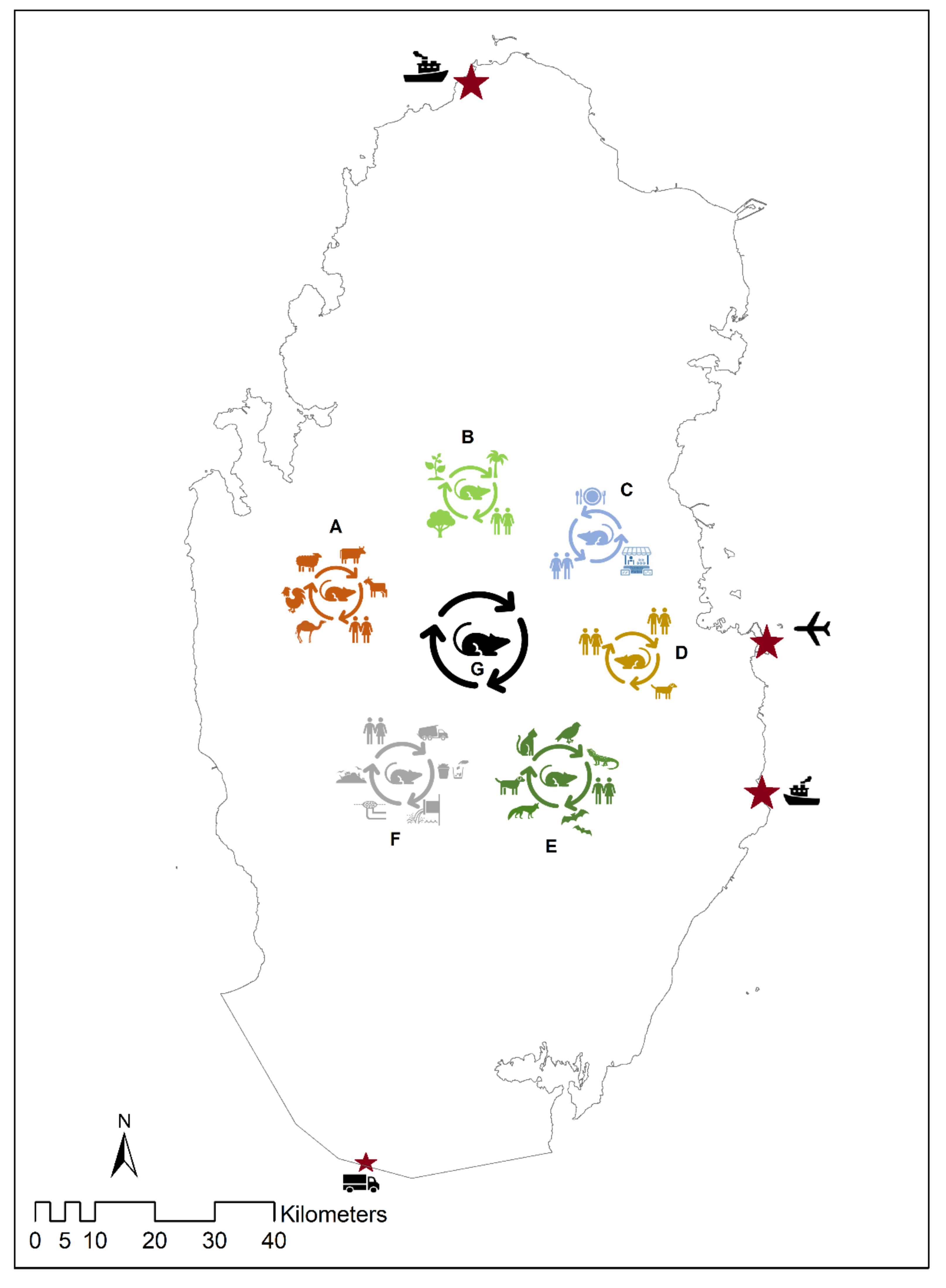
3.2. Possible Transmission Pathways of the Pathogens in Qatar
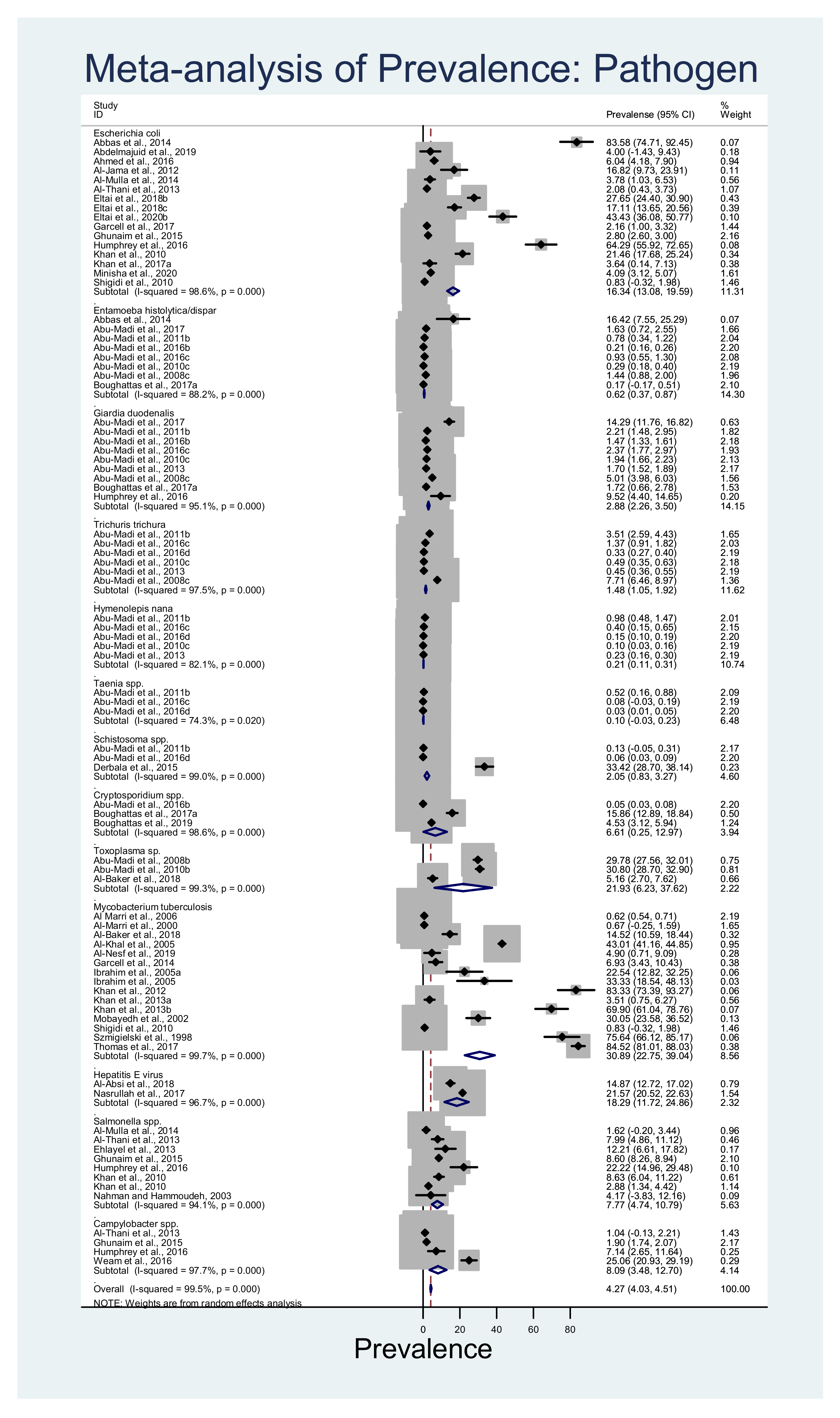
3.3. Estimated Pooled Prevalence of Pathogens
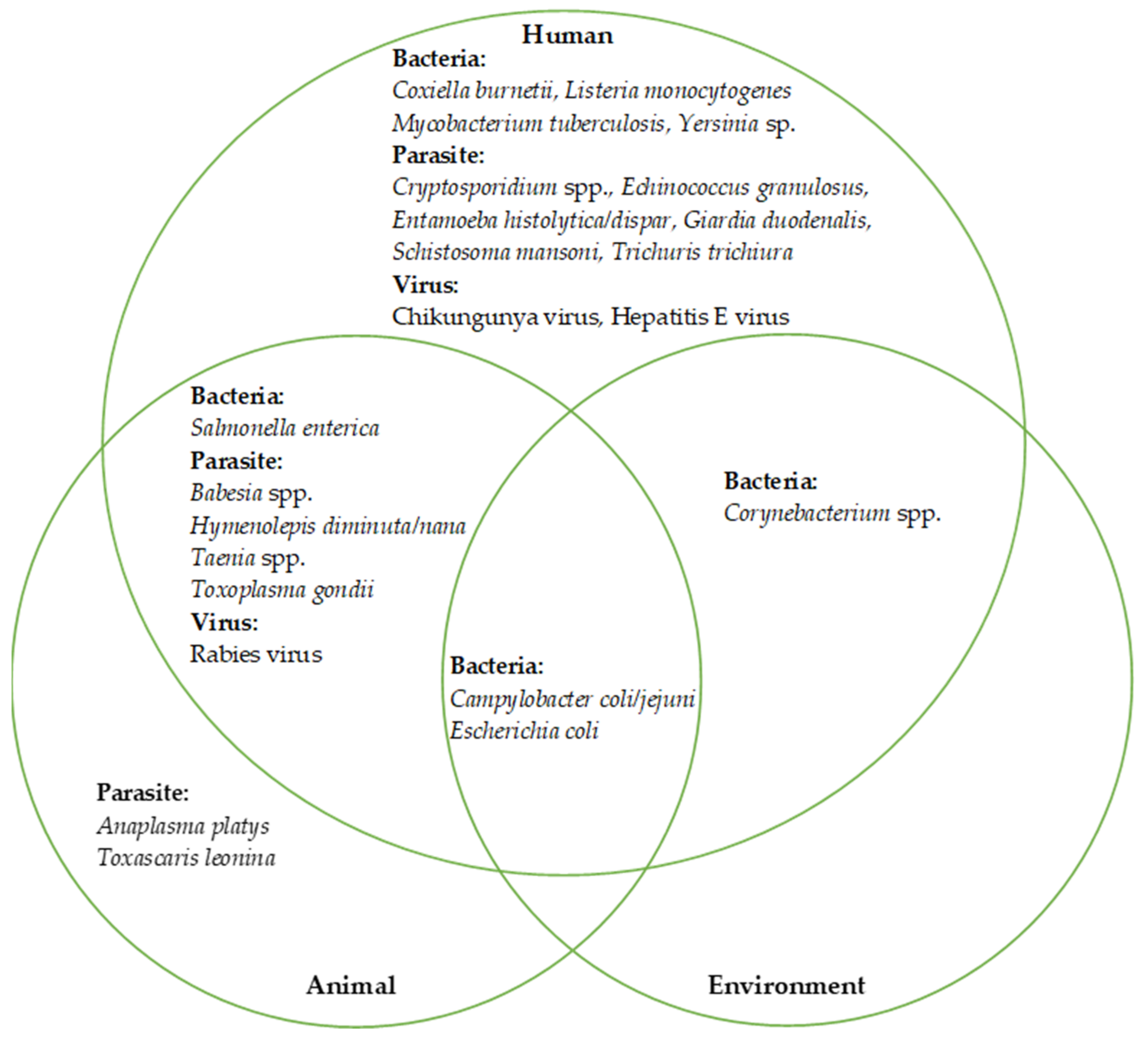
3.4. The Pathogens at the Human—Animal–Environmental Interface
4. Discussion
4.1. Characteristics of Rodent-Borne Pathogens
4.2. Bacterial Pathogens
4.3. Parasitic Pathogens
4.4. Viral Pathogens
4.5. Possible Transmission of Rodent-Borne Pathogens at the Human–Animal–Environment Interface
4.6. Limitations
5. Conclusions
Supplementary Materials
Author Contributions
Funding
Institutional Review Board Statement
Informed Consent Statement
Data Availability Statement
Acknowledgments
Conflicts of Interest
References
- Wilson, D.E.; Reeder, D.M. Mammal Species of the World: A Taxonomic and Geographic Reference; Johns Hopkins University Press: Baltimore, MD, USA, 2005. [Google Scholar]
- Burgin, C.J.; Colella, J.P.; Kahn, P.L.; Upham, N.S. How many species of mammals are there? J. Mammal. 2018, 99, 1–14. [Google Scholar] [CrossRef] [Green Version]
- Rabiee, M.H.; Mahmoudi, A.; Siahsarvie, R.; Kryštufek, B.; Mostafavi, E. Rodent-borne diseases and their public health importance in Iran. PLoS Negl. Trop. Dis. 2018, 12, e0006256. [Google Scholar] [CrossRef] [Green Version]
- Khaghani, R. The Economic and Health Impact of Rodent in Urban Zone and Harbours And Their Control Methods. Ann. Mil. Health Sci. Res. 2007, 4, 1071–1078. [Google Scholar]
- Meerburg, B.G.; Singleton, G.R.; Kijlstra, A. Rodent-borne diseases and their risks for public health. Crit. Rev. Microbiol. 2009, 35, 221–270. [Google Scholar] [CrossRef]
- Han, B.A.; Schmidt, J.P.; Bowden, S.E.; Drake, J.M. Rodent reservoirs of future zoonotic diseases. Proc. Natl. Acad. Sci. USA 2015, 112, 7039. [Google Scholar] [CrossRef] [Green Version]
- Hamidi, K. How do Rodents Play Role in Transmission of Foodborne Diseases? Nutr. Food Sci. Int. J. 2018, 6, 1–4. [Google Scholar] [CrossRef]
- Strand, T.M.; Lundkvist, Å. Rat-borne diseases at the horizon. A systematic review on infectious agents carried by rats in Europe 1995–2016. Infect. Ecol. Epidemiol. 2019, 9, 1553461. [Google Scholar] [CrossRef] [Green Version]
- Lalis, A.; Leblois, R.; Lecompte, E.; Denys, C.; Ter Meulen, J.; Wirth, T. The impact of human conflict on the genetics of Mastomys natalensis and Lassa virus in West Africa. PLoS ONE 2012, 7, e37068. [Google Scholar] [CrossRef] [Green Version]
- Zizi, M.; Heyman, P.; Vandenvelde, C. The assessment of human health risks from rodent-borne diseases by means of ecological studies of rodent reservoirs. Mil. Med. 2002, 167, 70–73. [Google Scholar]
- Gubler, D.J.; Reiter, P.; Ebi, K.L.; Yap, W.; Nasci, R.; Patz, J.A. Climate variability and change in the United States: Potential impacts on vector- and rodent-borne diseases. Environ. Health Perspect. 2001, 109 (Suppl. 2), 223–233. [Google Scholar] [CrossRef] [Green Version]
- Kilpatrick, A.M.; Randolph, S.E. Drivers, dynamics, and control of emerging vector-borne zoonotic diseases. Lancet 2012, 380, 1946–1955. [Google Scholar] [CrossRef] [Green Version]
- Mangili, A.; Gendreau, M.A. Transmission of infectious diseases during commercial air travel. Lancet 2005, 365, 989–996. [Google Scholar] [CrossRef]
- WTG. Qatar Weateher, Climate and Geography. Available online: https://www.worldtravelguide.net/guides/middle-east/qatar/weather-climate-geography/ (accessed on 7 June 2019).
- Snoj, J. Population of Qatar by Nationality—2019 Report; Priya D’Souza Communications. Available online: http://priyadsouza.com/population-of-qatar-by-nationality-in-2017/ (accessed on 17 June 2020).
- Planning and Statistics Authority. Monthly Figures on Total Population. 2020. Available online: https://www.psa.gov.qa/en/statistics1/StatisticsSite/pages/population.aspx (accessed on 17 June 2020).
- Planning and Statistics Authority. Labor Force Sample Survey. 2018. Available online: https://www.psa.gov.qa/en/statistics/Statistical%20Releases/Social/LaborForce/2018/statistical_analysis_labor_force_2018_En.pdf (accessed on 17 June 2020).
- FAO. Country Profile—Qatar. Available online: http://www.fao.org/3/ca0349en/CA0349EN.pdf (accessed on 1 May 2021).
- Miniaoui, H.; Irungu, P.; Kaitibie, S. Contemporary Issues in Qatar’s Food Security; MEI Insight No. 185; Middle East Institute, National University of Singapore: Singapore, 2018; Available online: https://mei.nus.edu.sg/publication/insight-185-contemporary-issues-in-qatars-food-security/ (accessed on 4 July 2020).
- Custom Department. The Annual Custom Report–2020; Ministry of Trading and Industries: Doha, Qatar, 2021.
- Abu-Madi, M.A.; Behnke, J.M.; Mikhail, M.; Lewis, J.W.; Al-Kaabi, M.L. Parasite populations in the brown rat Rattus norvegicus from Doha, Qatar between years: The effect of host age, sex and density. J. Helminthol. 2005, 79, 105–111. [Google Scholar] [CrossRef]
- Abu-Madi, M.A.; Lewis, J.W.; Mikhail, M.; El-Nagger, M.E.; Behnke, J.M. Monospecific helminth and arthropod infections in an urban population of brown rats from Doha, Qatar. J. Helminthol. 2001, 75, 313–320. [Google Scholar] [CrossRef] [PubMed]
- Al Thani, M.A. Irkaya; Ministy of Environment: Doha, Qatar, 2015.
- Noureldin, E.M.; Farrag, H. Rodent control strategy in animal farms (izzab) in Qatar. In Qatar Foundation Annual Research Forum Proceedings; Hamad bin Khalifa University Press: Doha, Qatar, 2010. [Google Scholar]
- Himsworth, C.G.; Parsons, K.L.; Jardine, C.; Patrick, D.M. Rats, cities, people, and pathogens: A systematic review and narrative synthesis of literature regarding the ecology of rat-associated zoonoses in urban centers. Vector Borne Zoonotic Dis. 2013, 13, 349–359. [Google Scholar] [CrossRef] [PubMed]
- World Health Organization. Status of Endemicity of Cutaneous Leishmaniasis. 2019. Available online: https://apps.who.int/neglected_diseases/ntddata/leishmaniasis/leishmaniasis.html (accessed on 4 July 2020).
- Public Health England. Guidance: Rabies Risks in Terrestrial Animals by Country. 2020. Available online: https://www.gov.uk/government/collections/rabies-risk-assessment-post-exposure-treatment-management (accessed on 4 January 2020).
- Centers for Disease Control and Prevention. Travelers’ Health; Centers for Disease Control and Prevention: Atlanta, GA, USA, 2020.
- Moher, D.; Liberati, A.; Tetzlaff, J.; Altman, D.G. Preferred reporting items for systematic reviews and meta-analyses: The PRISMA statement. PLoS Med. 2009, 6, e1000097. [Google Scholar] [CrossRef] [Green Version]
- Higgins, J.P.; Thompson, S.G. Quantifying heterogeneity in a meta—Analysis. Stat. Med. 2002, 21, 1539–1558. [Google Scholar] [CrossRef]
- Uwaydah, A.K.; Matar, I.; Chacko, K.C.; Davidson, J.C. The emergence of antimicrobial resistant Salmonella typhi in Qatar: Epidemiology and therapeutic implications. Trans. Royal Soc. Trop. Med. Hyg. 1991, 85, 790–792. [Google Scholar] [CrossRef]
- Al-Marri, M.R.; Almosleh, A.; Almoslmani, Y. Primary tuberculosis of the breast in Qatar: Ten year experience and review of the literature. Eur. J. Surg. 2000, 166, 687–690. [Google Scholar] [CrossRef]
- Al-Marri, M.; Kirkpatrick, M.B. Erythrocyte sedimentation rate in childhood tuberculosis: Is it still worthwhile? Int. J. Tuberc. Lung Dis. 2000, 4, 237–239. [Google Scholar]
- Szmiegielski, W.; Venkataraman, B.; Ejeckam, G.C.; Jarikre, L.N. Abdominal tuberculosis in Qatar: A clinico-radiological study. Int. J. Tuberc. Lung Dis. 1998, 2, 563–568. [Google Scholar]
- Al Marri, M.R.H.A.; Al Qatami, M.; Al Janahi, M. The tuberculin skin test in children with tuberculosis in the State of Qatar. Qatar Med J. 2002, 11, 19–22. [Google Scholar]
- Al-Jedah, J.H.; Robinson, R.K. Nutritional value and microbiological safety of fresh fruit juices sold through retail outlets in Qatar. Pak. J. Nutr. 2002, 1, 79–81. [Google Scholar] [CrossRef] [Green Version]
- Al-Khal, A.L.; Bener, A.; Enarson, D.A. Tuberculosis among garment workers in an Arabian developing country: State of Qatar. Arch. Environ. Occup. Health 2005, 60, 295–298. [Google Scholar] [CrossRef] [PubMed]
- Al-Marri, M. Childhood tuberculosis in the State of Qatar: The effect of a limited expatriate screening programme on the incidence of tuberculosis. Int. J. Tuberc. Lung Dis. 2001, 5, 831–837. [Google Scholar] [PubMed]
- Al-Marri, M. Pattern of mycobacterial resistance to four anti-tuberculosis drugs in pulmonary tuberculosis patients in the State of Qatar after the implementation of DOTS and a limited expatriate screening programme. Int. J. Tuberc. Lung Dis. 2001, 5, 1116–1121. [Google Scholar]
- Alsoub, H.; Al Alousi, F.S. Miliary tuberculosis in Qatar: A review of 32 adult cases. Ann. Saudi Med. 2001, 21, 16–20. [Google Scholar] [CrossRef] [Green Version]
- Howady, F.S.; Al Soub, H.; Al Khal, A.L. Spinal tuberculosis in Qatar. Qatar Med. J. 2003, 12, 85–88. [Google Scholar] [CrossRef]
- Ibrahim, A.S.; Allangawi, M.H.; Sattar, H.A.; Mobyed, H.S.; Almohammed, A.A. Indications, diagnostic yields and complications of transbronchial biopsy over 5 years in the State of Qatar. Saudi Med. J. 2005, 26, 641–645. [Google Scholar]
- Ibrahim, W.H.; Ghadban, W.; Khinji, A.; Yasin, R.; Soub, H.; Al-Khal, A.L.; Bener, A. Does pleural tuberculosis disease pattern differ among developed and developing countries. Respir. Med. 2005, 99, 1038–1045. [Google Scholar] [CrossRef] [Green Version]
- Mobayedh, H.M.S.; Sattar, H.A.; Ghadban, W.K.; Al Khal, A.L.; Al Soub, H.; Al Alousi, F.S.; Al Mohammed, A.A. Diagnostic value of fiberoptic bronchoscopy in suspected pulmonary tuberculosis in the state of Qatar. Qatar Med. J. 2002, 11, 39–42. [Google Scholar] [CrossRef]
- Nahman, A.; Hammoudeh, M. Pyogenic arthritis in Qatar. Qatar Med. J. 2003, 12, 25–27. [Google Scholar] [CrossRef]
- Abu-Madi, M.A.; Al-Ahbabi, D.A.; Al-Mashhadani, M.M.; Al-Ibrahim, R.; Pal, P.; Lewis, J.W. Patterns of parasitic infections in faecal samples from stray cat populations in Qatar. J. Helminthol. 2007, 81, 281–286. [Google Scholar] [CrossRef]
- Abu-Madi, M.A.; Pal, P.; Al-Thani, A.; Lewis, J.W. Descriptive epidemiology of intestinal helminth parasites from stray cat populations in Qatar. J. Helminthol. 2008, 82, 59–68. [Google Scholar] [CrossRef]
- Abu-Madi, M.A.; Behnke, J.M.; Prabhaker, K.S.; Al-Ibrahim, R.; Lewis, J.W. Intestinal helminths of feral cat populations from urban and suburban districts of Qatar. Vet. Parasitol. 2010, 168, 284–292. [Google Scholar] [CrossRef]
- Abu-Madi, M.A.; Al-Molawi, N.; Behnke, J.M. Seroprevalence and epidemiological correlates of Toxoplasma gondii infections among patients referred for hospital-based serological testing in Doha, Qatar. Parasit Vectors 2008, 1, 39. [Google Scholar] [CrossRef] [Green Version]
- Abu-Madi, M.A.; Behnke, J.M.; Dabritz, H.A. Toxoplasma gondii seropositivity and co-infection with TORCH pathogens in high-risk patients from Qatar. Am. J. Trop. Med. Hyg. 2010, 82, 626–633. [Google Scholar] [CrossRef] [Green Version]
- Abu-Madi, M.A.; Behnke, J.M.; Doiphode, S.H. Changing trends in intestinal parasitic infections among long-term-residents and settled immigrants in Qatar. Parasites Vectors 2010, 3, 98. [Google Scholar] [CrossRef] [Green Version]
- Abu-Madi, M.A.; Behnke, J.M.; Ismail, A. Patterns of infection with intestinal parasites in Qatar among food handlers and housemaids from different geographical regions of origin. Acta Trop. 2008, 106, 213–220. [Google Scholar] [CrossRef]
- Al Marri, M.R.H.A.; Al Hail, L.; Al Otaibi, S.; Al Marri, N.D. The time of reactivation of tuberculosis in expatriates in the State of Qatar. Qatar Med. J. 2006, 15, 21–23. [Google Scholar]
- Al-Aani, F.K.; Abusalah, S.; Al-Aqeedi, R.; Ibrahim, A. Salmonella meningitis in an adult with type B viral hepatitis and an incidental schwannoma. BMJ Case Rep. 2009, 2009, bcr1120081209. [Google Scholar] [CrossRef] [Green Version]
- Dubey, J.P.; Pas, A.; Rajendran, C.; Kwok, O.C.H.; Ferreira, L.R.; Martins, J.; Hebel, C.; Hammer, S.; Su, C. Toxoplasmosis in Sand cats (Felis margarita) and other animals in the Breeding Centre for Endangered Arabian Wildlife in the United Arab Emirates and Al Wabra Wildlife Preservation, the State of Qatar. Vet. Parasitol. 2010, 172, 195–203. [Google Scholar] [CrossRef] [PubMed]
- Ibrahim, A.S.; Alkhal, A.; Jacob, J.; Ghadban, W.; Almarri, A. Hepatitis E in Qatar imported by expatriate workers from Nepal: Epidemiological characteristics and clinical manifestations. J. Med. Virol. 2009, 81, 1047–1051. [Google Scholar] [CrossRef]
- Khan, F.Y.; Elshafie, S.S.; Almaslamani, M.; Abu-Khattab, M.; El Hiday, A.H.; Errayes, M.; Almaslamani, E. Epidemiology of bacteraemia in Hamad general hospital, Qatar: A one year hospital-based study. Travel. Med. Infect. Dis. 2010, 8, 377–387. [Google Scholar] [CrossRef] [PubMed]
- Shigidi, M.M.; Fituri, O.M.; Chandy, S.K.; Asim, M.; Al Malki, H.A.; Rashed, A.H. Microbial spectrum and outcome of peritoneal dialysis related peritonitis in Qatar. Saudi J. Kidney Dis. Transpl. 2010, 21, 168–173. [Google Scholar] [PubMed]
- Abbas, M.T.; Khan, F.Y.; Muhsin, S.A.; Al-Dehwe, B.; Abukamar, M.; Elzouki, A.N. Epidemiology, clinical features and outcome of liver abscess: A single reference center experience in Qatar. Oman Med. J. 2014, 29, 260–263. [Google Scholar] [CrossRef]
- Abu-Madi, M.A.; Behnke, J.M.; Ismail, A.; Al-Olaqi, N.; Al-Zaher, K.; El-Ibrahim, R. Comparison of intestinal parasitic infection in newly arrived and resident workers in Qatar. Parasit Vectors 2011, 4, 211. [Google Scholar] [CrossRef] [Green Version]
- Abu-Madi, M.A.; Behnke, J.M.; Doiphode, S.H. Intestinal parasitic infections among long-term-residents and settled immigrants in Qatar in the period 2005 to 2011. Am. J. Trop. Med. Hyg. 2013, 88, 1185–1195. [Google Scholar] [CrossRef]
- Al Alousi, F.S.; Abu Khattab, M.; Al Soub, H.; Al-Khal, A.L.; Al-Suwaidi, Z.D. Value of examining 3 sputum samples in the diagnosis of active pulmonary tuberculosis in qatar. Infect. Dis. Clin. Pract. 2012, 20, 188–191. [Google Scholar] [CrossRef]
- Al Ani, A.M.; Khan, F.Y.; Elzouki, A.N.; Al Hajri, M.; Ibrahim, W. Epidemiology of hydatid disease in Qatar: A hospital based study from 2000 to 2013. Asian Pac. J. Trop. Med. 2014, 7, S85–S87. [Google Scholar] [CrossRef] [Green Version]
- Al Jama, F.E. Risk factors for wound infection after lower segment cesarean section. Qatar Med. J. 2012, 2012, 26–31. [Google Scholar] [CrossRef]
- Al Marri, M.R.H.A. The tuberculin skin test in confirmed pulmonary tuberculosis in the state of Qatar: Where we stand? Qatar Med. J. 2012, 2012, 16–19. [Google Scholar] [CrossRef] [PubMed]
- Al-Mulla, N.A.; Taj-Aldeen, S.J.; El Shafie, S.; Janahi, M.; Al-Nasser, A.A.; Chandra, P. Bacterial bloodstream infections and antimicrobial susceptibility pattern in pediatric hematology/oncology patients after anticancer chemotherapy. Infect. Drug Resist. 2014, 7, 289–299. [Google Scholar] [CrossRef] [PubMed] [Green Version]
- Al-Thani, A.; Baris, M.; Al-Lawati, N.; Al-Dhahry, S. Characterising the aetiology of severe acute gastroenteritis among patients visiting a hospital in Qatar using real-time polymerase chain reaction. BMC Infect. Dis. 2013, 13, 329. [Google Scholar] [CrossRef] [Green Version]
- Derbala, M.; Elbadri, M.E.; Amer, A.M.; AlKaabi, S.; Sultan, K.H.; Kamel, Y.M.; Elsayed, E.H.; Avades, T.Y.; Chandra, P.; Shebl, F.M. Aspartate transaminase to platelet ratio index in hepatitis C virus and Schistosomiasis coinfection. World J. Gastroenterol. 2015, 21, 13132–13139. [Google Scholar] [CrossRef]
- Ehlayel, M.S.; Bener, A.; Laban, M.A. Primary immunodeficiency diseases in children: 15 year experience in a tertiary care medical center in Qatar. J. Clin. Immunol. 2013, 33, 317–324. [Google Scholar] [CrossRef]
- Garcell, H.G.; Ramirez, E.C.; Contreras, A.K.; Garcia, F.G. Latent tuberculosis infection in healthcare workers at a community hospital in Qatar. J. Infect. Public Health 2014, 7, 356–359. [Google Scholar] [CrossRef] [Green Version]
- Ghunaim, H.; Behnke, J.M.; Aigha, I.; Sharma, A.; Doiphode, S.H.; Deshmukh, A.; Abu-Madi, M.M. Analysis of resistance to antimicrobials and presence of virulence/stress response genes in Campylobacter isolates from patients with severe diarrhoea. PLoS ONE 2015, 10, e0119268. [Google Scholar] [CrossRef] [PubMed]
- Imam, Y.Z.B.; Ahmedullah, H.S.; Akhtar, N.; Chacko, K.C.; Kamran, S.; Al Alousi, F.; Alsuwaidi, Z.; Almaslmani, M.; Al Khal, A.L.; Deleu, D. Adult Tuberculous Meningitis in Qatar: A Descriptive Retrospective Study from its Referral Center. Eur. Neurol. 2015, 73, 90–97. [Google Scholar] [CrossRef] [PubMed]
- Khan, F.Y.; Al-Muzrakchi, A.M.; Elbedawi, M.M.; Al-Muzrakchi, A.A.; Al Tabeb, A. Peritoneal tuberculosis in Qatar: A five-year hospital-based study from 2005 to 2009. Travel Med. Infect. Dis. 2012, 10, 25–31. [Google Scholar] [CrossRef]
- Khan, F.Y.; Abu-Khattab, M.; Baagar, K.; Mohamed, S.F.; Elgendy, I.; Anand, D.; Malallah, H.; Sanjay, D. Characteristics of patients with definite septic arthritis at Hamad General Hospital, Qatar: A hospital-based study from 2006 to 2011. Clin. Rheumatol. 2013, 32, 969–973. [Google Scholar] [CrossRef] [PubMed]
- Khan, F.Y.; Hamza, M.; Omran, A.H.; Saleh, M.; Lingawi, M.; Alnaqdy, A.; Rahman, M.O.A.; Ahmedullah, H.S.; Hamza, A.; Al Ani, A.; et al. Diagnostic value of pleural fluid interferon-gamma and adenosine deaminase in patients with pleural tuberculosis in Qatar. Int. J. Gen. Med. 2013, 6, 13–18. [Google Scholar] [CrossRef] [PubMed] [Green Version]
- Mohammed, H.O.; Stipetic, K.; Salem, A.; McDonough, P.; Chang, Y.F.; Sultan, A. Risk of Escherichia coli O157:H7, Non-O157 Shiga Toxin-Producing Escherichia coli, and Campylobacter spp. in Food Animals and Their Products in Qatar. J. Food Prot. 2015, 78, 1812–1818. [Google Scholar] [CrossRef] [PubMed]
- Royal, J.; Riddle, M.S.; Mohareb, E.; Monteville, M.R.; Porter, C.K.; Faix, D.J. Seroepidemiologic Survey for Coxiella burnetii Among US Military Personnel Deployed to Southwest and Central Asia in 2005. Am. J. Trop. Med. Hyg. 2013, 89, 991–995. [Google Scholar] [CrossRef] [PubMed] [Green Version]
- Thandassery, R.B.; Sharma, M.; Abdelmola, A.; Derbala, M.F.M.; Al Kaabi, S.R. Uncommon gastrointestinal complications of enteric fever in a non-endemic country. Qatar Med. J. 2014, 2014. [Google Scholar] [CrossRef] [Green Version]
- Thomas, M.; AlGherbawe, M. Acute myeloid leukemia presenting with pulmonary tuberculosis. Case Rep. Infect. Dis. 2014, 2014, 865909. [Google Scholar] [CrossRef] [Green Version]
- Zowawi, H.M.; Sartor, A.L.; Balkhy, H.H.; Walsh, T.R.; Al Johani, S.M.; AlJindan, R.Y.; Alfaresi, M.; Ibrahim, E.; Al-Jardani, A.; Al-Abri, S.; et al. Molecular characterization of carbapenemase-producing Escherichia coli and Klebsiella pneumoniae in the countries of the Gulf cooperation council: Dominance of OXA-48 and NDM producers. Antimicrob. Agents Chemother. 2014, 58, 3085–3090. [Google Scholar] [CrossRef] [Green Version]
- Abdelmaguid, N.; Seleem, W.S.; Soliman, A.T.; Mohamed, R.S.; Elgharbawy, F.M.; Yassin, H.; De Sanctis, V. Clinical presentations, laboratory analysis and linear growth in 50 neonates and young infants with acute meningitis: One year experience of a single center in Qatar. Mediterr. J. Hematol. Infect. Dis. 2019, 11, e2019028. [Google Scholar] [CrossRef] [PubMed]
- Abu-Madi, M.; Behnke, J.M.; Sharma, A.; Bearden, R.; Al-Banna, N. Prevalence of Virulence/Stress Genes in Campylobacter jejuni from Chicken Meat Sold in Qatari Retail Outlets. PLoS ONE 2016, 11, e0156938. [Google Scholar] [CrossRef]
- Abu-Madi, M.; Boughattas, S.; Behnke, J.M.; Sharma, A.; Ismail, A. Coproscopy and molecular screening for detection of intestinal protozoa. Parasites Vectors 2017, 10, 414. [Google Scholar] [CrossRef] [Green Version]
- Abu-Madi, M.A.; Behnke, J.M.; Boughattas, S.; Al-Thani, A.; Doiphode, S.H. A decade of intestinal protozoan epidemiology among settled immigrants in Qatar. BMC Infect. Dis. 2016, 16, 370. [Google Scholar] [CrossRef] [PubMed] [Green Version]
- Abu-Madi, M.A.; Behnke, J.M.; Ismail, A.; Boughattas, S. Assessing the burden of intestinal parasites affecting newly arrived immigrants in Qatar. Parasit Vectors 2016, 9, 619. [Google Scholar] [CrossRef] [PubMed] [Green Version]
- Abu-Madi, M.A.; Behnke, J.M.; Boughattas, S.; Al-Thani, A.; Doiphode, S.H.; Deshmukh, A. Helminth infections among long-term-residents and settled immigrants in Qatar in the decade from 2005 to 2014: Temporal trends and varying prevalence among subjects from different regional origins. Parasites Vectors 2016, 9, 153. [Google Scholar] [CrossRef] [Green Version]
- Ahmed, M.A.S.; Bansal, D.; Acharya, A.; Elmi, A.A.; Hamid, J.M.; Ahmed, A.M.S.; Chandra, P.; Ibrahim, E.; Sultan, A.A.; Doiphode, S.; et al. Antimicrobial susceptibility and molecular epidemiology of extended-spectrum beta-lactamase-producing Enterobacteriaceae from intensive care units at Hamad Medical Corporation, Qatar. Antimicrob. Resist. Infect. Control. 2016, 5, 4. [Google Scholar] [CrossRef] [Green Version]
- Ahmedullah, H.; Khan, F.Y.; Al Maslamani, M.; Al Soub, H.; Chacko, K.; Abu Khattab, M.; Mahmoud, S.; Howaidy, F.; Thapur, M.; Al Madhoun, E.; et al. Epidemiological and Clinical Features of Salmonella Typhi Infection Among Adult Patients in Qatar: A Hospital-based Study. Oman Med. J. 2018, 33, 468–472. [Google Scholar] [CrossRef] [PubMed]
- Al Soub, H.; Al Maslamani, M.; Ahmedullah, H.S.; Shawkat, A.; Ibrahim, F.A.; Kanbar, N.A. First case of babesiosis in Qatar: Case report. Jordan Med. J. 2016, 50, 161–167. [Google Scholar] [CrossRef]
- Al-Absi, E.S.; Al-Sadeq, D.W.; Younis, M.H.; Yassine, H.M.; Abdalla, O.M.; Mesleh, A.G.; Hadwan, T.A.; Amimo, J.O.; Thalib, L.; Nasrallah, G.K. Performance evaluation of five commercial assays in assessing seroprevalence of HEV antibodies among blood donors. J. Med. Microbiol. 2018, 67, 1302–1309. [Google Scholar] [CrossRef] [PubMed]
- Al-Baker, Z.M.; Bodaghi, B.; Khan, S.A. Clinical Patterns and Causes of Uveitis in a Referral Eye Clinic in Qatar. Ocular Immunol. Inflamm. 2018, 26, 249–258. [Google Scholar] [CrossRef]
- Al-Dahshan, A.; Elyamani, R.; Naja, S.; Chehab, M.; Nour, M.; Elmagboul, E.; Saleh, T.; Al-Romaihi, H.; Farag, E. Epidemiological characteristics of a salmonella outbreak among infants in Qatar, 2017. Qatar Med. J. 2019, 2019, 12. [Google Scholar] [CrossRef] [PubMed]
- Alho, A.M.; Lima, C.; Latrofa, M.S.; Colella, V.; Ravagnan, S.; Capelli, G.; de Carvalho, L.M.; Cardoso, L.; Otranto, D. Molecular detection of vector-borne pathogens in dogs and cats from Qatar. Parasites Vectors 2017, 10, 298. [Google Scholar] [CrossRef] [Green Version]
- Ali, M.; Shaukat, A.; Ai-Suwaidi, Z.; Al-Maslamani, M. Tuberculosis of Pancreas, the First Case Reported from Qatar. Int. J. Mycobacteriol. 2019, 8, 101–103. [Google Scholar] [CrossRef]
- Al-Nesf, M.A.; Jerobin, J.; Al-Alawi, A.A.; El-Kassim, M.; Mobayed, H.; Mohammed, T.R.N. Etiology and outcome of hemoptysis in Qatar, a high-resource country with a large expatriate population: A retrospective study. Qatar Med. J. 2019, 2019. [Google Scholar] [CrossRef]
- Al-Shaer, M.H.; Mansour, H.; Elewa, H.; Salameh, P.; Iqbal, F. Treatment outcomes of fixed-dose combination versus separate tablet regimens in pulmonary tuberculosis patients with or without diabetes in Qatar. BMC Infect. Dis. 2017, 17, 118. [Google Scholar] [CrossRef] [PubMed] [Green Version]
- Al-Shaer, M.H.; Elewa, H.; Alkabab, Y.; Nazer, L.H.; Heysell, S.K. Fixed-dose combination associated with faster time to smear conversion compared to separate tablets of anti-tuberculosis drugs in patients with poorly controlled diabetes and pulmonary tuberculosis in Qatar. BMC Infect. Dis. 2018, 18, 384. [Google Scholar] [CrossRef] [PubMed]
- Ben Abid, F.; Abdel Rahman, S.A.S.H. A case report of TB versus idiopathic granulomatous mastitis with erythema nodosum, reactive arthritis, cough, and headache. Aging Male 2018, 23, 411–414. [Google Scholar] [CrossRef] [PubMed]
- Boughattas, S.; Behnke, J.M.; Al-Ansari, K.; Sharma, A.; Abu-Alainin, W.; Al-Thani, A.; Abu-Madi, M.A. Molecular Analysis of the Enteric Protozoa Associated with Acute Diarrhea in Hospitalized Children. Front. Cell. Infect. Microbiol. 2017, 7. [Google Scholar] [CrossRef] [PubMed]
- Boughattas, S.; Behnke, J.; Sharma, A.; Abu-Madi, M. Seroprevalence of Toxoplasma gondii infection in feral cats in Qatar. BMC Vet. Res. 2017, 13, 26. [Google Scholar] [CrossRef] [Green Version]
- Boughattas, S.; Behnke, J.M.; Al-Sadeq, D.; Ismail, A.; Abu-Madi, M. Cryptosporidium spp., prevalence, molecular characterisation and socio-demographic risk factors among immigrants in Qatar. PLoS Negl. Trop. Dis. 2019, 13, 343. [Google Scholar] [CrossRef] [PubMed]
- Chang, Y.C.; Scaria, J.; Ibraham, M.; Doiphode, S.; Chang, Y.-F.; Sultan, A.; Mohammed, H.O. Distribution and factors associated with Salmonella enterica genotypes in a diverse population of humans and animals in Qatar using multi-locus sequence typing (MLST). J. Infect. Public Health 2016, 9, 315–323. [Google Scholar] [CrossRef] [PubMed]
- Dousa, K.M.; Hamad, A.; Albirair, M.; Al Soub, H.; Elzouki, A.N.; Alwakeel, M.I.; Thiel, B.A.; Johnson, J.L. Impact of Diabetes Mellitus on the Presentation and Response to Treatment of Adults With Pulmonary Tuberculosis in Qatar. Open Forum Infect. Dis. 2019, 6. [Google Scholar] [CrossRef]
- El-Nemr, I.M.; Mushtaha, M.; Sundararaju, S.; Fontejon, C.; Suleiman, M.; Tang, P.; Goktepe, I.; Hasan, M.R. Application of MALDI Biotyper System for Rapid Identification of Bacteria Isolated from a Fresh Produce Market. Curr. Microbiol. 2019, 76, 290–296. [Google Scholar] [CrossRef]
- Eltai, N.; Al Thani, A.A.; Al-Hadidi, S.H.; Abdfarag, E.A.; Al-Romaihi, H.; Mahmoud, M.H.; Alawad, O.K.; Yassine, H.M. Antibiotic resistance profile of commensal Escherichia coli isolated from healthy sheep in Qatar. J. Infect. Dev. Ctries 2020, 14, 138–145. [Google Scholar] [CrossRef] [Green Version]
- Eltai, N.O.; Abdfarag, E.A.; Al-Romaihi, H.; Wehedy, E.; Mahmoud, M.H.; Alawad, O.K.; Al-Hajri, M.M.; Al Thani, A.A.; Yassine, H.M. Antibiotic Resistance Profile of Commensal Escherichia coli Isolated from Broiler Chickens in Qatar. J. Food Prot. 2018, 81, 302–307. [Google Scholar] [CrossRef] [PubMed]
- Eltai, N.O.; Al Thani, A.A.; Al-Ansari, K.; Deshmukh, A.S.; Wehedy, E.; Al-Hadidi, S.H.; Yassine, H.M. Molecular characterization of extended spectrum beta -lactamases enterobacteriaceae causing lower urinary tract infection among pediatric population. Antimicrob. Resist. Infect. Control. 2018, 7. [Google Scholar] [CrossRef] [PubMed]
- Eltai, N.O.; Yassine, H.M.; Al Thani, A.A.; Abu Madi, M.A.; Ismail, A.; Ibrahim, E.; Alali, W.Q. Prevalence of antibiotic resistant Escherichia coli isolates from fecal samples of food handlers in Qatar. Antimicrob. Resist. Infect. Control. 2018, 7, 78. [Google Scholar] [CrossRef] [PubMed]
- Eltai, N.O.; Al Thani, A.A.; Al Hadidi, S.H.; Al Ansari, K.; Yassine, H.M. Antibiotic resistance and virulence patterns of pathogenic Escherichia coli strains associated with acute gastroenteritis among children in Qatar. BMC Microbiol. 2020, 20, 54. [Google Scholar] [CrossRef]
- Farag, E.; Garcell, H.G.; Ganesan, N.; Ahmed, S.N.; Al-Hajri, M.; Al Thani, S.M.; Al-Marri, S.A.; Ibrahim, E.; Al-Romaihi, H.E. A retrospective epidemiological study on the incidence of salmonellosis in the State of Qatar during 2004–2012. Qatar Med. J. 2016, 2016, 3. [Google Scholar] [CrossRef] [Green Version]
- Garcell, H.G.; Arias, A.V.; Sandoval, C.A.; Garcia, E.G.; Gamboa, M.E.; Sado, A.B.; Serrano, R.N. Incidence and Etiology of Surgical Site Infections in Appendectomies: A 3-Year Prospective Study. Oman Med. J. 2017, 32, 31–35. [Google Scholar] [CrossRef]
- Humphrey, J.M.; Ranbhise, S.; Ibrahim, E.; Al-Romaihi, H.E.; Farag, E.; Abu-Raddad, L.J.; Glesby, M.J. Multiplex Polymerase Chain Reaction for Detection of Gastrointestinal Pathogens in Migrant Workers in Qatar. Am. J. Trop. Med. Hyg. 2016, 95, 1330–1337. [Google Scholar] [CrossRef] [Green Version]
- Humphrey, J.M.; Al-Absi, E.S.; Hamdan, M.M.; Okasha, S.S.; Al-Trmanini, D.M.; El-Dous, H.G.; Dargham, S.R.; Schieffelin, J.; Abu-Raddad, L.J.; Nasrallah, G.K. Dengue and chikungunya seroprevalence among Qatari nationals and immigrants residing in Qatar. PLoS ONE 2019, 14, e0211574. [Google Scholar] [CrossRef] [PubMed]
- Ibrahim, W.H.; Alousi, F.H.; Al-Khal, A.; Bener, A.; AlSalman, A.; Aamer, A.; Khaled, A.; Raza, T. Diagnostic Delay among Adults with Pulmonary Tuberculosis in a High Gross Domestic Product Per Capita Country: Reasons and Magnitude of the Problem. Int. J. Prev. Med. 2016, 7, 116. [Google Scholar] [CrossRef] [PubMed]
- Khan, F.Y.; Abu-Khattab, M.; Almaslamani, E.A.; Hassan, A.A.; Mohamed, S.F.; Elbuzdi, A.A.; Elmaki, N.Y.; Anand, D.; Sanjay, D. Acute Bacterial Meningitis in Qatar: A Hospital-Based Study from 2009 to 2013. BioMed Res. Int. 2017, 2017, 2975610. [Google Scholar] [CrossRef]
- Khan, F.Y. Clinical pattern of tuberculous adenitis in Qatar: Experience with 35 patients. Scand. J. Infect. Dis. 2009, 41, 128–134. [Google Scholar] [CrossRef]
- Minisha, F.; Mohamed, M.; Abdulmunem, D.; El Awad, S.; Zidan, M.; Abreo, M.; Ahmad, S.; Fender, G. Bacteriuria in pregnancy varies with the ambiance: A retrospective observational study at a tertiary hospital in Doha, Qatar. J. Perinat Med. 2019, 48, 46–52. [Google Scholar] [CrossRef] [PubMed]
- Nasrallah, G.K.; Al Absi, E.S.; Ghandour, R.; Ali, N.H.; Taleb, S.; Hedaya, L.; Ali, F.; Huwaidy, M.; Husseini, A. Seroprevalence of hepatitis E virus among blood donors in Qatar (2013–2016). Transfusion 2017, 57, 1801–1807. [Google Scholar] [CrossRef] [Green Version]
- Oude Munnink, B.B.; Farag, E.A.B.A.; GeurtsvanKessel, C.; Schapendonk, C.; van der Linden, A.; Kohl, R.; Arron, G.; Ziglam, H.; Goravey, W.G.M.; Coyle, P.V.; et al. First molecular analysis of rabies virus in Qatar and clinical cases imported into Qatar, a case report. Int. J. Infect. Dis. 2020, 96, 323–326. [Google Scholar] [CrossRef] [PubMed]
- Thomas, M.; Ibrahim, W.H.; Raza, T.; Mushtaq, K.; Arshad, A.; Ahmed, M.; Taha, S.; Al Sarafandi, S.; Karim, H.; Abdul-sattar, H.A. Medical thoracoscopy for exudative pleural effusion: An eight-year experience from a country with a young population. BMC Pulm. Med. 2017, 17, 151. [Google Scholar] [CrossRef] [Green Version]
- Weam, B.; Abraham, M.; Doiphode, S.; Peters, K.; Ibrahim, E.; Sultan, A.; Mohammed, H.O. Foodborne Bacterial Pathogens Associated with the Risk of Gastroenteritis in the State of Qatar. Int. J. Health Sci. 2016, 10, 197–207. [Google Scholar]
- Zaqout, A.; Abid, F.B.; Murshed, K.; Al-Bozom, I.; Al-Rumaihi, G.; Al Soub, H.; Al Maslamani, M.; Al Khal, A. Cerebral schistosomiasis: Case series from Qatar. Int. J. Infect. Dis. 2019, 86, 167–170. [Google Scholar] [CrossRef] [PubMed] [Green Version]
- Behr, M.A.; Gagneux, S. 24—The Rise and Fall of the Mycobacterium tuberculosis Complex. In Genetics and Evolution of Infectious Disease; Tibayrenc, M., Ed.; Elsevier: London, UK, 2011; pp. 651–667. [Google Scholar] [CrossRef]
- Moradi, E.; Mosavari, N.; Tebianian, M.; Tadayon, K.; Ghaderi, R.; Soleymani Babadi, K.; Mohammad Taheri, M.; Dashtipour, S.; Loni, R.; Moradi Garavand, M.; et al. Pest rodents as the essential elements of Mycobacterium bovis controlling programs. Int. J. Mycobacteriol. 2015, 4, 137. [Google Scholar] [CrossRef] [Green Version]
- Cavanagh, R.; Begon, M.; Bennett, M.; Ergon, T.; Graham, I.M.; De Haas, P.E.W.; Hart, C.A.; Koedam, M.; Kremer, K.; Lambin, X.; et al. Mycobacterium microti infection (vole tuberculosis) in wild rodent populations. J. Clin. Microbiol. 2002, 40, 3281–3285. [Google Scholar] [CrossRef] [PubMed] [Green Version]
- Blanco Crivelli, X.; Rumi, M.V.; Carfagnini, J.C.; Degregorio, O.; Bentancor, A.B. Synanthropic rodents as possible reservoirs of shigatoxigenic Escherichia coli strains. Front. Cell Infect. Microbiol. 2012, 2, 134. [Google Scholar] [CrossRef] [Green Version]
- Meerburg, B.G.; Jacobs-Reitsma, W.F.; Wagenaar, J.A.; Kijlstra, A. Presence of Salmonella and Campylobacter spp. in wild small mammals on organic farms. Appl. Environ. Microbiol. 2006, 72, 960–962. [Google Scholar] [CrossRef] [Green Version]
- Meerburg, B.G.; Kijlstra, A. Role of rodents in transmission of Salmonella and Campylobacter. J. Sci. Food Agric. 2007, 87, 2774–2781. [Google Scholar] [CrossRef]
- Loftis, A.D.; Reeves, W.K.; Szumlas, D.E.; Abbassy, M.M.; Helmy, I.M.; Moriarity, J.R.; Dasch, G.A. Surveillance of Egyptian fleas for agents of public health significance: Anaplasma, bartonella, coxiella, ehrlichia, rickettsia, and Yersinia pestis. Am. J. Trop. Med. Hyg. 2006, 75, 41–48. [Google Scholar] [CrossRef] [Green Version]
- Morick, D.; Baneth, G.; Avidor, B.; Kosoy, M.Y.; Mumcuoglu, K.Y.; Mintz, D.; Eyal, O.; Goethe, R.; Mietze, A.; Shpigel, N.; et al. Detection of Bartonella spp. in wild rodents in Israel using HRM real-time PCR. Vet. Microbiol. 2009, 139, 293–297. [Google Scholar] [CrossRef]
- Nekouie, H.; Razavi, M.; Seyedipoor, G. Investigation of Yersinia pestis in Xenopsylla astia. Southeast Asian J. Trop. Med. Public Health 2003, 34 (Suppl. 2), 158–161. [Google Scholar]
- He, J.; Innis, B.L.; Shrestha, M.P.; Clayson, E.T.; Scott, R.M.; Linthicum, K.J.; Musser, G.G.; Gigliotti, S.C.; Binn, L.N.; Kuschner, R.A.; et al. Evidence that rodents are a reservoir of hepatitis E virus for humans in Nepal. J. Clin. Microbiol. 2002, 40, 4493–4498. [Google Scholar] [CrossRef] [Green Version]
- Fitzpatrick, J.L.; Dyer, J.L.; Blanton, J.D.; Kuzmin, I.V.; Rupprecht, C.E. Rabies in rodents and lagomorphs in the United States, 1995–2010. J. Am. Vet. Med. Assoc. 2014, 245, 333–337. [Google Scholar] [CrossRef] [Green Version]
- Rohde, R.; Wilson, P. 8 Things You May Not Know About Rabies—But Should! Elsevier Connect: Amsterdam, The Netherlands, 2015. [Google Scholar]
- Qatar Pest Control Company. Rodent trapping catelogue. In Semco Building, Behind HSBC Building, Airport Road; Qatar Pest Control Company: Doha, Qatar, 2021. [Google Scholar]
- Social & Economic Survey Research Institute. Priminiary Records of Agricultural Census; Qatar University and the Ministry of Municipality and Environment, Social & Economic Survey Research Institute: Doha, Qatar, 2021. [Google Scholar]
- Farag, E.; Sikkema, R.S.; Vinks, T.; Islam, M.M.; Nour, M.; Al-Romaihi, H.; Al Thani, M.; Atta, M.; Alhajri, F.H.; Al-Marri, S.; et al. Drivers of MERS-CoV Emergence in Qatar. Viruses 2018, 11, 22. [Google Scholar] [CrossRef] [PubMed] [Green Version]
- Ben Hassen, T.; El Bilali, H.; Al-Maadeed, M. Agri-Food Markets in Qatar: Drivers, Trends, and Policy Responses. Sustainability 2020, 12, 3643. [Google Scholar] [CrossRef]
- Boussaa, D. Al Asmakh historic district in Doha, Qatar: From an urban slum to living heritage. J. Archit. Conserv. 2014, 20, 2–15. [Google Scholar] [CrossRef]
- Karanisa, T.; Amato, A.; Richer, R.; Abdul Majid, S.; Skelhorn, C.; Sayadi, S. Agricultural Production in Qatar’s Hot Arid Climate. Sustainability 2021, 13, 4059. [Google Scholar] [CrossRef]
- Kuo, C.-C.; Wardrop, N.; Chang, C.-T.; Wang, H.-C.; Atkinson, P.M. Significance of major international seaports in the distribution of murine typhus in Taiwan. PLOS Negl. Trop. Dis. 2017, 11, e0005430. [Google Scholar] [CrossRef] [Green Version]
- White, C.F. Plague: Modern Preventive Measures in Ships and Ports: (Section of Tropical Diseases and Parasitology). Proc. R Soc. Med. 1935, 28, 591–602. [Google Scholar]
- Banks, P.; Cleary, G.; Dickman, C. Sydney’s bubonic plague outbreak 1900-1910: A disaster for foreshore wildlife? Aust. Zool. 2011, 35, 1033–1039. [Google Scholar] [CrossRef]
- Sonne, O. Plague, rats, and ships The realisation of the infection routes of plague. Dan Medicinhist Arbog 2016, 44, 101–133. [Google Scholar]
- Buller, R.M.L. 170—Poxviruses. In Infectious Diseases, 4th ed.; Cohen, J., Powderly, W.G., Opal, S.M., Eds.; Elsevier: Amsterdam, The Netherlands, 2017; pp. 1452–1457.e1. [Google Scholar] [CrossRef]
- Cao, C.; Liang, L.; Wang, W.; Luo, H.; Huang, S.; Liu, D.; Xu, J.; Henk, D.A.; Fisher, M.C. Common reservoirs for Penicillium marneffei infection in humans and rodents, China. Emerg. Infect. Dis. 2011, 17, 209–214. [Google Scholar] [CrossRef]
- Da Costa Cordeiro, H.; de Vasconcelos Melo, F.T.; Giese, E.G.; Santos, J.N.D. Gongylonema Parasites of Rodents: A Key to Species and New Data on Gongylonema neoplasticum. J. Parasitol. 2018, 104, 51–59. [Google Scholar] [CrossRef] [PubMed]
- Da Silva, A.M. Human echinococcosis: A neglected disease. Gastroenterol. Res. Pr. 2010, 2010, 583297. [Google Scholar] [CrossRef] [PubMed]
- Deplazes, P.; Eichenberger, R.M.; Grimm, F. Wildlife-transmitted Taenia and Versteria cysticercosis and coenurosis in humans and other primates. Int. J. Parasitol. Parasites Wildl. 2019, 9, 342–358. [Google Scholar] [CrossRef]
- Dworkin, M.S.; Schwan, T.G.; Anderson, D.E., Jr.; Borchardt, S.M. Tick-borne relapsing fever. Infect. Dis. Clin. 2008, 22, 449–468. [Google Scholar] [CrossRef] [Green Version]
- Edmonds, P.; Patton, C.M.; Griffin, P.M.; Barrett, T.J.; Schmid, G.P.; Baker, C.N.; Lambert, M.A.; Brenner, D.J. Campylobacter hyointestinalis associated with human gastrointestinal disease in the United States. J. Clin. Microbiol. 1987, 25, 685–691. [Google Scholar] [CrossRef] [PubMed] [Green Version]
- Emmons, C.W.; Ashburn, L.L. Histoplasmosis in Wild Rats: Occurrence and Histopathology. Public Health Rep. 1948, 63, 1416–1422. [Google Scholar] [CrossRef]
- Favacho, A.R.d.M.; Andrade, M.N.; de Oliveira, R.C.; Bonvicino, C.R.; D’Andrea, P.S.; de Lemos, E.R.S. Zoonotic Bartonella species in wild rodents in the state of Mato Grosso do Sul, Brazil. Microbes Infect. 2015, 17, 889–892. [Google Scholar] [CrossRef] [PubMed]
- García-Livia, K.; Martín-Alonso, A.; Foronda, P. Diversity of Cryptosporidium spp. in wild rodents from the Canary Islands, Spain. Parasit Vectors 2020, 13, 445. [Google Scholar] [CrossRef] [PubMed]
- Gillespie, S.H.; Pearson, R.D. Principles and Practice of Clinical Parasitology; Wiley: New York, NY, USA, 2010. [Google Scholar]
- Gonçalves, L.R.; Favacho, A.R.d.M.; Roque, A.L.R.; Mendes, N.S.; Fidelis Junior, O.L.; Benevenute, J.L.; Herrera, H.M.; Andrea, P.S.; de Lemos, E.R.S.; Machado, R.Z.; et al. Association of Bartonella Species with Wild and Synanthropic Rodents in Different Brazilian Biomes. Appl. Environ. Microbiol. 2016, 82, 7154. [Google Scholar] [CrossRef] [Green Version]
- Gravinatti, M.L.; Barbosa, C.M.; Soares, R.M.; Gregori, F. Synanthropic rodents as virus reservoirs and transmitters. Rev. Soc. Bras. Med. Trop. 2020, 53, e20190486. [Google Scholar] [CrossRef] [PubMed] [Green Version]
- Hildebrand, J.; Adamczyk, M.; Laskowski, Z.; Zaleśny, G. Host-dependent morphology of Isthmiophora melis (Schrank, 1788) Luhe, 1909 (Digenea, Echinostomatinae)--morphological variation vs. molecular stability. Parasit Vectors 2015, 8, 481. [Google Scholar] [CrossRef] [Green Version]
- Kawahara, M.; Ito, T.; Suto, C.; Shibata, S.; Rikihisa, Y.; Hata, K.; Hirai, K. Comparison of Ehrlichia muris Strains Isolated from Wild Mice and Ticks and Serologic Survey of Humans and Animals with E. muris as Antigen. J. Clin. Microbiol. 1999, 37, 1123. [Google Scholar] [CrossRef] [Green Version]
- Lv, C.; Zhang, L.; Wang, R.; Jian, F.; Zhang, S.; Ning, C.; Wang, H.; Feng, C.; Wang, X.; Ren, X.; et al. Prevalence and molecular characterization of Cryptosporidium spp. in wild, laboratory, and pet rodents in China. Appl. Environ. Microbiol. 2009, 75, 7692. [Google Scholar] [CrossRef] [PubMed] [Green Version]
- Macnish, M.G.; Ryan, U.M.; Behnke, J.M.; Thompson, R.C. Detection of the rodent tapeworm Rodentolepis (=Hymenolepis) microstoma in humans. A new zoonosis? Int. J. Parasitol. 2003, 33, 1079–1085. [Google Scholar] [CrossRef]
- Möhl, K.; Grosse, K.; Hamedy, A.; Wüste, T.; Kabelitz, P.; Lücker, E. Biology of Alaria spp. and human exposition risk to Alaria mesocercariae-a review. Parasitol. Res. 2009, 105, 1–15. [Google Scholar] [CrossRef] [PubMed]
- Nkogwe, C.; Raletobana, J.; Stewart-Johnson, A.; Suepaul, S.; Adesiyun, A. Frequency of Detection of Escherichia coli, Salmonella spp., and Campylobacter spp. in the Faeces of Wild Rats (Rattus spp.) in Trinidad and Tobago. Vet. Med. Int. 2011, 2011, 686923. [Google Scholar] [CrossRef] [Green Version]
- Pozio, E. World distribution of Trichinella spp. infections in animals and humans. Vet. Parasitol. 2007, 149, 3–21. [Google Scholar] [CrossRef]
- Scharmann, W.; Heller, A. Survival and transmissibility of Pasteurella pneumotropica. Lab. Anim. 2001, 35, 163–166. [Google Scholar] [CrossRef]
- Spickler, A.R. Taeniasis, Cysticercosis, and Coenurosis. 2020. Available online: http://www.cfsph.iastate.edu/Factsheets/pdfs/taenia.pdf (accessed on 30 June 2020).
- Tappe, D.; Stich, A.; Frosch, M. Emergence of polycystic neotropical echinococcosis. Emerg. Infect. Dis. 2008, 14, 292–297. [Google Scholar] [CrossRef]
- Thomas, R.J.; Dumler, J.S.; Carlyon, J.A. Current management of human granulocytic anaplasmosis, human monocytic ehrlichiosis and Ehrlichia ewingii ehrlichiosis. Expert Rev. Anti Infect. Ther. 2009, 7, 709–722. [Google Scholar] [CrossRef] [PubMed] [Green Version]
- Toledo, R.; Esteban, J.G. An update on human echinostomiasis. Trans. R Soc. Trop. Med. Hyg. 2016, 110, 37–45. [Google Scholar] [CrossRef]
- Tominello, T.R.; Oliveira, E.R.A.; Hussain, S.S.; Elfert, A.; Wells, J.; Golden, B.; Ismail, N. Emerging Roles of Autophagy and Inflammasome in Ehrlichiosis. Front. Immunol. 2019, 10, 1011. [Google Scholar] [CrossRef] [Green Version]
- Van Soolingen, D.; van der Zanden, A.G.; de Haas, P.E.; Noordhoek, G.T.; Kiers, A.; Foudraine, N.A.; Portaels, F.; Kolk, A.H.; Kremer, K.; van Embden, J.D. Diagnosis of Mycobacterium microti infections among humans by using novel genetic markers. J. Clin. Microbiol. 1998, 36, 1840–1845. [Google Scholar] [CrossRef] [PubMed] [Green Version]
- Vourc’h, G.; Halos, L.; Desvars, A.; Boué, F.; Pascal, M.; Lecollinet, S.; Zientara, S.; Duval, T.; Nzonza, A.; Brémont, M. Chikungunya antibodies detected in non-human primates and rats in three Indian Ocean islands after the 2006 ChikV outbreak. Vet. Res. 2014, 45, 52. [Google Scholar] [CrossRef] [PubMed] [Green Version]
- Wang, Y.; Lu, L.; Lan, R.; Salazar, J.K.; Liu, J.; Xu, J.; Ye, C. Isolation and characterization of Listeria species from rodents in natural environments in China. Emerg. Microbes Infect. 2017, 6, e44. [Google Scholar] [CrossRef] [PubMed] [Green Version]
- Wasiluk, A. Alaria alata infection—Threating yet rarely detected trematodiasis. J. Lab. Diagn. 2013, 49, 33–37. [Google Scholar]


| Characteristics | Number of Articles (%; 95%CI) | References | |
|---|---|---|---|
| Publication Year | |||
| 1991–1995 | 1 (1.06; 0.027–5.79) | [31] | |
| 1996–2000 | 3 (3.19; 0.66–9.04) | [32,33,34] | |
| 2001–2005 | 12 (12.77; 6.77–21.24) | [21,35,36,37,38,39,40,41,42,43,44,45] | |
| 2006–2010 | 13 (13.83; 7.57–22.49) | [46,47,48,49,50,51,52,53,54,55,56,57,58] | |
| 2011–2015 | 23 (24.47; 16.19–34.42) | [22,59,60,61,62,63,64,65,66,67,68,69,70,71,72,73,74,75,76,77,78,79,80] | |
| 2016–2020 | 42 (44.68; 34.41–55.29) | [81,82,83,84,85,86,87,88,89,90,91,92,93,94,95,96,97,98,99,100,101,102,103,104,105,106,107,108,109,110,111,112,113,114,115,116,117,118,119,120,121,122] | |
| Host | |||
| Humans | 80 (85.11; 76.28–91.61) | [31,32,33,34,35,36,37,38,39,40,41,42,43,44,45,49,50,51,52,53,54,56,57,58,59,60,61,62,63,64,65,66,67,68,69,70,71,72,73,74,75,77,78,79,80,81,83,84,85,86,87,88,89,90,91,92,94,95,96,97,98,99,101,103,104,107,108,109,110,111,112,113,114,115,116,117,118,120,121,122] | |
| Animals | 10 (10.64; 5.22–18.70) | [21,22,46,47,48,55,82,93,105,106] | |
| Environment | 1 (1.06; 0.027–5.79) | [36] | |
| Humans + Animals | 1 (1.06; 0.027–5.79) | [119] | |
| Animals + Environment | 1 (1.06; 0.027–5.79) | [76] | |
| Humans + Environment | 1 (1.06; 0.027–5.79) | [104] | |
| Pathogen | |||
| Bacteria | 62 (65.96; 55.46–75.42) | [31,32,33,34,35,36,37,38,39,40,41,42,43,44,45,53,54,57,58,62,63,64,65,66,67,69,70,71,72,73,74,75,76,77,78,79,80,81,82,87,88,92,94,95,96,97,98,102,103,105,106,107,108,109,110,111,114,115,116,117,120,121] | |
| Helminth | 10 (10.64; 5.22–18.70) | [21,22,46,47,48,60,63,68,86,122] | |
| Protozoa | 9 (9.57; 4.47–17.40) | [49,50,55,83,84,89,99,100,101] | |
| Virus | 5 (5.32; 1.75–11.98) | [45,56,90,113,119] | |
| Helminth + Protozoa | 4 (4.25; 1.17–10.54) | [51,52,61,85] | |
| Bacteria + Protozoa | 4 (4.25; 1.17–10.54) | [59,91,93,112] | |
Publisher’s Note: MDPI stays neutral with regard to jurisdictional claims in published maps and institutional affiliations. |
© 2021 by the authors. Licensee MDPI, Basel, Switzerland. This article is an open access article distributed under the terms and conditions of the Creative Commons Attribution (CC BY) license (https://creativecommons.org/licenses/by/4.0/).
Share and Cite
Islam, M.M.; Farag, E.; Mahmoudi, A.; Hassan, M.M.; Mostafavi, E.; Enan, K.A.; Al-Romaihi, H.; Atta, M.; El Hussein, A.R.M.; Mkhize-Kwitshana, Z. Rodent-Related Zoonotic Pathogens at the Human–Animal–Environment Interface in Qatar: A Systematic Review and Meta-Analysis. Int. J. Environ. Res. Public Health 2021, 18, 5928. https://0-doi-org.brum.beds.ac.uk/10.3390/ijerph18115928
Islam MM, Farag E, Mahmoudi A, Hassan MM, Mostafavi E, Enan KA, Al-Romaihi H, Atta M, El Hussein ARM, Mkhize-Kwitshana Z. Rodent-Related Zoonotic Pathogens at the Human–Animal–Environment Interface in Qatar: A Systematic Review and Meta-Analysis. International Journal of Environmental Research and Public Health. 2021; 18(11):5928. https://0-doi-org.brum.beds.ac.uk/10.3390/ijerph18115928
Chicago/Turabian StyleIslam, Md Mazharul, Elmoubashar Farag, Ahmad Mahmoudi, Mohammad Mahmudul Hassan, Ehsan Mostafavi, Khalid A. Enan, Hamad Al-Romaihi, Muzzamil Atta, Abdel Rahim M. El Hussein, and Zilungile Mkhize-Kwitshana. 2021. "Rodent-Related Zoonotic Pathogens at the Human–Animal–Environment Interface in Qatar: A Systematic Review and Meta-Analysis" International Journal of Environmental Research and Public Health 18, no. 11: 5928. https://0-doi-org.brum.beds.ac.uk/10.3390/ijerph18115928