Temporomandibular Joint and Cervical Spine Mobility Assessment in the Prevention of Temporomandibular Disorders in Children with Osteogenesis Imperfecta: A Pilot Study


Abstract
:1. Introduction
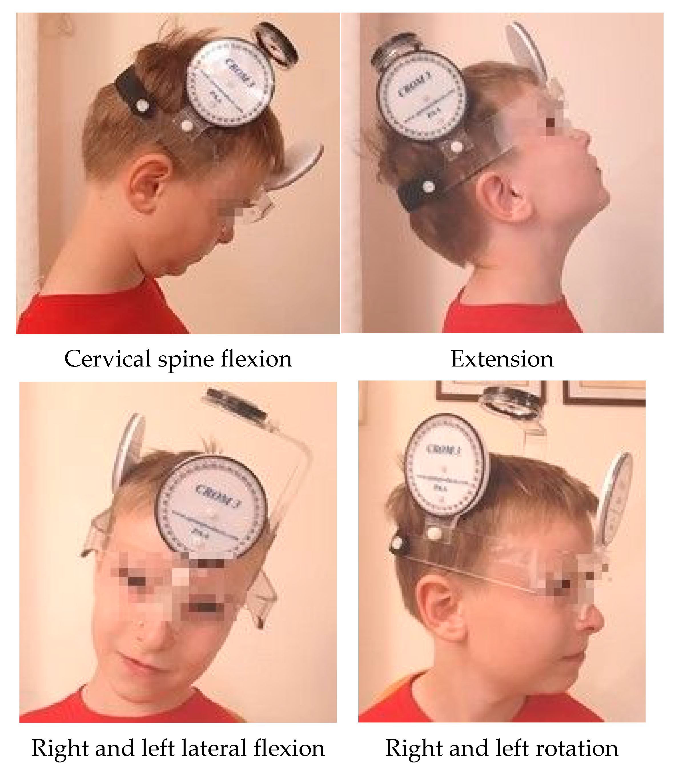
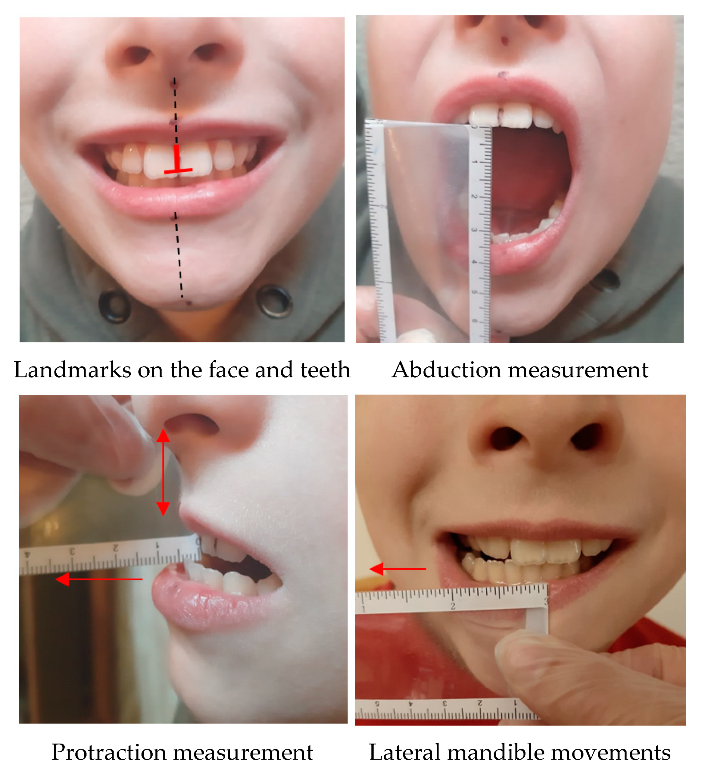
2. Materials and Methods
3. Results
4. Discussion
5. Conclusions
Author Contributions
Funding
Institutional Review Board Statement
Informed Consent Statement
Conflicts of Interest
References
- Sillence, D.O.; van Dijk, F.S. Osteogenesis Imperfecta: Clinical Diagnosis, Nomenclature and Severity Assessment. Am. J. Med. Genet. 2014, 164A, 1470–1481. [Google Scholar]
- Leotard, A.; Taytard, J.; Aouate, M.; Boule, M.; Forin, V.; Lallemant-Dudek, P. Diagnosis, follow-up and management of sleep-disordered breathing in children with osteogenesis imperfecta. Ann. Phys. Rehabil. Med. 2018, 61, 135–139. [Google Scholar] [CrossRef] [PubMed]
- Engelbert, R.H.H.; van der Graaf, Y.; van Empelen, R.; Beeme, F.A.; Helders, P.J.M. Osteogenesis Imperfecta in Childhood: Impairment and Disability. Pediatrics 1997, 99, 2. [Google Scholar] [CrossRef] [PubMed] [Green Version]
- Rusińska, A.; Jakubowska-Pietkiewicz, E.; Michałus, I.; Kurnatowska, O.; Rychłowska, E.; Beska, K.; Chlebna-Sokół, D. Zróżnicowanie objawów klinicznych wrodzonej łamliwości kości u dzieci—trudności diagnostyczne na podstawie doświadczeń własnych. Post N. Med. 2016, 29, 716–722. [Google Scholar]
- Galicka, A. Czynniki Determinujące Heterogenność Genotypową i Fenotypową Wrodzonej Łamliwości Kości; Rozprawa habilitacyjna: Białystok, Poland, 2009. [Google Scholar]
- Bregou Bourgeois, A.; Aubry-Rozier, B.; Bonafé, L.; Laurent-Applegate, L.A.; Pioletti, D.; Zambelli, P.Y. Osteogenesis imperfecta: From diagnosis and multidisciplinary treatment to future perspectives. Swiss Med. Wkly. 2016, 146, 14322. [Google Scholar]
- Sułko, J. Wrodzona Łamliwość Kości; Praca Habilitacyjna: Kraków, Poland, 2008. [Google Scholar]
- Vetter, U.; Pontz, B.; Zauner, E.; Brenner, R.E.; Spranger, J. Osteogenesis Imperfecta: A Clinical Study of the First Ten Years of Life. Calcif. Tissue Int. 1992, 50, 36–41. [Google Scholar] [CrossRef]
- Kozubska, A.; Szczepańska, J. Zaburzenia uzębienia u dzieci i młodzieży w przebiegu osteogenesis imperfecta. Nowa Stomatol. 2017, 22, 192–201. [Google Scholar] [CrossRef]
- Smoląg, D.; Kulesa-Mrowiecka, M.; Sułko, J. Evaluation of stomatognathic problems in children with osteogenesis imperfecta osteogenesis imperfecta. Preliminary study. Dev. Period Med. 2017, 21, 144–153. [Google Scholar]
- Rauch, F.; Glorieux, F.H. Osteogenesis imperfecta. Lancet 2004, 363, 1377–1385. [Google Scholar] [CrossRef]
- Sillence, D.O.; Senn, A.; Danks, D. Genetic heterogeneity in osteogenesis imperfecta. J. Med. Genet. 1979, 16, 101–116. [Google Scholar] [CrossRef] [Green Version]
- Chu, M.L.; Williams, C.J.; Pepe, G.; Hirsch, J.L.; Prockop, D.J.; Ramirez, F. Internal deletion in a collagen gene in a perinatal lethal form of osteogenesis imperfecta. Nature 1983, 304, 78–80. [Google Scholar] [CrossRef] [PubMed]
- Sułko, J.; Radło, W. OI-leczenie operacyjne deformacji kończyn dolnych u dzieci z wrodzoną łamliwością kości. Chir. Narządów Ruchu Ortop. Pol. 2005, 70, 189–193. [Google Scholar] [PubMed]
- Sułko, J.; Radło, W. OI-leczenie operacyjne kończyn górnych. Chir. Narządów Ruchu Ortop. Pol. 2005, 70, 195–199. [Google Scholar] [PubMed]
- Besio, R.; Forlino, A. Treatment options for osteogenesis imperfecta. Expert Opin. Orphan Drugs 2015, 3, 165–181. [Google Scholar] [CrossRef]
- Brizola, E.; Shapiro, J.R. Bisphosphonate Treatment of Children and Adults with Osteogenesis Imperfecta: Unanswered Questions. Calcif. Tissue Int. 2015, 97, 101–103. [Google Scholar] [CrossRef] [Green Version]
- Antoniazzi, F.; Monti, E.; Venturi, G.; Franceschi, R.; Doro, F.; Gatti, D.; Zamboni, G.; Tato, L.G.H. Growth Hormone in combination with bisphosphonate treatment in osteogenesis imperfecta. Eur. J. Endocrinol. 2010, 163, 479–487. [Google Scholar] [CrossRef] [Green Version]
- Jones, G.N.; Moschidou, D.; Abdulrazzak, H.; Kalirai, B.S.; Vanleene, M.; Osatis, S.; Shefelbine, S.J.; Horwood, N.J.; Marenzana, M.; De Coppi, P.; et al. Potential of human fetal chorionic stem cells for the treatment of osteogenesis imperfecta. Stem Cells Dev. 2014, 23, 262–276. [Google Scholar] [CrossRef] [Green Version]
- Van Brussel, M.; Takken, T.; Uiterwaal, C.S.; Pruijs, H.J.; Van Der Net, J.; Helders, P.J.; Engelbert, R.H. Physical Training in Children with Osteogenesis Imperfecta. J. Pediatr. 2008, 152, 111–117. [Google Scholar] [CrossRef]
- Brenner, R.E.; Schiller, B.; Pontz, B.F.; Lehmann, H.; Teller, W.M.; Spranger, J.; Vetter, U. Osteogenesis imperfecta in childhood and adolescence. Mon. Kinderheilkd 1993, 141, 940–945. (In German) [Google Scholar]
- Veilleux, L.-N.; Darsaklis, V.B.; Montpetit, K.; Glorieux, F.H.; Rauch, F. Muscle Function in Osteogenesis Imperfecta Type IV. Calcif. Tissue Int. 2017, 101, 362–370. [Google Scholar] [CrossRef]
- Hochschild, J. Anatomia Funkcjonalna dla Fizjoterapeutów; MedPharm Polska: Wrocław, Poland, 2018. [Google Scholar]
- Walocha, J. Anatomia Prawidłowa Człowieka; Osteologia; Wydawnictwo Uniwersytetu Jagiellońskiego: Kraków, Poland, 2013. [Google Scholar]
- Arbogast, K.B.; Gholve, P.A.; Friedman, J.E.; Maltese, M.R.; Tomasello, M.F.; Dormans, J.P. Normal Cervical Spine Range of Motion in Children 3–12 Years Old. SPINE 2007, 32, E309–E315. [Google Scholar] [CrossRef] [PubMed]
- Budelmann, K.; von Piekartz, H.; Hall, T. A normative study of cervical range of motion measures including the flexion–rotation test in asymptomatic children: Side-to-side variability and pain provocation. J. Man. Manip. Ther. 2016, 24, 185–191. [Google Scholar] [CrossRef] [PubMed] [Green Version]
- Raymond, A.; Swinkels-Meewisse, I.E.J. Normal Values for Cervical Range of Motion. SPINE 2014, 39, 362–367. [Google Scholar]
- Brizola, E.; Staub, A.L.P.; Félix, T.M. Muscle Strength, Joint Range of Motion, and Gait in Children and Adolescents with Osteogenesis Imperfecta. Pediatric Phys. Ther. 2014, 26, 245–252. [Google Scholar] [CrossRef] [PubMed]
- McKay, M.J.; Baldwin, J.N.; Ferreira, P.; Simic, M.; Vanicek, N.; Burns, J. Normative reference values for strength and flexibility of 1,000 children and adults; American Academy of Neurology. Neurology 2017, 3, 88. [Google Scholar]
- Kleinrok, M. Zaburzenia Czynnościowe Układu Ruchowego Narządu Żucia; WYD SanMedia: Warszawa, Poland, 1992. [Google Scholar]
- Czerwińska-Niezabitowska, B.; Kulesa-Mrowiecka, M. Diagnostyka i Leczenie Dysfunkcji Czaszkowo-Żuchwowych w Ujęciu Holistycznym; Medycyna Praktyczna: Krakow, Poland, 2016; pp. 46–64. [Google Scholar]
- Manfredini, D.; Arveda, N.; Guarda-Nardini, L.; Segù, M.; Collesano, V. Distribution of diagnoses in a population of patients with temporomandibular disorders. Oral Surg. Oral Med. Oral Pathol. Oral Radiol 2012, 114, 35–41. [Google Scholar] [CrossRef]
- Młynarska-Zduniak, E.; Zadurska, M.; Siemińska-Piekarczyk, B. Orthodontic problems in patients with Down syndrome from infancy to maturity based on own observations. J. Stoma 2015, 68, 703–717. [Google Scholar]
- Bendixen, K.H.; Gjørup, H.; Baad-Hansen, L.; Dahl Hald, J.; Harsløf, T.; Schmidt, M.H.; Langdahl, B.L.; Haubek, D. Temporomandibular disorders and psychosocial status in osteogenesis imperfecta—A cross-sectional study. BMC Oral Health 2018, 18, 35. [Google Scholar] [CrossRef]
- Balkefors, V.; Mattsson, E.; Pernow, Y.; Sääf, M. Functioning and quality of life in adults with mild-to-moderate osteogenesis imperfecta. Physiother Res Int. 2013, 18, 203–211. [Google Scholar] [CrossRef] [Green Version]
- Engelbert, R.H.; Gerves, W.J.M.; Breslau-Siderius, L.J.; van der Graaf, Y.; Pruijs, H.E.H.; van Doorne, J.M.; Beemer, F.A.; Helders, P.J.M. Spinal complications in osteogenesis imperfecta. Acfa Orihop. Scand. 1998, 69, 283–286. [Google Scholar] [CrossRef] [Green Version]
- Baba, T.; Shitoto, K.; Kaneko, K.; Maruyama, Y. Osteogenesis imperfecta associated with atlantoaxial rotatory fixation. Eur Orthop. Traumatol. 2011, 2, 121–125. [Google Scholar] [CrossRef]
- Germain-Lee, E.L.; Brennen, F.-S.; Stern, D.; Kantipuly, A.; Melvin, P.; Terkowitz, M.S.; Shapiro, J.R. Cross-sectional and longitudinal growth patterns in osteogenesis imperfecta: Implications for clinical care. Pediatr Res. 2016, 79, 489–495. [Google Scholar] [CrossRef] [PubMed]
- Zambrano, M.B.; Brizola, E.S.; Refosco, L.; Giugliani, R.; Félix, T.M. Anthropometry, nutritional status, and dietary intake in pediatric patients with osteogenesis imperfecta. J. Am. Coll. Nutr. 2014, 33, 18–25. [Google Scholar] [CrossRef] [PubMed]
- Kruger, K.M.; Caudill, A.; Rodriguez Celin, M.; Nagamani, S.C.S.; Shapiro, J.R.; Steiner, R.D.; Bober, M.B.; Hart, T.; Cuthbertson, D.; Krischer, J.; et al. Mobility in osteogenesis imperfecta: A multicenter North American study. Genet. Med. 2019, 21, 2311–2318. [Google Scholar] [CrossRef]


| Variables | Total | Female Children | Male Children |
|---|---|---|---|
| Number of participants [n] | 34 | 15 (44%) | 19 (56%) |
| Age [y ± SD] | 9.1 ± 3.8 | 8.9 ± 3.7 | 9.3 ± 3.9 |
| Age 3–5 years [n] | 5 | 1 (20%) | 4 (80%) |
| Age 6–8 years [n] | 12 | 8 (67%) | 4 (33%) |
| Age 10–12 years [n] | 7 | 2 (29%) | 5 (71%) |
| Age 13–15 years [n] | 10 | 4 (40%) | 6 (60%) |
| Type I OI [n] | 17 | 6 (35%) | 11 (65%) |
| Type III OI [n] | 12 | 7 (53%) | 5 (47%) |
| Type IV OI [n] | 4 | 2 (50%) | 2 (50%) |
| Regular physiotherapy [n] | 18 | 9 (50%) | 9 (50%) |
| Dentinogenesis imperfecta [n] | 16 | 8 (50%) | 8 (50%) |
| Deviation of the mandible to the right [n] | 16 | 8 (50%) | 8 (50%) |
| Deviation of the mandible to the left [n] | 11 | 5 (45%) | 6 (55%) |
| Crackles and Crepitations | Cervical Spine Rotation to the Right | |
|---|---|---|
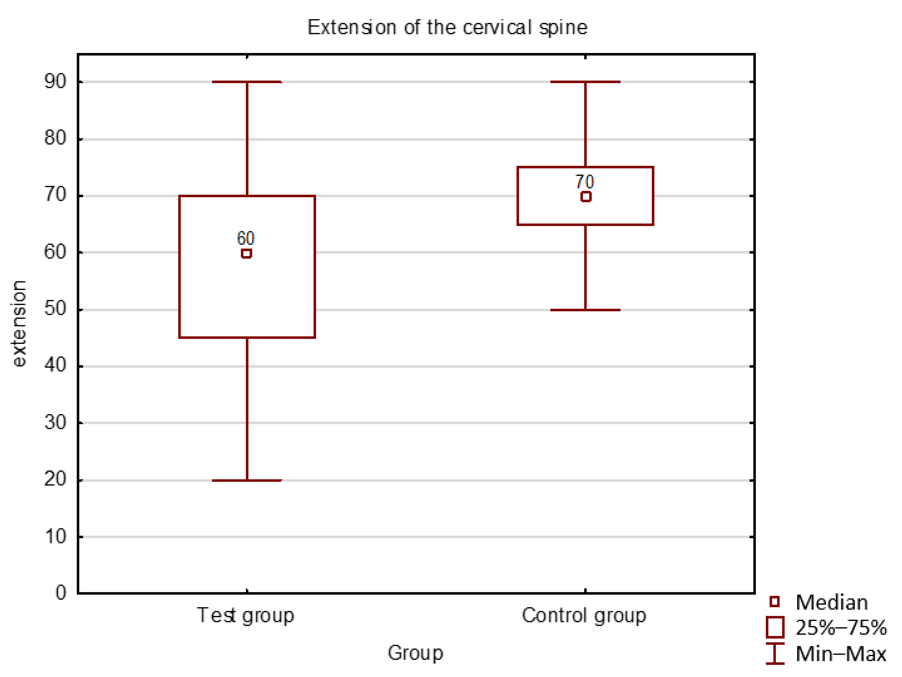
| Study group | 24% | 62 |
| Control group | 11% | 80 |
| Age | Jaw Opening [mm] ± SD | Functional Norm [mm] | Protrusion [mm] ± SD | Translation to the Right [mm] ± SD | Translation to the Left [mm] ± SD | |
|---|---|---|---|---|---|---|
| Test group | 3–5 | 37.2 ± 7 | 35.2 | 5.8 ± 2 | 5.9 ± 2 | 7.3 ± 2 |
| 6–8 | 39.5 ± 4 | 36.0 | 5 ± 2 | 7.8 ± 2 | 8.3 ± 2 | |
| 9–12 | 36.3 ± 9 | 40.1 | 6.3 ± 3 | 6.3 ± 2 | 5.6 ± 4 | |
| 13–15 | 40.8 ± 7 | 43.3 | 5 ± 2 | 7.5 ± 2 | 7 ± 2 | |
| Control group | 3–5 | 34.0 ± 2 | 35.0 | 5.3 ± 1 | 7.7 ± 2 | 7.0 ± 0 |
| 6–8 | 39.3 ± 9 | 39.0 | 6.0 ± 5 | 7.3 ± 3 | 7.3 ± 4 | |
| 9–12 | 41.8 ± 4 | 44.1 | 5.4 ± 2 | 8.3 ± 1 | 8.8 ± 3 | |
| 13–15 | 44.0 ± 5 | 46.0 | 4.2 ± 3 | 8.4 ± 4 | 9.6 ± 5 |
| Variables | Jaw Opening | Protrusion | Translation to the Right | Translation to the Left |
|---|---|---|---|---|
| flexion | 0.13 | −0.01 | 0.12 | 0.25 |
| extension | 0.37 | 0.13 | 0.13 | 0.10 |
| flexion to the right | 0.35 | 0.11 | −0.05 | 0.36 |
| flexion to the left | 0.25 | 0.04 | 0.11 | 0.35 |
| rotation to the right | 0.33 | −0.04 | 0.08 | 0.03 |
| rotation to the left | 0.31 | −0.09 | −0.02 | 0.14 |
Publisher’s Note: MDPI stays neutral with regard to jurisdictional claims in published maps and institutional affiliations. |
© 2021 by the authors. Licensee MDPI, Basel, Switzerland. This article is an open access article distributed under the terms and conditions of the Creative Commons Attribution (CC BY) license (http://creativecommons.org/licenses/by/4.0/).
Share and Cite
Małgorzata, K.-M.; Małgorzata, P.; Kinga, S.; Jerzy, S. Temporomandibular Joint and Cervical Spine Mobility Assessment in the Prevention of Temporomandibular Disorders in Children with Osteogenesis Imperfecta: A Pilot Study. Int. J. Environ. Res. Public Health 2021, 18, 1076. https://0-doi-org.brum.beds.ac.uk/10.3390/ijerph18031076
Małgorzata K-M, Małgorzata P, Kinga S, Jerzy S. Temporomandibular Joint and Cervical Spine Mobility Assessment in the Prevention of Temporomandibular Disorders in Children with Osteogenesis Imperfecta: A Pilot Study. International Journal of Environmental Research and Public Health. 2021; 18(3):1076. https://0-doi-org.brum.beds.ac.uk/10.3390/ijerph18031076
Chicago/Turabian StyleMałgorzata, Kulesa-Mrowiecka, Pihut Małgorzata, Słojewska Kinga, and Sułko Jerzy. 2021. "Temporomandibular Joint and Cervical Spine Mobility Assessment in the Prevention of Temporomandibular Disorders in Children with Osteogenesis Imperfecta: A Pilot Study" International Journal of Environmental Research and Public Health 18, no. 3: 1076. https://0-doi-org.brum.beds.ac.uk/10.3390/ijerph18031076