
Nanoporous Titanium Enriched with Calcium and Phosphorus Promotes Human Oral Osteoblast Bioactivity

 , ,
, ,  ,
, 
Abstract
:1. Introduction
2. Materials and Methods
2.1. Dental Implant Discs
- -
- Machined implant surface with no roughening or CaP treatments applied in cold-drawn titanium GR4;
- -
- Osteopore implant surface was treated by double acid-etching in order to create surface structures and roughness at the micro level. This treatment was followed by washing and final decontamination by plasma;
- -
- Nanopore implant surface was treated by double acid-etching in order to create surface structures and roughness at the micro level and were further enriched in calcium and phosphorus (CaP treatment) by process of inorganic salts in aqueous solution, to superimpose a nanotopographic complexity. This treatment was followed by washing and final decontamination by plasma.
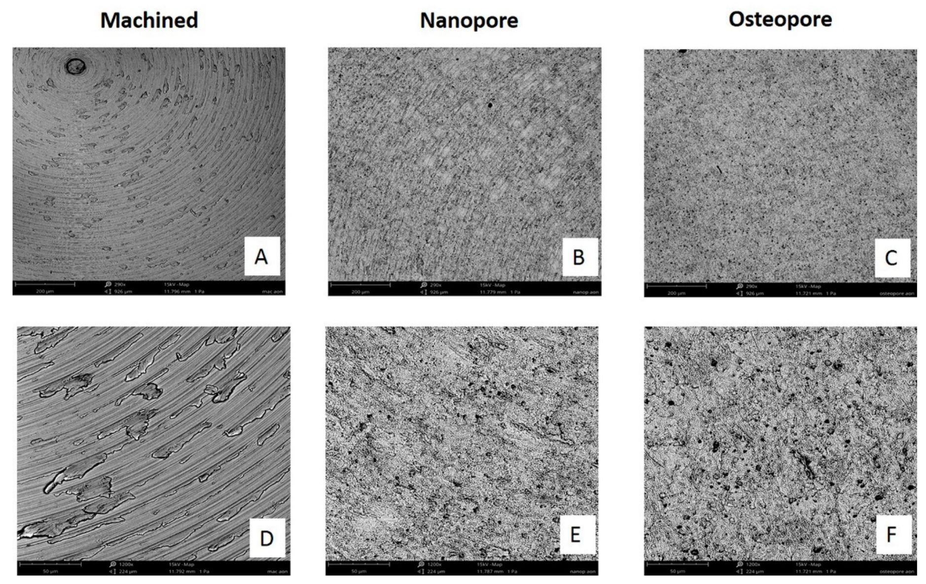
2.2. Scanning Electron Microscopy (SEM) Analysis
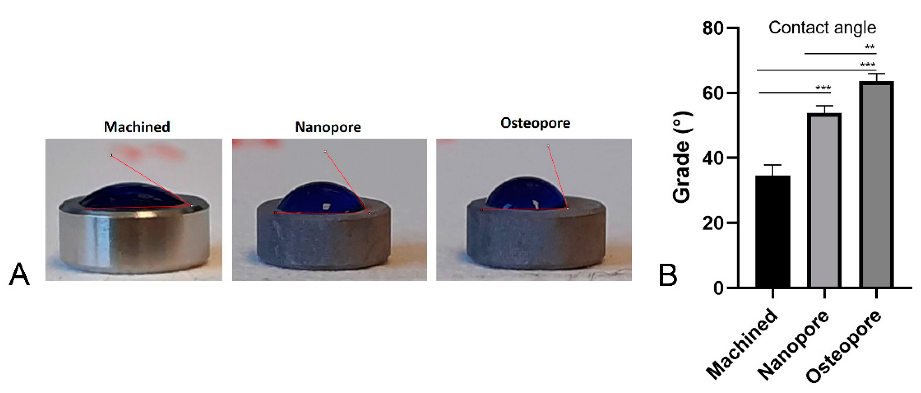
2.3. Water Contact Angle Measurement
2.4. Oral Osteoblasts Culture
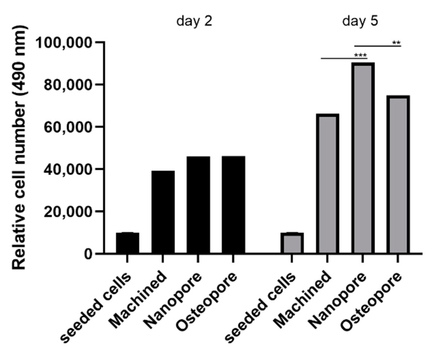
2.5. Proliferation Study
2.6. Multiphoton Microscopy
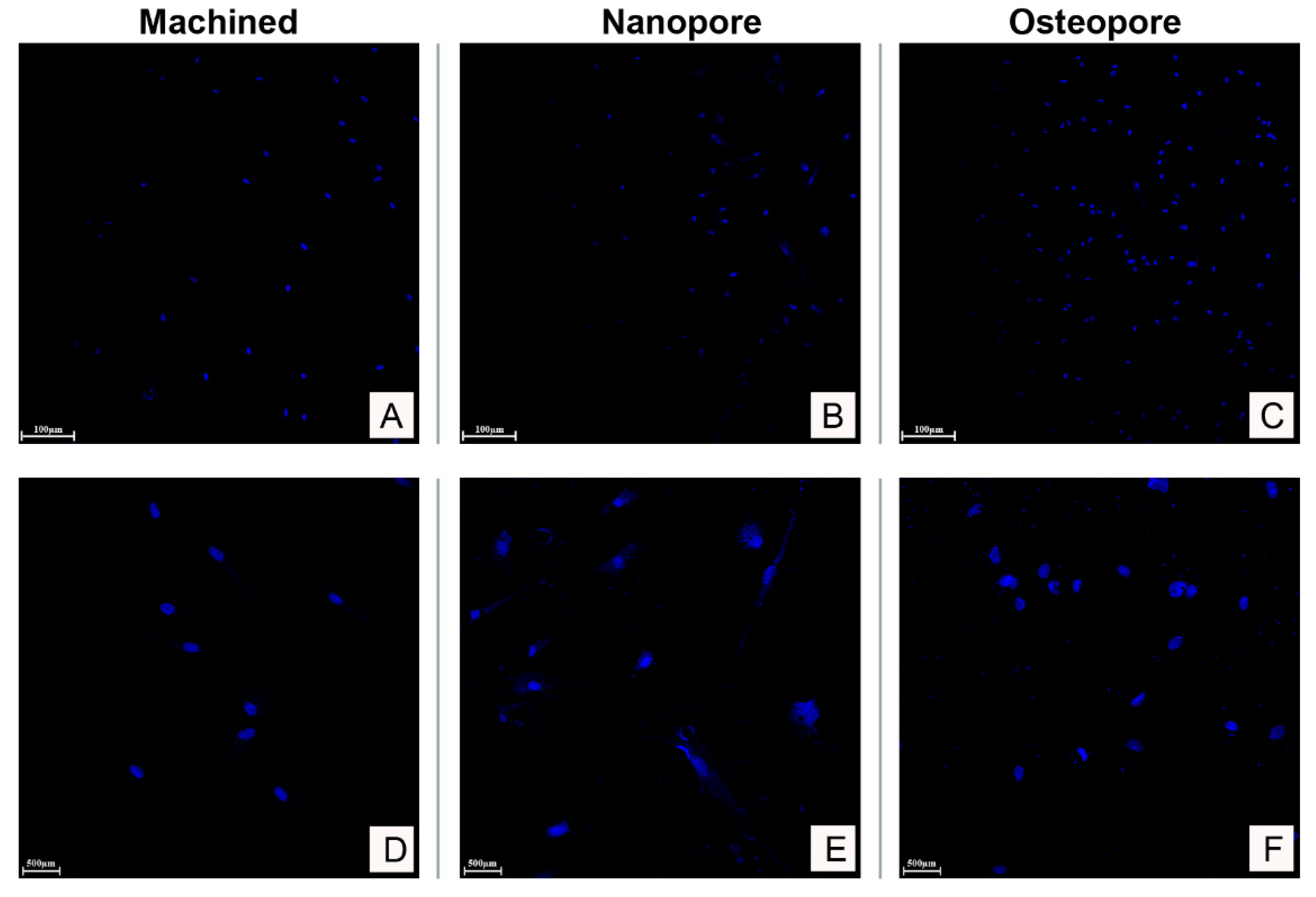
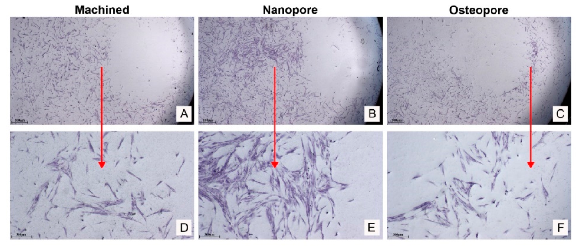
2.7. Cell Staining
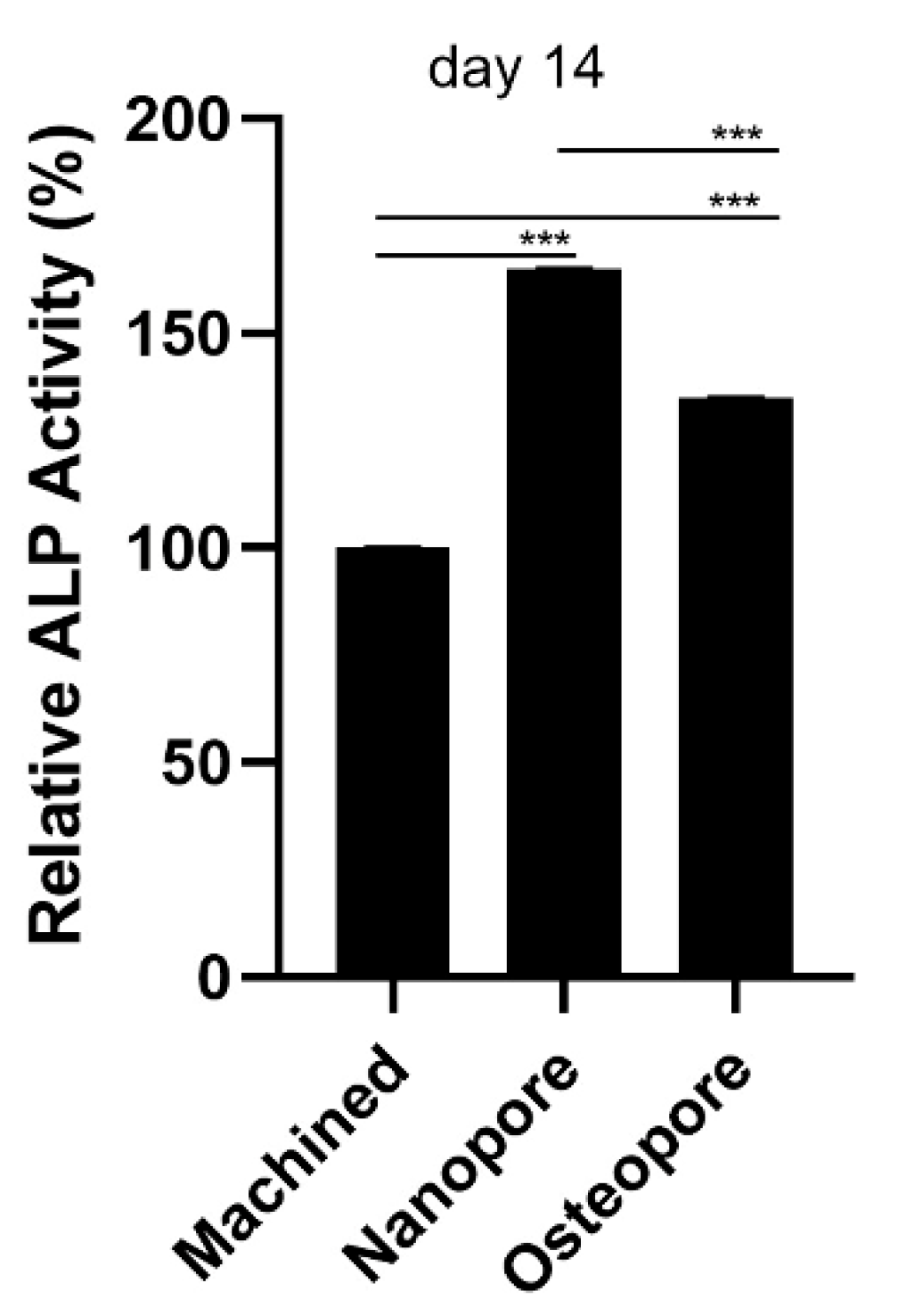
2.8. ALP Assay
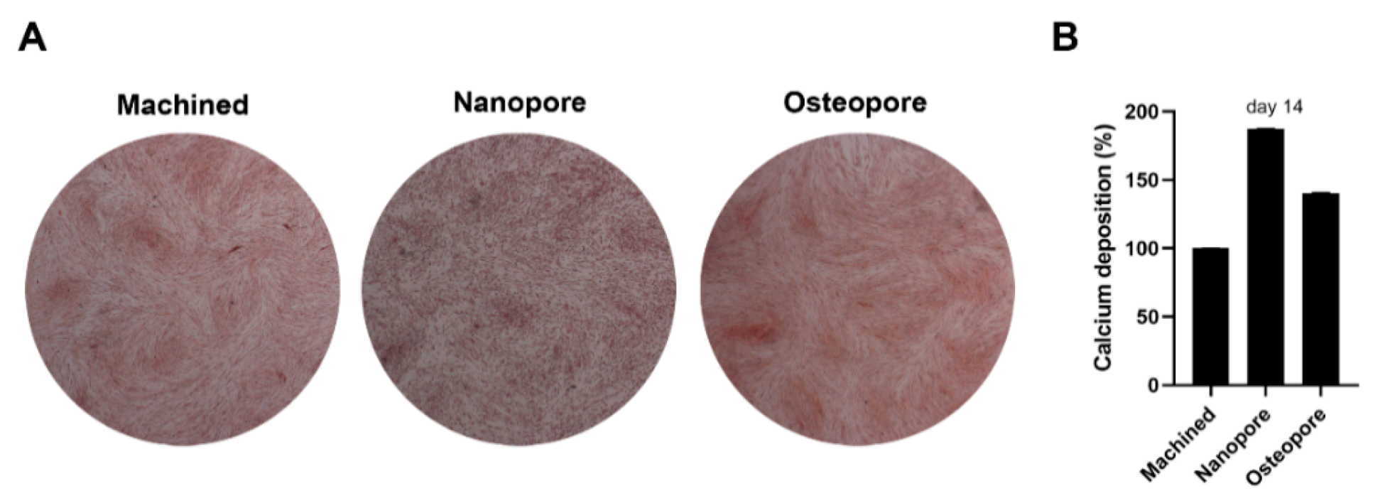
2.9. Alizarin Red Staining
2.10. Calcium Deposition
2.11. Statistical Analysis
3. Results
3.1. Surface Characterization
3.2. Osteoblasts Proliferation
3.3. Adhesion of Osteoblast on Titanium Discs
3.4. Interaction between Osteoblasts and the Tested Surfaces
3.5. Alkaline Phosphatase Activity
3.6. Calcium Deposition
4. Discussion
5. Conclusions
Author Contributions
Funding
Institutional Review Board Statement
Informed Consent Statement
Data Availability Statement
Conflicts of Interest
Abbreviations
| hOBs | human oral osteoblasts |
| GR4 | grade 4 |
| CaP | calcium and phosphorus |
| SEM | scanning electron microscopy |
| MTS | (4,5-dimethylthiazolyl-2)-2,5-diphenyltetrazolium bromide |
| Ti | titanium |
| ALP | alkaline phosphatase |
| DAE | double acid-etching |
| BIC | bone-to-implant contact |
| FBS | fetal bovine serum |
| DMEM | Dulbecco’s Modified Eagle Medium |
| OD | optical density |
| DAPI | 4′,6-diamidino-2-phenylindole |
| pNPP | p-nitrophenyl phosphate |
| ARS | alizarin red staining |
| PBS | phosphate-buffered saline |
| CPC | cetylpyridinium chloride |
| SD | standard deviation |
| cpTi | commercially pure titanium |
References
- Bai, L.; Chen, P.; Zhao, Y.; Hang, R.; Yao, X.; Tang, B.; Liu, C.; Xiao, Y.; Hang, R. A Micro/Nano-Biomimetic Coating on Titanium Orchestrates Osteo/Angio-Genesis and Osteoimmunomodulation for Advanced Osseointegration. Biomaterials 2021, 278, 121162. [Google Scholar] [CrossRef] [PubMed]
- Shibli, J.A.; Pires, J.T.; Piattelli, A.; Iezzi, G.; Mangano, C.; Mangano, F.; de Souza, S.L.S.; Gehrke, S.A.; Wang, H.-L.; Dohan Ehrenfest, D.M. Impact of Different Implant Surfaces Topographies on Peri-Implant Tissues: An Update of Current Available Data on Dental Implants Retrieved from Human Jaws. Curr. Pharm. Biotechnol. 2017, 18, 76–84. [Google Scholar] [CrossRef] [PubMed]
- Petrini, M.; Giuliani, A.; di Campli, E.; di Lodovico, S.; Iezzi, G.; Piattelli, A.; D’ercole, S. The Bacterial Anti-Adhesive Activity of Double-Etched Titanium (DAE) as a Dental Implant Surface. Int. J. Mol. Sci. 2020, 21, 8315. [Google Scholar] [CrossRef] [PubMed]
- Petrini, M.; Pierfelice, T.V.; D’amico, E.; di Pietro, N.; Pandolfi, A.; D’arcangelo, C.; de Angelis, F.; Mandatori, D.; Schiavone, V.; Piattelli, A.; et al. Influence of Nano, Micro, and Macro Topography of Dental Implant Surfaces on Human Gingival Fibroblasts. Int. J. Mol. Sci. 2021, 22, 9871. [Google Scholar] [CrossRef]
- Chouirfa, H.; Bouloussa, H.; Migonney, V.; Falentin-Daudré, C. Review of Titanium Surface Modification Techniques and Coatings for Antibacterial Applications. Acta Biomater. 2019, 83, 37–54. [Google Scholar] [CrossRef]
- Scarano, A.; Piattelli, A.; Quaranta, A.; Lorusso, F. Bone Response to Two Dental Implants with Different Sandblasted/Acid-Etched Implant Surfaces: A Histological and Histomorphometrical Study in Rabbits. Biomed. Res. Int. 2017, 2017, 8724951. [Google Scholar] [CrossRef] [Green Version]
- Ma, L.; Li, G.; Lei, J.; Song, Y.; Feng, X.; Tan, L.; Luo, R.; Liao, Z.; Shi, Y.; Zhang, W.; et al. Nanotopography Sequentially Mediates Human Mesenchymal Stem Cell-Derived Small Extracellular Vesicles for Enhancing Osteogenesis. ACS Nano 2021, 16, 415–430. [Google Scholar] [CrossRef]
- Dabare, P.R.L.; Bachhuka, A.; Visalakshan, R.M.; Shirazi, H.S.; Ostriko, K.; Smith, L.E.; Vasilev, K. Mechanistic Insight in Surface Nanotopography Driven Cellular Migration. ACS Biomater. Sci. Eng. 2021, 7, 4921–4932. [Google Scholar] [CrossRef]
- Lopes, H.B.; Freitas, G.P.; Fantacini, D.M.C.; Picanço-Castro, V.; Covas, D.T.; Rosa, A.L.; Beloti, M.M. Titanium with Nanotopography Induces Osteoblast Differentiation through Regulation of Integrin AV. J. Cell Biochem. 2019, 120, 16723–16732. [Google Scholar] [CrossRef]
- Le Guéhennec, L.; Soueidan, A.; Layrolle, P.; Amouriq, Y. Surface Treatments of Titanium Dental Implants for Rapid Osseointegration. Dent. Mater. 2007, 23, 844–854. [Google Scholar] [CrossRef]
- Mendonça, G.; Mendonça, D.B.S.; Aragão, F.J.L.; Cooper, L.F. Advancing Dental Implant Surface Technology--from Micron- to Nanotopography. Biomaterials 2008, 29, 3822–3835. [Google Scholar] [CrossRef] [PubMed]
- Bandyopadhyay, A.; Mitra, I.; Shivaram, A.; Dasgupta, N.; Bose, S. Direct Comparison of Additively Manufactured Porous Titanium and Tantalum Implants towards in Vivo Osseointegration. Addit. Manuf. 2019, 28, 259–266. [Google Scholar] [CrossRef] [PubMed]
- Souza, J.C.M.; Sordi, M.B.; Kanazawa, M.; Ravindran, S.; Henriques, B.; Silva, F.S.; Aparicio, C.; Cooper, L.F. Nano-Scale Modification of Titanium Implant Surfaces to Enhance Osseointegration. Acta Biomater. 2019, 94, 112–131. [Google Scholar] [CrossRef] [PubMed]
- Komori, T. Regulation of Proliferation, Differentiation and Functions of Osteoblasts by Runx2. Int. J. Mol. Sci. 2019, 20, 1694. [Google Scholar] [CrossRef] [PubMed] [Green Version]
- Coelho, P.G.; Jimbo, R.; Tovar, N.; Bonfante, E.A. Osseointegration: Hierarchical Designing Encompassing the Macrometer, Micrometer, and Nanometer Length Scales. Dent. Mater. 2015, 31, 37–52. [Google Scholar] [CrossRef] [PubMed]
- D’Ercole, S.; Cellini, L.; Pilato, S.; di Lodovico, S.; Iezzi, G.; Piattelli, A.; Petrini, M. Material Characterization and Streptococcus Oralis Adhesion on Polyetheretherketone (PEEK) and Titanium Surfaces Used in Implantology. J. Mater. Sci. Mater. Med. 2020, 31, 84. [Google Scholar] [CrossRef]
- Novaes, A.B.; de Souza, S.L.S.; de Barros, R.R.M.; Pereira, K.K.Y.; Iezzi, G.; Piattelli, A. Influence of Implant Surfaces on Osseointegration. Braz. Dent. J. 2010, 21, 471–481. [Google Scholar] [CrossRef]
- Mavrogenis, A.F.; Dimitriou, R.; Parvizi, J.; Babis, G.C. Biology of Implant Osseointegration. J. Musculoskelet. Neuronal Interact. 2009, 9, 61–71. [Google Scholar]
- Cai, S.; Wu, C.; Yang, W.; Liang, W.; Yu, H.; Liu, L. Recent Advance in Surface Modification for Regulating Cell Adhesion and Behaviors. Nanotechnol. Rev. 2020, 9, 971–989. [Google Scholar] [CrossRef]
- Rupp, F.; Scheideler, L.; Rehbein, D.; Axmann, D.; Geis-Gerstorfer, J. Roughness Induced Dynamic Changes of Wettability of Acid Etched Titanium Implant Modifications. Biomaterials 2004, 25, 1429–1438. [Google Scholar] [CrossRef]
- Becker, M.; Schmied, F.; Kadem, L.F.; Freitag-Wolf, S.; Naujokat, H.; Mehl, C.; Kern, M.; Harder, S. Single-Cell Adhesion of Human Osteoblasts on Plasma-Conditioned Titanium Implant Surfaces in Vitro. J. Mech. Behav. Biomed. Mater. 2020, 109, 103841. [Google Scholar] [CrossRef] [PubMed]
- Mendes, V.C.; Moineddin, R.; Davies, J.E. Discrete Calcium Phosphate Nanocrystalline Deposition Enhances Osteoconduction on Titanium-Based Implant Surfaces. J. Biomed. Mater. Res. A 2009, 90, 577–585. [Google Scholar] [CrossRef] [PubMed]
- Rodriguez y Baena, R.; Arciola, C.R.; Selan, L.; Battaglia, R.; Imbriani, M.; Rizzo, S.; Visai, L. Evaluation of Bacterial Adhesion on Machined Titanium, Osseotite® and Nanotite® Discs. Int. J. Artif. Organs 2012, 35, 754–761. [Google Scholar] [CrossRef] [PubMed]


Publisher’s Note: MDPI stays neutral with regard to jurisdictional claims in published maps and institutional affiliations. |
© 2022 by the authors. Licensee MDPI, Basel, Switzerland. This article is an open access article distributed under the terms and conditions of the Creative Commons Attribution (CC BY) license (https://creativecommons.org/licenses/by/4.0/).
Share and Cite
Pierfelice, T.V.; D’Amico, E.; Iezzi, G.; Piattelli, A.; Di Pietro, N.; D’Arcangelo, C.; Comuzzi, L.; Petrini, M. Nanoporous Titanium Enriched with Calcium and Phosphorus Promotes Human Oral Osteoblast Bioactivity. Int. J. Environ. Res. Public Health 2022, 19, 6212. https://0-doi-org.brum.beds.ac.uk/10.3390/ijerph19106212
Pierfelice TV, D’Amico E, Iezzi G, Piattelli A, Di Pietro N, D’Arcangelo C, Comuzzi L, Petrini M. Nanoporous Titanium Enriched with Calcium and Phosphorus Promotes Human Oral Osteoblast Bioactivity. International Journal of Environmental Research and Public Health. 2022; 19(10):6212. https://0-doi-org.brum.beds.ac.uk/10.3390/ijerph19106212
Chicago/Turabian StylePierfelice, Tania Vanessa, Emira D’Amico, Giovanna Iezzi, Adriano Piattelli, Natalia Di Pietro, Camillo D’Arcangelo, Luca Comuzzi, and Morena Petrini. 2022. "Nanoporous Titanium Enriched with Calcium and Phosphorus Promotes Human Oral Osteoblast Bioactivity" International Journal of Environmental Research and Public Health 19, no. 10: 6212. https://0-doi-org.brum.beds.ac.uk/10.3390/ijerph19106212