Article

First Report of an Asymptomatic Leishmania (Viannia) shawi Infection Using a Nasal Swab in Amazon, Brazil

by
Luciana P. Oliveira
1,*,
Luciana C. S. Nascimento
1,
Fabiola S. Santos
1,
Jaqueline L. C. Takamatsu
2,
Luiz R. P. Sanchez
2,
Walter S. Santos
1 and
Lourdes M. Garcez
1,3
1
Ministério da Saúde, Secretaria de Vigilância em Saúde, Instituto Evandro Chagas, Ananindeua 67015-120, PA, Brazil
2
Hospital Municipal de Tomé Açu, Tomé-Açu 68680-000, PA, Brazil
3
Departamento de Patologia, Universidade do Estado do Pará, Belém 66050-540, PA, Brazil
*
Author to whom correspondence should be addressed.
Int. J. Environ. Res. Public Health 2022, 19(10), 6346; https://0-doi-org.brum.beds.ac.uk/10.3390/ijerph19106346
Submission received: 17 February 2022 / Revised: 30 March 2022 / Accepted: 20 April 2022 / Published: 23 May 2022
(This article belongs to the Section Infectious Disease Epidemiology)

Abstract

:
The state of Pará has recorded seven Leishmania species that cause tegumentary leishmaniasis (TL). Leishmania species induce distinct immunological responses from the host and exhibit resistance to Glucantime, the first-line drug treatment for TL in Brazil. Objective: Identify the etiology of TL in an Amazonian city in the state of Pará. Material and methods: Eleven patients with TL were recruited and nasal swabs, lesion swabs, and skin fragments samples were collected. In the control group (n = 6), only the nasal swabs were collected. Polymerase Chain Reaction (PCR) amplification of the gene region hsp70-234 was performed using the extracted DNA from the samples, from which nine patients with TL and five in the control group were positive. Products were sequenced, mounted in CAP3 software, aligned using MAFFT v.7.221, edited in Geneious software v.8.1.7, and compared and aligned with sequences available in GenBank using the BLAST tool. Results: For patients with TL, six molecular diagnosis at the species level (L. (Viannia) braziliensis (n = 5/9), L. (Viannia) shawi (n = 1/9)) and three at the genus level (Leishmania sp. (n = 3/9)) were obtained. In the control group, four individuals were infected with Leishmania sp. (n = 4/5) and L. (V.) shawi (n = 1/5). Conclusion: This is the first report of L. (V.) shawi infection in the mucosal secretion of a healthy person in Brazil. Moreover, genetic variants were identified in the haplotypes of L. (V.) braziliensis in the gene sequence hsp70-234.

1. Introduction

In the Brazilian Amazon, several species of Leishmania cause TL. The state of Pará, the second-largest extension in this region, is the only state in Brazil to record seven dermotropic species of Leishmania pathogenic to humans, presenting variable levels of pathogenicity and drug resistance [1,2].
Lesional pleomorphism in patients with TL results from a balance in the parasite–host relationship. Leishmania species induce different immunological responses in the host. Three important species are associated with the severe evolution of the disease: Leishmania (Viannia) braziliensis, Leishmania (Viannia) guyanensis, and Leishmania (Leishmania) amazonensis [3,4].
Some Leishmania species are more prone to hematogenous dissemination. L. (V.) braziliensis causes more frequently disseminated conditions, being the main species causing mucosal leishmaniasis [4]. L. (V.) braziliensis can be detected in the nasal mucosa of both sick and healthy people living in areas with endemic TL [5]. L. (V.) guyanensis, which is predominant in certain endemic areas of northern Brazil, notably in the states of Amazonas and Amapá [6], can also cause mucosal lesions [7] and is highly resistant to Glucantime, the first-line drug treatment for TL in Brazil [2]. Meanwhile, L. (L.) amazonensis can induce severely disseminated disease and anergic diffuse leishmaniasis [8]. Other pathogenic species, including Leishmania (Viannia) lainsoni, Leishmannia (Viannia) shawi, L. (Viannia) naiffi, and L. (Viannia) lindenbergi, do not usually cause complications. Leishmania (V.) lainsoni and L. (V.) naiffi usually present as a single lesion. L. (V.) lindenbergi has localized cutaneous lesions. No case of the mucocutaneous disease has been reported in any of the species [9].
L. (V.) shawi was isolated for the first time from sloths and monkeys from the Serra dos Carajás, Pará, Brazil, which is one of the etiological agents of TL in Brazil, and is associated with the cutaneous form of TL, usually observed as a single lesion without mucous or other complications [9,10]. L. (V.) shawi has been identified in sandflies, Lutzomyia (Nyssomyia) whitmani [11], and Lutzomyia gomezi [12]. There are reports of L. (V.) shawi in the Amazon region identified in TL carriers [2,13].
The molecular characterization of Leishmania sp. that causes TL in the Amazon is important not only to define a prognosis for the patient but also to guide the therapeutic approach. In addition, it is essential to evaluate the distribution of Leishmania sp. and the occurrence of variants, and alert surveillance services to support transmission prevention [6,14]. The great challenge in the Amazon is to provide access and resources of scientific advances in communities to improve molecular diagnostic techniques [15]. Molecular methods have received increasing attention due to the possibility of distinguishing species in material obtained directly from the lesion. The enzymatic techniques that are effective for species distinction depend on the isolation of Leishmania, which makes their use in routine diagnostics impossible. The PCR in its various versions and with different markers are able to discriminate Leishmania species, but the sequencing of products with certain markers is what confirms the species distinction. Among the most used markers in PCR are G6PhD, ITS1, and Hsp70-234 [16]. In the Brazilian Amazon region, it obtained excellent results using the marker Hsp70-234 [6,17]. The study aimed to identify the etiology of TL in an Amazonian city in the state of Pará. Despite the difficulties in performing the study during the COVID-19 pandemic, molecular analysis of several clinical samples revealed human infection by L. (V.) shawi in nasal swabs for the first time. Moreover, we discuss the importance of this finding on the epidemiological surveillance of TL.

2. Material and Method
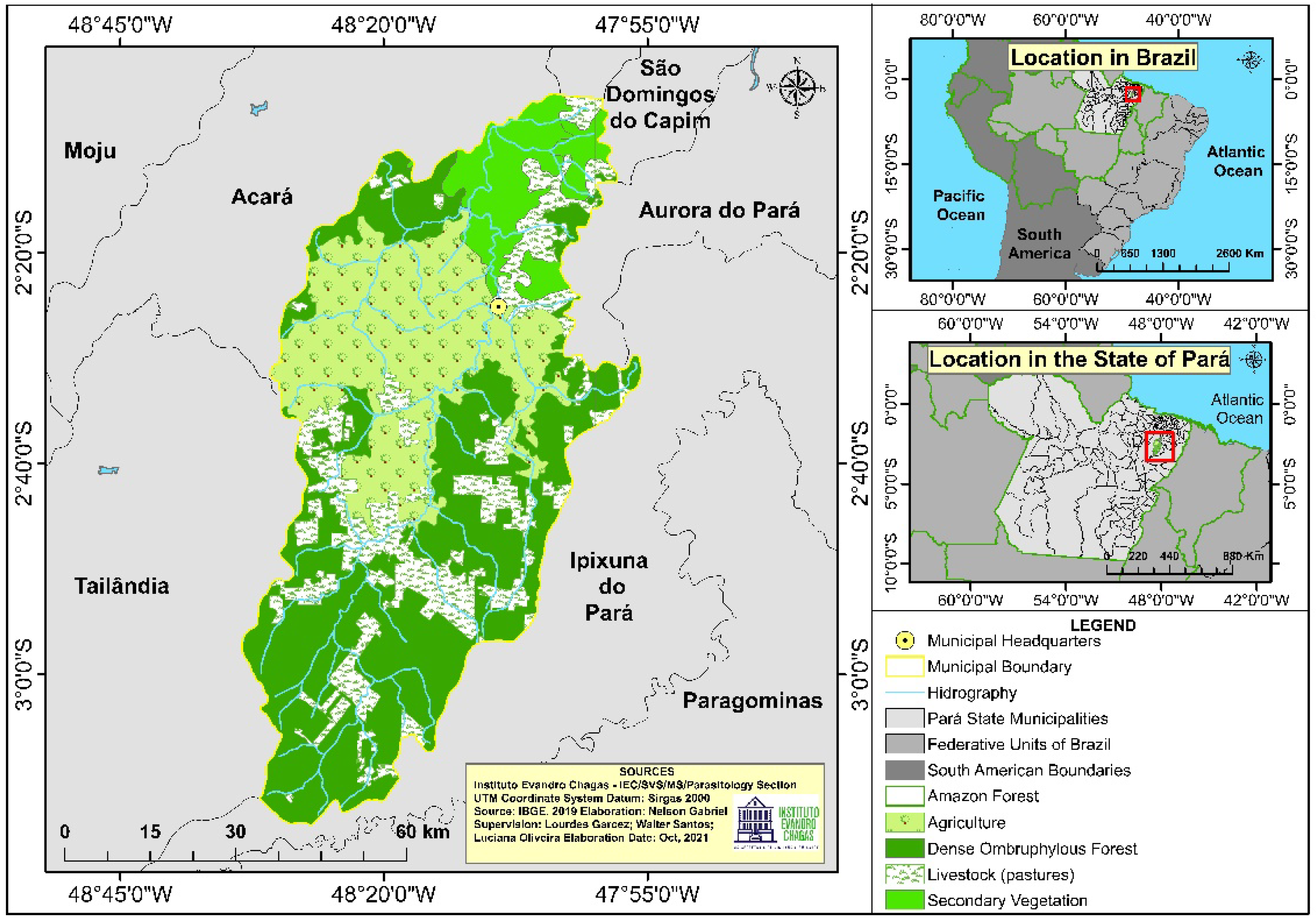
2.1. Study Site

The research was conducted at the Municipal Hospital of Tomé-Açu. The city of Tomé-Açu is located in the northeastern region of the state of Pará, Brazilian Amazon, and is an important agricultural and mining center in the state (Figure 1) [18,19]. In the municipality of Tomé-Açu there are two rainy seasons a year, one from December to May, representing 80% of the rainfall, and another from June to November. The locality features the following general characteristics: average annual temperature of 26 °C and rainfall average of 2300 mm/year [19].

2.2. Participant Recruitment

Participants were recruited in two stages: the first stage from October 2019 to February 2020, and the second from August 2020 to October 2020. The stages chosen were related to the increase in cases during the rainy season, and also to changes in health service due to the established pandemic.
Men and women over 18 years of age with a confirmed diagnosis of TL and controls were included in the study. The controls were individuals without injury, companions of the patients with TL who agreed to participate in the research, or those who live with the patients.

2.3. Participants and Sample Types

For patients with TL (n = 11), nasal swab, lesion swab, and skin fragment samples were collected. In the control group (n = 6), only nasal swabs were collected. All participants gave their consent for inclusion in the study. The study was conducted according to the Declaration of Helsinki, and the protocol was approved by the Research Ethics Committee of the Instituto Evandro Chagas (Opinion number: 3.601.679).

2.4. Sample Collection

Sample collection was performed after site cleaning and antisepsis, in the following order: (1) nasal swab, (2) lesion swab, and (3) skin biopsy. For the nasal swab, the absorbent segment of the swab was inserted into the anterior third of the nose and rotated clockwise (360°) over the nasal mucosa. The same procedure was performed in both teins with the same swab. For the lesion swab, the absorbent segment was pressed parallel to the edge of the lesion and rotated on its surface clockwise [20,21]. Meanwhile, the skin sample was obtained using a disposable punch (5 mm) after local anesthesia (2% lidocaine). This was performed on the infiltrated and eysthematous edge of the lesion [14].

2.5. Sample Storage

The end rod of each swab containing the sample was cut with the aid of sterile scissors and placed in a 1.5 mL microtube containing 500 μL of NET (NaCl 0.15 mM; EDTA 50 mM; Tris-HCl 0.1 M, pH 7.5). Skin samples were also preserved in NET. All tubes containing NET samples were stored at 4–8 °C.

2.6. DNA Extraction

DNA extraction from the samples was performed using the Wizard® Genomic DNA Purification Kit (Promega, Madison, WI, USA) according to the manufacturer’s recommendations, with a final volume of 25 μL.

2.7. Polymerase Chain Reaction (PCR) for Target Region Hsp70-234

The target region hsp70-234 (234 bp) was amplified using conventional PCR. Briefly, the PCR mix, with a final volume of 50 μL, contained 0.03 U/μL Taq DNA polymerase, 1.5 mM MgCl2, 0.25 mM of each dNTP), 1X buffer solution with KCl, 0.25% DMSO, 0.2 pMol of primers F (5′-GGACGAGATCGAGCGCATGGT-3′) and R (5′-TCCTTCGACGCCTCCTGGTTG-3′), and 3.0 μL of DNA from samples. The PCR conditions were as follows: initial denaturation at 94 °C for 5 min, followed by 32 cycles of denaturation at 94 °C for 30 s, amplification at 61 °C for 1 min, and extension at 72 °C for 1 min; and a final extension at 72 °C for 10 min. Two positive controls containing the genomic DNA of L. (L.) infantum (CEPA 6445) and L. (V.) braziliensis (CEPA 2904), and a control without DNA (white) were used. The amplified samples electrophoresed in 2% agarose in TAE buffer (Tris- acetate 0.004 M; EDTA 0.001 M, pH 8.0) containing GelRed™ (Biotium®, Fremont, CA, USA) at a concentration of 0.5 μg/mL and subsequently observed in a transilluminator for qualitative analysis [16].

2.8. Phylogenetic Sequencing and Analysis
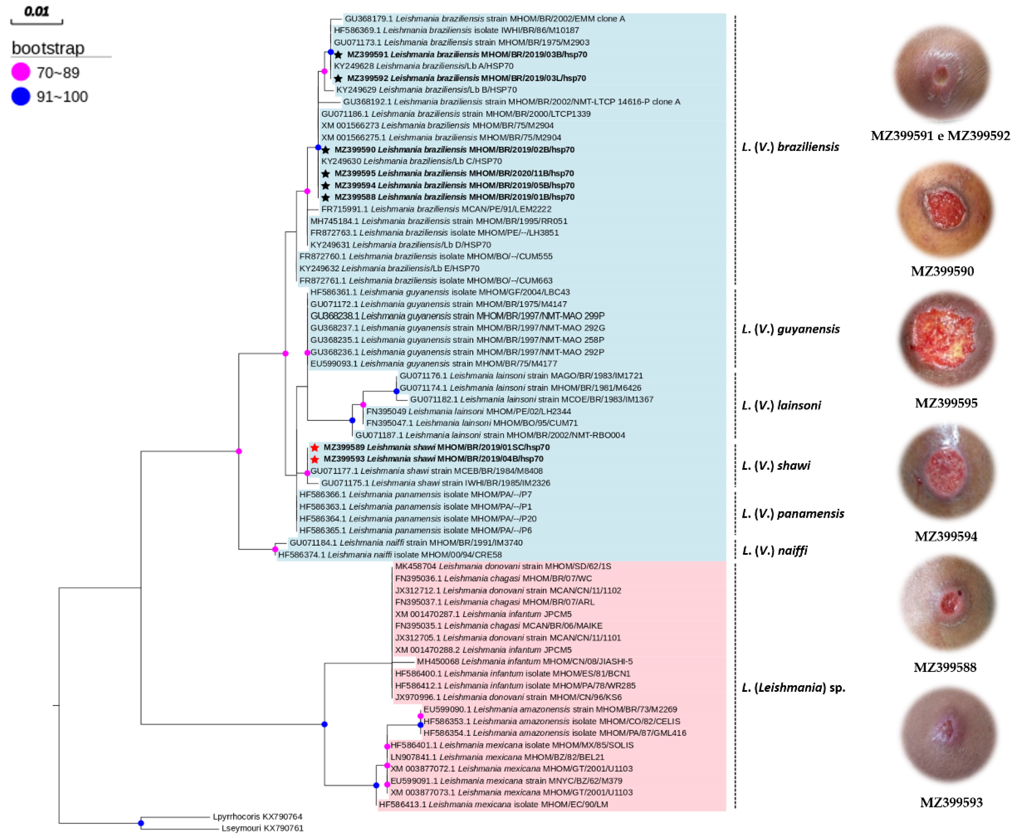
PCR-hsp70-234 products were purified with the EXOSAP-ITTM Express PCR Product Cleanup (AppliedBioystems®, University Park, IL, USA) and prepared for sequencing using the BigDye Terminator Kit v3.1 (Thermo Fisher Scientific®, Waltham, MA, USA) according to the manufacturer’s instructions. The forward and reverse sequences were obtained using the ABI3500XL automatic DNA analyzer. The sequences obtained were mounted in CAP3 software (Iowa State University, Ames, IA, USA), aligned using MAFFT v.7.221 (Universitu of Osaka, Osaka, Japan) [22], edited in Geneious v.8.1.7 software (Biomatters Ltd., Auckland, New Zealand) [23], and compared with sequences available in GenBank using the BLAST tool. Phylogenetic analyses were performed in three stages: (i) determination of the evolutionary model and likelihood analysis using the IQ-TREE v.1.3.2; (ii) phylogenetic reconstruction in IQ-TREE v.1.3.2 software (Center for Integrative Bioinformatics Vienna, Wien, Austria) using a non-parametric reliability test of 1000 replicates and bootstraps; and (iii) editing of the phylogenetic tree using FigTree v.1.4.2 software (University of Edinburgh, Edinburgh, UK) [24].

3. Results

Among the 11 patients with TL included in the study, six received molecular diagnosis at the species level (L. (V.) braziliensis (n = 5/9), L. (V.) shawi (n = 1/9)), and three at the genus level (Leishmania sp. (n = 3/9)). For two individuals, it was not possible to amplify the deoxyribonucleic acid (DNA) of the respective samples. Furthermore, the sensitivity of PCR to amplify hsp70-234 varied according to the sample collection. The highest number of positive results was obtained from the biopsy samples (n = 7/11), followed by injury swabs (n = 6/11) and nasal swabs (n = 4/11). Species identification also occurred in a greater proportion of the biopsy samples (n = 6/7). In the group of healthy individuals (n = 6), five had hsp70-234-positive PCR results in nasal secretion swabs, where L. (V.) shawi (n = 1/5) and Leishmania sp. (n = 4/5) were identified (Table 1).
A healthy participant diagnosed with L. (V.) shawi (4S) in nasal secretion was a parent that was accompanied the individuals with TL (4D), who also had the infection confirmed by L. (V.) shawi.
Most patients were men (8/11). The eight individuals were infected by L. (V.) braziliensis (n = 4/8), L. (V.) shawi (n = 1/8), and Leishmania sp. (n = 3/8). The nine patients with a molecular diagnosis of TL had ulcerated lesions and mostly localized lesions in the lower limbs (n = 7/9). The size of the lesions varied between 2 cm and 32 cm, and smaller lesions (2–6 cm in diameter) were more frequent (n = 4/9). The sociodemographic and clinical data of the nine individuals, for whom molecular diagnosis was possible, are shown in Table 2.
Eight nucleotide sequences were obtained: six were identified as L. (V.) braziliensis, with four haplotypes; and two were L. (V.) shawi, corresponding to only one haplotype (Table 3).
The sequences of L. (V.) braziliensis from the study (MZ399588, MZ399590, MZ399591, MZ399592, MZ399594, and MZ399595) clustered with GenBank sequences (KY249628 and KY249630) that also originated in Tomé-Açu (Amazon Biome), with values between 90 and 100 bootstraps. KY249629, KY249631, and KY249632 did not align with the nucleotide sequences in the present study, indicating a probable mutation in the gene region hsp70-234 of L. (V.) braziliensis circulating in the municipality. In the phylogenetic analysis, sequences MZ399591 and MZ399592, which belonged to the same patient, were grouped into a different clade from the other sequences of L. (V.) braziliensis (Figure 2).
The sequences of L. (V.) shawi identified in this study (MZ399593 and MZ399589) were grouped with nucleotide sequences of the same locality (GU071177.1 and GU071175.1) in a clade with values between 70 and 89 bootstraps, proving the discrimination of the species.

4. Discussion

A higher amplification rate of the gene region hsp70-234 was observed in DNA samples from skin biopsy, which also favored the discrimination of Leishmania species using sequence analysis. Swab is a collection method considered useful for public health because it is painless and easy to perform and can be performed by trained professionals of the health surveillance team and not necessarily physicians. Since it is not invasive, swab sampling offers more comfort and less risks, and is important for health surveillance in places with structural difficulties [21,25,26,27,28]. Moreover, the efficacy of swab compared to biopsy can be used for the discrimination of Leishmania species, and swab could be indicated as an aid in health services, in support of the diagnosis of the disease, especially when the parasitological examination of the exudate obtained through scarification of the lesion (blade-colored smear) is negative [29].
In this study, Leishmania DNA was found in nasal secretions, including the first report of L. (V.) shawi in the nasal mucosa of a healthy woman, accompanied by a male patient infected with L. (V.) shawi. The species was identified in patients with TL at the study site [2], and in other locations in the Brazilian Amazon, in the city of Roraima [13]. In the municipality of Tomé-Açu, the sandfly species are present in extradomiciliary environments [18].
Leishmania RNA virus 1 (LRV1) was found in patients diagnosed with TL due to L. (V.) shawi infection in the Brazilian Amazon [30]. Leishmania infection by LRV1, a double-taped RNA virus of the Totiviridae family, is associated with increased mucosal injury [29]. There are strong indications that viruses infecting Leishmania spp. can increase the survival of the parasite in humans and influence the pathogenicity of the disease, as well as the drug resistance of the parasite [31,32]. The frequency of this virus is twice as high in mucosal lesions than in cutaneous lesions [32,33]. LRV1 mainly infects L. (V.) braziliensis and L. (V.) guyanensis, both of which are capable of producing mucous lesions and are resistant to the first-line of treatment for TL—Glucantime® [2]. These two species were reported in the same area as in the present study. We previously identified cases of human CL caused by L. (V.) shawi [1].
The findings of this study provide a warning for the possible association between L. (V.) shawi and mucous lesions, since L. (V.) braziliensis can be found both in normal mucosa and in injured mucosa [5]. Experimental studies showed that infection by L. (V.) shawi without symptoms in mice can occur through the activation of antigens capable of inducing a protective immune profile, which is important for the development of vaccines against TL [34,35]. These observations allow for further studies in the fields of genomics and bioinformatics in this species of Leishmania.
The nucleotide sequences of L. (V.) shawi identified in this study (MZ399593 and MZ399589) corresponded to haplotype V and were grouped in the same clade as other sequences obtained from GenBank, suggesting that there was no genetic variation in the sample group.
The presence of the parasite in individuals without active disease can be observed in endemic areas, especially in the blood samples, nasal mucosa smears, conjunctiva, or tonsils. The presence of Leishmania in mucous tissues without signs and symptoms may also be considered a common feature of the natural history of infection by Leishmania species of the subgenus Viannia [36]. Studies on the role of asymptomatic Leishmania carriers in the transmission of leishmaniasis are limited, and whether community and population control interfere with the transmission requires further understanding [37].
L. (V.) braziliensis was the most frequent species in the individuals and was also found in the mucosal secretion of patients, but only L. (V.) shawi was detected both in a male patient (skin lesion) and in a healthy individual (nasal swab). We have previously identified a high frequency of L. (V.) braziliensis in skin biopsies of TL carriers in the city of Tomé-Açu [2]. In this same locality, we detected canine infection by L. (V.) braziliensis and reported the first case of L. (V.) infection. guyanensis in dogs [17].
In this small series of cases, the clinical profile (single ulcerated lesion) and demographics of the patients resembled that in the commonly observed areas with endemic TL [38]. Multiple injuries were identified in three patients, which may have occurred due to infection with L. (V.) braziliensis.
Normally, species from the same geographical regions tend to cluster in the same clade [39]. Therefore, the topology of the phylogenetic tree estimated for the L. (V.) braziliensis sequences grouped into distinct clades may be the result of possible genetic mutations in the locality. In the Nordeste region of Brazil, there are reports of genetic variability and polymorphisms of the species, resulting in phylogenetic differences, although they present close genetic content [40,41].

5. Conclusions

Here, we report the first L. (V.) shawi infection in mucosal secretion in Brazil; particularly, from a healthy woman living in the same household with one of the patients with TL participating in the study also infected with L. (V.) shawi. This finding represents a warning for the possible association between L. (V.) shawi and mucous lesions in the Amazon. We also highlighted the importance of metagenomic studies in investigating Leishmania sp. infection by LRV1 in cases of mucosal leishmaniasis, which could elucidate the pathogenic action of L. (V.) shawi and other Leishmania species.
Four haplotypes or genetic variants of L. (V.) braziliensis based on the gene sequence hsp70-234 were identified, and the same genetic variability was not detected in L. (V.) shawi. The relationship between genetic variants and the severity of TL still needs to be studied, since mutations could influence the evolution and outcome of TL treatment. Clinical studies to establish this relationship are necessary and cannot be dispensed with the molecular characterization of Leishmania.

Author Contributions

Conceptualization, L.M.G.; data collection: J.L.C.T. and L.R.P.S.; formal analysis, L.P.O., L.C.S.N., W.S.S., F.S.S. and L.M.G.; methodology, L.P.O., L.C.S.N., F.S.S. and L.M.G.; project administration, L.C.S.N. and L.M.G.; supervision, L.M.G.; writing—original draft, L.P.O. and L.M.G.; writing—review and editing, L.P.O., L.C.S.N., W.S.S., L.P.O. and L.M.G. All authors have read and agreed to the published version of the manuscript.

Funding

This study was financed in part by the Coordenação for the Aperfeiçoamento de Pessoal de Nível Superior—Brazil (CAPES), through the Programa de Pós-Graduação em Epidemiologia e Vigilância em Saúde (PPGEVS/IEC/SVS/MS), and the Conselho Nacional de Desenvolvimento Científico e Tecnológico (CNPq), which awarded a scholarship to the first author, and by the Instituto Evandro Chagas/SVS/MS, who paid for the translation and publication fees.

Institutional Review Board Statement

The study was conducted according to the guidelines of the Declaration of Helsinki, and approved by the Research Ethics Committee of the Instituto Evandro Chagas (Protocol code: 3.601.679, 26 September 2019).

Informed Consent Statement

Informed consent was obtained from all subjects involved in the study.

Acknowledgments

We thank the Municipal Health Department of Tomé-Açu (State of Pará, Brazil) for logistical support during field activities.

Conflicts of Interest

The authors declare no conflict of interest.

References

  1. Garcez, L.M.; Hueb, M.; Cordies, N.; Sanchez, L.; Nascimento, L.; Santos, R. Cutaneous Leishmaniasis in Brazil: Undeniable diversity of species is still poorly known. In Proceedings of the 6th World Congress on Leishmaniasis, Toledo, Spain, 16–20 May 2017; p. 175. Available online: http://worldleish2017.org/documentos/Abstracts_Book_WL6_2017.pdf (accessed on 29 November 2020).
  2. Romero, G.A.; Guerra, M.V.; Paes, M.G.; Macêdo, V.O. Comparison of cutaneous leishmaniasis due to Leishmania (Viannia) Braziliensis and L. (V.) guyanensis in Brazil: Therapeutic response to meglumine antimoniate. Am. J. Trop. Med. Hyg. 2001, 65, 456–465. [Google Scholar] [CrossRef] [Green Version]
  3. Da-Cruz, A.M.; Bittar, R.; Mattos, M.; Oliveira-Neto, M.; Nogueira, R.; Pinho-Ribeiro, V.; Azeredo-Coutinho, R.; Coutinho, S. T-cell-mediated imune responses with cutaneous or mucosal leishmaniasis: Long-term evaluation after therapy. Clin. Diagn. Immunol. 2002, 9, 251–256. [Google Scholar]
  4. Mota, L.A.A.; Miranda, R.R. Dermatologic and otorhinolaryngologic manifestations in leishmaniasis. Arq. Int. Otorrinolaringol. 2011, 15, 376–381. [Google Scholar] [CrossRef] [Green Version]
  5. Barroso, D.H.; Nóbrega, O.T.; de Araujo, C.N.; Freire, G.S.M.; Martins, S.S.; Rodrigues, B.C.; Gomes, C.M.; Sampaio, R.N.R. The Presence of Leishmania Braziliensis DNA in the Nasal Mucosa of Cutaneous Leishmaniasis Patients and the Search for Possible Clinical and Immunological Patterns of Disease Progression: A Cross Sectional Study. Front. Cell. Infect. Microbiol. 2021, 11, 744163. [Google Scholar] [CrossRef]
  6. Almeida, A.N.F.; Nascimento, L.C.S.D.; Sousa, E.S.M.M.; Oliveira, A.J.D.; Sena, M.G.; Resende, B.M.; Chaves, R.C.G.; Garcez, L.M. Surveillance of cutaneous leishmaniasis in clinical samples: Distribution of Leishmania Guyanensis in the state of Amapá, Brazil, 2018. Epidemiol. Serviços Saúde 2020, 29, e2018504. [Google Scholar]
  7. Guerra, J.A.; Prestes, S.R.; Silveira, H.; Coelho, L.I.; Gama, P.; Moura, A.; Amato, V.; Barbosa, M.D.; Ferreira, L.C. Mucosal Leishmaniasis caused by Leishmania (Viannia) Braziliensis and Leishmania (Viannia) Guyanensis in the Brazilian Amazon. PLoS Negl. Trop. Dis. 2011, 8, e980. [Google Scholar] [CrossRef] [Green Version]
  8. Silveira, F.T.; Lainson, R.; Corbett, C.E.P. Clinical and immunopathological spectrum of American cutaneous leishmaniasis with special reference to the disease in Amazonian Brazil a review. Memórias Inst. Oswaldo Cruz 2004, 99, 239–251. [Google Scholar] [CrossRef]
  9. Lainson, R. Espécies neotropicais de Leishmania: Uma breve revisão histórica sobre sua descoberta, ecologia e taxonomia. Rev. Pan-Amaz. Saúde 2010, 1, 13–32. [Google Scholar]
  10. Lainson, R.; Braga, R.R.; Souza, A.A.; Povoa, M.M.; Ishikawa, E.A.; Silveira, F.T. Leishmania (Viannia) Shawi sp.n., a parasite of monkeys, sloths and procyonids in Amazonian Brazil. Ann. Parasitol. Hum. Comp. 1989, 64, 200–207. [Google Scholar] [CrossRef] [Green Version]
  11. Rangel, E.F.; Lainson, R.; Souza, A.A.; Ready, P.; Azevedo, A.C.R. Variation between geographical populations of Lutzomyia (Nyssomyia) whitmani (Antunes & Coutinho, 1939) sensu lato (Diptera: Psychodidae: Phlebotominae) in Brazil. Mem. Inst. Oswaldo Cruz 1996, 91, 43–50. [Google Scholar]
  12. de Souza, A.A.; Dos Santos, T.V.; Jennings, Y.L.; Ishikawa, E.A.; Barata, I.; Silva, M.D.; Lima, J.A.; Shaw, J.; Lainson, R.; Silveira, F.T. Natural Leishmania (Viannia) spp. infections in phlebotomine sand flies (Diptera: Psychodidae) from the Brazilian Amazon region reveal new putative transmission cycles of American cutaneous leishmaniasis. Parasite 2016, 23, 22. [Google Scholar] [CrossRef] [Green Version]
  13. de Almeida, J.V.; de Souza, C.F.; Fuzari, A.A.; Joya, C.A.; Valdivia, H.O.; Bartholomeu, D.C.; Brazil, R.P. Diagnosis and identification of Leishmania species in patients with cutaneous leishmaniasis in the state of Roraima, Brazil’s Amazon Region. Parasites Vectors 2021, 14, 32. [Google Scholar] [CrossRef]
  14. Brasil. Ministério da Saúde. Secretaria de Vigilância em Saúde. Departamento de Vigilância das Doenças Transmissíveis. Manual de Vigilância da Leishmaniose Tegumentar; Ministério da Saúde: Brasilia, Brazil, 2017; 189p.
  15. Faber, W.R.; Oskam, L.; van Gool, T.; Kroon, N.C.; Knegt-Junk, K.J.; Hofwegen, H.; van der Wal, A.C.; Kager, P.A. Value of diagnostic techniques for cutaneous leishmaniasis. J. Am. Acad. Dermatol. 2003, 49, 70–74. [Google Scholar] [CrossRef]
  16. Graça, G.C.; Volpini, A.C.; Romero, G.A.; Oliveira Neto, M.P.; Hueb, M.; Porrozzi, R.; Boité, M.C.; Cupolillo, E. Development and validation of PCR-based assays for diagnosis of American cutaneous leishmaniasis and identification of the parasite species. Mem. Inst. Oswaldo Cruz 2012, 107, 664–674. [Google Scholar] [CrossRef]
  17. Santos, F.J.A.; Nascimento, L.C.S.; Silva, W.B.; Oliveira, L.P.; Santos, W.S.; Aguiar, D.C.F.; Garcez, L.M. First report of canine infection by Leishmania (Viannia) Guyanensis in the Brazilian Amazon. Int. J. Environ. Res. Public Health 2020, 17, 8488. [Google Scholar] [CrossRef]
  18. Santos, W.S.; Ortega, F.D.; Alves, V.R.; Garcez, L.M. Flebotomíneos (Psychodidae: Phlebotominae) de área endêmica para leishmaniose cutânea e visceral no nordeste do estado do Pará, Brasil. Rev. Pan-Amaz. Saúde 2019, 10, e201900059. [Google Scholar] [CrossRef] [Green Version]
  19. Pachêco, N.A.; Bastos, T.X.; Creão, L. Boletim Agrometeorológico de 2008 Para Tomé-Açu, PA. Embrapa Amazônia Oriental-Documentos (INFOTECA-E). 2009. Available online: https://www.infoteca.cnptia.embrapa.br/bitstream/doc/697089/1/Doc361.pdf (accessed on 30 October 2021).
  20. Mimori, T.; Matsumoto, T.; Calvopiña, M.H.; Gomez, E.A.; Saya, H.; Katakura, K.; Nonaka, S.; Shamsuzzaman, S.M.; Hashiguchi, Y. Usefulness of sampling with cotton swab for PCR-diagnosis of cutaneous leishmaniasis in the New World. Acta Trop. 2002, 81, 197–202. [Google Scholar] [CrossRef]
  21. Boggild, A.K.; Valencia, B.M.; Veland, N.; Pilar Ramos, A.; Calderon, F.; Arevalo, J.; Loe, D.E.; Llanos-Cuentas, A. Non-invasive cytology brush PCR diagnostic testing in mucosal leishmaniasis: Superior performance to conventional biopsy with histopathology. PLoS ONE 2011, 6, e26395. [Google Scholar] [CrossRef]
  22. Katoh, K.; Standley, D.M. MAFFT multiple sequence alignment software version 7: Improvements in performance and usability. Mol. Biol. Evol. 2013, 30, 772–780. [Google Scholar] [CrossRef] [Green Version]
  23. Kearse, M.; Moir, R.; Wilson, A.; Stones-Havas, S.; Cheung, M.; Sturrock, S.; Buxton, S.; Cooper, A.; Markowitz, S.; Duran, C.; et al. Geneious Basic: An integrated and extendable desktop software platform for the organization and analysis of sequence data. Bioinformatics 2012, 28, 1647–1649. [Google Scholar] [CrossRef]
  24. Nguyen, L.-T.; Schmidt, H.A.; von Haeseler, A.; Minh, B.Q. IQ-TREE: A fast and effective stochastic algorithm for estimating maximum-likelihood phylogenies. Mol. Biol. Evol. 2015, 32, 268–274. [Google Scholar] [CrossRef] [PubMed]
  25. Sevilha-Santos, L.; Dos Santos Júnior, A.C.M.; Medeiros-Silva, V.; Bergmann, J.O.; da Silva, E.F.; Segato, L.F.; Arabi, A.Y.M.; de Paula, N.A.; Sampaio, R.N.R.; Lima, B.D.; et al. Accuracy of qPCR for quantifying Leishmania kDNA in different skin layers of patients with American tegumentary leishmaniasis. Clin. Microbiol. Infect. 2019, 25, 242–247. [Google Scholar] [CrossRef] [PubMed] [Green Version]
  26. Gomes, C.M.; Cesetti, M.V.; de Paula, N.A.; Vernal, S.; Gupta, G.; Sampaio, R.N.; Roselino, A.M. Field Validation of SYBR Green- and TaqMan-Based Real-Time PCR Using Biopsy and Swab Samples to Diagnose American Tegumentary Leishmaniasis in an Area Where Leishmania (Viannia) Braziliensis Is Endemic. J. Clin. Microbiol. 2017, 55, 526–534. [Google Scholar] [CrossRef] [PubMed] [Green Version]
  27. Boni, S.M.; Oyafuso, L.K.; Soler, R.C.; Lindoso, J.A.L. Efficiency of noninvasive sampling methods (swab) together with Polymerase Chain Reaction (PCR) for diagnosing American Tegumentary Leishmaniasis. Rev. Inst. Med. Trop. 2017, 59, e38. [Google Scholar] [CrossRef] [PubMed] [Green Version]
  28. Mesa, L.E.; Manrique, R.; Muskus, C.; Robledo, S.M. Test accuracy of polymerase chain reaction methods against conventional diagnostic techniques for Cutaneous Leishmaniasis (CL) in patients with clinical or epidemiological suspicion of CL: Systematic review and meta-analysis. PLoS Negl. Trop. Dis. 2020, 14, e0007981. [Google Scholar] [CrossRef] [Green Version]
  29. Araujo, A.I.F. Evaluation of the Collection Method by Cotton Swab to the Molecular Diagnosis of American Cutaneous Leishmaniasis in Patients of Endemic Areas of Pernambuco State, Brazil. Master’s Thesis, Federal University of Pernambuco, Recife, Brazil, 2013; 90p. [Google Scholar]
  30. Cantanhede, L.M.; da Silva Júnior, C.F.; Ito, M.M.; Felipin, K.P.; Nicolete, R.; Salcedo, J.M.V.; Porrozi, R.; Cupolillo, E.; Ferreira, R.G.M. Further evidence of an association between the presence of Leishmania RNA virus 1 and the mucosal manifestations in tegumentary leishmaniasis patients. PLoS Negl. Trop. Dis. 2015, 9, e0004079. [Google Scholar] [CrossRef]
  31. Ives, A.; Ronet, C.; Prevel, F.; Ruzzante, G.; Fuertes-Marraco, S.; Schutz, F.; Zangger, H.; Revaz-Breton, M.; Lye, L.F.; Hickerson, S.M.; et al. Leishmania RNA virus controls the severity of mucocutaneous leishmaniasis. Science 2011, 331, 775–778. [Google Scholar] [CrossRef] [Green Version]
  32. Cantanhêde, L.M.; Mata-Somarribas, C.; Chourabi, K.; Pereira da Silva, G.; Dias das Chagas, B.; de Oliveira, R.; Pereira, L.; Côrtes Boité, M.; Cupolillo, E. The Maze Pathway of Coevolution: A Critical Review over the Leishmania and Its Endosymbiotic History. Genes 2021, 12, 657. [Google Scholar] [CrossRef]
  33. Ito, M.M.; Catanhêde, L.M.; Katsuragawa, T.H.; Silva, C.F., Jr.; Camargo, L.M.; de Mattos, R.G.; Vilallobos-Salcedo, J.M. Correlation between presence of Leishmania RNA virus 1 and clinical characteristics of nasal mucosal leishmaniosis. Braz. J. Otorhinolaryngol. 2015, 81, 533–540. [Google Scholar] [CrossRef] [Green Version]
  34. Passero, L.F.D.; Carvalho, A.K.; Bordon, M.L.A.C.; Melo-Bonfim, A.; Toyama, M.H.; Corbertt, C.E.P.; Laurenti, M.D. Leishmania (Viannia) shawi purified antigens confer protection against murine cutaneous leishmaniasis. Inflamm. Res. 2012, 61, 255–263. [Google Scholar] [CrossRef]
  35. Passero, L.F.D.; Carvalho, A.K.; Bordon, M.L.A.C.; Melo-Bonfim, A.; Carvalho, K.; Kallás, E.G.; Santos, B.B.A.; Toyama, M.H.; Paes-Leme, A.; Corbertt, C.E.P.; et al. Proteins of Leishmania (Viannia) shawi confer protection associated with Th1 immune response and memory generation. Parasites Vectors 2012, 5, 64. [Google Scholar] [CrossRef] [PubMed] [Green Version]
  36. Figueroa, R.A.; Lozano, L.E.; Romero, I.C.; Cardona, M.T.; Prager, M.; Pacheco, R.; Diaz, Y.R.; Tellez, J.A.; Saravia, N.G. Detection of Leishmania in Unaffected Mucosal Tissues of Patients with Cutaneous Leishmaniasis Caused by Leishmania (Viannia) Species. J. Infect. Dis. 2009, 200, 638–646. [Google Scholar] [CrossRef] [PubMed] [Green Version]
  37. Rosales-Chilama, M.; Gongora, R.E.; Valderrama, L.; Jojoa, J.; Alexander, N.; Rubiano, L.C.; Cossio, A.; Adams, E.R.; Saravia, N.G.; Gomez, M.A. Parasitological Confirmation and Analysis of Leishmania Diversity in Asymptomatic and Subclinical Infection following Resolution of Cutaneous Leishmaniasis. PLoS Negl. Trop. Dis. 2015, 9, e0004273. [Google Scholar] [CrossRef] [PubMed]
  38. Melchior, L.A.K.; Brilhante, A.F.; Chiaravalloti-Neto, F. Spatial and temporal distribution of American cutaneous leishmaniasis in Acre state, Brazil. Infect. Dis. Poverty 2017, 6, 99. [Google Scholar] [CrossRef] [PubMed] [Green Version]
  39. Quaresma, P.F.; de Brito, C.F.A.; Rugani, J.M.N.; Freire, J.M.; Baptista, R.P.; Moreno, E.C.; Gontijo, R.C.; Rego, F.D.; Diniz, J.E.; Melo, M.N.; et al. Distinct genetic profiles of Leishmania (Viannia) Braziliensis associate with clinical variations in cutaneous-leishmaniasis patients from an endemic area in Brazil. Parasitology 2018, 145, 1161–1169. [Google Scholar] [CrossRef] [PubMed]
  40. Schriefer, A.; Schriefer, A.L.; Góes-Neto, A.; Guimarães, L.H.; Carvalho, L.P.; Almeida, R.P.; Machado, P.R.; Lessa, H.A.; de Jesus, A.R.; Riley, L.W.; et al. Multiclonal Leishmania Braziliensis population structure and its clinical implication in a region of endemicity for American tegumentary leishmaniasis. Infect. Immun. 2004, 72, 508–514. [Google Scholar] [CrossRef] [Green Version]
  41. Queiroz, A.; Sousa, R.; Heine, C.; Cardoso, M.; Guimarães, L.H.; Machado, P.R.; Carvalho, E.M.; Riley, L.W.; Wilson, M.E.; Schriefer, A. Association between an emerging disseminated form of leishmaniasis and Leishmania (Viannia) Braziliensis strain polymorphisms. J. Clin. Microbiol. 2012, 50, 4028–4034. [Google Scholar] [CrossRef] [Green Version]
Figure 1. Location and vegetation map of Tomé-Açu, Pará, Amazon, Brazil.
Ijerph 19 06346 g001
Figure 2. Phylogenetic tree of the genus Leishmania estimated using maximum likelihood of hsp70-234 (234 bp). Sequences of Leptomonas pyrrhocons and Leptomonas seymourii were used as an external group. The tree involved 67 nucleotide sequences, 7 of which were obtained in this study. The identified sequences of L. (V.) shawi are identified with a star (★) and L. (V.) braziliensis also marked by a (★), and were related to the clinical characteristics of the lesions observed during the study.
Ijerph 19 06346 g002
Table 1. TL in an endemic area of the Amazon: etiology of Leishmania sp. infection in individuals from the city of Tomé-Açu, in northeastern Pará, Brazil. (+) Amplified DNA; (−) Unamplified DNA; (•) DNA sequenced.
SamplesPCR-Hsp70-234Diagnosis
Biopsy of the LesionInjury SwabNasal Swab
Patient with TL1D+•+L. (V.) braziliensis
2D+•++L. (V.) braziliensis
3D+•+•+L. (V.) braziliensis
4D+•L. (V.) shawi
5D+•L. (V.) braziliensis
6D+Leishmania sp.
7D+Leishmania sp.
8DUndetectable DNA
9D++Leishmania sp.
10DUndetectable DNA
11D+•++L. (V.) braziliensis
Healthy Individuals1S +Leishmania sp.
2S Undetectable DNA
3S +Leishmania sp.
4S +•L. (V.) shawi
5S +Leishmania sp.
6S +Leishmania sp.
Table 2. Sociodemographic information of patients with TL (n = 9) and characteristics of lesions in the localized cutaneous form and their etiological agents.
VariablesSpecies
L. (V.) braziliensisL. (V.) shawiLeishmania sp.
Sex
Male413
Female100
Age
21–30313
31–40200
Number of injuries
One site313
Multiple200
Kind
Ulcerated513
Size (cm)
2–6310
7–10101
11–14100
20–24001
31–34001
Location
Head100
Upper limbs010
Lower Limbs403
N513
Table 3. Haplotypes of Leishmania species for the hsp70-234gene.
SpeciesHaplotypeGenBank Access NumberNucleotide Position
88122125167191
L. (V.) braziliensisHap IMZ399588 and MZ399590TAGGG
Hap IIMZ399591 and MZ399592CGTTA
Hap IIIMZ399594CGTTA
Hap IVMZ399595CGTTR
L. (V.) shawiHap VMZ399593 and MZ399589CGTTG
Publisher’s Note: MDPI stays neutral with regard to jurisdictional claims in published maps and institutional affiliations.

