
Effect of Rotational Speed on the Structural, Morphological, and Optical Properties of Biosynthesized Nickel Oxide Thin Films for Selective Solar Absorber Nanocoatings
 , ,
, ,
Abstract
:1. Introduction
2. Experimental Details
2.1. Preparation of Plant Extract
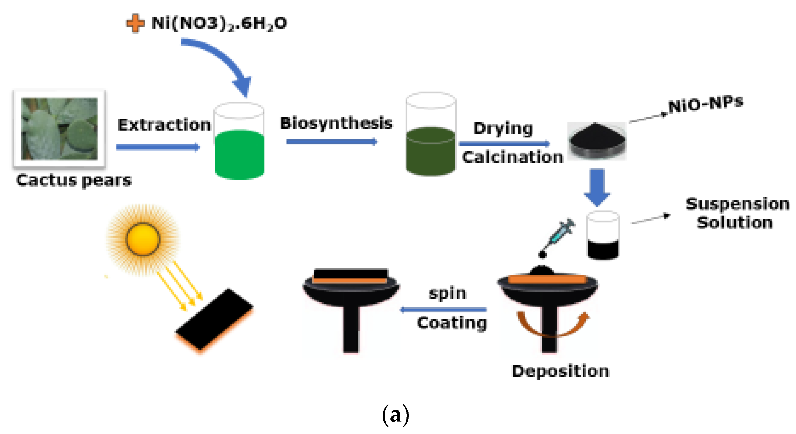
2.2. Green Synthesis and Thin Film Deposition of NiO Nanoparticles
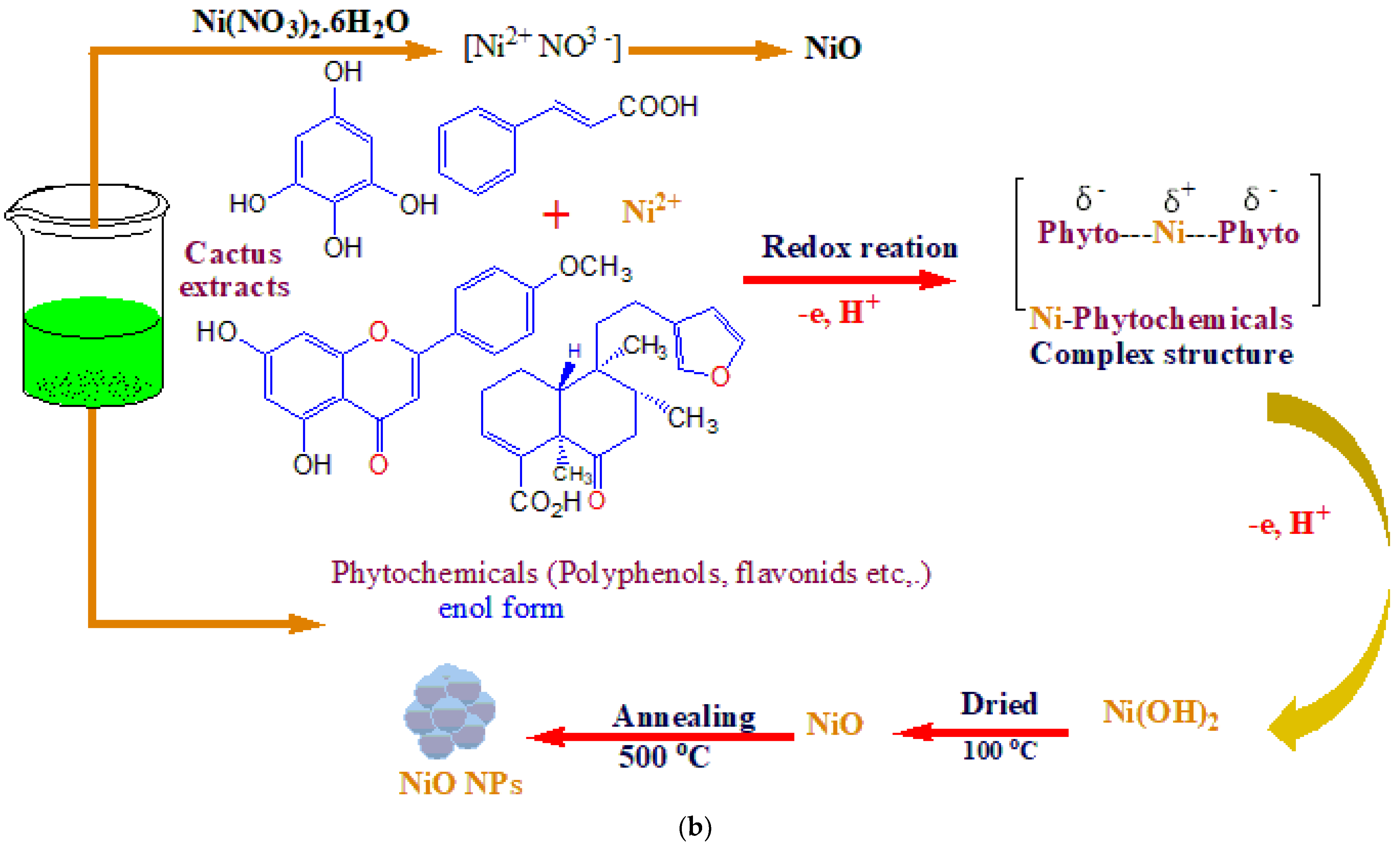
2.3. Possible Reaction Mechanism of Green Synthesized NiO Nanoparticles
2.4. Characterization Techniques
3. Results and Discussion
4. Conclusions
Author Contributions
Funding
Data Availability Statement
Acknowledgments
Conflicts of Interest
References
- Tesfamichael, T. Characterization of Selective Solar Absorbers: Experimental and Theoretical Modeling. Ph.D. Thesis, Universitatis Upsaliensis, Uppsala, Sweden, 2000. [Google Scholar]
- Zhang, K.; Hao, L.; Du, M.; Mi, J.; Wang, J.-N.; Meng, J.-P. A review on thermal stability and high temperature induced ageing mechanisms of solar absorber coatings. Renew. Sustain. Energy Rev. 2017, 67, 1282–1299. [Google Scholar] [CrossRef]
- Dan, A.; Barshilia, H.C.; Chattopadhyay, K.; Basu, B. Solar energy absorption mediated by surface plasma polaritons in spectrally selective dielectric-metal-dielectric coatings: A critical review. Renew. Sustain. Energy Rev. 2017, 79, 1050–1077. [Google Scholar] [CrossRef]
- Alam, T.; Balam, N.B.; Kulkarni, K.S.; Siddiqui, M.I.H.; Kapoor, N.R.; Meena, C.S.; Kumar, A.; Cozzolino, R. Performance Augmentation of the Flat Plate Solar Thermal Collector: A Review. Energies 2021, 14, 6203. [Google Scholar] [CrossRef]
- Dias, D.; Rebouta, L.; Costa, P.; Al-Rjoub, A.; Benelmeki, M.; Tavares, C.; Barradas, N.; Alves, E.; Santilli, P.; Pischow, K. Optical and structural analysis of solar selective absorbing coatings based on AlSiOx: W cermets. Sol. Energy 2017, 150, 335–344. [Google Scholar] [CrossRef] [Green Version]
- Wang, H.; Haechler, I.; Kaur, S.; Freedman, J.; Prasher, R. Spectrally selective solar absorber stable up to 900 °C for 120 h under ambient conditions. Sol. Energy 2018, 174, 305–311. [Google Scholar] [CrossRef] [Green Version]
- Cao, F.; Kraemer, D.; Sun, T.; Lan, Y.; Chen, G.; Ren, Z. Enhanced Thermal Stability of W-Ni-Al2O3 Cermet-Based Spectrally Selective Solar Absorbers with Tungsten Infrared Reflectors. Adv. Energy Mater. 2015, 5, 1401042. [Google Scholar] [CrossRef]
- Bilokur, M.; Gentle, A.; Arnold, M.D.; Cortie, M.B.; Smith, G.B. High Temperature Spectrally Selective Solar Absorbers Using Plasmonic AuAl2: AlN Nanoparticle Composites. Sol. RRL 2017, 1, 1700092. [Google Scholar] [CrossRef]
- Amri, A.; Jiang, Z.T.; Pryor, T.; Yin, C.-Y.; Djordjevic, S. Developments in the synthesis of flat plate solar selective absorber materials via sol–gel methods: A review. Renew. Sustain. Energy Rev. 2014, 36, 316–328. [Google Scholar] [CrossRef] [Green Version]
- Farchado, M.; Rodríguez, J.; San Vicente, G.; Germán, N.; Morales, A. Optical parameters of a novel competitive selective absorber for low temperature solar thermal applications. Sol. Energy Mater. Sol. Cells 2018, 178, 234–239. [Google Scholar] [CrossRef]
- Karoro, A.; Nuru, Z.; Kotsedi, L.; Bouziane, K.; Mothudi, B.M.; Maaza, M. Laser nanostructured Co nanocylinders-Al2O3 cermets for enhanced & flexible solar selective absorbers applications. Appl. Surf. Sci. 2015, 347, 679–684. [Google Scholar]
- Nuru, Z.; Perez, D.; Kaviyarasu, K.; Vantomme, A.; Maaza, M. Annealing effect on the optical properties and interdiffusion of MgO/Zr/MgO multilayered selective solar absorber coatings. Sol. Energy 2015, 120, 123–130. [Google Scholar] [CrossRef]
- Li, C.-Y.; Sari, F.N.I.; Ting, J.-M. Reactive magnetron sputter-deposited TiNxOy multilayered solar selective coatings. Sol. Energy 2019, 181, 178–186. [Google Scholar] [CrossRef]
- De Maio, D.; D’Alessandro, C.; Caldarelli, A.; De Luca, D.; Di Gennaro, E.; Russo, R.; Musto, M. A selective solar absorber for unconcentrated solar thermal panels. Energies 2021, 14, 900. [Google Scholar] [CrossRef]
- Kennedy, C.E. Review of Mid-to High-Temperature Solar Selective Absorber Materials; National Renewable Energy Lab.: Golden, CO, USA, 2002.
- Wäckelgård, E.; Hultmark, G. Industrially sputtered solar absorber surface. Sol. Energy Mater. Sol. Cells 1998, 54, 165–170. [Google Scholar] [CrossRef]
- Gelin, K. Preparation and Characterization of Sputter Deposited Spectrally Selective Solar Absorbers. Ph.D. Thesis, Universitatis Upsaliensis, Uppsala, Sweden, 2004. [Google Scholar]
- Nuru, Z.; Kotsedi, L.; Arendse, C.; Motaung, D.; Mwakikunga, B.; Roro, K.; Maaza, M. Thermal stability of multilayered Pt-Al2O3 nanocoatings for high temperature CSP systems. Vacuum 2015, 120, 115–120. [Google Scholar] [CrossRef]
- Qiu, X.-L.; Gao, X.-H.; Zhou, T.-H.; Chen, B.-H.; Lu, J.-Z.; Guo, H.-X.; Li, X.-T.; Liu, G. Structure, thermal stability and chromaticity investigation of TiB2 based high temperature solar selective absorbing coatings. Sol. Energy 2019, 181, 88–94. [Google Scholar] [CrossRef]
- Welegergs, G.; Gebretnisae, H.; Tsegay, M.; Nuru, Z.; Dube, S.; Maaza, M. Thickness dependent morphological, structural and optical properties of SS/CuO nanocoatings as selective solar absorber. Infrared Phys. Technol. 2021, 113, 103619. [Google Scholar] [CrossRef]
- Kumar, S.K.; Suresh, S.; Murugesan, S.; Raj, S.P. CuO thin films made of nanofibers for solar selective absorber applications. Sol. Energy 2013, 94, 299–304. [Google Scholar] [CrossRef]
- Randich, E.; Pettit, R. Solar selective properties and high temperature stability of CVD ZrB2. Sol. Energy Mater. 1981, 5, 425–435. [Google Scholar] [CrossRef]
- Seraphin, B. Chemical vapor deposition of thin semiconductor films for solar energy conversion. Thin Solid Film. 1976, 39, 87–94. [Google Scholar] [CrossRef]
- Prasadam, V.P.; Gautier, N.; Bahlawane, N. CNT nanoengineering for thermally stable selective solar absorption. Mater. Today Commun. 2021, 28, 102552. [Google Scholar] [CrossRef]
- Selvakumar, N.; Barshilia, H.C. Review of physical vapor deposited (PVD) spectrally selective coatings for mid-and high-temperature solar thermal applications. Sol. Energy Mater. Sol. Cells 2012, 98, 1–23. [Google Scholar] [CrossRef]
- Nuru, Z.; Arendse, C.; Muller, T.; Maaza, M. Structural and optical properties of AlxOy/Pt/AlxOy multilayer absorber. Mater. Sci. Eng. B 2012, 177, 1194–1199. [Google Scholar] [CrossRef]
- Nuru, Z.; Msimanga, M.; Muller, T.; Arendse, C.; Mtshali, C.; Maaza, M. Microstructural, optical properties and thermal stability of MgO/Zr/MgO multilayered selective solar absorber coatings. Sol. Energy 2015, 111, 357–363. [Google Scholar] [CrossRef]
- Kotsedi, L.; Mthunzi, P.; Nuru, Z.; Eaton, S.; Sechoghela, P.; Mongwaketsi, N.; Ramponi, R.; Maaza, M. Femtosecond laser surface structuring of molybdenum thin films. Appl. Surf. Sci. 2015, 353, 1334–1341. [Google Scholar] [CrossRef]
- Khan, S.; Wu, Z.; Yuan, G.; Khan, M.; Song, C.; Han, G.; Liu, Y. Study of annealing effect upon the structural and solar-selective properties of C/Ni/NiO composite coatings prepared by sol–gel method. J. Sol-Gel Sci. Technol. 2019, 89, 120–127. [Google Scholar] [CrossRef]
- Katumba, G.; Olumekor, L.; Forbes, A.; Makiwa, G.; Mwakikunga, B.; Lu, J.; Wäckelgård, E. Optical, thermal and structural characteristics of carbon nanoparticles embedded in ZnO and NiO as selective solar absorbers. Sol. Energy Mater. Sol. Cells 2008, 92, 1285–1292. [Google Scholar] [CrossRef]
- Roro, K.T.; Tile, N.; Forbes, A. Preparation and characterization of carbon/nickel oxide nanocomposite coatings for solar absorber applications. Appl. Surf. Sci. 2012, 258, 7174–7180. [Google Scholar] [CrossRef]
- Gebretinsae, H.; Tsegay, M.; Nuru, Z. Biosynthesis of nickel oxide (NiO) nanoparticles from cactus plant extract. Mater. Today Proc. 2021, 36, 566–570. [Google Scholar] [CrossRef]
- Ventura-Aguilar, R.I.; Bosquez-Molina, E.; Bautista-Baños, S.; Rivera-Cabrera, F. Cactus stem (Opuntia ficus-indica Mill): Anatomy, physiology and chemical composition with emphasis on its biofunctional properties. J. Sci. Food Agric. 2017, 97, 5065–5073. [Google Scholar] [CrossRef]
- Duffie, J.; Beckman, W. Solar Thermal Engineering Processes; A Wiley Interscience Publication: New York, NY, USA, 1980. [Google Scholar]
- Liao, L.; Cui, Z. Thermal Processes, in Solution Processed Metal Oxide Thin Films for Electronic Applications; Elsevier: Amsterdam, The Netherlands, 2020; pp. 99–107. [Google Scholar]
- Visweswaran, S.; Venkatachalapathy, R.; Haris, M.; Murugesan, R. Structural, morphological, optical and magnetic properties of sprayed NiO thin films by perfume atomizer. Appl. Phys. A 2020, 126, 524. [Google Scholar] [CrossRef]
- Ahmed, A.A.; Hashim, M.; Rashid, M. Control of the structural, electrical and optical properties of spin coated NiO films by varying precursor molarity. Thin Solid Film. 2019, 690, 137554. [Google Scholar] [CrossRef]
- Ganesh, V.; Haritha, L.; Anis, M.; Shkir, M.; Yahia, I.; Singh, A.; AlFaify, S. Structural, morphological, optical and third order nonlinear optical response of spin-coated NiO thin films: An effect of N doping. Solid State Sci. 2018, 86, 98–106. [Google Scholar] [CrossRef]
- Chtouki, T.; El Mrabet, M.; Tarbi, A.; Goncharova, I.; Erguig, H. Comprehensive review of the morphological, linear and nonlinear optical characterization of spin-coated NiO thin films for optoelectronic applications. Opt. Mater. 2021, 118, 111294. [Google Scholar] [CrossRef]
- El Sayed, A.M.; Shaban, M. Morphological, surface and optical properties of spin-coated IrOx films; influence of spin speed, annealing and (Cr, La) codoping. Ceram. Int. 2019, 45, 8460–8470. [Google Scholar] [CrossRef]
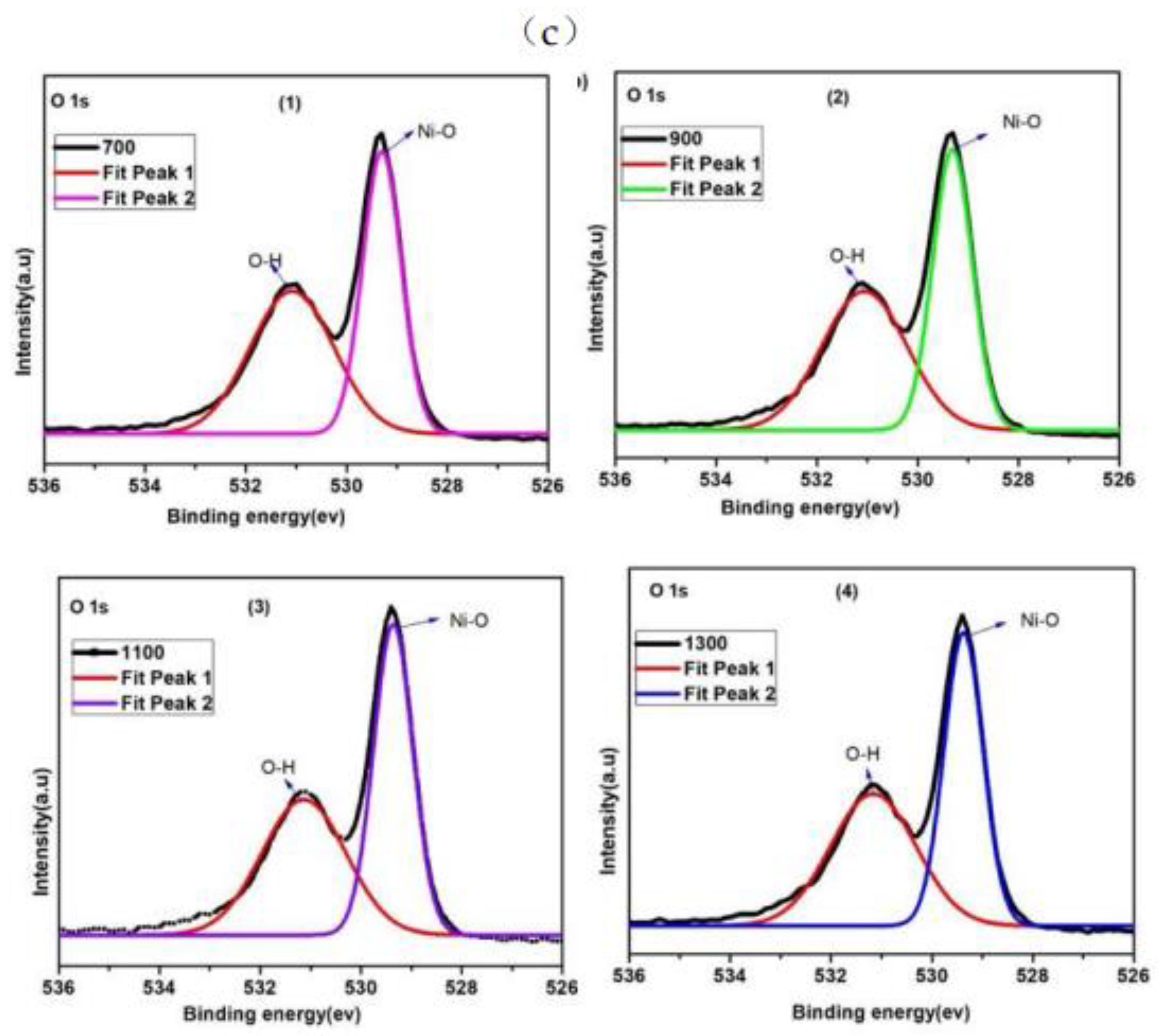
- Scharfschwerdtt, C.; Neumannt, M.; Illing, G.; Freund, H. The influence of defects on the Ni 2p and O 1s XPS of NiO. J. Phys. Condens. Matter 1992, 4, 7973–7978. [Google Scholar]
- Roberts, M.W.; Smart, R.S.C. The defect structure of nickel oxide surfaces as revealed by photoelectron spectroscopy. Journal of the Chemical Society, Faraday Transactions 1: Physical Chemistry in Condensed. Phases 1984, 80, 2957–2968. [Google Scholar]
- Biju, V. Ni 2p X-ray photoelectron spectroscopy study of nanostructured nickel oxide. Mater. Res. Bull. 2007, 42, 791–796. [Google Scholar] [CrossRef]
- Salunkhe, P.; AV, M.A.; Kekuda, D. Investigation on tailoring physical properties of Nickel Oxide thin films grown by dc magnetron sputtering. Mater. Res. Express 2020, 7, 016427. [Google Scholar] [CrossRef]
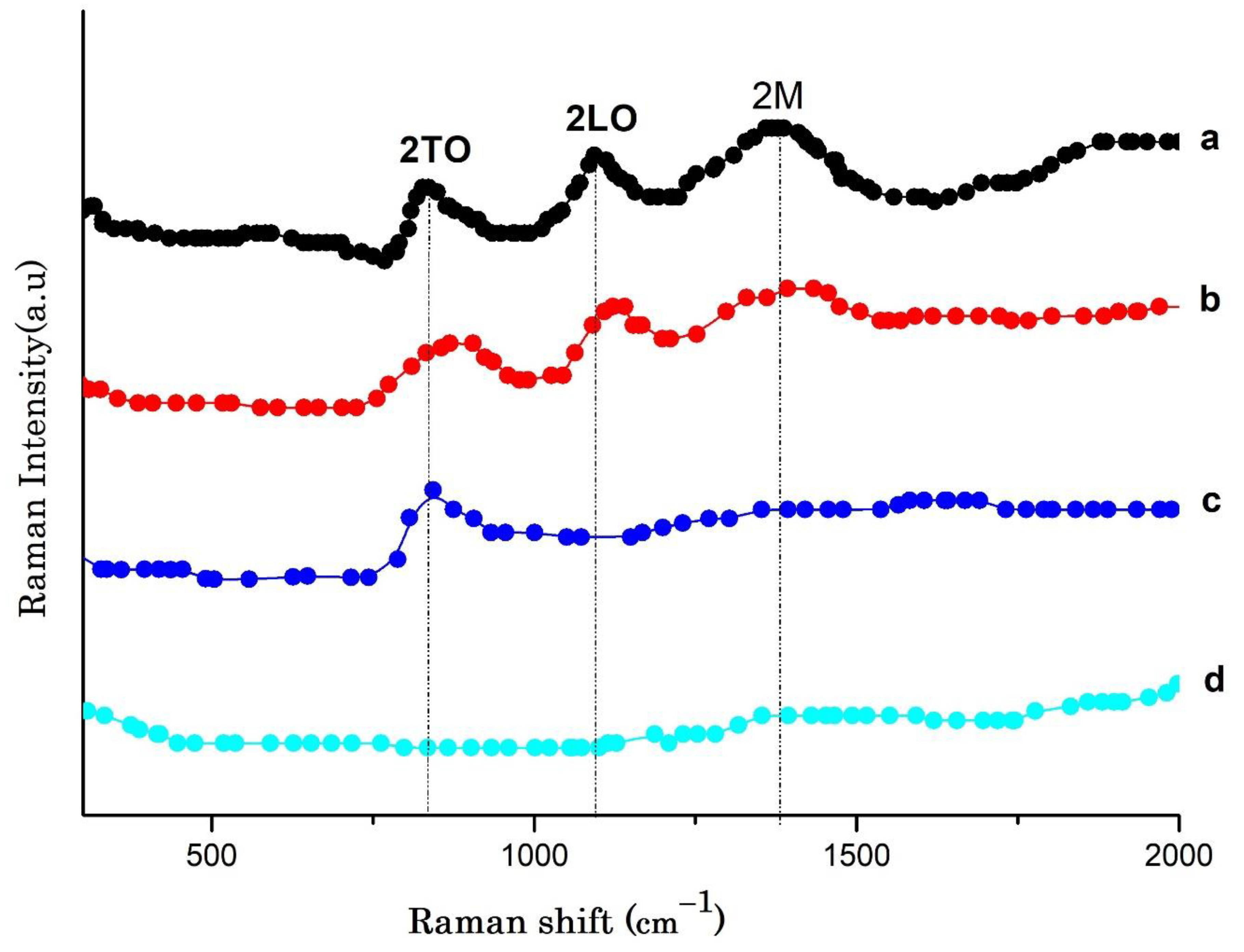
- Mironova-Ulmane, N.; Kuzmin, A.; Steins, I.; Grabis, J.; Sildos, I.; Pärs, M. Raman scattering in nanosized nickel oxide NiO. J. Phys. Conf. Ser. 2007, 93, 012039. [Google Scholar] [CrossRef]
- Ascencio, F.; Bobadilla, A.; Escudero, R. Study of NiO nanoparticles, structural and magnetic characteristics. Appl. Phys. A 2019, 125, 279. [Google Scholar] [CrossRef]
- Cazzanelli, E.; Kuzmin, A.; Mariotto, G.; Mironova-Ulmane, N. Study of vibrational and magnetic excitations in Nic Mg1-cO solid solutions by Raman spectroscopy. J. Phys. Condens. Matter 2003, 15, 2045. [Google Scholar] [CrossRef] [Green Version]
- Kumar, R.; Baratto, C.; Faglia, G.; Sberveglieri, G.; Bontempi, E.; Borgese, L. Tailoring the textured surface of porous nanostructured NiO thin films for the detection of pollutant gases. Thin Solid Film. 2015, 583, 233–238. [Google Scholar] [CrossRef]
- Jiang, D.; Qin, J.; Wang, X.; Gao, S.; Liang, Q.; Zhao, J. Optical properties of NiO thin films fabricated by electron beam evaporation. Vacuum 2012, 86, 1083–1086. [Google Scholar] [CrossRef]
- Ivanova, T.; Harizanova, A.; Shipochka, M.; Vitanov, P. Nickel Oxide Films Deposited by Sol-Gel Method: Effect of Annealing Temperature on Structural, Optical, and Electrical Properties. Materials 2022, 15, 1742. [Google Scholar] [CrossRef]
- Khodasevych, I.E.; Wang, L.; Mitchell, A.; Rosengarten, G. Micro-and nanostructured surfaces for selective solar absorption. Adv. Opt. Mater. 2015, 3, 852–881. [Google Scholar] [CrossRef]
- Wang, W.; Wen, H.; Huan, X.; Shi, J.; Li, Z.; Su, J.; Wang, C. Single layer WOx films for efficient solar selective absorber. Mater. Des. 2020, 186, 108351. [Google Scholar] [CrossRef]
- El Mahallawy, N.; Shoeib, M.; Ali, Y. Application of CuCoMnO x coat by sol gel technique on aluminum and copper substrates for solar absorber application. J. Coat. Technol. Res. 2014, 11, 979–991. [Google Scholar] [CrossRef]
- Kim, M.J.; Kim, H.T.; Kang, J.K.; Kim, D.H.; Lee, D.H.; Lee, S.H.; Sohn, S.H. Effects of the surface roughness on optical properties of CdS thin films. Mol. Cryst. Liq. Cryst. 2010, 532, 21/[437]–28/[444]. [Google Scholar] [CrossRef]
- Goldschmidt, D. Determination of the absorption edge of a thin film from transmission measurements. JOSA A 1984, 1, 275–277. [Google Scholar] [CrossRef]
- Tsegay, M.; Gebretinsae, H.; Welegergs, G.; Maaza, M.; Nuru, Z. Novel green synthesized Cr2O3 for selective solar absorber: Investigation of structural, morphological, chemical, and optical properties. Sol. Energy 2022, 236, 308–319. [Google Scholar] [CrossRef]


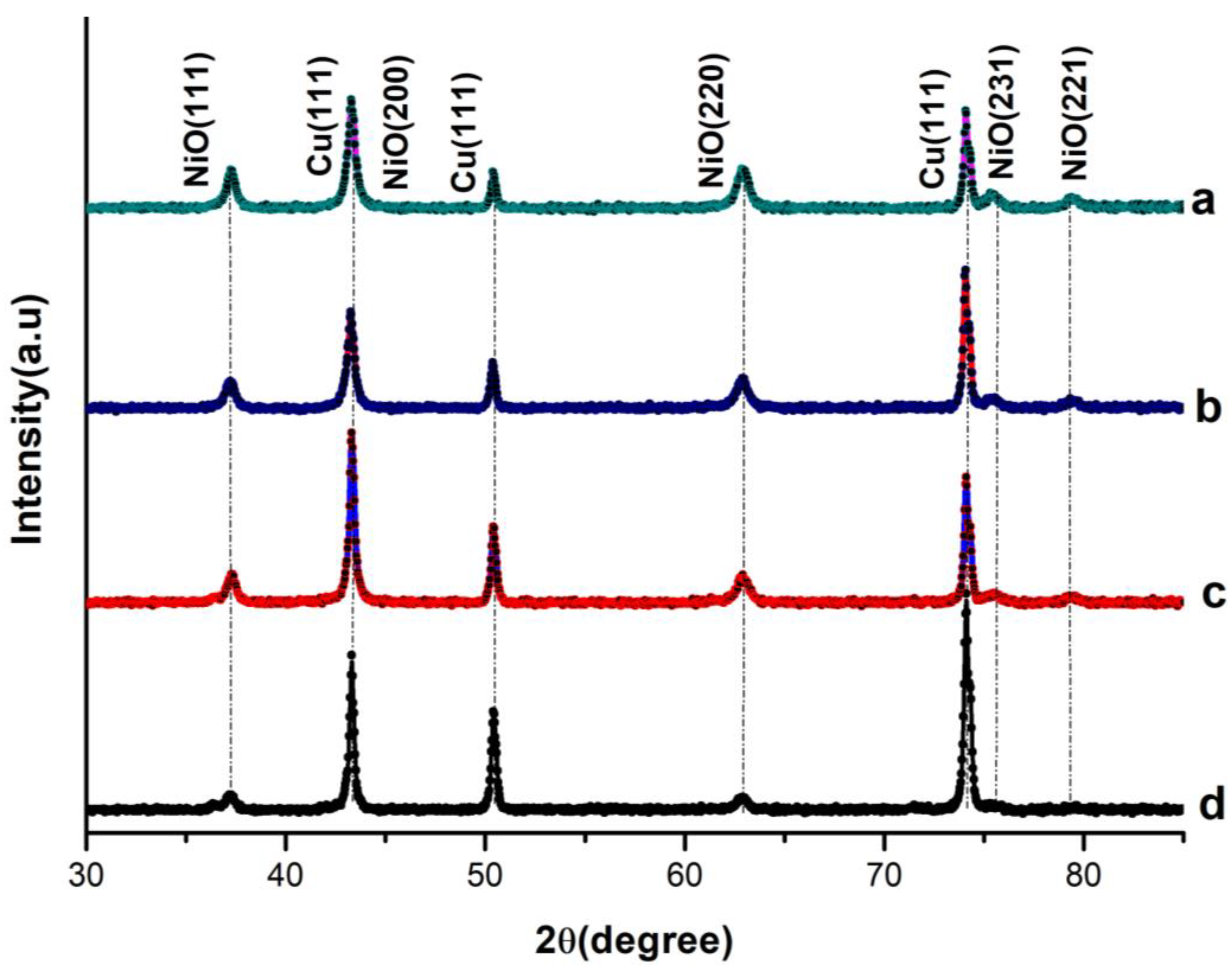
| RS | 2θ(111) Deg | FWHM | Crystal Size (nm) | d(111)(Å) | ||
|---|---|---|---|---|---|---|
| a | 700 | 37.26° | 0.486 | 18.01168 | 2.411 | 0.00652 |
| b | 900 | 37.28° | 0.51 | 17.1699 | 2.4100 | 0.00684 |
| c | 1100 | 37.30° | 0.552 | 15.86 | 2.4087 | 0.007399 |
| d | 1300 | 37.31° | 0.559 | 13.66 | 2.4081 | 0.007491 |
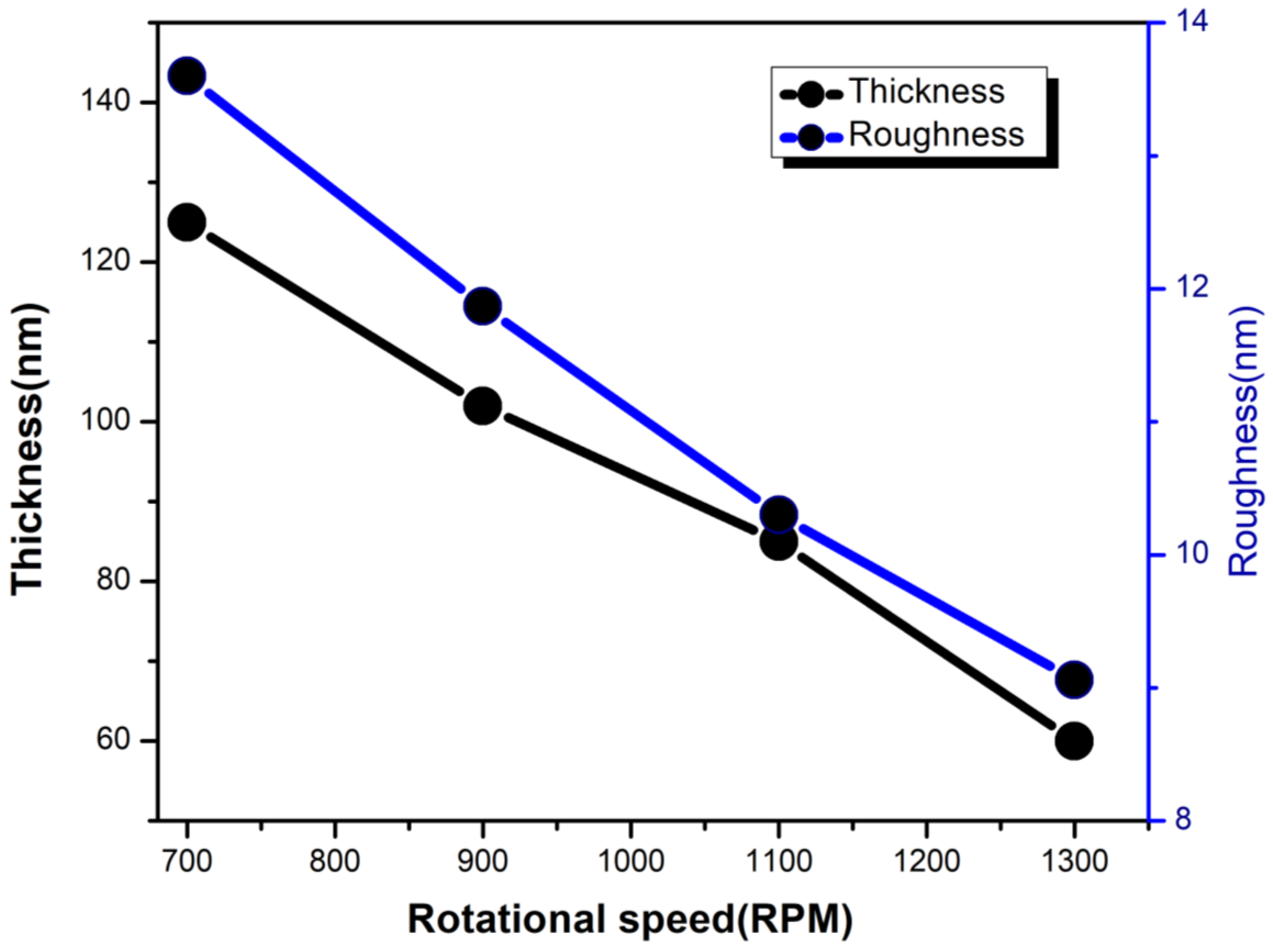
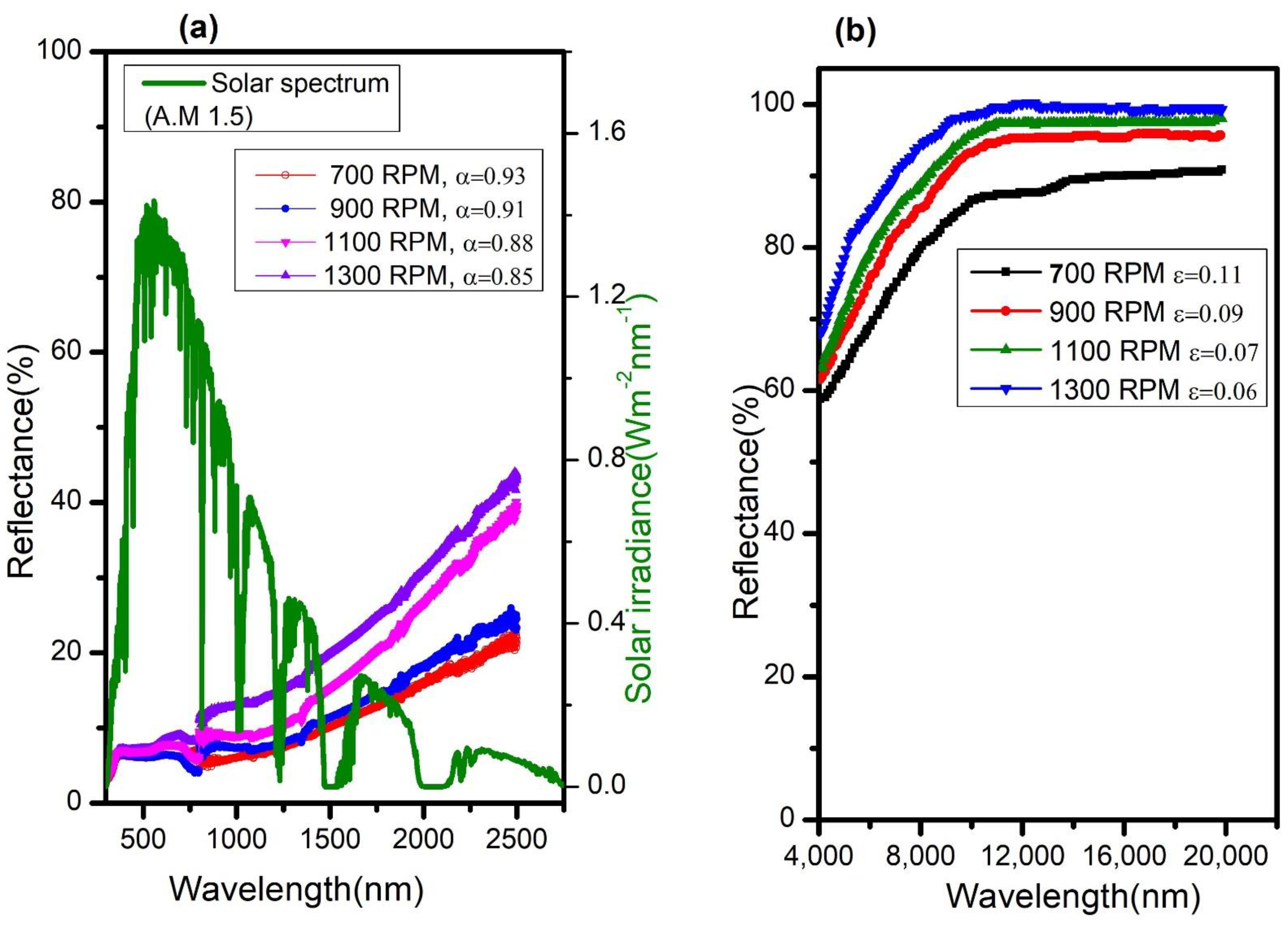
| RS (RPM) | Thickness (nm) | Absorptance (α) | Emittance (ε) (100 °C) | Selectivity (η) |
|---|---|---|---|---|
| 700 | 125 | 0.92 | 0.11 | 8.4 |
| 900 | 102 | 0.91 | 0.09 | 10.1 |
| 1100 | 85 | 0.88 | 0.07 | 12.6 |
| 1300 | 60 | 0.85 | 0.06 | 14.2 |
Publisher’s Note: MDPI stays neutral with regard to jurisdictional claims in published maps and institutional affiliations. |
© 2022 by the authors. Licensee MDPI, Basel, Switzerland. This article is an open access article distributed under the terms and conditions of the Creative Commons Attribution (CC BY) license (https://creativecommons.org/licenses/by/4.0/).
Share and Cite
Gebretinsae, H.G.; Tsegay, M.G.; Welegergs, G.G.; Maaza, M.; Nuru, Z.Y. Effect of Rotational Speed on the Structural, Morphological, and Optical Properties of Biosynthesized Nickel Oxide Thin Films for Selective Solar Absorber Nanocoatings. Energies 2022, 15, 8960. https://0-doi-org.brum.beds.ac.uk/10.3390/en15238960
Gebretinsae HG, Tsegay MG, Welegergs GG, Maaza M, Nuru ZY. Effect of Rotational Speed on the Structural, Morphological, and Optical Properties of Biosynthesized Nickel Oxide Thin Films for Selective Solar Absorber Nanocoatings. Energies. 2022; 15(23):8960. https://0-doi-org.brum.beds.ac.uk/10.3390/en15238960
Chicago/Turabian StyleGebretinsae, Henok G., Meresa G. Tsegay, Giday G. Welegergs, Malik Maaza, and Zebib Y. Nuru. 2022. "Effect of Rotational Speed on the Structural, Morphological, and Optical Properties of Biosynthesized Nickel Oxide Thin Films for Selective Solar Absorber Nanocoatings" Energies 15, no. 23: 8960. https://0-doi-org.brum.beds.ac.uk/10.3390/en15238960