Heteroleptic Pt(II)-dithiolene-based Colorimetric Chemosensors: Selectivity Control for Hg(II) Ion Sensing


1
Department of Chemistry, Seoul Women’s University, Seoul 01797, Korea
2
School of Chemical Engineering and Materials Science, Institute of Energy Converting Soft Materials, Chung-Ang University, Seoul 06974, Korea
*
Authors to whom correspondence should be addressed.
†
Present address: Western Seoul Center, Korea Basic Science Institute (KBSI), Seoul 03759, Korea.
Materials 2020, 13(6), 1385; https://0-doi-org.brum.beds.ac.uk/10.3390/ma13061385
Submission received: 22 February 2020
/
Revised: 10 March 2020
/
Accepted: 16 March 2020
/
Published: 19 March 2020
(This article belongs to the Special Issue Application of Nanoparticles as Biosensors in the Biomedical Field)
Abstract
:Hg2+ ions can accumulate in the natural environment and in organisms, where they cause damage to the central nervous system. Therefore, the detection of Hg2+ ions is essential for monitoring environmental contamination and human health. Herein, we demonstrate a simple method for tuning chemosensor signal ratios that significantly increased chemosensor selectivity for Hg2+ detection. Selectivity tuning was accomplished for chemosensors of the type (diphosphine)Pt(dmit), bearing the two different terminal groups 1,2-bis(diphenylphosphino)ethane (dppe) and 1,2-bis[bis(pentafluorophenyl)phosphino]ethane) (dfppe) due to the modulation of specific intermolecular interactions between the dmit ligand and Hg2+ ion. The structure exhibited a large pseudo-Stokes shift, which was advantageous for the internal reference signal and for eliminating potential artifacts. Straightforward chain-end manipulation enabled the tuning of chemosensor properties without additional chemical alterations. Based on these findings, we propose a new platform for improving the selectivity and sensitivity of colorimetric cation sensors. The results of this study will facilitate the designing of organic materials whose certain properties can be enhanced through precise control of the materials’ chemical hybridization by simple functional end-group manipulation.
1. Introduction
Ionic Hg (Hg2+) is well known for its high toxicity, and can be found in water, soil, and food [1]. The ions accumulate in the organism and interact with thiol groups of proteins, causing serious damage to the central nervous system and posing a significant threat to human health and the natural environment. Therefore, the detection of Hg2+ ions is a fundamental requirement for monitoring the environment and human health [2]. Various sensor platforms for Hg2+ detection have been developed to date, based on inorganic substances, oligonucleotides, liposomes, proteins, polymers, DNA glymes, small fluorescent organic molecules, and other compounds [2,3,4,5,6,7,8,9]. In supramolecular chemistry, the fluorescent chemosensor is one of the most significant designs because of the possibility of straightforward manipulation of photophysical processes that enables sensitive and selective signaling of targeted ion components [10]. Previously, we have reported a series of studies regarding (diphosphine)Pt(dmit) complexes (dmit: 1,3-dithiole-2-thione-4,5-dithiolate) as cation chemosensors that can be converted to operate via a ratiometric internal charge transfer, especially for Hg2+ detection [11,12,13]. These complexes created a large pseudo-Stokes shift between the analyte-free and analyte-bound emissive states. It rendered them wavelength-ratiometric, which means that potential artifacts were eliminated based on the internal referencing of the signal. Selectivity can be achieved only when the emission peaks for the free and bound-chemosensor shows clearly distinct intensities. However, the ratio of intensities is related to an inherent characteristic of a particular analyte and chemosensor interaction; hence, a systematic methodology for the control of signal ratio has rarely been practiced hitherto [10].
Herein, we demonstrate a simple method for chemosensor signal ratio tuning that significantly increased the chemosensor selectivity for Hg2+ detection. In order to achieve and explore signal ratio tuning of emission intensities, we set out to synthesize (dxpe)Pt(dmit) complexes with diphosphine-groups where dxpe = dppe (1,2-bis(diphenylphosphino)ethane) and dfppe (1,2-bis[bis(pentafluorophenyl)phosphino]ethane), while all other chemical functionalities remained constant. Both materials contained π-electrons in the >C=S double bond and a lone pair of electrons on the S atom; only the diphosphine end-groups were altered to obtain distinct electronegativities. A direct comparison of these two samples was made to elucidate the relationship between embedded terminal groups and the tunability of the sensing probe of the colorimetric sensors, based primarily on their electronic affinities and interfacial interactions with analyte cations. To the best of our knowledge, this is the first experimental report demonstrating the successful tuning of chemosensor properties by chain-end manipulation, without requiring further chemical alterations. We, therefore, propose a new platform for the improvement of selectivity of colorimetric cation sensors. This systematic investigation of end-functionalized chemicals serves to broaden the applicability and understanding of polymer science and provides new insights into advanced sensor technologies.
2. Materials and Methods
2.1. Synthesis
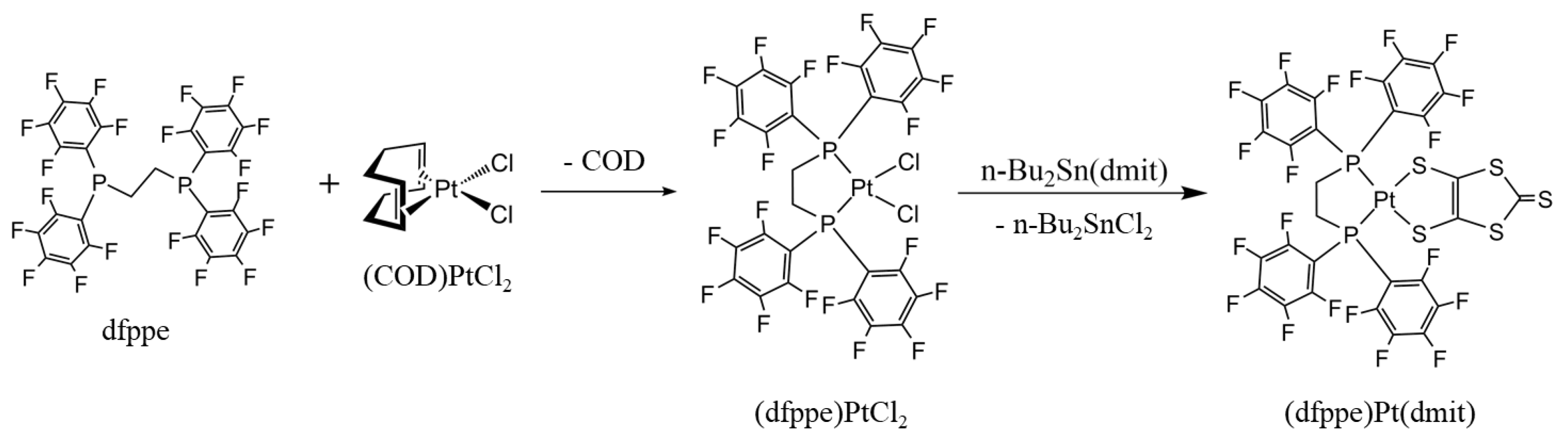
Synthesis of (dfppe)PtCl2. A dichloromethane (DCM) solution (30 mL) of (COD)PtCl2 (1.0 mmol, 374 mg) was added to an acetone solution (80 mL) of dfppe (1.0 mmol, 758 mg) and stirred overnight at room temperature. Following solvent removal, the solid was dissolved in methanol, filtered, and dried in air to yield 96.5% (988 mg) of the final product. Fourier Transform Infrared Spectroscopy (FT-IR) (KBr, cm−1) 2958, 2905 (C–H str.), 1646, 1524, 1482 (Ar C=C), 1100 (P–Ph), 982 (Ar C–F str.), and 538 (Ar ring oop def).
Synthesis of (dfppe)Pt(dmit). A DCM solution (30 mL) of (dfppe)PtCl2 (0.5 mmol, 512 mg) and an acetone solution (30 mL) of n-Bu2Sn(dmit) (0.5 mmol, 214 mg) were mixed and stirred overnight at room temperature under an Ar atmosphere [14,15]. Evaporation of the solvent to 5 mL, followed by the addition of DCM, precipitated the red solid under vacuum. Following filtration and washing with DCM, the product was recrystallized from DCM/MeOH by slow diffusion, yielding 65.0% (374 mg) of a red product. Mp. > 278 °C (decomp.). EA cal. (obs.) for C29H4F20P2PtS5: C 30.30 (30.37); H 0.35 (0.39); S 13.95 (14.64). FT-IR (KBr, cm−1) 1644, 1522, 1478 (Ar C=C), 1101 (P–Ph), 1054 (C=S), 980 (Ar C–F str.), and 536 (Ar ring o.o.p. def).
Synthesis of (dppe)Pt(dmit). The (dppe)Pt(dmit) complex was prepared using (dppe)PtCl2 and Na2(dmit) according to a previously reported procedure, and its spectroscopic data were in good agreement with the reported values [13].
2.2. General Characterization
Elemental analysis was performed using a FLASH2000 (Thermo Fisher Scienctific, Waltham, MA, USA) and PerkinElmer 2400 Series II installed at the National Center for Inter-University Research Facilities (NCIRF) at Seoul National University, Seoul, Korea. The infrared spectra were recorded using KBr pellets on a Perkin Elmer Spectrum 100 in the range of 400 ~ 4000 cm−1. UV-Vis absorption spectra were obtained on an S-3100 Photodiode Array spectrophotometer (SCINCO CO., Ltd., Seoul, Korea), using an H2O/CH3CN mixed solution (1:1 v/v %).
2.3. Electrochemical Analysis
Cyclic voltammetry measurements were conducted at room temperature with a computer-controlled potentiostat (CHI 620A Electrochemical Analyzer, CHI Instrument Inc., Bee Cave, TX, USA), using a standard three-electrode cell system with a Pt-button working electrode, a Pt wire as a counter electrode, and saturated Ag/AgCl as a reference electrode. Cyclic voltammograms were obtained in a DCM solution with 1.0 mM sample and 0.1 M n-Bu4N·ClO4 supporting electrolyte at a scan rate of 100 mV s−1. Ferrocene was used as a calibrant after each set of measurements, and all potentials reported were quoted with reference to the Fc/Fc+ redox couple (E1/2 = 0.532 V).
2.4. X-ray Diffraction (XRD) for Molecular Structure Analysis
Crystal structure analysis was performed by single-crystal diffraction methods at the Korea Basic Science Institute (KBSI, Western Seoul Center, Seoul, Korea). A single needle-like crystal (0.23 × 0.16 × 0.08 mm3) of (dfppe)Pt(dmit) was taken up with paratone oil and mounted on a Bruker SMART CCD diffractometer equipped with a graphite-monochromated Mo Kα (λ = 0.71073 Å) radiation source and a nitrogen cold stream (200(2) K). Data collection and integration was performed on a SMART (Bruker, Madison, WI, USA, 2000) and SAINT-Plus (Bruker, 2001) [16]. Absorption correction was performed by a multiscan method implemented in SADABS [17]. The structure was solved by direct methods and refined by full-matrix least-squares on F2 using SHELXTL [18]. All the nonhydrogen atoms were refined anisotropically, and hydrogen atoms were added to their geometrically ideal positions.
2.5. Density Functional Theory Calculations
First-principles calculations were carried out on the basis of density functional theory (DFT) using a generalized gradient approximation (GGA) within the Perdew–Burke–Ernzerhof (PBE) functional [19,20]. We used the projector-augmented wave (PAW) method for describing ionic cores as implemented in the Vienna ab initio simulation package [21]. A plane-wave basis set with a kinetic-energy cut-off of 600 eV and a 4 × 4 × 4 Monkhorst–Pack k-point mesh was used. The electronic optimization steps were converged self-consistently over 10−4 eV per formula unit. The cation adsorbed on each (dxpe)Pt(dmit) in the most stable state was directly relaxed by energy optimization from any initial positions. The charge accumulations and charge reductions around the molecules are shown in Figure 1, as yellow and blue surfaces, respectively. In this study, the unit of charge densities was e a0−3, where e was the elementary charge and a0 was the Bohr radius.
3. Results and Discussion
The selective metal ion-sensing properties of (dxpe)Pt(dmit) complexes were demonstrated by unique color changes. To validate the selectivity of the chemosensors in practice, a series of metal cations were added to a solution of the chemosensor under identical conditions. The protocol was as follows: (1) the materials analyzed were fabricated with differing end-functional groups and (2) the color changes were observed with the naked eye and by UV-visible spectrometry.
3.1. Synthesis and XRD for Structural Analysis
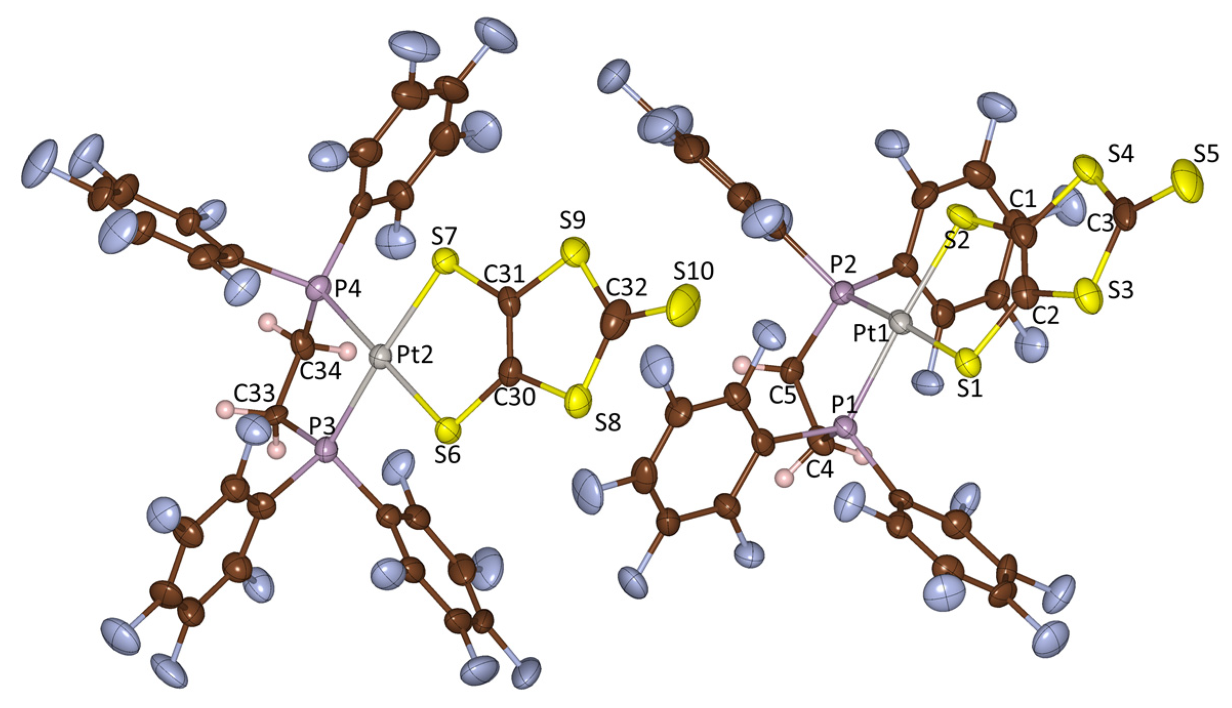
The (dppe)Pt(dmit) complex was prepared from (dppe)PtCl2 and Na2(dmit) according to a previously reported procedure [12]. The synthesis of the (dfppe)Pt(dmit) complex, having an F-enriched end-functional group, involved (dfppe)PtCl2 and n-Bu2Sn(dmit), following an analogous salt elimination process (Scheme 1). Product identification was confirmed by spectroscopic analyses. The structure of the as-synthesized (dfppe)Pt(dmit) complex was additionally confirmed by X-ray diffraction for molecular structure analysis (Table 1 and Table 2, and Figure 1). The molecular structure of (dfppe)Pt(dmit) shown in Figure 1 is virtually isostructural with that of (dppe)Pt(dmit) [22], with the former bearing F, and the latter H atoms on the diphosphine chelate ligands. Their principal bond lengths and angles were likewise analogous, as compared in Table 1.
3.2. Electrochemical Analysis
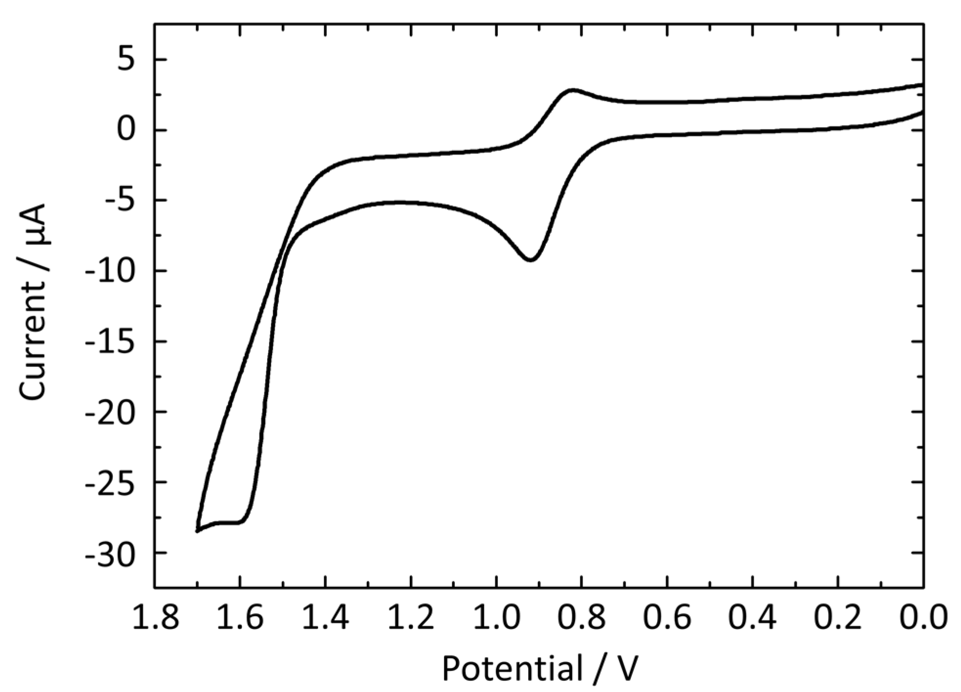
The (dxpe)Pt(dmit) complexes (dxpe = dppe and dfppe) exhibited identical cyclic voltammograms with one reversible cycle at a lower potential region and one irreversible oxidation peak at a higher region. However, their redox potential values were not identical due to differences in the donor capability of the dithiolate ligand, which is affected by the diphosphine ligand. The cyclic voltammogram of (dfppe)Pt(dmit) exhibits a reversible cycle at = 0.871 V ( = 0.921 V and = 0.820 V), and an irreversible anodic peak at = 1.600 V (Figure 2). This half-wave potential can be attributed to the redox process of the Pt(dmit) moiety [23], which suggests that [(dfppe)Pt(dmit)]+, formed during the first redox process at = 0.871 V, is well stabilized by the Pt(dmit) moiety. This potential is higher than that of (dppe)Pt(dmit) ( = 0.777 V), indicating that the F-ended functional groups of (dfppe)Pt(dmit) decreased the donor capability of the Pt(dmit) moiety compared to H-ended functional groups of (dppe)Pt(dmit). This variance is likely to endow distinct sensing properties to these chemosensors.
3.3. Colorimetric Sensing Properties
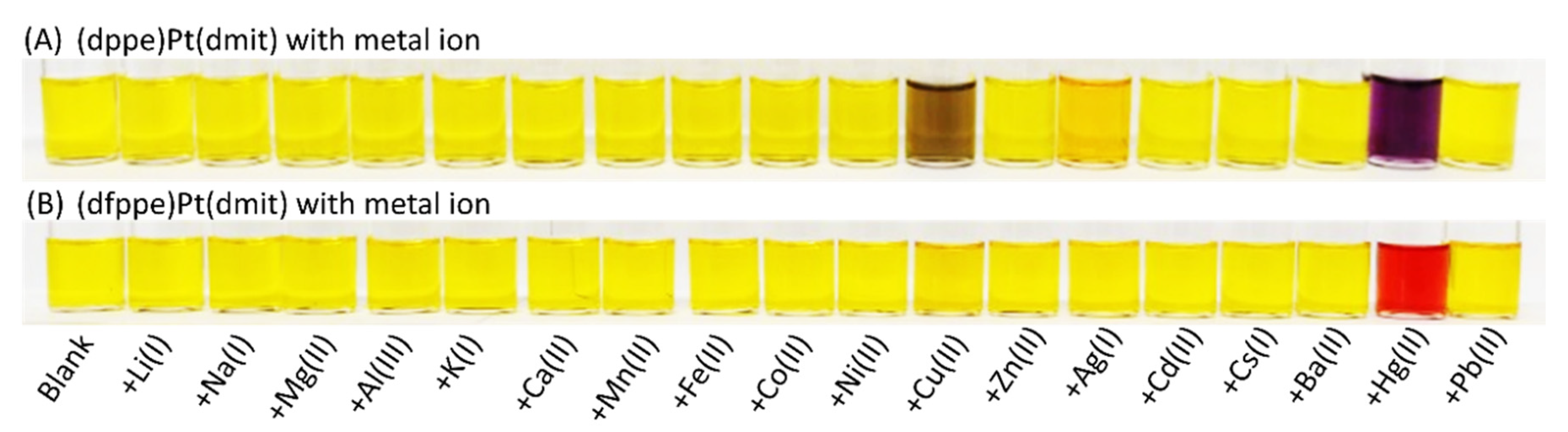
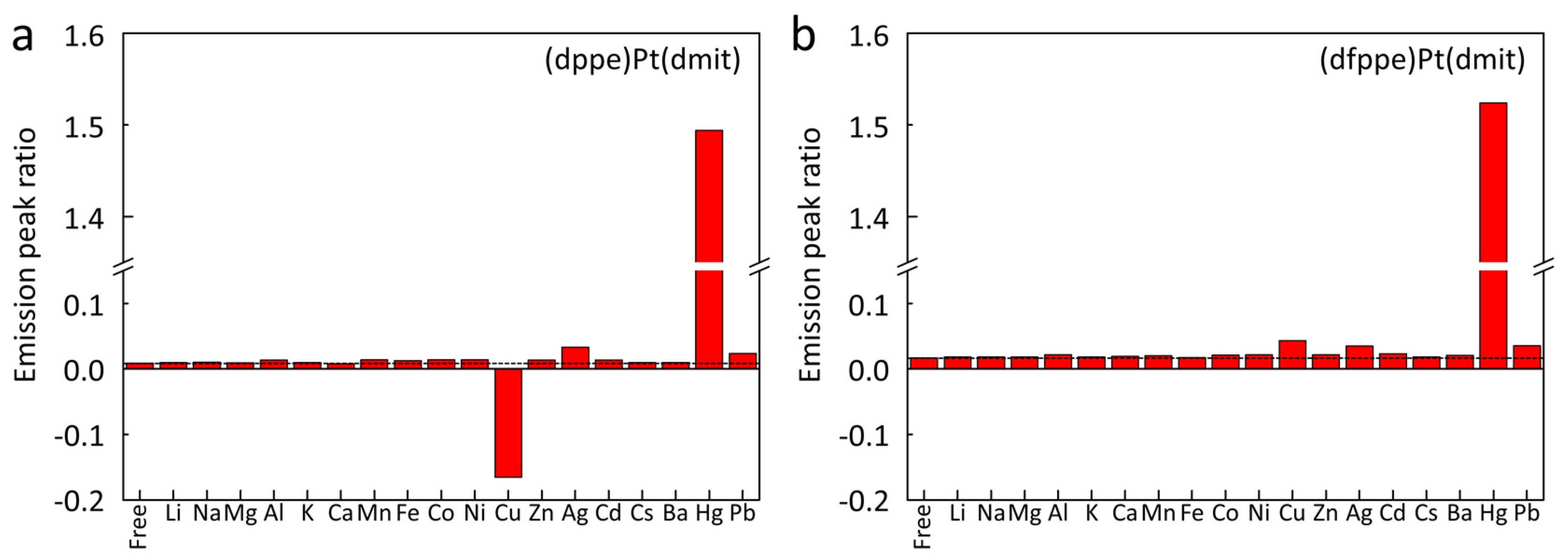
To compare the selectivity of the chemosensors, 18 commercially available metal perchlorate compounds were considered (Figure 3). Both chemosensors exhibited the most efficient sensing capabilities for the Hg2+ ion, but their selectivity differed significantly based on the nature of the end-functional group. The color change of the chemosensor in H2O/CH3CN solution (0.2 mM, 1:1 v/v %) with various metal cations is clearly depicted in Figure 3. Both (dfppe)Pt(dmit) and (dppe)Pt(dmit) complexes in aqueous solution exhibited virtually no color change with hard acids by Pearson’s acid-base concept, such as Na+, K+, and Ca2+ [24]. These observations are understandable, as coordination between hard Lewis acids and thione moiety (>C=S) is unfavorable. The dmit ligand is an electron-rich moiety, containing π-electrons in the >C=S double bond and a lone pair of electrons on the S atom; therefore, it is categorized as a soft base. On the other hand, the binding of several cations, such as Cu2+ and Ag+, occurs with the (dppe)Pt(dmit) chemosensor, in which the cation receptor is an electron donor as well as part of the π-system of the chromophore. Cu2+ and Ag+ ions are soft acids and can thus chelate weakly with the thione moiety. Therefore, although the response was minimal compared to that observed for the Hg2+ cation, a color change with a blue shift was observed for Cu2+ and Ag+ in an aqueous solution of (dppe)Pt(dmit), even by naked eye, as shown in Figure 3A.
It is worth noting the selective sensing properties of the (dppe)Pt(dmit) complex toward Hg2+, Cu2+ and Ag+ cations among the 18 metal ions, in light of the standard reduction potentials (E°) of the metal ions. The three metal cations have positive E° values (E° = +0.854 V for Hg2+/Hg, +0.34 V for Cu2+/Cu, and +0.80 V for Ag+/Ag at 298 K), while the values of the other 15 metal cations are negative [25]. Therefore, during the sensing process, the three metal cations with positive reduction potentials would be more susceptible to reduction and would, consequently, withdraw electrons more readily from the dmit ligand, inducing the color change in the chemosensor.
Altering the diphosphine end-group of the chemosensor from dppe to dfppe dramatically influenced its selectivity for metal ions. Notably, alkali metals, alkaline earth metals, and transition metal ions did not induce any apparent color change, including Cu2+ and Ag+ cations. Of the 18 metal cations tested, only Hg2+ ions induced a color change with the (dfppe)Pt(dmit) chemosensor, from yellow to vivid red (Figure 3B). This suggests that the interaction between the >C=S moiety and metal ions was dependent on the properties of the end-functional group of the chemosensor. That is, the F-enriched sensor, which has the highest reduction potential among the 18 metal ions, exhibited excellent selectivity for Hg2+ detection.
3.4. Absorption Spectroscopy on Sensing of Metal Ions
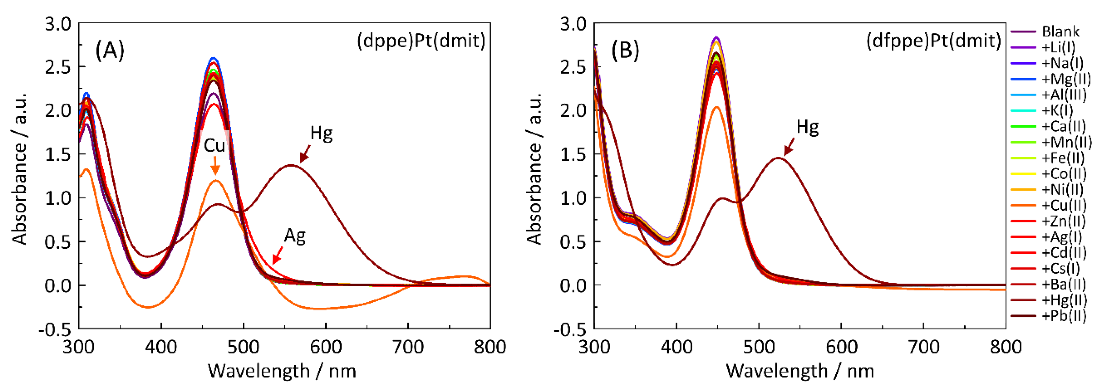
The Hg2+ sensing properties of the (dxpe)Pt(dmit) complexes were quantitatively investigated by monitoring the shift in their absorption peak by UV-visible spectrometry, which corresponds to the shift of the π→π* transition of the >C=S moiety in the dmit ligand (Figure 4 and Figure 5). In the absence of cations, the absorption spectra of (dppe)Pt(dmit) and (dfppe)Pt(dmit) contained one peak of medium intensity at λ1 = 463 nm and λ2 = 448 nm, respectively. The peaks in the spectra resemble those of complexes containing the Pt(dmit) moiety, such as (Ph3P)2Pt(dmit) and (P2)Pt(dmit) (P2 = 3,4-dimethyl-3’,4’-bis(diphenylphosphino)tetrathiafulvalene), indicating that they originated from the absorption of the >C=S moiety [23]. As various metal cations were added to the yellow solution of (dppe)Pt(dmit), the absorption intensity at λ1 decreased, with concurrent evolution of new peaks for the three metal ions (Hg2+, Cu2+, and Ag+) in the higher wavelength region: the characteristic bathochromic shift to 558 nm for Hg2+, a shift to 700–800 nm for Cu2+, and a slight increase (and consequently, a weak color change in Figure 3A) at approximately 530 nm for Ag+. In the case of the (dfppe)Pt(dmit) chemosensor, however, only the Hg2+ ion led to the evolution of a new absorption peak at 523 nm with a decrease in intensity at λ2. This bathochromic shift was accompanied by a color change of the solution to vivid red, as seen in Figure 3.
3.5. DFT Calculation
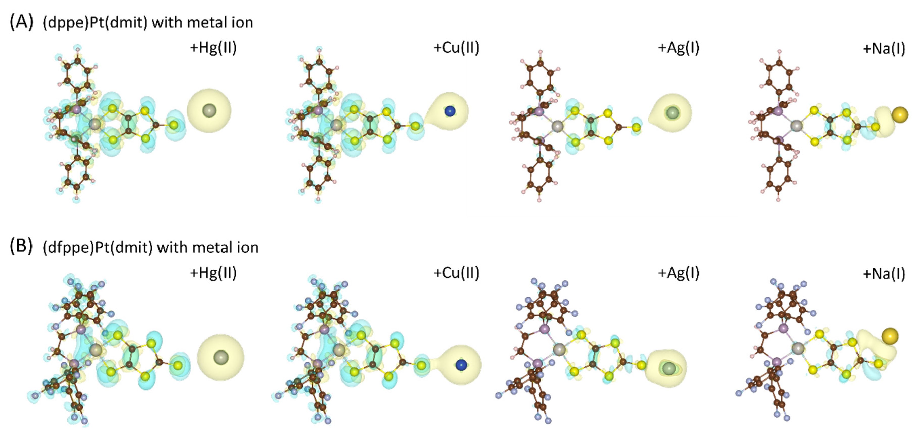
To rationalize the differential selectivity between (dppe)Pt(dmit) and (dfppe)Pt(dmit) for Hg2+ chemosensing, we investigated the mechanism involved in the interaction between the reactants at the atomic level using DFT calculations. The exposed S with a lone pair of electrons on the >C=S moiety had effective charges of 5.11e and 4.87e for (dppe)Pt(dmit) and (dfppe)Pt(dmit), respectively. The difference in these values was caused by the differing electronegativities of H(2.1) and F(4.0) protruding from the diphosphine ligands, which have an average effective charge of 0.95e and 7.81e, respectively. In the plots of differential charge densities shown in Figure 6, both cases exhibit a hybridized region between the chemosensor and the metal ion via intermolecular >C=S···M (M: metal ion) interactions in the solvent, indicating the occurrence of chemical adhesion. The differential charge density, ρdiff, of >C=S···M adhesion is defined by
where ρtotal is the charge density of S···M adhesion, ρ(dxpe)Pt(dmit) is the charge density for a single (dxpe)Pt(dmit) unit, and ρM is the charge density of the metal ions [26]. Charge accumulation (yellow) and charge reduction (blue) around (dxpe)Pt(dmit) and the metal ions are explicitly shown in Figure 6.
ρdiff = ρtotal − (ρ(dxpe)Pt(dmit) + ρM)
The two distinct charge densities of the (dxpe)Pt(dmit) chemosensors share a common feature: charge accumulation and reduction were horizontally superposed between the metal ions(M) and the S of the dmit moiety, which suggests an electrostatic interaction at the >C=S···M site. In this molecule, the interaction energy was dependent on the nature of the metal ion. In the case of Hg2+ ions, electronic hybridization significantly increased. On the other hand, hybridization decreased in the order of Ag+, Cu2+, and Na+, which is in good agreement with the results of the colorimetric sensing property analysis of (dxpe)Pt(dmit). The interaction characteristics between (dppe)Pt(dmit) and (dfppe)Pt(dmit) were analogous; however, their absolute values varied significantly. The variance in the effective charge on the S atom of the dmit moiety, with and without various metal ions, is shown in Table 3. The (dfppe)Pt(dmit) complex exhibited a notably smaller variance compared to (dppe)Pt(dmit), based on the all-metal ions. It should be noted that the selectivity of the chemosensor for other cations can be controlled by the hybridization shielding effect of F-ended functional groups.
4. Conclusions
We presented a novel methodology for the enhancement of Hg2+ detection by the introduction of differing functional terminal groups on (diphosphine)Pt(dmit) chemosensors. The use of distinct terminal groups enabled selectivity tuning, which is attributed to the modulation of specific intermolecular interactions. Organization of the end groups on the ligand moiety proved beneficial for altering whole-material properties. The results hold implications for the design of organic materials, wherein crucial properties can be enhanced through precise control of their chemical hybridization by functional end-group manipulation.
Author Contributions
D.-Y.N. conceived and designed the experiments. H.J., H.R., and I.N. carried out the synthesis of the materials, performed chemical characterization, and conducted simulations. I.N. and D.-Y.N. wrote the paper. All authors read and agreed to the published version of the manuscript.
Funding
This work was supported by the Basic Science Research Program through the National Research Foundation of Korea (NRF), funded by the Ministry of Education, Science, and Technology (NRF2010-0011478).
Acknowledgments
This work was supported by the National Institute of Supercomputing and Network/Korea Institute of Science and Technology Information with supercomputing resources including technical support (KSC-2019-CRE-0106).
Conflicts of Interest
The authors declare no conflict of interest.
References
- Valko, M.; Morris, H.; Cronin, M.T.D. Metals, toxicity and oxidative stress. Curr. Med. Chem. 2005, 12, 1161–1208. [Google Scholar] [CrossRef] [Green Version]
- Guo, C.; Irudayaraj, J. Fluorescent ag clusters via a protein-directed approach as a Hg(II) ion sensor. Anal. Chem. 2011, 83, 2883–2889. [Google Scholar] [CrossRef] [PubMed]
- Chen, P.; He, C.A. A General strategy to convert the merr family proteins into highly sensitive and selective fluorescent biosensors for metal ions. J. Am. Chem. Soc. 2004, 126, 728–729. [Google Scholar] [CrossRef] [PubMed]
- Chen, Y.Q.; Bai, H.; Hong, W.J.; Shi, G.Q. Fluorescence detection of mercury ions in aqueous media with the complex of a cationic oligopyrene derivative and oligothymine. Analyst 2009, 134, 2081–2086. [Google Scholar] [CrossRef] [PubMed]
- Huang, C.-C.; Yang, Z.; Lee, K.-H.; Chang, H.-T. Synthesis of highly fluorescent gold nanoparticles for sensing mercury(II). Angew. Chem. Int. Ed. 2007, 46, 6824–6828. [Google Scholar] [CrossRef]
- Li, D.; Wieckowska, A.; Willner, I. Optical analysis of Hg2+ ions by oligonucleotide-gold-nanoparticle hybrids and DNA-based machines. Angew. Chem. Int. Ed. 2008, 47, 3927–3931. [Google Scholar] [CrossRef]
- Wang, Z.D.; Lee, J.H.; Lu, Y. Highly sensitive “turn-on” fluorescent sensor for Hg2+ in aqueous solution based on structure-switching DNA. Chem. Commun. 2008, 44, 6005–6007. [Google Scholar] [CrossRef]
- Yigit, M.V.; Mishra, A.; Tong, R.; Cheng, J.J.; Wong, G.C.L.; Lu, Y. Inorganic mercury detection and controlled release of chelating agents from ion-responsive liposomes. Chem. Biol. 2009, 16, 937–942. [Google Scholar] [CrossRef] [Green Version]
- Yoon, S.; Miller, E.W.; He, Q.; Do, P.H.; Chang, C.J. A bright and specific fluorescent sensor for mercury in water, cells, and tissue. Angew. Chem. Int. Ed. 2007, 46, 6658–6661. [Google Scholar] [CrossRef]
- Coskun, A.; Akkaya, E.U. Signal ratio amplification via modulation of resonance energy transfer: Proof of principle in an emission ratiometric Hg(II) sensor. J. Am. Chem. Soc. 2006, 128, 14474–14475. [Google Scholar] [CrossRef]
- Nam, H.J.; Lee, H.-J.; Noh, D.-Y. Novel mercury(II) complexes of 1,3-dithiole-2-thiones containing the 2-pyridyl moiety: Syntheses, X-ray crystal structures and solution behavior. Polyhedron 2004, 23, 115–123. [Google Scholar] [CrossRef]
- Jeon, S.; Suh, W.; Noh, D.-Y. Anion-dependent Hg2+-sensing of colorimetric (dppe)Pt(dmit) chemosensor (dppe: 1,2-bis(diphenylphosphino)ethane; dmit: 1,3-dithiole-2-thione-4,5-dithiolate). Inorg. Chem. Commun. 2017, 81, 43–46. [Google Scholar] [CrossRef]
- Jeon, H.; Suh, W.; Noh, D.-Y. Hg(II) sensing properties of (diphosphine)Pt(dmit) complexes (dmit: C3S52−: 1,3-dithiole-2-thione-4,5-dithiolate). Inorg. Chem. Commun. 2012, 24, 181–185. [Google Scholar] [CrossRef]
- Nomura, M.; Fourmigué, M. Dinuclear Cp* Cobalt Complexes of the 1,2,4,5-Benzenetetrathiolate Bischelating Ligand. Inorg. Chem. 2008, 47, 1301–1312. [Google Scholar] [CrossRef] [PubMed]
- Doidge-Harrison, S.M.; Irvine, J.T.; Khan, A.; Spencer, G.M.; Wardell, J.L.; Aupers, J.H. Diorganotin 1,3-dithiole-2-thione-4,5-dithiolate compounds, R2Sn(dmit): The crystal structure of MePhSn(dmit). J. Organomet. Chem. 1996, 516, 199–205. [Google Scholar] [CrossRef]
- Bruker AXS Inc. SMART, SAINT-Plus v 6.22 and XPREP; Bruker AXS Inc.: Madison, WI, USA, 2000. [Google Scholar]
- Sheldrick, G.M. SADABS v 2.03; University of Göttingen: Göttingen, Germany, 2002. [Google Scholar]
- Bruker AXS Inc. SHELXTL v 6.10; Bruker AXS Inc.: Madison, WI, USA, 2000. [Google Scholar]
- Perdew, J.P.; Burke, K.; Ernzerhof, M. Generalized gradient approximation made simple. Phys. Rev. Lett. 1996, 77, 3865–3868. [Google Scholar] [CrossRef] [Green Version]
- Kresse, G.; Furthmüller, J. Efficient iterative schemes for ab initio total-energy calculations using a plane-wave basis set. Phys. Rev. B Condens. Matter Mater. Phys. 1996, 54, 11169–11186. [Google Scholar] [CrossRef]
- Grimme, S. Semiempirical GGA-Type density functional constructed with a long-range dispersion correction. J. Comput. Chem. 2006, 27, 1787–1799. [Google Scholar] [CrossRef]
- Vicente, R.; Ribas, J.; Solans, X.; Font-Altaba, M.; Mari, A.; Loth, P.; Cassoux, P. Electrochemical, EPR, and crystal structure studies on mixed-ligand 4,5-dimercapto-l,3-dithia-2-thione phosphine complexes of nickel, palladium and platinum, M(dmit)(dppe) and Pt(dmit)(PPh3)2. Inorg. Chim. Acta 1987, 132, 229–236. [Google Scholar] [CrossRef]
- Shin, K.S.; Jung, Y.; Lee, S.K.; Fourmigué, M.; Barrière, F.; Bergamini, J.F.; Noh, D.Y. Redox bifunctionality in a Pt(II) dithiolene complex of a tetrathiafulvalene diphosphine ligand. Dalton Trans. 2008, 5869–5871. [Google Scholar] [CrossRef]
- Pearson, R.G. Hard and soft acids and bases. J. Am. Chem. Soc. 1963, 85, 3533–3539. [Google Scholar] [CrossRef]
- Rumble, J. CRC Handbook of Chemistry and Physics, 100th ed.; CRC Press: Boca Raton, FL, USA, 2019. [Google Scholar]
- Bae, S.; Nam, I.; Park, S.; Yoo, Y.G.; Yu, S.; Lee, J.M.; Han, J.W.; Yi, J. Interfacial adsorption and redox coupling of Li4Ti5O12 with nanographene for high-rate lithium storage. ACS Appl. Mater. Interfaces 2015, 7, 16565–16572. [Google Scholar] [CrossRef] [PubMed]
Scheme 1.
Synthesis of (dfppe)Pt(dmit).

Figure 1.
Molecular structure of (dfppe)Pt(dmit) with the selected numbering scheme. Bond lengths (Å) and angles (°): Pt1-P1 2.2473(18), Pt1-P2 2.251(2), Pt1-S1 2.3023(19), Pt1-S2 2.3094(19), Pt2-P3 2.2554(19), Pt2-P4 2.2549(19), Pt2-S6 2.3107(19), Pt2-S7 2.3094(18), C1-C2 1.347(11), C3-S5 1.650(8), C30-C31 1.349(11), C32-S10 1.648(8), P1-Pt1-P2 86.59(7), P2-Pt1-S2 90.05(7), P1-Pt1-S2 175.51(7), P2-Pt1-S1 172.59(7), P1-Pt1-S1 90.38(7), S1-Pt1-S2 90.45(7), P3-Pt2-P4 86.48(7), P3-Pt2-S6 91.64(7), P4-Pt2-S6 175.93(7), P3-Pt2-S7 172.11(7), P4-Pt2-S7 91.51(7), and S6-Pt2-S7 89.88(7).

Figure 2.
Cyclic voltammogram of (dfppe)Pt(dmit) measured in dichloromethane (DCM) (Fc/Fc+ = 0.532 V vs Ag/AgCl, 0.1 M n-Bu4N⋅ClO4, Pt-button electrode, Ag/AgCl, 100 mV/s).
Figure 2.
Cyclic voltammogram of (dfppe)Pt(dmit) measured in dichloromethane (DCM) (Fc/Fc+ = 0.532 V vs Ag/AgCl, 0.1 M n-Bu4N⋅ClO4, Pt-button electrode, Ag/AgCl, 100 mV/s).

Figure 3.
Color changes of H2O/CH3CN solution (0.2 mM, 1:1 v/v %) of (A) (dppe)Pt(dmit) and (B) (dfppe)Pt(dmit) upon the addition of one equivalent of metal ions.
Figure 3.
Color changes of H2O/CH3CN solution (0.2 mM, 1:1 v/v %) of (A) (dppe)Pt(dmit) and (B) (dfppe)Pt(dmit) upon the addition of one equivalent of metal ions.

Figure 4.
UV-vis spectra of (A) (dppe)Pt(dmit) and (B) (dfppe)Pt(dmit) complexes with or without metal ions (Li+, Na+, K+, Cs+, Mg2+, Ca2+, Ba2+, Mn2+, Fe2+, Co2+, Ni2+, Cu2+, Zn2+, Ag+, Cd2+, Hg2+, Al3+, and Pb2+) dissolved in H2O/CH3CN solution (1:1 v/v %). The absorption peaks due to the >C=S moiety observed at (A) 463 nm and (B) 448 nm show a noticeable bathochromic shift upon the addition of Hg2+ ions.

Figure 5.
Absorbance ratios (A558/A464 and A524/A448) from the UV-Vis spectra of (a) (dppe)Pt(dmit) and (b) (dfppe)Pt(dmit), respectively, with or without metal ions dissolved in H2O/CH3CN solution (1:1 v/v %).
Figure 5.
Absorbance ratios (A558/A464 and A524/A448) from the UV-Vis spectra of (a) (dppe)Pt(dmit) and (b) (dfppe)Pt(dmit), respectively, with or without metal ions dissolved in H2O/CH3CN solution (1:1 v/v %).

Figure 6.
Charge analyses of (A) (dppe)Pt(dmit) and (B) (dfppe)Pt(dmit) complexes with representative metal ions. The charge accumulations and charge reductions around the molecules are shown as yellow and blue surfaces, respectively. The isosurface is 0.0015e a0−3, where a0 is the Bohr radius.
Figure 6.
Charge analyses of (A) (dppe)Pt(dmit) and (B) (dfppe)Pt(dmit) complexes with representative metal ions. The charge accumulations and charge reductions around the molecules are shown as yellow and blue surfaces, respectively. The isosurface is 0.0015e a0−3, where a0 is the Bohr radius.


Table 1.
Crystal data and structure refinement parameters for (dfppe)Pt(dmit).
| Empirical Formula | C29 H4 F20 P2 Pt S5 |
|---|---|
| Formula weight | 1149.65 |
| Crystal system | Monoclinic |
| Space group | P2(1)/n |
| a (Å) | 12.2555(10) |
| b (Å) | 13.7690(11) |
| c (Å) | 40.429(3) |
| β (°) | 90.906(2) |
| Volume (Å3) | 6821.4(9) |
| Z | 8 |
| Density Cal. (Mg/m3) | 2.239 |
| Absorption coefficient (mm−1) | 4.650 |
| F(000) | 4368 |
| Crystal size (mm3) | 0.23 × 0.16 × 0.08 |
| Theta range for data collection (°) | 1.007 to 26.017 |
| Index ranges | −15 ≤ h ≤ 8, −16 ≤ k ≤ 16, −49 ≤ l ≤ 46 |
| Reflections collected | 41778 |
| Independent reflections | 13361 [R(int) = 0.0618] |
| Completeness to theta = 25.242° (%) | 99.8 |
| Absorption correction | Semiempirical from equivalents |
| Refinement method | Full-matrix least-squares on F2 |
| Data/restraints/parameters | 13361/0/1027 |
| Goodness-of-fit on F2 | 1.037 |
| Final R indices [I > 2σ(I)] | R1 = 0.0442, wR2 = 0.0938 |
| R indices (all data) | R1 = 0.0757, wR2 = 0.1147 |
| Largest diff. peak and hole (e.Å−3) | 2.062 and −1.187 |
Table 2.
Comparison of selected bond lengths (Å) and angles (°) between (dppe)Pt(dmit) and (dfppe)Pt(dmit).
Table 2.
Comparison of selected bond lengths (Å) and angles (°) between (dppe)Pt(dmit) and (dfppe)Pt(dmit).
| Complexes | Bond Lengths (Å) | Bond Angles (°) | ||||
|---|---|---|---|---|---|---|
| Pt1-P1 | Pt1-P2 | Pt1-S1 | Pt1-S2 | P1-Pt-P2 | S1-Pt-S2 | |
| (dppe)Pt(dmit) 1) | 2.251(3) | 2.258(3) | 2.315(3) | 2.308(3) | 85.2(1) | 90.0(1) |
| (dfppe)Pt(dmit) | 2.2473(18) | 2.251(2) | 2.3023(19) | 2.3094(19) | 86.59(7) | 90.45(7) |
| 2.2554(19) | 2.2549(19) | 2.3107(19) | 2.3094(19) | 86.48(7) | 89.88(7) | |
1) Ref. [22].
Table 3.
Bader atomic charge analyses for the effective charges of (dppe)Pt(dmit) and (dfppe)Pt(dmit) complexes with and without representative metal ions.
Table 3.
Bader atomic charge analyses for the effective charges of (dppe)Pt(dmit) and (dfppe)Pt(dmit) complexes with and without representative metal ions.
| Complexes | −e 1) | Δ(−e) 2) | |||
|---|---|---|---|---|---|
| Hg2+ | Cu2+ | Ag+ | Na+ | ||
| (dppe)Pt(dmit) | 5.11 | −1.76 | −1.42 | −0.66 | −0.08 |
| (dfppe)Pt(dmit) | 4.87 | −1.73 | −1.18 | −0.35 | −0.08 |
1) Effective charge of S on >C=S moiety in (dxpe)Pt(dmit) without metal ions; 2) Differential effective charge of S on >C=S moiety in (dxpe)Pt(dmit) with metal ions.
© 2020 by the authors. Licensee MDPI, Basel, Switzerland. This article is an open access article distributed under the terms and conditions of the Creative Commons Attribution (CC BY) license (http://creativecommons.org/licenses/by/4.0/).
Share and Cite
MDPI and ACS Style
Jeon, H.; Ryu, H.; Nam, I.; Noh, D.-Y. Heteroleptic Pt(II)-dithiolene-based Colorimetric Chemosensors: Selectivity Control for Hg(II) Ion Sensing. Materials 2020, 13, 1385. https://0-doi-org.brum.beds.ac.uk/10.3390/ma13061385
AMA Style
Jeon H, Ryu H, Nam I, Noh D-Y. Heteroleptic Pt(II)-dithiolene-based Colorimetric Chemosensors: Selectivity Control for Hg(II) Ion Sensing. Materials. 2020; 13(6):1385. https://0-doi-org.brum.beds.ac.uk/10.3390/ma13061385
Chicago/Turabian StyleJeon, Hyokyung, Hwahui Ryu, Inho Nam, and Dong-Youn Noh. 2020. "Heteroleptic Pt(II)-dithiolene-based Colorimetric Chemosensors: Selectivity Control for Hg(II) Ion Sensing" Materials 13, no. 6: 1385. https://0-doi-org.brum.beds.ac.uk/10.3390/ma13061385
Note that from the first issue of 2016, this journal uses article numbers instead of page numbers. See further details here.