
Vertical and Orovestibular Forces Generated by Beta-Titanium and Stainless-Steel Rectangular Wires in Labial and Fully Customized Lingual Bracket Systems


Abstract
:
1. Introduction
2. Materials and Methods
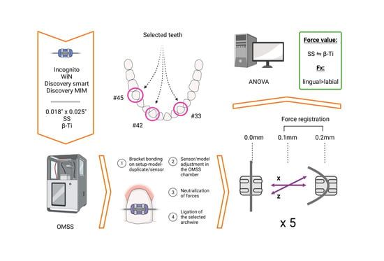
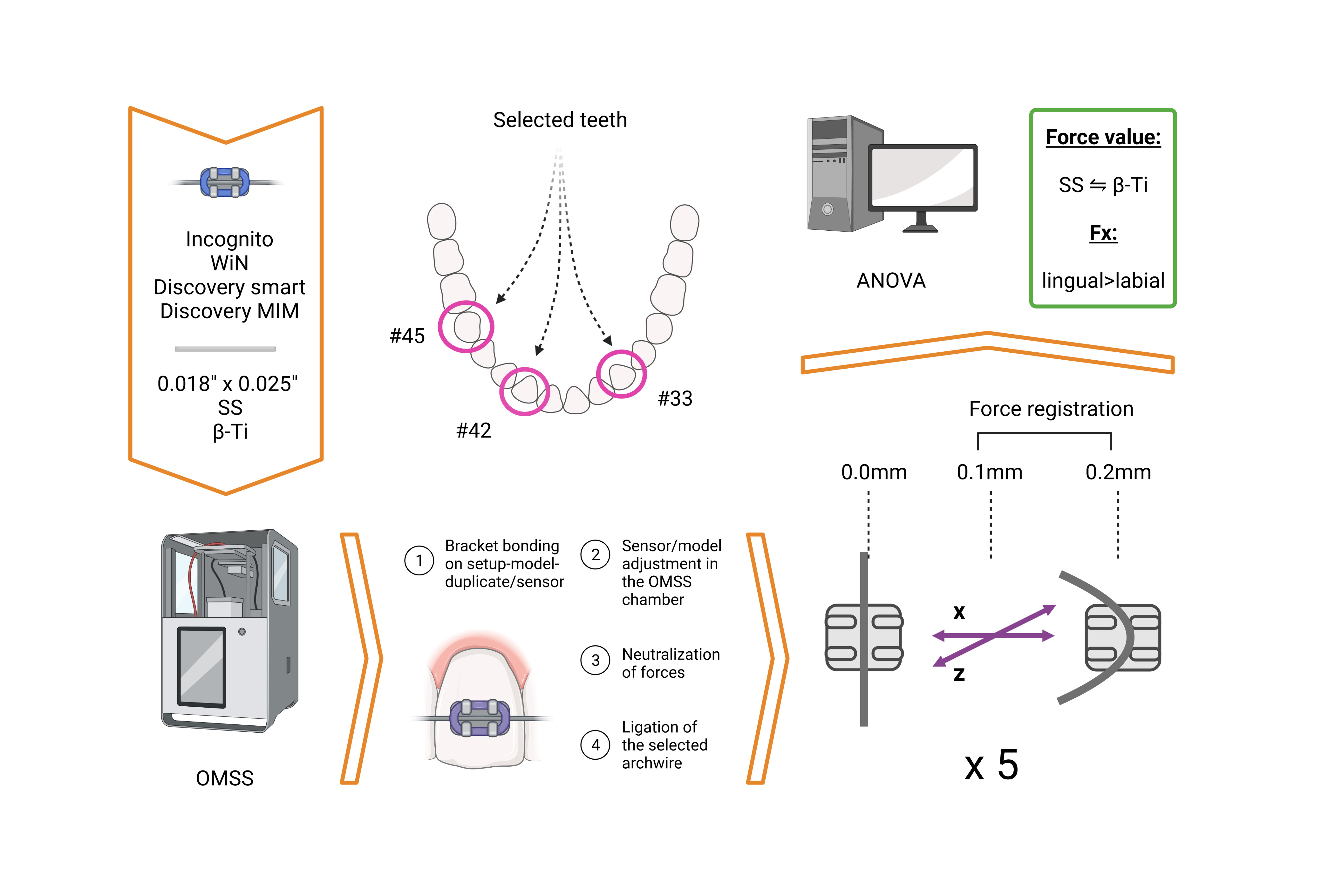
2.1. Bracket Appliances and Wires
2.2. Model
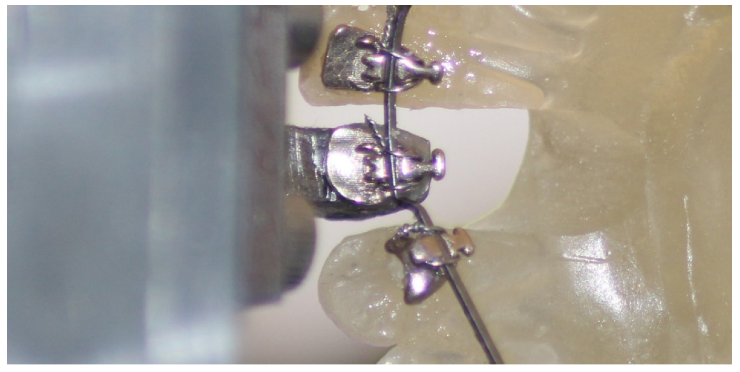
2.3. Apparatus
2.4. Activation Procedure
2.5. Statistical Analysis
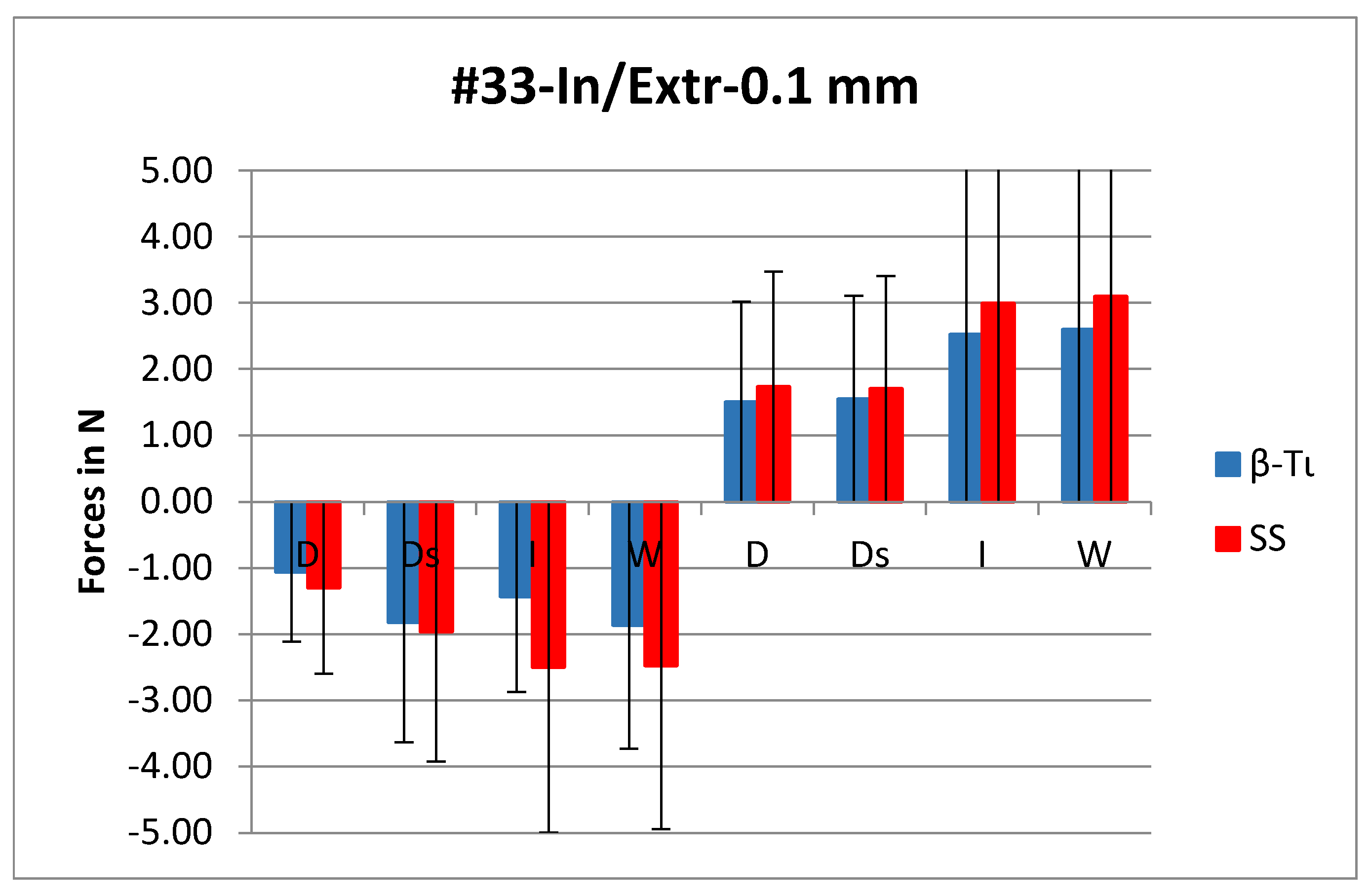
3. Results
3.1. Stainless-Steel Wires
3.2. Beta-Titanium Wires
4. Discussion
4.1. Sources of Error
4.2. Limitations
5. Conclusions
Author Contributions
Funding
Institutional Review Board Statement
Informed Consent Statement
Data Availability Statement
Acknowledgments
Conflicts of Interest
References
- D’Amario, M.; Bernardi, S.; Di Lauro, D.; Marzo, G.; Macchiarelli, G.; Capogreco, M. Debonding and Clean-Up in Orthodontics: Evaluation of Different Techniques and Micro-Morphological Aspects of the Enamel Surface. Dent. J. 2020, 17, 58. [Google Scholar] [CrossRef]
- Perinetti, G.; Varvara, G.; Salini, L.; Tetè, S. Alkaline phosphatase activity in dental pulp of orthodontically treated teeth. Am. J. Orthod. Dentofac. Orthop. 2005, 128, 492–496. [Google Scholar] [CrossRef]
- Knösel, M.; Klang, E.; Helms, H.J.; Wiechmann, D. Occurrence and severity of enamel decalcification adjacent to bracket bases and sub-bracket lesions during orthodontic treatment with two different lingual appliances. Eur. J. Orthod. 2016, 38, 485–492. [Google Scholar] [CrossRef] [Green Version]
- Maués, C.P.; do Nascimento, R.R.; Vilella Ode, V. Severe root resorption resulting from orthodontic treatment: Prevalence and risk factors. Dent. Press J. Orthod. 2015, 20, 52–58. [Google Scholar] [CrossRef] [PubMed]
- Kurz, C.; Swartz, M.L.; Andreiko, C. Lingual orthodontics: A status report. Part 2: Research and development. J. Clin. Orthod. 1982, 16, 735–740. [Google Scholar] [PubMed]
- Wiechmann, D. A new bracket system for lingual orthodontic treatment. Part 1: Theoretical background and development. J. Orofac. Orthop. 2002, 63, 234–245. [Google Scholar] [CrossRef]
- Choi, S.; Kim, J.H.; Kim, N.J.; Lee, S.H.; Kim, H.; Kim, K.A.; Park, K.H.; Park, Y.G. Morphological investigation of various orthodontic lingual bracket slots using scanning electron microscopy and atomic force microscopy. Microsc. Res. Tech. 2016, 79, 1193–1199. [Google Scholar] [CrossRef]
- Daratsianos, N.; Bourauel, C.; Fimmers, R.; Jäger, A.; Schwestka-Polly, R. In vitro biomechanical analysis of torque capabilities of various 0.018″ lingual bracket-wire systems: Total torque play and slot size. Eur. J. Orthod. 2016, 38, 459–469. [Google Scholar] [CrossRef] [PubMed]
- Fuck, L.M.; Wiechmann, D.; Drescher, D. Comparison of the initial orthodontic force systems produced by a new lingual bracket system and a straight-wire appliance. J. Orofac. Orthop. 2005, 66, 363–376. [Google Scholar] [CrossRef]
- Sifakakis, I.; Pandis, N.; Makou, M.; Katsaros, C.; Eliades, T.; Bourauel, C. A comparative assessment of forces and moments generated by lingual and conventional brackets. Eur. J. Orthod. 2013, 35, 82–86. [Google Scholar] [CrossRef]
- Alobeid, A.; El-Bialy, T.; Khawatmi, S.; Dirk, C.; Jäger, A.; Bourauel, C. Comparison of the force levels among labial and lingual self-ligating and conventional brackets in simulated misaligned teeth. Eur. J. Orthod. 2017, 39, 419–425. [Google Scholar] [CrossRef] [PubMed]
- Papageorgiou, S.N.; Konstantinidis, I.; Papadopoulou, K.; Jäger, A.; Bourauel, C. A systematic review and meta-analysis of experimental clinical evidence on initial aligning archwires and archwire sequences. Orthod. Craniofac. Res. 2014, 17, 197–215. [Google Scholar] [CrossRef]
- Proffit, W.R.; Fields Junior, H.W.; Sarver, D.M. Contemporary Orthodontics, 4th ed.; Mosby: St. Louis, MO, USA, 2007; pp. 362; 365–366. [Google Scholar]
- Morinaga, M.; Yukawa, N.; Maya, T.; Sone, K.; Adachi, H. Theoretical design of titanium alloys. In Proceedings of the 6th World Conference on Titanium, Cannes, France, 6–9 June 1988; pp. 1601–1606. [Google Scholar]
- Kim, H.Y.; Kim, J.I.; Inamura, T.; Hosoda, H.; Miyazaki, S. Effect of thermo-mechanical treatment on mechanical properties and shape memory behavior of Ti–(26–28)at.% Nb alloys. Mater. Sci. Eng. A 2006, 438, 839–843. [Google Scholar] [CrossRef]
- Sun, F.; Nowak, S.; Gloriant, T.; Laheurte, P.; Eberhardt, A.; Prima, F. Influence of a short thermal treatment on the superelastic properties of a titanium-based alloy. Scr. Mater. 2010, 63, 1053–1056. [Google Scholar] [CrossRef]
- Kusy, R.P. On the use of nomograms to determine the elastic property ratios of orthodontic archwires. Am. J. Orthod. 1983, 83, 374–381. [Google Scholar] [CrossRef]
- Verstrynge, A.; Van Humbeeck, J.; Willems, G. In-vitro evaluation of the material characteristics of stainless steel and beta-titanium orthodontic wires. Am. J. Orthod. Dentofac. Orthop. 2006, 130, 460–470. [Google Scholar] [CrossRef]
- Lombardo, L.; Carlucci, A.; Palone, M.; Mollica, F.; Siciliani, G. Stiffness comparison of mushroom and straight SS and TMA lingual archwires. Prog. Orthod. 2016, 17, 27. [Google Scholar] [CrossRef] [Green Version]
- Johnson, E. Relative stiffness of beta titanium archwires. Angle Orthod. 2003, 73, 259–269. [Google Scholar] [CrossRef]
- Alsabti, N.; Bourauel, C.; Talic, N. Comparison of force loss during sliding of low friction and conventional TMA orthodontic archwires: An in vitro study. J. Orofac. Orthop. 2021, 82, 218–225. [Google Scholar] [CrossRef]
- Whitley, J.Q.; Kusy, R.P. Resistance to sliding of titanium brackets tested against stainless steel and beta-titanium archwires with second-order angulation in the dry and wet states. Am. J. Orthod. Dentofac. Orthop. 2007, 131, 400–411. [Google Scholar] [CrossRef]
- Seixas, M.R.; Konatu, R.T.; Rangel, A.L.R.; Hilsdorf, G.O.R.; Pereirajr, A.; Alves Claro, A.P.R. Study of a new titanium alloy for orthodontic applications. Materials 2013, 29 (Suppl. 1), e1–e96. [Google Scholar] [CrossRef]
- Tran, B.; Nobes, D.S.; Major, P.W.; Carey, J.P.; Romanyk, D.L. Research paper: The three-dimensional mechanical response of orthodontic archwires and brackets in vitro during simulated orthodontic torque. J. Mech. Behav. Biomed. Mater. 2021, 114, 104196. [Google Scholar] [CrossRef]
- Mandall, N.; Lowe, C.; Worthington, H.; Sandler, J.; Derwent, S.; Abdi-Oskouei, M.; Ward, S. Which orthodontic archwire sequence? A randomized clinical trial. Eur. J. Orthod. 2006, 28, 561–566. [Google Scholar] [CrossRef] [Green Version]
- Kerosuo, H.; Kullaa, A.; Kerosuo, E.; Kanerva, L.; Hensten-Pettersen, A. Nickel allergy in adolescents in relation to orthodontic treatment and piercing of ears. Am. J. Orthod. Dentofac. Orthop. 1996, 109, 148–154. [Google Scholar] [CrossRef]
- Mortz, C.G.; Lauritsen, J.M.; Bindslev-Jensen, C.; Andersen, K.E. Nickel sensitization in adolescents and association with ear piercing, use of dental braces and hand eczema. The Odense Adolescence Cohort Study on Atopic Diseases and Dermatitis (TOACS). Acta Derm. Venereol. 2002, 82, 359–364. [Google Scholar] [CrossRef] [PubMed] [Green Version]
- Kyprianou, C.; Chatzigianni, A.; Daratsianos, N.; Bourauel, C. Comparative investigation of fully customized lingual bracket systems and conventional labial appliances: Analysis of forces/moments and final tooth positions. Am. J. Orthod. Dentofac. Orthop. 2021. Accepted for publication. [Google Scholar]
- Drescher, D.; Bourauel, C.; Thier, M. Application of the orthodontic measurement and simulation system (OMSS) in orthodontics. Eur. J. Orthod. 1991, 13, 169–178. [Google Scholar] [CrossRef] [PubMed]
- Bourauel, C.; Drescher, D.; Thier, M. An experimental apparatus for the simulation of three-dimensional movements in orthodontics. J. Biomed. Eng. 1992, 14, 371–378. [Google Scholar] [CrossRef]
- Burstone, C.J.; Koenig, H.A. Force systems from an ideal arch. Am. J. Orthod. Dentofac. Orthop. 1974, 65, 270–289. [Google Scholar] [CrossRef]
- Creekmore, T.D. The importance of interbracket width in orthodontic tooth movement. J. Clin. Orthod. 1976, 10, 530–534. [Google Scholar]
- Moran, K.I. Relative wire stiffness due to lingual versus labial interbracket distance. Am. J. Orthod. Dentofac. Orthop. 1987, 92, 24–32. [Google Scholar] [CrossRef]
- Abtahi, M.; Eslami, N.; Abadi, R.Z.; Rezaei, S.P. The effect of intrusive orthodontic force on dental pulp of adults versus adolescents. Dent. Res. J. 2016, 13, 367–372. [Google Scholar] [CrossRef]
- Wang, H.; Li, T.; Wang, X.; Guan, Y.; Jiang, Y.; Chen, S.; Zou, S.; Duan, P. Mechanisms of sphingosine-1-phosphate (S1P) signaling on excessive stress-induced root resorption during orthodontic molar intrusion. Clin. Oral Investig. 2021. Epub ahead of print. [Google Scholar] [CrossRef]
- Akl, H.E.; El-Beialy, A.R.; El-Ghafour, M.A.; Abouelezz, A.M.; El Sharaby, F.A. Root resorption associated with maxillary buccal segment intrusion using variable force magnitudes: A randomized clinical trial. Angle Orthod. 2021. Epub ahead of print. [Google Scholar] [CrossRef]
- Bayir, F.; Bolat Gumus, E. External apical root resorption after orthodontic treatment: Incidence, severity and risk factors. J. Dent. Res. Dent. Clin. Dent. Prospect. 2021, 15, 100–105. [Google Scholar] [CrossRef] [PubMed]
- Pinheiro, L.H.M.; Guimarães, L.S.; Antunes, L.S.; Küchler, E.C.; Kirschneck, C.; Antunes, L.A.A. Genetic variation involved in the risk to external apical root resorption in orthodontic patients: A systematic review. Clin. Oral Investig. 2021, 25, 5613–5627. [Google Scholar] [CrossRef] [PubMed]
- Pompei-Reynolds, R.C.; Kanavakis, G. Interslot variations of transition temperature range and force delivery in copper-nickel-titanium orthodontic wires. Am. J. Orthod. Dentofac. Orthop. 2014, 146, 215–226. [Google Scholar] [CrossRef] [PubMed]
- Fansa, M.; Keilig, L.; Reimann, S.; Jäger, A.; Bourauel, C. The leveling effectiveness of self-ligating and conventional brackets for complex tooth malalignments. J. Orofac. Orthop. 2009, 70, 285–296. [Google Scholar] [CrossRef]
- Kusy, R.P. A review of contemporary archwires: Their properties and characteristics. Angle Orthod. 1997, 67, 197–207. [Google Scholar] [CrossRef] [PubMed]
- Cacciafesta, V.; Sfondrini, M.F.; Ricciardi, A.; Scribante, A.; Klersy, C.; Auricchio, F. Evaluation of friction of stainless steel and esthetic self-ligating brackets in various bracket-archwire combinations. Am. J. Orthod. Dentofac. Orthop. 2003, 124, 395–402. [Google Scholar] [CrossRef]
- Cash, A.; Curtis, R.; Garrigia-Majo, D.; McDonald, F. A comparative study of the static and kinetic frictional resistance of titanium molybdenum alloy archwires in stainless steel brackets. Eur. J. Orthod. 2004, 26, 105–111. [Google Scholar] [CrossRef] [Green Version]
- Doshi, U.H.; Bhad-Patil, W.A. Static frictional force and surface roughness of various bracket and wire combinations. Am. J. Orthod. Dentofac. Orthop. 2011, 139, 74–79. [Google Scholar] [CrossRef] [PubMed]
- Drescher, D.; Bourauel, C.; Schumacher, H.A. Frictional forces between bracket and arch wire. Am. J. Orthod. Dentofac. Orthop. 1989, 96, 397–404. [Google Scholar] [CrossRef]
- Kusy, R.P.; Whitley, J.Q. Coefficients of friction for arch wires in stainless steel and polycrystalline alumina bracket slots. I. The dry state. Am. J. Orthod. Dentofac. Orthop. 1990, 98, 300–312. [Google Scholar] [CrossRef]
- Fidalgo, T.K.; Pithon, M.M.; Maciel, J.V.; Bolognese, A.M. Friction between different wire bracket combinations in artificial saliva—An in vitro evaluation. J. Appl. Oral Sci. 2011, 19, 57–62. [Google Scholar] [CrossRef]
- Nishio, C.; da Motta, A.F.; Elias, C.N.; Mucha, J.N. In vitro evaluation of frictional forces between archwires and ceramic brackets. Am. J. Orthod. Dentofac. Orthop. 2004, 125, 56–64. [Google Scholar] [CrossRef]
- Amuk, N.G.; Karsli, E.; Kurt, G. Comparison of dental measurements between conventional plaster models, digital models obtained by impression scanning and plaster model scanning. Int. Orthod. 2019, 17, 151–158. [Google Scholar] [CrossRef]
- Kirschneck, C.; Kamuf, B.; Putsch, C.; Chhatwani, S.; Bizhang, M.; Danesh, G. Conformity, reliability and validity of digital dental models created by clinical intraoral scanning and extraoral plaster model digitization workflows. Comput. Biol. Med. 2018, 100, 114–122. [Google Scholar] [CrossRef]
- Peutzfeldt, A.; Asmussen, E. Accuracy of alginate and elastomeric impression materials. Scand. J. Dent. Res. 1989, 97, 375–379. [Google Scholar] [CrossRef] [PubMed]
- Vitti, R.P.; da Silva, M.A.; Consani, R.L.; Sinhoreti, M.A. Dimensional accuracy of stone casts made from silicone-based impression materials and three impression techniques. Braz. Dent. J. 2013, 24, 498–502. [Google Scholar] [CrossRef] [PubMed] [Green Version]
- Cho, S.H.; Schaefer, O.; Thompson, G.A.; Guentsch, A. Comparison of accuracy and reproducibility of casts made by digital and conventional methods. J. Prosthet. Dent. 2015, 113, 310–315. [Google Scholar] [CrossRef] [PubMed] [Green Version]
- Wan Hassan, W.N.; Othman, S.A.; Chan, C.S.; Ahmad, R.; Ali, S.N.; Abd Rohim, A. Assessing agreement in measurements of orthodontic study models: Digital caliper on plaster models vs. 3-dimensional software on models scanned by structured-light scanner. Am. J. Orthod. Dentofac. Orthop. 2016, 150, 886–895. [Google Scholar] [CrossRef] [Green Version]
- Vinay, K.; Venkatesh, M.J.; Nayak, R.S.; Pasha, A.; Rajesh, M.; Kumar, P. A comparative study to evaluate the effects of ligation methods on friction in sliding mechanics using 0.022" slot brackets in dry state: An In-vitro study. J. Int. Oral Health 2014, 6, 76–83. [Google Scholar] [PubMed]
- Thorstenson, G.A.; Kusy, R.P. Resistance to sliding of self-ligating brackets versus conventional stainless steel twin brackets with second-order angulation in the dry and wet (saliva) states. Am. J. Orthod. Orofac. Orthop. 2001, 120, 361–370. [Google Scholar] [CrossRef] [PubMed]
- Phukaoluan, A.; Khantachawana, A.; Kaewtatip, P.; Dechkunakorn, S.; Anuwongnukroh, N.; Santiwong, P.; Kajornchaiyakul, J. Comparison of friction forces between stainless orthodontic steel brackets and TiNi wires in wet and dry conditions. Int. Orthod. 2017, 15, 13–24. [Google Scholar] [CrossRef]


| Dimensions (mm) | Young’s Modulus (GPa) | Temperature Dependence | ||
|---|---|---|---|---|
| β-Ti | IncognitoTM | 0.635 × 0.462 | 72 | None |
| WiN | 0.635 × 0.457 | |||
| Dentaurum | 0.457 × 0.635 | |||
| SS | IncognitoTM | 0.635 × 0.457 | 200 | None |
| WiN | 0.635 × 0.457 | |||
| Dentaurum | 0.457 × 0.635 |
| Slot Length (mm) | ||||
| Discovery® MIM | Discovery® Smart | WiN | Incognito™ | |
| Lower left 1st bicuspid (#34) | 3.3 | 2.7 | 3.1 | 2.5 |
| Lower left cuspid (#33) | 2.9 | 2.7 | 2.5 | 2.3 |
| Lower left lateral incisor (#32) | 2.6 | 2.3 | 2.1 | 2.4 |
| Lower right central incisor (#41) | 2.6 | 2.3 | 2.1 | 2.4 |
| Lower right lateral incisor (#42) | 2.6 | 2.3 | 2.1 | 2.4 |
| Lower right cuspid (#43) | 3.0 | 2.7 | 2.5 | 2.4 |
| Lower right 1st bicuspid (#44) | 3.3 | 2.8 | 3.0 | 2.4 |
| Lower right 2nd bicuspid (#45) | 3.3 | 2.7 | 3.1 | 2.4 |
| Lower right 1st molar (#46) | 3.2 | 3.2 | 3.1 | 3.9 |
| Distance Between Adjacent Slots (mm) | ||||
| Discovery® MIM | Discovery® Smart | WiN | Incognito™ | |
| #34–#33 | 4.5 | 4.9 | 3.0 | 3.4 |
| #33–#32 | 4.2 | 4.9 | 2.8 | 3.0 |
| #41–#42 | 3.1 | 3.7 | 3.0 | 2.9 |
| #42–#43 | 4.6 | 5.3 | 2.2 | 2.2 |
| #44–#45 | 3.6 | 4.0 | 3.3 | 4.1 |
| #45–#46 | 5.7 | 6.6 | 4.7 | 4.4 |
| Wire Dimensions (mm) | ||||
|---|---|---|---|---|
| Discovery® MIM | Discovery® Smart | WiN | Incognito™ | |
| SS1 | 0.440 × 0.624 | 0.442 × 0.626 | 0.632 × 0.457 | 0.622 × 0.451 |
| SS2 | 0.441 × 0.624 | 0.440 × 0.622 | 0.623 × 0.450 | |
| SS3 | 0.441 × 0.622 | 0.444 × 0.620 | 0.623 × 0.453 | |
| SS4 | 0.445 × 0.623 | 0.443 × 0.621 | 0.620 × 0.452 | |
| SS5 | 0.445 × 0.622 | 0.440 × 0.622 | 0.620 × 0.452 | |
| β-Ti1 | 0.450 × 0.631 | 0.450 × 0.633 | 0.635 × 0.445 | 0.623 × 0.455 |
| β-Ti2 | 0.443 × 0.628 | 0.450 × 0.628 | 0.623 × 0.455 | |
| β-Ti3 | 0.446 × 0.632 | 0.449 × 0.632 | 0.623 × 0.454 | |
| β-Ti4 | 0.449 × 0.630 | 0.450 × 0.632 | 0.624 × 0.455 | |
| β-Ti5 | 0.446 × 0.631 | 0.450 × 0.630 | 0.620 × 0.456 | |
| Act+ | Teeth under Examination | ||||||||||||
|---|---|---|---|---|---|---|---|---|---|---|---|---|---|
| #33 | #42 | #45 | |||||||||||
| 0.1 mm | 0.2 mm | 0.1 mm | 0.2 mm | 0.1 mm | 0.2 mm | ||||||||
| Brackets | Wires | Fx | Fz | Fx | Fz | Fx | Fz | Fx | Fz | Fx | Fz | Fx | Fz |
| (N) | (N) | (N) | (N) | (N) | (N) | (N) | (N) | (N) | (N) | (N) | (N) | ||
| D | SS | −1.3 a | −1.1 r | −2.7 c | −1.8 u | −1.7 f | −1.5 x | −3.4 i,k | −2.2 j | −1.9 m,n | −0.7 φ | −3.3 p | −1.5 ω |
| Ds | SS | −2.0 b | −1.0 r | −3.5 d | −1.6 u | −1.3 g | −1.0 y | −3.0 k | −1.9 l | −1.5 m | −1.3 ψ | −2.7 p | −2.2 β |
| W | SS | −2.5 b | −0.6 s | −3.0 c,d | −1.0 v | −2.1 f | −0.9 y | −2.8 k | −1.4 θ | −2.9 n | −1.1 ψ | −4.7 q | −1.7 ω,β |
| I | SS | −2.5 b | −1.7 t | −4.2 e | −2.8 w | −2.6 h | −1.7 z | −3.9 i | −2.8 λ | −2.3 o | −1.2 ψ | −4.7 q | −1.9 ω,β |
| p value for ANOVA | 0.000 | 0.000 | 0.000 | 0.000 | 0.000 | 0.000 | 0.004 | 0.000 | 0.000 | 0.001 | 0.000 | 0.013 | |
| Act- | Teeth under Examination | ||||||||||||
|---|---|---|---|---|---|---|---|---|---|---|---|---|---|
| #33 | #42 | #45 | |||||||||||
| 0.1 mm | 0.2 mm | 0.1 mm | 0.2 mm | 0.1 mm | 0.2 mm | ||||||||
| Brackets | Wires | Fx | Fz | Fx | Fz | Fx | Fz | Fx | Fz | Fx | Fz | Fx | Fz |
| (N) | (N) | (N) | (N) | (N) | (N) | (N) | (N) | (N) | (N) | (N) | (N) | ||
| D | SS | 1.7 a | 1.6 q | 2.3 c | 2.5 t | 2.4 f | 2.0 w | 3.2 i | 2.8 x | 1.8 l | 1.3 y | 2.5 o | 2.1 θ |
| Ds | SS | 1.7 a | 1.3 q | 2.3 c | 1.9 u | 1.4 g | 2.2 w | 2.2 j | 3.0 x | 1.8 l | 1.3 y | 2.4 o | 2.3 θ |
| W | SS | 3.1 b | 3.1 r | 3.9 d | 3.4 v | 3.0 h | 2.2 w | 4.1 k | 2.8 x | 3.0 m | 1.8 z | 3.6 p | 2.2 θ |
| I | SS | 3.0 b | 2.0 s | 4.6 e | 2.7 t | 2.8 h | 2.4 w | 3.9 k | 3.0 x | 2.4 n | 1.5 y,z | 3.3 p | 2.4 θ |
| p value for ANOVA | 0.000 | 0.000 | 0.000 | 0.000 | 0.000 | 0.699 | 0.000 | 0.451 | 0.000 | 0.000 | 0.022 | 0.388 | |
| Act+ | Teeth under Examination | ||||||||||||
|---|---|---|---|---|---|---|---|---|---|---|---|---|---|
| #33 | #42 | #45 | |||||||||||
| 0.1 mm | 0.2 mm | 0.1 mm | 0.2 mm | 0.1 mm | 0.2 mm | ||||||||
| Brackets | Wires | Fx | Fz | Fx | Fz | Fx | Fz | Fx | Fz | Fx | Fz | Fx | Fz |
| (N) | (N) | (N) | (N) | (N) | (N) | (N) | (N) | (N) | (N) | (N) | (N) | ||
| D | β-Ti | −1.1 b | −1.0 s | −2.1 d | −1.5 u | −1.4 f | −1.2 x | −2.7 i | −2.0 y | −1.4 k | −1.0 θ | −2.6 o | −1.5 ψ |
| Ds | β-Ti | −1.8 a | −0.9 s | −3.4 e | −1.5 u | −1.1 f | −1.2 x | −2.6 i | −1.9 y | −1.2 l | −1.4 λ | −2.3 p | −2.1 ω |
| W | β-Ti | −1.9 a | −1.7 t | −2.3 d | −2.8 v | −2.2 g | −1.1 x | −3.0 i | −1.8 y | −2.1 m | −0.4 φ | −4.0 q | −0.7 γ |
| I | β-Ti | −1.4 c | −1.4 t | −2.5 d | −2.2 w | −2.6 h | −1.4 x | −4.1 j | −2.5 z | −1.9 n | −0.8 θ | −3.8 r | −1.4 ψ |
| p value for ANOVA | 0.000 | 0.000 | 0.000 | 0.000 | 0.000 | 0.264 | 0.001 | 0.000 | 0.000 | 0.001 | 0.000 | 0.013 | |
| Act- | Teeth under Examination | ||||||||||||
|---|---|---|---|---|---|---|---|---|---|---|---|---|---|
| #33 | #42 | #45 | |||||||||||
| 0.1 mm | 0.2 mm | 0.1 mm | 0.2 mm | 0.1 mm | 0.2 mm | ||||||||
| Brackets | Wires | Fx | Fz | Fx | Fz | Fx | Fz | Fx | Fz | Fx | Fz | Fx | Fz |
| (N) | (N) | (N) | (N) | (N) | (N) | (N) | (N) | (N) | (N) | (N) | (N) | ||
| D | β-Ti | 1.5 a | 1.4 q | 2.1 c | 2.2 t | 1.9 e | 1.8 w | 2.7 h | 2.5 y,z | 1.5 k | 1.5 θ | 2.1 n | 2.4 λ |
| Ds | β-Ti | 1.6 a | 1.3 q | 2.2 c | 1.9 t | 1.4 f | 1.9 w,x | 2.2 i | 2.7 y | 1.7 k | 1.9 θ,ω | 2.3 n | 2.8 λ |
| W | β-Ti | 2.6 b | 2.6 r | 3.4 d | 3.5 u | 2.6 g | 1.6 w,x | 3.4 j | 2.9 z | 2.7 l | 2.1 ω | 3.2 o | 2.5 λ |
| I | β-Ti | 2.5 b | 2.1 s | 3.1 d | 2.7 v | 2.5 g | 2.2 x | 3.3 j | 2.2 y | 2.1 m | 1.6 θ | 2.8 p | 2.0 ψ |
| p value for ANOVA | 0.000 | 0.000 | 0.000 | 0.000 | 0.000 | 0.054 | 0.000 | 0.006 | 0.000 | 0.004 | 0.000 | 0.002 | |
Publisher’s Note: MDPI stays neutral with regard to jurisdictional claims in published maps and institutional affiliations. |
© 2021 by the authors. Licensee MDPI, Basel, Switzerland. This article is an open access article distributed under the terms and conditions of the Creative Commons Attribution (CC BY) license (https://creativecommons.org/licenses/by/4.0/).
Share and Cite
Kyprianou, C.; Chatzigianni, A.; Daratsianos, N.; Bourauel, C. Vertical and Orovestibular Forces Generated by Beta-Titanium and Stainless-Steel Rectangular Wires in Labial and Fully Customized Lingual Bracket Systems. Materials 2021, 14, 5632. https://0-doi-org.brum.beds.ac.uk/10.3390/ma14195632
Kyprianou C, Chatzigianni A, Daratsianos N, Bourauel C. Vertical and Orovestibular Forces Generated by Beta-Titanium and Stainless-Steel Rectangular Wires in Labial and Fully Customized Lingual Bracket Systems. Materials. 2021; 14(19):5632. https://0-doi-org.brum.beds.ac.uk/10.3390/ma14195632
Chicago/Turabian StyleKyprianou, Chrystalla, Athina Chatzigianni, Nikolaos Daratsianos, and Christoph Bourauel. 2021. "Vertical and Orovestibular Forces Generated by Beta-Titanium and Stainless-Steel Rectangular Wires in Labial and Fully Customized Lingual Bracket Systems" Materials 14, no. 19: 5632. https://0-doi-org.brum.beds.ac.uk/10.3390/ma14195632