
Physico-Chemical Properties of Calcium-Silicate vs. Resin Based Sealers—A Systematic Review and Meta-Analysis of Laboratory-Based Studies


Abstract
:1. Introduction
2. Materials and Methods
Literature Research
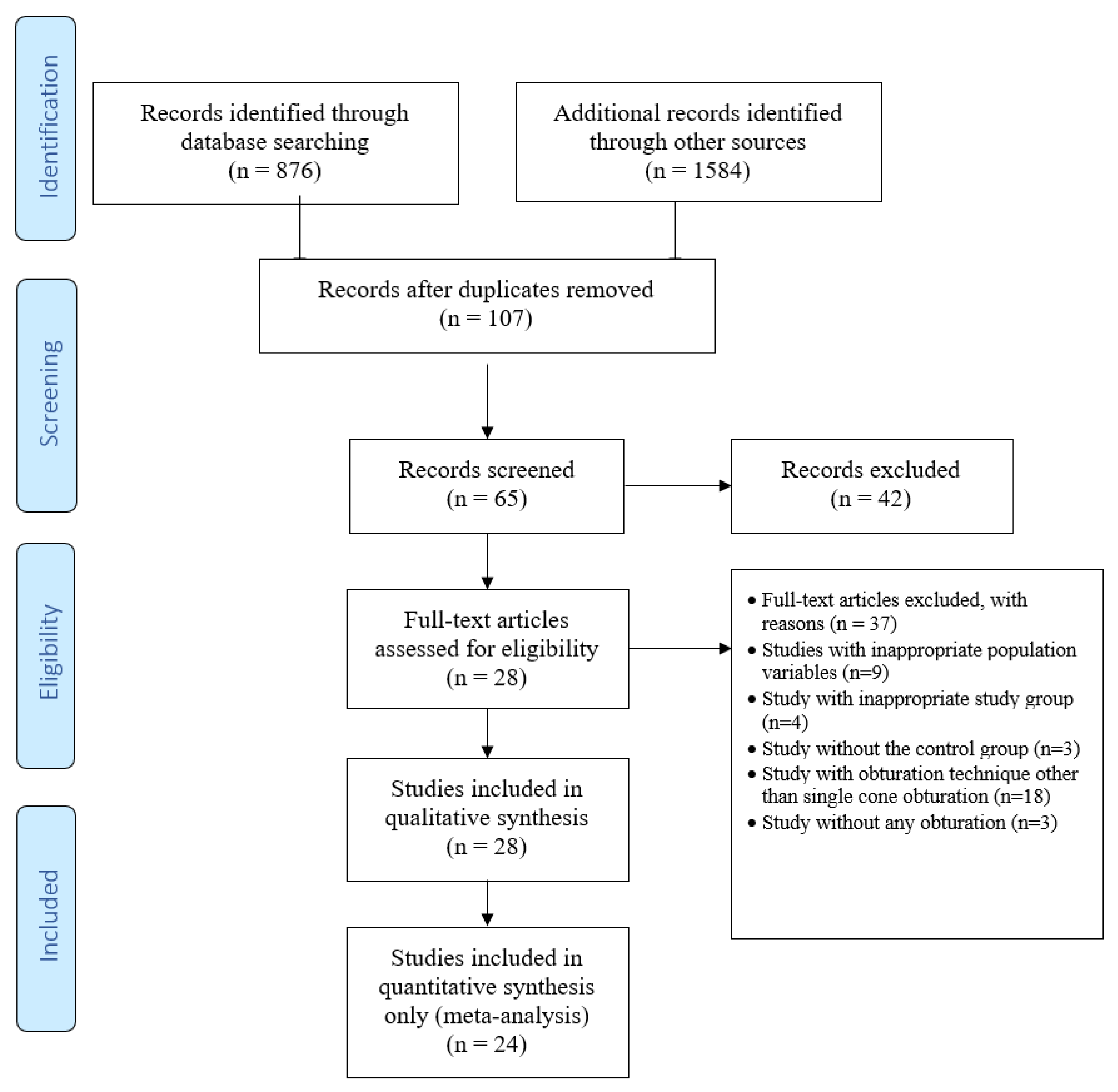
3. Literature Screening and Study Selection
4. Data Extraction
Quality Assessment and Risk of Bias Analysis
5. Results
5.1. Physico-Chemical Properties and Meta-Analysis
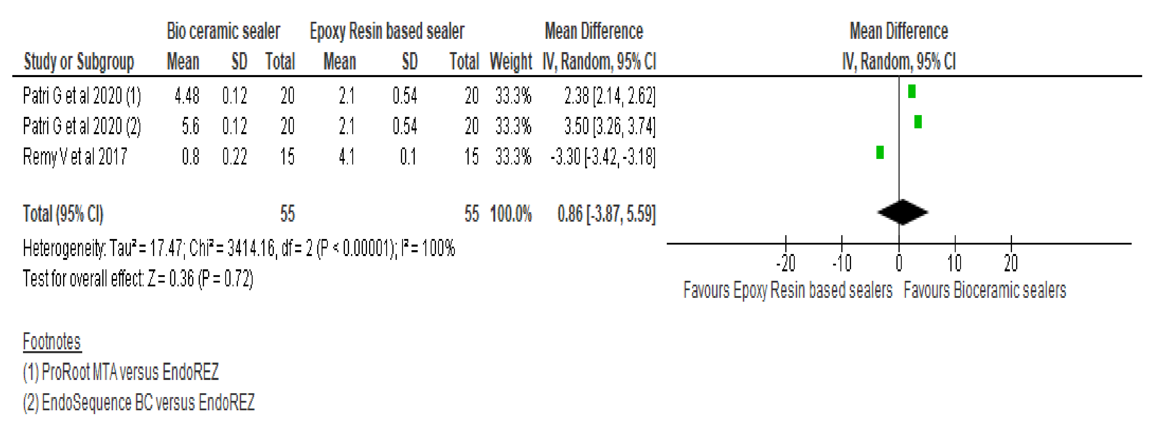
5.1.1. Marginal Adaptation
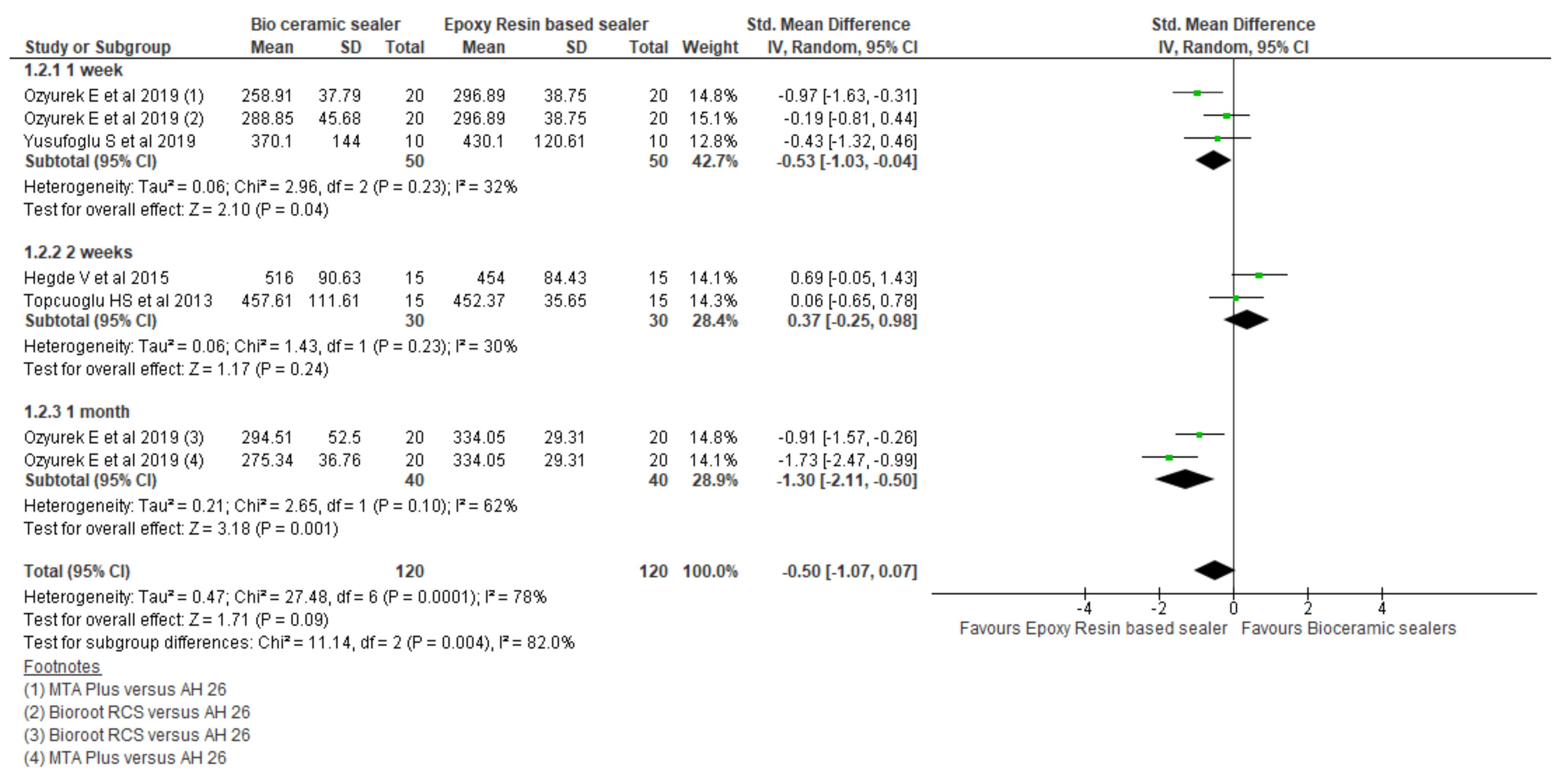
5.1.2. Fracture Resistance
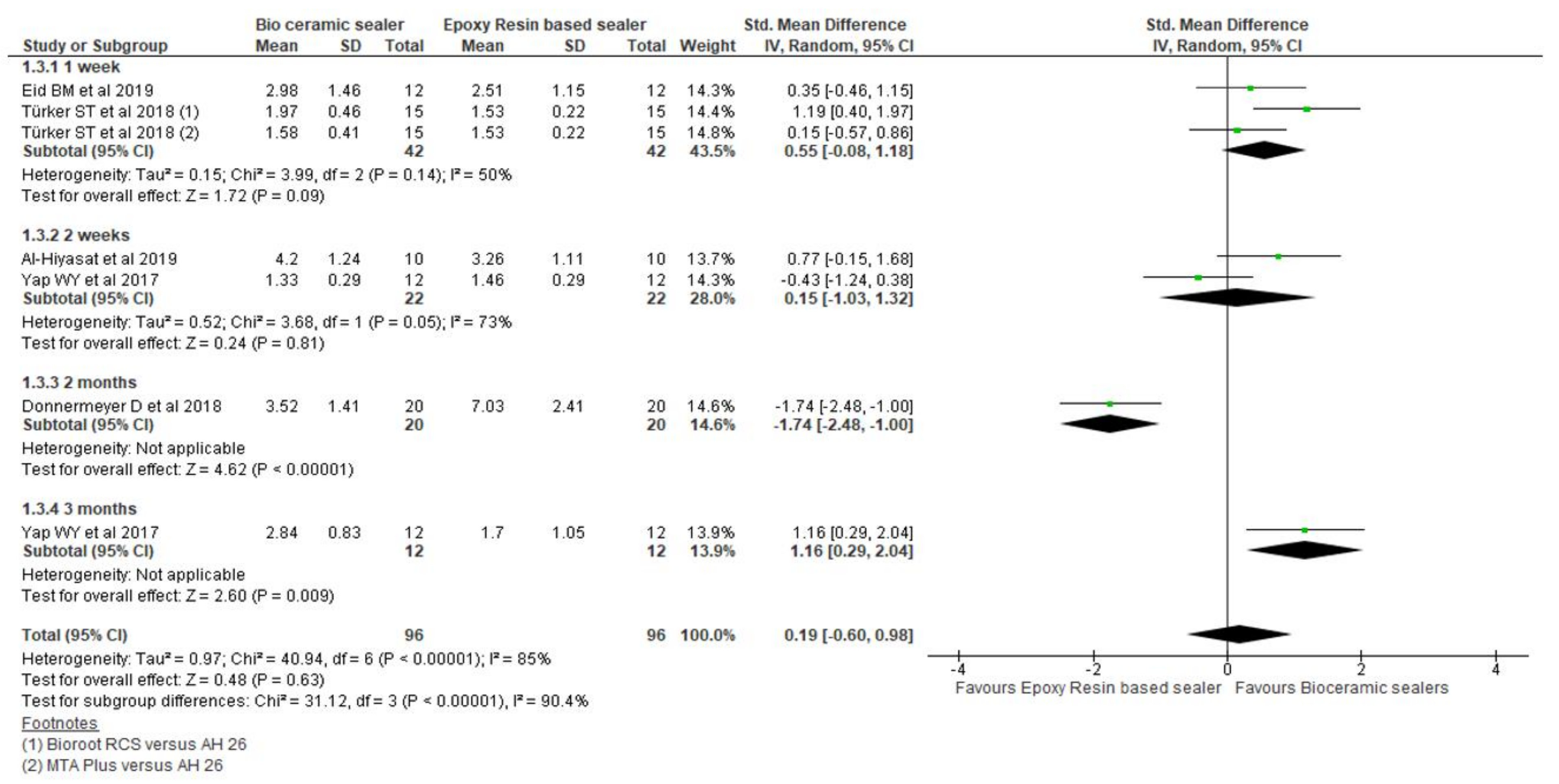
5.1.3. Pushout Bond Strength
5.1.4. Penetration Depth
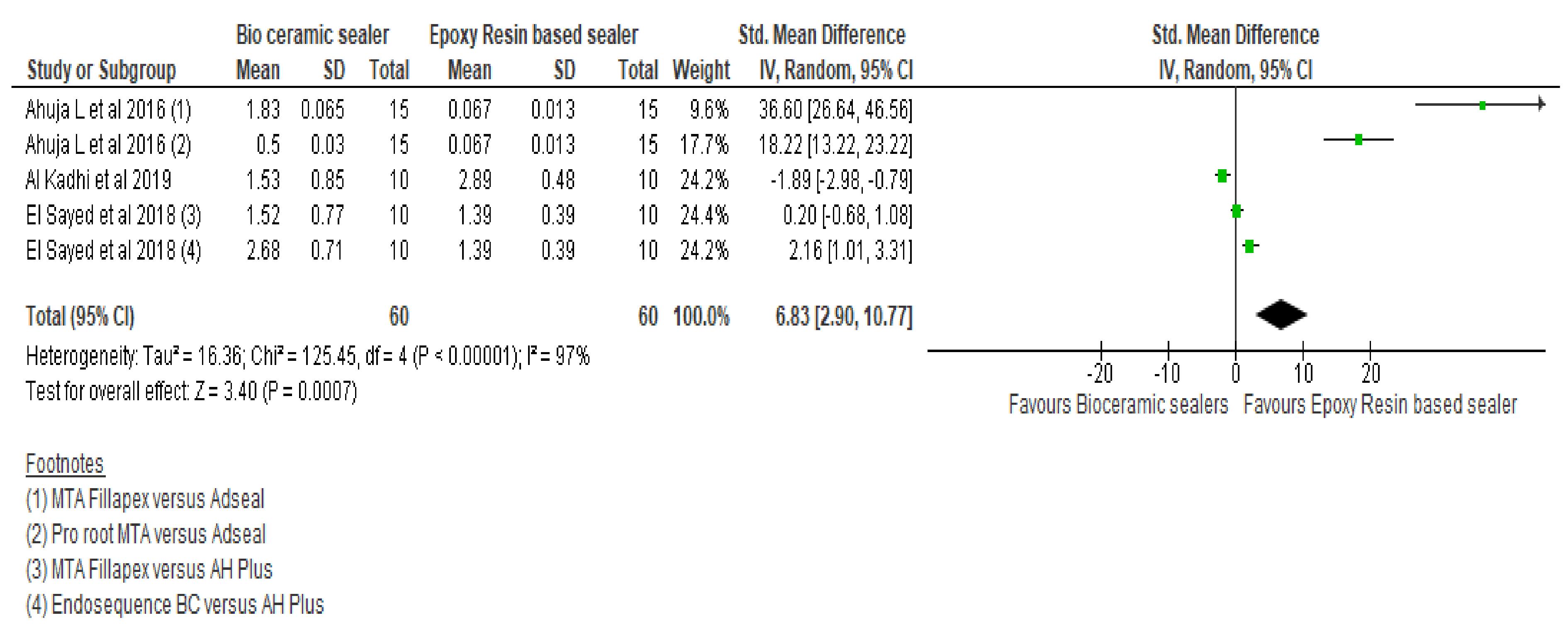
5.1.5. Apical Microleakage
5.1.6. Coronal Discoloration
5.1.7. Bacterial Leakage
6. Discussion
7. Conclusions
Author Contributions
Funding
Institutional Review Board Statement.
Informed Consent Statement
Data Availability Statement
Conflicts of Interest
References
- D’Souza, L.; Sharma, N.; Chander, S.; Singh, S.; D’Souza, R. Root canal sealers and its role in successful endodontics—A review. Ann. Dent. Res. 2021, 2, 68–78. [Google Scholar]
- Sjögren, U.; Figdor, D.; Persson, S.; Sundqvist, G. Influence of infection at the time of root filling on the outcome of endodontic treatment of teeth with apical periodontitis. Int. Endod. J. 1997, 30, 297–306. [Google Scholar] [CrossRef]
- Katge, F.; Patil, D.; Poojari, M.; Pimpale, J.; Shitoot, A.; Rusawat, B. Comparison of instrumentation time and cleaning efficacy of manual instrumentation, rotary systems and reciprocating systems in primary teeth: An in vitro study. J. Indian Soc. Pedod. Prev. Dent. 2014, 32, 311–316. [Google Scholar] [CrossRef]
- Baez, A.; Dextre, T.; Pinheiro, C.; Nishiyama, C. Advantages and disadvantages of the single cone technique. Rev. Asoc. Dent. Mex. 2016, 73, 170–174. [Google Scholar]
- Washio, A.; Morotomi, T.; Yoshii, S.; Kitamura, C. Bioactive glass-based endodontic sealer as a promising root canal filling material without semisolid core materials. Materials 2019, 12, 3967. [Google Scholar] [CrossRef] [Green Version]
- Li, G.; Niu, L.; Zhang, W.; Olsen, M.; De-Deus, G.; Eid, A. Ability of new obturation materials to improve the seal of the root canal system: A review. Acta Biomater. 2014, 10, 1050–1063. [Google Scholar] [CrossRef] [PubMed] [Green Version]
- Silva Almeida, L.; Moraes, R.; Morgental, R.; Pappen, F. Are premixed calcium silicate-based endodontic sealers comparable to conventional materials? A systematic review of in vitro studies. J. Endod. 2017, 43, 527–535. [Google Scholar] [CrossRef]
- Ersahan, S.; Aydin, C. Dislocation resistance of iRoot SP, a calcium silicate-based sealer, from radicular dentine. J. Endod. 2010, 36, 2000–2002. [Google Scholar] [CrossRef]
- Muharsya, Y.; Usman, M.; Suprastiwi, E. Comparison sealability of root canal obturation using bioceramic sealer and methacrylate resin-based sealer. J. Phys.: Conf. Ser. 2017, 884, 012111. [Google Scholar] [CrossRef]
- Tanomaru-Filho, M.; Cristine Prado, M.; Torres, F.F.E.; Viapiana, R.; Pivoto-João, M.M.B.; Guerreiro-Tanomaru, J.M. Physicochemical properties and bioactive potential of a new epoxy resin-based root canal sealer. Braz. Dent. J. 2019, 30, 563–568. [Google Scholar] [CrossRef] [PubMed]
- Donnermeyer, D.; Bürklein, S.; Dammaschke, T.; Schäfer, E. Endodontic sealers based on calcium silicates: A systematic review. Odontology 2019, 107, 421–436. [Google Scholar] [CrossRef] [PubMed]
- Best, S.M.; Porter, A.E.; Thian, E.S.; Huang, J. Bioceramics: Past, present and for the future. J. Eur. Ceram. Soc. 2008, 28, 1319–1327. [Google Scholar] [CrossRef]
- Al-Hiyasat, A.; Alfirjani, S. The effect of obturation techniques on the push-out bond strength of a premixed bioceramic root canal sealer. J. Dent. 2019, 89, 103169. [Google Scholar] [CrossRef]
- Zavattini, A.; Knight, A.; Foschi, F.; Mannocci, F. Outcome of root canal treatments using a new calcium silicate root canal sealer: A non-randomized clinical trial. J. Clin. Med. 2020, 9, 782. [Google Scholar] [CrossRef] [Green Version]
- Alim, B.; Berker, Y. Evaluation of different root canal filling techniques in severely curved canals by micro-computed tomography. Saudi Dent. J. 2020, 32, 200–205. [Google Scholar] [CrossRef]
- Jo, S.; Kim, H.; Lee, H.; Kim, Y.; Dev Patel, K.; Campbell Knowles, J.; Lee, J.; Song, M. Physical Properties and Biofunctionalities of Bioactive Root Canal Sealers In Vitro. Nanomaterials 2020, 10, 1750. [Google Scholar] [CrossRef]
- Economides, N.; Kokorikos, I.; Kolokouris, I.; Panagiotis, B.; Gogos, C. Comparative study of apical sealing ability of a new resin-based root canal sealer. J. Endod. 2004, 30, 403–405. [Google Scholar] [CrossRef]
- Pereira, A.; Nishiyama, C.; De Castro Pinto, L. Single-cone obturation technique: A literature review. RSBO Rev. Sul-Bras. Odontol. 2012, 9, 442–447. [Google Scholar]
- Markowitz, K.; Moynihan, M.; Liu, M.; Kim, S. Biologic properties of eugenol and zinc oxide-eugenol: A clinically oriented review. Oral Surg. Oral Med. Oral Pathol. 1992, 73, 729–737. [Google Scholar] [CrossRef]
- DeLong, C.; He, J.; Woodmansey, K. The effect of obturation technique on the push-out bond strength of calcium silicate sealers. J. Endod. 2015, 41, 385–388. [Google Scholar] [CrossRef]
- Tuncer, A.; Tuncer, S.; Gökyay, S. Correlation between sealer penetration into dentinal tubules and bond strength of two new calcium silicate-based and an epoxy resin-based, endodontic sealer. J. Adhes. Sci. Technol. 2014, 28, 702–710. [Google Scholar] [CrossRef]
- Nagas, E.; Uyanik, M.; Eymirli, A.; Cehreli, Z.; Vallittu, P.; Lassila, L. Dentin moisture conditions affect the adhesion of root canal sealers. J. Endod. 2021, 38, 240–244. [Google Scholar] [CrossRef]
- Carvalho, C.; Grazziotin-Soares, R.; De Miranda Candeiro, G.; Martinez, L.; De Souza, J.; Oliveira, P. Micro push-out bond strength and bioactivity analysis of a bioceramic root canal sealer. Iran. Endod. J. 2017, 12, 343. [Google Scholar] [CrossRef] [PubMed]
- Silva, E.; Carvalho, N.; Prado, M.; Zanon, M.; Senna, P.; Souza, E.; De-Deus, G. Push-out bond strength of injectable pozzolan-based root canal sealer. J. Endod. 2016, 42, 1656–1659. [Google Scholar] [CrossRef] [PubMed]
- Shokouhinejad, N.; Gorjestani, H.; Nasseh, A.; Hoseini, A.; Mohammadi, M.; Shamshiri, A. Push-out bond strength of gutta-percha with a new bioceramic sealer in the presence or absence of smear layer. Aust. Endod. J. 2013, 39, 102–106. [Google Scholar] [CrossRef] [PubMed]
- Wiesse, P.; Silva-Sousa, Y.; Pereira, R.; Estrela, C.; Domingues, L.; Pécora, J.; Sousa-Neto, M. Effect of ultrasonic and sonic activation of root canal sealers on the push-out bond strength and interfacial adaptation to root canal dentine. Int. Endod. J. 2018, 51, 102–111. [Google Scholar] [CrossRef]
- Qaiser, S.; Devadiga, D.; Hegde, M.; Shetty, N. Bioceramics as Sealers in Endodontics—A Research. J. Pharm. Biol. Chem. Sci. 2019, 10, 363–368. [Google Scholar]
- Silva, E.; Canabarro, A.; Andrade, M.; Cavalcante, D.; Von Stetten, O.; Da Silva Fidalgo, T. Dislodgment Resistance of Bioceramic and Epoxy Sealers: A Systematic Review and Meta-analysis. J. Evid. Based Dent. Pract. 2019, 19, 221–235. [Google Scholar] [CrossRef]
- Uzunoglu-Özyürek, E.; Eren, S.; Karahan, S. Effect of root canal sealers on the fracture resistance of endodontically treated teeth: A systematic review of in vitro studies. Clin. Oral Investig. 2018, 22, 2475–2485. [Google Scholar] [CrossRef]
- Kim, J.; Moe, M.; Kim, S. A micro-computed tomographic evaluation of root canal filling with a single gutta-percha cone and calcium silicate sealer. Restor. Dent. Endod. 2020, 45, e18. [Google Scholar] [CrossRef] [Green Version]
- Patri, G.; Agrawal, P.; Anushree, N.; Arora, S.; Kunjappu, J.J.; Shamsuddin, S.V. A Scanning Electron Microscope Analysis of Sealing Potential and Marginal Adaptation of Different Root Canal Sealers to Dentin: An In Vitro study. J. Contemp. Dent. Pract. 2020, 21, 73–77. [Google Scholar]
- Al-Kadhi, A.M.A.; Al-Ani, Z.B.M.A.; Al-Eanizi, J.A.J. Comparison of Apical Microleakage of Four Contemporary Endodontic Sealers by Dye Penetration Method. Int. Med. J. 2019, 26, 237–401. [Google Scholar]
- Alotaibi, R.M.; Altuwaijri, S.M.; Ahmed, D.R. The degree of crown discoloration induced by a bioceramic and other conventional endodontic sealers. Saudi Endod. J. 2019, 9, 192. [Google Scholar]
- Eid, B.M.; Abdel Gawad, R.A. Assessment of Two Root Canal Sealers Push-out Bond Strength in Root Canals Obturated Using Two Different Obturation Techniques. Egypt. Dent. J. 2019, 65, 1487–1494. [Google Scholar] [CrossRef] [Green Version]
- El Hachem, R.; Khalil, I.; Le Brun, G.; Pellen, F.; Le Jeune, B.; Daou, M. Dentinal tubule penetration of AH Plus, BC Sealer and a novel tricalcium silicate sealer: A confocal laser scanning microscopy study. Clin. Oral Inves. 2019, 23, 1871–1876. [Google Scholar] [CrossRef]
- Özyürek, E.U.; Türker, S.A. Evaluation of fracture resistance of roots-filled with various root canal sealers at different time periods. Eur. Oral Res. 2019, 53, 6–11. [Google Scholar]
- Yusufoglu, S.İ.; Akman, M.; Akbulut, M.B.; Eldeniz, A.Ü. Fracture resistance of roots enlarged with various rotary systems and obturated with different sealers. J. Dent. Res. Dent. Clin. Dent. Prospect. 2019, 13, 215–220. [Google Scholar] [CrossRef] [PubMed] [Green Version]
- Donnermeyer, D.; Dornseifer, P.; Schäfer, E.; Dammaschke, T. The push-out bond strength of calcium silicate-based endodontic sealers. Head Face Med. 2018, 14, 1–7. [Google Scholar] [CrossRef]
- Eltair, M.; Pitchika, V.; Hickel, R.; Kühnisch, J.; Diegritz, C. Evaluation of the interface between gutta-percha and two types of sealers using scanning electron microscopy (SEM). Clin. Oral Investig. 2018, 22, 1631–1639. [Google Scholar] [CrossRef]
- El Sayed, M.A.A.M.; Al Husseini, H. Apical dye leakage of two single-cone root canal core materials (hydrophilic core material and gutta-percha) sealed by different types of endodontic sealers: An in vitro study. J. Conserv. Dent. 2018, 21, 147–152. [Google Scholar] [CrossRef]
- Germain, S.; Meetu, K.; Issam, K.; Alfred, N.; Carla, Z. Impact of the root canal taper on the apical adaptability of sealers used in a single-cone technique: A micro-computed tomography study. J. Contemp. Dent. Pract 2018, 19, 808–815. [Google Scholar]
- Huang, Y.; Orhan, K.; Celikten, B.; Orhan, A.I.; Tufenkci, P.; Sevimay, S. Evaluation of the sealing ability of different root canal sealers: A combined SEM and micro-CT study. J. Appl. Oral Sci. 2018, 26, e20160584. [Google Scholar] [CrossRef] [Green Version]
- Salem, A.S.; Saleh, A.R.M.; Elmasmari, H.A. Assessment of Apical Leakage of Bioceramic Endodontic Sealer with Two Obturation Techniques. Open Dent. J. 2018, 12, 1162–1168. [Google Scholar] [CrossRef] [Green Version]
- Türker, S.A.; Uzunoğlu, E.; Purali, N. Evaluation of dentinal tubule penetration depth and push-out bond strength of AH 26, BioRoot RCS, and MTA Plus root canal sealers in presence or absence of smear layer. J. Dent. Res. Dent. Clin. Dent. Prospect. 2018, 12, 294–298. [Google Scholar] [CrossRef]
- Yanpiset, K.; Banomyong, D.; Chotvorrarak, K.; Srisatjaluk, R.L. Bacterial leakage and micro-computed tomography evaluation in round-shaped canals obturated with bioceramic cone and sealer using matched single cone technique. Restor. Dent. Endod. 2018, 43, e30. [Google Scholar] [CrossRef] [PubMed]
- Russell, A.; Friedlander, L.; Chandler, N. Sealer penetration and adaptation in root canals with the butterfly effect. Aust. Endod. J. 2018, 44, 225–234. [Google Scholar] [CrossRef]
- Huang, Y.; Celikten, B.; De Faria Vasconcelos, K.; Ferreira Pinheiro Nicolielo, L.; Lippiatt, N.; Buyuksungur, A.; Jacobs, R.; Orhan, K. Micro-CT and nano-CT analysis of filling quality of three different endodontic sealers. Dentomaxillofacial Radiol. 2017, 46. [Google Scholar] [CrossRef] [PubMed]
- Remy, V.; Krishnan, V.; Job, T.V.; Ravisankar, M.S.; Raj, C.R.; John, S. Assessment of Marginal Adaptation and Sealing Ability of Root Canal Sealers: An in vitro Study. J. Contemp. Dent. Pract. 2017, 18, 1130–1134. [Google Scholar] [CrossRef]
- Yap, W.Y.; Ab Aziz, Z.A.C.; Azami, N.H.; Al-Haddad, A.Y.; Khan, A.A. An in vitro comparison of bond strength of different sealers/obturation systems to root dentin using the push-out test at 2 weeks and 3 months after obturation. Med. Princ. Pract. 2017, 26, 464–469. [Google Scholar] [CrossRef] [PubMed] [Green Version]
- Ahuja, L.; Jasuja, P.; Verma, K.G.; Juneja, S.; Mathur, A.; Walia, R.; Kakkar, A.; Singla, M. A comparative evaluation of sealing ability of new MTA based sealers with conventional resin based sealer: An in-vitro study. J. Clin. Diagn. Res. 2016, 10, ZC76–ZC79. [Google Scholar] [CrossRef]
- Celikten, B.; Uzuntas, C.F.; Orhan, A.I.; Orhan, K.; Tufenkci, P.; Kursun, S.; Demiralp, K.Ö. Evaluation of root canal sealer filling quality using a single-cone technique in oval shaped canals: An In vitro Micro-CT study. Scanning 2016, 38, 133–140. [Google Scholar] [CrossRef]
- Madhuri, G.V.; Varri, S.; Bolla, N.; Mandava, P.; Akkala, L.S.; Shaik, J. Comparison of bond strength of different endodontic sealers to root dentin: An in vitro push-out test. J. Conserv. Dent. 2016, 19, 461–464. [Google Scholar] [CrossRef] [Green Version]
- Polineni, S.; Bolla, N.; Mandava, P.; Vemuri, S.; Mallela, M.; Gandham, V.M. Marginal adaptation of newer root canal sealers to dentin: A SEM study. J. Conserv. Dent. JCD 2016, 19, 360–363. [Google Scholar] [CrossRef]
- Hegde, V.; Arora, S. Fracture resistance of roots obturated with novel hydrophilic obturation systems. J. Conserv. Dent. 2015, 18, 261–264. [Google Scholar] [CrossRef] [Green Version]
- Shinde, A.; Kokate, S.; Hegde, V. Comparative assessment of apical sealing ability of three different endodontic sealers: A scanning electron microscopic study. J. Pierre Fauchard Acad. (India Sect.) 2014, 28, 78–82. [Google Scholar] [CrossRef]
- Topçuoğlu, H.S.; Tuncay, Ö.; Karataş, E.; Arslan, H.; Yeter, K. In vitro fracture resistance of roots obturated with epoxy resin-based, mineral trioxide aggregate-based, and bioceramic root canal sealers. J. Endod. 2013, 39, 1630–1633. [Google Scholar] [CrossRef] [PubMed]
- Pawar, S.S.; Pujar, M.A.; Makandar, S.D. Evaluation of the apical sealing ability of bioceramic sealer, AH plus & epiphany: An in vitro study. J. Conserv. Dent.: JCD 2014, 17, 579–582. [Google Scholar] [CrossRef] [Green Version]
- de Miranda Candeiro, G.T.; Correia, F.C.; Duarte, M.A.H.; Ribeiro-Siqueira, D.C.; Gavini, G. Evaluation of radiopacity, pH, release of calcium ions, and flow of a bioceramic root canal sealer. J. Endod. 2012, 38, 842–845. [Google Scholar] [CrossRef] [PubMed] [Green Version]
- Atmeh, A.; Chong, E.; Richard, G.; Festy, F.; Watson, T. Dentin-cement interfacial interaction: Calcium silicates and polyalkenoates. J. Dent. Res. 2012, 91, 454–459. [Google Scholar] [CrossRef] [PubMed]
- McMichael, G.E.; Primus, C.M.; Opperman, L.A. Dentinal tubule penetration of tricalcium silicate sealers. J. Endod. 2016, 42, 632–636. [Google Scholar] [CrossRef] [Green Version]
- Çobankara, F.K.; Üngör, M.; Belli, S. The effect of two different root canal sealers and smear layer on resistance to root fracture. J. Endod. 2002, 28, 606–609. [Google Scholar] [CrossRef]
- Tedesco, M.; Chain, M.C.; Bortoluzzi, E.A.; Garcia, L.d.F.R.; Alves, A.M.H.; Teixeira, C.S. Comparison of two observational methods, scanning electron and confocal laser scanning microscopies, in the adhesive interface analysis of endodontic sealers to root dentine. Clin. Oral Investig. 2018, 22, 2353–2361. [Google Scholar] [CrossRef] [PubMed]
- Al-Haddad, A.; Kasim, N.H.A.; Ab Aziz, Z.A.C. Interfacial adaptation and thickness of bioceramic-based root canal sealers. Dent. Mater. J. 2015, 34, 516–521. [Google Scholar] [CrossRef] [PubMed] [Green Version]
- Šimundić Munitić, M.; Poklepović Peričić, T.; Utrobičić, A.; Bago, I.; Puljak, L. Antimicrobial efficacy of commercially available endodontic bioceramic root canal sealers: A systematic review. PLoS ONE 2019, 14, e0223575. [Google Scholar] [CrossRef] [PubMed] [Green Version]


| Search Strategy | |
|---|---|
| Focused Question | Is there a difference between calcium silicate based sealers and resin-based sealers in terms of physico-chemical properties on the outcome of root canal treatment using a single cone obturation technique for extracted permanent teeth? |
| PICO Strategy | |
| Population | (Permanent Dentition [MeSH] OR Adult Dentition OR Secondary Dentition OR Permanent teeth OR Teeth OR Extracted teeth OR Root Canal Obturation [MeSH] OR Single cone obturation |
| Intervention (#1) | (Bioceramic sealer OR Endosquence BC OR iRoot Plus OR MTA fillapex OR Totalfill BC OR tricalcium phosphate OR tricalcium phosphate ceramic sealer OR Calcium silicate sealer OR Calcium phosphate sealer OR Endodontic sealer OR Root canal sealer |
| Comparisons (#2) | (Epoxy resin-based root canal sealer OR AH Plus OR AH 26 |
| Outcomes (#3) | (Depth of penetration OR Adaptability OR Void volume OR Seal ability OR Adhesiveness OR Tooth discoloration OR Fracture resistance OR Fracture strength OR Bond strength OR Push-out bond strength OR Root fracture OR Anti-microbial OR Penetration |
| Study design (#4) | (In Vitro Techniques [MeSH] OR In vitro studies OR In vitro studies OR Ex vivo studies |
| Search Combination | #1 AND #2 AND #3 AND #4 |
| Database Search | |
| Language | No restriction (Articles in English language or other language where English translation is possible.) |
| Electronic Databases | PubMed/MEDLINE, Cochrane Central Register of Controlled Trials, Web of Science |
| Journals | Journal of Endodontics, International Endodontic Journal, Australian Endodontic Journal, Clinical Oral Investigations, Journal of Conservative Dentistry, Journal of American Dental Association. Brazilian dental journal, Journal of physics, Materials, Dental materials etc. |
| Period of Publication | 1 January 2011 to 31 August 2020 |
| Sr. No | Study Id | Sample Size Calculation | Samples with Similar Dimensions | Teeth Randomization | Standardization of Instrumentation Procedures | Standardization of Filling Procedures | Endodontic Treatment Performed by a Single Operator | Blinding of the Observer | Statistical Analysis Carried Out | Risk of Bias |
|---|---|---|---|---|---|---|---|---|---|---|
| 1. | Kim J et al. (2020) [30] | No | Yes | Yes | Yes | Yes | No | No | Yes | Medium risk |
| 2. | Patri G et al. (2020) [31] | No | Yes | Yes | Yes | Yes | No | Yes | Yes | Medium risk |
| 3. | Al-Hiyasat et al. (2019) [13] | No | Yes | Yes | Yes | Yes | No | No | Yes | Medium risk |
| 4. | Al-Kadhi AM et al. (2019) [32] | No | Yes | Yes | Yes | Yes | No | No | Yes | Medium risk |
| 5. | Alotaibi RM et al. (2019) [33] | No | Yes | Yes | Yes | Yes | Yes | No | Yes | Medium risk |
| 6. | Eid BM et al. (2019) [34] | No | Yes | Yes | Yes | Yes | No | No | Yes | Medium risk |
| 7. | El Hachem R et al. (2019) [35] | No | Yes | Yes | Yes | Yes | Yes | No | Yes | Medium risk |
| 8. | Ozyurek E et al. (2019) [36] | No | Yes | Yes | Yes | Yes | Yes | No | Yes | Medium risk |
| 9. | Yusufoglu S et al. (2019) [37] | No | Yes | Yes | Yes | Yes | Yes | No | Yes | Medium risk |
| 10. | Donnermeyer D et al. (2018) [38] | Yes | Yes | Yes | Yes | Yes | No | No | Yes | Medium risk |
| 11. | Eltair M et al. (2018) [39] | No | Yes | Yes | Yes | Yes | Yes | Yes | Yes | Low risk |
| 12. | El Sayed et al. (2018) [40] | No | Yes | Yes | Yes | Yes | Yes | Yes | Yes | Low risk |
| 13. | Germain S et al. (2018) [41] | No | Yes | Yes | Yes | Yes | No | No | Yes | Medium risk |
| 14. | Huang Y et al. (2018) [42] | No | Yes | Yes | Yes | Yes | No | No | Yes | Medium risk |
| 15. | Salem AS et al. (2018) [43] | Yes | Yes | Yes | Yes | Yes | No | No | Yes | Medium risk |
| 16. | Türker ST et al. (2018) [44] | No | Yes | Yes | Yes | Yes | No | No | Yes | Medium risk |
| 17. | Yanpiset K et al. (2018) [45] | No | Yes | Yes | Yes | Yes | No | No | Yes | Medium risk |
| 18. | Russell A et al. (2018) [46] | Yes | Yes | Yes | Yes | Yes | Yes | Yes | Yes | Low risk |
| 19. | Huang Y et al. (2017) [47] | Yes | Yes | Yes | Yes | Yes | Yes | Yes | Yes | Low risk |
| 20. | Remy V et al. (2017) [48] | Yes | Yes | No | Yes | Yes | No | No | Yes | Medium risk |
| 21. | Yap WY et al. (2017) [49] | Yes | Yes | Yes | Yes | Yes | No | No | Yes | Medium risk |
| 22. | Ahuja L et al. (2016) [50] | No | Yes | Yes | Yes | Yes | No | No | Yes | Medium risk |
| 23. | Celikten B et al. (2016) [51] | Yes | Yes | Yes | Yes | Yes | Yes | Yes | Yes | Low risk |
| 24. | Madhuri GV et al. (2016) [52] | No | Yes | No | Yes | Yes | No | No | Yes | Medium risk |
| 25. | Polineni S et al. (2016) [53] | No | Yes | Yes | Yes | Yes | No | Yes | Yes | Medium risk |
| 26. | Hegde V et al. (2015) [54] | No | Yes | Yes | Yes | Yes | No | No | Yes | Medium risk |
| 27. | Shinde A et al. (2014) [55] | No | Yes | Yes | Yes | Yes | No | No | Yes | Medium risk |
| 28. | Topcuoglu HS et al. (2013) [56] | No | Yes | Yes | Yes | Yes | No | No | Yes | Medium risk |
| Property | Author, Year | Method | Material | Author’s Conclusion |
|---|---|---|---|---|
| Adaptation to root canal wall | Kim J. et al. (2020) | Void percentage | EndosealMTA AH Plus Jet | Endoseal MTA does not seem to reduce the voids over time when it was used with a single gutta-percha cone technique. |
| Patri G. (2020) | Sealing potential and marginal adaptation | EndoSequence BC ProRoot MTA EndoREZ | Significant and better sealing ability and marginal adaptation was demonstrated by EndoSequence BC (bioceramic sealer) when compared to ProRoot MTA sealer (MTA-based sealer) and EndoREZ sealer (resin-based sealer). | |
| Eltair M. et al. (2018) | Areas and interfacial gaps between sealer and dentine | TotalFill BC AH Plus | All tested root canal fillings exhibited minor interfacial gaps. The BC sealer showed better adaptability than the AH Plus sealer. | |
| Germain S. et al. (2018) | Voids volume in the apical third | TotalFill bioceramic (NB) sealers AHPlus | Bioceramic (BC) sealers showed good all-round performance demonstrating good adaptability, and reduced voids while maintaining similar characteristics when compared with conventional resin sealer. | |
| Huang Y. et al. (2018) | Total ROI volume (mm3), object volume (dentin volume, mm3), volume of closed pores (mm3), surface of closed pores (mm2), volume of open pores (mm3), and open porosity (%) | EndoSequence BC AHPlus | By using the single cone technique, neither endoSequence or AH Plus provides a porosity-free root canal filling. The EndoSequence BC sealer may have similar sealing abilities regarding the whole root canal as the AH Plus sealer. A better sealing effect could be obtained in the coronal and middle sections of a root canal than the apical part by using the tested sealers. | |
| Huang Y. et al. (2017) | Void volume in (mm3) | Sure Seal Root Total BC Sealer AH Plus | A high incidence rate of voids was found within each sealer material and no significant difference was found among the root filling sealers. | |
| Remy V. et al. (2017) | Marginal adaptation | MTA Fillapex AH Plus | AH Plus sealer shows a good marginal adaptation. | |
| Celikten B. et al. (2016) | Voids in 3D volumes | EndoSequence BC AH Plus | All root canal sealers tested resulted in voids. The bioceramic sealers (Endo Sequence BCSealer, Smartpastebio) produced similar voids which had the fewest in the apical third of root canals. | |
| Polineni S. et al. (2016) | Maximum gap width (nm) | MTA Fillapex EndoSequence BC MM-Seal | Epoxy resin-based MM-Seal showedgood marginal adaptation than the MTA Fillapex. apical halves showed poor adaptation regardless of the material used than the coronal halves | |
| Shinde A. et al. (2014) | Mean distance from the radicular dentin to the root canal fillings was in (mm) | Endo- Sequence BC AH Plus | Endosequence BC endodontic sealers showed better adaptation to the radicular dentin as compared to AH Plus sealer. | |
| Fracture Resistance | Ozyurek E. et al. (2019) | Fracture resistance values (FRV) in Newtons | MTA Plus BioRoot RCS AH 26 | Root canal preparation lowered the fracture resistance values. All sealers increased the force values needed to fracture the filled samples compared to unfilled ones but the time factor had no effect on the reinforcement effect of root canal sealers. |
| Yusufoglu S. et al. (2019) | Push-out bond strength | BioRoot RCS AH Plus | All the three root canal sealers examined in this study strengthened the prepared root canals with increased fracture resistance | |
| Hegde V. et al. (2015) | Forces in Newton | EndoSequence BC AH Plus | Hydrophilic obturations have shown to reinforce the strength of the root canal after instrumentation, and thus increasing the fracture resistance of the root to the stresses encountered | |
| Topcuoglu H.S. et al. (2013) | Forces in newtons | EndoSequence BC AH Plus | Endosequence BC sealer and AH Plus Jet were able to increase the force to fracture in single-rooted endodontically treated premolar teeth. | |
| Bond strength | Al-Hiyasat et al. (2019) | Push-out bond strength | TotalFill AH plus | Overall the push-out bond strength of TotalFill BC sealer was significantly higher than that of AH plus sealer. |
| Eid B.M. et al. (2019) | Push-out bond strength | Totalfill bioceramic Adseal sealer | The push-out bond strength of the tested TotalFill root canal sealer was higher than the pushout bond strength of Adseal resin sealer | |
| Donnermeyer D. et al. (2018) | Push-out bond strength | Total Fill BC AH Plus | The push-out bond strength of the investigated calcium silicate-based sealers was lower than of AH Plus. Total Fill BC showed the highest push-out bond strength of the calcium silicate-based sealers. | |
| Türker S.T. et al. (2018) | Push-out bond strength | BioRoot RCSMTA Plus AH 26 | Dentinal tubule penetration had limited effect on the push-out bond strength of the root canal sealers. | |
| Yap W.Y. et al. (2017) | Push-out bond strength | TotalFill BC AH Plus | TotalFill BC TM sealer (G3) showed comparable bond strengths to AH Plus. The bond strength also exhibited an increase over a 3-month post-obturation period. | |
| Madhuri G.V. et al. (2016) | Push-out bond strength | Bioceramic Sealer Epoxy resin-based sealer | Endosequence BC (Bioceramic Sealer) showed the highest push-out bond strength among all the four groups. MM (Epoxy resin based sealer) showed the second highest bond strength followed by Hybrid seal (Dual cure resin based sealer) | |
| Penetration depth | El Hachem R. et al. (2019) | Dentinal penetration depth | BC Sealer AH Plus | BC Sealer and NTS demonstrated better dentinal tubule penetration results compared to AH Plus. |
| Türker S.T. et al. (2018) | Dentinal penetration depth | BioRoot RCSMTA Plus AH 26 | Dentinal tubule penetration had limited effect on the push-out bond strength of the root canal sealers. | |
| Russell A. et al. (2017) | Dentinal penetration depth | MTA Fillapex AH Plus | Coronal sections of roots have superior adaptation and penetration compared with middle sections. Penetration in middle sections was significantly more favourable in teeth without the butterfly effect. | |
| Apical Microleakage | Al-Kadhi et al. (2019) | Apical linear dye penetration | Total fill BC Acroseal | No sealer can completely prevent microleakage, but the bioceramic is superior in performance to the other Sealers |
| El Sayed et al. (2018) | Apical linear dye penetration | MTA FillapexEndoSequence BC AH Plus | Higher apical leakage values were observed with single-cone gutta-percha/EndoSequence BC as compared gutta-percha/AH Plus, single-cone gutta-percha/MTA Fillapex | |
| Salem A.S. et al. (2018) | Apical linear dye penetration | Total fill BC AH Plus | Total Fill BC was equivalent to AH Plus in apical sealing ability when using single cone. | |
| Ahuja L. et al. (2016) | Apical linear dye penetration | MTA Fillapex Pro RootMTA Adseal sealer | Adseal sealer was better in providing the apical seal than Proroot MTA and MTA Fillapex | |
| Coronal discoloration | Alotaibi R.M. et al. (2019) | Coronal color change | TotalFill AH Plus | All sealers tested result in a measurable and gradual tooth color change. While the bioceramic sealer resulted in a slightly higher color change compared to calcium hydroxide- and resin-based sealers, the difference was not considerable. |
| Apical Bacterial leakage | Yanpiset K. et al. (2018) | Bacterial leakage test with E. faecalis | Bioceramic sealer AH Plus | In roundly-prepared canals, the epoxy resin sealer had lower amount of leaked samples as compared to bioceramic sealers using single cone gutta percha for bacterial leakage at 60 days. |
Publisher’s Note: MDPI stays neutral with regard to jurisdictional claims in published maps and institutional affiliations. |
© 2021 by the authors. Licensee MDPI, Basel, Switzerland. This article is an open access article distributed under the terms and conditions of the Creative Commons Attribution (CC BY) license (https://creativecommons.org/licenses/by/4.0/).
Share and Cite
Chopra, V.; Davis, G.; Baysan, A. Physico-Chemical Properties of Calcium-Silicate vs. Resin Based Sealers—A Systematic Review and Meta-Analysis of Laboratory-Based Studies. Materials 2022, 15, 229. https://0-doi-org.brum.beds.ac.uk/10.3390/ma15010229
Chopra V, Davis G, Baysan A. Physico-Chemical Properties of Calcium-Silicate vs. Resin Based Sealers—A Systematic Review and Meta-Analysis of Laboratory-Based Studies. Materials. 2022; 15(1):229. https://0-doi-org.brum.beds.ac.uk/10.3390/ma15010229
Chicago/Turabian StyleChopra, Viresh, Graham Davis, and Aylin Baysan. 2022. "Physico-Chemical Properties of Calcium-Silicate vs. Resin Based Sealers—A Systematic Review and Meta-Analysis of Laboratory-Based Studies" Materials 15, no. 1: 229. https://0-doi-org.brum.beds.ac.uk/10.3390/ma15010229