
Mapping Bone Marrow Cell Response from Senile Female Rats on Ca-P-Doped Titanium Coating

 ,
,  , , , , and
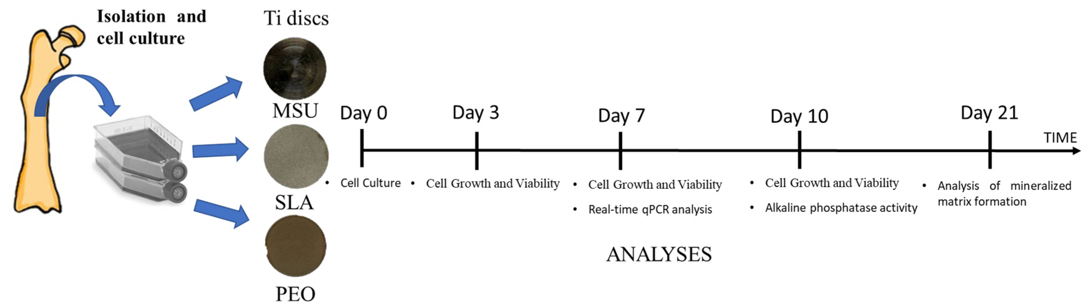
Abstract
:1. Introduction
2. Materials and Methods
2.1. Specimens
2.2. Plasma Electrolytic Oxidation Treatment
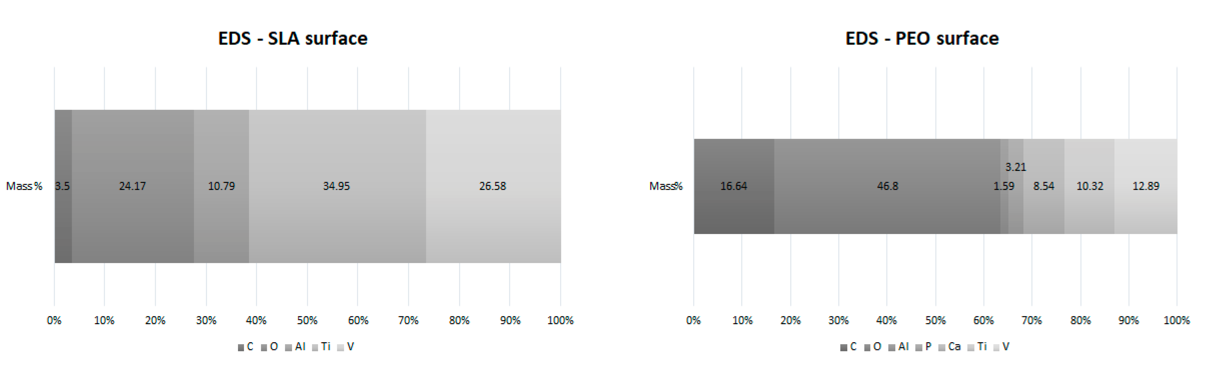
2.3. Chemical Characterization
2.4. Animals
2.5. Isolation and Cell Culture
2.6. Cell Plating on Ti Discs
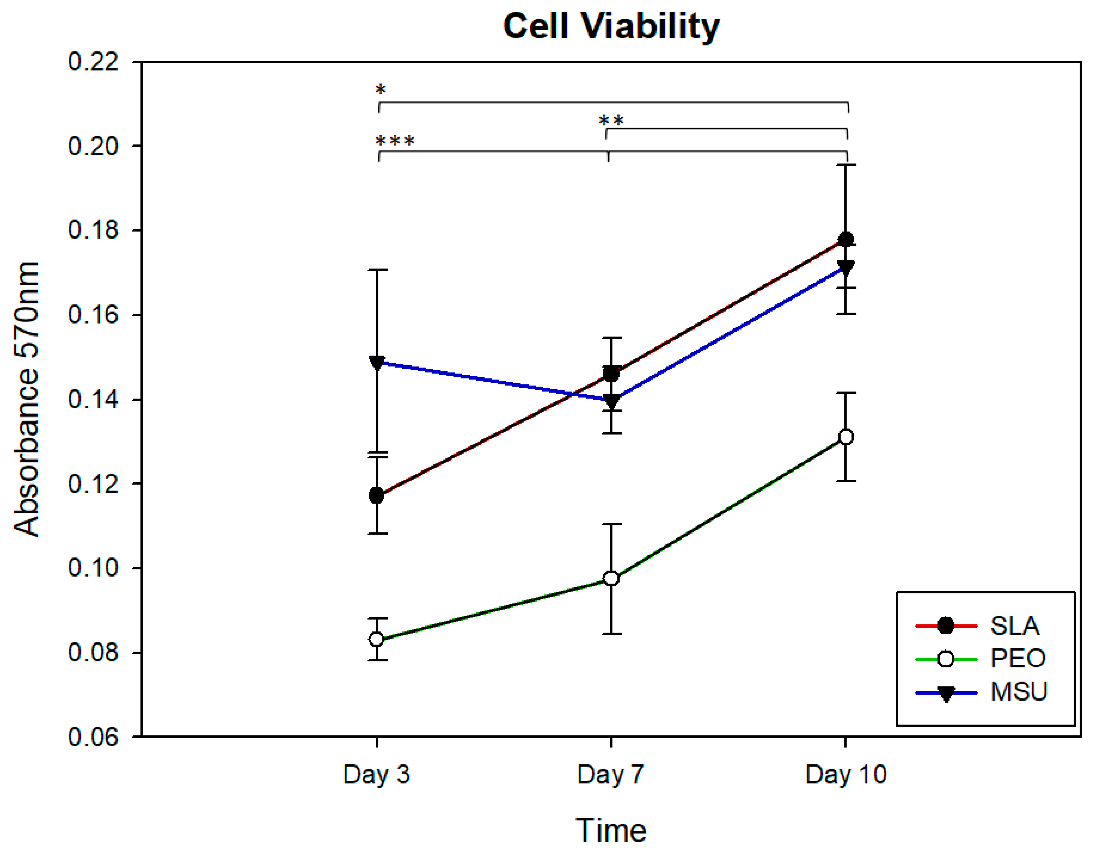
2.7. Cell Growth and Viability
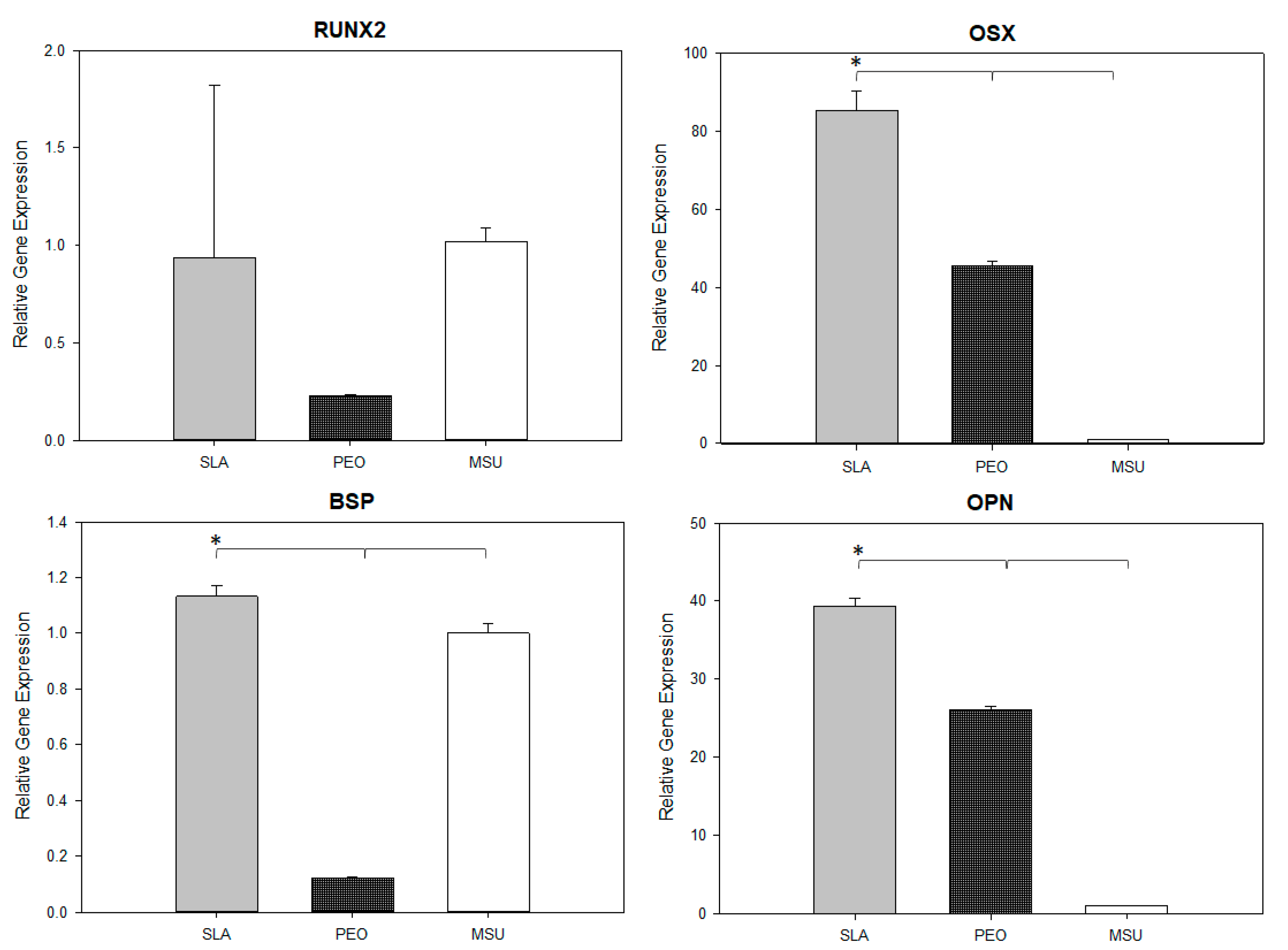
2.8. Real-Time qPCR Analysis
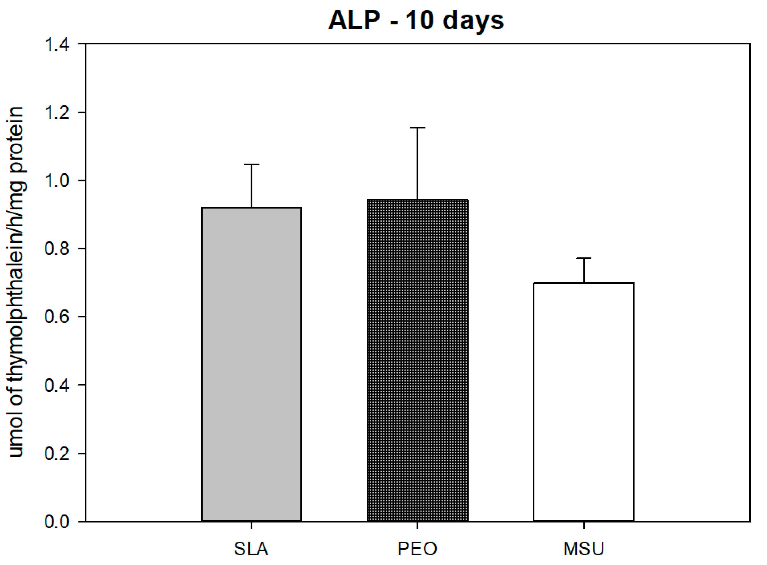
2.9. Alkaline Phosphatase Activity
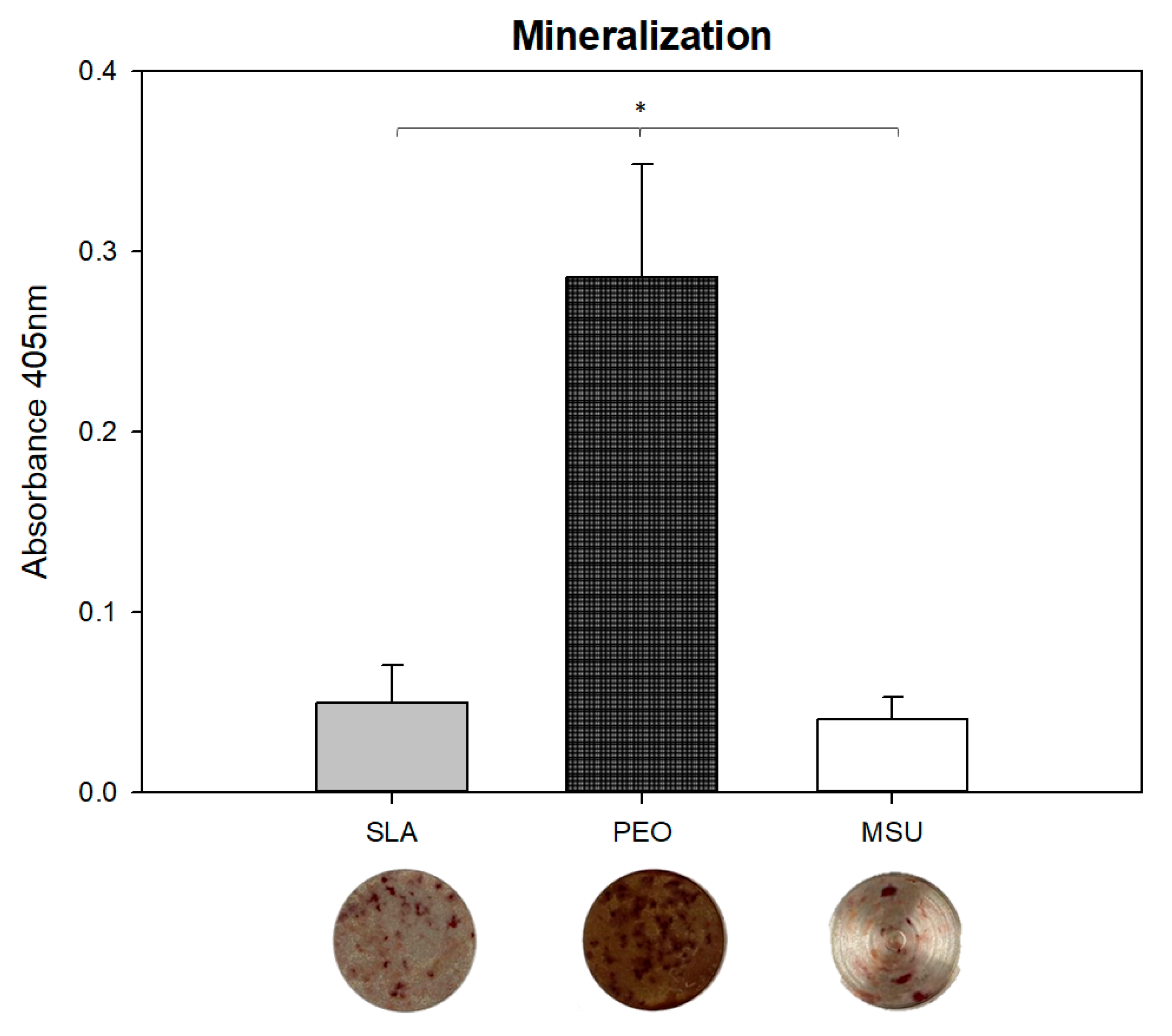
2.10. Analysis of Mineralized Matrix Formation
2.11. Statistical Analysis
3. Results
3.1. Chemical Characterization
3.2. Cell Growth and Viability
4. Gene Expression
4.1. Alkaline Phosphatase Activity
4.2. Mineralized Matrix Formation
5. Discussion
6. Conclusions
Author Contributions
Funding
Institutional Review Board Statement
Data Availability Statement
Acknowledgments
Conflicts of Interest
References
- Fini, M.; Giavaresi, G.; Torricelli, P.; Borsari, V.; Giardino, R.; Nicolini, A.; Carpi, A. Osteoporosis and biomaterial osteointegration. Biomed. Pharmacother. 2004, 58, 487–493. [Google Scholar] [CrossRef] [PubMed]
- Aghaloo, T.; Pi-Anfruns, J.; Moshaverinia, A.; Sim, D.; Grogan, T.; Hadaya, D. The Effects of Systemic Diseases and Medications on Implant Osseointegration: A Systematic Review. Int. J. Oral Maxillofac. Implant. 2019, 34, s35–s49. [Google Scholar] [CrossRef] [PubMed]
- Aguilar, E.A.; Barry, S.D.; Cefalu, C.A.; Abdo, A.; Hudson, W.P.; Campbell, J.S.; Reske, T.M.; Bonafede, M.; Wilson, K.; Stolshek, B.S.; et al. Osteoporosis Diagnosis and Management in Long-Term Care Facility. Am. J. Med. Sci. 2015, 350, 357–363. [Google Scholar] [CrossRef] [PubMed]
- Briot, K.; Geusens, P.; Em Bultink, I.; Lems, W.F.; Roux, C. Inflammatory diseases and bone fragility. Osteoporos. Int. 2017, 28, 3301–3314. [Google Scholar] [CrossRef] [PubMed]
- Chrcanovic, B.; Albrektsson, T.; Wennerberg, A. Diabetes and Oral Implant Failure: A systematic review. J. Dent. Res. 2014, 93, 859–867. [Google Scholar] [CrossRef] [PubMed]
- Qadir, A.; Liang, S.; Wu, Z.; Chen, Z.; Hu, L.; Qian, A. Senile Osteoporosis: The Involvement of Differentiation and Senescence of Bone Marrow Stromal Cells. Int. J. Mol. Sci. 2020, 21, 349. [Google Scholar] [CrossRef] [Green Version]
- Wang, H.; Jiang, Z.; Zhang, J.; Xie, Z.; Wang, Y.; Yang, G. Enhanced osteogenic differentiation of rat bone marrow mesenchymal stem cells on titanium substrates by inhibiting Notch3. Arch. Oral Biol. 2017, 80, 34–40. [Google Scholar] [CrossRef]
- Lotz, E.M.; Berger, M.; Schwartz, Z.; Boyan, B.D. Regulation of osteoclasts by osteoblast lineage cells depends on titanium implant surface properties. Acta Biomater. 2018, 68, 296–307. [Google Scholar] [CrossRef]
- Walsh, F.C.; Low, C.T.J.; Wood, R.; Stevens, K.T.; Archer, J.; Poeton, A.R.; Ryder, A. Plasma electrolytic oxidation (PEO) for production of anodised coatings on lightweight metal (Al, Mg, Ti) alloys. Trans. IMF 2009, 87, 122–135. [Google Scholar] [CrossRef]
- Albrektsson, T.; Wennerberg, A. Oral implant surfaces: Part 1—Review focusing on topographic and chemical properties of different surfaces and in vivo responses to them. Int. J. Prosthodont. 2004, 17, 536–543. [Google Scholar]
- Buser, D.; Broggini, N.; Wieland, M.; Schenk, R.K.; Denzer, A.J.; Cochran, D.L.; Hoffmann, B.; Lussi, A.; Steinemann, S.G. Enhanced Bone Apposition to a Chemically Modified SLA Titanium Surface. J. Dent. Res. 2004, 83, 529–533. [Google Scholar] [CrossRef] [PubMed]
- Combe, E.C. A protocol for determining the surface free energy of dental materials. Dent. Mater. 2004, 20, 262–268. [Google Scholar] [CrossRef]
- Cordeiro, J.M.; Beline, T.; Ribeiro, A.L.R.; Rangel, E.C.; da Cruz, N.C.; Landers, R.; Faverani, L.P.; Vaz, L.G.; Fais, L.M.; Vicente, F.B.; et al. Development of binary and ternary titanium alloys for dental implants. Dent. Mater. 2017, 33, 1244–1257. [Google Scholar] [CrossRef] [PubMed] [Green Version]
- Akatsu, T.; Yamada, Y.; Hoshikawa, Y.; Onoki, T.; Shinoda, Y.; Wakai, F. Multifunctional porous titanium oxide coating with apatite forming ability and photocatalytic activity on a titanium substrate formed by plasma electrolytic oxidation. Mater. Sci. Eng. C 2013, 33, 4871–4875. [Google Scholar] [CrossRef] [PubMed]
- Santos-Coquillat, A.; Mohedano, M.; Martinez-Campos, E.; Arrabal, R.; Pardo, A.; Matykina, E. Bioactive multi-elemental PEO-coatings on titanium for dental implant applications. Mater. Sci. Eng. C 2019, 97, 738–752. [Google Scholar] [CrossRef] [PubMed]
- Marques, I.D.S.V.; Barão, V.A.R.; da Cruz, N.C.; Yuan, J.C.-C.; Mesquita, M.F.; Ricomini-Filho, A.P.; Sukotjo, C.; Mathew, M.T. Electrochemical behavior of bioactive coatings on cp-Ti surface for dental application. Corros. Sci. 2015, 100, 133–146. [Google Scholar] [CrossRef] [Green Version]
- Yerokhin, A.L.; Nie, X.; Leyland, A.; Matthews, A.; Dowey, S.J. Plasma electrolysis for surface engineering. Surf. Coat. Technol. 1999, 122, 73–93. [Google Scholar] [CrossRef]
- Ammarullah, M.I.; Afif, I.Y.; Maula, M.I.; Winarni, T.I.; Tauviqirrahman, M.; Akbar, I.; Basri, H.; van der Heide, E.; Jamari, J. Tresca Stress Simulation of Metal-on-Metal Total Hip Arthroplasty during Normal Walking Activity. Materials 2021, 14, 7554. [Google Scholar] [CrossRef]
- Jamari, J.; Ammarullah, M.; Saad, A.; Syahrom, A.; Uddin, M.; van der Heide, E.; Basri, H. The Effect of Bottom Profile Dimples on the Femoral Head on Wear in Metal-on-Metal Total Hip Arthroplasty. J. Funct. Biomater. 2021, 12, 38. [Google Scholar] [CrossRef]
- Jung, O.; Smeets, R.; Kopp, A.; Porchetta, D.; Hiester, P.; Heiland, M.; Friedrich, R.E.; Precht, C.; Hanken, H.; Gröbe, A.; et al. PEO-generated Surfaces Support Attachment and Growth of Cells In Vitro with No Additional Benefit for Micro-roughness in Sa (0.2–4 μm). In Vivo 2016, 30, 27–33. [Google Scholar]
- Krząkała, A.; Służalska, K.; Widziołek, M.; Szade, J.; Winiarski, A.; Dercz, G.; Kazek, A.; Tylko, G.; Michalska, J.; Iwaniak, A.; et al. Formation of bioactive coatings on a Ti–6Al–7Nb alloy by plasma electrolytic oxidation. Electrochim. Acta 2013, 104, 407–424. [Google Scholar] [CrossRef]
- Laurindo, C.A.; Torres, R.; Mali, S.A.; Gilbert, J.L.; Soares, P. Incorporation of Ca and P on anodized titanium surface: Effect of high current density. Mater. Sci. Eng. C 2014, 37, 223–231. [Google Scholar] [CrossRef] [PubMed]
- Luo, W.; Zhao, J.; Meng, X.; Ma, S.; Sun, Q.; Guo, T.; Wang, Y.; Zhou, Y. [Effect of Zinc Doped Calcium Phosphate Coating on Bone Formation and the Underlying Biological Mechanism]. Sheng Wu Yi Xue Gong Cheng Xue Za Zhi/J. Biomed. Eng. 2015, 32, 1359–1363. [Google Scholar]
- Krząkała, A.; Kazek-Kęsik, A.; Simka, W. Application of plasma electrolytic oxidation to bioactive surface formation on titanium and its alloys. RSC Adv. 2013, 3, 19725–19743. [Google Scholar] [CrossRef]
- Marques, I.D.S.V.; da Cruz, N.C.; Landers, R.; Yuan, J.C.C.; Mesquita, M.F.; Sukotjo, C.; Mathew, M.T.; Barão, V.A.R. Incorporation of Ca, P, and Si on bioactive coatings produced by plasma electrolytic oxidation: The role of electrolyte concentration and treatment duration. Biointerphases 2015, 10, 041002. [Google Scholar] [CrossRef]
- Queiroz, T.P.; Souza, F.Á.; Guastaldi, A.C.; Margonar, R.; Garcia-Júnior, I.R.; Hochuli-Vieira, E. Commercially pure titanium implants with surfaces modified by laser beam with and without chemical deposition of apatite. Biomechanical and topographical analysis in rabbits. Clin. Oral Implant. Res. 2012, 24, 896–903. [Google Scholar] [CrossRef]
- Souza, F.A.; Queiroz, T.; Guastaldi, A.C.; Garcia-Júnior, I.R.; Filho, O.M.; Nishioka, R.S.; Sisti, K.E.; Sonoda, C. Comparativein vivostudy of commercially pure Ti implants with surfaces modified by laser with and without silicate deposition: Biomechanical and scanning electron microscopy analysis. J. Biomed. Mater. Res. Part B Appl. Biomater. 2012, 101, 76–84. [Google Scholar] [CrossRef]
- Rodriguez, L.L.; Sundaram, P.A.; Rosim-Fachini, E.; Padovani, A.M.; Diffoot-Carlo, N. Plasma electrolytic oxidation coatings on γTiAl alloy for potential biomedical applications. J. Biomed. Mater. Res. Part B Appl. Biomater. 2014, 102, 988–1001. [Google Scholar] [CrossRef]
- Freitas, G.P.; Lopes, H.B.; Almeida, A.L.G.; Abuna, R.P.F.; Gimenes, R.; Souza, L.E.B.; Covas, D.T.; Beloti, M.M.; Rosa, A.L. Potential of Osteoblastic Cells Derived from Bone Marrow and Adipose Tissue Associated with a Polymer/Ceramic Composite to Repair Bone Tissue. Calcif. Tissue Res. 2017, 101, 312–320. [Google Scholar] [CrossRef]
- Glösel, B.; Kuchler, U.; Watzek, G.; Gruber, R. Review of dental implant rat research models simulating osteoporosis or diabetes. Int. J. Oral Maxillofac. Implant. 2010, 25, 516–524. [Google Scholar]
- Thompson, D.D.; Simmons, H.A.; Pirie, C.M.; Ke, H.Z. FDA guidelines and animal models for osteoporosis. Bone 1995, 17 (Suppl. S4), S125–S133. [Google Scholar] [CrossRef]
- Costa, D.G.; Ferraz, E.; Abuna, R.P.F.; de Oliveira, P.T.; Morra, M.; Beloti, M.M.; Rosa, A.L. The effect of collagen coating on titanium with nanotopography on in vitro osteogenesis. J. Biomed. Mater. Res. Part A 2017, 105, 2783–2788. [Google Scholar] [CrossRef] [PubMed]
- Owen, T.A.; Aronow, M.; Shalhoub, V.; Barone, L.M.; Wilming, L.; Tassinari, M.S.; Kennedy, M.B.; Pockwinse, S.; Lian, J.B.; Stein, G.S. Progressive development of the rat osteoblast phenotype in vitro: Reciprocal relationships in expression of genes associated with osteoblast proliferation and differentiation during formation of the bone extracellular matrix. J. Cell. Physiol. 1990, 143, 420–430. [Google Scholar] [CrossRef] [PubMed]
- Manolagas, S.C. Cellular and molecular mechanisms of osteoporosis. Aging Clin. Exp. Res. 1998, 10, 182–190. [Google Scholar] [CrossRef]
- Lincks, J. Response of MG63 osteoblast-like cells to titanium and titanium alloy is dependent on surface roughness and composition. Biomaterials 1998, 19, 2219–2232. [Google Scholar] [CrossRef]
- Fo, H.O.S.; Novaes, A.B.; De Castro, L.M.S.; Rosa, A.L.; De Oliveira, P.T. In vitro osteogenesis on a microstructured titanium surface with additional submicron-scale topography. Clin. Oral Implant. Res. 2007, 18, 333–344. [Google Scholar] [CrossRef]
- Sisti, K.E.; de Andrés, M.C.; Johnston, D.; Almeida-Filho, E.; Guastaldi, A.C.; Oreffo, R.O. Skeletal stem cell and bone implant interactions are enhanced by LASER titanium modification. Biochem. Biophys. Res. Commun. 2016, 473, 719–725. [Google Scholar] [CrossRef] [Green Version]
- Xavier, S.; Carvalho, P.S.; Beloti, M.M.; Rosa, A.L. Response of rat bone marrow cells to commercially pure titanium submitted to different surface treatments. J. Dent. 2003, 31, 173–180. [Google Scholar] [CrossRef]
- Xing, H.; Komasa, S.; Taguchi, Y.; Sekino, T.; Okazaki, J. Osteogenic activity of titanium surfaces with nanonetwork structures. Int. J. Nanomed. 2014, 9, 1741–1755. [Google Scholar] [CrossRef] [Green Version]
- Das, K.; Bose, S.; Bandyopadhyay, A. Surface modifications and cell–materials interactions with anodized Ti. Acta Biomater. 2007, 3, 573–585. [Google Scholar] [CrossRef]
- Ribeiro, C.F.; Cogo-Muller, K.; Franco, G.C.; Concilio, L.S.; Campos, M.S.; Rode, S.D.M.; Neves, A.C.C. Initial oral biofilm formation on titanium implants with different surface treatments: An in vivo study. Arch. Oral Biol. 2016, 69, 33–39. [Google Scholar] [CrossRef] [PubMed] [Green Version]
- Momesso, G.A.C.; de Souza Santos, A.M.; Fonseca e Santos, J.M.; da Cruz, N.C.; Okamoto, R.; Barão, V.A.R.; Siroma, R.S.; Shibli, J.A.; Perez Faverani, L. Comparison between Plasma Electrolytic Oxidation Coating and Sandblasted Acid-Etched Surface Treatment: Histometric, Tomographic, and Expression Levels of Osteoclastogenic Factors in Osteoporotic Rats. Materials 2020, 13, 1604. [Google Scholar] [CrossRef] [PubMed] [Green Version]
- Trisi, P.; Lazzara, R.; Rebaudi, A.; Rao, W.; Testori, T.; Porter, S.S. Bone-Implant Contact on Machined and Dual Acid-Etched Surfaces After 2 Months of Healing in the Human Maxilla. J. Periodontol. 2003, 74, 945–956. [Google Scholar] [CrossRef]
- Wennerberg, A.; Albrektsson, T. Effects of titanium surface topography on bone integration: A systematic review. Clin. Oral Implant. Res. 2009, 20 (Suppl. 4), 172–184. [Google Scholar] [CrossRef] [PubMed]
- Wan, P.; Tan, L.; Yang, K. Surface Modification on Biodegradable Magnesium Alloys as Orthopedic Implant Materials to Improve the Bio-adaptability: A Review. J. Mater. Sci. Technol. 2016, 32, 827–834. [Google Scholar] [CrossRef]
- Conserva, E.; Menini, M.; Ravera, G.; Pera, P. The role of surface implant treatments on the biological behavior of SaOS-2 osteoblast-like cells. Anin vitrocomparative study. Clin. Oral Implant. Res. 2012, 24, 880–889. [Google Scholar] [CrossRef]
- Plekhova, N.G.; Lyapun, I.N.; Drobot, E.I.; Shevchuk, D.V.; Sinebryukhov, S.L.; Mashtalyar, D.V.; Gnedenkov, S.V. Functional State of Mesenchymal Stem Cells upon Exposure to Bioactive Coatings on Titanium Alloys. Bull. Exp. Biol. Med. 2020, 169, 147–156. [Google Scholar] [CrossRef]
- Polo, T.O.B.; da Silva, W.P.P.; Momesso, G.A.C.; Lima-Neto, T.J.; Barbosa, S.; Cordeiro, J.M.; Hassumi, J.S.; Da Cruz, N.C.; Okamoto, R.; Barão, V.A.R.; et al. Plasma Electrolytic Oxidation as a Feasible Surface Treatment for Biomedical Applications: An in vivo study. Sci. Rep. 2020, 10, 10000. [Google Scholar] [CrossRef]
- Andreollo, N.A.; Santos, E.F.; Araujo, M.R.; Lopes, L.R. Rat’s age versus human’s age: What is the relationship? ABCD. Arq. Bras. Cir. Dig. 2012, 25, 49–51. [Google Scholar] [CrossRef] [Green Version]
- Busenlechner, D.; Fürhauser, R.; Haas, R.; Watzek, G.; Mailath, G.; Pommer, B. Long-term implant success at the Academy for Oral Implantology: 8-year follow-up and risk factor analysis. J. Periodontal Implant. Sci. 2014, 44, 102–108. [Google Scholar] [CrossRef]
- Chung, D.-J.; Hayashi, K.; Toupadakis, C.A.; Wong, A.; Yellowley, C.E. Osteogenic proliferation and differentiation of canine bone marrow and adipose tissue derived mesenchymal stromal cells and the influence of hypoxia. Res. Veter. Sci. 2012, 92, 66–75. [Google Scholar] [CrossRef]
- Dvorak, G.; Fügl, A.; Watzek, G.; Tangl, S.; Pokorny, P.; Gruber, R. Impact of dietary vitamin D on osseointegration in the ovariectomized rat. Clin. Oral Implant. Res. 2012, 23, 1308–1313. [Google Scholar] [CrossRef] [PubMed]
- Grisa, A.; Veitz-Keenan, A. Is osteoporosis a risk factor for implant survival or failure? Evid.-Based Dent. 2018, 19, 51–52. [Google Scholar] [CrossRef]
- Harvey, N.; Dennison, E.; Cooper, C. Osteoporosis: Impact on health and economics. Nat. Rev. Rheumatol. 2010, 6, 99–105. [Google Scholar] [CrossRef] [PubMed]
- Lerner, U. Bone Remodeling in Post-menopausal Osteoporosis. J. Dent. Res. 2006, 85, 584–595. [Google Scholar] [CrossRef] [PubMed]
- LeRoith, D. Endocrinology and Metabolism Clinics of North America. Pediatric endocrinology. Foreword. Endocrinol. Metab. Clin. N. Am. 2012, 41, xi–xiii. [Google Scholar] [CrossRef]
- Kazek-Kęsik, A.; Kuna, K.; Dec, W.; Widziołek, M.; Tylko, G.; Osyczka, A.M.; Simka, W. In vitrobioactivity investigations of Ti-15Mo alloy after electrochemical surface modification. J. Biomed. Mater. Res. Part B Appl. Biomater. 2016, 104, 903–913. [Google Scholar] [CrossRef]
- Branemark, P.-I. Osseointegration and its experimental background. J. Prosthet. Dent. 1983, 50, 399–410. [Google Scholar] [CrossRef]
- Choi, J.; Kim, S.; Bin Jo, S.; Kang, H.K.; Jung, S.Y.; Kim, S.W.; Min, B.; Yeo, I.L. A laminin-211-derived bioactive peptide promotes the osseointegration of a sandblasted, large-grit, acid-etched titanium implant. J. Biomed. Mater. Res. Part A 2020, 108, 1214–1222. [Google Scholar] [CrossRef]
- Degirmenci, K.; Saridag, S. Effect of different surface treatments on the shear bond strength of luting cements used with implant-supported prosthesis: An in vitro study. J. Adv. Prosthodont. 2020, 12, 75–82. [Google Scholar] [CrossRef] [Green Version]
- López-Valverde, N.; Flores-Fraile, J.; Ramírez, J.M.; De Sousa, B.M.; Herrero-Hernández, S.; López-Valverde, A. Bioactive Surfaces vs. Conventional Surfaces in Titanium Dental Implants: A Comparative Systematic Review. J. Clin. Med. 2020, 9, 2047. [Google Scholar] [CrossRef] [PubMed]
- Wang, Q.; Zhou, P.; Liu, S.; Attarilar, S.; Ma, R.L.-W.; Zhong, Y.; Wang, L. Multi-Scale Surface Treatments of Titanium Implants for Rapid Osseointegration: A Review. Nanomaterials 2020, 10, 1244. [Google Scholar] [CrossRef] [PubMed]


Publisher’s Note: MDPI stays neutral with regard to jurisdictional claims in published maps and institutional affiliations. |
© 2022 by the authors. Licensee MDPI, Basel, Switzerland. This article is an open access article distributed under the terms and conditions of the Creative Commons Attribution (CC BY) license (https://creativecommons.org/licenses/by/4.0/).
Share and Cite
Faverani, L.P.; Silva, W.P.P.; de Sousa, C.A.; Freitas, G.; Bassi, A.P.F.; Shibli, J.A.; Barão, V.A.R.; Rosa, A.L.; Sukotjo, C.; Assunção, W.G. Mapping Bone Marrow Cell Response from Senile Female Rats on Ca-P-Doped Titanium Coating. Materials 2022, 15, 1094. https://0-doi-org.brum.beds.ac.uk/10.3390/ma15031094
Faverani LP, Silva WPP, de Sousa CA, Freitas G, Bassi APF, Shibli JA, Barão VAR, Rosa AL, Sukotjo C, Assunção WG. Mapping Bone Marrow Cell Response from Senile Female Rats on Ca-P-Doped Titanium Coating. Materials. 2022; 15(3):1094. https://0-doi-org.brum.beds.ac.uk/10.3390/ma15031094
Chicago/Turabian StyleFaverani, Leonardo P., William P. P. Silva, Cecília Alves de Sousa, Gileade Freitas, Ana Paula F. Bassi, Jamil A. Shibli, Valentim A. R. Barão, Adalberto L. Rosa, Cortino Sukotjo, and Wirley G. Assunção. 2022. "Mapping Bone Marrow Cell Response from Senile Female Rats on Ca-P-Doped Titanium Coating" Materials 15, no. 3: 1094. https://0-doi-org.brum.beds.ac.uk/10.3390/ma15031094