Article

Molecular Characterization, Pathogenicity and Biological Characterization of Colletotrichum Species Associated with Anthracnose of Camellia yuhsienensis Hu in China

1
Key Laboratory of National Forestry and Grassland Administration for Control of Diseases and Pests of South Plantation, Changsha 410004, China
2
Hunan Provincial Key Laboratory for Control of Forest Diseases and Pests, Changsha 410004, China
3
Key Laboratory for Non-Wood Forest Cultivation and Conservation of Ministry of Education, Changsha 410004, China
4
College of Life Science and Technology, Central South University of Forestry and Technology, Changsha 410004, China
*
Author to whom correspondence should be addressed.
Submission received: 16 November 2021 / Revised: 30 November 2021 / Accepted: 2 December 2021 / Published: 6 December 2021
(This article belongs to the Special Issue Detection and Analysis of Forest Pathogens)

Abstract

:
Camellia yuhsienensis Hu, a species of tea oil tree with resistance to anthracnose, is widely used to breed disease-resistant Camellia varieties. In 2019, anthracnose symptoms were observed on Ca. yuhsienensis for the first time. However, the species and biological characteristics of Colletotrichum spp. isolated from Ca. yuhsienensis (YX-Colletotrichum spp.) have not been elucidated. In this study, five isolates (YX2-5-2, 2YX-3-1, 2YX-5-1, 2YX-8-1-1 and 2YX-8-1-2), which were consistent with the morphological characteristics of Colletotrichum spp., were obtained from Ca. yuhsienensis. A phylogenetic analysis demonstrated that YX2-5-2, 2YX-3-1 and 2YX-8-1-2 belonged to first clade along with Colletotrichum fructicola. 2YX-8-1-1 belonged to the second clade along with Colletotrichum siamense. 2YX-5-1 belonged to the third clade with Colletotrichum camelliae. Pathogenicity tests revealed that the pathogenicity of YX-Colletotrichum spp. was stronger than that of Colletotrichum spp. isolated from Camellia oleifera (GD-Colletotrichum spp.). Biological characteristics illustrated that the mycelial growth of YX-Co. camelliae (2YX-5-1) was slower than that of GD-Co. camelliae when the temperature exceeded 20 °C. In addition, in the presence of ions, the mycelial growth of YX-Co. fructicola (YX2-5-2) and YX-Co. siamense (2YX-8-1-1) was also slower than that of GD-Co. fructicola and GD-Co. siamense. Furthermore, the ability of YX-Colletotrichum spp. to utilize lactose and mannitol was weaker than that of GD-Colletotrichum spp., while the ability to utilize NH4+ was generally stronger than that of GD-Colletotrichum spp. This is the first report of anthracnose of Ca. yuhsienensis induced by Co. fructicola, Co. siamense and Co. camelliae in China. These results will provide theoretical guidance for the study of the pathogenesis and control of anthracnose on Ca. yuhsienensis.

1. Introduction

The tea oil tree generally refers to the Camellia genus, which has rich seed oil content that is produced and highly valuable [1]. The genus includes such species as Ca. oleifera, Ca. yuhsienensis, Camellia vietnamensis and Ca. oleifera var. monosperma, among others [2,3]. Tea oil extracted from the seed of tea oil tree is rich in unsaturated fatty acids and vitamin E and has unique nutritional value [4]. Thus, the tea oil tree is as famous as coconut, palm and olive, and is also known as one of the four major woody oil plants in the world [5,6]. Moreover, the United Nations Food and Agriculture Organization (FAO) recommended tea oil as a high-quality and healthy vegetable oil owing to its nutritional value and excellent storage quality [7]. In 2020, the area in China planted with tea oil trees reached 45,333.3 km2; the output of tea oil reached 627,000 tons, and the output value of tea oil industry reached 18 billion U.S. dollars, indicating that tea oil is highly valuable [8].
Anthracnose of the tea oil tree is an important factor that limits the yield of tea oil [9,10]. Colletotrichum spp. primarily infects the leaves and fruits of the tea oil tree, leading to a 20% to 40% fruit drop and up to 40% seed loss [11]. It can also lead to the death of branches and even entire plants, causing substantial economic losses and seriously damaging the safety of edible oil in China [12]. In addition, Colletotrichum spp. are also important pathogens of a variety of plants, such as tea plants (Camellia sinensis) and apple trees (Malus domestica), among others [13,14,15]. Colletotrichum spp. are also regarded as among the top 10 plant pathogenic fungi in the field of molecular plant pathology because of their strong pathogenicity and wide spread [16].
The species and biological characteristics of Colletotrichum spp. vary according to the host. The different species and biological characteristics of Colletotrichum spp. cause great obstacles to the targeted control of anthracnose. A previous study revealed that the destructive pathogen that causes anthracnose of the tea oil tree (Ca. oleifera) is in the Colletotrichum gloeosporioides species complex [17].Li (2016) [18] further isolated 406 strains of Colletotrichum spp. from Ca. oleifera in 10 provinces of China, including Co. fructicola, Co. siamense, Co. gloeosporioides, Co. camelliae and C. horii, with Co. fructicola the most widely distributed. Fu (2019) [19] isolated 488 strains of Colletotrichum spp. from pear in seven provinces of China. It was found that Co. fructicola was the most distributed in Asian pear (Pyrus pyrifolia), and Co. siamense was the most distributed in European pear (P. communis), indicating that the species and pathogenicity of Colletotrichum spp. vary on different species of pear. Lu (2018) [20] isolated Co. camelliae and Co. fructicola from Ca. sinensis, and their biological characteristics showed that they were substantially different. Consequently, the identification of the species and biological characteristics of Colletotrichum spp. on the host is the basis of targeted control of anthracnose.
Breeding and planting resistant plants is an important measure to control anthracnose [21,22].
Camellia yuhsienensis Hu, a species of tea oil tree, was once widely cultivated in central China because of its high quality, yield and strong resistance to anthracnose. [23,24,25,26,27,28,29,30]. Consequently, Ca. yuhsienensis, as a wild relative of Ca. oleifera, is widely used to breed varieties of tea oil tree [23].
Unfortunately, anthracnose symptoms have been observed on the leaves of Ca. yuhsienensis for the first time. Therefore, anthracnose of Ca. yuhsienensis, as a new disease, merits urgent study. The aim of the present study was to investigate the cause of anthracnose associated with Ca. yuhsienensis. Following surveys, morphological studies and DNA phylogenies were used to identify the disease causal agent. Moreover, pathogenicity and biological characterization studies were performed to determine the virulence of diverse fungal isolates in Ca. yuhsienensis and provide guidance for the targeted control of Ca. yuhsienensis anthracnose. In summary, this study provides a theoretical basis for further understanding the pathogenic mechanism of tea oil tree anthracnose and provides theoretical guidance for the prevention and control of tea oil tree anthracnose.

2. Materials and Methods

2.1. Fungal Isolates and Plant Material

Colletotrichum spp. isolated from Ca. oleifera, Guangdong Province, China (GD-Colletotrichum spp.) were all obtained from the Key Laboratory of National Forestry and Grassland Administration for the Control of Diseases and Pests of South Plantation, Changsha, China.
The infected leaves of Ca. yuhsienensis were collected from a Ca. yuhsienensis plantation in Youxian, Hunan Province, China (113.3°2′16″ E, 26.7°15′14″ N).
Three-year-old specimens of Ca. yuhsienensis were used as the experimental material. Trees were originally obtained from the Key Laboratory of Cultivation and Protection for Non-Wood Forest Trees of Ministry of Education, Changsha, Hunan Province, China, and transplanted into a greenhouse (28 °C, 12 h light, 90% humidity).

2.2. Molecular Characterization

Colletotrichum spp. Were incubated on potato dextrose broth (PDB) at 28 °C for 5 days. The genomic DNA of Colletotrichum spp. Was extracted from the mycelia using a Plant Genomic DNA Extraction Kit DP305 (TIANGEN, Biotech, China). The DNA samples were used as the templates for PCR amplification. The partial actin (ACT), glyceraldehyde-3-phosphate dehydrogenase (GAPDH), chitin synthase (CHSI) and manganese-superoxide dismutase (SOD2) genes were amplified by PCR [31,32]. The PCR products were sequenced by Tsingke Biotechnology Co., Ltd., Changsha, China. A Maximum Likelihood (ML) phylogenetic tree based on the combined ACT, CHSI, SOD2 and GAPDH sequences using MEGA 5.05 (https://www.megasoftware.net, accessed on 4 November 2021; AZ, USA) was established.

2.3. Morphological Characterization

Colletotrichum spp. Were cultured on potato dextrose agar (PDA) at 5-day post-inoculation (dpi). A 5 mm mycelial plug was transferred from the edge of actively growing cultures to fresh PDA plates. Morphological characters, including the shape and color of the colony and mycelia, were visually observed at 5 dpi [33]. Microscopic characters were examined by microscopy (Eclipse Ni-U; Nikon, Tokyo, Japan) at 10 dpi.

2.4. Koch’s Postulates Verification

Attached Ca. yuhsienensis leaves were washed with deionized water and then sterilized with 1% sodium hypochlorite for 3 min. Nonwounded tests were conducted by inoculating leaves with a YX2-5-2 conidial suspension (1 × 10⁶ conidia/mL) that were used as the treated samples, and leaves inoculated with sterile water were used as the controls. Wounding tests were conducted by scratching the leaves with sterilized needles and then inoculating them with a YX2-5-2 conidial suspension (1 × 10⁶ conidia/mL). Leaves inoculated with sterile water were used as the controls. Finally, anthracnose symptoms were photographed after 5 dpi. Both tests were repeated three times.

2.5. Pathogenicity Tests

Nonwounded and unattached Ca. yuhsienensis or Ca. oleifera leaves were washed with deionized water and then sterilized with 1% sodium hypochlorite for 3 min. Pathogenicity tests were conducted by scratching the leaves with sterilized needles and then inoculating them with a conidial suspension (1 × 10⁶ conidia/mL). Leaves inoculated with sterile water were used as the controls. The inoculated samples were placed in 12 cm plastic Petri dishes and cultured in an incubator for 2 (For Ca. oleifera) or 4 (For Ca. yuhsienensis) days at 28 °C. Finally, the diameter of the lesions was measured. Each isolate was measured in triplicate.

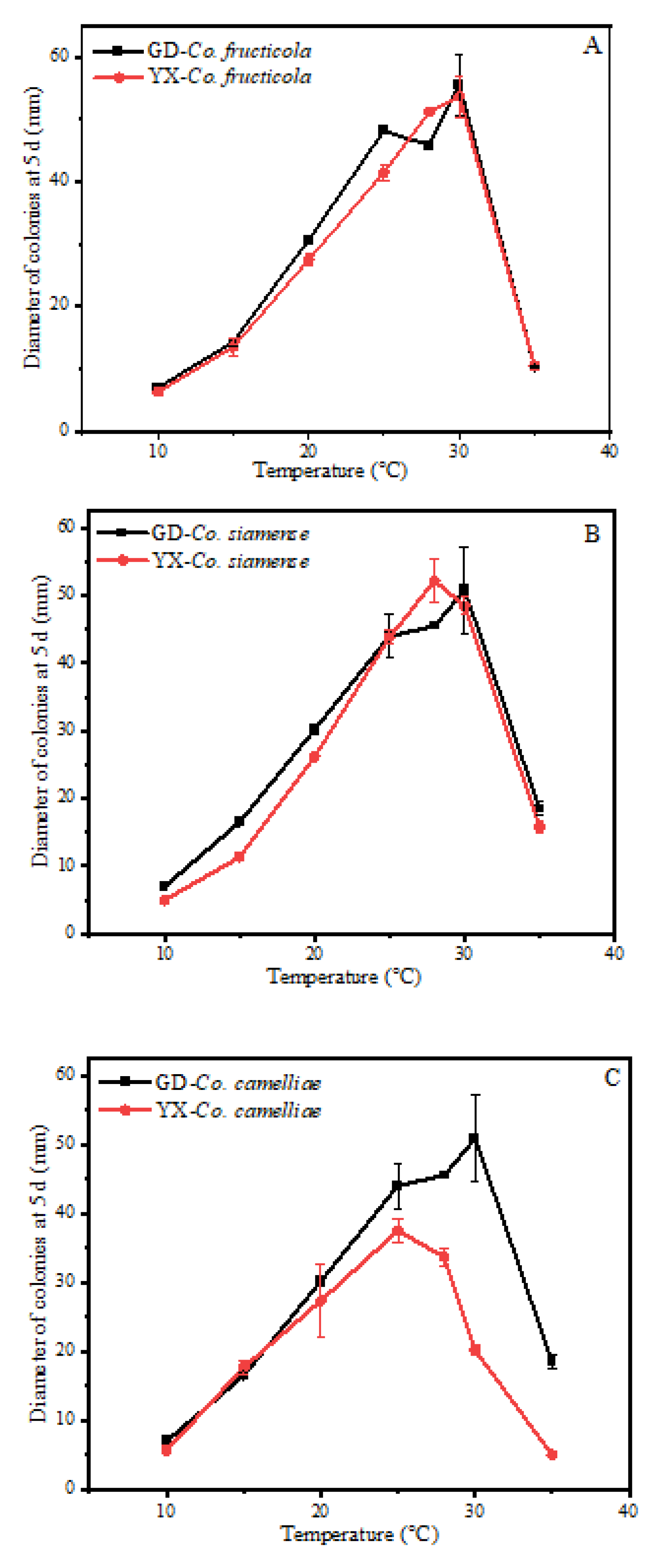
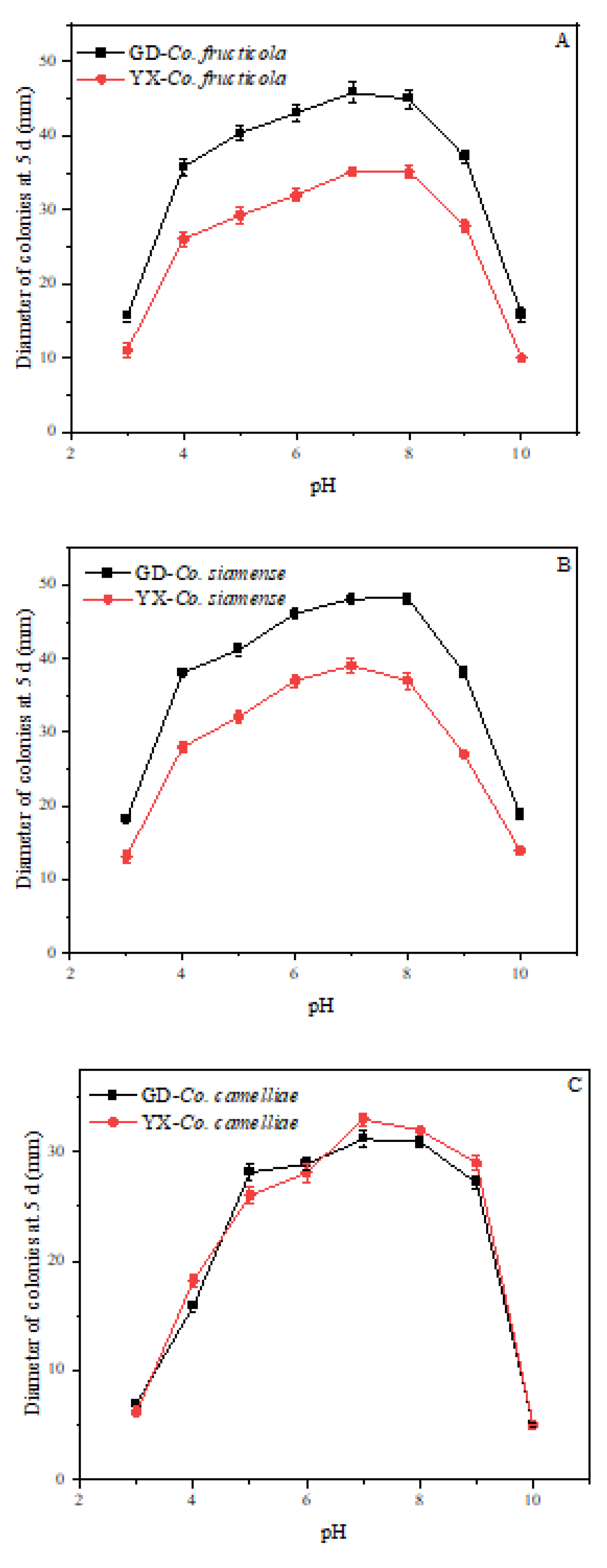
2.6. Effect of Temperature and pH on Mycelial Growth

Mycelial plugs (5 mm) from PDA were placed in the center of PDA plates and cultured in incubators set at different temperatures (10, 15, 20, 25, 28, 30, and 35 °C) for 5 days. Moreover, 5 mm mycelial plugs from PDA were placed in the center of PDA plates adjusted to a range of pH values from 3.0 to 10.0 for 5 days in an incubator at 28 °C. Na2HPO4–citric acid buffer was used to prepare PDA with pH values of 3.0–8.0, while Na2CO3-NaHCO3 buffer was used to prepare PDA at pH values 9.0 and 10.0. Finally, the colony diameter was measured. Each isolate was measured in triplicate.

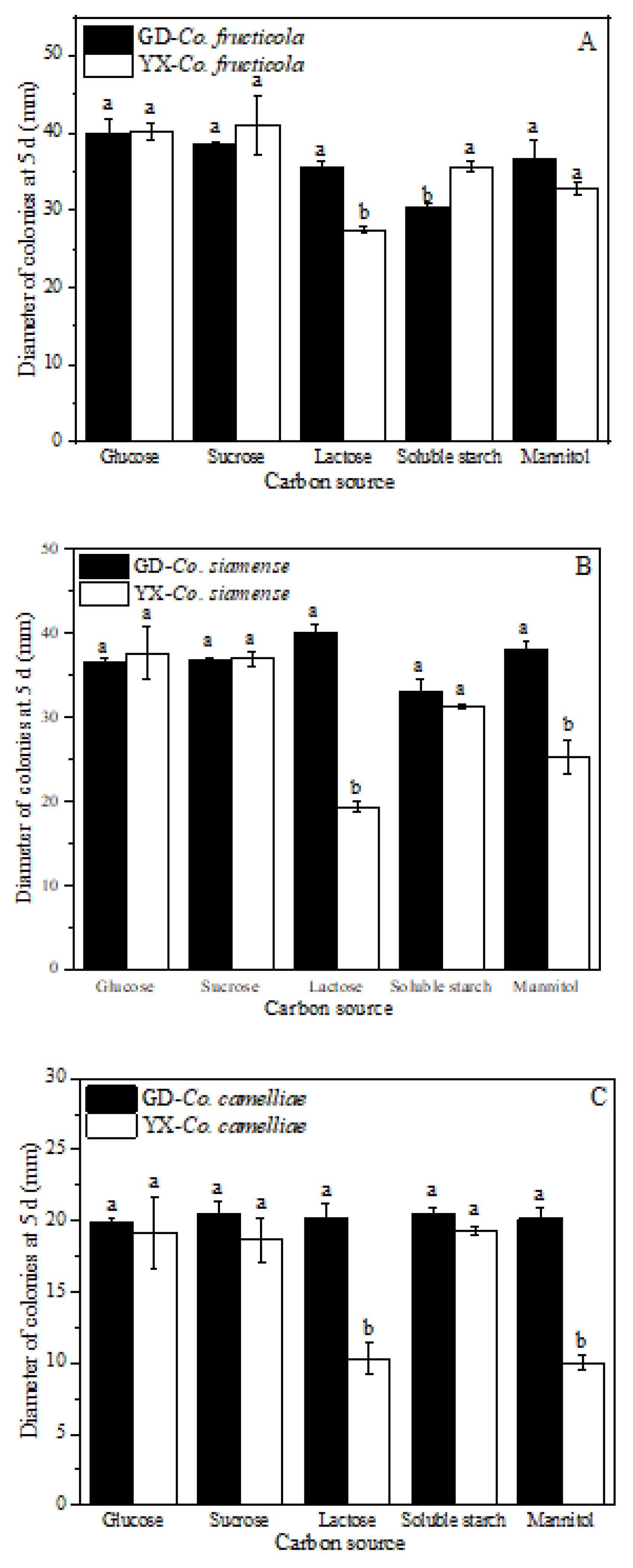
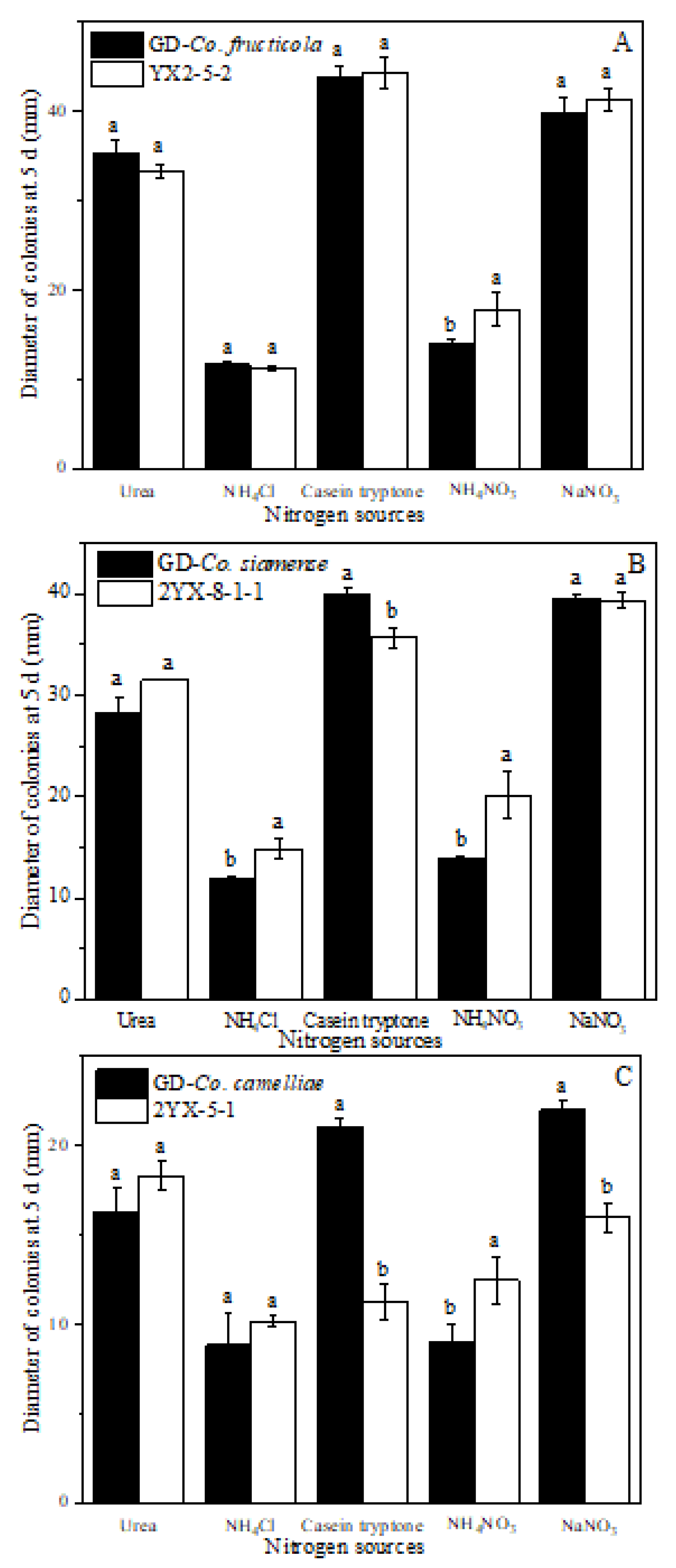
2.7. Effect of Carbon and Nitrogen Sources on Mycelial Growth

Czapek-Dox Agar (3 g/L NaNO3, 1 g/L K2HPO4, 0.5 g/L MgSO4·7H2O, 0.5 g/L KCl, 0.01 g/L FeSO4, and 30 g/L sucrose) was used as the basic medium. To analyze the carbon sources, sucrose in the basic medium was replaced by the same quantity of glucose, mannitol, lactose, or soluble starch. To analyze the nitrogen source, sodium nitrate (NaNO3) in the basic medium was replaced by the same quantity of casein tryptone, urea, ammonium chloride (NH4Cl), or ammonium nitrate (NH4NO3). Mycelial plugs (5 mm) from PDA were transferred onto the media containing different carbon or nitrogen sources. The cultures were incubated at 28 °C for 5 days. The colony diameter was measured, and each experiment was conducted in triplicate.

3. Results and Discussion

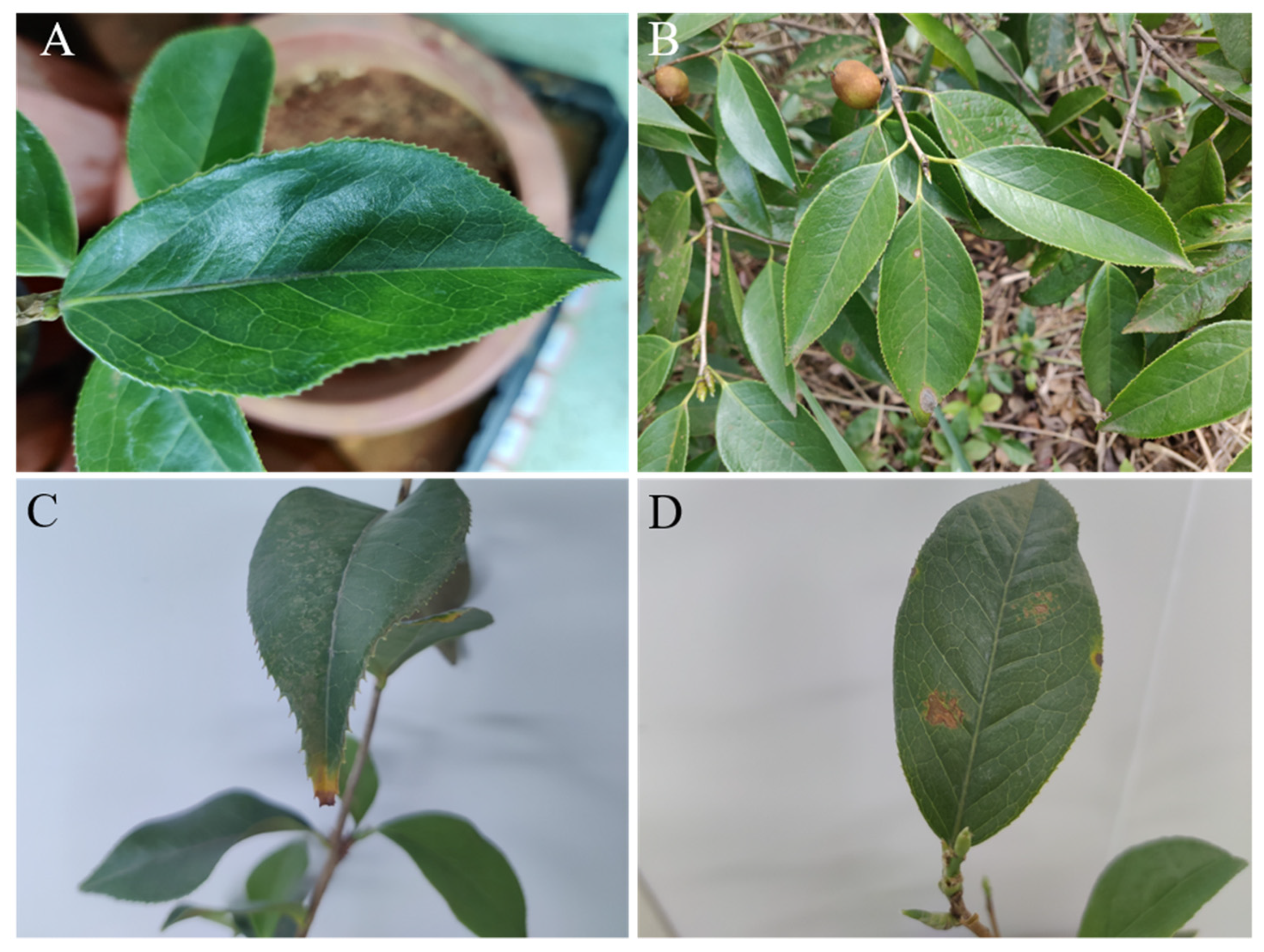
3.1. Symptom Characteristics

In April 2019, typical anthracnose symptoms were first observed on Ca. yuhsienensis in a plantation in Youxian, Hunan Province, China (113.3°2′16″ E, 26.7°15′14″ N). Most of the diseased leaves had wounds, such as those caused by insect bites [34,35,36]. Therefore, Colletotrichum spp. are more likely to infect leaves through these wounds. It is common for resistant plants to be infected through wounds. For instance, Silva (2021) [37] found that on the relatively resistant host Capsicum chinense PBC932, pathogenicity was dependent on both the inoculation method (with or without wounding) and the stage of maturity of the fruit. It was difficult to infect PBC932 with Colletotrichum spp. without injury but relatively easy to infect PBC932 with injury.
However, five unwounded Ca. yuhsienensis leaves also showed symptoms of anthracnose (Figure 1B). The isolate YX2-5-2 was reinoculated on Ca. yuhsienensis, and the same symptoms occurred, confirming Koch’s postulates (Figure 1C,D). All of the diseased leaves had irregular grayish brown spots with dark brown edges and dark brown undersides, similar to previous reports [38,39]. Ca. yuhsienensis is famous for its resistance to anthracnose, and there have been no reports of anthracnose on Ca. yuhsienensis to date. Therefore, the phenomenon that the healthy leaves of Ca. yuhsienensis were infected by Colletotrichum spp. attracted our attention. Five leaves of Ca. yuhsienensis were collected to obtain the pathogens.

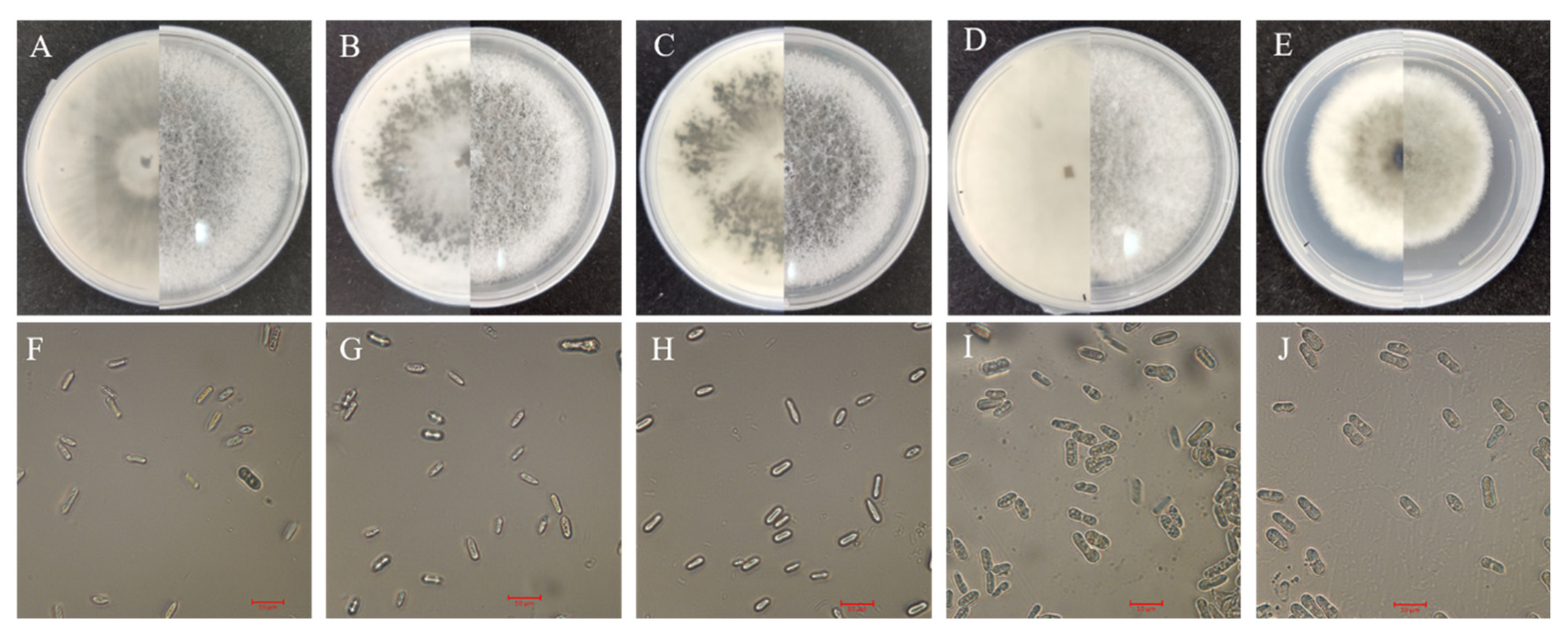
3.2. Cultural and Morphological Characteristics

Five isolates (YX2-5-2, 2YX-3-1, 2YX-5-1, 2YX-8-1-1 and 2YX-8-1-2) were obtained from Ca. yuhsienensis for the first time. Their morphological characteristics are shown in Figure 2. Few differences in colony morphology were clearly observed among the five isolates. Figure 2 shows that the upper side of these colonies on PDA was fluffy, cottony and white at first, then became light gray, whereas the reverse side slowly turned dark gray. Thus, the color of the upper side of the colony was generally lighter than that of the reverse side. There were significant differences in the rate of mycelial growth between 2YX-5-1 and the other isolates. The mycelial growth of 2YX-5-1 was generally slower than that of the other four isolates (Table 1). The conidia were all hyaline, guttulate, smooth, one-celled, and cylindrical (Figure 2). In addition, the conidial sizes of 2YX-8-1-1 and 2YX-5-1 were larger than those of other isolates (Table 1). An interesting phenomenon was observed that larger conidia can result in slower mycelial growth. A similar phenomenon has been described in a previous study; the conidia of isolate C07046 was larger than those of isolate C96002, and the mycelial growth was slower than that of C96002 even though both C07046 and C96002 are isolates of Colletotrichum coccodes [40]. In conclusion, the characteristics of the five isolates were consistent with the morphological characteristics of Colletotrichum spp. [31,41].

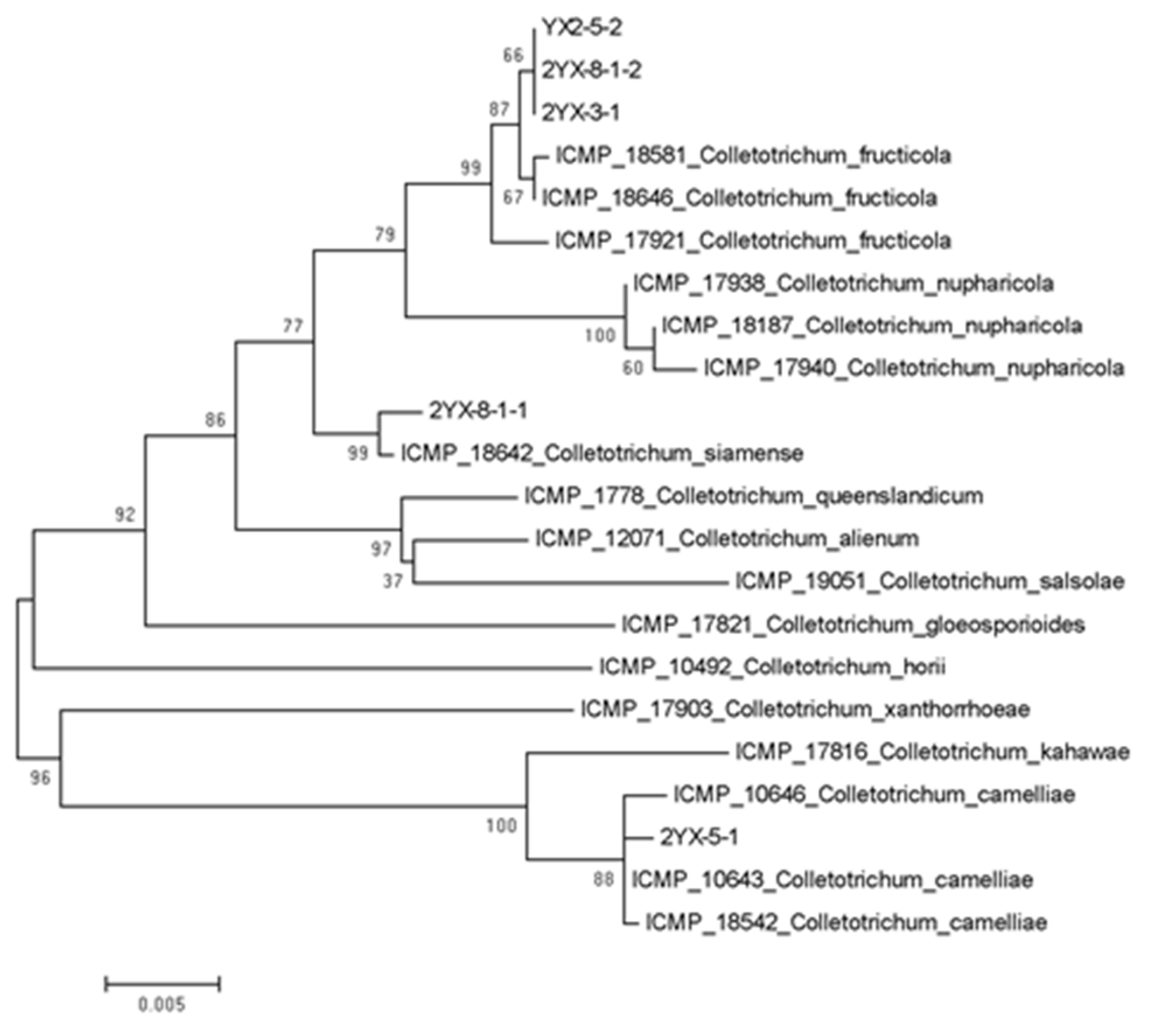
3.3. Phylogenetic Analysis

For molecular identification, internal transcribed spacer (ITS), partial actin (ACT), glyceraldehyde-3-phosphate dehydrogenase (GAPDH), manganese-superoxide dismutase (SOD2) and chitin synthase (CHS-1) genes/region of all the isolates were successfully amplified and sequenced (Table 2). The sequences at individual loci were insufficient to separate closely related species within the Co. gloeosporioides species complex. Thus, the five genes/region of each isolate were combined in the order of ITS-ACT-GAPDH-SOD2-CHS-1. A phylogenetic tree of these five isolates in this study indicated that there were three well separated clades (Figure 3). YX2-5-2, 2YX-3-1 and 2YX-8-1-2 belonged to first clade along with Co. fructicola ICMP 18581, ICMP 17,921 and ICMP 18646. 2YX-8-1-1 belonged to the second clade along with Co. siamense ICMP 18642. Lastly, 2YX-5-1 belonged to the third clade with Co. camelliae ICMP 10646, ICMP 10,643 and ICMP 18542. These results suggest that Co. fructicola is probably the most distributed species on Ca. yuhsienensis. Li (2016) [18] also found that Co. fructicola was the dominant species on Ca. oleifera. Wang (2020) [38] further discovered that Co. fructicola was common and a widely distributed species on the leaves of Ca. oleifera, which indicated that the tea oil tree is probably the most susceptible to Co. fructicola. The anthracnose of Ca. yuhsienensis was first discovered with a small rate of incidence and a few samples, and the disease has not yet been found in other areas. Consequently, the deduction above merits further verification with more samples in the future. However, this result is still helpful for the study of anthracnose on Ca. yuhsienensis.

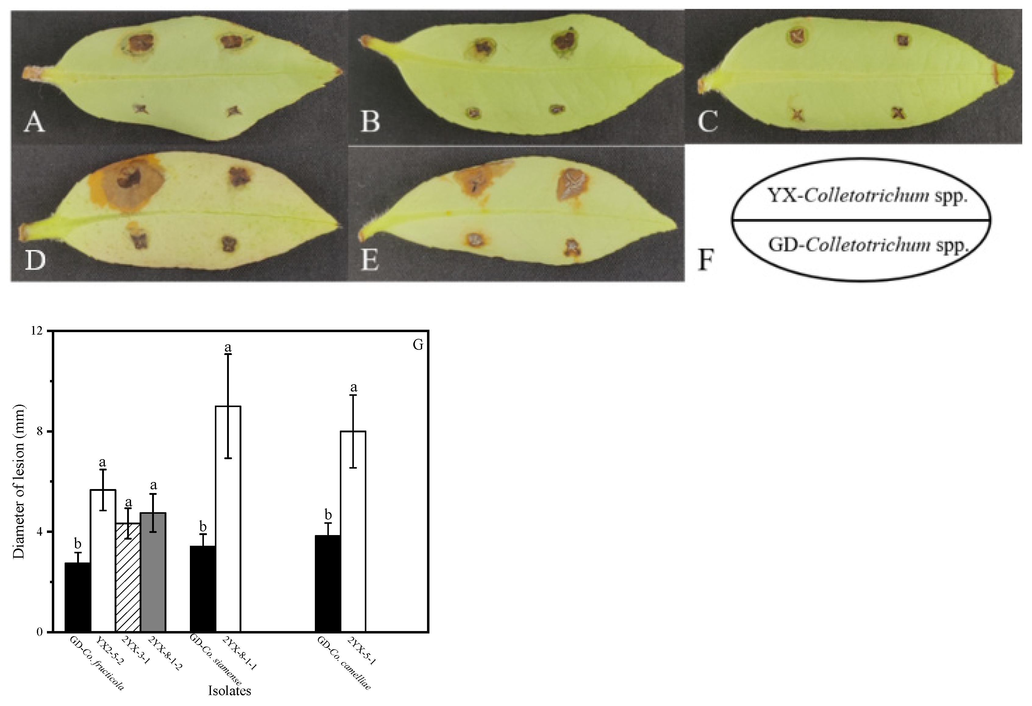
3.4. Pathogenicity Tests

To explore the reason why the Colletotrichum spp. isolated from Ca. yuhsienensis, Youxian, China (YX-Colletotrichum spp.) could infect Ca. yuhsienensis without requiring an injury to the tree, the Colletotrichum spp. isolated from Ca. oleifera, Guangdong, China (GD-Colletotrichum spp.) and YX-Colletotrichum spp. were used for pathogenicity tests. First, GD-Colletotrichum spp. infected Ca. yuhsienensis without injury for 15 days. There were no typical symptoms of anthracnose, proving that Ca. yuhsienensis could not be infected by GD-Colletotrichum spp. Therefore, we suspected that some changes may have taken place in the biological characteristics, such as pathogenicity, optimal temperature, pH, carbon and nitrogen source, of the YX-Colletotrichum spp., so that they could infect Ca. yuhsienensis without injury, while GD-Colletotrichum spp. could not.
Secondly, wounded leaves of Ca. yuhsienensis were used for pathogenicity. The results are shown in Figure 4. Different species of Colletotrichum differ in their degree of pathogenicity to Ca. yuhsienensis. The pathogenicity of Co. fructicola was the weakest, in which the diameter of lesion formed by GD-Co. fructicola was 2.45 mm and those of YX-Co. fructicola (YX2-5-2, 2YX-3-1, 2YX-8-1-2) were 4.97 mm, 4.6 mm, and 4.95 mm, respectively. Co. camelliae was the most aggressive at causing infection, in which the diameter of the lesion formed by GD-Co. camelliae was 4.95 mm and that by YX- Co. camelliae (2YX-5-1) was 7.83 mm (Figure 4G). These results showed that the pathogenicity of Co. fructicola was weaker than that of the other species of Colletotrichum spp., which is consistent with previous studies [13,20,38,42]. Lu (2018) [20] concluded that the difference in appressorium development between Co. camelliae and Co. fructicola led to pathogenic variation between these two species. However, a phenomenon was observed that YX-Co. camelliae (2YX-5-1) with larger conidia and slower mycelial growth may be more pathogenic. Choi (2011) [40] also found that the pathogenicity of isolate C07046 with larger conidia and slower mycelial growth was stronger than that of isolate C96002, even though they are all members of Co. coccodes.
Significantly, in contrast to the previous experimental results, only one isolate of YX-Co. camelliae (2YX-5-1) and YX-Co. siamense (2YX-8-1-1) with stronger pathogenicity was obtained, while three isolates of YX-Co. fructicola with weaker pathogenicity were obtained. Li (2016) [18] also found that although Co. camelliae had strong pathogenicity, the prevalent Colletotrichum spp. on Ca. oleifera was Co. fructicola, which indicated that the strong pathogenicity may be owing to the loss of other abilities, such as transmission and mycelial growth. This phenomenon is also consistent with the law of infectious diseases in animals. For instance, influenza has strong transmissibility, but the mortality rate is poor. In contrast, Creutzfeldt -Jacob disease has high mortality but poor transmission. In addition, these results also indicated that during the same incubation time, the diameter of lesions caused by YX-Colletotrichum spp. were generally larger than those of GD-Colletotrichum spp. (Figure 4). Thus, the pathogenicity of YX-Colletotrichum spp. to Ca. yuhsienensis was stronger than that of Colletotrichum spp.
The pathogenicity of Colletotrichum spp. to different hosts varies. For example, Han (2016) [43] found that Co. fructicola was more pathogenic to strawberry than other Colletotrichum spp., which differs from the results of this study. Owing to the fact that Ca. yuhsienensis is more resistant to anthracnose than Ca. oleifera, we hypothesized that YX-Colletotrichum spp. was also more pathogenic on Ca. oleifera than GD-Colletotrichum spp. [44]. Thus, wounded leaves of Ca. oleifera were used for pathogenicity. Figure 5 shows similar results. Co. fructicola was the weakest pathogen. The diameter of the lesion formed by GD-Co. fructicola was 2.75 mm and by those of YX-Co. fructicola (YX2-5-2, 2YX-3-1, 2YX-8-1-2) were 5.67 mm, 4.33 mm, and 4.75 mm, respectively. Co. camelliae was the most pathogenic. The diameter of the lesion formed by GD-Co. camelliae was 3.83 mm and that of YX-Co. camelliae (2YX-5-1) was 8.0 mm (Figure 5G). The fact that the pathogenicity of Co. fructicola was weaker than that of the other Colletotrichum spp. is also consistent with the results. The diameter of lesions caused by YX-Colletotrichum spp. were generally larger than those of GD-Colletotrichum spp. during the same amount of incubation (Figure 5).
These results prove that whether the host was Ca. oleifera or Ca. yuhsienensis, the pathogenicity of YX-Colletotrichum spp. was stronger than that of GD-Colletotrichum spp., which could be the reason why YX-Colletotrichum spp. could infect Ca. yuhsienensis without injury. Different plants respond differently to pathogens, which leads to different growth environments after the pathogen has colonized. For instance, Wang (2018) [45] found two varieties of Ca. sinensis Zhongcha 108 and Longjing 43 with different resistances to anthracnose. Among them, Zhongcha 108, having strong resistance could be due to the important role of H2O2. When the same isolate of Colletotrichum spp. infected Zhongcha 108, only the Colletotrichum spp. that were more resistant to H2O2 could colonize. The formation of resistance to H2O2 requires the cooperation of multiple metabolic pathways of Colletotrichum spp. During this process, the biological characteristics of Colletotrichum spp. may change. Similarly, increased pathogenicity of YX-Colletotrichum spp. may also cause changes in some biological characteristics. Consequently, the other biological characteristics of YX-Colletotrichum spp. were studied. Owing to the strong pathogenicity of YX2-5-2 in YX-Co. fructicola, YX2-5-2 was selected as the representative isolate of YX-Co. fructicola for the convenience of the follow-up experiments.

3.5. Effect of Temperature and pH on Mycelial Growth

Figure 6 indicates that the mycelia of isolates grew more quickly as the temperature increased. When the temperature reached 25 °C~30 °C, the diameter of colonies decreased with an increase in temperature. Lima (2015) [46] also found that both high and low temperatures significantly affected the development of the pathogen in vitro and in vivo, and that a high temperature (35 °C) can completely inactivate the virulence of Colletotrichum acutatum, while low temperatures (≤2 °C) can inactivate the virulence of Co. gloeosporioides. The results illustrate that the optimal temperature of different isolates differed, but the growth trend was similar. The optimal temperature of GD-Co. fructicola and YX-Co. fructicola (YX2-5-2) was 30 °C. The optimal temperature of GD-Co. siamense was 30 °C, while the optimal temperature of YX-Co. siamense (2YX-8-1-1) was 28 °C. The optimal temperature of YX-Co. camelliae (2YX-5-1) was 25 °C, while the optimal temperature of GD-Co. camelliae was 30 °C (Table 3). These results prove that the optimal growth temperature of GD-Colletotrichum spp. and YX-Colletotrichum spp. were slightly different. This could be because GD-Colletotrichum spp. and YX-Colletotrichum spp. originated from different latitudes. GD-Colletotrichum spp. was isolated from Guangdong Province in southern China, while YX-Colletotrichum spp. was isolated from Youxian, Hunan Province in central China. Therefore, the optimal growth temperature of GD-Colletotrichum spp. may be slightly higher than that of YX-Colletotrichum spp. A similar phenomenon was also found by Han, in that isolates of Colletotrichum nymphaeae, which are only distributed in areas of higher altitude (1,000 m), were highly sensitive to higher temperatures [43].
However, Figure 6C indicates an interesting phenomenon, in that the mycelial growth of YX-Co. camelliae (2YX-5-1) was slower than that of GD-Co. camelliae when the temperature exceeded 20 °C. The results of 3.1 and 3.2 show that YX-Co. camelliae with stronger pathogenicity has mycelia that grow more slowly than those of the other species of YX-Colletotrichum spp. A previous study also displayed the strong pathogenicity and slow mycelial growth of Co. camelliae [38]. Therefore, we hypothesized that the increased pathogenicity of YX-Co. camelliae (2YX-5-1) was at the expense of its mycelial growth rate. Xue (2019) [47] also discovered a similar phenomenon, that the mycelial growth of Colletotrichum fioriniae with stronger pathogenicity was slower than that of Co. fructicola. Li (2008) [48] also discovered that the mycelial growth of Colletotrichum gloeosporioides with stronger pathogenicity was slower than that of Colletotrichum gloeosporioides with weaker pathogenicity. Thus, these phenomena further proved the previous hypothesis that the improvement in pathogenicity may come at the expense of other adaptive abilities.
Figure 7 illustrates that all of Colletotrichum spp. could grow at pH 3~10, but the growth was greatly limited when the pH was 3 or 10. The optimal pH for the growth of the six isolates was between 6 and 8, which indicates that Colletotrichum spp. can grow in acidic, alkaline and neutral environments. He (2016) [49] also found that the optimal growth pH of Colletotrichum truncatum was 5~8.
A previous study indicated that Colletotrichum spp. alkalinizes its surroundings during the colonization of host tissue [50,51]. Tardi-Ovadia (2017) [52] also found that the pH of the area infected by Co. coccodes and Helminthosporium solani increased from the native pH of approximately 6.0 for potatoes to 7.4 to 8.0, which proved that Colletotrichum spp. grow better in an alkaline environment. However, De Costa (2014) [53] found that the optimal pH for the mycelial growth of Colletotrichum musae was 4.5. These results prove that different species of Colletotrichum respond differently to environmental factors, such as pH. Consequently, it is necessary to explore the biological characteristics of YX-Colletotrichum spp.
An interesting phenomenon appeared in that the mycelial growth of YX-Co. fructicola (YX2-5-2) and YX-Co. siamense (2YX-8-1-1) was slower than that of GD-Co. fructicola and GD-Co. siamense at pH 3~10. We hypothesized that the existence of ions inhibits their growth. We further hypothesized that the enhancement of their pathogenicity may come at the cost of the reduction of ion resistance, just as YX-Co. camelliae (2YX-5-1) increased its pathogenicity at the cost of its ability to grow.

3.6. Effect of Carbon and Nitrogen Sources on Mycelial Growth

When glucose, sucrose and mannitol were used as carbon sources, there was no significant difference in the mycelial growth of GD-Co. fructicola and YX-Co. fructicola (YX2-5-2) (Figure 8A). When lactose was used as the carbon source, the mycelial growth of YX-Co. fructicola (YX2-5-2) decreased, and when soluble starch was used as the carbon source, the mycelial growth of YX-Co. fructicola (YX2-5-2) increased (Figure 8A). There was no significant difference between YX-Co. siamense (2YX-8-1-1) and GD-Co. siamense in the utilization of glucose, sucrose and soluble starch, but YX-Co. siamense (2YX-8-1-1) was less effective than GD-Co. siamense at utilizing lactose and mannitol (Figure 8B). Furthermore, compared with GD-Co. camelliae, YX-Co. camelliae (2YX-5-1) did not differ significantly in the utilization of glucose, sucrose and soluble starch, but YX-Co. camelliae (2YX-5-1) was significantly less effective than GD-Co. camelliae in the utilization of lactose and mannitol (Figure 8C). Therefore, there was no significant difference in the utilization of glucose, sucrose and soluble starch between YX-Colletotrichum spp. and GD-Colletotrichum spp. Nevertheless, the ability of YX-Colletotrichum spp. to utilize lactose and mannitol was generally less than that of GD-Colletotrichum spp. The reason for this phenomenon could be that Ca. yuhsienensis was difficult to infect and colonize. Thus, YX-Colletotrichum spp. may sacrifice some functions not normally used, such as the utilization of mannitol and lactose, to improve its pathogenicity.
When urea, NH4Cl, casein tryptone and NaNO3 were used as nitrogen sources, there was no significant difference in the mycelial growth of GD-Co. fructicola and YX-Co. fructicola (YX2-5-2). When NH4NO3 was used as the nitrogen source, the mycelial growth of YX-Co. fructicola (YX2-5-2) increased (Figure 9A). There was no significant difference between YX-Co. siamense (2YX-8-1-1) and GD-Co. siamense in the utilization of urea and NaNO3, but YX-Co. siamense (2YX-8-1-1) was less effective than GD-Co. siamense at utilizing casein tryptone (Figure 9B). In addition, YX-Co. siamense (2YX-8-1-1) was significantly more effective than GD-Co. siamense at utilizing NH4Cl and NH4NO3 (Figure 9B). YX-Co. camelliae (2YX-5-1) was less effective than GD-Co. camelliae at utilizing casein tryptone and NaNO3 but slightly more effective at utilizing NH4NO3 (Figure 9C). These results illustrate that the utilization of NH4+ of YX-Colletotrichum spp. was generally stronger than that of GD-Colletotrichum spp., while the utilization of casein tryptone was generally less effective than that of Colletotrichum spp. We hypothesized that there are fewer proteins or amino acids that can be directly used in the leaves of Ca. yuhsienensis, but more NH4+, resulting in the stronger utilization of NH4+ by YX-Colletotrichum spp. Prusky (2001) [54] also believed that NH4+ was an important pathogenic factor of Colletotrichum. Therefore, the increased utilization of NH4+ may increase the pathogenicity of YX-Colletotrichum spp.

4. Conclusions

This study presents the first research on anthracnose in Ca. yuhsienensis leaves caused by Colletotrichum spp. in Hunan Province, China. Five isolates (YX2-5-2, 2YX-3-1, 2YX-5-1, 2YX-8-1-1 and 2YX-8-1-2), having the morphological characteristics of Colletotrichum spp., were obtained from Ca. yuhsienensis. A phylogenetic analysis demonstrated that YX2-5-2, 2YX-3-1 and 2YX-8-1-2 belonged to Co. fructicola, 2YX-8-1-1 belonged to Co. siamense and 2YX-5-1 belonged to Co. camelliae. A pathogenicity test indicated that the pathogenicity of YX-Colletotrichum spp. to Ca. yuhsienensis and Ca. oleifera was stronger than that of GD-Colletotrichum spp. The results of biological characteristics indicated that the mycelial growth and ionic resistance of YX-Colletotrichum spp. were generally less than that of Colletotrichum spp. Further research illustrated that the ability of YX-Colletotrichum spp. to utilize lactose and mannitol was less than that of Colletotrichum spp., while the ability to utilize NH4+ was generally greater than that in Colletotrichum spp. In summary, the results will provide theoretical guidance for the study of the pathogenesis and control schemes of anthracnose in Ca. yuhsienensis.

Author Contributions

Conceptualization, G.Z. and J.L.; methodology, X.C. and C.L.; validation, X.C. and L.J.; formal analysis, L.J. and A.B.; investigation, X.C. and L.J.; resources, G.Z. and J.L.; data curation, X.C. and C.L.; writing—original draft preparation, X.C.; writing—review and editing, L.J. visualization, X.C.; supervision, G.Z. and J.L.; project administration, G.Z. and J.L.; funding acquisition, G.Z. All authors have read and agreed to the published version of the manuscript.

Funding

This research was funded by the National Natural Science Foundation of China grant number 31971661, Natural Science Foundation of Hunan Province, China grant number 2021JJ31145, Postgraduate Scientific Research Innovation Project of Hunan Province grant number CX20200712 and Scientific Innovation Fund for Post-graduates of Central South University of Forestry and Technology grant number CX20201008.

Institutional Review Board Statement

Not applicable.

Informed Consent Statement

Not applicable.

Data Availability Statement

Data available in a publicly accessible repository. The data presented in this study are openly available in Dryad at https://0-doi-org.brum.beds.ac.uk/10.5061/dryad.n02v6wwzx, reference number (https://datadryad.org/stash/share/kcfEYPaXFxXHqwiLf1SH_bcdTqjh8FDpAVXQgva9Cos) accessed on 4 November 2021.

Acknowledgments

The authors would like to thank Deyi Yuan, a researcher at the Central South University of Forestry and Technology for his great help in providing Camellia yuhsienensis Hu.

Conflicts of Interest

The authors declare no conflict of interest. The funders had no role in the design of the study; in the collection, analyses, or interpretation of data; in the writing of the manuscript, or in the decision to publish the results.

References

  1. Chen, Y.Z. Oil Tea Camellia Superior Germplasm Resources; China Forestry Publishing House: Beijing, China, 2008. [Google Scholar]
  2. Qin, S.; Rong, J.; Zhang, W.; Chen, J. Cultivation history of Camellia oleifera and genetic resources in the Yangtze River Basin. Biodivers. Sci. 2018, 26, 384–395. [Google Scholar] [CrossRef]
  3. Li, Y.; Ye, T.; Han, C.; Ye, Z.; Zhang, J.; Xiao, S.; Yuan, D. Cytogenetic analysis of interspecific hybridization in oil-tea (Camellia oleifera). Euphytica 2021, 217, 28. [Google Scholar] [CrossRef]
  4. Shi, T.; Wu, G.; Jin, Q.; Wang, X. Camellia oil authentication: A comparative analysis and recent analytical techniques developed for its assessment. A review. Trends Food Sci. Technol. 2020, 97, 88–99. [Google Scholar] [CrossRef]
  5. Yang, C.; Liu, X.; Chen, Z.; Lin, Y.; Wang, S. Comparison of Oil Content and Fatty Acid Profile of Ten New Camellia oleifera Cultivars. J. Lipids 2016, 2016, 3982486. [Google Scholar] [CrossRef] [Green Version]
  6. Yang, S.; Liang, K.; Wang, A.; Zhang, M.; Qiu, J.; Zhang, L. Physiological Characterization and Transcriptome Analysis of Camellia oleifera Abel. during Leaf Senescence. Forests 2020, 11, 812. [Google Scholar] [CrossRef]
  7. Chen, Y.; Deng, S.; Chen, L.; Li, M.; He, H.; Wang, X.; Peng, S.; Liu, C.; Wang, R.; Xu, Y.; et al. A new view on the development of oil tea camellia industry. J. Nanjing For. Univ. Nat. Sci. Edit 2020, 44, 1–10. [Google Scholar] [CrossRef]
  8. Wang, J.C. The total output value of Camellia oleifera Industry in China reached 116 billion yuan. China Food Newspaper, 20 November 2020; 2. [Google Scholar]
  9. Li, S.Z.; Zhang, S.P.; Li, H. A HOPS protein, CfVps39, is required for appressorium formation, environmental stress response and vacuolar fusion of Colletotrichum fructicola. For. Pathol. 2021, 51, e12692. [Google Scholar] [CrossRef]
  10. Chen, Y.; Liu, J.; Jiang, S.; Li, H.; Zhou, G. Colletotrichum fructicola STE50 is required for vegetative growth, asexual reproduction, appressorium formation, pathogenicity and the response to external stress. J. Plant. Pathol. 2020, 102, 335–342. [Google Scholar] [CrossRef]
  11. Jin, A.X.; Zhou, G.Y.; Li, H. Progress, problem and prospect of oil camelliae anthracnose (Colletotrichum gloeosporioides) research. For. Pest. Dis. 2009, 28, 27–31. [Google Scholar]
  12. Zhu, Y.; Liao, W.; Zou, D.; Wu, Y.; Yan, D. Identification and biological characteristics of the pathogen from Camellia oleifera anthracnose in Guangxi. J. Plant. Prot. 2015, 42, 382–389. [Google Scholar] [CrossRef]
  13. Wang, Y.-C.; Hao, X.-Y.; Wang, L.; Bin, X.; Wang, X.-C.; Yang, Y.-J. Diverse Colletotrichum species cause anthracnose of tea plants (Camellia sinensis (L.) O. Kuntze) in China. Sci. Rep. 2016, 6, 35287. [Google Scholar] [CrossRef] [PubMed] [Green Version]
  14. Mongkolporn, O.; Taylor, P.W.J. Chili anthracnose: Colletotrichum taxonomy and pathogenicity. Plant. Pathol. 2018, 67, 1255–1263. [Google Scholar] [CrossRef]
  15. Hyde, K.D.; Cai, L.; Cannon, P.F.; Crouch, J.A.; Crous, P.W.; Damm, U.; Goodwin, P.H.; Chen, H.; Johnston, P.R.; Jones, E.B.G.; et al. Colletotrichum—Names in current use. Fungal Divers. 2009, 39, 147–182. [Google Scholar]
  16. Dean, R.; Van Kan, J.A.; Pretorius, Z.A.; Hammond-Kosack, K.E.; Di Pietro, A.; Spanu, P.D.; Rudd, J.J.; Dickman, M.; Kahmann, R.; Ellis, J. The Top 10 fungal pathogens in molecular plant pathology. Mol. Plant. Pathol. 2012, 13, 414–430. [Google Scholar] [CrossRef] [Green Version]
  17. Li, H.; Zhu, D.; Xu, J.; Zhou, G.; Hu, M.; Tian, F. Population genetic structure of Colletotrichum gloeosporioides causing anthracnose of Camellia oleifera in China. Acta Phytopathol. Sin. 2014, 44, 620–628. [Google Scholar] [CrossRef]
  18. Li, H.; Zhou, G.-Y.; Liu, J.-A.; Xu, J. Population Genetic Analyses of the Fungal Pathogen Colletotrichum fructicola on Tea-Oil Trees in China. PLoS ONE 2016, 11, e0156841. [Google Scholar] [CrossRef] [Green Version]
  19. Fu, M.; Crous, P.W.; Bai, Q.; Zhang, P.F.; Xiang, J.; Guo, Y.S.; Zhao, F.F.; Yang, M.M.; Hong, N.; Xu, W.X.; et al. Colletotrichum species associated with anthracnose of Pyrus spp. in China. Persoonia 2019, 42, 1–35. [Google Scholar] [CrossRef] [Green Version]
  20. Lu, Q.; Wang, Y.; Li, N.; Ni, D.; Yang, Y.; Wang, X. Differences in the Characteristics and Pathogenicity of Colletotrichum camelliae and C. fructicola Isolated From the Tea Plant [Camellia sinensis (L.) O. Kuntze]. Front. Microbiol. 2018, 9, 3060. [Google Scholar] [CrossRef]
  21. Holtappels, D.; Fortuna, K.; Lavigne, R.; Wagemans, J. The future of phage biocontrol in integrated plant protection for sustainable crop production. Curr. Opin. Biotechnol. 2021, 68, 60–71. [Google Scholar] [CrossRef]
  22. Savchenko, I.V. Breeding New Varieties and Hybrids of Agricultural Plants. Her. Russ. Acad. Sci. 2017, 87, 104–110. [Google Scholar] [CrossRef]
  23. Nie, Z.; Huang, X.; Hu, Z.; Li, X.; Yin, H.; Li, J. Characterization of the complete chloroplast genome of Camellia yuhsienensis Hu, a resilient shrub with strong floral fragrance. Mitochondrial DNA B 2020, 5, 3016–3017. [Google Scholar] [CrossRef] [PubMed]
  24. Cao, Y.; Yao, X.; Ren, H.; Wang, K. Determination of fatty acid composition and metallic element content of four Camellia species used for edible oil extraction in China. J. Consum Prot. Food Saf. 2017, 12, 165–169. [Google Scholar] [CrossRef]
  25. Denton-Giles, M.; Bradshaw, R.E.; Dijkwel, P.P. Ciborinia camelliae (Sclerotiniaceae) Induces Variable Plant Resistance Responses in Selected Species of Camellia. Phytopathology 2013, 103, 725–732. [Google Scholar] [CrossRef] [PubMed]
  26. Denton-Giles, M. Characterization of Incompatible and Compatible Camellia-Ciborinia Camelliae Plant-Pathogen Interactions. Ph.D. Thesis, Massey University, Palmerston North, New Zealand, 2014. [Google Scholar]
  27. Yang, G.; Shu, Q.; Duan, L.; Chen, C.; Zheng, H. Resistance of main cultivars of oil tea to Colletotrichum gloeosporioides. J. Anhui Agric. Univ. 2004, 31, 480–483. [Google Scholar] [CrossRef]
  28. Duan, L.; Yang, G.; Shu, Q.; Hongbing, Z. Relationship of Peel Color with Resistance to Anthracnose in Oiltea Camellia. Nonwood For. Res. 2005, 23, 9–12+20. [Google Scholar] [CrossRef]
  29. Li, J.; Luo, Z.; Zhang, C.; Qu, X.; Chen, M.; Song, T.; Yuan, J. Seasonal Variation in the Rhizosphere and Non-Rhizosphere Microbial Community Structures and Functions of Camellia yuhsienensis Hu. Microorganisms 2020, 8, 1385. [Google Scholar] [CrossRef]
  30. Saracchi, M.; Locati, D.; Colombo, E.M.; Pasquali, M. Updates on Ciborinia camelliae, the causal agent of camellia flower blight. J. Plant. Pathol. 2019, 101, 215–223. [Google Scholar] [CrossRef]
  31. Weir, B.; Johnston, P.; Damm, U. The Colletotrichum gloeosporioides species complex. Stud. Mycol. 2012, 73, 115–180. [Google Scholar] [CrossRef] [PubMed] [Green Version]
  32. Liu, F.; Weir, B.S.; Damm, U.; Crous, P.W.; Wang, Y.; Liu, B.; Wang, M.; Zhang, M.; Cai, L. Unravelling Colletotrichum species associated with Camellia: Employing ApMat and GS loci to resolve species in the C. gloeosporioides complex. Persoonia 2015, 35, 63–86. [Google Scholar] [CrossRef] [Green Version]
  33. Cai, L.; Hyde, K.D.; Taylor, P.W.J.; Weir, B.S.; Waller, J.M.; Abang, M.M.; Zhang, J.Z.; Yang, Y.L.; Phoulivong, S.; Liu, Z.Y.; et al. A polyphasic approach for studying Colletotrichum. Fungal Divers. 2009, 39, 183–204. [Google Scholar]
  34. Piesik, D.; Rochat, D.; Delaney, K.J.; Marion-Poll, F. Orientation of European corn borer first instar larvae to synthetic green leaf volatiles. J. Appl. Entomol. 2013, 137, 234–240. [Google Scholar] [CrossRef] [Green Version]
  35. Skoczek, A.; Piesik, D.; Wenda-Piesik, A.; Buszewski, B.; Bocianowski, J.; Wawrzyniak, M. Volatile organic compounds released by maize following herbivory or insect extract application and communication between plants. J. Appl. Entomol. 2017, 141, 630–643. [Google Scholar] [CrossRef]
  36. Piesik, D.; Rochat, D.; van der Pers, J.; Marion-Poll, F. Pulsed Odors from Maize or Spinach Elicit Orientation in European Corn Borer Neonate Larvae. J. Chem. Ecol. 2009, 35, 1032–1042. [Google Scholar] [CrossRef]
  37. De Silva, D.D.; Ades, P.K.; Taylor, P.W.J. Pathogenicity of Colletotrichum species causing anthracnose of Capsicum in Asia. Plant. Pathol. 2021, 70, 875–884. [Google Scholar] [CrossRef]
  38. Wang, Y.; Chen, J.-Y.; Xu, X.; Cheng, J.; Zheng, L.; Huang, J.; Li, D.-W. Identification and Characterization of Colletotrichum Species Associated with Anthracnose Disease of Camellia oleifera in China. Plant. Dis. 2020, 104, 474–482. [Google Scholar] [CrossRef] [PubMed]
  39. Moral, J.; Agustí-Brisach, C.; Raya, M.C.; Jurado-Bello, J.; López-Moral, A.; Roca, L.F.; Chattaoui, M.; Rhouma, A.; Nigro, F.; Sergeeva, V.; et al. Diversity of Colletotrichum Species Associated with Olive Anthracnose Worldwide. J. Fungi 2021, 7, 741. [Google Scholar] [CrossRef] [PubMed]
  40. Choi, K.J.; Kim, W.G.; Kim, H.G.; Choi, H.W.; Lee, Y.K.; Lee, B.D.; Lee, S.Y.; Hong, S.K. Morphology, Molecular Phylogeny and Pathogenicity of Colletotrichum panacicola Causing Anthracnose of Korean Ginseng. Plant. Pathol. J. 2011, 27, 1–7. [Google Scholar] [CrossRef] [Green Version]
  41. Chen, X.; Liu, C.; Liu, J.A.; Zhou, G.Y. First Report of Colletotrichum fructicola Causing Anthracnose on Camellia yuhsienensis Hu in China. Plant. Dis. 2021. [Google Scholar] [CrossRef] [PubMed]
  42. Sharma, G.; Pinnaka, A.K.; Shenoy, B.D. Resolving the Colletotrichum siamense species complex using ApMat marker. Fungal Divers. 2015, 71, 247–264. [Google Scholar] [CrossRef]
  43. Han, Y.C.; Zeng, X.G.; Xiang, F.Y.; Ren, L.; Chen, F.Y.; Gu, Y.C. Distribution and Characteristics of Colletotrichum spp. Associated with Anthracnose of Strawberry in Hubei, China. Plant. Dis. 2016, 100, 996–1006. [Google Scholar] [CrossRef] [Green Version]
  44. Zhu, J.; Ye, G. A Study on the Peroxidase in Camellia Species and Their Resistance to Anthracnoso Causing Fungus. J. Fujian Coll. For. 1990, 10, 368–375. [Google Scholar] [CrossRef]
  45. Wang, Y.; Hao, X.; Lu, Q.; Wang, L.; Qian, W.; Li, N.; Ding, C.; Wang, X.; Yang, Y. Transcriptional analysis and histochemistry reveal that hypersensitive cell death and H2O2 have crucial roles in the resistance of tea plant (Camellia sinensis (L.) O. Kuntze) to anthracnose. Hort. Res. 2018, 5, 18. [Google Scholar] [CrossRef] [Green Version]
  46. Lima, N.B.; Lima, W.G.; Tovar-Pedraza, J.M.; Michereff, S.J.; Camara, M.P.S. Comparative epidemiology of Colletotrichum species from mango in northeastern Brazil. Eur. J. Plant. Pathol. 2015, 141, 679–688. [Google Scholar] [CrossRef]
  47. Xue, L.; Zhang, L.; Yang, X.X.; Huang, X.; Wu, W.; Zhou, X.; White, J.F.; Liu, Y.; Li, C. Characterization, Phylogenetic Analyses, and Pathogenicity of Colletotrichum Species on Morus alba in Sichuan Province, China. Plant. Dis. 2019, 103, 2624–2633. [Google Scholar] [CrossRef] [PubMed]
  48. Li, N.; Tan, G.; Li, Z. Studies on the Biological Characteristics of Collectotrichum gloeosporiodes Strain Related to Virulence and Hypovirulence on Apple Anthracnose. Acta Laser Biol. Sin. 2008, 17, 64–69. [Google Scholar]
  49. He, Y.Y.; Chen, Q.G.; Shu, C.W.; Yang, M.; Zhou, E.X. Colletotrichum truncatum, a new cause of anthracnose on Chinese flowering cabbage (Brassica parachinensis) in China. Trop. Plant. Pathol. 2016, 41, 183–192. [Google Scholar] [CrossRef]
  50. Miyara, I.; Shafran, H.; Davidzon, M.; Sherman, A.; Prusky, D. pH Regulation of Ammonia Secretion by Colletotrichum gloeosporioides and Its Effect on Appressorium Formation and Pathogenicity. Mol. Plant.-Microbe Interact. 2010, 23, 304–316. [Google Scholar] [CrossRef] [Green Version]
  51. Alkan, N.; Meng, X.; Friedlander, G.; Reuveni, E.; Sukno, S.; Sherman, A.; Thon, M.; Fluhr, R.; Prusky, D. Global Aspects of pacC Regulation of Pathogenicity Genes in Colletotrichum gloeosporioides as Revealed by Transcriptome Analysis. Mol. Plant.-Microbe Interact. 2013, 26, 1345–1358. [Google Scholar] [CrossRef] [Green Version]
  52. Tardi-Ovadia, R.; Linker, R.; Tsror, L. Direct Estimation of Local pH Change at Infection Sites of Fungi in Potato Tubers. Phytopathology 2017, 107, 132–137. [Google Scholar] [CrossRef] [PubMed] [Green Version]
  53. De Costa, D.M.; Chandima, A.A.G. Effect of exogenous pH on development and growth of Colletotrichum musae and development of anthracnose in different banana cultivars in Sri Lanka. J. Natl. Sci. Found. Sri Lanka 2014, 42, 203–214. [Google Scholar] [CrossRef]
  54. Prusky, D.; McEvoy, J.L.; Leverentz, B.; Conway, W.S. Local Modulation of Host pH by Colletotrichum Species as a Mechanism to Increase Virulence. Mol. Plant.-Microbe Interact. 2001, 14, 1105–1113. [Google Scholar] [CrossRef] [PubMed] [Green Version]
Figure 1. Typical symptoms of anthracnose on Camellia yuhsienensis. (A) No symptoms were observed on control leaves treated with sterilized water; (B) Anthracnose symptoms on Ca. yuhsienensis in a plantation in Youxian, Hunnan Province, China; (C) Anthracnose symptoms were seen on attached, unwounded leaves treated with a conidial suspension (1 × 10⁶ conidial/mL) of isolate YX2-5-2; (D) Anthracnose symptoms were seen on attached wounded leaves treated with a conidial suspension (1 × 10⁶ conidial/mL) of isolate YX2-5-2.
Forests 12 01712 g001
Figure 2. (AE) Colony morphology of isolates YX2-5-2, 2YX-8-1-2, 2YX-3-1, 2YX-8-1-1 and 2YX-5-1 on PDA after 5 days at 28 °C, respectively; (FJ) Conidia of isolates YX2-5-2, 2YX-8-1-2, 2YX-3-1, 2YX-8-1-1 and 2YX-5-1, respectively; scale bar = 10 μm.
Forests 12 01712 g002
Figure 3. A Maximum Likelihood phylogenetic tree using isolates YX2-5-2, 2YX-8-1-2, 2YX-3-1, 2YX-8-1-1, 2YX-5-1 and type strains of the Colletotrichum gloeosporioides complex based on the combined CHS-1, ACT, ITS, SOD2 and GAPDH gene sequences.
Forests 12 01712 g003
Figure 4. Pathogenicity of GD-Colletotrichum spp. (isolated from Camellia oleifera) and YX-Colletotrichum spp. (isolated from Ca. yuhsienensis) on Ca. yuhsienensis. (AC) Lesion development by YX-Co. fructicola (YX2-5-2, 2YX-3-1, 2YX-8-1-2) and GD-Co. fructicola was photographed at 4 days post infiltration (dpi); (D) Lesion development by YX-Co. siamense (2YX-8-1-1) and GD-Co. siamense was photographed at 4 dpi; (E) Lesion development by YX-Co. camelliae (2YX-5-1) and GD-Co. camelliae was photographed at 4 dpi; (F) Schematic diagram of the pathogenicity test in which the conidia of GD-Colletotrichum spp. were infiltrated into the underside panel of the leaf, while those of YX-Colletotrichum spp. were infiltrated into the upper side panel of the same leaf; (G) Lesion diameters were measured at 4 dpi. The error bars represent standard deviations based on six biological replicates. Lesion diameters followed by the same lowercase letter are not significantly different at p < 0.05 using an ANOVA.
Forests 12 01712 g004
Figure 5. Pathogenicity of GD-Colletotrichum spp. and YX-Colletotrichum spp. on Camellia. oleifera. (AC) Lesion development by YX-Co. fructicola (YX2-5-2, 2YX-3-1, 2YX-8-1-2) and GD-Co. fructicola was photographed at two days post inoculation (dpi); (D) Lesion development by YX-Co. siamense (2YX-8-1-1) and GD-Co. siamense were photographed at 2 dpi; (E) Lesion development by YX-Co. camelliae (2YX-5-1) and GD-Co. camelliae were photographed at 2 dpi; (F) Schematic diagram of pathogenicity. The conidia of GD-Colletotrichum spp. were infiltrated into the underside panel of the leaf, while the YX-Colletotrichum spp. was infiltrated into the upper side panel of the same leaf; (G) Lesion diameters were measured at 2 dpi. The error bars represent standard deviations based on six biological replicates. Lesion diameters followed by the same lowercase letter are not significantly different at p < 0.05 using an ANOVA.
Forests 12 01712 g005
Figure 6. Effect of temperature on the growth of (A) YX-Co. fructicola (YX2-5-2) and GD-Co. fructicola; (B) YX-Co. siamense (2YX-8-1-1) and GD-Co. siamense; (C) YX-Co. camelliae (2YX-5-1) and GD-Co. camelliae. The error bars represent standard deviations based on three biological replicates.
Forests 12 01712 g006
Figure 7. Effect of pH on the mycelial growth of (A) YX-Co. fructicola (YX2-5-2) and GD-Co. fructicola; (B) YX-Co. siamense (2YX-8-1-1) and GD-Co. siamense; (C) YX-Co. camelliae (2YX-5-1) and GD-Co. camelliae. The error bars represent standard deviations based on three biological replicates.
Forests 12 01712 g007
Figure 8. Effect of carbon sources on the growth of (A) YX-Co. fructicola (YX2-5-2) and GD-Co. fructicola; (B) YX-Co. siamense (2YX-8-1-1) and GD-Co. siamense; (C) YX-Co. camelliae (2YX-5-1) and GD-Co. camelliae. The error bars represent standard deviations based on three biological replicates. Diameters of colonies followed by the same lowercase letter are not significantly different at p < 0.05 using an ANOVA.
Forests 12 01712 g008
Figure 9. Effect of nitrogen sources on the growth of (A) YX-Co. fructicola (YX2-5-2) and GD-Co. fructicola; (B) YX-Co. siamense (2YX-8-1-1) and GD-Co. siamense; (C) YX-Co. camelliae (2YX-5-1) and GD-Co. camelliae. The error bars represent standard deviations based on three biological replicates. Diameters of colonies followed by the same lowercase letter are not significantly different at p < 0.05 using an ANOVA.
Forests 12 01712 g009
Table 1. Summary of the morphological data of the five isolates.
IsolatesConidial Size (μm)Mycelial Growth (mm/d)
YX2-5-27.27 ± 0.52 × 1.81 ± 0.3113.9 ± 0.6
2YX-8-1-27.27 ± 0.48 × 2.42 ± 0.2713.7 ± 0.3
2YX-3-17.27 ± 0.55 × 1.81 ± 0.2113.1 ± 0.8
2YX-8-1-19.09 ± 0.45 × 3.64 ± 0.4313.4 ± 0.3
2YX-5-19.09 ± 0.36× 3.64 ± 0.398.5 ± 0.9
Table 2. GenBank accession number of the five isolates.
IsolateGene NameGenbank Accession Number
YX2-5-2GAPDHMW398864
YX2-5-2ACTMW398863
YX2-5-2CHSIMW886232
YX2-5-2SOD2MW398866
YX2-5-2ITSMW398865
2YX-8-1-2GAPDHMZ224482
2YX-8-1-2ACTMZ224483
2YX-8-1-2CHSIOL310498
2YX-8-1-2SOD2MZ224480
2YX-8-1-2ITSMZ224481
2YX-8-1-1GAPDHMW398861
2YX-8-1-1ACTMW398860
2YX-8-1-1CHSIOL310500
2YX-8-1-1SOD2MZ048745.1
2YX-8-1-1ITSMW398862
2YX-5-1GAPDHMZ048746
2YX-5-1ACTMW924872
2YX-5-1CHSIMW924874
2YX-5-1SOD2MW924873
2YX-5-1ITSMW911446
2YX-3-1GAPDHMW924878
2YX-3-1ACTMW924879
2YX-3-1CHSIOL310499
2YX-3-1SOD2MW924877
2YX-3-1ITSMW924880
Table 3. Optimum mycelial growth temperature of the six isolates.
IsolatesOptimal Temperatures (°C)
YX-Colletotrichum spp.YX-Co. fructicola30
YX-Co. siamense28
YX-Co. camelliae25
GD-Colletotrichum spp.GD-Co. fructicola30
GD-Co. siamense30
GD-Co. camelliae30
Publisher’s Note: MDPI stays neutral with regard to jurisdictional claims in published maps and institutional affiliations.

