Article

Development and Comparison of Seminested PCR, qPCR, and LAMP for the Rapid Detection of Arthrinium phaeospermum, the Causal Agent of Bamboo Blight

1
College of Forestry, Sichuan Agricultural University, Chengdu 611130, China
2
National Forestry and Grassland Administration Key Laboratory of Forest Resources Conservation and Ecological Safety on the Upper Reaches of the Yangtze River, Chengdu 611130, China
*
Author to whom correspondence should be addressed.
Submission received: 27 April 2022 / Revised: 25 May 2022 / Accepted: 27 May 2022 / Published: 29 May 2022
(This article belongs to the Special Issue Detection and Analysis of Forest Pathogens)

Abstract

:
Bambusa pervariabilis × Dendrocalamopsis grandis blight is a newly discovered disease in bamboos that has caused substantial economic loss to the affected areas. With the purpose of carrying out rapid detection of Bambusa pervariabilis × Dendrocalamopsis grandis blight caused by Arthrinium phaeospermum during the incubation period, three sets of detection assays were established: seminested PCR, real-time quantitative PCR, and LAMP. The specificity, sensitivity, and effectiveness of these assays were also detected. The results showed that the three assays were able to specifically amplify the target bands from five strains of Arthrinium phaeospermum from different sources, but none of the other 18 strains were able to obtain the specific bands. The sensitivity of the established seminested PCR, LAMP, and real-time quantitative PCR assays were 100, 10, and 1 pg/μL, respectively. The presence of A. phaeospermum could be detected in the early stage of disease using the total DNA of infected hybrid bamboo tissue as a template. The three systems established in this study are of great significance for the early diagnosis and rapid detection of hybrid bamboo blight.

1. Introduction

Bambusa pervariabilis × Dendrocalamopsis grandis is an improved hybrid bamboo species derived from the inbred parental lines of B. pervariabilis McClure and D. grandis Keng f. that is commonly found in Sichuan, Zhejiang, and Fujian provinces. In addition to the characteristics of having a male parent and a female parent, it also has the characteristics of rapid growth and easy reproduction. This hybrid bamboo species is good for the use of both its bamboo shoots and timber, and it has important economic, ecological, and social benefits [1]. At present, there are many studies on shoot blight in bamboos. However, as a new disease that has emerged in recent years, related research on B. pervariabilis × D. grandis shoot blight is not comprehensive. At present, there is a lack of effective high-quality disease-resistant varieties, so shoot blight in hybrid bamboos is mainly controlled using chemical and silvicultural techniques. In recent years, research on biological control of shoot blight in hybrid bamboos has made progress.
This hybrid bamboo blight is found in an area where farmland is being restored to forests in Tianquan county, Sichuan province, and the disease occurrence area reaches more than 3000 hm2 [2], thus threatening the process of restoring farmlands to forests and the construction of ecological barriers [3]. The pathogenic fungus, Arthrinium phaeospermum (Corda) M.B.Ellis, has been found to cause wilting and death of hybrid bamboos [4]. A. phaeospermum is a pathogenic fungus distributed all over the world with a wide range of host species, including plants, humans [5,6], and animals. In addition to B. pervariabilis × D. grandis [3], its host plants include cowpea, garden pea [7], sugarcane [8], Phyllostachys prominens [9,10], new olive [11], Phyllostachys viridis [12], etc.
Traditional disease diagnosis is mainly based on the symptoms of the plant and the pathogen’s morphology, and diagnosis is difficult and time-consuming to conduct. Moreover, when the disease shows obvious symptoms, it has already caused damage to the plants and inflicted considerable losses to the forestry industry. To overcome these problems, rapid and accurate detection technology has been gradually developed. With the advantages of high precision, fast speed, and easy operation [13], nucleic acid amplification technology is now widely used in the detection of pathogens. It is necessary to use sensitive and accurate molecular detection methods for early diagnosis of diseases during the incubation period in order to prevent and control them in time.
The development of polymerase chain reaction (PCR) specifically for pathogens has greatly improved the accuracy and sensitivity of disease diagnosis. Nested PCR (NPCR) is one of the most sensitive methods for detecting pathogens, even at a very low concentration [14,15]. Nested PCR consists of two rounds of PCR. The first round targets a wide range of DNA regions, while the second round targets a narrow subregion of the product using the first round as a template [16]. Compared with the traditional PCR method, qPCR assay has the advantages of high sensitivity and specificity and can be used to detect a variety of pathogens [17]. Real-time quantitative PCR (qPCR) has been widely used in the quantitative detection and diagnosis of many plant pathogens. Coats et al. [18] developed a sensitive and specific quantitative PCR (qPCR) assay to detect Rhizoctonia tuliparum and tested its effectiveness with bulb and soil samples. Wang et al. [19] developed a TaqMan qPCR assay of Fusarium virguliforme, which is the causal agent of soybean sudden death syndrome. With the development of nucleic acid amplification technology, various isothermal amplification techniques have been established. Loop-mediated isothermal amplification (LAMP) is one of the most widely used isothermal amplification techniques. Loop-mediated isothermal amplification (LAMP) is a newly developed gene amplification method that has the advantages of simplicity and high specificity [20].
These three detection methods each have their own unique advantages and have been successfully used in the detection of pathogens. In this study, seminested PCR, real-time quantitative PCR (qPCR), and LAMP were used for A. phaeospermum detection. We evaluated and compared the three detection methods in an attempt to find an effective and suitable method for the detection of A. phaeospermum. To the best of our knowledge, there is no previous study comparing seminested PCR, qPCR, and LAMP assays for the detection of A. phaeospermum.

2. Materials and Methods

2.1. Strains

A total of 18 strains were tested. A. phaeospermum strains were isolated from samples of diseased hybrid bamboos in Ya’an and Yibin, Sichuan Province, China. Strains 4 and 5 were blight pathogens of different bamboo species; strains 6–9 were different fungi of the same genus (Arthrinium); strains 15–18 were pathogens isolated from diseased hybrid bamboos from Changning and Jiang’an, Sichuan Province, China; strains 13, 14, 17, and 18, similar to A. phaeospermum, are also present in Apiosporaceae, and the corresponding genus is a fungus of the most recently related genus. All strains were preserved at the College of Forestry, Sichuan Agricultural University (Table 1).

2.2. Culture of the Tested Strains and DNA Extraction
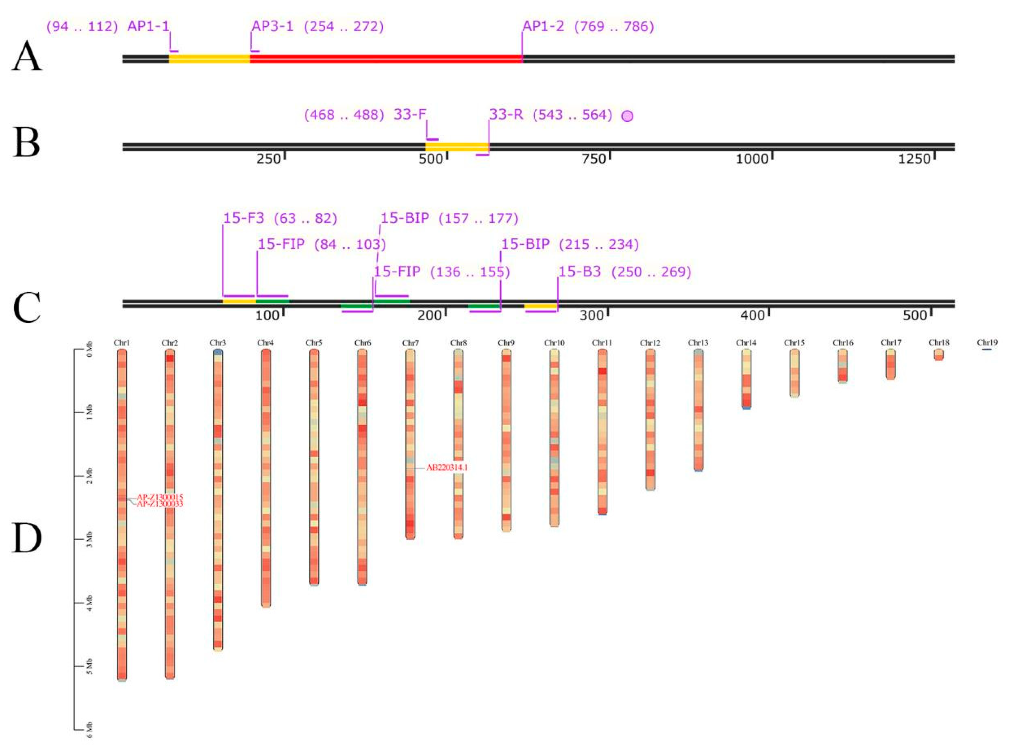
All strains were inoculated on potato dextrose agar (PDA) culture at a temperature of 25 °C for 7 days. DNA was extracted from hyphae using the Fungi Genomic DNA Extraction Kit (Solarbio, China) following the manufacturer’s instructions. The DNA was detected by agarose gel electrophoresis and stored at −20 °C until use.
For strains with few hyphae, a small amount of hyphae was picked and shake-cultured in the sterilized PDB at a constant temperature of 28 °C for 5 d. Then, we obtained the hyphae before DNA extraction.

2.3. Primer Design

The seminested PCR primers were designed on known sequences of the beta tubulin gene sequences of A. phaeospermum (GeneBank accession number: AB220314.1) by Primer Premier 5.0 and validated with BLAST-N (http://blast.ncbi.nlm.nih.gov (accessed on 11 April 2021)). The AP1-1 and AP1-2 primers were designed for initial PCR amplification, and AP1-2 and AP3-1 were used to amplify the second fragment inside the fragment produced in the initial reaction. Primers for qPCR and LAMP detection methods were designed based on the whole genome of A. phaeospermum [21]. The primers of qPCR were also designed in a similar manner according to the gene AP-Z1300033 (GeneBank accession number: QYRS01000001.1). The LAMP primers, including two external (15-F3 and 15-B3) and two internal (15-FIP and 15-BIP) primers, were designed according to the gene AP-Z1300015 (GeneBank accession number: QYRS01000001.1) with online PrimerExplorer V4 software (Eiken Chemical Co., Tokyo, Japan). The nine primers for seminested PCR, qPCR, and LAMP assays are listed in Table 2, and the target positions of the primers are illustrated in Figure 1. Figure 1D shows the chromosomal location of AP-Z1300015, AP-Z1300033, and AB220314.1 in A. phaeospermum. All designed primers were synthesized by Tsingke, Beijing, China.

2.4. Seminested PCR, qPCR, and LAMP System Assays

2.4.1. Seminested PCR Conditions

To determine the most effective conditions for seminested PCR, gradient PCR amplifications were performed at different annealing temperatures (ranging from 57 to 62 °C) for two primer sets. To ensure the yield of the amplification products and the specificity of the primers, appropriate annealing temperatures were selected.
For seminested PCR, the initial reaction was performed at a volume of 25 µL with 22 μL of 1 × Taq PCR MasterMix, 400 nM of each primer (AP1-1 and AP1-2), and 1 μL of the DNA template. The PCR amplification conditions of the first round were as follows: initial denaturation step of 5 min at 94 °C; followed by 34 cycles of 94 °C (45 s), 55 °C (45 s), and 72 °C (20 s); and then a final extension at 72 °C for 2 min.
As with the initial reaction, the second round was also conducted in a volume of 25 μL using the AP1-2 and AP3-1 primers and 1 μL of the amplified PCR product from the first round as template. The amplification program consisted of 5 min at 94 °C; followed by 34 cycles of 94 °C (45 s), 60 °C (45 s), and 72 °C (20 s); and then a final extension at 72 °C for 2 min. Genomic DNA extracted from A. phaeospermum was used for the positive control, and ddH2O was used as the negative control. Amplification products were visualized in 1% agarose gel containing GoldView™ (Solarbio, Beijing, China).

2.4.2. qPCR Conditions

The qPCR reactions were performed in a 20 μL reaction system consisting of 10 μL 2×TSINGKE Master qPCR Mix (Tsingke, Beijing, China), 400 nM of each primer (33-F and 33-R), and 1 μL of the DNA template. The thermal cycling procedure included denaturation at 95 °C for 1 min and 40 cycles of amplification at 10 s at 95 °C and 15 s at 60 °C. The reaction process was carried out in a fluorescence quantitative polymerase chain reaction (CFX96 Real-Time System, Bio-Rad, Hercules, CA, USA).
The purified PCR product was inserted into the pEASY®-Blunt Zero Cloning Vector (TransGen Biotech, Beijing, China) to construct a standard plasmid. The recombinant plasmid was transformed with Trelief™ 5α Chemically Competent Cell (Tsingke, Beijing, China). The plasmid was added to 100 µL Trelief ™ 5α, then ice-bathed for 30 min, then made to undergo a procedure at 42 °C for 45 s, and then ice-bathed again for 2 min. Additionally, 500 μL of the LB liquid medium was added, followed by shake-culturing at a speed of 200 rpm at 37 °C for 1 h, and colony PCR was used for detection. The recombinant plasmid DNA was quantified by a NanoPhotometer N60 Touch and diluted 10-fold with the TE buffer before use.

2.4.3. LAMP Conditions

To increase the accuracy and sensitivity of the results, the concentration of each component in the LAMP assay was optimized. The LAMP reaction conditions were optimized in terms of the concentration of dNTPs, Mg2+, and betaine as well as the concentration ratio of the external and internal primers.
For the optimization of the LAMP assay, a range of concentration gradients of dNTP (0.9, 1.0, 1.1, 1,2, and 1.3 mM), Mg2+ (1.5, 1.8, 2.1, 2.4, and 2.7 mM), and betaine (0.5, 0.6, 0.7, 0.8, and 0.9 mM) and a range of concentration ratio gradients of the external and internal primers (the external primer concentration was fixed at 0.1 μM, while the internal primer concentration gradient was set to 0.2, 0.4, 0.8, 1.6, and 3.2 μM) were evaluated under similar conditions. The LAMP reactions were incubated at 65 °C for 60 min, followed by incubation at 85 °C for 10 min for enzyme denaturation. The products were analyzed by 1.5% agarose gel electrophoresis, and the positive samples showed a typical ladder-like pattern. After amplification, 1 μL of hydroxy naphthol blue (HNB) was added to observe color change of the reaction tube. The LAMP reaction can be detected by both color change and electrophoresis.

2.5. Specificity of the Assays

The specificities of the three assays were tested with the genomic DNA of 18 fungus strains noted in Table 1, including strains belonging to the Arthrinium genus and other non-Arthrinium species. The detection was reacted under the previously optimized conditions, and ddH2O was used as a negative control.
Primer specificity of qPCR can be determined by the presence of a single absorption peak presented by the melting curves. The same primers (33-F/33-R) were used for conventional PCR, and the results of agarose gel electrophoresis were used to determine primer specificity. Additionally, HNB was added to observe amplification to determine the specificity of the LAMP assay.

2.6. Sensitivity of the Assays

To compare the sensitivity of seminested PCR, qPCR, and LAMP for A. phaeospermum detection, the DNA concentration was detected using the NanoPhotometer N60 Touch, and the DNA was diluted to 10 ng/μL, 1 ng/μL, 100 pg/μL, 10 pg/μL, 1 pg/μL, 100 fg/μL, and 10 fg/μL at a concentration gradient 10 times that of the reaction templates of the sensitivity test. The reaction procedures were the same as above. A series of plasmid standard samples were prepared by 10-fold dilution of the A. phaeospermum recombinant plasmid solution. The standard curve of qPCR was established with the threshold cycle (Ct) values against the dilution concentration logarithm of the recombinant plasmid.

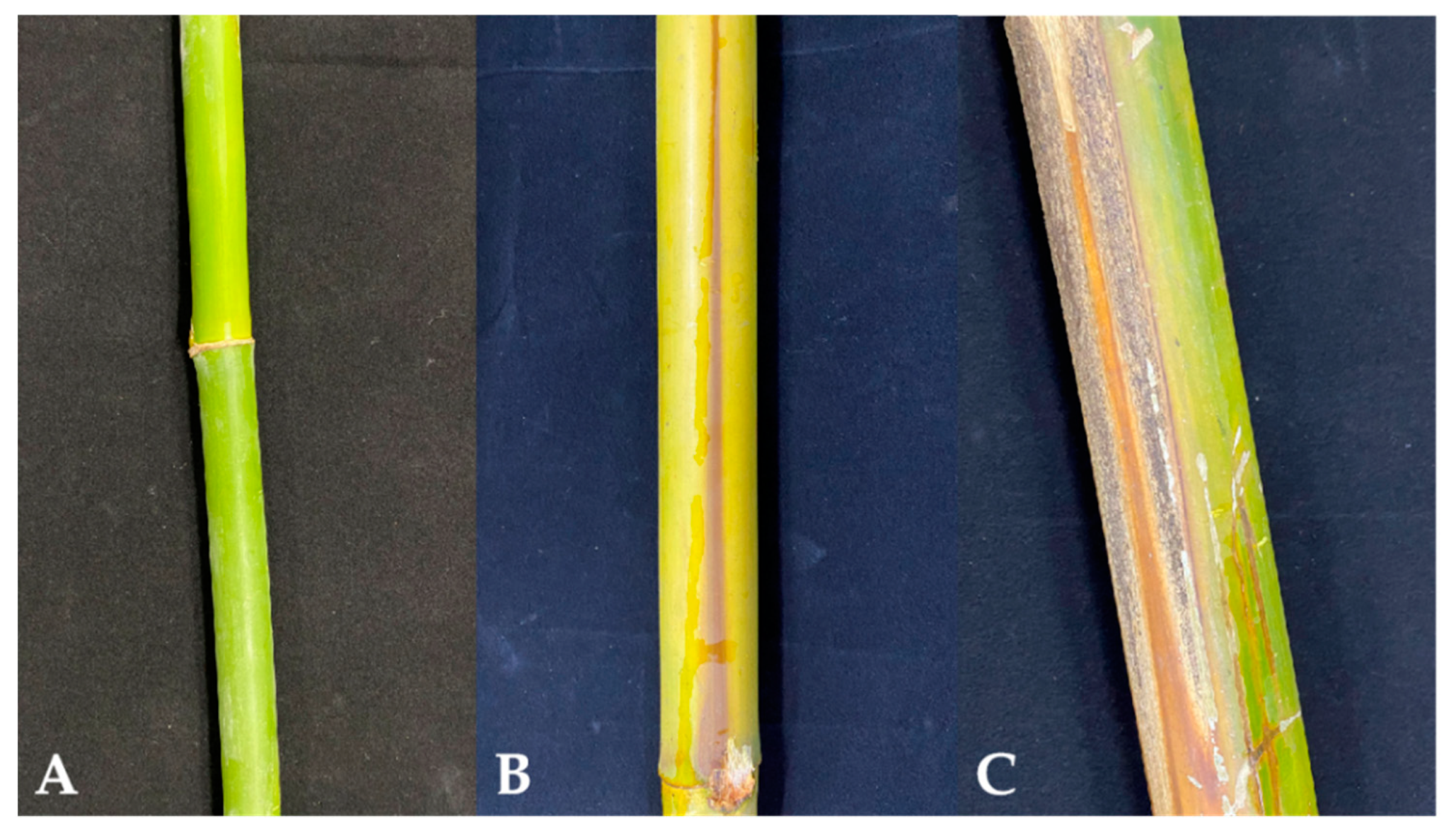
2.7. Detection of Artificially Inoculated and Field-Collected Samples

2.7.1. Artificially Inoculated Sample Preparation

A number of fresh and healthy hybrid bamboo shoots were randomly selected from the nursery of Sichuan Agricultural University for artificial inoculation. The strain was inoculated on PDA and cultured at 25 °C for 7 days to prepare the 1 × 10⁵ spores·mL−1 suspension. The hybrid bamboos were surface sterilized with 75% ethanol, then dried naturally after rinsing with sterile water. Additionally, the surface-sterilized bamboos were wounded with a sterile needle at a depth of 1 mm in the stems and twigs and inoculated with the prepared spore suspension. Healthy hybrid bamboos were also inoculated with sterile water as a negative control. All plants were kept at 25 to 28 °C and covered with plastic bags to maintain high relative humidity (90 to 95%). DNA was extracted from diseased bamboos at 4, 8, 12, and 16 days after inoculation and used as template for evaluating the effectiveness of the assays. At the same time, DNA extracted from noninoculated healthy hybrid bamboos was used as template for the negative control.

2.7.2. Field-Collected Sample Preparation

The field-collected diseased hybrid bamboos, which were samples from Meishan, Sichuan Province, China (29°41′ N, 104°11′ E), were classified as asymptomatic, mild, and severe according to the degree of incidence of different grades. DNA extracted from diseased and healthy branches was amplified by seminested PCR, real-time quantitative PCR, and LAMP. The reaction procedures were the same as above.

3. Results

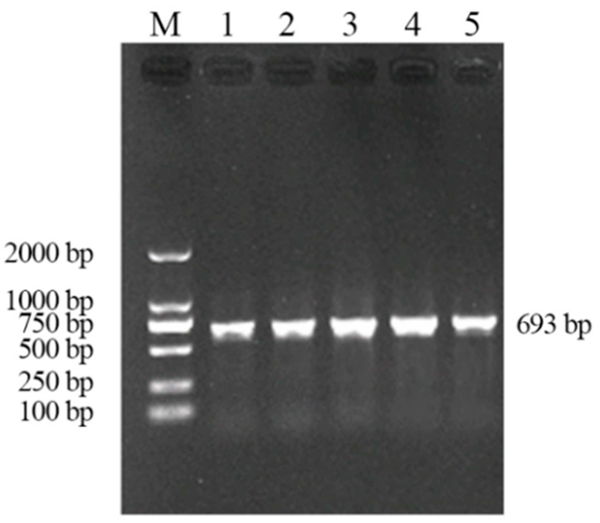
3.1. Establishment of Seminested PCR

3.1.1. Optimization of the Reaction Conditions of Seminested PCR

Primer AP1-1/AP1-2 was used to amplify the genomic DNA of A. phaeospermum at different annealing temperatures to identify the most effective conditions for seminested PCR. The results of amplification (Figure 2) showed that 693 bp fragments could be amplified at 57–61 °C, and no impurity bands were produced. In order to ensure the yield of the amplified product, 59 °C was selected as the annealing temperature of the outer primer.
The products from the first round were used as template and amplified with the inner primer AP1-2/AP3-1. The result in Figure 3 shows that 533 bp fragments could be amplified at 58–62 °C. In order to ensure the yield of the amplified product, 60 °C was selected as the annealing temperature of the inner primer.

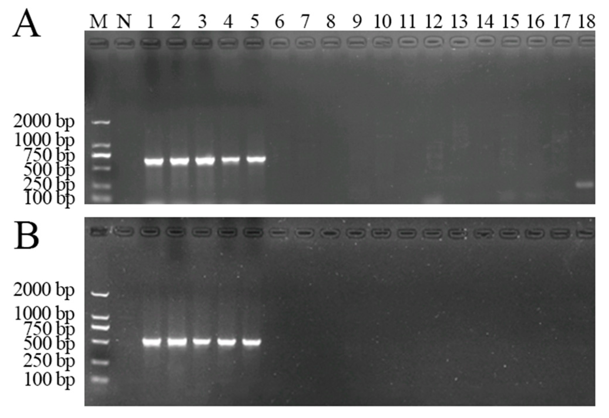
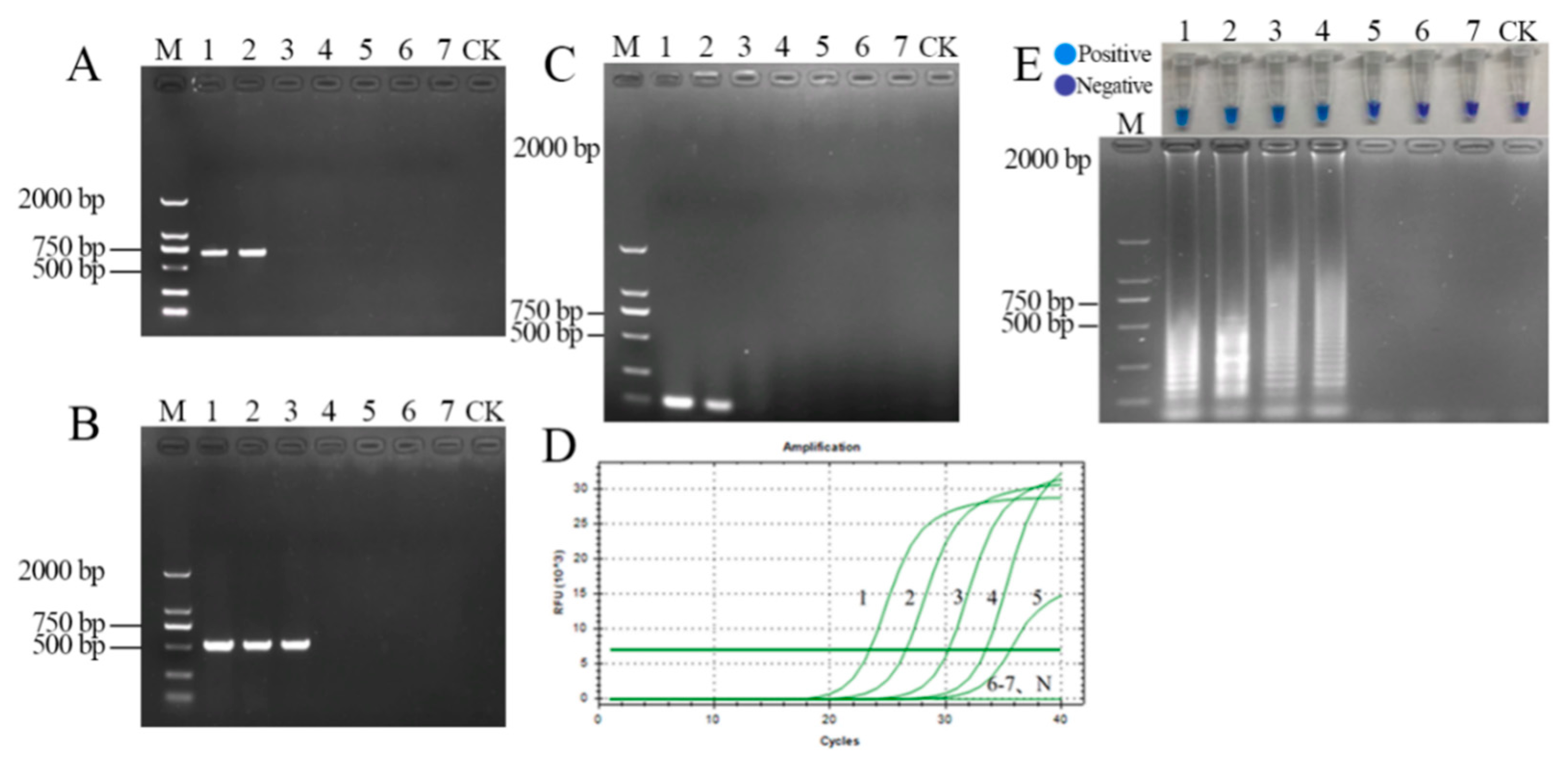
3.1.2. Specificity of the Seminested PCR Assay

To determine the specificity of the reaction, the outer primer AP1-1/AP1-2 was used to amplify the genomic DNA of A. phaeospermum and the other tested strains. The genomic DNA of five isolates from different sources revealed a DNA band, and there were no specific bands in the DNA of other strains and the negative control (Figure 4A). The products from the first round were amplified with the inner primer AP1-2/AP3-1 for the second round. The results are shown in Figure 4B. The genomic DNA of A. phaeospermum from five different sources amplified the specific bands of 533 bp, while no nonspecific amplifications appeared in the other tested strains. Therefore, the specificity of the seminested PCR detection of A. phaeospermum was confirmed.

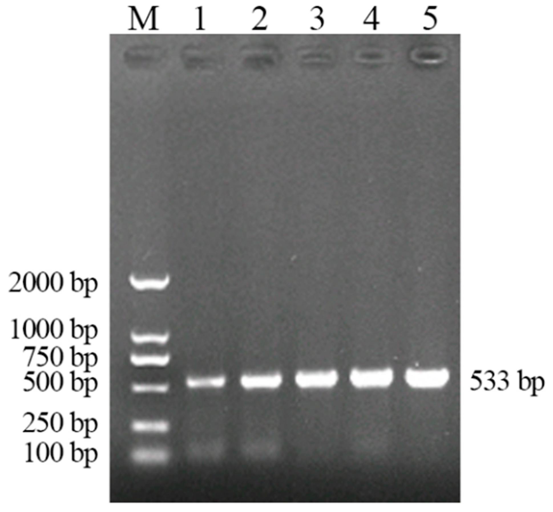
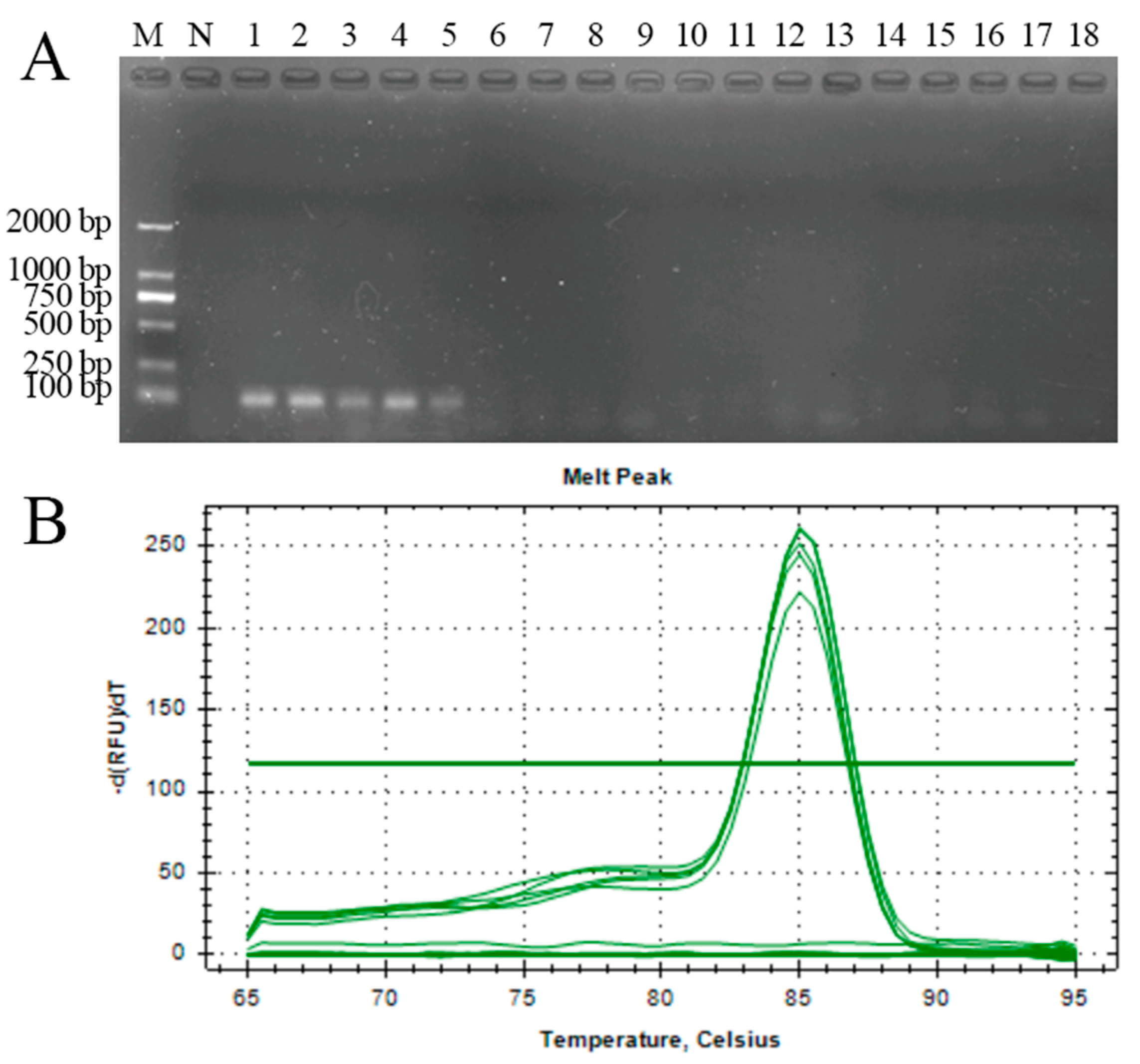
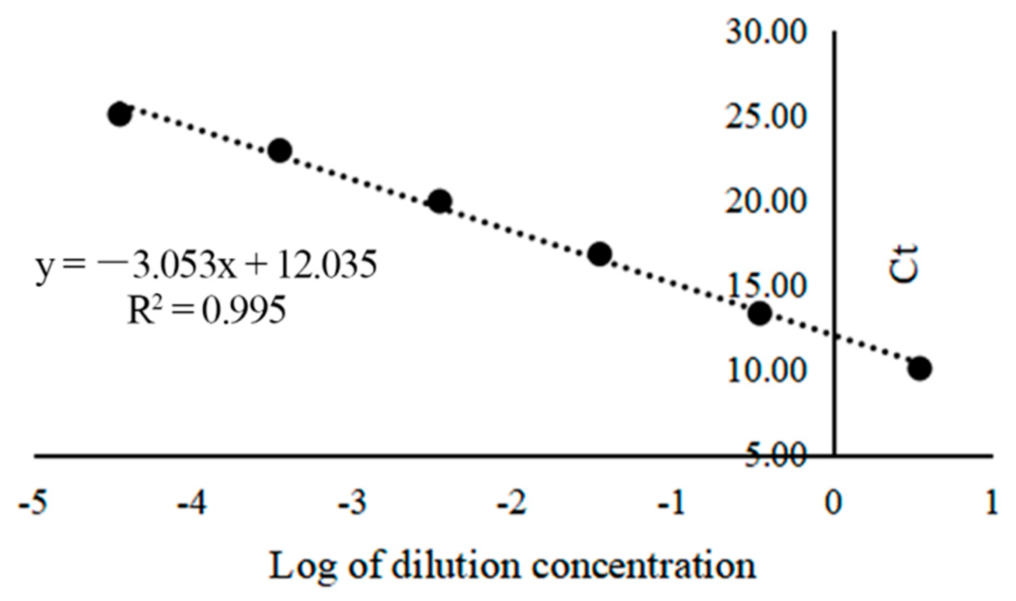
3.2. Establishment of Real-Time Quantitative PCR

The genomic DNA of A. phaeospermum and the DNA of the isolates were amplified by conventional PCR and real-time quantitative PCR with the 33-F/33-R primer. The results are shown in Figure 5. It was found that the genomic DNA of A. phaeospermum of five strains from different sources amplified the specific bands of 97 bp, but there were no specific bands in the other strains or the negative control (Figure 5A). Real-time quantitative PCR also showed that the designed primer only had a single absorption peak for A. phaeospermum (Figure 5B). At the end of the cyclic reaction, the melting curve of the PCR product showed a single sharp peak at 85 °C, indicating the specificity of the 33-F/33-R primer.
The standard curve of the qPCR was established with the threshold cycle (Ct) values against the dilution concentration logarithm of the recombinant plasmid (Figure 6). The regression equation for recombinant plasmids was y = −3.053x + 12.035 (R2 = 0.995), in which x indicated the dilution concentration logarithm of the recombinant plasmid, while y indicated the Ct value. The standard curve between the log of the dilution concentration and Ct value generated a linear fit with a slope of −3.053. The linear regression coefficient (R2) was 0.995, indicating a good correlation. The PCR efficiency was 112.59% based on the equation e = 101/s − 1 [22]. This illustrates the reliability of the qPCR assay method, and the target DNA can be quantified by the standard curves.

3.3. Establishment of LAMP

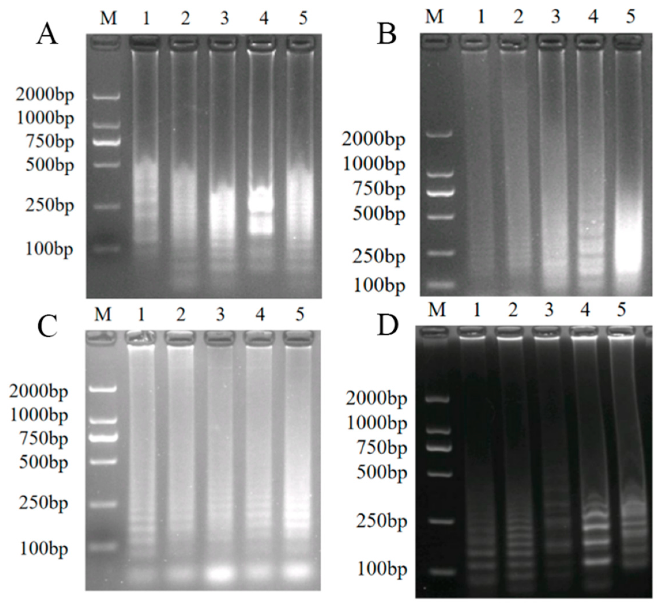
3.3.1. Optimization of the Reaction Conditions of LAMP

In this experiment, the system optimization experiment was carried out using the genomic DNA of A. phaeospermum as a template, and the accuracy of the results was confirmed by agarose gel electrophoresis.
dNTP concentration: Under this experimental condition, the dNTP concentration was set with a total of five concentration gradients of 0.9, 1.0, 1.1, 1,2, and 1.3 mM, and the DNA amplification product reached the maximum when the dNTP concentration was 1.1 mM (Figure 7A). Therefore, 1.1 mM was established as the suitable concentration of dNTPs.
Mg2+ concentration: Under the conditions of this experiment, the Mg2+ concentration was set with a total of five concentration gradients of 1.5, 1.8, 2.1, 2.4, and 2.7 mM. The amount of DNA amplification product gradually increased when the Mg2+ concentration was 1.5 to 2.4 mM. The amount of DNA amplification product reached the maximum at 2.4 mM, and the amplification product at a concentration of 2.7 mM was not much different (Figure 7B). Therefore, 2.4 mM was used as the optimal concentration of Mg2+.
Betaine concentration: Under the conditions of this experiment, the betaine concentration was set with a total of five concentration gradients of 0.5, 0.6, 0.7, 0.8, and 0.9 mM, and the amount of DNA amplification product increased with the increase in the set concentration (Figure 7C). Therefore, 0.9 mM was used as the optimal concentration of betaine.
Primer concentration ratio: Under the conditions of this experiment, the concentration of the external primers (B3 and F3) was fixed at 0.1 μM. The ratio of external primers to internal primers was 1:2, 1:4, 1:8, 1:16, and 1:32, meaning the internal primers (BIP and FIP) were set with a total of five concentration gradients of 0.2, 0.4, 0.8, 1.6, and 3.2 μM. When the concentration ratio of external primers to internal primers was 1:16, that is, the internal primer concentration was 1.6 μM, the amount of DNA amplification product reached the maximum, and the amount of DNA amplification product did not change as the concentration of internal primers continued to increase (Figure 7D). Therefore, the concentration of primers was optimized using 1.6 μM of internal primers and 0.1 μM of external primers.
In summary, the optimized LAMP reaction system (25 μL) of A. phaeospermum was 2.5 μL of 10× isothermal amplification buffer (New England Biolabs (Beijing) Ltd., Beijing, China.), 0.9 mM betaine, 2.4 mM of MgSO4, 1.1 mM of dNTPs, 320 U/mL Bst 2.0 DNA polymerase, 0.1 μM external primers F3 and B3, 1.6 μM of internal primers FIP and BIP, and 2 μL of template DNA (10 to 50 ng/μL). The LAMP reactions were incubated at 65 °C for 60 min, followed by incubation at 85 °C for 10 min.

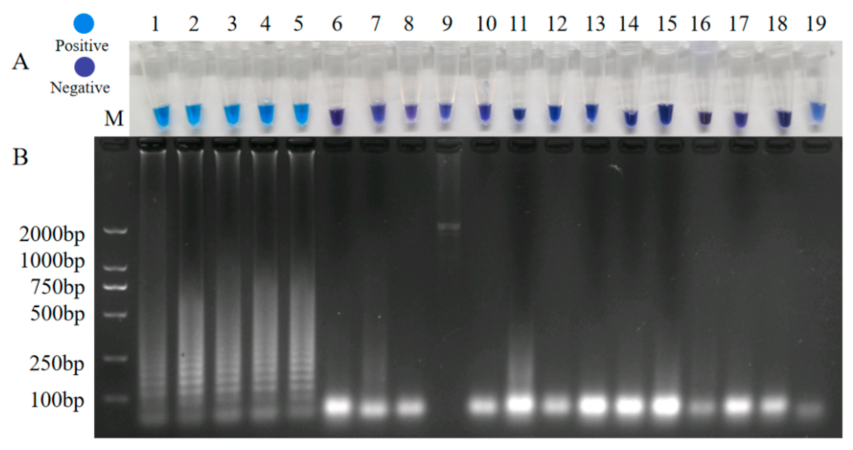
3.3.2. Specificity Test of LAMP for the Detection of A. phaeospermum

In order to verify the specificity of the LAMP assay, the genomic DNA of A. phaeospermum and the other tested strains were amplified under the same reaction conditions. After amplification, HNB was added to observe the color change of the reaction tubes. The color of the five A. phaeospermum tubes was sky blue, indicating that there was a large amount of DNA amplification product in the reaction tubes, while the color of the reaction tubes of other tested strains and the negative control was dark purple, indicating that there was no amplification of the target DNA product in the reaction tubes (Figure 8). Therefore, the specificity of the LAMP assay of A. phaeospermum was verified.

3.4. Sensitivity Test of the Seminested PCR, Real-Time Quantitative PCR, and LAMP for the Detection of A. phaeospermum

The limits of seminested PCR, real-time quantitative PCR, and LAMP were evaluated with a series of concentrations (10 to 105 ng/μL) of the A. phaeospermum DNA template under optimized conditions.
Sensitivity of seminested PCR: The results showed that conventional PCR with AP1-1/AP1-2 as primers could detect 1 ng/μL of A. phaeospermum genomic DNA (Figure 9A), whereas the seminested PCR assay was able to detect 100 pg/uL of the genomic DNA of A. phaeospermum (Figure 9B), illustrating that the sensitivity of seminested PCR was 10 times that of conventional PCR.
Sensitivity of real-time quantitative PCR: Conventional PCR and real-time quantitative PCR were performed on genomic DNA of different concentration gradients with the 33-F/33-R primer. In conventional PCR, bands with a DNA concentration of 1 ng/μL were indistinctly visible, while no band appeared when the DNA was further diluted, indicating that the sensitivity of conventional PCR detection was 1 ng/μL with the 33-F/33-R primer (Figure 9C). In the real-time quantitative PCR amplification curve, the DNA concentrations of 10 ng/μL, 1 ng/μL, 100 pg/μL, 10 pg/μL, and 1 pg/μL showed a smooth S-shaped dissolution curve, and the exponential growth and plateau periods were obvious. With the increase in dilution, the number of cycles required for the exponential growth period decreased, while the DNA concentrations of 100 and 10 fg/μL and the negative control did not have any amplification curves, indicating that the sensitivity of real-time quantitative PCR detection was 1 pg/μL (Figure 9D). The sensitivity of real-time quantitative PCR was therefore 1000 times that of conventional PCR.
Sensitivity of real-time quantitative LAMP: The limit of the LAMP assay was evaluated with the concentration range from 10 ng/μL to 10 fg/μL. The results of the chromatic reaction showed that the limit of the LAMP assay was 10 pg/μL for the genomic DNA of A. phaeospermum (Figure 9E), and the results of electrophoresis were consistent with that of LAMP. However, the sensitivity of conventional PCR (primer AP1-1/AP1-2) was every 25 uL system of A. phaeospermum DNA purified by 1 ng/uL (Figure 9A). Therefore, LAMP was about 100 times more sensitive than conventional PCR.
Overall, all three methods had relatively high sensitivity, enabling efficient amplification at a low concentration of the target DNA. Among the three assays, seminested PCR had the lowest sensitivity, while qPCR had the highest sensitivity. With the increase in sensitivity, the possibility of false positives and contamination would also increase [23], especially if the lid of the PCR tube is opened twice during LAMP and the seminested PCR operation. Therefore, attention should be paid to the operation specifications and negative control setting during the experiment.

3.5. Detection of Artificially Inoculated and Field-Collected Samples

Three rapid detection systems for A. phaeospermum were established to detect 16 artificially inoculated samples (4, 8, 12, and 16 days), and the detection effects were compared (Table 3). The positive rate was 50% for PCR, 83.3% for LAMP, and 91.7% for qPCR. LAMP and qPCR assays could detect A. phaeospermum from inoculated twigs collected 4 days after inoculation but not from healthy samples inoculated with sterile water as the negative control. The results showed that the positive detection rate of real-time quantitative PCR was the highest, while that of seminested PCR was the lowest. The timely detection of latent infection in plant tissues is essential for estimating the potential for the initial inoculum of an epidemic [24]. Therefore, the established detection assays in this study can be used in conjunction with traditional methods of disease diagnosis to estimate the level of latent infection.
Three rapid detection systems for A. phaeospermum were established to detect nine hybrid bamboos with different incidence degrees (Figure 10). The test results (Table 4) showed that for the hybrid bamboo tissue with milder pathology, it was almost impossible to detect A. phaeospermum via seminested PCR. However, both LAMP and qPCR could detect A. phaeospermum from asymptomatic branches, thus enabling rapid and accurate detection of latent infections of A. phaeospermum in the hybrid bamboo.
The presence of A. phaeospermum could be detected in hybrid bamboo samples without any obvious symptoms (the group of asymptomatic samples). This phenomenon might be due to the fact that the sample collection site was an area of high incidence of hybrid bamboo shoot blight and the amount of A. phaeospermum had been relatively high, so asymptomatic samples of hybrid bamboo were infected by A. phaeospermum. Therefore, as an efficient and sensitive molecular detection method, the qPCR assay enables more accurate and sensitive detection of A. phaeospermum in the incubation stage and can be used to predict the occurrence of diseases in the coming years so as to prevent an outbreak and epidemic of a large-scale in a timely manner.

4. Discussion

Bambusa pervariabilis × Dendrocalamopsis grandis blight is one of the most severe diseases in bamboos and has caused damage to China’s forest resources. However, the effect of the current control methods for alleviating the occurrence and development of this disease is limited, and these methods cannot fundamentally solve the problem. Additionally, there is no reported assay method for the rapid detection of A. phaeospermum. Therefore, it is particularly important to establish a rapid detection system to achieve rapid detection of the hybrid bamboo shoot blight pathogen A. phaeospermum in the incubation period. In this study, seminested PCR, qPCR, and LAMP methods were all effective in detecting A. phaeospermum. The purpose of the study was to develop a simple and efficient detection assay with the advantages of accuracy, sensitivity, high feasibility, and low cost.
The sensitivity and accuracy are key considerations for detection methods. After comparing the sensitivity and accuracy of the three methods, it can be seen that real-time quantitative PCR has the advantages of high sensitivity, specificity, and repeatability, which makes it suitable for the quantitative detection and dynamic monitoring of A. phaeospermum. Real-time quantitative PCR has great potential to be widely utilized in laboratories due to the specificity, sensitivity, and quantification of the target DNA. Because the second amplification template is derived from the first amplification product, the entire amplification system is not in a closed state, which would greatly increase the possibility of laboratory aerosol contamination and cross-contamination of the products from the first round. The binding of fluorescent dyes to dsDNA is not specific [25], the field environment is complex, and the fragments of the amplification product are large and not easily degradable, resulting in LAMP detection being prone to false positives and low experimental reproducibility in practical applications.
There is no doubt that the LAMP assay has the advantage of high feasibility compared to the other two detection assays, and this method is suitable for the field diagnosis of hybrid bamboo blight [26]. Due to the design of four unique primers, more amplification products can be produced in less time than conventional PCR without a temperature cycle. Moreover, it can be applied under a simple isothermal heater without the need for a laboratory environment [27]. Compared to conventional PCR and seminested PCR, LAMP detection technology has the main advantages of having a short reaction time, not requiring special instruments, having simple operation steps, and being able to observe the reaction with the naked eye. While maintaining the advantages of PCR technology, the specificity of the reaction was further enhanced, and the detection time was shortened. The variety of devices needed for real-time quantitative PCR detection limits its potential for field applications. Although real-time PCR has been able to be used for pathogen diagnosis in the field, this method is not feasible as it requires the purchase of an additional expensive, dedicated portable thermal cycler [28]. Because seminested PCR requires two rounds of amplification, it takes a relatively long time. The entire process of seminested PCR assay takes at least three hours, which is twice as long as LAMP assays. Therefore, seminested PCR is not suitable for field detection.
The cost of detection should also be taken into account. Seminested PCR only needs conventional PCR enzymes and instruments. Thus, this method can achieve detection with higher sensitivity and higher specificity, help save on detection instruments and costs, and completely meet the needs of sensitive and low-cost detection in basic laboratories. In contrast, due to the need for specialized enzymes and reagents, cost control is a problem that needs to be solved for LAMP detection methods. Although the cost of real-time quantitative PCR is gradually decreasing, expenditure on specialized equipment such as real-time thermocyclers still needs to be considered. In contrast, seminested PCR is the best assay for detection when it comes to limited costs.
In summary, this study established seminested PCR, qPCR, and LAMP assays for the detection of A. phaeospermum and analyzed the assays as comprehensively as possible. These assays are of great significance for the early diagnosis and rapid detection of hybrid bamboo blight. However, there are different detection methods for different detection situations, and the appropriate detection method can be selected according to the actual situation. Sufficient time and conditions (equipment and reagents) permitting, the use of LAMP for preliminary field testing and qPCR detection in the laboratory is the best choice. If laboratory conditions are limited, seminested PCR can be used for rapid testing.
When rapid detection of hybrid bamboo blight is required in the field, the LAMP detection method is a good option [29]. With the continued use of LAMP rapid test kits for practical diagnosis in recent years [30,31], it is possible that an A. phaeospermum LAMP kit could be developed and used in the field on a large scale. When accuracy and sensitivity are not highly required and the budget is limited, seminested PCR is an option. At present, it has been widely used in the diagnosis of pathogens such as plant diseases [32,33] and animal diseases [16,34]. Compared to the current diagnostic methods, qPCR has higher sensitivity and specificity and is therefore more suitable for the early detection of infected bamboo tissues. Based on the qPCR detection assay, monitoring and prediction of field hybrid bamboo shoot blight can be carried out in the field. Hybrid bamboo tissue is only required to be collected in the field and used as template for real-time quantitative PCR detection to determine the presence or absence of A. phaeospermum and its degree of infestation, which can provide a basis for disease epidemic monitoring. Although seminested PCR and LAMP assays can successfully detect infected samples in the field, qPCR is overall superior when time, safety, cost, and simplicity are taken into account. However, the qPCR assay for detecting A. phaeospermum still needs to be further developed for wider applications. Firstly, as different extraction protocols result in different qualities of extracted DNA, the extraction protocol of genomic DNA should be improved to enhance the efficiency of DNA extraction [35,36]. The high contents of fiber, pigments, and phenols in bamboo make the isolation and purification of DNA more difficult. Successful preparation of high-purity genomic DNA templates is crucial for pathogenic detection [37]. A simple and easy method of extracting total DNA from bamboos for PCR analysis would make this method more feasible.
The actual efficiency of detection assays is determined by appropriate primers, which is the reason for the low sensitivity of some primer sets [38]. In order to further enhance the efficiency of detection, further research is needed to find well-conserved gene sequences to design more specific primers for real-time quantitative PCR with the aim of establishing a more efficient and sensitive real-time quantitative PCR assay for the detection of A. phaeospermum in hybrid bamboo tissues. In addition, the rapid detection methods developed in this study can be combined, such as the establishment of qLAMP technology [39], for efficient and rapid on-site diagnostics.

Author Contributions

Conceptualization, H.Z. and W.Y.; Data curation, L.X. and S.L. (Shuying Li); Formal analysis, H.Z. and W.Y.; Funding acquisition, S.L. (Shujiang Li); Investigation, W.Y. and T.L.; Methodology, H.Z.; Project administration, T.Z.; Resources, L.X. and T.Z.; Software, H.Z.; Supervision, S.H.; Validation, T.Z. and S.L. (Shuying Li); Visualization, W.Y. and S.H.; Writing – original draft, H.Z. and L.X.; Writing – review & editing, T.L. and S.L. (Shujiang Li). All authors have read and agreed to the published version of the manuscript.

Funding

This research was funded by the National Natural Science Foundation of China, grant number 32171795, and General Program of Science and Technology Department of Sichuan, grant number 2020YJ0400.

Data Availability Statement

Whole gene sequences for primer design are available online in the GenBank database under the accession number QYRS01000001.1.

Acknowledgments

We thank MDPI English Editing for its linguistic assistance during the preparation of this manuscript.

Conflicts of Interest

The authors declare there is no conflict of interest.

References

  1. Yang, Q. Research Status of B. pervariabilis × D. Daii and Cost-benefifit analysis. J. Shandong For. Sci. Technol. 2007, 3, 101–103. [Google Scholar]
  2. Yang, Z.Z.; Ye, J.R. Identifification on pathogen of hybrid bamboo blight. J. Sichuan Agric. Univ. 2004, 22, 225–227. [Google Scholar]
  3. Li, S.; He, Q.; Peng, Q.; Fang, X.; Zhu, T.; Qiao, T.; Han, S. Metabolomics responses of Bambusa pervariabilis × Dendrocalamopsis grandis varieties to Biotic (pathogenic fungus) stress. Phytochemistry 2019, 167, 112087. [Google Scholar] [CrossRef] [PubMed]
  4. Zhu, T.H.; Huang, Z.C.; Gao, Q.Z. Pathogen and occurrence regularity of Bambusa ervariabilis × Dendrocalamopsis daii blight. For. Pest Dis. 2009, 28, 10–12. [Google Scholar]
  5. Zhao, Y.M.; Deng, C.R.; Chen, X. Arthrinium phaeospermum causing dermatomycosis, a new record of China. Mycosystema 1990, 3, 232–235. [Google Scholar]
  6. Rai, M.K. Mycosis in man due to Arthrinium phaeospermum var. indicum. First Case Report: Mykose durch Arthrinium phaeospermum var. indicum beim Menschen. Erstbericht. Mycoses 2010, 32, 472–475. [Google Scholar] [CrossRef]
  7. Khan, K.R.; Sullia, S.B. Arthrinium phaeospermium var. indicum var. nov., a new market pathogen of cowpea, garden pea and french bean. Acta Bot. Indica 1980, 8, 103–104. [Google Scholar]
  8. Liu, X.J.; Luo, X.Y.; Li, X.F.; Chen, Q.T. Taxonomic determination of toxigenic Arthrinium strains isolated from poisoning sugarcane. Mycosystema 1988, 7, 221–225. [Google Scholar]
  9. Xia, L.M.; Zhang, S.X.; Huang, J.H.; Zhang, W.M. Studies on Arthrinium phaeospermum causing moso bamboo foot rot. J. Nanjing Fores. Univ. 1995, 19, 1–7. [Google Scholar]
  10. Ma, G.L.; Hu, G.L.; Yu, C.Z.; Wu, J.L.; Xu, B.C. Phyllostachys prominens plum shoot wilt pathogenic fungoid and its biological characteristics. Zhejiang For. Coll. 2003, 20, 44–48. [Google Scholar]
  11. Piccolo, S.L.; Mondello, V.; Giambra, S.; Conigliaro, G.; Torta, L.; Burruano, S. Arthrinium phaeospermum, Phoma cladoniicola and Ulocladium consortiale, New Olive Pathogens in Italy. J. Phytopathol. 2014, 162, 258–263. [Google Scholar] [CrossRef]
  12. Li, B.J.; Liu, P.Q.; Jiang, Y.; Weng, Q.Y.; Chen, Q.H. First Report of Culm Rot Caused by Arthrinium phaeospermum on Phyllostachys viridis in China. Plant Dis. 2016, 100, 10–13. [Google Scholar] [CrossRef]
  13. Gong, H.Y.; Li, Q.Y.; Zhang, H.; Ye, L.; Shi, L.; Feng, Y.H. Development and comparison of qPCR and qLAMP for rapid detection of the decapod iridescent virus 1 (DIV1). J. Invertebr. Pathol. 2021, 182, 107567. [Google Scholar] [CrossRef] [PubMed]
  14. Huang, S.Y.; Gong, J.Z.; Yang, B.; Fan, Y.M.; Yao, N.; Wang, C.R. Development of a nest-PCR for detection of Fasciola hepatica DNA in the intermediate snail host, Radix cucunorica, and the prevalence in northwestern China. Infect. Genet. Evol. 2019, 75, 103984. [Google Scholar] [CrossRef] [PubMed]
  15. Matthew, M.A.; Christie, J.; Yang, N.; Yao, C. A Loop-Mediated Isothermal Amplification (LAMP) Assay Specific to Trichomonas tenax Is Suitable for Use at Point-of-Care. Microorganisms 2022, 10, 594. [Google Scholar] [CrossRef] [PubMed]
  16. Smolejová, M.; Cihová, I.; Sulo, P. Reliable and Sensitive Nested PCR for the Detection of Chlamydia in Sputum. Microorganisms 2021, 9, 935. [Google Scholar] [CrossRef]
  17. Kamber, T.; Malpica-López, N.; Messmer, M.M.; Oberhänsli, T.; Arncken, C.; Alkemade, J.A.; Hohmann, P. A qPCR Assay for the Fast Detection and Quantification of Colletotrichum lupini. Plants 2021, 10, 1548. [Google Scholar] [CrossRef]
  18. Coats, K.; DeBauw, A.; Lakshman, D.K.; Roberts, D.P.; Ismaiel, A.; Chastagner, G. Detection and Molecular Phylogenetic-Morphometric Characterization of Rhizoctonia tuliparum, Causal Agent of Gray Bulb Rot of Tulips and Bulbous Iris. J. Fungi 2022, 8, 163. [Google Scholar] [CrossRef]
  19. Wang, J.; Jacobs, J.L.; Byrne, J.M.; Chilvers, M.I. Improved diagnoses and quantification of Fusarium virguliforme, causal agent of soybean sudden death syndrome. Phytopathology 2015, 105, 378–387. [Google Scholar] [CrossRef] [Green Version]
  20. Notomi, T.; Mori, Y.; Tomita, N.; Kanda, H. Loop-mediated isothermal amplification (LAMP): Principle, features, and future prospects. J. Microbiol. 2015, 53, 1–5. [Google Scholar] [CrossRef]
  21. Li, S.; Tang, Y.; Fang, X.; Qiao, T.; Han, S.; Zhu, T. Whole-genome sequence of Arthrinium phaeospermum, a globally distributed pathogenic fungus. Genomics 2020, 112, 919–929. [Google Scholar] [CrossRef] [PubMed]
  22. Garrido-Maestu, A.; Azinheiro, S.; Carvalho, J.; Prado, M. Rapid and sensitive detection of viable Listeria monocytogenes in food products by a filtration-based protocol and qPCR. Food Microbiol. 2018, 73, 254–263. [Google Scholar] [CrossRef] [PubMed]
  23. Schneider, L.; Blakely, H.; Tripathi, A. Mathematical model to reduce loop mediated isothermal amplification (LAMP) false-positive diagnosis. Electrophoresis 2019, 40, 2706–2717. [Google Scholar] [CrossRef] [PubMed] [Green Version]
  24. Huang, C.; Sun, Z.; Yan, J.; Luo, Y.; Wang, H.; Ma, Z. Rapid and Precise Detection of Latent Infections of Wheat Stripe Rust in Wheat Leaves using Loop-Mediated Isothermal Amplification. J. Phytopathol. 2011, 159, 582–584. [Google Scholar] [CrossRef]
  25. Yan, H.; Zhang, J.; Ma, D.; Yin, J. qPCR and loop mediated isothermal amplification for rapid detection of Ustilago tritici. PeerJ 2019, 7, e7766. [Google Scholar] [CrossRef] [Green Version]
  26. Almasi, M.A.; Dehabadi, S.H.; Moradi, A.; Eftekhari, Z.; Ojaghkandi, M.A.; Aghaei, S. Development and application of loop-mediated isothermal amplifification assay for rapid detection of Fusarium oxysporum f. sp. lycopersici. J. Plant Pathol. Microbiol. 2013, 4, 177. [Google Scholar]
  27. Sagcan, H.; Turgut Kara, N. Detection of Potato ring rot Pathogen Clavibacter michiganensis subsp. sepedonicus by Loop-mediated isothermal amplification (LAMP) assay. Sci. Rep. 2019, 9, 20393. [Google Scholar] [CrossRef] [Green Version]
  28. Durand, L.; La Carbona, S.; Geffard, A.; Possenti, A.; Dubey, J.P.; Lalle, M. Comparative evaluation of loop-mediated isothermal amplification (LAMP) vs qPCR for detection of Toxoplasma gondii oocysts DNA in mussels. Exp. Parasitol. 2020, 208, 107809. [Google Scholar] [CrossRef]
  29. Gill, P.; Ghaemi, A. Nucleic acid isothermal amplification technologies—A review. Nucleosides Nucleotides Nucleic Acids 2008, 27, 224–243. [Google Scholar] [CrossRef]
  30. Zhao, B.; Yang, D.; Zhang, Y.; Xu, Y.; Zhao, X.; Liang, J.; Fan, X.; Du, Y.; Zhu, Z.; Shi, B.; et al. Rapid visual detection of lily mottle virus using a loop-mediated isothermal amplification method. Arch. Virol. 2018, 163, 545–548. [Google Scholar] [CrossRef]
  31. Hopkins, H.; González, I.J.; Polley, S.D.; Angutoko, P.; Ategeka, J.; Asiimwe, C.; Bell, D. Highly sensitive detection of malaria parasitemia in a malaria-endemic setting: Performance of a new loop-mediated isothermal amplification kit in a remote clinic in Uganda. J. Infect. Dis. 2013, 208, 645–652. [Google Scholar] [CrossRef] [PubMed]
  32. Cesbron, S.; Dupas, E.; Beaurepère, Q.; Briand, M.; Montes-Borrego, M.; Velasco-Amo, M.D.P.; Jacques, M.A. Development of a Nested-MultiLocus Sequence Typing approach for a highly sensitive and specific identification of Xylella fastidiosa subspecies directly from plant samples. Agronomy 2020, 10, 1099. [Google Scholar] [CrossRef]
  33. Puig, A.S. Detection of Cacao Mild Mosaic Virus (CaMMV) Using Nested PCR and Evidence of Uneven Distribution in Leaf Tissue. Agronomy 2021, 11, 1842. [Google Scholar] [CrossRef]
  34. Coelho, C.; Vieira-Pinto, M.; Vilares, A.; Gargaté, M.J.; Rodrigues, M.; Cardoso, L.; Lopes, A.P. PCR detection of Toxoplasma gondii in European wild rabbit (Oryctolagus cuniculus) from Portugal. Microorganisms 2020, 8, 1926. [Google Scholar] [CrossRef]
  35. Yuan, J.; Mi, T.; Zhen, Y.; Yu, Z. Development of a rapid detection and quantification method of Karenia mikimotoi by real-time quantitative PCR. Harmful Algae 2012, 17, 83–91. [Google Scholar] [CrossRef]
  36. Foulds, I.V.; Granacki, A.; Xiao, C.; Krull, U.J.; Castle, A.; Horgen, P.A. Quantification of microcystin-producing cyanobacteria and E. coli in water by 5′-nuclease PCR. J. Appl. Microbiol. 2002, 93, 825–834. [Google Scholar] [CrossRef] [Green Version]
  37. Abd-Elsalam, K.; Bahkali, A.; Moslem, M.; Amin, O.E.; Niessen, L. An optimized protocol for DNA extraction from wheat seeds and loop-mediated isothermal amplification (LAMP) to detect Fusarium graminearum contamination of wheat grain. Int. J. Mol. Sci. 2011, 12, 3459–3472. [Google Scholar] [CrossRef] [Green Version]
  38. Bustin, S.; Huggett, J. qPCR primer design revisited. Biomol. Detect. Quantif. 2017, 14, 19–28. [Google Scholar] [CrossRef]
  39. Garrido-Maestu, A.; Azinheiro, S.; Fuciños, P.; Carvalho, J.; Prado, M. Highly sensitive detection of gluten-containing cereals in food samples by real-time Loop-mediated isothermal AMPlification (qLAMP) and real-time polymerase chain reaction (qPCR). Food Chem. 2018, 246, 156–163. [Google Scholar] [CrossRef]
Figure 1. Schematic of primer design. (A) Seminested PCR: AP1-1, AP1-2, and AP3-1 binding sites; (B) qPCR: 33-F/33-R binding sites; (C) LAMP: two external (15-F3 and 15-B3) and two internal (15-FIP and 15-BIP) primer binding sites; (D) chromosomal location of AP-Z1300015, AP-Z1300033, and AB220314.1 in A. phaeospermum.
Forests 13 00850 g001
Figure 2. Optimization of the annealing temperature for the primer AP1-1/AP1-2. M: DL 2000 DNA marker; 1–5: annealing temperatures of 57, 58, 59, 60, and 61 °C, respectively.
Forests 13 00850 g002
Figure 3. Optimization of the annealing temperature for the primer AP1-2/AP3-1. M: DL 2000 DNA marker; 1–6: annealing temperatures of 58, 59, 60, 61 and 62 °C, respectively.
Forests 13 00850 g003
Figure 4. Specificity of seminested PCR detection of A. phaeospermum. (A) Specificity of the AP1-1/AP1-2 primer; (B) specificity of the AP1-2/AP3-1 primer. M: DL2000 marker; N: negative control; 1–18: strain number is the same as in Table 1.
Forests 13 00850 g004
Figure 5. Specificity of qPCR detection of A. phaeospermum (A) Conventional PCR amplification with the 33-F/33-R primer; (B) melting curves of the real-time quantitative PCR. M: DL2000 marker; N: negative control; 1–18: strain number is the same as in Table 1.
Forests 13 00850 g005
Figure 6. Standard curves showing the relationship between the log of the dilution concentration and the Ct value.
Forests 13 00850 g006
Figure 7. Optimization of LAMP reaction conditions. (A) Optimization of the LAMP assay’s dNTP concentrations for the detection of A. phaeospermum. M: DL 2000 DNA marker; 1–5: dNTP concentrations of 0.9, 1.0, 1.1, 1.2, and 1.3 mM, respectively. (B) Optimization of the LAMP assay’s Mg2+ concentrations for the detection of A. phaeospermum. M: DL 2000 DNA marker; 1–5: Mg2+ concentrations of 1.5, 1.8, 2.1, 2.4, and 2.7 mM, respectively. (C) Optimization of the LAMP assay’s betaine concentrations for the detection of A. phaeospermum. M: DL 2000 DNA marker; 1–5: betaine concentrations of 0.5, 0.6, 0.7, 0.8, and 0.9 mM, respectively. (D) Optimization of the LAMP assay’s primer concentration ratio for the detection of A. phaeospermum. M: DL 2000 DNA marker; 1–5: the external primer concentration was fixed at 0.1 μM, while the internal primer concentrations were 0.2, 0.4, 0.8, 1.6, and 3.2 μM, respectively.
Forests 13 00850 g007
Figure 8. The specificity test of the LAMP assay of A. phaeospermum. (A) Color rendering change plot; (B) agarose gel electrophoresis. M: DL 2000 DNA marker; 1–18: strain number is the same as in Table 1; 19: negative control.
Forests 13 00850 g008
Figure 9. Sensitivity test of seminested PCR, real-time quantitative PCR, and LAMP for the detection of A. phaeospermum (A) Sensitivity of conventional PCR for the detection of A. phaeospermum with the AP1-1/AP1-2 primer; (B) sensitivity of seminested PCR for the detection of A. phaeospermum with the AP1-1, AP1-2, and AP1-3 primers; (C) sensitivity of conventional PCR for the detection of A. phaeospermum with the 33-F/33-R primer; (D) sensitivity of qPCR for the detection of A. phaeospermum; (E) sensitivity of LAMP for the detection of A. phaeospermum. M: DL 2000 DNA marker; 1–7: DNA concentrations were 10 ng/μL, 1 ng/μL, 100 pg/μL, 10 pg/μL, 1 pg/μL, 100 fg/μL, and 10 fg/μL, respectively; CK: negative control.
Forests 13 00850 g009
Figure 10. Field-collected hybrid bamboos samples classified as asymptomatic, mild, and severe according to the degree of incidence of different grades. (A) asymptomatic samples; (B) mild samples; (C) severe samples.
Forests 13 00850 g010
Table 1. Strains used in this study to test the specificity of seminested PCR, qPCR, and LAMP assays.
No.SpeciesHostSource
1Arthrinium phaeospermum (Corda) M.B. EllisB. pervariabilis × D. grandisTianquan, Sichuan, China
2Arthrinium phaeospermum (Corda) M.B. EllisB. pervariabilis × D. grandisYa’an, Sichuan, China
3Arthrinium phaeospermum (Corda) M.B. EllisB. pervariabilis × D. grandisChangning, Sichuan, China
4Arthrinium phaeospermum (Corda) M.B. EllisPhyllostachys edulisBaoxing, Sichuan, China
5Arthrinium phaeospermum (Corda) M.B. EllisPhyllostachys edulisXinyang, Henan, China
6Arthrinium kogelbergenseDendrocalamus farinosusChangning, Sichuan, China
7Arthrinium marii Larrondo & CalvoPhyllostachys edulisQingdao, Shandong, China
8Arthrinium arundinis (Corda) Dyko & B. SuttonArundo donaxXinyang, Henan, China
9Arthrinium haricotospora (Sacc.) Y.L. Zhang & T.Y. Zhang, comb. nov.Phyllostachys edulisQingdao, Shandong, China
10Diaporthe guangxinsisDendrocalamus latifloruYibin, Sichuan, China
11Neofusicoccum parvumJuglans regiaMianyang, Sichuan, China
12Fusarium tricinctum (Corda) Sacc.Zanthoxylum bungeanumHanyuan, Sichuan, China
13Nigrospora sphaerica (Sacc.) E.W. MasonArundo donaxXinyang, Henan, China
14Apiospora montagnei Sacc.Phyllostachys praecoxTianshui, Gansu, China
15Arthrinium rasikravindrii Shiv M. Singh, L.S. Yadav, P.N. SinB. pervariabilis × D. grandisJiangan, Sichuan, China
16Fusarium proliferatum (Matsush.) NirenbergB. pervariabilis × D. grandisChangning, Sichuan, China
17Nigrospora guilinensis Mei Wang & L. CaiB. pervariabilis × D. grandisChangning, Sichuan, China
18Nigrospora oryzae (Berk. & Broome) PetchB. pervariabilis × D. grandisJiangan, Sichuan, China
Table 2. Primer sequences of seminested PCR, qPCR, and LAMP for A. phaeospermum detection.
AssayPrimer NameSequence (5′–3′)Length
qPCR33-FCACTCCGAGCCATGCTACTAC 21
33-RGCATAGCGATCCAACAGGTAGA22
Seminested PCRAP1-1GGCACCGACCCCTTGATTG19
AP1-2CGAAGTTGTCGGGGCGGA18
AP3-1CTCCAGACCGGTCAATGCG19
LAMP15-F3GCATCGAACAAGACGGAAAC20
15-B3GTTGACCCTGCTCTCGTTTC20
15-FIPCTCTTCGCCGGGGCGTATGCCCTGGACCTCGGCATCAC38
15-BIPTTGTGTGGACCGTCTTCGTCGCCACCAGGGCCAGTTTC38
Table 3. The results of artificial inoculation for detecting A. phaeospermum.
No.The Time of Inoculation Seminested PCRLAMPqPCR
14 d+
2
3++
48 d++
5++
6++
712 d+++
8+++
9+++
1016 d+++
11+++
12+++
Note: “+” means the positive amplification result, “−” means the negative amplification result.
Table 4. The results of field-collected samples for detecting A. phaeospermum.
No.Symptom RankSeminested PCRLAMPqPCR
1asymptomatic++
2++
3
4Mild+++
5+++
6+++
7severe+++
8+++
9+++
Note: “+” means the positive amplification result, “−” means the negative amplification result.
Publisher’s Note: MDPI stays neutral with regard to jurisdictional claims in published maps and institutional affiliations.

