Brief Report

Metagenomic Analysis Reveals High Abundance of Torque Teno Mini Virus in the Respiratory Tract of Children with Acute Respiratory Illness

1
Laboratoire de Virologie, Institut des Agents Infectieux, Laboratoire Associé au Centre National de Référence des Virus des Infections Respiratoires, Hospices Civils de Lyon, 69004 Lyon, France
2
Univ Lyon, Université Lyon 1, CIRI, Inserm U1111 CNRS UMR5308, Virpath, 69007 Lyon, France
3
GenEPII Platform, Institut des Agents Infectieux, Hospices Civils de Lyon, 69004 Lyon, France
4
Bacteriology Laboratory, Infectious Agents Department, Centre Hospitalier Universitaire de Poitiers, 86021 Poitiers, France
5
Inserm U1070 Pharmacology of Antimicrobial Agents and Resistance, University of Poitiers, 86073 Poitiers, France
6
Laboratoire Commun de Recherche HCL-bioMerieux, Centre Hospitalier Lyon Sud, 69495 Pierre-Bénite, France
7
Hospices Civils de Lyon, Urgences Pédiatriques, Hôpital Femme Mère Enfant, 69500 Bron, France
*
Authors to whom correspondence should be addressed.
Submission received: 21 March 2022 / Revised: 25 April 2022 / Accepted: 26 April 2022 / Published: 3 May 2022
(This article belongs to the Special Issue Applications of Next-Generation Sequencing in Virus Discovery)

Abstract

:
Human Anelloviridae is a highly prevalent viral family, including three main genera—Alphatorquevirus (Torque teno virus, TTV), Betatorquevirus (Torque teno mini virus, TTMV), and Gammatorquevirus (Torque teno midi virus, TTMDV). To date, the characterization of Anelloviridae in the respiratory tract of children with acute respiratory infection (ARI) has been poorly reported and mainly focused on TTV. We performed a metagenomic analysis of eight respiratory samples collected from children with an ARI of unknown etiology (eight samples tested negative with a multiplex PCR assay, out of the 39 samples initially selected based on negative routine diagnostic testing). A total of 19 pediatric respiratory samples that tested positive for respiratory syncytial virus (RSV, n = 13) or influenza virus (n = 6) were also sequenced. Anelloviridae reads were detected in 16/27 samples, including 6/8 negative samples, 7/13 RSV samples and 3/6 influenza samples. For samples with a detection of at least one Anelloviridae genus, TTMV represented 87.1 (66.1–99.2)% of Anelloviridae reads, while TTV and TTMDV represented 0.8 (0.0–9.6)% and 0.7 (0.0–7.1)%, respectively (p < 0.001). Our findings highlight a high prevalence of TTMV in respiratory samples of children with an ARI of unknown etiology, as well as in samples with an RSV or influenza infection. Larger studies are needed to explore the role of TTMV in childhood respiratory diseases.

1. Introduction

Human Anelloviridae is a highly prevalent viral family characterized by an important genetic diversity. More than eighty species are grouped into three main genera: Alphatorquevirus (Torque teno virus, TTV), Betatorquevirus (Torque teno mini virus, TTMV) and Gammatorquevirus (Torque teno midi virus, TTMDV) [1,2]. The viral load of TTV in plasma or blood is considered as a surrogate marker of immune competence and might be used to assess the immune status of transplant patients [3,4]. TTMV and TTMDV have been less studied, but the prevalence in blood is considerably lower than that of TTV [5,6]. Anelloviridae has also been detected in almost all parts of the human body, including the respiratory tract, and is recognized as the main component of the human viral flora [5,7,8]. While considered to be non-pathogenic, Anellovriridae might be associated with the occurrence of some disorders, including respiratory disorders in childhood [9,10,11,12,13]. Co-infection between TTV and common respiratory viruses have been reported, but the detailed composition of Anelloviridae, including the characterization of TTMV and TTMDV, has been poorly explored in children with respiratory virus infections [14,15].
Due to a complex genetic diversity, PCR-based detection of Anelloviridae genera might be difficult to optimize, and no diagnostic PCR has been validated in respiratory samples so far [5,8,16]. As untargeted methods, metagenomic next-generation sequencing (mNGS) has become a powerful tool for the characterization of the whole viral communities in clinical samples, and has been used to describe the abundance and/or dynamics of TTV, TTMV and TTMDV [4,6,17,18]. Herein, we aim to describe the Anelloviridae composition within the respiratory virome of children under five years presenting an ARI with or without identified etiologic agents.

2. Materials and Methods

2.1. Samples Selection

A retrospective study was conducted on samples received at the virology laboratory at the University Hospital of Lyon, France, between 2010 and 2016. Nasopharyngeal aspirates from hospitalized children under five years old with ARI were selected according to the following criteria: (1) absence of documented infections on two consecutive samples (collected up to two weeks apart) with routine techniques that included bacterial/viral cultures, reverse-transcriptase polymerase chain reaction (RT-PCR) assay detecting human rhinovirus, and respiratory syncytial (RSV) and influenza viruses (MWS r-gene™ respiratory panel; bioMérieux, Lyon, France); (2) negative results with a large screening multiplex PCR (FilmArray® Respiratory Panel, bioMérieux, Lyon, France). This assay allows to detect the main viral and bacterial respiratory pathogens, including adenovirus, human coronavirus (229E, HKU1, OC43, NL63), human metapneumovirus, human rhinovirus/enterovirus, influenza (A, A/H1, A/H1-2009, A/H3, B), human parainfluenza virus, RSV, bordetella pertussis, chlamydophila pneumonia and mycoplasma pneumonia.
Nineteen respiratory samples from hospitalized children under five with ARI that tested positive with RSV RT-PCR (n = 13) or influenza RT-PCR (n = 6) were also sequenced.

2.2. Metagenomic Workflow

A metagenomic workflow including quality controls, evaluated for the detection of a comprehensive panel of DNA and RNA viruses in respiratory samples, was used [19]. Briefly, after sample viral enrichment, total nucleic acid was extracted, randomly amplified, and Illumina libraries were prepared using the Nextera XT DNA Library preparation kit, according to the manufacturer’s recommendations (Illumina, San Diego, CA, USA). Libraries were sequenced on Illumina NextSeq500™ platform with mid-output 2 × 150 bp flowcells. Raw fastq files generated were cleaned with cutadapt (version 1.18). The host reads were then removed using bwa mem (version 0.7.8) alignment to the human genome (GRCh37.p2). Unmapped reads extracted with samtools tool (version 1.3.1) were then mapped to the nr database (non-redundant protein sequences database) (downloaded on 25 September 2018) using Diamond (version 0.9.22). We also explored the prevalence of Anelloviridae species, as previously described [6]. Briefly, non-human NGS reads were aligned using BLAST on a manually curated database composed of 56 reference sequences of human Anelloviridae (TTV-1 to TTV-29, TTMV-1 to TTMV-12, TTMDV-1 and TTMDV-15).
To correct the difference in sequencing depth between samples, the number of reads were normalized in reads per million mapped reads (RPM). To reduce false positive results, only viruses with a count of >1 RPM were considered.

2.3. Statistical Analysis

Statistical analyses were conducted using R software, version 4.0.5 (R Foundation for Statistical Computing). Continuous variables are presented as the median with interquartile range (IQR) and compared using non-parametric Kruskal–Wallis or Mann–Whitney tests. Differences are considered significant at p < 0.05.

3. Results

3.1. Study Population

Between 2010 and 2016, 39 hospitalized patients with ARI were tested negative on two successive samples with routine diagnostic techniques. Among them, eight remained negative after testing with a large multiplex PCR assay and were analyzed in mNGS. The median (IQR) age was 12.0 (6.3–22.5) months; three patients had comorbidities, including spinal muscular atrophy, sickle cell anemia and cardiac congenital disease. Two patients presented severe respiratory distress that required admission in the intensive care unit (ICU).
Nineteen respiratory samples from hospitalized children with ARI that tested positive for RSV (n = 13) or influenza (n = 6) were sequenced. The median age was 2.3 (1.3–2.8) months and 1.3 (1.0–4.1) months for RSV and influenza patients, respectively; no ICU admission nor comorbidities were noticed.

3.2. Composition of Metagenomic Sequences

Libraries were sequenced to a median of 11,880,612 (9,085,799–15,865,360) reads, passing quality filters. Viral reads represented 0.7 (0.2–1.8)% of the total reads generated from samples that tested negative (vs. 1.2 (0.3–4.2)% and 0.3 (0.03–0.7)% for RSV and influenza samples, respectively, p = 0.63). Of note, viral reads mapping to RSV or influenza were detected in all samples of the RSV or influenza groups, respectively.
Viral contamination represented 0.04% of total reads detected in the no-template control and mainly (97.5%) derived from bacteriophages. General metagenomic data are summarized in Table 1.

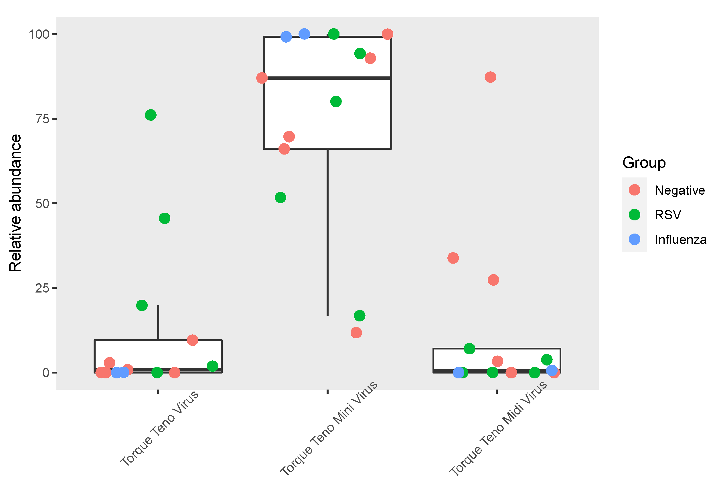
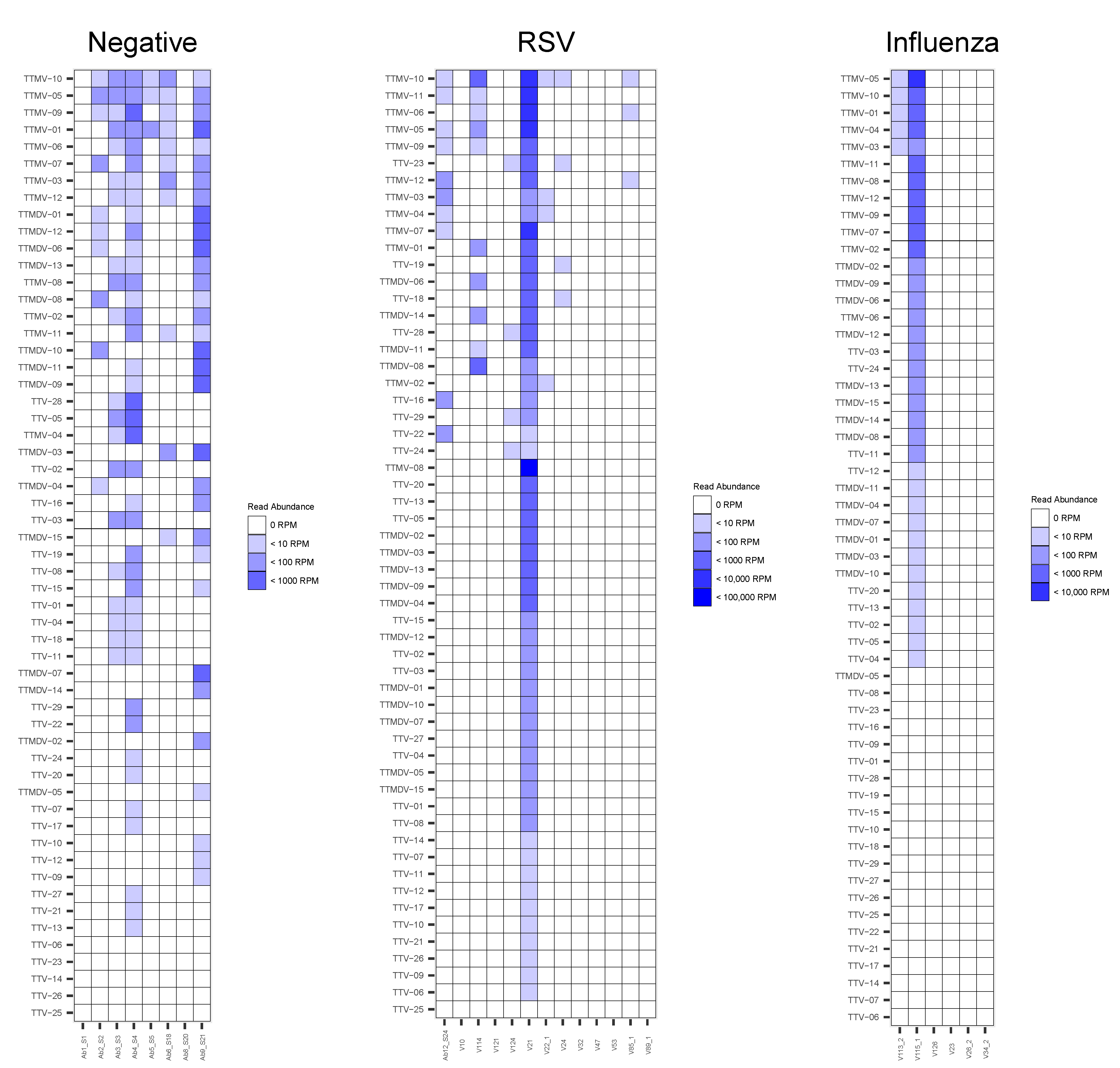
3.3. Anelloviridae Abundance and Composition within the Respiratory Virome

Among the 27 patients included in the present study, Anelloviridae reads were detected in 16/27 (59.3%) samples, including 6/8 (75%) negative samples, 7/13 (53.8%) RSV samples and 3/6 (50%) influenza samples (Figure 1). The median abundances of Anelloviridae reads were 48.2 (24.6–6066.0) RPM for influenza samples, 51.4 (27.0–370.3) RPM for RSV samples and 1131.8 (181.6–2761.7) RPM for negative samples (p = 0.4).
To investigate the composition of Anelloviridae, relative abundances of TTMV, TTV and TTMDV were computed according to the normalized number of reads identified as genus level. For samples with a detection of specific reads mapping to at least one Anelloviridae genus (n = 13), TTMV represented 87.1 (66.1–99.2)% of Anelloviridae reads, while TTV and TTMDV represented 0.8 (0.0–9.6)% and 0.7 (0.0–7.1)%, respectively (p < 0.001; Figure 2). No significant differences were observed between the relative abundance of TTV vs. TTMDV (p = 0.57). Of note, for 2/13 samples, the main genus detected was not TTMV (TTMDV for one negative sample and TTV for one RSV sample, Figure 2). No significant differences regarding Anelloviridae genus abundances were noticed between negative, RSV and influenza samples.
At the species level, a high inter-individual variability was noticed with multiple species detection for most individuals. TTMV-10 and TTMV-5 were the two most frequent species detected in the negative group, while the most prevalent species in the RSV group were TTMV-10, followed by TTMV-11, TTMV-6, and TTMV-5. Regarding the two patients with a detection of Anelloviridae species in the influenza group, TTMV-10, TTMV-5, TTMV-4, TTMV-3 and TTMV-1 were co-detected (Figure 3).

4. Discussion

In this present study, Anelloviridae was detected in about 60% of respiratory samples of children under five, highlighting a high inter-individual variability in Anelloviridae detection, as previously reported in blood or plasma samples [6,8,14]. Furthermore, we found a high abundance of TTMV in samples associated with or without viral pathogen detection (RSV or influenza). Although considered as commensal virome, Anelloviridae might contribute to respiratory diseases by activating the production of inflammatory cytokines [11,13]. TTMV has already been identified in several pathogenic settings, including empyema, encephalitis and periodontitis, suggesting a possible association of TTMV infection with inflammation [10,20,21]. In addition, in vitro replication of TTMV in alveolar epithelial cells has been demonstrated [10] and a recent mNGS study found that the upper respiratory virome composition of children with pneumonia was mainly represented by TTMV [12].
In contrast, Wang et al., showed that the majority of Anelloviridae reads mapped to TTV in children with severe ARI as well as in the control group constituted of 15 children without respiratory symptoms [9]. In addition, no differences in TTMV detection were noticed in respiratory samples of febrile pediatric patients compared to afebrile controls [22]. The differences in the Anelloviridae composition within the respiratory tract might be explained by clinical features, including the level of immunosuppression and the presence of viral co-infections, as well as by age, sex, geographical, or environmental factors which may influence the virome composition [23]. Importantly, we used the same mNGS methods as a previous study performed on the plasma samples of autologous stem cell transplant patients where TTV represented the most abundant genus (91.7% of Anelloviridae reads), while TTMDV and TTMV represented 6.4% and 1.9%, respectively [6].
As TTMV were found in all groups herein and in line with the limited number of patients included, we could not draw a conclusion regarding the respiratory pathogenicity of TTMV. This needs to be investigated in larger pediatric cohorts, including a healthy control group. The impact of TTMV levels, TTMV species diversity and TTMV co-infection (found in about 50% of influenza and RSV samples herein) on disease severity also need to be explored. Studies focusing on TTV found that a higher level or a co-infection with common respiratory viruses were associated with a more severe disease [14,24].
Overall, the present study emphasizes the high prevalence of Anelloviridae within the respiratory tract of children with ARI, and that the Anelloviridae composition is mainly represented by TTMV in the presence, or not, of common respiratory viruses. The characterization of whole viral communities is crucial for understanding the complex role of the human virome in respiratory diseases.

Author Contributions

A.B., G.D., L.J., F.M. and K.B.-P. conceived the study. A.B., M.S., G.D. and M.P., performed the sample preparations and sequencing. L.J., H.R. and G.O., performed bioinformatics analysis. L.J. is the guarantor for the NGS data. Y.G., B.L. and F.M. are the guarantors for the clinical data and sample collection. A.B. and G.D. were the main writers of the manuscript. All authors have read and agreed to the published version of the manuscript.

Funding

This study was funded by a metagenomic grant received in 2014 from the French foundation of innovation in infectious diseases (FINOVI, fondation innovation en infectiologie). This work was also supported by the LABEX ECOFECT (ANR-11-LABX-0048) of Université de Lyon, within the program “Investissements d’Avenir” (ANR-11-IDEX-0007), operated by the French National Research Agency (ANR).

Institutional Review Board Statement

This monocentric retrospective study received approval from HCL board of the French data protection authority (Commission Nationale de l’Informatique et des Libertés) and was registered with the national data protection agency, number 17-024.

Informed Consent Statement

During their hospitalization in the HCL, patients were made aware that their de-identified data, including clinical samples, may be used for research purposes, and they could opt out if they objected to the use of their data.

Data Availability Statement

The data that support the findings of this study are available upon request from the authors.

Acknowledgments

We thank Audrey Guichard, Gwendolyne Burfin, Delphine Falcon and Cecile Darley for their technical assistance.

Conflicts of Interest

K.B.-P. and G.O. are employees of bioMérieux. A.B. has served as a consultant to bioMérieux, and received a research grant from bioMérieux. The other authors declare that the research was conducted in the absence of any commercial or financial relationships that could be construed as a potential conflict of interest.

References

  1. Biagini, P. Classification of TTV and related viruses (anelloviruses). Curr. Top. Microbiol. Immunol. 2009, 331, 21–33. [Google Scholar] [CrossRef]
  2. Anelloviridae—ssDNA Viruses (2011)—ssDNA Viruses (2011)—ICTV. Available online: https://talk.ictvonline.org/ictv-reports/ictv_9th_report/ssdna-viruses-2011/w/ssdna_viruses/139/anelloviridae (accessed on 8 March 2022).
  3. Jaksch, P.; Kundi, M.; Görzer, I.; Muraközy, G.; Lambers, C.; Benazzo, A.; Hoetzenecker, K.; Klepetko, W.; Puchhammer-Stöckl, E. Torque teno virus as a novel biomarker targeting the efficacy of immunosuppression after lung transplantation. J. Infect. Dis. 2018, 218, 1922–1928. [Google Scholar] [CrossRef] [PubMed]
  4. De Vlaminck, I.; Khush, K.K.; Strehl, C.; Kohli, B.; Luikart, H.; Neff, N.F.; Okamoto, J.; Snyder, T.M.; Cornfield, D.N.; Nicolls, M.R.; et al. Temporal response of the human virome to immunosuppression and antiviral therapy. Cell 2013, 155, 1178–1187. [Google Scholar] [CrossRef] [PubMed] [Green Version]
  5. Spandole, S.; Cimponeriu, D.; Berca, L.M.; Mihăescu, G. Human anelloviruses: An update of molecular, epidemiological and clinical aspects. Arch. Virol. 2015, 160, 893–908. [Google Scholar] [CrossRef] [PubMed]
  6. Bal, A.; Sarkozy, C.; Josset, L.; Cheynet, V.; Oriol, G.; Becker, J.; Vilchez, G.; Sesques, P.; Mallet, F.; Pachot, A.; et al. Metagenomic next-generation sequencing reveals individual composition and dynamics of anelloviruses during autologous stem cell transplant recipient management. Viruses 2018, 10, 633. [Google Scholar] [CrossRef] [Green Version]
  7. Popgeorgiev, N.; Temmam, S.; Raoult, D.; Desnues, C. Describing the silent human virome with an emphasis on giant viruses. Intervirology 2013, 56, 395–412. [Google Scholar] [CrossRef]
  8. Arze, C.A.; Springer, S.; Dudas, G.; Patel, S.; Bhattacharyya, A.; Swaminathan, H.; Brugnara, C.; Delagrave, S.; Ong, T.; Kahvejian, A.; et al. Global genome analysis reveals a vast and dynamic anellovirus landscape within the human virome. Cell Host Microbe 2021, 29, 1305–1315.e6. [Google Scholar] [CrossRef]
  9. Wang, Y.; Zhu, N.; Li, Y.; Lu, R.; Wang, H.; Liu, G.; Zou, X.; Xie, Z.; Tan, W. Metagenomic analysis of viral genetic diversity in respiratory samples from children with severe acute respiratory infection in China. Clin. Microbiol. Infect. 2016, 22, 458.e1–458.e9. [Google Scholar] [CrossRef] [Green Version]
  10. Galmès, J.; Li, Y.; Rajoharison, A.; Ren, L.; Dollet, S.; Richard, N.; Vernet, G.; Javouhey, E.; Wang, J.; Telles, J.-N.; et al. Potential implication of new torque teno mini viruses in parapneumonic empyema in children. Eur. Respir. J. 2013, 42, 470–479. [Google Scholar] [CrossRef] [Green Version]
  11. Freer, G.; Maggi, F.; Pifferi, M.; Di Cicco, M.E.; Peroni, D.G.; Pistello, M. The virome and its major component, anellovirus, a convoluted system molding human immune defenses and possibly affecting the development of asthma and respiratory diseases in childhood. Front. Microbiol. 2018, 9, 686. [Google Scholar] [CrossRef] [Green Version]
  12. Espinoza, J.A.; Moreno-Valencia, Y.; Coronel-Tellez, R.H.; Castillejos-López, M.; Hernández, A.; Dominguez, A.; Miliar-García, A.; Barbachano-Guerrero, A.; Perez-Padilla, R.; Alejandre-Garcia, A.; et al. Virome and bacteriome characterization of children with pneumonia and asthma in Mexico City during winter seasons 2014 and 2015. PLoS ONE 2018, 13, e0192878. [Google Scholar] [CrossRef] [Green Version]
  13. Dodi, G.; Attanasi, M.; Di Filippo, P.; Di Pillo, S.; Chiarelli, F. Virome in the lungs: The role of anelloviruses in childhood respiratory diseases. Microorganisms 2021, 9, 1357. [Google Scholar] [CrossRef] [PubMed]
  14. Del Rosal, T.; García-García, M.L.; Casas, I.; Alcolea, S.; Iglesias-Caballero, M.; Pozo, F.; Rodrigo-Muñoz, J.M.; del Pozo, V.; Calvo, C. Torque teno virus in nasopharyngeal aspirate of children with respiratory infections. Res. Square 2022, preprint. [Google Scholar] [CrossRef]
  15. Prasetyo, A.A.; Desyardi, M.N.; Tanamas, J.; Suradi; Reviono; Harsini; Kageyama, S.; Chikumi, H.; Shimizu, E. Respiratory viruses and torque teno virus in adults with acute respiratory infections. Intervirology 2015, 58, 57–68. [Google Scholar] [CrossRef] [PubMed]
  16. Leijonhufvud, G.; Bajalan, A.; Soratto, T.A.T.; Gustafsson, B.; Bogdanovic, G.; Bjerkner, A.; Allander, T.; Ljungman, G.; Andersson, B. Better detection of Torque teno virus in children with leukemia by metagenomic sequencing than by quantitative PCR. J. Med Virol. 2022, 94, 634–641. [Google Scholar] [CrossRef] [PubMed]
  17. Mokili, J.L.; Rohwer, F.; Dutilh, B.E. Metagenomics and future perspectives in virus discovery. Curr. Opin. Virol. 2012, 2, 63–77. [Google Scholar] [CrossRef]
  18. Taboada, B.; Morán, P.; Serrano-Vázquez, A.; Iša, P.; Rojas-Velázquez, L.; Pérez-Juárez, H.; López, S.; Torres, J.; Ximenez, C.; Arias, C.F. The gut virome of healthy children during the first year of life is diverse and dynamic. PLoS ONE 2021, 16, e0240958. [Google Scholar] [CrossRef]
  19. Bal, A.; Pichon, M.; Picard, C.; Casalegno, J.S.; Valette, M.; Schuffenecker, I.; Billard, L.; Vallet, S.; Vilchez, G.; Cheynet, V.; et al. Quality control implementation for universal characterization of DNA and RNA viruses in clinical respiratory samples using single metagenomic next-generation sequencing workflow. BMC Infect. Dis. 2018, 18, 537. [Google Scholar] [CrossRef]
  20. Eibach, D.; Hogan, B.; Sarpong, N.; Winter, D.; Struck, N.S.; Adu-Sarkodie, Y.; Owusu-Dabo, E.; Schmidt-Chanasit, J.; May, J.; Cadar, D. Viral metagenomics revealed novel betatorquevirus species in pediatric inpatients with encephalitis/meningoencephalitis from Ghana. Sci. Rep. 2019, 9, 2360. [Google Scholar] [CrossRef]
  21. Zhang, Y.; Li, F.; Shan, T.-L.; Deng, X.; Delwart, E.; Feng, X.-P. A novel species of torque teno mini virus (TTMV) in gingival tissue from chronic periodontitis patients. Sci. Rep. 2016, 6, 26739. [Google Scholar] [CrossRef] [Green Version]
  22. TeKippe, E.M.; Wylie, K.M.; Deych, E.; Sodergren, E.; Weinstock, G.; Storch, G.A. Increased prevalence of anellovirus in pediatric patients with fever. PLoS ONE 2012, 7, e50937. [Google Scholar] [CrossRef]
  23. Unger, S.A.; Bogaert, D. The respiratory microbiome and respiratory infections. J. Infect. 2017, 74, S84–S88. [Google Scholar] [CrossRef]
  24. Maggi, F.; Pifferi, M.; Fornai, C.; Andreoli, E.; Tempestini, E.; Vatteroni, M.; Presciuttini, S.; Marchi, S.; Pietrobelli, A.; Boner, A.; et al. TT Virus in the nasal secretions of children with acute respiratory diseases: Relations to viremia and disease severity. J. Virol. 2003, 77, 2418–2425. [Google Scholar] [CrossRef] [PubMed] [Green Version]
Figure 1. Abundance of Anelloviridae, TTV, TTMV and TTMDV in negative, RSV and influenza samples. Anelloviridae reads include TTV, TTMV, TTMDV reads, as well as reads from unclassified Anelloviridae. To correct the difference of sequencing depth between samples, the number of reads were normalized in reads per million mapped reads (RPM). Only viruses with a count of >1 RPM were considered.
Viruses 14 00955 g001
Figure 2. Relative abundance of Anelloviridae genera among negative, RSV and influenza samples. Relative abundances of TTMV, TTV and TTMDV were computed according to the normalized number of reads identified as genus level. The three groups (negative, RSV and influenza) are represented by dots of different colors.
Viruses 14 00955 g002
Figure 3. Abundance of Anelloviridae species in negative, RSV and Influenza samples. Species are ranked by frequency of detection in each group. To correct difference of sequencing depth between samples, number of reads were normalized in read per millon mapped read (RPM). Only viruses with a count >1 RPM were considered.
Viruses 14 00955 g003
Table 1. Composition of metagenomic reads generated from samples that tested negative and samples that tested positive for RSV or influenza.
GroupSample IDTotal
Number of Reads
Number of Reads
for PCR-Positive
Virus
%
Human
%
Viruses
%
Bacteria
Negative samples (n = 8)Ab1_S113,242,872NA68.10.239.8
Ab2_S221,124,748NA53.71.036.1
Ab3_S318,613,428NA88.52.732.7
Ab4_S45,729,468NA40.92.138.3
Ab5_S56,506,898NA74.60.438.6
Ab6_S189,719,338NA58.10.236.8
Ab8_S2012,453,484NA58.90.250.2
Ab9_S215,124,554NA42.21.742.1
RSV samples (n = 13)Ab12_S2410,533,108232,82767.61.635.2
V104,547,41010287.00.03.7
V3214,911,09657,60688.50.42.1
V5310,826,666361,36588.83.41.5
V89_16,622,86458,28487.50.92.2
V4711,747,124182,77685.91.63.1
V22_116,819,6247787.60.02.8
V12419,598,588540,93484.32.83.0
V12114,490,59062,27987.90.42.1
V248,452,26081,2538.624.07.0
V85_119,138,76089283.80.04.2
V11412,516,2841,673,60252.013.69.7
V2116,928,282406,59464.96.66.0
Influenza samples (n = 6)V1267,910,142112587.20.013.6
V2318,131,47210390.70.0010.6
V26_213,672,72653,55682.70.54.8
V34_211,880,612805890.00.11.4
V113_210,944,27678,19349.60.716.0
V115_111,403,04621083.059.96.7
NTCNTC7,210,744NA8.50.0446.5
NTC: No-template control, RSV: respiratory syncytial virus, NA: not applicable.
Publisher’s Note: MDPI stays neutral with regard to jurisdictional claims in published maps and institutional affiliations.

