Review

Viral Interference with Functions of the Cellular Receptor Tyrosine Phosphatase CD45

Department of Virology, Hannover Medical School, Carl-Neuberg-Strasse 1, 30625 Hannover, Germany
*
Author to whom correspondence should be addressed.
Submission received: 20 January 2015 / Revised: 17 March 2015 / Accepted: 19 March 2015 / Published: 23 March 2015
(This article belongs to the Section Animal Viruses)

Abstract

:
The receptor tyrosine phosphatase CD45 is expressed on the surface of almost all cells of hematopoietic origin. CD45 functions are central to the development of T cells and determine the threshold at which T and B lymphocytes can become activated. Given this pivotal role of CD45 in the immune system, it is probably not surprising that viruses interfere with the activity of CD45 in lymphocytes to dampen the immune response and that they also utilize this molecule to accomplish their replication cycle. Here we report what is known about the interaction of viral proteins with CD45. Moreover, we debate putative interactions of viruses with CD45 in myeloid cells and the resulting consequences—subjects that remain to be investigated. Finally, we summarize the evidence that pathogens were the driving force for the evolution of CD45.

1. Introduction

The receptor-like protein tyrosine phosphatase CD45 (also known as leukocyte common antigen (LCA) or protein tyrosine phosphatase receptor type C (PTPRC)) has been long acknowledged for its remarkably strong expression level on the surface of all nucleated cells of hematopoietic origin. In T cells it can make up to 10% of the cell surface proteins [1]. In immunological research, CD45 is therefore widely used as a marker for cells of myeloid and lymphoid lineage. CD45 is a typical type I transmembrane protein; it has two intracellular phosphatase domains and although only domain D1 is enzymatically active, both domains are required for optimal phosphatase activity [2]. A single transmembrane domain connects the intracellular part with the ectodomain, which consists of a cysteine-rich domain followed by three fibronectin III-like repeats. Due to alternative splicing of exons 4 to 6 of the CD45 gene, one or more of the three domains A to C can be added to the amino-terminal region of CD45, leading to the expression of different isoforms [3]. Although potentially eight CD45 isoforms could be formed by variations in splicing, only five have been found in nature (RABC, RBC, RB, RAB, R0) [4]. The domains A to C are O-glycosylated [5] and the cysteine-rich domain is highly N-glycosylated [6]. Depending on the isoform and the glycosylation state, the molecular masses of the CD45 isoforms reach 180–240 kDa. CD45 isoform expression and glycosylation are dependent on cell type and also on cellular maturation and activation state. These differences in cell surface-expressed CD45 may have an influence on its distribution in the plasma membrane and interactions with other proteins [7].
For many years, the function of CD45 was predominantly investigated in T lymphocytes and to a lesser extent also in B lymphocytes. Here we briefly summarize the essentials of CD45 functions, and for details we refer the reader to the many excellent reviews on the topic (for example references [8,9,10]). In T cells, one functionally important substrate of CD45 is the Src family kinase Lck [11,12], which activates downstream signaling following triggering of the T cell receptor (TCR) [13,14]. Lck can be both positively and negatively regulated by tyrosine phosphorylation leading to hyperactivation of T cells and reduced TCR signaling, respectively. A phosphorylated tyrosine residue in the active site (Y394) increases the kinase activity of Lck, whereas phosphorylation of a tyrosine in the regulatory carboxy-terminal domain (Y505) favors the formation of a closed conformation, resulting in diminished activity [9,15,16,17,18]. Upon engagement of the TCR, active Lck phosphorylates the immunoreceptor tyrosine-based activation motifs (ITAM) of the CD3 ζ-chain, which initiates a signaling cascade, resulting in the phosphorylation of downstream signaling proteins, such as ZAP-70, LAT and SPL76. Ultimately, this can induce cytokine production, differentiation and proliferation of T cells [19]. CD45 and the Csk kinase have opposing effects on the phosphorylation state of the negative regulatory tyrosine Y505, CD45 acts to dephosphorylate Y505, thus maintaining Lck in a “primed” state. The balance of the activities of CD45 and Csk determines the threshold at which a T cell can become activated [9,20,21]. Beautiful studies by the Vale group who reconstituted the TCR and the associated signaling network in a non-immune cell or in liposomes imply that in addition to Lck the ITAMs in the CD3 ζ-chain are a major target of the CD45 phosphatase and that spatial segregation of the TCR and of CD45 is the mechanism resulting in TCR phosphorylation [22,23]. Deletion of the CD45 gene disrupts signaling via the TCR and CD45-deficient mice develop severe-combined immunodeficiency (SCID) with defects in the development and function of lymphocytes [24,25,26]. Similar phenotypes have also been observed in patients carrying defective CD45 alleles [27,28,29].
In contrast to T cells, the development of Natural Killer (NK) cells is not disrupted in CD45-deficient mice, rather an increased number of NK cells was observed [30,31]. Although NK cells differ from lymphocytes of the adaptive immune system in that their receptors used for detection of molecular patterns of pathogens are not rearranged during development, the downstream pathways for transduction of activating signals in these different immune cells have many features in common, including the utilization of Src kinases and their regulation by CD45. Many NK cell-activating receptors signal via the adapter molecules DAP12, CD3ζ and FcεRIγ, which carry an ITAM motif. Upon receptor ligation the ITAM is phosphorylated by a Src family kinase, followed by the recruitment of one of the two Syk family kinases expressed in NK cells, Syk and ZAP-70. Those functions of NK cells that are induced by engagement of receptors associated with ITAM-containing adapters are either completely abrogated or strongly affected in the absence of CD45. This applies to cytokine production, cytotoxicity and proliferation of NK cells in response to viral infection [31,32,33,34]. Since Syk function is partially independent of Src kinase activity, cell-mediated cytotoxicity is still detectable in CD45-deficient NK cells, although at a reduced level. Cytokine (IFN-γ) production by NK cells in response to stimulation with pro-inflammatory cytokines (IL2, IL12, IL18) is CD45-independent [31], as this pathway employs different receptors and adapter molecules. Although not all NK cell functions have an absolute requirement for CD45, a recent study demonstrated that those functions remaining active in CD45-deficient NK cells were not sufficient to protect mice against an infection with mouse cytomegalovirus (CMV) [34], underlining the vulnerability of these innate effector cells to viral interference via CD45.

2. Viral Interference with CD45 Affecting the Activity of Immune Cells

Almost all viruses have to modulate the immune response—at least to some extent—to gain a foothold in their hosts. Immunomodulatory strategies, also termed viral immune evasion, seem to be particularly important for viruses that establish long-lasting persistent infections. Examples of viral immune evasion range from interference with the recognition of molecular patterns of viruses by cellular receptors of the innate immune system to modulation of effector mechanisms of the adaptive immune response.
A recent genome-wide mutagenesis screen in mice revealed that CD45 functions are essential for mounting a protective immune response against Herpes simplex virus type 1 (HSV-1) [35]. Since lymphocytes do not develop normally in the absence of CD45, this study underlines their pivotal role in controlling HSV-1 infection and in preventing HSV-1 associated encephalitis. Beyond this fact the study also suggests that CD45 represents a potential Achilles’ heel of the immune system that could be an attractive target for viral counteraction. Notably, excessive CD45 activity can also result in potentially lethal immune pathology, as observed during the infection of mice with Ebola virus infection [36] and with Bacillus anthracis [37]. It may therefore be not only to the benefit of the pathogen but also of the host to regulate CD45 activity.
CD45 regulation is a topic that has been actively discussed over decades by authors with many different viewpoints (for review, see reference [10]). The search for cellular ligands of CD45 has led only to the discovery of cellular interaction partners, such as lectins, whose binding to CD45 is mediated via glycans and is thus typically of low specificity. This does not, however, exclude physiologically relevant roles for such molecules in influencing CD45 activity as shown—for instance—by the modulation of effector T cell function caused by the macrophage galactose-type lectin expressed by immature antigen presenting cells [38].
Only recently, the first viral proteins that bind to CD45 have been identified [39,40]. Some members of the CMV RL11 gene family display hypervariability between different CMV strains, suggesting that they evolved under selection pressure and possibly interact with variable host proteins [41,42,43]. One of these hypervariable RL11 proteins is UL11, a predicted type I transmembrane protein [44]. Using a soluble fusion protein consisting of the UL11 ectodomain and the Fc-binding domain of human IgG, we detected that UL11 binds to the surface of various leukocytes, including B and T lymphocytes [39]. The interaction partner of UL11 turned out to be CD45, and the interaction could be confirmed not only for the soluble UL11-Fc protein but also for the full-length UL11 transmembrane protein when expressed at the cell surface using an adenoviral vector. UL11 binds to different CD45 isoforms with comparable efficiencies, suggesting that the binding site is within a domain common to all isoforms, which is consistent with the observed blocking of the interaction with an antibody specific for the conserved region of CD45 [39]. Pretreatment of T cells with UL11-Fc reduced their proliferation capacity upon TCR stimulation. This was associated with reduced tyrosine phosphorylation of signaling proteins, suggesting that binding of UL11 reduces the phosphatase activity of CD45, which in turn affects the pool of active Lck molecules and thus the transduction of signals received by the TCR. Based on these observations we hypothesized that UL11 protects CMV-infected cells against the effector mechanisms of virus-specific T lymphocytes. We could confirm that UL11 is exposed on the surface of CMV-infected fibroblasts, although at relatively low level [45]. Unexpectedly, CMV-expressed UL11 had no effect on the activation of CMV-specific T lymphocytes when assessed by co-cultivation with CMV-infected cells. This leaves a number of questions for further investigation. It remains to be analyzed whether UL11 is expressed at higher levels in cell types other than fibroblasts or under physiologic conditions in vivo. Another possibility is that UL11 does not principally target mature effector T cells but rather has effects on the priming of naïve T cells, under conditions where the presented antigens are possibly only available in limiting amounts and the activation threshold may be considerably higher. Given that a substantial amount of UL11 seemed to reside in the endoplasmic reticulum of infected cells, it is also conceivable that UL11 rather interacts with CD45 present within CMV-infected cells than on neighboring cells. CD45-expressing myeloid cells such as macrophages and dendritic cells have been shown to be important target cells of CMV infection [46]. One of our aims is therefore to examine the potential impact of UL11 on the functions of CD45 in these cell types.
The second viral protein for which binding to CD45 has been reported is the E3/49K protein of adenovirus (AdV) type 19a, belonging to the AdV species D [40]. Interestingly, specific domains (CR1) within the N-terminal ectodomain of the E3/49K transmembrane protein [47] share some characteristics with a domain (RL11D) present in CMV RL11 proteins [41]. In contrast to the CMV UL11 protein, the Ad19a E3/49K protein is proteolytically cleaved, leading to the secretion of the ectodomain (sec49K). It is this soluble sec49K protein that binds to CD45 on the surface of T lymphocytes and NK cells as well as of B cells and monocytes [40]. Treatment of CD4 T cells with sec49K impaired their activation and led to a decrease in cytokine secretion upon stimulation with specific antigens. Functional impairment of T cells was associated with diminished phosphorylation of the signaling molecule ZAP-70 and of the downstream target molecules ERK-1/2. The strongest inhibitory effects of sec49K were seen for NK cells (Figure 1). Activation of these cells—induced by IL2 or by ligation of the NKG2D and 2B4 receptors—was reduced in the presence of sec49K, and NK cell-mediated lysis of MHC-negative target cells was strongly inhibited. The exact mechanisms of sec49K activity remain to be elucidated; it will be of interest to compare its effects in detail with those of CMV UL11 and of other lectin ligands for CD45. Altogether, this study points to NK cells as another population of immune cells that depend on CD45 function and that can be influenced by a viral protein.
Figure 1. Modulation of NK cell functions by the secreted adenovirus type 19a sec49K protein (as described in reference [40]). Binding of sec49K to CD45 results in altered activity of Src kinases, affecting the phosphorylation of the adapter molecule DAP12 that is associated with the activating NK cell receptor NKG2D. CD45 can also regulate the phosphorylation of JAK kinases associated with the interleukin-2 receptor (IL-2R).
Viruses 07 01540 g001
One report has implicated CD45 in the apoptosis of CD4 T lymphocytes induced by the gp120 envelope protein of human immunodeficiency virus type 1 (HIV-1) [48]. Depletion of both infected and uninfected CD4 T cells occurs already early after infection in HIV-1 infected individuals and is later one of the hallmarks of progression to AIDS [49]. Engagement of CD45 has been linked with apoptosis, although the underlying mechanisms have not been completely elucidated and cell death may be due to triggering of unconventional apoptosis pathways (for review see References [50,51]). If CD45-mediated apoptosis contributes to gp120-dependent CD4 T cell depletion—in conjunction with several other mechanisms that have been suggested [52]—this would provide another example how a viral protein influences viral pathogenesis by means of CD45.
Polymorphism in the human CD45 gene—for instance the C77G and A138G mutations—change the expression patterns of CD45 isoforms and can result in an altered threshold for activation of T cells (reviewed in reference [53]). CD45 polymorphisms have been connected with altered susceptibility to autoimmune diseases and viral infections, for example with HIV-1 [54,55], hepatitis C virus [56] and hepatitis B virus [57]. Some of these studies may have been underpowered and have therefore been critically discussed and sometimes challenged; the association of CD45 polymorphisms with autoimmune diseases in particular has been disputed [58]. However, in view of the central role of CD45 in the immune response and in the control of viral infections there is a rational basis for this hypothesis, warranting larger epidemiological studies.

3. Utilization of CD45 to the Benefit of Viruses

In some circumstances viruses go beyond the functional inhibition of proteins involved in cellular defense mechanisms and even manage to integrate such molecules into their lifecycle in order to promote viral replication. One example that applies to CD45 is the human T cell leukemia virus type 1 (HTLV-1). Mazurov et al. (2012) detected that cell-to-cell transmission of HTLV-1 is inhibited by monoclonal antibodies which recognize a specific glycan signature (the so-called Tn antigen) attached to CD45 on Jurkat T cells (and also to another surface protein, CD43) [59]. Moreover, high levels of fully O-glycosylated CD45 and CD43 promoted HTLV-1 infection of neighboring cells. CD45 and CD43 are large rod-like proteins, which protrude from the cell surface and due to their large size, and the negative charge resulting from sialic acid present within their O-linked glycans favor repulsion of cells. Accordingly, CD45 has to be excluded from the immunological synapse [60,61] and perhaps also from virological synapses [62] in order to allow activation of immune cells and efficient cell-to-cell transmission of viruses, respectively. Based on the observed clustering of newly formed HTLV-1 virions on the surface of Jurkat T cells that express high levels of CD45 and CD43 and mechanisms of HTLV-1 transmission previously described by others [63], Mazurov et al. (2012) proposed that the high abundance of these proteins forces the virus to assemble only at specific sites of the outer membrane. These clusters might then serve as contact sites to the target cell, mediating efficient cell-to-cell spread. In cells expressing lower amounts of these proteins HTLV-1 budding occurs along the whole cell surface, which makes transmission less efficient. Although blocking of CD45 with antibodies promotes adhesion between neighboring cells, the authors argued that the inhibition of HTLV-1 infection observed in this case is due to exclusion of the virions from the contact sites [59]. It remains to be seen whether the proposed mechanism also plays a role in HTLV-1 infection of primary T cells. Interestingly, the authors also observed inhibition of HIV-1 transmission in Jurkat cells upon knock-down of CD45 expression [59]. However, the mechanism appeared to be different from the one described for HTLV-1 and details remain to be elucidated.
HTLV-1 does not seem to be the only virus that employs CD45-like proteins for infection. Upon inoculation into the arthropod vector Aedes aegypti, West Nile virus particles associate with the C-type lectin mosGCTL-1 present in the hemolymph of the mosquito [64]. The lectin-decorated virions are then recruited to a mosquito homolog of CD45 (mosPTP-1) on the surface of cells of various mosquito tissues, promoting attachment and viral entry. West Nile virus upregulates both the expression of the lectin in infected cells and also its secretion thereby facilitating further viral spread within the mosquito. It appears that evolution taught viruses at least twice to utilize CD45 or CD45-like molecules in such diverse hosts as humans and insects.

4. Further Possible Interaction of Viruses with CD45 in Myeloid Cells

CD45 is not only expressed by lymphoid cells, but also by myeloid cells, such as monocytes, macrophages and dendritic cells (DC). This is of interest as these cells are central to the initiation of immune responses and can be infected by some viruses, such as CMV, which utilizes them to modulate immunity, to disseminate within the organism [65] and to establish latency [66]. The state of knowledge of CD45 functions in these cells is, however, still limited in comparison to the profusion of publications describing the role of CD45 in lymphoid cells.
While T cells cannot develop normally in the absence of CD45, CD45 deletion has little impact on the development of DC with only a slight shift seen in the composition of DC subsets [67,68]. CD45-negative DC seem to be slightly pre-activated as assessed by the surface expression of co-stimulatory molecules. Piercy et al. (2006) observed that bone marrow-derived DC (BMDC) and splenic DC from CD45 knock-out mice responded with increased production of the pro-inflammatory cytokines TNFα and IL6 following stimulation with pattern recognition receptor TLR3 and TLR9 agonists [69]. In contrast, the levels of type I interferons (IFN) produced by plasmacytoid DC in the spleen upon infection with lymphocytic choriomeningitis virus were found to be diminished in CD45-/- mice [67]. These apparently contradictory outcomes in terms of the production of different cytokines could at least in part be explained by the results of another study [68], where it was shown that CD45 has a negative regulatory effect on TLR signaling pathways that depend on the adapter protein MyD88 and a positive regulatory role on MyD88-independent signaling (Figure 2). Mechanisms that were discussed [69] include the direct dephosphorylation of an adapter molecule by CD45 or—more indirectly—dephosphorylation of the negatively regulatory tyrosine residue of Src kinases, resulting in their activation. An involvement of Src kinases in TLR signaling has indeed been suggested [70]. Piercy et al. (2006) also found that cytokine production following stimulation with TNFα or IFNα was enhanced in CD45-negative BMDC. CD45 can apparently modulate the positive feedback loop of cytokine production and dampen the signaling induced by cytokine receptors. This is supported by the discovery that CD45 can dephosphorylate JAK kinases in various cells of hematopoietic origin, thereby negatively regulating cytokine-induced signaling [71]. A recent publication has implicated the adapter protein DOK-1 in the negative regulation of JAK-STAT signaling by CD45 [72].
Figure 2. CD45 regulates TLR signaling in a positive or negative manner depending on the adapter molecules (MyD88 or TRIF) involved [68]. Viruses may modulate these pathways in order to influence cytokine production (IRFs, interferon response factors).
Viruses 07 01540 g002
The induction of a cellular antiviral state by cytokines (e.g., by type I interferons) is an important defense mechanism against many viruses; in line with the mechanism outlined above, replication of an interferon-sensitive Coxsackievirus was more strongly inhibited by IFNα in CD45-negative cells and CD45-/- mice were found to be much more resistant to infection with the Coxsackievirus than their CD45-positive littermates [71]. However, other factors than the diminished regulation of JAK-STAT signaling may have contributed to this phenotype of CD45-/- mice as they also lack T cells which are known to mediate immunopathological tissue damage [73]. To sum up, CD45 obviously plays a complex role in the sensing of pathogens and the orchestration of the antiviral immune response by myeloid cells. These functions of CD45 may potentially be modulated by viral proteins, although this remains speculative and has to be the subject of further experimental investigation.
Modulation of cell adhesion by CD45 in macrophages [74] is another interesting subject in the context of viral infection. Altered adhesion has also been reported for CD45-negative T cells [75,76], and it seems possible that the underlying mechanisms are similar, as in both cell types integrin signaling is affected upon CD45 deletion. In macrophages CD45 was found to co-localize with ß2-integrin and Src kinases in focal contact sites. Roach et al. (1997) reported that CD45-negative macrophages adhere and spread more rapidly than normal macrophages when plated on plastic dishes, however the CD45-negative cells were unable to maintain this β2 integrin-dependent adherence as they detached after 48 h. Moreover, the CD45-negative macrophages displayed altered morphology. These changes were associated with hyperphosphorylation and hyperactivation of the Src kinases Hck and Lyn. Binding of macrophages to the extracellular matrix or to neighboring cells via integrins induces a signaling process (outside-in signaling), which is necessary for adhesion, migration and also for immunologic activation. Upon integrin clustering Src kinases are rapidly activated and phosphorylate and activate focal adhesion kinases (FAK) and FAK-binding proteins (for a short overview see reference [77]). Based on the observations that the phosphatase activity of CD45 is required for its function in cell adhesion [75] and that CD45 and Src kinases are intimately connected in TCR signaling, a role for CD45 upstream of Src kinases has been proposed. However, in this process CD45 acts rather as a negative regulator, limiting the activity of the Src kinases by dephosphorylating the autocatalytic site [74]. A recent study sheds some light on the molecular mechanism by which CD45 regulates macrophage adhesion [78]. Using bone marrow-derived macrophages, St-Pierre and Ostergaard (2013) confirmed the phenotypic characteristics of CD45-/- macrophages described by Roach et al. (1997), and also detected impaired mobility of these cells [78]. Interestingly, the level of paxillin was found to be strongly decreased in CD45-negative cells due to its degradation by calpain, probably in response to tyrosine-phosphorylation of paxillin by members of the focal adhesion kinase family (FAK or Pyk2). Paxillin acts as a scaffold for coordination of the signaling processes at sites of adhesion [77]. As outlined above, FAK kinases are themselves targets for phosphorylation by Src kinases, which are hyperactive in the absence of negative regulation by CD45. Given that macrophages can serve as vehicles for dissemination of viruses, which involves the release of macrophages from cellular tissues into the blood stream followed by their subsequent adherence in other organs, it could be postulated that regulation of CD45 by viral proteins is a mechanism to govern this process.

5. CD45 Evolution in the Light of Recently Identified Putative Viral Ligands

Pathogen-host interactions are often marked by signatures of positive selection on host genes encoding the interacting proteins. These signatures are characterized by increased rates of non-synonymous (Ka) as opposed to synonymous (Ks) nucleotide substitutions, resulting in amino acid differences in the encoded proteins. The evolution of CD45 has been the focus of several studies [79,80,81], which revealed that the CD45 extracellular domain has been evolving under strong positive selection, whereas the intracellular domain is well conserved. An evolutionary study of primate genes potentially involved in viral pathogenesis identified ten residues in CD45 that were under positive selection [81]. Interestingly, all of these hot spots were located in the extracellular domain of CD45, mostly at the surface of the predicted structure (Figure 3A) and thus potentially accessible to contact by interacting proteins. Filip et al. (2004) compared the Ka/Ks rates in the alternatively spliced exons and two common exons encoding the cysteine-rich and fibronectin III-like domains of CD45 in primates. Alternatively spliced exons could be expected to evolve faster than constitutively spliced ones as the encoded protein domains may have more freedom to diverge, but the reverse was observed. The common exons displayed even higher rates of nucleotide substitutions than an intron of the primate CD45 genes (intron 6), although the latter would plausibly be able to tolerate a greater number of nucleotide changes. In both primate studies [80,81] it was hypothesized that the ectodomain of CD45 is targeted by pathogens—although at that time no pathogen was known to encode a CD45 interacting protein. The fact that the amino acid changes were mainly found in domains common to all CD45 isoforms suggests that pathogens may need to modulate the activity of all of the different CD45 molecules.
Notably, the HCMV UL11 and the AdV 19a E3/49K proteins bind to the constant region of CD45 [39,40], making these viral proteins candidates to have driven CD45 evolution. In a virus-host arms race model, one would not only expect signatures of positive selection in the host gene but also in the corresponding virus gene, particularly in the sequences encoding the part of the viral protein that mediates the interaction. The few studies that exist on CMV UL11 primarily revealed the highly polymorphic characteristics of the viral protein [42,44]. Alignment of the approximately 50 UL11 amino acid sequences available in the NCBI database and construction of a phylogeny confirms the reported divergence (Figure 3B). Interestingly, the polymorphism in the UL11 sequences is particularly pronounced at certain positions of the predicted extracellular domain (Figure 3C), whereas other parts of the UL11 protein, e.g., the signal peptide, the threonine-rich domain, the membrane-proximal part of the ectodomain and also the transmembrane region and the cytoplasmic tail are comparably well-conserved. It would be interesting to learn whether the divergent amino acids form part of the contact zone with CD45 and whether UL11 variants display different capacity for interaction with CD45. The few UL11 proteins which we tested were all able to bind to CD45 [39]; however, more careful evaluation would be needed to determine potential quantitative differences. Another interesting question is whether CMV UL11 proteins can bind to CD45 variants from primates, especially since the development of the RL11 gene family dates back to CMVs of primates [82]. Possibly, orthologous RL11 proteins of those primates CMVs exist that also bind to CD45 proteins. Since the high sequence variability of the RL11 proteins impedes the simple identification of UL11 orthologs, experimental testing of this hypothesis is required.
The apparently higher divergence of UL11 sequences compared to that of CD45 is in agreement with the predictions of a virus-host arms race as a virus has a much shorter generation time than its host, resulting in higher mutation rates. In addition, a nonstructural viral protein that is not absolutely required for virus replication and assembly has probably much more freedom to diverge than a host protein with importance for the immune response. In addition, abundant and broad expression in different cell types as is seen for CD45 can be expected to negatively affect the selection rate [83].
Figure 3. (A) The positions of those residues in the extracellular domain of CD45RO that were shown to be under positive selection [81] are labeled in red and marked with asterisks. The model of the CD45 structure was generated with the RaptorX program [84,85] and depicted using UCSF Chimera [86]. (B) Phylogenetic representation of 53 UL11 amino acid sequences derived from clinical and laboratory adapted HCMV strains. The phylogeny tree was calculated with MEGA 6 [87]. (C) The identity scores of the amino acids along the different positions of UL11 were calculated with the Jalview program [88].
Viruses 07 01540 g003
E3/49K-like proteins are apparently expressed by all AdV species of subgenus D [89], not just those causing epidemic keratoconjunctivitis as initially proposed. Whether all of the homologs of the AdV type 19a E3/49K protein share the ability of CD45 binding remains to be examined. Sequencing of whole genomes of species D human AdV revealed one of the highest amino acid variability in the predicted 49K (CR1-β) homologs among the AdV proteins [90]. Interestingly, homologous recombination between E3 genes of human AdV seems to be a driving force generating this diversity [90,91]. Functional analysis of these additional AdV E3/49K proteins and perhaps the identification of proteins from other viruses with similar properties may strengthen the support for the long-standing hypothesis that pathogens were the driving force behind the evolution of CD45.

6. Conclusions and Further Directions

A series of publications have reported on the interference of viruses from a range of different virus families with the functions of the receptor tyrosine phosphatase CD45. In view of the important and essential position of CD45 in various signaling networks of immune cells, it will not be surprising if more viruses and viral proteins are detected that take over command of these networks by either recruiting CD45 or modulating its functions. With the CMV UL11 and the AdV E3/49K proteins, now two proteins are known that can clearly bind to CD45. AdV E3/49K might be the first secreted viral protein identified that acts on CD45 in non-infected neighboring cells, possibly also on cells distant from infected tissues [40]. The precise function of the CMV UL11 protein is less clear; we hypothesize that it rather binds to CD45 in cis within CMV-infected cells. This is particularly interesting since CMV infects myeloid cells for which several specific functions of CD45 have been reported. However, the consequences of this interaction remain to be elucidated. For both the CMV UL11 and the AdV E3/49K protein the mode of binding and the resulting molecular effects for CD45 still have to be determined [92]. Both UL11 and E3/49K carry O- and N-linked carbohydrates and it is not clear whether and how these glycans contribute to the interaction. It is possible that the binding strength of these viral proteins is relatively weak and is mediated via the many glycans present in the extracellular domain of CD45, resembling the known interaction with several cellular lectins [93]. Engagement of CD45 by the viral proteins definitely has consequences for downstream pathways, but this could arguably be due to either increased or diminished phosphatase activity. The mechanism by which the activity of CD45 is regulated has been debated for many years. Rather than a conformational change of CD45, induction of dimerization or changed spatial distribution within the plasma membrane have been discussed as regulatory mechanisms. Experiments to determine the mode of action of the viral CD45 interaction partners could shed light on this issue.
Targeting of CD45 isoforms with specific antibodies has been proposed as a strategy to selectively modulate immune responses, for instance to induce tolerance against transplanted organs and to prevent rejection [94]—although with limited success. It is quite possible that the results of the long evolutionary interplay between viruses and their hosts can teach us about therapeutic options to allow us to intervene in the delicate and sophisticated signaling networks of immune cells in a much smarter way than has previously been conceivable.

Acknowledgments

We thank Andrew Davison for hints on the evolution of CMV RL11 proteins. Molecular graphics and analyses were performed with the UCSF Chimera package. Chimera is developed by the Resource for Biocomputing, Visualization, and Informatics at the University of California, San Francisco (supported by NIGMS P41-GM103311).
Our work on the viral interaction with CD45 is supported by the VISTRIE project (VH-VI-424) of the Impuls- und Vernetzungsfond from the Helmholtz Association. N.T., J.Z., and E.E. are supported by fellowships of the Hannover Biomedical Research School and the International Research Training Group 1273 from the Deutsche Forschungsgemeinschaft.

Conflicts of Interest

The authors declare no conflict of interest.

References

  1. Williams, A.F.; Barclay, A.N. Glycoprotein antigens of the lymphocyte surface and their purification by antibody affinity chromatography. In Handbook of Experimental Immunology; Weir, D.M., Herzenberg, L.A., Eds.; Blackwell Scientific Publications: Oxford, UK, 1986; pp. 1–24. [Google Scholar]
  2. Desai, D.M.; Sap, J.; Silvennoinen, O.; Schlessinger, J.; Weiss, A. The catalytic activity of the CD45 membrane-proximal phosphatase domain is required for TCR signaling and regulation. EMBO J. 1994, 13, 4002–4010. [Google Scholar] [PubMed]
  3. Streuli, M.; Hall, L.R.; Saga, Y.; Schlossman, S.F.; Saito, H. Differential usage of three exons generates at least five different mRNAs encoding human leukocyte common antigens. J. Exp. Med. 1987, 166, 1548–1566. [Google Scholar] [CrossRef] [PubMed]
  4. Trowbridge, I.S.; Thomas, M.L. CD45: An emerging role as a protein tyrosine phosphatase required for lymphocyte activation and development. Annu. Rev. Immunol. 1994, 12, 85–116. [Google Scholar] [CrossRef] [PubMed]
  5. Furukawa, K.; Funakoshi, Y.; Autero, M.; Horejsi, V.; Kobata, A.; Gahmberg, C.G. Structural study of the O-linked sugar chains of human leukocyte tyrosine phosphatase CD45. Eur. J. Biochem. 1998, 251, 288–294. [Google Scholar] [CrossRef] [PubMed]
  6. Sato, T.; Furukawa, K.; Autero, M.; Gahmberg, C.G.; Kobata, A. Structural study of the sugar chains of human leukocyte common antigen CD45. Biochemistry 1993, 32, 12694–12704. [Google Scholar] [CrossRef] [PubMed]
  7. Dianzani, U.; Redoglia, V.; Malavasi, F.; Bragardo, M.; Pileri, A.; Janeway, C.A., Jr.; Bottomly, K. Isoform-specific associations of CD45 with accessory molecules in human T lymphocytes. Eur. J. Immunol. 1992, 22, 365–371. [Google Scholar] [CrossRef] [PubMed]
  8. Hermiston, M.L.; Xu, Z.; Weiss, A. CD45: A critical regulator of signaling thresholds in immune cells. Annu. Rev. Immunol. 2003, 21, 107–137. [Google Scholar] [CrossRef] [PubMed]
  9. Palacios, E.H.; Weiss, A. Function of the Src-family kinases, Lck and Fyn, in T-cell development and activation. Oncogene 2004, 23, 7990–8000. [Google Scholar] [CrossRef] [PubMed]
  10. Hermiston, M.L.; Zikherman, J.; Zhu, J.W. CD45, CD148, and Lyp/Pep: Critical phosphatases regulating Src family kinase signaling networks in immune cells. Immunol. Rev. 2009, 228, 288–311. [Google Scholar] [CrossRef] [PubMed]
  11. Mustelin, T.; Coggeshall, K.M.; Altman, A. Rapid activation of the T-cell tyrosine protein kinase pp56lck by the CD45 phosphotyrosine phosphatase. Proc. Natl. Acad. Sci. USA 1989, 86, 6302–6306. [Google Scholar] [CrossRef] [PubMed]
  12. Ostergaard, H.L.; Shackelford, D.A.; Hurley, T.R.; Johnson, P.; Hyman, R.; Sefton, B.M.; Trowbridge, I.S. Expression of CD45 alters phosphorylation of the lck-encoded tyrosine protein kinase in murine lymphoma T-cell lines. Proc. Natl. Acad. Sci. USA 1989, 86, 8959–8963. [Google Scholar] [CrossRef] [PubMed]
  13. Danielian, S.; Fagard, R.; Alcover, A.; Acuto, O.; Fischer, S. The lymphocyte-specific protein tyrosine kinase p56lck is hyperphosphorylated on serine and tyrosine residues within minutes after activation via T cell receptor or CD2. Eur. J. Immunol. 1989, 19, 2183–2189. [Google Scholar] [CrossRef] [PubMed]
  14. Rossy, J.; Williamson, D.J.; Gaus, K. How does the kinase Lck phosphorylate the T cell receptor? Spatial organization as a regulatory mechanism. Front Immunol. 2012, 3, 167. [Google Scholar] [PubMed]
  15. Bergman, M.; Mustelin, T.; Oetken, C.; Partanen, J.; Flint, N.A.; Amrein, K.E.; Autero, M.; Burn, P.; Alitalo, K. The human p50csk tyrosine kinase phosphorylates p56lck at Tyr-505 and down regulates its catalytic activity. EMBO J. 1992, 11, 2919–2924. [Google Scholar] [PubMed]
  16. Yamaguchi, H.; Hendrickson, W.A. Structural basis for activation of human lymphocyte kinase Lck upon tyrosine phosphorylation. Nature 1996, 384, 484–489. [Google Scholar] [CrossRef] [PubMed]
  17. D’Oro, U.; Sakaguchi, K.; Appella, E.; Ashwell, J.D. Mutational analysis of Lck in CD45-negative T cells: Dominant role of tyrosine 394 phosphorylation in kinase activity. Mol. Cell. Biol. 1996, 16, 4996–5003. [Google Scholar] [PubMed]
  18. McNeill, L.; Salmond, R.J.; Cooper, J.C.; Carret, C.K.; Cassady-Cain, R.L.; Roche-Molina, M.; Tandon, P.; Holmes, N.; Alexander, D.R. The differential regulation of Lck kinase phosphorylation sites by CD45 is critical for T cell receptor signaling responses. Immunity 2007, 27, 425–437. [Google Scholar] [CrossRef] [PubMed]
  19. Brownlie, R.J.; Zamoyska, R. T cell receptor signalling networks: Branched, diversified and bounded. Nat. Rev. Immunol. 2013, 13, 257–269. [Google Scholar] [CrossRef] [PubMed]
  20. Zikherman, J.; Jenne, C.; Watson, S.; Doan, K.; Raschke, W.; Goodnow, C.C.; Weiss, A. CD45-Csk phosphatase-kinase titration uncouples basal and inducible T cell receptor signaling during thymic development. Immunity 2010, 32, 342–354. [Google Scholar] [CrossRef] [PubMed]
  21. Schoenborn, J.R.; Tan, Y.X.; Zhang, C.; Shokat, K.M.; Weiss, A. Feedback circuits monitor and adjust basal Lck-dependent events in T cell receptor signaling. Sci. Signal. 2011, 4, ra59. [Google Scholar] [PubMed]
  22. Hui, E.; Vale, R.D. In vitro membrane reconstitution of the T-cell receptor proximal signaling network. Nat. Struct. Mol. Biol. 2014, 21, 133–142. [Google Scholar] [CrossRef] [PubMed]
  23. James, J.R.; Vale, R.D. Biophysical mechanism of T-cell receptor triggering in a reconstituted system. Nature 2012, 487, 64–69. [Google Scholar] [CrossRef] [PubMed]
  24. Byth, K.F.; Conroy, L.A.; Howlett, S.; Smith, A.J.; May, J.; Alexander, D.R.; Holmes, N. CD45-null transgenic mice reveal a positive regulatory role for CD45 in early thymocyte development, in the selection of CD4+CD8+ thymocytes, and B cell maturation. J. Exp. Med. 1996, 183, 1707–1718. [Google Scholar] [CrossRef] [PubMed]
  25. Kishihara, K.; Penninger, J.; Wallace, V.A.; Kundig, T.M.; Kawai, K.; Wakeham, A.; Timms, E.; Pfeffer, K.; Ohashi, P.S.; Thomas, M.L.; et al. Normal B lymphocyte development but impaired T cell maturation in CD45-exon6 protein tyrosine phosphatase-deficient mice. Cell 1993, 74, 143–156. [Google Scholar] [CrossRef] [PubMed]
  26. Mee, P.J.; Turner, M.; Basson, M.A.; Costello, P.S.; Zamoyska, R.; Tybulewicz, V.L. Greatly reduced efficiency of both positive and negative selection of thymocytes in CD45 tyrosine phosphatase-deficient mice. Eur. J. Immunol. 1999, 29, 2923–2933. [Google Scholar] [CrossRef] [PubMed]
  27. Cale, C.M.; Klein, N.J.; Novelli, V.; Veys, P.; Jones, A.M.; Morgan, G. Severe combined immunodeficiency with abnormalities in expression of the common leucocyte antigen, CD45. Arch. Dis. Child 1997, 76, 163–164. [Google Scholar] [CrossRef] [PubMed]
  28. Kung, C.; Pingel, J.T.; Heikinheimo, M.; Klemola, T.; Varkila, K.; Yoo, L.I.; Vuopala, K.; Poyhonen, M.; Uhari, M.; Rogers, M.; et al. Mutations in the tyrosine phosphatase CD45 gene in a child with severe combined immunodeficiency disease. Nat. Med. 2000, 6, 343–345. [Google Scholar] [CrossRef] [PubMed]
  29. Tchilian, E.Z.; Wallace, D.L.; Wells, R.S.; Flower, D.R.; Morgan, G.; Beverley, P.C. A deletion in the gene encoding the CD45 antigen in a patient with SCID. J. Immunol. 2001, 166, 1308–1313. [Google Scholar] [CrossRef] [PubMed]
  30. Yamada, H.; Kishihara, K.; Kong, Y.Y.; Nomoto, K. Enhanced generation of NK cells with intact cytotoxic function in CD45 exon 6-deficient mice. J. Immunol. 1996, 157, 1523–1528. [Google Scholar] [PubMed]
  31. Huntington, N.D.; Xu, Y.; Nutt, S.L.; Tarlinton, D.M. A requirement for CD45 distinguishes Ly49D-mediated cytokine and chemokine production from killing in primary natural killer cells. J. Exp. Med. 2005, 201, 1421–1433. [Google Scholar] [CrossRef] [PubMed]
  32. Mason, L.H.; Willette-Brown, J.; Taylor, L.S.; McVicar, D.W. Regulation of Ly49D/DAP12 signal transduction by Src-family kinases and CD45. J. Immunol. 2006, 176, 6615–6623. [Google Scholar] [CrossRef] [PubMed]
  33. Hesslein, D.G.; Takaki, R.; Hermiston, M.L.; Weiss, A.; Lanier, L.L. Dysregulation of signaling pathways in CD45-deficient NK cells leads to differentially regulated cytotoxicity and cytokine production. Proc. Natl. Acad. Sci. USA 2006, 103, 7012–7017. [Google Scholar] [CrossRef] [PubMed]
  34. Hesslein, D.G.; Palacios, E.H.; Sun, J.C.; Beilke, J.N.; Watson, S.R.; Weiss, A.; Lanier, L.L. Differential requirements for CD45 in NK-cell function reveal distinct roles for Syk-family kinases. Blood 2011, 117, 3087–3095. [Google Scholar] [CrossRef] [PubMed]
  35. Caignard, G.; Leiva-Torres, G.A.; Leney-Greene, M.; Charbonneau, B.; Dumaine, A.; Fodil-Cornu, N.; Pyzik, M.; Cingolani, P.; Schwartzentruber, J.; Dupaul-Chicoine, J.; et al. Genome-wide mouse mutagenesis reveals CD45-mediated T cell function as critical in protective immunity to HSV-1. PLoS. Pathog. 2013, 9, e1003637. [Google Scholar] [CrossRef] [PubMed]
  36. Panchal, R.G.; Bradfute, S.B.; Peyser, B.D.; Warfield, K.L.; Ruthel, G.; Lane, D.; Kenny, T.A.; Anderson, A.O.; Raschke, W.C.; Bavari, S. Reduced levels of protein tyrosine phosphatase CD45 protect mice from the lethal effects of Ebola virus infection. Cell Host. Microbe 2009, 6, 162–173. [Google Scholar] [CrossRef] [PubMed]
  37. Panchal, R.G.; Ulrich, R.L.; Bradfute, S.B.; Lane, D.; Ruthel, G.; Kenny, T.A.; Iversen, P.L.; Anderson, A.O.; Gussio, R.; Raschke, W.C.; et al. Reduced expression of CD45 protein-tyrosine phosphatase provides protection against anthrax pathogenesis. J. Biol. Chem. 2009, 284, 12874–12885. [Google Scholar] [CrossRef] [PubMed]
  38. Van Vliet, S.J.; Gringhuis, S.I.; Geijtenbeek, T.B.; van Kooyk, Y. Regulation of effector T cells by antigen-presenting cells via interaction of the C-type lectin MGL with CD45. Nat. Immunol. 2006, 7, 1200–1208. [Google Scholar] [CrossRef] [PubMed]
  39. Gabaev, I.; Steinbrück, L.; Pokoyski, C.; Pich, A.; Stanton, R.J.; Schwinzer, R.; Schulz, T.F.; Jacobs, R.; Messerle, M.; Kay-Fedorov, P.C. The human cytomegalovirus UL11 protein interacts with the receptor tyrosine phosphatase CD45, resulting in functional paralysis of T cells. PLoS. Pathog. 2011, 7, e1002432. [Google Scholar] [CrossRef] [PubMed]
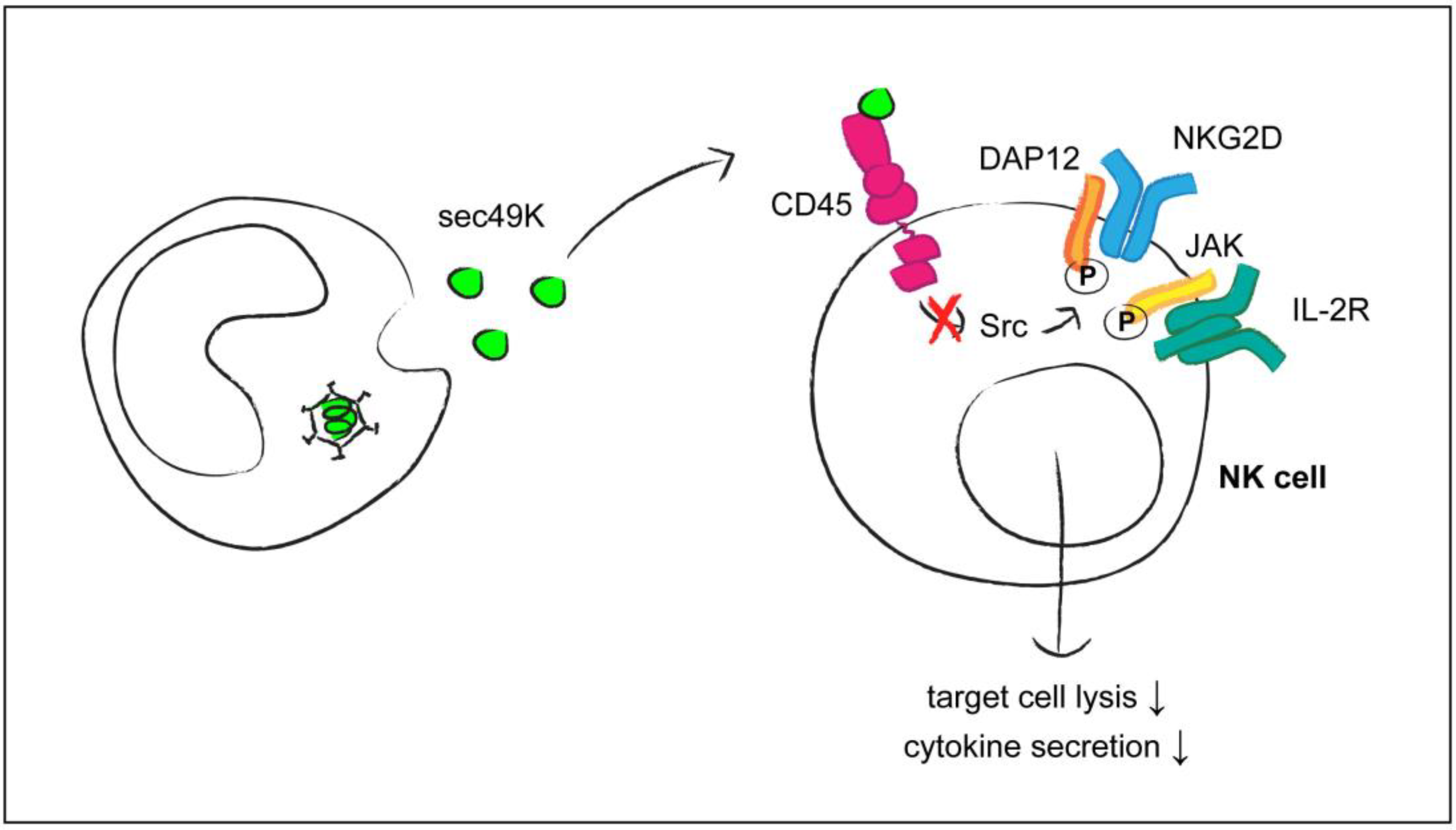
  40. Windheim, M.; Southcombe, J.H.; Kremmer, E.; Chaplin, L.; Urlaub, D.; Falk, C.S.; Claus, M.; Mihm, J.; Braithwaite, M.; Dennehy, K.; et al. A unique secreted adenovirus E3 protein binds to the leukocyte common antigen CD45 and modulates leukocyte functions. Proc. Natl. Acad. Sci. USA 2013, 110, e4884–e4893. [Google Scholar] [CrossRef] [PubMed]
  41. Davison, A.J.; Akter, P.; Cunningham, C.; Dolan, A.; Addison, C.; Dargan, D.J.; Hassan-Walker, A.F.; Emery, V.C.; Griffiths, P.D.; Wilkinson, G.W. Homology between the human cytomegalovirus RL11 gene family and human adenovirus E3 genes. J. Gen. Virol. 2003, 84, 657–663. [Google Scholar] [CrossRef] [PubMed]
  42. Dolan, A.; Cunningham, C.; Hector, R.D.; Hassan-Walker, A.F.; Lee, L.; Addison, C.; Dargan, D.J.; McGeoch, D.J.; Gatherer, D.; Emery, V.C.; et al. Genetic content of wild-type human cytomegalovirus. J. Gen. Virol. 2004, 85, 1301–1312. [Google Scholar] [CrossRef] [PubMed]
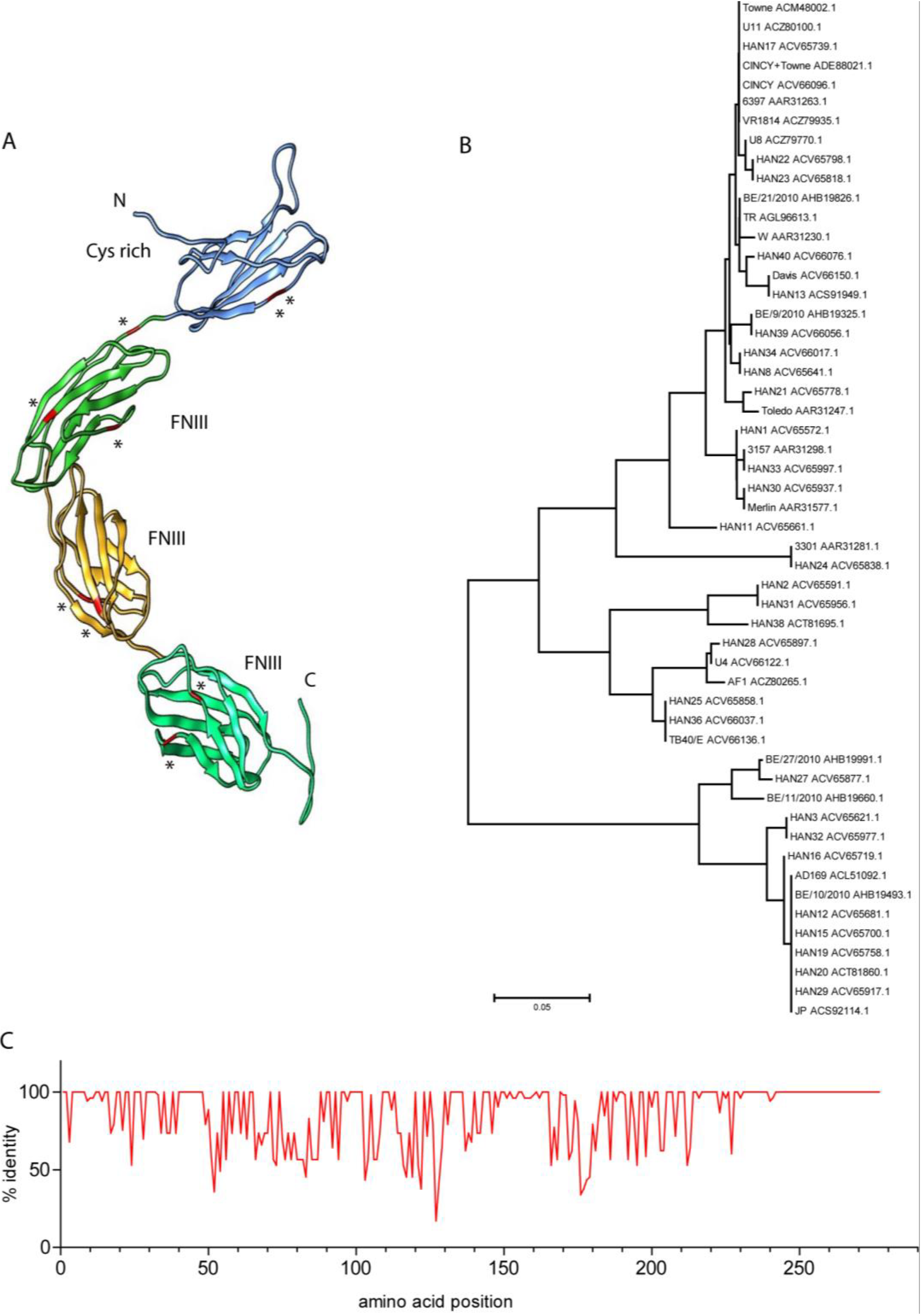
  43. Sekulin, K.; Görzer, I.; Heiss-Czedik, D.; Puchhammer-Stöckl, E. Analysis of the variability of CMV strains in the RL11D domain of the RL11 multigene family. Virus Genes 2007, 35, 577–583. [Google Scholar] [CrossRef] [PubMed]
  44. Hitomi, S.; Kozuka-Hata, H.; Chen, Z.; Sugano, S.; Yamaguchi, N.; Watanabe, S. Human cytomegalovirus open reading frame UL11 encodes a highly polymorphic protein expressed on the infected cell surface. Arch. Virol. 1997, 142, 1407–1427. [Google Scholar] [CrossRef] [PubMed]
  45. Gabaev, I.; Elbasani, E.; Ameres, S.; Steinbrück, L.; Stanton, R.; Döring, M.; Lenac-Rovis, T.; Kalinke, U.; Jonjic, S.; Moosmann, A.; et al. Expression of the Human Cytomegalovirus UL11 Glycoprotein in Viral Infection and Evaluation of Its Effect on Virus-Specific CD8 T Cells. J. Virol. 2014, 88, 14326–14339. [Google Scholar] [CrossRef] [PubMed]
  46. Jahn, G.; Stenglein, S.; Riegler, S.; Einsele, H.; Sinzger, C. Human cytomegalovirus infection of immature dendritic cells and macrophages. Intervirology 1999, 42, 365–372. [Google Scholar] [CrossRef] [PubMed]
  47. Deryckere, F.; Burgert, H.G. Early region 3 of adenovirus type 19 (subgroup D) encodes an HLA-binding protein distinct from that of subgroups B and C. J. Virol. 1996, 70, 2832–2841. [Google Scholar] [PubMed]
  48. Anand, A.R.; Ganju, R.K. HIV-1 gp120-mediated apoptosis of T cells is regulated by the membrane tyrosine phosphatase CD45. J. Biol. Chem. 2006, 281, 12289–12299. [Google Scholar] [CrossRef] [PubMed]
  49. Okoye, A.A.; Picker, L.J. CD4(+) T-cell depletion in HIV infection: Mechanisms of immunological failure. Immunol. Rev. 2013, 254, 54–64. [Google Scholar] [CrossRef] [PubMed]
  50. Dupere-Minier, G.; Desharnais, P.; Bernier, J. Involvement of tyrosine phosphatase CD45 in apoptosis. Apoptosis. 2010, 15, 1–13. [Google Scholar] [CrossRef] [PubMed]
  51. Earl, L.A.; Baum, L.G. CD45 glycosylation controls T-cell life and death. Immunol. Cell Biol. 2008, 86, 608–615. [Google Scholar] [CrossRef] [PubMed]
  52. Grossman, Z.; Meier-Schellersheim, M.; Sousa, A.E.; Victorino, R.M.; Paul, W.E. CD4+ T-cell depletion in HIV infection: Are we closer to understanding the cause? Nat. Med. 2002, 8, 319–323. [Google Scholar] [CrossRef] [PubMed]
  53. Tchilian, E.Z.; Beverley, P.C. Altered CD45 expression and disease. Trends Immunol. 2006, 27, 146–153. [Google Scholar] [CrossRef] [PubMed]
  54. Tchilian, E.Z.; Wallace, D.L.; Dawes, R.; Imami, N.; Burton, C.; Gotch, F.; Beverley, P.C. A point mutation in CD45 may be associated with an increased risk of HIV-1 infection. AIDS 2001, 15, 1892–1894. [Google Scholar] [CrossRef] [PubMed]
  55. Stanton, T.; Boxall, S.; Bennett, A.; Kaleebu, P.; Watera, C.; Whitworth, J.; French, N.; Dawes, R.; Hill, A.V.; Bodmer, W.; et al. CD45 variant alleles: Possibly increased frequency of a novel exon 4 CD45 polymorphism in HIV seropositive Ugandans. Immunogenetics 2004, 56, 107–110. [Google Scholar] [CrossRef] [PubMed]
  56. Dawes, R.; Hennig, B.; Irving, W.; Petrova, S.; Boxall, S.; Ward, V.; Wallace, D.; Macallan, D.C.; Thursz, M.; Hill, A.; et al. Altered CD45 expression in C77G carriers influences immune function and outcome of hepatitis C infection. J. Med. Genet. 2006, 43, 678–684. [Google Scholar] [CrossRef] [PubMed]
  57. Boxall, S.; Stanton, T.; Hirai, K.; Ward, V.; Yasui, T.; Tahara, H.; Tamori, A.; Nishiguchi, S.; Shiomi, S.; Ishiko, O.; et al. Disease associations and altered immune function in CD45 138G variant carriers. Hum. Mol. Genet. 2004, 13, 2377–2384. [Google Scholar] [CrossRef] [PubMed]
  58. Barcellos, L.F.; Caillier, S.; Dragone, L.; Elder, M.; Vittinghoff, E.; Bucher, P.; Lincoln, R.R.; Pericak-Vance, M.; Haines, J.L.; Weiss, A.; et al. PTPRC (CD45) is not associated with the development of multiple sclerosis in U.S. patients. Nat. Genet. 2001, 29, 23–24. [Google Scholar] [CrossRef] [PubMed]
  59. Mazurov, D.; Ilinskaya, A.; Heidecker, G.; Filatov, A. Role of O-glycosylation and expression of CD43 and CD45 on the surfaces of effector T cells in human T cell leukemia virus type 1 cell-to-cell infection. J. Virol. 2012, 86, 2447–2458. [Google Scholar] [CrossRef] [PubMed]
  60. Graf, B.; Bushnell, T.; Miller, J. LFA-1-mediated T cell costimulation through increased localization of TCR/class II complexes to the central supramolecular activation cluster and exclusion of CD45 from the immunological synapse. J. Immunol. 2007, 179, 1616–1624. [Google Scholar] [CrossRef] [PubMed]
  61. Dustin, M.L. Signaling at neuro/immune synapses. J. Clin. Investig. 2012, 122, 1149–1155. [Google Scholar] [CrossRef] [PubMed]
  62. Jolly, C. T cell polarization at the virological synapse. Viruses 2010, 2, 1261–1278. [Google Scholar] [CrossRef] [PubMed]
  63. Igakura, T.; Stinchcombe, J.C.; Goon, P.K.; Taylor, G.P.; Weber, J.N.; Griffiths, G.M.; Tanaka, Y.; Osame, M.; Bangham, C.R. Spread of HTLV-I between lymphocytes by virus-induced polarization of the cytoskeleton. Science 2003, 299, 1713–1716. [Google Scholar] [CrossRef] [PubMed]
  64. Cheng, G.; Cox, J.; Wang, P.; Krishnan, M.N.; Dai, J.; Qian, F.; Anderson, J.F.; Fikrig, E. A C-type lectin collaborates with a CD45 phosphatase homolog to facilitate West Nile virus infection of mosquitoes. Cell 2010, 142, 714–725. [Google Scholar] [CrossRef] [PubMed]
  65. Daley-Bauer, L.P.; Roback, L.J.; Wynn, G.M.; Mocarski, E.S. Cytomegalovirus hijacks CX3CR1(hi) patrolling monocytes as immune-privileged vehicles for dissemination in mice. Cell Host. Microbe 2014, 15, 351–362. [Google Scholar] [CrossRef] [PubMed]
  66. Sinclair, J.; Reeves, M. The intimate relationship between human cytomegalovirus and the dendritic cell lineage. Front Microbiol. 2014, 5, 389. [Google Scholar] [PubMed]
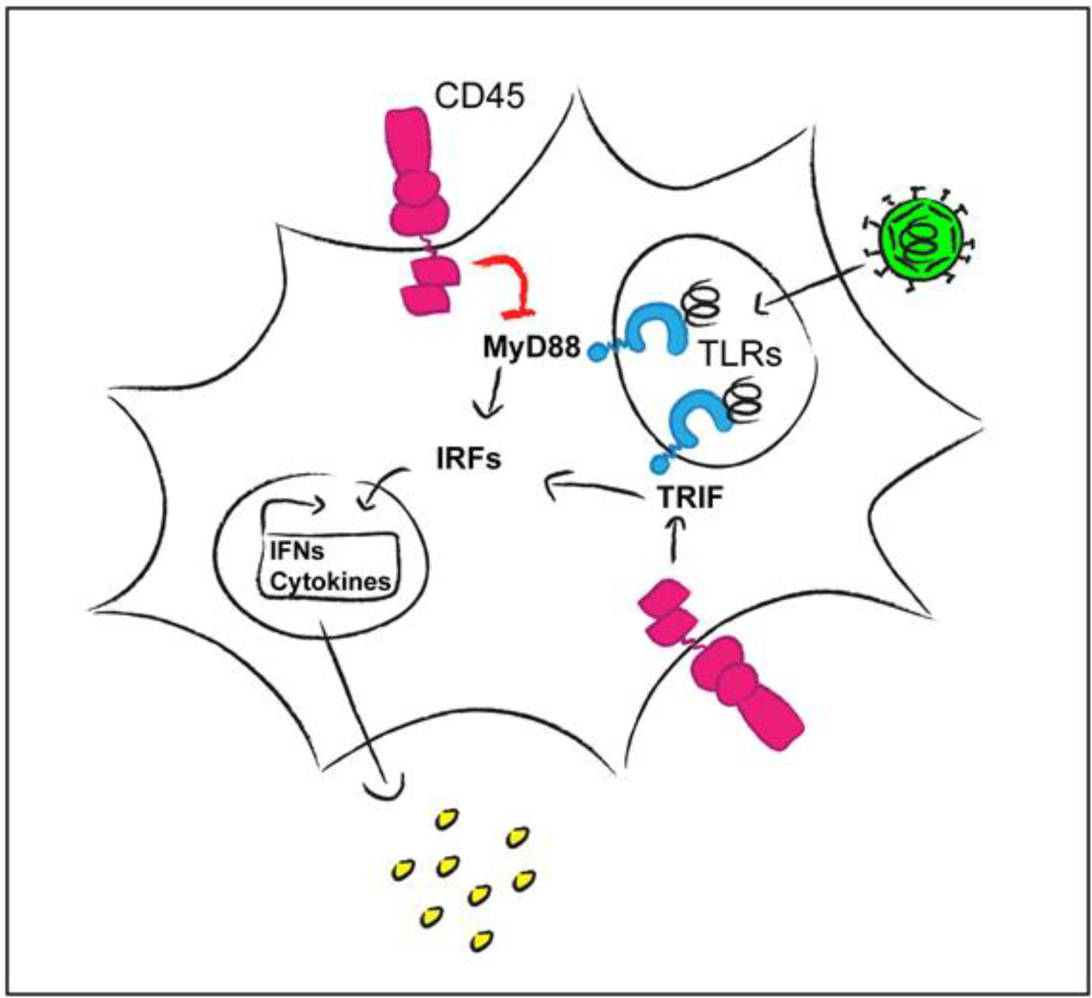
  67. Montoya, M.; Dawes, R.; Reid, D.; Lee, L.N.; Piercy, J.; Borrow, P.; Tchilian, E.Z.; Beverley, P.C. CD45 is required for type I IFN production by dendritic cells. Eur. J. Immunol. 2006, 36, 2150–2158. [Google Scholar] [CrossRef] [PubMed]
  68. Cross, J.L.; Kott, K.; Miletic, T.; Johnson, P. CD45 regulates TLR-induced proinflammatory cytokine and IFN-beta secretion in dendritic cells. J. Immunol. 2008, 180, 8020–8029. [Google Scholar] [CrossRef] [PubMed]
  69. Piercy, J.; Petrova, S.; Tchilian, E.Z.; Beverley, P.C. CD45 negatively regulates tumour necrosis factor and interleukin-6 production in dendritic cells. Immunology 2006, 118, 250–256. [Google Scholar] [CrossRef] [PubMed]
  70. Page, T.H.; Smolinska, M.; Gillespie, J.; Urbaniak, A.M.; Foxwell, B.M. Tyrosine kinases and inflammatory signalling. Curr. Mol. Med. 2009, 9, 69–85. [Google Scholar] [CrossRef] [PubMed]
  71. Irie-Sasaki, J.; Sasaki, T.; Matsumoto, W.; Opavsky, A.; Cheng, M.; Welstead, G.; Griffiths, E.; Krawczyk, C.; Richardson, C.D.; Aitken, K.; et al. CD45 is a JAK phosphatase and negatively regulates cytokine receptor signalling. Nature 2001, 409, 349–354. [Google Scholar] [CrossRef] [PubMed]
  72. Wu, L.; Bijian, K.; Shen, S.H. CD45 recruits adapter protein DOK-1 and negatively regulates JAK-STAT signaling in hematopoietic cells. Mol. Immunol. 2009, 46, 2167–2177. [Google Scholar] [CrossRef] [PubMed]
  73. Blay, R.; Simpson, K.; Leslie, K.; Huber, S. Coxsackievirus-induced disease. CD4+ cells initiate both myocarditis and pancreatitis in DBA/2 mice. Am. J. Pathol. 1989, 135, 899–907. [Google Scholar] [PubMed]
  74. Roach, T.; Slater, S.; Koval, M.; White, L.; Cahir McFarland, E.D.; Okumura, M.; Thomas, M.; Brown, E. CD45 regulates Src family member kinase activity associated with macrophage integrin-mediated adhesion. Curr. Biol. 1997, 7, 408–417. [Google Scholar] [CrossRef] [PubMed]
  75. Shenoi, H.; Seavitt, J.; Zheleznyak, A.; Thomas, M.L.; Brown, E.J. Regulation of integrin-mediated T cell adhesion by the transmembrane protein tyrosine phosphatase CD45. J. Immunol. 1999, 162, 7120–7127. [Google Scholar] [PubMed]
  76. Li, R.; Wong, N.; Jabali, M.D.; Johnson, P. CD44-initiated cell spreading induces Pyk2 phosphorylation, is mediated by Src family kinases, and is negatively regulated by CD45. J. Biol. Chem. 2001, 276, 28767–28773. [Google Scholar] [CrossRef] [PubMed]
  77. Harburger, D.S.; Calderwood, D.A. Integrin signalling at a glance. J. Cell Sci. 2009, 122, 159–163. [Google Scholar] [CrossRef] [PubMed]
  78. St Pierre, J.; Ostergaard, H.L. A role for the protein tyrosine phosphatase CD45 in macrophage adhesion through the regulation of paxillin degradation. PLoS ONE 2013, 8, e71531. [Google Scholar] [CrossRef] [PubMed]
  79. Ballingall, K.T.; Waibochi, L.; Holmes, E.C.; Woelk, C.H.; MacHugh, N.D.; Lutje, V.; McKeever, D.J. The CD45 locus in cattle: Allelic polymorphism and evidence for exceptional positive natural selection. Immunogenetics 2001, 52, 276–283. [Google Scholar] [CrossRef] [PubMed]
  80. Filip, L.C.; Mundy, N.I. Rapid evolution by positive Darwinian selection in the extracellular domain of the abundant lymphocyte protein CD45 in primates. Mol. Biol. Evol. 2004, 21, 1504–1511. [Google Scholar] [CrossRef] [PubMed]
  81. Ortiz, M.; Guex, N.; Patin, E.; Martin, O.; Xenarios, I.; Ciuffi, A.; Quintana-Murci, L.; Telenti, A. Evolutionary trajectories of primate genes involved in HIV pathogenesis. Mol. Biol. Evol. 2009, 26, 2865–2875. [Google Scholar] [CrossRef] [PubMed]
  82. Davison, A.J.; Dolan, A.; Akter, P.; Addison, C.; Dargan, D.J.; Alcendor, D.J.; McGeoch, D.J.; Hayward, G.S. The human cytomegalovirus genome revisited: Comparison with the chimpanzee cytomegalovirus genome. J. Gen. Virol. 2003, 84, 17–28. [Google Scholar] [CrossRef] [PubMed]
  83. Pal, C.; Papp, B.; Lercher, M.J. An integrated view of protein evolution. Nat. Rev. Genet. 2006, 7, 337–348. [Google Scholar] [CrossRef] [PubMed]
  84. Källberg, M.; Wang, H.; Wang, S.; Peng, J.; Wang, Z.; Lu, H.; Xu, J. Template-based protein structure modeling using the RaptorX web server. Nat. Protoc. 2012, 7, 1511–1522. [Google Scholar] [CrossRef] [PubMed]
  85. Ma, J.; Wang, S.; Zhao, F.; Xu, J. Protein threading using context-specific alignment potential. Bioinformatics. 2013, 29, i257–i265. [Google Scholar] [CrossRef] [PubMed]
  86. Meng, E.C.; Pettersen, E.F.; Couch, G.S.; Huang, C.C.; Ferrin, T.E. Tools for integrated sequence-structure analysis with UCSF Chimera. BMC Bioinformatics. 2006, 7, 339. [Google Scholar] [CrossRef] [PubMed]
  87. Tamura, K.; Stecher, G.; Peterson, D.; Filipski, A.; Kumar, S. MEGA6: Molecular Evolutionary Genetics Analysis version 6.0. Mol. Biol. Evol. 2013, 30, 2725–2729. [Google Scholar] [CrossRef] [PubMed]
  88. Waterhouse, A.M.; Procter, J.B.; Martin, D.M.; Clamp, M.; Barton, G.J. Jalview Version 2-a multiple sequence alignment editor and analysis workbench. Bioinformatics 2009, 25, 1189–1191. [Google Scholar] [CrossRef] [PubMed]
  89. Blusch, J.H.; Deryckere, F.; Windheim, M.; Ruzsics, Z.; Arnberg, N.; Adrian, T.; Burgert, H.G. The novel early region 3 protein E3/49K is specifically expressed by adenoviruses of subgenus D: Implications for epidemic keratoconjunctivitis and adenovirus evolution. Virology 2002, 296, 94–106. [Google Scholar] [CrossRef] [PubMed]
  90. Robinson, C.M.; Singh, G.; Lee, J.Y.; Dehghan, S.; Rajaiya, J.; Liu, E.B.; Yousuf, M.A.; Betensky, R.A.; Jones, M.S.; Dyer, D.W.; et al. Molecular evolution of human adenoviruses. Sci. Rep. 2013, 3, 1812. [Google Scholar] [CrossRef] [PubMed]
  91. Singh, G.; Robinson, C.M.; Dehghan, S.; Jones, M.S.; Dyer, D.W.; Seto, D.; Chodosh, J. Homologous recombination in E3 genes of human adenovirus species D. J. Virol. 2013, 87, 12481–12488. [Google Scholar] [CrossRef] [PubMed]
  92. Arnberg, N. Adenovirus E3 protein modulates leukocyte functions. Proc. Natl. Acad. Sci. USA 2013, 110, 19976–19977. [Google Scholar] [CrossRef] [PubMed]
  93. Symons, A.; Cooper, D.N.; Barclay, A.N. Characterization of the interaction between galectin-1 and lymphocyte glycoproteins CD45 and Thy-1. Glycobiology 2000, 10, 559–563. [Google Scholar] [CrossRef] [PubMed]
  94. Chen, G.; Luke, P.P.; Yang, H.; Visser, L.; Sun, H.; Garcia, B.; Qian, H.; Xiang, Y.; Huang, X.; Liu, W.; et al. Anti-CD45RB monoclonal antibody prolongs renal allograft survival in cynomolgus monkeys. Am. J. Transplant. 2007, 7, 27–37. [Google Scholar] [CrossRef] [PubMed]

