Correction: Ci et al. Enhanced Delivery of Imatinib into Vaginal Mucosa via a New Positively Charged Nanocrystal-Loaded in Situ Hydrogel Formulation for Treatment of Cervical Cancer. Pharmaceutics 2019, 11, 15

 and
and 
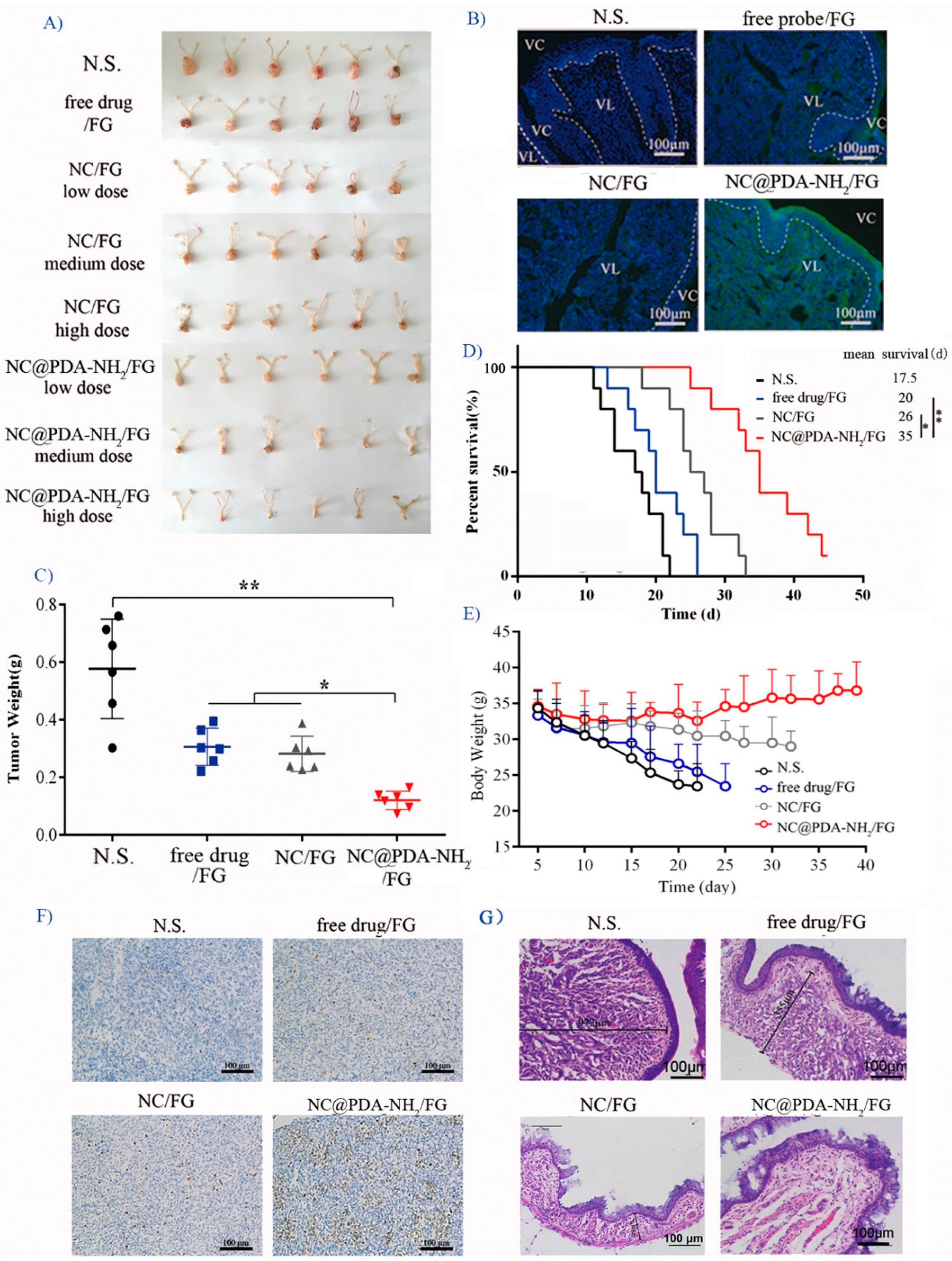
1. Error in Figure
Reference
- Ci, L.-q.; Huang, Z.-g.; Lv, F.-m.; Wang, J.; Feng, L.-l.; Sun, F.; Cao, S.-j.; Liu, Z.-p.; Liu, Y.; Wei, G.; et al. Enhanced Delivery of Imatinib into Vaginal Mucosa via a New Positively Charged Nanocrystal-Loaded in Situ Hydrogel Formulation for Treatment of Cervical Cancer. Pharmaceutics 2019, 11, 15. [Google Scholar] [CrossRef] [PubMed]


Disclaimer/Publisher’s Note: The statements, opinions and data contained in all publications are solely those of the individual author(s) and contributor(s) and not of MDPI and/or the editor(s). MDPI and/or the editor(s) disclaim responsibility for any injury to people or property resulting from any ideas, methods, instructions or products referred to in the content. |
© 2023 by the authors. Licensee MDPI, Basel, Switzerland. This article is an open access article distributed under the terms and conditions of the Creative Commons Attribution (CC BY) license (https://creativecommons.org/licenses/by/4.0/).
Share and Cite
Ci, L.-q.; Huang, Z.-g.; Lv, F.-m.; Wang, J.; Feng, L.-l.; Sun, F.; Cao, S.-j.; Liu, Z.-p.; Liu, Y.; Wei, G.; et al. Correction: Ci et al. Enhanced Delivery of Imatinib into Vaginal Mucosa via a New Positively Charged Nanocrystal-Loaded in Situ Hydrogel Formulation for Treatment of Cervical Cancer. Pharmaceutics 2019, 11, 15. Pharmaceutics 2023, 15, 2188. https://0-doi-org.brum.beds.ac.uk/10.3390/pharmaceutics15092188
Ci L-q, Huang Z-g, Lv F-m, Wang J, Feng L-l, Sun F, Cao S-j, Liu Z-p, Liu Y, Wei G, et al. Correction: Ci et al. Enhanced Delivery of Imatinib into Vaginal Mucosa via a New Positively Charged Nanocrystal-Loaded in Situ Hydrogel Formulation for Treatment of Cervical Cancer. Pharmaceutics 2019, 11, 15. Pharmaceutics. 2023; 15(9):2188. https://0-doi-org.brum.beds.ac.uk/10.3390/pharmaceutics15092188
Chicago/Turabian StyleCi, Li-qian, Zhi-gang Huang, Feng-mei Lv, Jun Wang, Ling-lin Feng, Feng Sun, Shui-juan Cao, Zhe-peng Liu, Yu Liu, Gang Wei, and et al. 2023. "Correction: Ci et al. Enhanced Delivery of Imatinib into Vaginal Mucosa via a New Positively Charged Nanocrystal-Loaded in Situ Hydrogel Formulation for Treatment of Cervical Cancer. Pharmaceutics 2019, 11, 15" Pharmaceutics 15, no. 9: 2188. https://0-doi-org.brum.beds.ac.uk/10.3390/pharmaceutics15092188