Next Article in Journal
Fatty Acid Indices and the Nutritional Properties of Karakul Sheep Meat
Next Article in Special Issue
Oral intake of Lactiplantibacillus pentosus LPG1 Produces a Beneficial Regulation of Gut Microbiota in Healthy Persons: A Randomised, Placebo-Controlled, Single-Blind Trial
Previous Article in Journal
Effects of Endurance Exercise Intensities on Autonomic and Metabolic Controls in Children with Obesity: A Feasibility Study Employing Online Exercise Training
Previous Article in Special Issue
Latilactobacillus sakei Wikim0066 Protects Skin through MMP Regulation on UVB-Irradiated In Vitro and In Vivo Model
 
 
nutrients-logo
Article Menu

Article Menu

Article

Short-Term Intake of Theobroma grandiflorum Juice Fermented with Lacticaseibacillus rhamnosus ATCC 9595 Amended the Outcome of Endotoxemia Induced by Lipopolysaccharide

by
Adrielle Zagmignan
1,2,3,*,
Yasmim Costa Mendes
1,
Gabrielle Pereira Mesquita
1,
Gabrielle Damasceno Costa dos Santos
1,
Lucas dos Santos Silva
1,
Amanda Caroline de Souza Sales
1,
Simeone Júlio dos Santos Castelo Branco
1,
Alexsander Rodrigues Carvalho Junior
4,
José Manuel Noguera Bazán
1,
Edinalva Rodrigues Alves
2,
Bárbara Lima de Almeida
3,
Anne Karoline Maiorana Santos
5,
Wellyson da Cunha Araújo Firmo
2,6,
Maria Raimunda Chagas Silva
7,
Antônio José Cantanhede Filho
5,
Rita de Cássia Mendonça de Miranda
3 and
Luís Cláudio Nascimento da Silva
1
1
Laboratório de Patogenicidade Microbiana, Universidade CEUMA, São Luís 65075-120, Brazil
2
Programa de Pós-Graduação em Gestão de Serviços e Programas de Saúde, Universidade CEUMA, São Luís 65075-120, Brazil
3
Laboratório de Microbiologia Ambiental, Universidade CEUMA, São Luís 65075-120, Brazil
4
Instituto de Ciências Biomédicas, Universidade de São Paulo, São Paulo 05508-000, Brazil
5
Laboratório de Extração e Cromatografia, Instituto Federal de Educação, Ciência e Tecnologia do Maranhão, Campus Monte Castelo, São Luís 65030-005, MA, Brazil
6
Centro de Ciências da Saúde, Campus Imperatriz, Universidade Estadual da Região Tocantina do Maranhão, Imperatriz 65900-000, MA, Brazil
7
Laboratório de Química Ambiental, Universidade CEUMA, São Luís 65075-120, Brazil
*
Author to whom correspondence should be addressed.
Submission received: 3 December 2022 / Revised: 8 February 2023 / Accepted: 14 February 2023 / Published: 20 February 2023
(This article belongs to the Special Issue Role of Lactobacillus and Probiotics in Human Health and Diseases)

Abstract

:
Endotoxemia is a condition caused by increasing levels of lipopolysaccharide (LPS) characterized by an impaired systemic response that causes multiple organ dysfunction. Lacticaseibacillus rhamnosus ATCC 9595 is a strain with probiotic potential which shows immunomodulatory properties. The incorporation of this bacterium in food rich in bioactive compounds, such as cupuaçu juice (Theobroma grandiflorum), could result in a product with interesting health properties. This work evaluated the effects of the oral administration of cupuaçu juice fermented with L. rhamnosus on the outcome of LPS-induced endotoxemia in mice. C57BL/6 mice (12/group) received oral doses (100 µL) of saline solution and unfermented or fermented cupuaçu juice (108 CFU/mL). After 5 days, the endotoxemia was induced by an intraperitoneal injection of LPS (10 mg/kg). The endotoxemia severity was evaluated daily using a score based on grooming behavior, mobility, presence of piloerection, and weeping eyes. After 6 h and 120 h, the mice (6/group) were euthanized for analysis of cell counts (in peritoneal lavage and serum) and organ weight. L. rhamnosus grew in cupuaçu juice and produced organic acids without the need for supplementation. The bacteria counts were stable in the juice during storage at 4 °C for 28 days. The fermentation with L. rhamnosus ATCC 9595 changed the metabolites profile of cupuaçu juice due to the biotransformation and enhancement of some compounds. In general, the administration of L. rhamnosus-fermented juice allowed a significant improvement in several characteristics of endotoxemic status (weight loss, hypothermia, severity index, cell migration). In addition, treatment with fermented juice significantly reduced the weight of the spleen, liver, intestine, and kidneys compared to the saline-treated endotoxemic group. Taken together, our data show that short-term intake therapy of cupuaçu juice fermented with L. rhamnosus ATCC 9595 can reduce systemic inflammation in an experimental model of LPS-induced endotoxemia in mice.

1. Introduction

Lipopolysaccharide (LPS) is a structural part of the outer membrane of Gram-negative bacteria [1] that can be recognized by Toll-like Receptor 4 (TLR-4) [2,3]. Increased levels of LPS in the bloodstream are classified as endotoxemia, which may result from bacteremia and/or metabolic dysfunctions [4,5]. Metabolic endotoxemia is closely related to changes in intestinal microbiota homeostasis, due to obesity, liver damage, or other inflammatory-related chronic conditions [4,6].
Regardless of its causes, endotoxemia is a life-threatening clinical condition characterized by an impaired systemic response that provokes the dysfunction of multiple organs. Endotoxemia is a complex issue for health systems worldwide [7]. Due to the complexity of endotoxin shock physiopathology, the development of effective therapy is still a huge challenge [8].
Some evidence has shown the strong immunomodulatory effects induced by probiotic bacteria (particularly certain Lactobacillus sp. Strains) in experimental models of inflammatory disorders [9,10]. These results advocate for their alternative use in the treatment of clinical conditions related to endotoxemia [11,12]. These bacteria may modulate host response due to direct stimulation of immune cells and/or modification of gut microbiota [11,13,14]. A recent study showed that bacuri juice fermented with L. rhamnosus ATCC 9595 prolongs the lifespan of Tenebrio molitor larvae infected by enteroaggregative Escherichia coli 042 [15].
Lactobacilli are lactic acid bacteria that are normal inhabitants of the human gut and they have been also consumed as food for a long time [16]. However, most Lactobacillus-derived products are derived from milk, imposing an obstacle for some individuals (such as those with lactose intolerance, allergy to milk protein, and vegans), pointing to the importance of plant-derived material for the obtention of new probiotic products [17]. In this sense, the use of juice as a vehicle for these probiotic bacteria is appearing as a suitable alternative [18,19]. Furthermore, the availability of various bioactive compounds in juices may cooperate with the probiotic strains to promote beneficial effects for consumers [20,21].
The juice from Theobroma grandiflorum (Cupuassu or Cupuaçu) is very appreciated in Brazil in nature or industrial preparation for the production of juices, ice creams, popsicles, jams, chocolates, sweets, and is an important source of vitamins and minerals [22]; it has antioxidant properties such as vitamins C (ascorbic acid) and E (tocopherols), flavonoids, anthocyanins, and polyphenols, in addition to saccharides (glucose, fructose, and sucrose) and minerals (such as Na, K, Ca, Mg, P, Fe, Zn, and Cu) [22,23]. This work analyzed the growth of L. rhamnosus in cupuaçu juice and lactic acid production. We then evaluated the effects of the short-term intake of L. rhamnosus-fermented juice in mice submitted to endotoxemia induced by LPS.

2. Materials and Methods

2.1. Biological Materials

2.1.1. Probiotic Strain

Samples of L. rhamnosus ATCC 9595 are kept refrigerated at −80 °C. For each experiment, aliquots were activated in MRS broth (De Man, Rogosa, and Sharpe).

2.1.2. Pulp Characterization

The fruits of T. grandiflorum were obtained in the city of São Luís (Maranhão, Brazil). A sample of the plant (branch with leaf, flower, and fruit) was sent to the Herbarium “Ático Seabra” of the Federal University of Maranhão (UFMA) for identification. The proximate composition of T. grandiflorum was analyzed in triplicate [24]. The next fraction (carbohydrates) was obtained by calculating the difference from the other fractions analyzed. The composition of fatty acids was analyzed by the Soxhlet method, while the determination of moisture and ash was performed as described in the physical–chemical methods for food analysis [25].

2.2. Fermentation

The pulp was removed and stored at −20 °C. In the first fermentation assays, the pulp sample (30 g) was dissolved in 250 mL of distilled water to reach a concentration of 120 mg/mL. The pH was adjusted to 6.0 before sterilization. In parallel, a pre-inoculum of L. rhamnosus ATCC 9595 was prepared in MRS broth. The bacterial cells were grown at 37 °C under agitation (120 rpm). After 24 h, bacterial suspension of L. rhamnosus ATCC 9595 were prepared at an optical density at 600 nm (OD600nm) of 1.0. Then, 1 mL of this suspension was inoculated in juice or MRS broth. The cultures were incubated with shaking at 120 rpm for 48 h.
The quantification of bacterial growth was performed by plating on MRS agar. Serial dilutions were made in phosphate-buffered saline (PBS) solution after each determined period (0, 7, 14, 21, and 28 days of refrigeration). The Petri dishes were then incubated at 37 °C for 48 h, and the colony-forming units (CFU) were expressed in CFU/mL.

2.3. Optimization of Cultivation Conditions

The optimization of cultivation conditions was conducted through a Central Rotational Composite Design (DCCR). Inoculum (x1) and pulp concentrations (x2) were the two selected variables for the optimization (Table 1). After each test, the following parameters were determined: (i) pH values, (ii) microbial population, (iii) the relationship between bacterial growth and pH (G/pH), and (iv) lactic acid production [26].

2.3.1. Quantification of Lactic Acid Content

The quantification of lactic acid in the fermentative liquid was performed using a Shimadzu high-performance liquid chromatograph, equipped with a quaternary pump r coupled to a degassing system (DGU-20A5r). The system holds an oven to control the column temperature (set at 28 °C) and an automatic injector (20 µL injection) with a diode array detector (SPD-M20A; range 190–800 nm). An ion exchange column (300 mm × 7.8 mm × 9 µm; Aminex® HPX-87H, Bio-Rad, Hercules, CA, USA) was used. The elution was conducted isocratically with a mobile phase composed of 5 mM H2SO4 and with a flow of 0.6 mL/min. The software used was LC-Solutions, manufactured by Shimadzu Corporation (Kyoto, Japan) [26]. For each test, the concentration of lactic acid in an unfermented juice with the same pulp concentration (unfermented controls) was also detected. The production of lactic acid (g/L) was determined by the difference between the concentration of lactic acid in each fermented liquid and its respective unfermented control. The fermentative conditions with the best results were selected for further assays.

2.3.2. Chemical Characterization of Ethyl Phases from Unfermented and Fermented Juices

The juices from the fermentative conditions with the best results were submitted to metabolites extraction using ethyl acetate as a solvent (1:1, v/v). The obtained extracts were used for chemical characterization. In this assay, the solvents used were purchased from J.T. Baker (Phillipsburg, USA), with all solvents of analytical and HPLC grades. The LC-HRMS system was composed of UHPLC, 1260 Infinity II system (Agilent, Barueri, SP, Brazil), and it was equipped with a high-resolution mass spectrometer (HRMS) containing a quadrupole time-of-flight mass analyzer (QTOF, Impact HD) with an electrospray ionization (ESI) source (Bruker Daltonics, Bremen, Germany).
The characterization was performed in LC-QqToF. Briefly, 1 mg of sample was dissolved in 1 mL of methanol/water (1:1, v/v). Samples were filtered and 20 μL aliquots were injected into the apparatus. For the analysis, a Luna C-18-HST (2.5 µm particle size; 10 × 0.21 cm) (Waters, Milford, MA, USA) analytical column was used with a mobile phase composed of water (A) and acetonitrile (B), with 0.1% v/v of formic acid added to both solvents. A linear gradient of 5-to-100% B in 20 min was used at a flow rate of 0.4 mL/min, with a temperature of 40.0 °C. The ionization experiments were carried out at negative mode [M-H]. The parameters used for the mass spectrometry ionization source were as follows: nebulizer, 4.0 bar; dry gas flow, 8.0 L min−1; dry heater temperature, 180 °C; capillary voltage, 4500 V; end plate offset, 500 V; collision cell energy, 5 eV; and full-MS scan range, m/z 100–1000.

2.4. Animal Experimentation

2.4.1. Animals

This study used male C57BL/6 mice aged 6–8 weeks and weighing 20–25 g. The animals were housed in plastic cages at room temperature (23 ± 1 °C) and submitted to a 12 h light–dark cycle. They received balanced laboratory food and water ad libitum. All in vivo procedures were conducted following the laboratory animal care standards of the CEUMA University. The protocol was approved by the Ethics Committee on the use of Animals from CEUMA University (Protocol number 68/17).

2.4.2. Short-Term Administration of L. rhamnosus-Fermented and Unfermented Cupuaçu Juice

Before the endotoxemia induction, the animals were distributed in four experimental groups that were pre-treated as follows: (i) control group (CON; n = 8) and (ii) LPS group (n = 12) received oral doses of PBS (100 µL/mouse); (iii) LPS + CUP group (n = 12) received oral doses of unfermented cupuaçu juice (100 µL/mouse); (iv) LPS + Lrh-CUP group received the L. rhamnosus-fermented juice at 108 CFU/mL (100 µL/mouse). The oral administration of each sample was performed for 5 days prior to endotoxemia induction.

2.4.3. Induction and Evaluation of LPS-Mediated Endotoxemia

Each animal received an intraperitoneal injection of LPS (10 mg/kg in saline; obtained from E. coli serotype O111:B4; Sigma-Aldrich) [27]. Every day, the body weight and temperature were recorded and compared with the data obtained before the LPS inoculation (baseline). The severity of endotoxemia was also evaluated daily using a score as reported by Mendes et al. [28], which is based on the observation of grooming behavior, mobility, presence of piloerection, and weeping eyes. The animals (n = 6/group) were euthanized after 6 h and 120 h following LPS inoculation using lethal doses of 80 mg/kg ketamine hydrochloride and 10 mg/kg xylazine hydrochloride.

2.4.4. Determination of Cell Population in the Blood and Peritoneal Cavity

The blood was collected by cardiac puncture using tubes having EDTA and aliquots were reserved for analysis of the total and differential cell population. The samples were then centrifuged (2000 rpm, 4 °C, 20 min) and the plasma samples were obtained and stored at −80 °C. In parallel, the mice were submitted to laparotomy followed by the introduction of 3 mL of EDTA (1 mM in PBS) into the abdominal cavity. The peritoneal lavage fluids (PELF) were transferred to a tube and stored at −80 °C. Total and differential measurements of the cell population were also performed in PELF samples.
The leukocytes present in each sample were counted in a Neubauer chamber under microscopy (×10) after proper dilution in Türk solution. The differential determination of polymorphonuclear (PMN) and mononuclear (MN) leukocytes was performed using a 100 µm hanging drop of the sample obtained by cytocentrifugation at 600 rpm for 10 min. The slides were Giemsa stained and 100 cells were counted by optical microscopy at 1000 × using an oil immersion.

2.5. Statistical Analysis

Data were presented as means ± standard variation (SD) or percentages. The data were analyzed using the software GraphPad Prism® (version 7.0) or Statistica®. The normality of distributions was determined by the Shapiro–Wilk test, and the differences between groups were evaluated by analysis of variance (ANOVA) followed by Tukey’s multiple comparison test using the Graph Prism 6.0 software. The values were considered significant when p < 0.05. Correlations were determined using Pearson’s coefficient (p) and classified as very strong (p ≥ 0.9), strong (0.7 ≤ p ≤ 0.89), moderate (0.5 ≤ p ≤ 0.69), weak (0.3 ≤ p ≤ 0.49), and negligible (p ≤ 0.29) [29].

3. Results

3.1. Nutritional Composition of Cupuaçu Pulp

The composition analysis (moisture, ashes content, proteins, lipids, and carbohydrates) showed that Cupuaçu pulp has a high moisture content (84.77 ± 0.2%) and dry mass (DM) of 15.23% (Table 2). The ashes content was 3.3 ± 0.9% (21.77% of DM). In addition, the pulp had 9.45% ± 0.0 of carbohydrates (62.05% of DM), 2.53% ± 0.5 of proteins (16.62% of DM), and 0.35% ± 0.0 of lipids (2.30% of DM).

3.2. Growth and Production of Lactic Acid by Lacticaseibacillus rhamnosus in Cupuaçu Juice

We then analyzed the growth of L. rhamnosus ATCC 9595 in Cupuaçu juice (120 mg/mL). This strain was able to grow in the juice without the addition of any supplement. Further, the viability of these strains was kept after 28 days of storage at 4 °C (Figure 1A). Based on these preliminary results, we evaluated the effects of inoculum concentration (x1) and the pulp concentration (x2) in the growth and organic acids production by L. rhamnosus ATCC 9595.
In all assays, L. rhamnosus ATCC 9595 produced organic acid (as seen by reductions in pH media) displaying G/pH ratios ranging from 1.36 to 2.45 (Table 3). The best results were seen in the conditions used in assays 4 and 6. These data were used to generate the surface response graph with a linearity coefficient (R2) of 0.88661 (Figure 1B). Both studied variables significantly influenced the G/pH ratios (p < 0.05); however, the inoculum concentration had the most important effect. This was also shown by p values that indicated strong (p = 0.74) and weak (p = 0.47) correlations between G/pH ratios and inoculum and pulp concentrations, respectively.
Regarding the production of lactic acid, the yields ranged from 1.32 g/L to 5.72 g/L (Table 3). The surface response showed an R2 value of 0.74475 (Figure 1C). In this case, the tested variables did not significantly impact lactic acid levels (p > 0.05). However, a moderate correlation was seen by the Pearson coefficient between inoculum concentration and lactic acid production (p = 0.65), while the correlation was weak for pulp concentration (p = 0.47). Higher levels of lactic acid were found in assays 6 and 4. We selected the conditions of assay 4 to perform the animal assays.

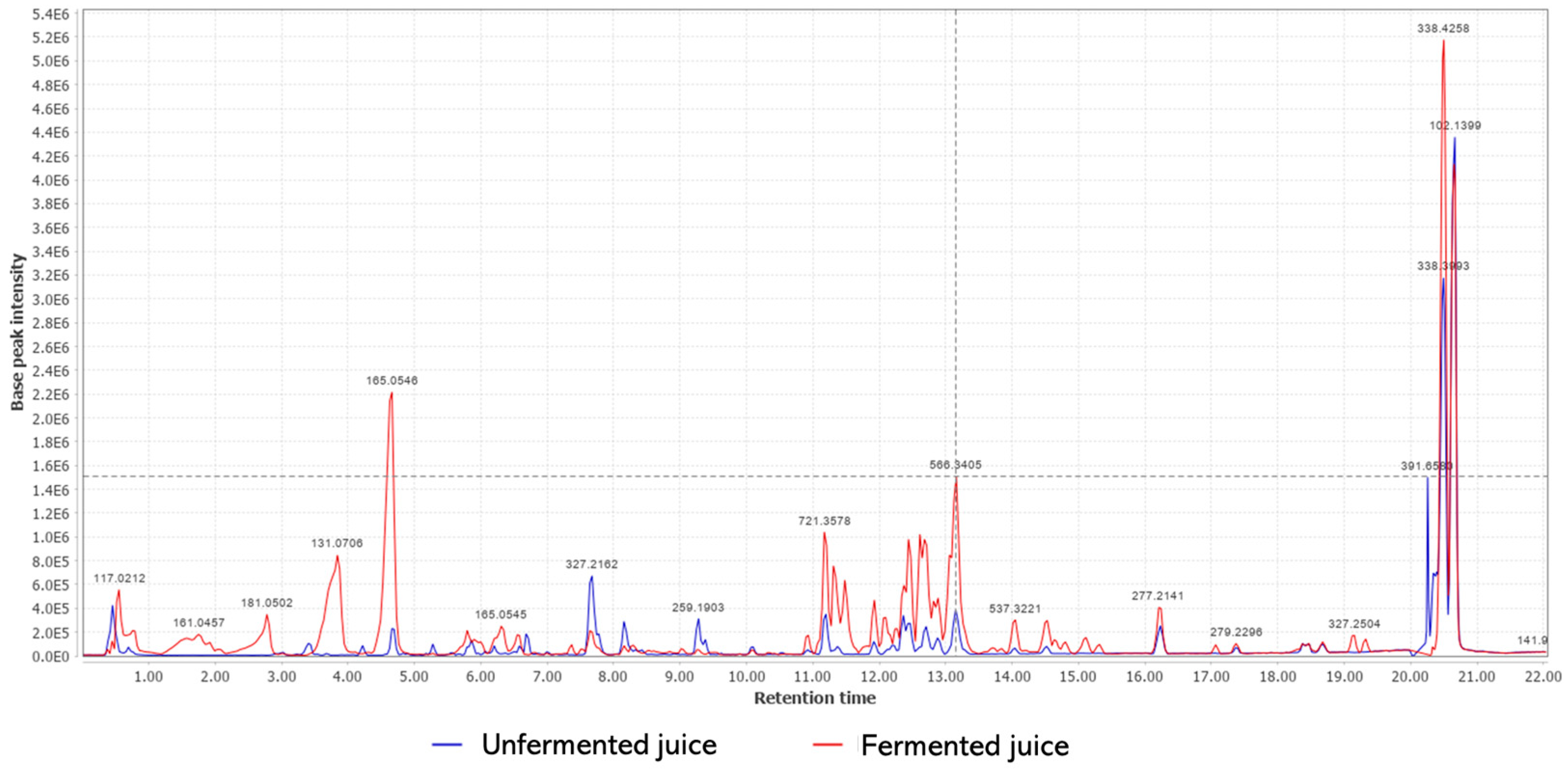
3.3. Chemical Composition

To perform the chemical characterization of fermented and unfermented juices, the samples were submitted to extraction using ethyl acetate as a solvent. It is possible to observe, by superimposing the obtained chromatograms (Figure 2), changes in the profiles of the metabolites for each sample. It indicates that some metabolites were biotransformed by L. rhamnosus. Furthermore, it is possible to verify a greater production of some metabolites in the fermented medium.
Based on the analysis of molecular ion peaks (m/z) in the obtained chromatogram and in comparison with the literature data, some metabolites differentially present in each sample were identified (Table 4). Saccharose, quinic acid, tyrosol, chlorogenic acid, and diethyl succinate were only identified in unfermented juice, while Theobromine, vanillic acid glucoside, and epicatechin-(2beta- > 5,4beta- > 6)-ent-epicatechin (or epicatechin-(2beta- > 7,4beta- > 6)-ent-epicatechin) were only detected in L. rhamnosus-fermented juice.

3.4. The Administration of L. rhamnosus-Fermented Juice Reduced the Severity of LPS-Mediated Endotoxemia

We used the median severity score (SSmedian) to evaluate the progression of the LPS-mediated endotoxemia (Figure 3A). In the first 72 h, the LPS-inoculated animals without juice treatment showed higher severity scores (SSmedian ranging from 6.0 to 9.0) than the healthy mice (those that received only PBS) (SSmedian= 4.0). After 72 h, all the groups exhibited SSmedian equal to 4. The short-time intake of both unfermented and L. rhamnosus-fermented juice showed a score significantly lower (p < 0.0001) than the untreated endotoxemic group in this period. Importantly, the treatment with L. rhamnosus-fermented juice (SSmedian= 4.0) also significantly improved the outcome of disease compared to the group treated with unfermented cupuaçu juice (SSmedian= 5.0) (p < 0.0001). These positive effects of fermentation are seen by the analysis of data from the calculation of the Area Under Curve (AUC) (Figure 3B).

3.5. The Administration of L. rhamnosus-Fermented Juice Reduced the Weight Decrease Associated with Endotoxemia Induced by LPS

The induction of endotoxemia was also confirmed by the measurement of body weight and temperature. As expected, the animals had a marked reduction in body weight (Figure 3C,D) and hypothermia (Figure 3E,F) after the LPS injection. Regarding body weight, the maximum reduction was detected after 48 h for all groups (ranging from 13.58% to 17.63%). The mice treated with L. rhamnosus-fermented juice showed the lowest values of body weight reduction in all evaluated periods; however, significant differences were only detected after 48 h (p < 0.05), 72 h, and 96 h (p < 0.01 for both) compared with endotoxemic animals (Figure 3C). The data from AUC analysis confirmed these beneficial effects of fermented cupuaçu juice (Figure 3D). The treatment using unfermented cupuaçu juice did not affect body weight decrease.
The three groups exposed to endotoxemia presented higher levels of hypothermia compared with healthy animals (Figure 3E,F). However, significant differences were only detected among the control groups (LPS-untreated vs. healthy animals). Although the mouse treated with L. rhamnosus-fermented juice displayed lower levels of hypothermia, it was not possible to observe significant differences with the other experimental groups.

3.6. The Administration of L. rhamnosus-Fermented Juice Amended the Weight Reduction of Some Organs Associated with Endotoxemia Induced by LPS

The weight of some organs was also evaluated after 6 h of endotoxemia induced by LPS (Figure 4). The endotoxemia was associated with significant reductions in the weight of spleens (Figure 4A; p < 0.0001), liver (Figure 4B; p < 0.0001), gut (Figure 4C; p < 0.05), and kidneys (Figure 4D; p < 0.0001). The treatment with L. rhamnosus-fermented juice significantly reduced the weight of the spleen (Figure 4A; p < 0.0001), liver (Figure 4B; p < 0.01), gut (Figure 4C; p < 0.05), and kidneys (Figure 4D; p < 0.0001) compared with the endotoxemic group. In turn, mice treated with unfermented juice also exhibited reductions in the spleen (Figure 4A; p < 0.0001) and kidney weights (Figure 4D; p < 0.0001). Other organs (lung, brain, stomach) did not present significant differences after 6 h of endotoxemia induction. No treatment-related alteration was observed for organ weights after 120 h.

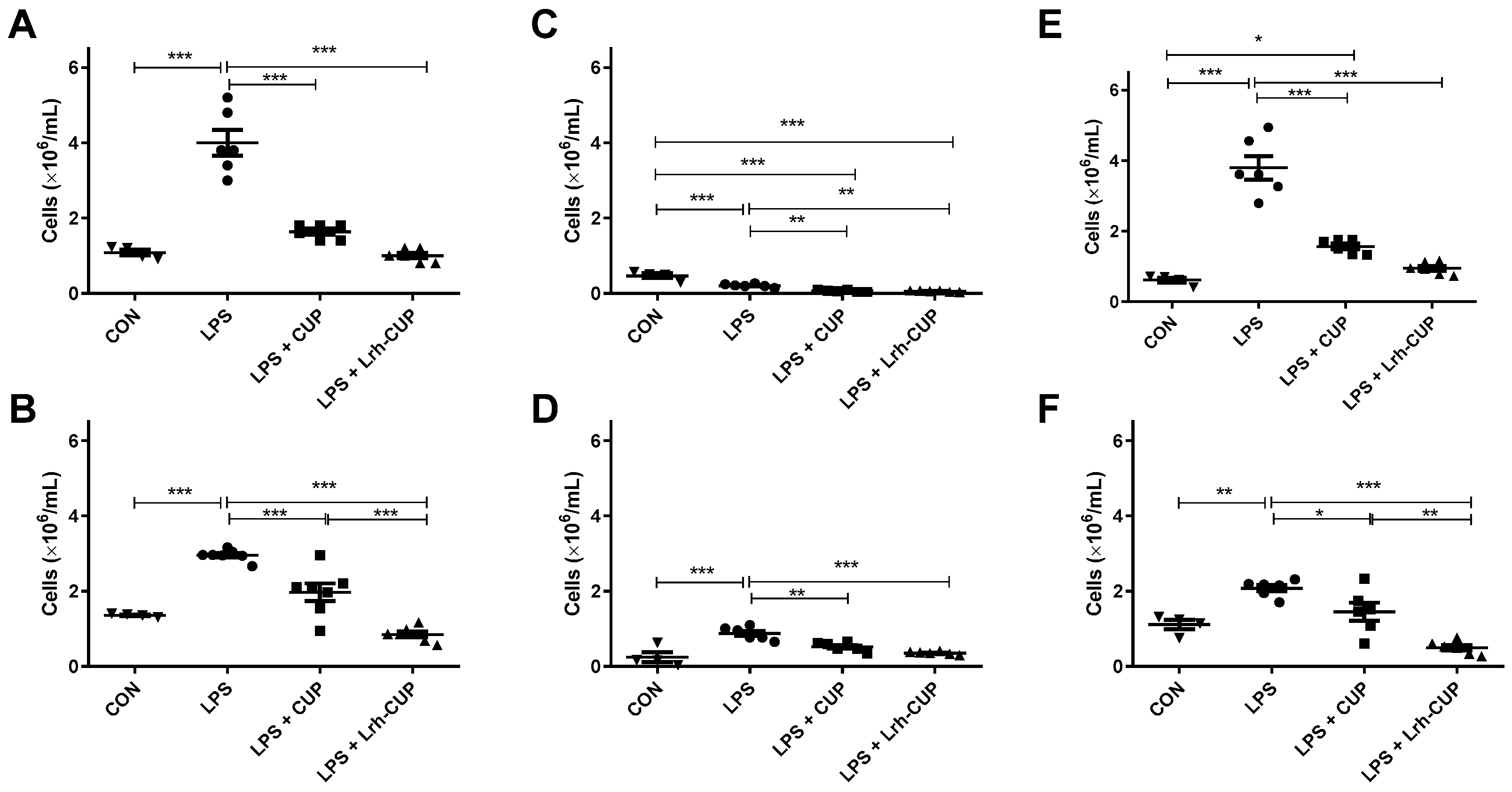
3.7. The Administration of L. rhamnosus-Fermented Juice Amended the Migration of Cells to the Peritoneal Cavity in Mice Submitted to Endotoxemia

The peritonitis was then evaluated by the migration of cells to the peritoneal cavity (Figure 5). As expected, the endotoxemia induction significantly increased the number of leukocytes in relation to healthy animals (3.49-fold and 5.21-fold increases for 6 h and 120 h, respectively) (Figure 5A,B). The peritoneal fluids of endotoxemic mice also exhibited higher counts of leukocytes after 6 h and 120 h of LPS injection than the other animals treated with L. rhamnosus-fermented or unfermented juices (Figure 5).
After 6 h, the total number of cells in PELF were reduced 3.39-fold by the short-term use of L. rhamnosus-fermented juice (p < 0.0001) and 1.97-fold (p < 0.01) in mice that received unfermented juice (Figure 5A). Similar results were observed after 120 h of endotoxemia induction, with 3.45-fold and 1.79-fold reductions for groups administrated with fermented and unfermented juices, respectively (Figure 5B). At this period, the administration of L. rhamnosus-fermented juice significantly reduced the number of cells in the peritoneal cavity concerning the treatment with unfermented juice (p < 0.0001).
Consequently, the administration of L. rhamnosus-fermented juice also significantly reduced the number of PMN cells in both evaluated periods compared to untreated endotoxemic mice (p < 0.0001; 7.57-fold and 4.68-fold after 6 h and 120 h, respectively) (Figure 5C,D). Mice that received unfermented juice also displayed lower levels of PMN cells than LPS-untreated groups, with 1.84-fold and 1.51-fold reductions after 6 h (p < 0.01) and 120 h, respectively. Significant differences between the number of PMN leukocytes among the groups treated with unfermented and fermented juices were only observed after 6 h of endotoxemia induction (Figure 5C; p < 0.05).
The groups that received fermented and unfermented juices also had lower levels of MN leukocyte migration to the peritoneal cavity after 6 h and 120 h of LPS inoculation than untreated endotoxemic animals (Figure 5E,F). Again, higher rates of inhibition were seen for L. rhamnosus-fermented juice (2.91-fold and 3.37-fold reductions; p < 0.0001) than unfermented juice (2.03-fold and 1.81-fold reductions; p < 0.0001). Regarding the treatment, statistical differences were only observed after 120 h (Figure 5F; p < 0.01).

3.8. The Administration of L. rhamnosus-Fermented Juice Reduced the Increase of Cells in the Blood Associated with LPS-Mediated Endotoxemia

We also evaluated the effects of fermented and unfermented cupuaçu juices on the population of cells in the blood (Figure 6). The blood of endotoxemic animals showed a higher number of circulating leukocytes than all other groups in the analyzed periods (p < 0.0001 for all groups) (Figure 6A,B). The reductions were higher in the mice treated with L. rhamnosus-fermented juice (4-fold and 3.47-fold) than those treated with unfermented juice (2.45-fold and 1.50-fold). However, significant differences among the groups that received unfermented and fermented juices were only seen after 120 h (Figure 6B; p < 0.0001).
The treatment with both fermented and unfermented juices significantly reduced the levels of PMN leukocytes in the blood in relation to endotoxemic mice (Figure 6C, D; p < 0.0001). Although the reductions in PMN cells were higher in mice treated with fermented juice (3.78-fold and 2.46-fold after 6 h and 120 h of endotoxemia induction, respectively) than in those that received unfermented juice (3.19-fold after 6 h and 1.68-fold after 120 h), no significant differences were detected between both types of treatment.
Finally, we analyzed the alterations in MN leukocytes in the blood of mice submitted to each treatment schedule. We observed higher inhibitory effects for both types of treatment after 6 h of LPS-induction, with 4.01-fold and 2.42-fold reductions for mice treated with fermented and unfermented juices, respectively (Figure 6E; p < 0.0001). After 120 h (Figure 6F), the mice that received L. rhamnosus-fermented juice had a lower number of MN leukocytes in the blood in relation to both endotoxemic groups (4.20-fold reduction; p < 0.0001) and unfermented-treated mice (p < 0.01). The treatment with unfermented mice resulted in a 1.43-fold reduction compared with endotoxemic mice (p < 0.05).

4. Discussion

Despite many efforts, the harmful consequences of endotoxemia remain among the leading causes of mortality [34,35]. This scenario highlights the urgent need for new alternatives to prevent and/or treat this condition [8,36]. Some probiotic strains with immunomodulatory properties are indicated as attractive candidates for the management of sepsis [11,12]. Herein, we show the beneficial effects of the short-term intake of L. rhamnosus-fermented cupuaçu juice in a model of septic shock.
In the first stage of this study, we evaluated whether cupuaçu juice could support the growth of L. rhamnosus ATCC 9595. The juice was suitable for bacteria growth and production of organic acids (such as lactic acid) without any supplementation. These results are similar to those recently reported for the fermentation of bacuri juice by L. rhamnosus ATCC 9595 [15]. Several studies have proposed that fruit juices are considered excellent food matrices for probiotics [18,26,37,38,39]. On the other hand, fermentation can improve the activity of the phytochemicals present in the juice [18,19].
Indeed, the fermentation with L. rhamnosus ATCC 9595 changed the metabolites profile of cupuaçu juice due to the biotransformation and enhancement of some compounds. For instance, the production of lactic acid was significantly increased by fermentation. The increase of lactic acid content has been reported after the fermentation of bacuri juice by L. rhamnosus ATCC 9595 [15] and passion fruit by L. rhamnosus ATCC 7469 [26]. We have also showed that some compounds were specifically found in cupuaçu juice fermented by L. rhamnosus ATCC 9595. The biotransformative properties have been described for lactic acid bacteria during the fermentation of blueberry [18] and jujube juices [19].
We then evaluated whether the administration of cupuaçu fermented juice could improve the outcome of endotoxemic shock. Animal models that mimic the metabolic alterations seen in endotoxemic individuals are typically used for the preclinical evaluation of new therapeutic candidates [1], even though all these models have advantages and disadvantages regarding translational applications [8,40]. An example of an experimental model commonly used for mimicking endotoxemic shock is the peritoneal administration of LPS, resulting in an exaggerated inflammatory response seen in endotoxin shock, followed by dysregulation of several organs [41,42].
Our results showed that both unfermented and fermented cupuaçu juice were effective in reducing septic shock, due to their anti-inflammatory effects. Some constituents previously reported in cupuaçu juice (such as ascorbic acid (vitamin C), tocopherols (vitamin E), flavonoids, anthocyanins, and polyphenols) have antioxidant, immunomodulatory, anti-angiogenic, and anti-proliferative activities [22,23,43] Among the tested groups, the higher effects were observed for mice treated by L. rhamnosus-fermented juice. These effects may be related to compounds produced during the fermentation and the beneficial properties of L. rhamnosus ATCC 9595. Theobromine, one of the compounds specifically found in fermented juice, is a caffeine derivative which is also reported as an anti-inflammatory agent [44,45].
Furthermore, some L. rhamnosus strains have shown the ability to modulate the host immune system in different clinical situations [46,47]. The probiotic strains can also adhere to mucous membranes and protect against microbial infections that could be related to LPS-induced dysbiosis [48,49]. Additionally, L. rhamnosus strains can relieve hypersensitivity reactions and intestinal inflammation [50,51], and are also indicated as an adjuvant in cases of neoplasms, eczema, diarrhea, lactose intolerance, intestinal inflammation, and infections of the vaginal and urinary tracts [52,53,54].
Concerning L. rhamnosus ATCC 9595, this strain increased the survival time of Galleria mellonella larvae infected with Candida albicans. This action was related to the inhibition of fungal virulence factors and modulation of the immune system, as seen by the recruitment of hemocytes for hemolymph [55]. Moreover, exopolysaccharides (EPS) produced by L. rhamnosus ATCC 9595 were able to exhibit immunosuppressive activity when tested on peritoneal macrophages of mice stimulated with LPS. The treatment with EPS induced the production of elevated levels of IL-10 and reduced the secretion of TNF-α [56].
Taken together, the data obtained in this study showed that cupuaçu juice is a suitable vehicle for L. rhamnosus ATCC 9595, which showed high viability in the optimized conditions. During the fermentation, L. rhamnosus ATCC 9595 produced lactic acid without the need of any supplementation. Indeed, the fermentation changed the metabolites profile of cupuaçu juice due to the biotransformation and enhancement of some compounds. Moreover, the short-term administration of L. rhamnosus-fermented cupuaçu juice reduced the systemic inflammation induced by LPS in mice. These results may help in the development of probiotic therapies for the treatment and prevention of sepsis.

Author Contributions

Conceptualization, A.Z., A.J.C.F., R.d.C.M.d.M. and L.C.N.d.S.; Formal analysis, A.Z., R.d.C.M.d.M. and L.C.N.d.S.; Funding acquisition, A.Z. and L.C.N.d.S.; Investigation, A.Z., Y.C.M., G.P.M., G.D.C.d.S., L.d.S.S., A.R.C.J., A.C.d.S.S., A.K.M.S., B.L.d.A., S.J.d.S.C.B., J.M.N.B., E.R.A., W.d.C.A.F., A.J.C.F. and M.R.C.S.; Writing—original draft preparation, A.Z., Y.C.M., A.J.C.F. and R.d.C.M.d.M.; Writing—review and editing, L.C.N.d.S. All authors have read and agreed to the published version of the manuscript.

Funding

This research was funded by Fundação de Amparo à Pesquisa e ao Desenvolvimento Científico e Tecnológico do Maranhão (FAPEMA) (Grant numbers: UNIVERSAL 01011/17; INFRA-02032/21; POS-GRAD-02460/21; ACT 05691/21 FAPEMA/CAPES) and Conselho Nacional de Desenvolvimento Científico e Tecnológico (Grant number: 312349/2020-3).

Institutional Review Board Statement

The study was conducted following the rules of the Ethics Committee on the Use of Animals from CEUMA University (Protocol number 68/17).

Informed Consent Statement

Not applicable.

Data Availability Statement

Not applicable.

Acknowledgments

The authors would like to thank Valério Monteiro Neto for sharing the probiotic strain with us. We also thank the staff of the animal facility care of Universidade CEUMA.

Conflicts of Interest

The authors declare no conflict of interest.

References

  1. Kamisoglu, K.; Haimovich, B.; Calvano, S.E.; Coyle, S.M.; Corbett, S.A.; Langley, R.J.; Kingsmore, S.F.; Androulakis, I.P. Human Metabolic Response to Systemic Inflammation: Assessment of the Concordance between Experimental Endotoxemia and Clinical Cases of Sepsis/SIRS. Crit. Care 2015, 19, 71. [Google Scholar] [CrossRef] [PubMed] [Green Version]
  2. Cochet, F.; Peri, F. The Role of Carbohydrates in the Lipopolysaccharide (LPS)/Toll-Like Receptor 4 (TLR4) Signalling. Int. J. Mol. Sci. 2017, 18, 2318. [Google Scholar] [CrossRef] [Green Version]
  3. Bertani, B.; Ruiz, N. Function and Biogenesis of Lipopolysaccharides. EcoSal Plus 2018, 8, 1–19. [Google Scholar] [CrossRef] [PubMed]
  4. Fuke, N.; Nagata, N.; Suganuma, H.; Ota, T. Regulation of Gut Microbiota and Metabolic Endotoxemia with Dietary Factors. Nutrients 2019, 11, 2277. [Google Scholar] [CrossRef] [PubMed] [Green Version]
  5. Moludi, J.; Maleki, V.; Jafari-Vayghyan, H.; Vaghef-Mehrabany, E.; Alizadeh, M. Metabolic Endotoxemia and Cardiovascular Disease: A Systematic Review about Potential Roles of Prebiotics and Probiotics. Clin. Exp. Pharmacol. Physiol. 2020, 47, 927–939. [Google Scholar] [CrossRef] [PubMed] [Green Version]
  6. Satokari, R. High Intake of Sugar and the Balance between Pro- and Anti-Inflammatory Gut Bacteria. Nutrients 2020, 12, 1348. [Google Scholar] [CrossRef]
  7. Koutroulis, I.; Batabyal, R.; McNamara, B.; Ledda, M.; Hoptay, C.; Freishtat, R.J. Sepsis Immunometabolism: From Defining Sepsis to Understanding How Energy Production Affects Immune Response. Crit Care Explor. 2019, 1, e0061. [Google Scholar] [CrossRef]
  8. Cavaillon, J.; Singer, M.; Skirecki, T. Sepsis Therapies: Learning from 30 Years of Failure of Translational Research to Propose New Leads. EMBO Mol. Med. 2020, 12, e10128. [Google Scholar] [CrossRef]
  9. Abdo, Z.; LeCureux, J.; LaVoy, A.; Eklund, B.; Ryan, E.P.; Dean, G.A. Impact of Oral Probiotic Lactobacillus Acidophilus Vaccine Strains on the Immune Response and Gut Microbiome of Mice. PloS ONE 2019, 14, e0225842. [Google Scholar] [CrossRef] [Green Version]
  10. Sireswar, S.; Biswas, S.; Dey, G. Adhesion and Anti-Inflammatory Potential of Lactobacillus Rhamnosus GG in a Sea Buckthorn Based Beverage Matrix. Food Funct. 2020, 11, 2555–2572. [Google Scholar] [CrossRef]
  11. Ávila, P.R.M.; Michels, M.; Vuolo, F.; Bilésimo, R.; Burger, H.; Milioli, M.V.M.; Sonai, B.; Borges, H.; Carneiro, C.; Abatti, M.; et al. Protective Effects of Fecal Microbiota Transplantation in Sepsis Are Independent of the Modulation of the Intestinal Flora. Nutrition 2020, 73, 110727. [Google Scholar] [CrossRef]
  12. Haro, C.; Mónaco, M.E.; Medina, M. Lactobacillus Casei Beneficially Modulates Immuno-Coagulative Response in an Endotoxemia Model. Blood Coagul. Fibrinolysis 2018, 29, 104–110. [Google Scholar] [CrossRef] [PubMed]
  13. Nation, M.L.; Dunne, E.M.; Joseph, S.J.; Mensah, F.K.; Sung, V.; Satzke, C.; Tang, M.L.K. Impact of Lactobacillus Reuteri Colonization on Gut Microbiota, Inflammation, and Crying Time in Infant Colic. Sci. Rep. 2017, 7, 15047. [Google Scholar] [CrossRef] [PubMed] [Green Version]
  14. Mao, J.; Qi, S.; Cui, Y.; Dou, X.; Luo, X.M.; Liu, J.; Zhu, T.; Ma, Y.; Wang, H. Lactobacillus Rhamnosus GG Attenuates Lipopolysaccharide-Induced Inflammation and Barrier Dysfunction by Regulating MAPK/NF-ΚB Signaling and Modulating Metabolome in the Piglet Intestine. J. Nutr. 2020, 150, 1313–1323. [Google Scholar] [CrossRef]
  15. Mendes, Y.C.; Mesquita, G.P.; Costa, G.D.E.; Barbosa da Silva, A.C.; Gouveia, E.; Silva, M.R.C.; Monteiro-Neto, V.; de Miranda, R.d.C.M.; Nascimento da Silva, L.C.; Zagmignan, A. Evaluation of Growth, Viability, Lactic Acid Production and Anti-Infective Effects of Lacticaseibacillus Rhamnosus ATCC 9595 in Bacuri Juice (Platonia Insignis). Foods 2021, 10, 603. [Google Scholar] [CrossRef] [PubMed]
  16. de Sousa, M.A.; Rama, G.R.; Volken de Souza, C.F.; Granada, C.E. Acid Lactic Lactobacilli as a Biotechnological Toll to Improve Food Quality and Human Health. Biotechnol. Prog. 2020, 36, e2937. [Google Scholar] [CrossRef] [PubMed]
  17. Kim, J.; Choi, K.B.; Park, J.H.; Kim, K.H. Metabolite Profile Changes and Increased Antioxidative and Antiinflammatory Activities of Mixed Vegetables after Fermentation by Lactobacillus Plantarum. PLoS ONE 2019, 14, e0217180. [Google Scholar] [CrossRef] [PubMed] [Green Version]
  18. Li, S.; Tao, Y.; Li, D.; Wen, G.; Zhou, J.; Manickam, S.; Han, Y.; Chai, W.S. Fermentation of Blueberry Juices Using Autochthonous Lactic Acid Bacteria Isolated from Fruit Environment: Fermentation Characteristics and Evolution of Phenolic Profiles. Chemosphere 2021, 276, 130090. [Google Scholar] [CrossRef]
  19. Li, T.; Jiang, T.; Liu, N.; Wu, C.; Xu, H.; Lei, H. Biotransformation of Phenolic Profiles and Improvement of Antioxidant Capacities in Jujube Juice by Select Lactic Acid Bacteria. Food Chem. 2021, 339, 127859. [Google Scholar] [CrossRef]
  20. Lu, Y.; Tan, C.W.; Chen, D.; Liu, S.Q. Potential of Three Probiotic Lactobacilli in Transforming Star Fruit Juice into Functional Beverages. Food Sci. Nutr. 2018, 6, 2141–2150. [Google Scholar] [CrossRef]
  21. Kwaw, E.; Ma, Y.; Tchabo, W.; Apaliya, M.T.; Wu, M.; Sackey, A.S.; Xiao, L.; Tahir, H.E. Effect of Lactobacillus Strains on Phenolic Profile, Color Attributes and Antioxidant Activities of Lactic-Acid-Fermented Mulberry Juice. Food Chem. 2018, 250, 148–154. [Google Scholar] [CrossRef]
  22. Pugliese, A.G.; Tomas-Barberan, F.A.; Truchado, P.; Genovese, M.I. Flavonoids, Proanthocyanidins, Vitamin C, and Antioxidant Activity of Theobroma Grandiflorum (Cupuassu) Pulp and Seeds. J. Agric. Food Chem. 2013, 61, 2720–2728. [Google Scholar] [CrossRef]
  23. Barros, H.R.d.M.; García-Villalba, R.; Tomás-Barberán, F.A.; Genovese, M.I. Evaluation of the Distribution and Metabolism of Polyphenols Derived from Cupuassu (Theobroma Grandiflorum) in Mice Gastrointestinal Tract by UPLC-ESI-QTOF. J. Funct. Foods 2016, 22, 477–489. [Google Scholar] [CrossRef]
  24. Helrich, K. Official Methods of Analysis of the Association of Official Analytical Chemists; Association of Official Analytical Chemists: Arlington, VA, USA, 1990. [Google Scholar]
  25. Zenebon, O.; Pascuet, N.S.; Tiglea, P. Métodos Físico-Químicos Para Análise de Alimentos, 4th ed.; Instituto Adolfo Lutz: São Paulo, Brazil, 2008; 1020p. [Google Scholar]
  26. Farias, N.; Soares, M.; Gouveia, E. Enhancement of the Viability of Lactobacillus Rhamnosus ATCC 7469 in Passion Fruit Juice: Application of a Central Composite Rotatable Design. LWT 2016, 71, 149–154. [Google Scholar] [CrossRef]
  27. Zhang, Y.; Zhou, F.; Wang, Z.; Li, Z.; Li, J. PNU-282987 Attenuates Intestinal Epithelial Barrier Dysfunction in LPS-Induced Endotoxemia. Inflammation 2020, 43, 417–424. [Google Scholar] [CrossRef] [PubMed]
  28. Mendes, S.J.F.; Sousa, F.I.A.B.; Pereira, D.M.S.; Ferro, T.A.F.; Pereira, I.C.P.; Silva, B.L.R.; Pinheiro, A.J.M.C.R.; Mouchrek, A.Q.S.; Monteiro-Neto, V.; Costa, S.K.P.; et al. Cinnamaldehyde Modulates LPS-Induced Systemic Inflammatory Response Syndrome through TRPA1-Dependent and Independent Mechanisms. Int. Immunopharmacol. 2016, 34, 60–70. [Google Scholar] [CrossRef]
  29. Mukaka, M. A Guide to Appropriate Use of Correlation Coefficient in Medical Research. Malawi Med. J. 2012, 24, 69–71. [Google Scholar] [CrossRef] [PubMed]
  30. D’Souza, R.N.; Grimbs, S.; Behrends, B.; Bernaert, H.; Ullrich, M.S.; Kuhnert, N. Origin-Based Polyphenolic Fingerprinting of Theobroma Cacao in Unfermented and Fermented Beans. Food Res. Int. 2017, 99, 550–559. [Google Scholar] [CrossRef]
  31. Duarte, W.F.; Dias, D.R.; Oliveira, J.M.; Teixeira, J.A.; de Almeida e Silva, J.B.; Schwan, R.F. Characterization of Different Fruit Wines Made from Cacao, Cupuassu, Gabiroba, Jaboticaba and Umbu. LWT-Food Sci. Technol. 2010, 43, 1564–1572. [Google Scholar] [CrossRef]
  32. Pereira-Caro, G.; Borges, G.; Nagai, C.; Jackson, M.C.; Yokota, T.; Crozier, A.; Ashihara, H. Profiles of Phenolic Compounds and Purine Alkaloids during the Development of Seeds of Theobroma Cacao Cv. Trinitario. J. Agric. Food Chem. 2013, 61, 427–434. [Google Scholar] [CrossRef]
  33. Porter, L.J.; Ma, Z.; Chan, B.G. Cacao Procyanidins: Major Flavanoids and Identification of Some Minor Metabolites. Phytochemistry 1991, 30, 1657–1663. [Google Scholar] [CrossRef]
  34. Neiverth, A.; Prim, L.R.; Franck, C.L.; Nisihara, R. Sepsis in Burned Adult Patients: Study of Serie of Cases in Brazil. J. Burn Care Res. 2020, 41, 900–904. [Google Scholar] [CrossRef] [PubMed]
  35. de Almeida, N.R.C.; Pontes, G.F.; Jacob, F.L.; Deprá, J.V.S.; Porto, J.P.P.; de Lima, F.R.; de Albuquerque, M.R.T.C. Analysis of Trends in Sepsis Mortality in Brazil and by Regions from 2010 to 2019. Rev. Saude Publica 2022, 56, 25. [Google Scholar] [CrossRef]
  36. van der Poll, T.; van de Veerdonk, F.L.; Scicluna, B.P.; Netea, M.G. The Immunopathology of Sepsis and Potential Therapeutic Targets. Nat. Rev. Immunol. 2017, 17, 407–420. [Google Scholar] [CrossRef] [PubMed]
  37. Liu, Y.; Chen, H.; Chen, W.; Zhong, Q.; Zhang, G.; Chen, W. Beneficial Effects of Tomato Juice Fermented by Lactobacillus Plantarum and Lactobacillus Casei: Antioxidation, Antimicrobial Effect, and Volatile Profiles. Molecules 2018, 23, 2366. [Google Scholar] [CrossRef] [Green Version]
  38. Nguyen, B.T.; Bujna, E.; Fekete, N.; Tran, A.T.M.; Rezessy-Szabo, J.M.; Prasad, R.; Nguyen, Q.D. Probiotic Beverage From Pineapple Juice Fermented With Lactobacillus and Bifidobacterium Strains. Front. Nutr. 2019, 6, 54. [Google Scholar] [CrossRef] [Green Version]
  39. Bancalari, E.; Castellone, V.; Bottari, B.; Gatti, M. Wild Lactobacillus Casei Group Strains: Potentiality to Ferment Plant Derived Juices. Foods 2020, 9, 314. [Google Scholar] [CrossRef] [Green Version]
  40. Stortz, J.A.; Raymond, S.L.; Mira, J.C.; Moldawer, L.L.; Mohr, A.M.; Efron, P.A. Murine Models of Sepsis and Trauma: Can We Bridge the Gap? ILAR J. 2017, 58, 90–105. [Google Scholar] [CrossRef] [PubMed] [Green Version]
  41. Pohlmann, S.; Scheu, S.; Ziegler, V.; Schupp, N.; Henninger, C.; Fritz, G. Hepatic Rac1 GTPase Contributes to Liver-Mediated Basal Immune Homeostasis and LPS-Induced Endotoxemia. Biochim. Biophys. Acta Mol. Cell Res. 2018, 1865, 1277–1292. [Google Scholar] [CrossRef]
  42. Fitzpatrick, S.F.; Lambden, S.; Macias, D.; Puthucheary, Z.; Pietsch, S.; Mendil, L.; McPhail, M.J.W.; Johnson, R.S. 2-Hydroxyglutarate Metabolism Is Altered in an in Vivo Model of LPS Induced Endotoxemia. Front. Physiol. 2020, 11, 147. [Google Scholar] [CrossRef] [Green Version]
  43. Rodríguez-Pérez, C.; García-Villanova, B.; Guerra-Hernández, E.; Verardo, V. Grape Seeds Proanthocyanidins: An Overview of In Vivo Bioactivity in Animal Models. Nutrients 2019, 11, 2435. [Google Scholar] [CrossRef] [PubMed] [Green Version]
  44. Cadoná, F.C.; Dantas, R.F.; de Mello, G.H.; Silva, F.P., Jr. Natural Products Targeting into Cancer Hallmarks: An Update on Caffeine, Theobromine, and (+)-Catechin. Crit. Rev. Food Sci. Nutr. 2022, 62, 7222–7241. [Google Scholar] [CrossRef]
  45. Gu, R.; Shi, Y.; Huang, W.; Lao, C.; Zou, Z.; Pan, S.; Huang, Z. Theobromine Mitigates IL-1β-Induced Oxidative Stress, Inflammatory Response, and Degradation of Type II Collagen in Human Chondrocytes. Int. Immunopharmacol. 2020, 82, 106226. [Google Scholar] [CrossRef]
  46. Costabile, A.; Bergillos-Meca, T.; Rasinkangas, P.; Korpela, K.; de Vs, W.M.; Gibson, G.R. Effects of Soluble Corn Fiber Alone or in Synbiotic Combination with Lactobacillus Rhamnosus GG and the Pilus-Deficient Derivative GG-PB12 on Fecal Microbiota, Metabolism, and Markers of Immune Function: A Randomized, Double-Blind, Placebo-Controlled, Crossover Study in Healthy Elderly (Saimes Study). Front. Immunol. 2017, 8, 1443. [Google Scholar] [CrossRef] [PubMed]
  47. Saliganti, V.; Kapila, R.; Kapila, S. Consumption of Probiotic Lactobacillus Rhamnosus (MTCC: 5897) Containing Fermented Milk Plays a Key Role in Development of the Immune System in Newborn Mice during the Suckling-Weaning Transition. Microbiol. Immunol. 2016, 60, 261–267. [Google Scholar] [CrossRef] [PubMed] [Green Version]
  48. Spacova, I.; Lievens, E.; Verhoeven, T.; Steenackers, H.; Vanderleyden, J.; Lebeer, S.; Petrova, M.I. Expression of Fluorescent Proteins in Lactobacillus Rhamnosus to Study Host-Microbe and Microbe-Microbe Interactions. Microb. Biotechnol. 2018, 11, 317–331. [Google Scholar] [CrossRef] [Green Version]
  49. Petrova, M.I.; Imholz, N.C.E.; Verhoeven, T.L.A.; Balzarini, J.; van Damme, E.J.M.; Schols, D.; Vanderleyden, J.; Lebeer, S. Lectin-Like Molecules of Lactobacillus Rhamnosus GG Inhibit Pathogenic Escherichia Coli and Salmonella Biofilm Formation. PLoS ONE 2016, 11, e0161337. [Google Scholar] [CrossRef] [Green Version]
  50. Saliganti, V.; Kapila, R.; Sharma, R.; Kapila, S. Feeding Probiotic Lactobacillus Rhamnosus (MTCC 5897) Fermented Milk to Suckling Mothers Alleviates Ovalbumin-Induced Allergic Sensitisation in Mice Offspring. Br. J. Nutr. 2015, 114, 1168–1179. [Google Scholar] [CrossRef] [PubMed] [Green Version]
  51. Yan, F.; Liu, L.; Cao, H.; Moore, D.J.; Washington, M.K.; Wang, B.; Peek, R.M.; Acra, S.A.; Polk, D.B. Neonatal Colonization of Mice with LGG Promotes Intestinal Development and Decreases Susceptibility to Colitis in Adulthood. Mucosal. Immunol. 2017, 10, 117–127. [Google Scholar] [CrossRef] [Green Version]
  52. Makrgeorgou, A.; Leonardi-Bee, J.; Bath-Hextall, F.J.; Murrell, D.F.; Tang, M.L.K.; Roberts, A.; Boyle, R.J. Probiotics for Treating Eczema. Cochrane Database Syst. Rev. 2018, 11, CD006135. [Google Scholar] [CrossRef]
  53. Panpetch, W.; Chancharoenthana, W.; Bootdee, K.; Nilgate, S.; Finkelman, M.; Tumwasorn, S.; Leelahavanichkul, A. Lactobacillus Rhamnosus L34 Attenuates Gut Translocation-Induced Bacterial Sepsis in Murine Models of Leaky Gut. Infect. Immun. 2017, 86, e00700-17. [Google Scholar] [CrossRef] [PubMed] [Green Version]
  54. Vajro, P.; Mandato, C.; Licenziati, M.R.; Franzese, A.; Vitale, D.F.; Lenta, S.; Caropreso, M.; Vallone, G.; Meli, R. Effects of Lactobacillus Rhamnosus Strain GG in Pediatric Obesity-Related Liver Disease. J. Pediatr. Gastroenterol. Nutr. 2011, 52, 740–743. [Google Scholar] [CrossRef] [PubMed] [Green Version]
  55. Ribeiro, F.C.; de Barros, P.P.; Rossoni, R.D.; Junqueira, J.C.; Jorge, A.O.C. Lactobacillus Rhamnosus Inhibits Candida Albicans Virulence Factors in Vitro and Modulates Immune System in Galleria Mellonella. J. Appl. Microbiol. 2017, 122, 201–211. [Google Scholar] [CrossRef]
  56. Bleau, C.; Monges, A.; Rashidan, K.; Laverdure, J.P.; Lacroix, M.; van Calsteren, M.R.; Millette, M.; Savard, R.; Lamontagne, L. Intermediate Chains of Exopolysaccharides from Lactobacillus Rhamnosus RW-9595M Increase IL-10 Production by Macrophages. J. Appl. Microbiol. 2010, 108, 666–675. [Google Scholar] [CrossRef] [PubMed]
Figure 1. Growth and production of lactic acid by Lacticaseibacillus rhamnosus in cupuaçu juice. (A) Survival of L. rhamnosus ATCC 9595 in the fermentations of cupuaçu juice by L. rhamnosus ATCC 9595 during the storage period. (B) Response surface obtained for RVpH as a function of pulp and inoculum concentrations in cupuaçu juice fermentations by L. rhamnosus ATCC 9595. (C) Response surface obtained for lactic acid concentration as a function of pulp and inoculum concentrations in cupuaçu juice fermentations by L. rhamnosus ATCC 9595.
Nutrients 15 01059 g001
Figure 2. Comparative chromatograms of the ethyl acetate phase of cupuaçu juice (Theobroma grandiflorum) with and without fermentation by Lacticaseibacillus rhamnosus ATCC 9595.
Nutrients 15 01059 g002
Figure 3. Effects of fermented and unfermented Theobroma grandiflorum juice on some pathologic parameters associated with LPS-mediated endotoxemia. (A) Kinects of variation of severity score; (B) Area under curve from data of severity score analysis; (C) Kinects of variation of weight loss; (D) Area under curve from data of weight loss analysis; (E) Kinects of variation of body temperature reduction; (F) Area under curve from data of body temperature reduction analysis. * Significant differences with p < 0.05; *** Significant differences with p < 0.0001. LPS: mice submitted to LPS-mediated endotoxemia; LPS + CUP: mice treated with unfermented T. grandiflorum juice and submitted to LPS-mediated endotoxemia; LPS + Lrh-CUP: mice treated with L. rhamnosus-fermented T. grandiflorum juice and submitted to LPS-mediated endotoxemia.
Nutrients 15 01059 g003
Figure 4. Effects of fermented and unfermented Theobroma grandiflorum juice on the weight of some organs of mice submitted to LPS-mediated endotoxemia. (A) Spleen; (B) Liver; (C) Gut; (D) Kidneys. * Significant differences with p < 0.05; ** Significant differences with p < 0.01; *** Significant differences with p < 0.0001. LPS: mice submitted to LPS-mediated endotoxemia; LPS + CUP: mice treated with unfermented T. grandiflorum juice and submitted to LPS-mediated endotoxemia; LPS + Lrh-CUP: mice treated with L. rhamnosus-fermented T. grandiflorum juice and submitted to LPS-mediated endotoxemia.
Nutrients 15 01059 g004
Figure 5. Effects of fermented and unfermented Theobroma grandiflorum juice in the migration of cells to the peritoneal cavity in mice submitted to LPS-mediated endotoxemia. (A) Total leukocytes in the peritoneal cavity after 6 h of LPS-mediated endotoxemia; (B) Polymorphonuclear leukocytes in the peritoneal cavity after 6 h of LPS-mediated endotoxemia; (C) Mononuclear cells in the peritoneal cavity after 6 h of LPS-mediated endotoxemia; (D) Total leukocytes in the peritoneal cavity after 120 h of LPS-mediated endotoxemia; (E) Polymorphonuclear leukocytes in the peritoneal cavity after 120 h of LPS-mediated endotoxemia; (F) Mononuclear cells in the peritoneal cavity after 120 h of LPS-mediated endotoxemia. * Significant differences with p < 0.05; ** Significant differences with p < 0.01; *** Significant differences with p < 0.0001. LPS: mice submitted to LPS-mediated endotoxemia; LPS + CUP: mice treated with unfermented T. grandiflorum juice and submitted to LPS-mediated endotoxemia; LPS + Lrh-CUP: mice treated with L. rhamnosus-fermented T. grandiflorum juice and submitted to LPS-mediated endotoxemia.
Nutrients 15 01059 g005
Figure 6. Effects of fermented and unfermented Theobroma grandiflorum juice in leukocytes population in the blood of mice submitted to LPS-mediated endotoxemia. (A) Total leukocytes in the peritoneal cavity after 6 h of LPS-mediated endotoxemia; (B) Polymorphonuclear leukocytes in the peritoneal cavity after 6 h of LPS-mediated endotoxemia; (C) Mononuclear cells in the peritoneal cavity after 6 h of LPS-mediated endotoxemia; (D) Total leukocytes in the peritoneal cavity after 120 h of LPS-mediated endotoxemia; (E) Polymorphonuclear leukocytes in the peritoneal cavity after 120 h of LPS-mediated endotoxemia; (F) Mononuclear cells in the peritoneal cavity after 120 h of LPS-mediated endotoxemia. * Significant differences with p < 0.05; ** Significant differences with p < 0.01; *** Significant differences with p < 0.0001. LPS: mice submitted to LPS-mediated endotoxemia; LPS + CUP: mice treated with unfermented T. grandiflorum juice and submitted to LPS-mediated endotoxemia; LPS + Lrh-CUP: mice treated with L. rhamnosus-fermented T. grandiflorum juice and submitted to LPS-mediated endotoxemia.
Nutrients 15 01059 g006
Table 1. Coded and decoded levels of independent variables.
Variables−1.41−10+1+1.41
Inoculum density (OD600)0.771.001.552.102.33
Pulp concentration (mg/mL)135.25163.00230.00297.00324.75
Table 2. Nutritional Composition of Cupuaçu Pulp.
AttributeMoistureAshesProteinsLipidsCarbohydrates *
Content (%)84.77 ± 0.23.3 ± 0.92.53 ± 0.050.35 ± 0.09.45 ± 0.0
% Of Dry Mass-21.7716.622.3062.05
* Calculated using the equation = 100 − (protein + lipids + ash + moisture).
Table 3. Effects of inoculum density and pulp concentration on growth and lactic acid production by L. rhamnosus ATCC 9595 in cupuaçu juice.
AssayOD600nmPulp
(mg/mL)
Growth
(CFU/mL)
Final pHLactic Acid
(g/L)
G/pH RatioG/[La] Ratio
11.00163.006.134.51.321.364.65
21.00297.007.324.43.961.661.85
32.10163.008.414.93.251.722.59
42.10297.009.073.74.982.451.82
50.77230.007.294.70.651.5511.25
62.33230.008.463.95.722.171.48
71.55135.257.4441.441.865.17
81.55324.759.434.61.132.058.34
91.55230.008.674.21.252.066.92
101.55230.009.194.51.142.048.05
Table 4. Identification of substances differentially present in the ethyl acetate phase of cupuaçu juice (Theobroma grandiflorum), with and without fermentation by Lacticaseibacillus rhamnosus ATCC 9595.
SampleRT (min)[M-H]Proposed SubstanceReference
Unfermented0.46341.1159Saccharose[30]
0.70191.0227Quinic acid[30]
3.03137.0244Tyrosol[31]
5.17353.1801Chlorogenic acid[30]
5.86173.1176Diethyl succinate[31]
Fermented2.78181.0502Theobromine[32]
8.16329.2318Vanillic acid glucoside[30]
13.84633.3425Epicatechin-(2beta- > 5,4beta- > 6)-ent-epicatechin
Epicatechin-(2beta- > 7,4beta- > 6)-ent-epicatechin
[33]
RT: retention time; [M-H]: negative mode.
Disclaimer/Publisher’s Note: The statements, opinions and data contained in all publications are solely those of the individual author(s) and contributor(s) and not of MDPI and/or the editor(s). MDPI and/or the editor(s) disclaim responsibility for any injury to people or property resulting from any ideas, methods, instructions or products referred to in the content.

