Review

Review on the Visible Light Photocatalysis for the Decomposition of Ciprofloxacin, Norfloxacin, Tetracyclines, and Sulfonamides Antibiotics in Wastewater

by
Samar Shurbaji
1,
Pham Thi Huong
2,3 and
Talal Mohammed Altahtamouni
4,*
1
Biomedical Research Center, Qatar University, Doha 2713, Qatar
2
Institute of Research and Development, Duy Tan University, DaNang 550000, Vietnam
3
Faculty of Environment and Chemical Engineering, Duy Tan University, Danang 550000, Vietnam
4
Materials Science and Technology Program, College of Arts and Sciences, Qatar University, Doha 2713, Qatar
*
Author to whom correspondence should be addressed.
Submission received: 5 December 2020 / Revised: 23 January 2021 / Accepted: 27 January 2021 / Published: 29 March 2021
(This article belongs to the Special Issue Photocatalytic Oxidation/Ozonation Processes)

Abstract

:
Antibiotics are chemical compounds that are used to kill or prevent bacterial growth. They are used in different fields, such as the medical field, agriculture, and veterinary. Antibiotics end up in wastewater, which causes the threat of developing antibacterial resistance; therefore, antibiotics must be eliminated from wastewater. Different conventional elimination methods are limited due to their high cost and effort, or incomplete elimination. Semiconductor-assisted photocatalysis arises as an effective elimination method for different organic wastes including antibiotics. A variety of semiconducting materials were tested to eliminate antibiotics from wastewater; nevertheless, research is still ongoing due to some limitations. This review summarizes the recent studies regarding semiconducting material modifications for antibiotic degradation using visible light irradiation.

1. Introduction

Benjamin Franklin said once “When the well is dry, we know the worth of water” to encourage us to value what we have before water resources become limited. Although water covers more than two-thirds of the earth’s surface, not all of the available water is accessible for use [1]. Water quality is crucial when it comes to human utilization, to sustain good health and a disease-free community. Water has been consumed in many industries, which are all linked to human health, such as the agricultural, electronic, food, and pharmaceutical industries [2]. The increased population growth, along with the plentiful human activities, has led to a shortage of freshwater resources, as water consumption has increased by six-fold in the 20th century [3,4]. As a result, there is extensive research into finding methods to reuse and purify wastewater from different contaminants [5]. There are a variety of water pollutants that come from different resources, such as sewage wastewater, industrial waste, oil pollution, radioactive waste, and pharmaceutical wastes [3,4,6]. Nowadays, pharmaceutical wastes, mainly antibiotics, are considered a major concern. Antibiotics are chemical compounds that are intended to kill or slow the growth of bacteria [7,8]. They are used in a variety of fields, including the medical field, agriculture, and veterinary [9,10]. The overconsumption of antibiotics and their massive presence in wastewater can cause a serious situation where the bacteria induce certain mutations and become antibiotic-resistant [11].
Conventional elimination methods, such as filtration, biodegradation, or reverse osmosis, are limited, due either to their high cost or the incomplete elimination of pollutants. To overcome these limitations, advanced oxidative process (AOP) has been used extensively to degrade different organic pollutants [12]. AOP involves homogenous photocatalysis and heterogeneous photocatalysis. Homogenous photocatalysis has been widely studied, so there is more motive to study heterogeneous photocatalysis, which involves semiconductor-assisted photocatalysis [13]. Semiconductor-assisted photocatalysis involves the photosensitization of semiconducting material to generate free radicals that degrade pollutants in the presence of light [14]. TiO2 is one of the most semiconducting materials that has been used for photocatalysis for the degradation of different pollutants including antibiotics from wastewater. TiO2 is safe, efficient, and cost-effective; however, its light-absorption capability in the visible light range is limited, which increases costs and limits its use on an industrial scale. To overcome such limitations, there is intensive research into finding methods to modify or use alternative semiconducting materials, which can degrade antibiotics using solar energy [15].
Although there are a few reviews on semiconductor-assisted photocatalysis for antibiotic or pharmaceuticals removal with visible light irradiation [16,17], a comprehensive review of recent reports is still needed, as the research is still ongoing, which will help to update the readers about this research field.
This review summarizes the basic principle of photocatalysis, different antibiotics decomposition approaches, and the essential characteristics of semiconducting materials for photocatalytic decomposition of antibiotics in wastewater, as well as recent reports on different antibiotic degradation, including the most studied and consumed antibiotics (ciprofloxacin, norfloxacin, tetracyclines, and sulfonamides). Different semiconductor modification approaches are discussed, along with the mechanism of degradation for the most common antibiotics.

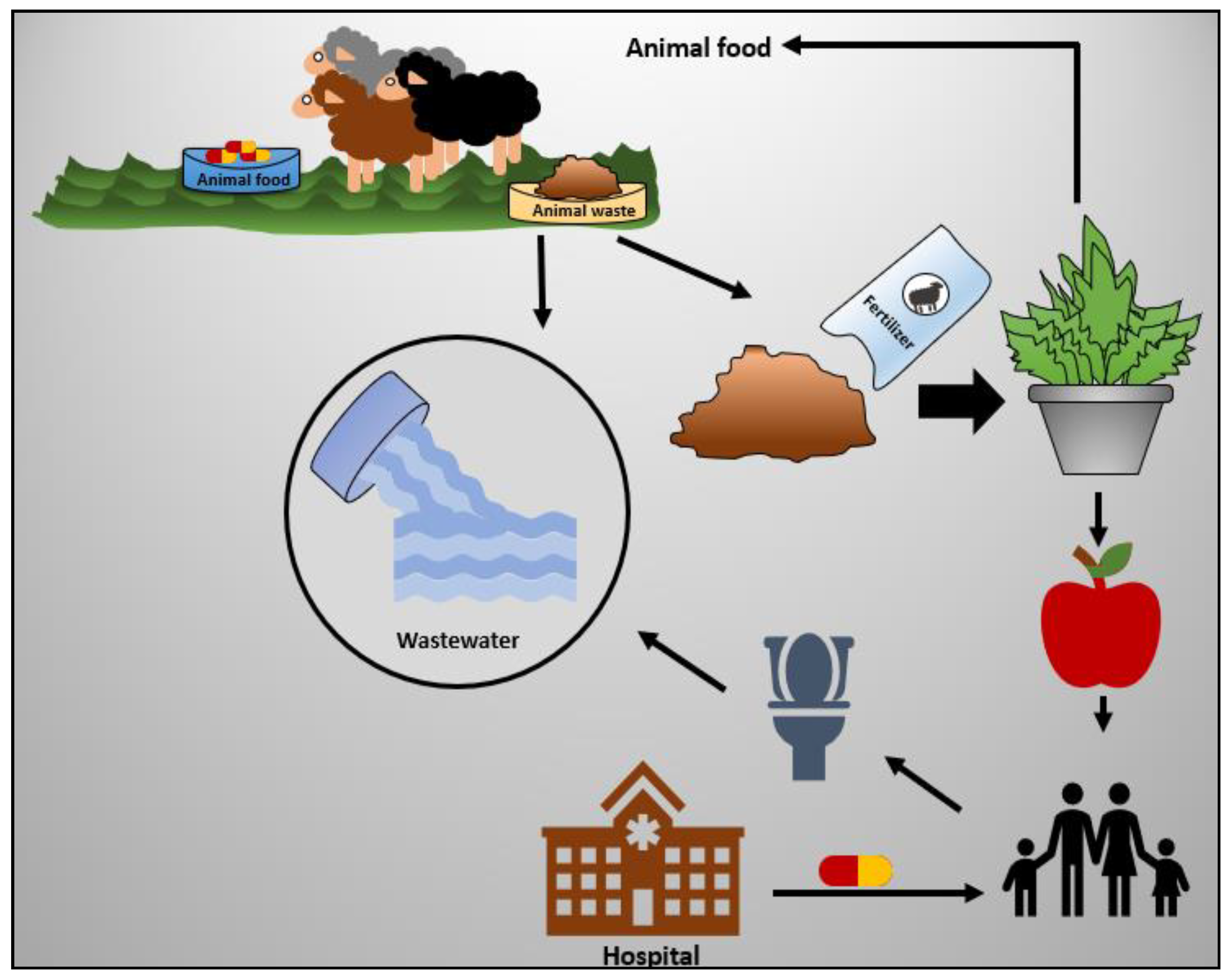
2. Antibiotics Routes of Entry into Wastewater and Its Consequences

Human consumption of antibiotics has increased significantly [18], which might not seem alarming, as it is being used for treating diseases [19]. However, the over-consumption of antibiotics is not human-friendly or environmentally safe [20]. Antibiotics’ disadvantages are not only limited to their consumption, but also their disposal. In most cases, antibiotics end up in wastewater, which cannot easily be eliminated by different degradation methods [21].
Due to the growing population demands, antibiotics are being used in different fields other than the medical field. In particular, antibiotics are extensively used in livestock and animal veterinary [22]. In some countries, antibiotics are not only used for animal treatment but also to promote animal growth and increase production [23]. However, there will not be considerable antibiotic amounts in animal meat, as all the antibiotics will be completely released from the animal’s blood if the treatment is restricted for a few days [24]. Nevertheless, antibiotics will be released as animal waste due to its incomplete digestion, which either used as fertilizers in agriculture or dumped into wastewater [25]. Few studies were conducted to assess the uptake of antibiotics from fertilizers by plants. For example, a study was done concerning assessing the amount of antibiotics in different vegetables after being fertilized with animal waste. They found that the concentration of different antibiotics was less than 10 μg\Kg, which is below the detection limit. Nevertheless, this result is alarming if antibiotics are used extensively [26]. Further study revealed that chlortetracycline antibiotic was uptaken by onions, cabbage, and corn. However, those vegetables did not take up the tyrosine antibiotic, probably due to its large molecular size [27]. The outcomes of these studies suggest that different plants can uptake different antibiotics at different rates depending on the plant type and the molecule size of the antibiotic.
The fundamental problem of having antibiotics in wastewater is the development of bacterial resistance to antibiotics. Those antibiotic-resistant bacterial strains are very difficult to treat, which causes a health and environmental threat. The bacterial resistance mostly occurs in hospital wastewater, due to antibiotics’ extensive usage at high concentrations [28,29]. As bacterial resistance becomes a major concern, antibiotic degradation becomes necessary. Figure 1 summarizes antibiotics’ possible routes of entry to wastewater.

3. Current and Conventional Elimination Methods of Antibiotics

Biodegradation is one of the methods that were used to eliminate antibiotics from wastewater. The principle of biodegradation depends upon the use of microorganisms to biologically degrade the antibiotics to safer compounds [31]. Some microorganisms showed promising behavior in the degradation of certain antibiotics; conversely, in other cases, antibiotics could not be removed completely. Generally, this approach is not considered to be effective, not only in terms of antibiotics degradation but also due to the associated risks of the direct use of microorganisms [32]. Another method is reverse osmosis, in which water is compressed across a semipermeable membrane (e.g., cellulose acetate membrane), allowing pure water to pass and eliminating other impurities [33]. This method is efficient but not cost-effective [34]. Furthermore, different filtration methods were conventionally used, such as ultra-filtration, which purifies water by using membranes with a pore size from 10 to 1000 Å. This technique is an efficient but unreliable approach to eliminate some organic wastes [35]. The advanced oxidation process is a further approach to eliminate antibiotics from wastewater. This approach depends upon the generation of free radicals, such as hydroxyl radical or singlet oxygen. Free radicals are very reactive; they react with organic compounds, leading to oxidation reaction and degradation of that compound. Free radicals can be produced using a variety of methods; some are photochemical, and others are non-photochemical, such as ozonation [19,35]. The photochemical process involves semiconductor-assisted photocatalysis which is the main topic of this review.

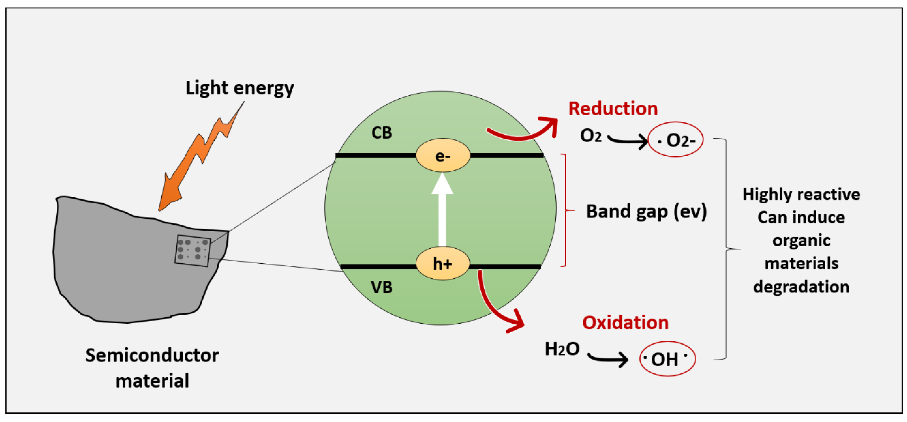
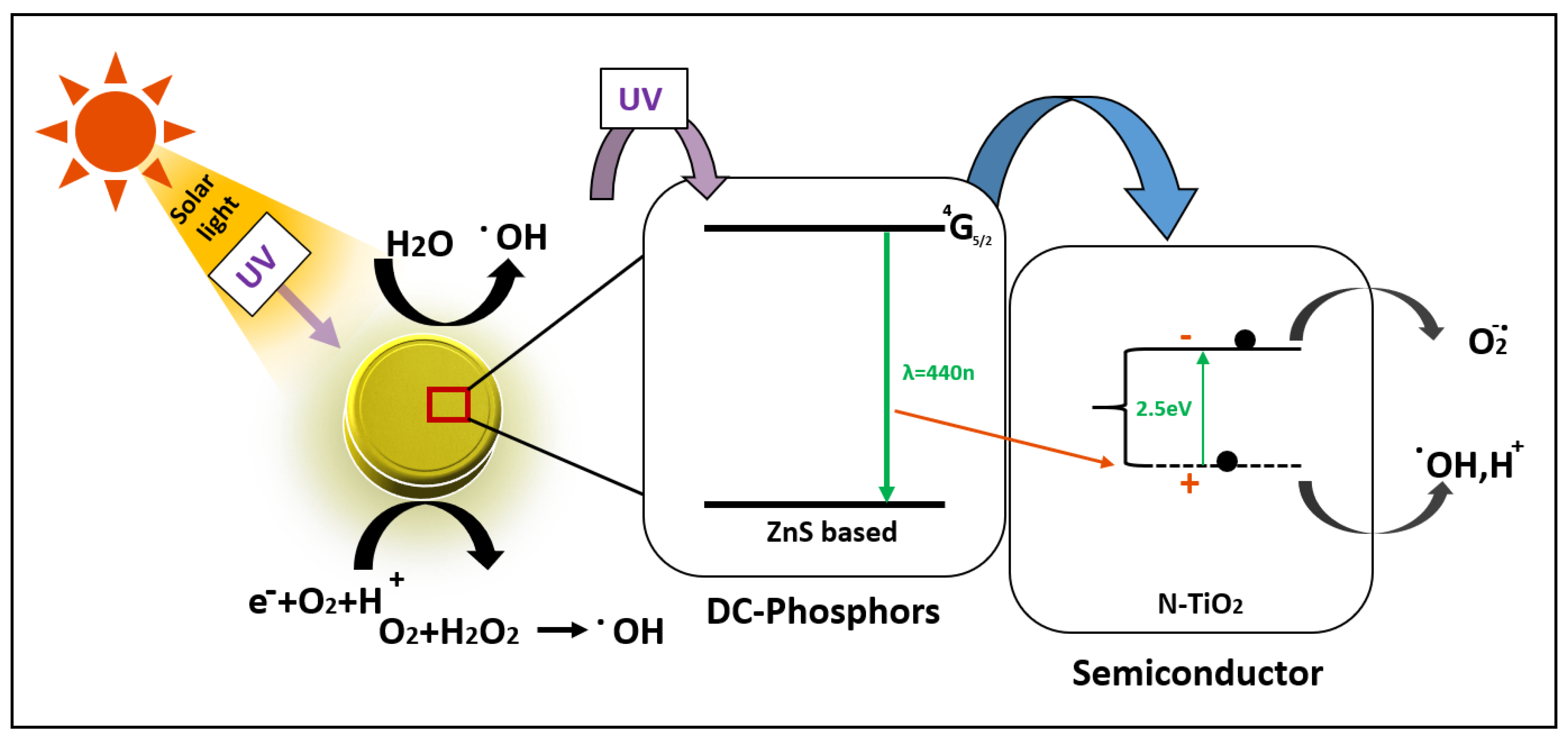
4. Semiconductor Assisted Photocatalysis

Photocatalysis is a term derived from a Greek word, where photo means light and catalysis signifies a process where the rate of a chemical reaction is accelerated by a substance without being altered. Therefore, photocatalysis depends upon the simultaneous use of light and catalysts to speed up a chemical reaction. The concept of photocatalysis was first introduced by Fujishima and Honda in 1972, where water was split using a TiO2 electrode under UV light. The main idea of applying photocatalysis to purify wastewater depends upon the generation of free radicals, which can react with organic components in wastewater, resulting in the degradation of that component [36].
The general mechanism of semiconductor-assisted photocatalysis is represented in Figure 2, when light energy (photons) hit the semiconductor surface with energy equal to or higher than the bandgap energy. Electrons from the valance band will get excited to the conduction band, leaving holes at the valance band. The holes at the valance band can react with water molecules, generating hydroxyl radicals, which have a strong oxidizing capability that is used to degrade organic matters. However, electrons at the conduction band can interact with oxygen-generating superoxide anion. Therefore, alternative oxidation and reduction reactions can occur by the formed electrons and holes [37].
Although semiconductor photocatalysis has many advantages compared to other wastewater removal techniques, many aspects need improvement. Particularly, the problem of fast recombination of the electron–hole pair, which significantly affects the photocatalytic performance [39]. Furthermore, the utilization of natural energy resources is limited, since some of the photocatalysts have a maximum absorption wavelength in the UV range [15]. Additionally, the morphology of some nanomaterials might not provide an efficient surface–light interaction; this can be improved by increasing the surface area/volume ratio. It has also been shown that the shape of the nanomaterial can influence the electron mobility, as in TiO2 nanoparticles, which showed lower electron mobility compared to highly ordered TiO2 nanotubes [39]. Besides this, the overall cost and safety of the material must be considered [36]; although TiO2 suffers from many of the mentioned limitations, it is still used due to its affordable cost [15]. TiO2 is also more chemically stable compared to other semiconducting materials with a lower energy bandgap, such as g-C3N4, which is also vulnerable to degradation by hydroxyl radicals [40].
Generally, increasing the overall photocatalytic efficiency would enhance the degradation of antibiotics. There are variety of studies testing different material characteristics to overcome such limitations. For example, surface engineering and morphology optimization can greatly affect photocatalytic performance. The photocatalytic activity would be enhanced by maximizing the active sites, mainly by increasing the surface area and increasing the proportion of crystal facets. [41,42]. Inducing defects such as anion or cation vacancies can affect the photocatalytic activity as well [43]; for example, oxygen vacancy was shown to enhance light absorption and charge separation in BiOBr. [44]. Doping semiconducting materials is another approach used to narrow the bandgap; doping can reduce the recombination rate as well as shift the absorption wavelength to a visible range [17]. Different dopants can be used, such as Ag, Mg, Cu, Rb, S, N and P. Additionally, semiconductor photocatalysts can be either doped with a single dopant or co-doped with two dopants [45]. Heterojunction charge transfer is another method where the photocatalytic performance is improved by lowering the recombination rate of electron–hole pairs. Semiconductor heterojunction depends upon combining the semiconductor material with other material, which can be metallic, carbon group, or a semiconductor. When coupling a semiconductor with another semiconductor, the photocatalytic efficiency would be enhanced, as photoexcited electrons will transfer from the semiconductor with more negative positions in the conduction band to the other semiconductor, whereas holes will be transferred from the more positive positions in the valance band to the other semiconductor with lower positions, which leads to overall enhanced charge separation, and, thus, better photocatalytic activity. [46]. A further approach to improve photocatalytic activity is the use of the plasmonic effect by involving metallic nanoparticles. Metallic nanomaterials such as Au and Ag have a property known as surface plasmon resonance (SPR). Upon irradiation, if the incident energy is enough to prevent the positive nuclei-restoring force, the valance band of the metallic particle will be generated. When metallic nanoparticles are combined with semiconductor material, the light absorption will be enhanced, as electrons will migrate to the conjugated semiconducting material; thus, overall photocatalytic efficiency will be improved. [17]. Non-Nobel metals such as bismuth, aluminum, and copper were also reported to have a plasmonic effect. They can be used as an alternative to nobel metals due to their convenience and low cost [47].
There are further approaches used to enhance the photocatalytic activity; adding a sensitizer is one of them. Organic metal complexes, such as Ru(bpy) 2 + 3 (bpy = 2′,2 bipyridine), can be used as a sensitizer. However, sensitization is highly dependent on the solution pH, as it involves the adsorption of the molecule on the surface. Furthermore, a sensitizer might be degraded if an electron donor is not present. Sensitization has not been commonly used recently; rather, research focuses on other approaches to enhance photocatalytic activity [41,48]. Hydrogenation is another approach to enhancing the photocatalytic performance. Hydrogenation, as the name implies, involves adding hydrogen molecules to the semiconductor’s surface by a chemical reaction using catalysts or by applying H2 plasma treatment [30]. The surface of the semiconducting material can be either fully or partially hydrogenated, i.e., different degrees of hydrogenation can be achieved. A study conducted by Yan, Han et.al revealed that slightly hydrogenated TiO2 nanoparticles enhanced the photocatalytic degradation of organic dyes by enhancing the light absorption at the UV range. However, highly hydrogenated TiO2 nanoparticles showed a lower photocatalytic performance compared to the pristine form [49]. Another study showed an enhancement of g-C3N4 performance after hydrogenation; this enhancement was mainly attributed to the redistribution of charge density in valance and conduction bands, which leads to efficient charge separation [50].
A novel approach to enhancing photocatalytic performance is coupling the semiconducting material with phosphor. The photocatalytic process involves the use of a photoreactor that ensures an appropriate interaction between photons, the chemical reactant, and the photocatalyst. The photoreactor must be properly designed, so that all the photons emitted from the light source are collected. Unlike classical chemical reactors, different reaction parameters, like pressure, temperature, and fluid dynamics, are less important in the photoreactor, and controlling these parameters will not give the best photocatalytic results. However, the optical path length is an important parameter in the photoreactor’s design. This makes sure that the photon flux is uniform inside the reactor. This parameter is critical as, when the light travels through the photoreactor, the photons are absorbed by the photocatalyst. This leads to scattering and decaying of the photon intensity, causing a non-uniform reaction rate in the photoreactor. This problem can be overcome by coupling the photocatalyst with light-emitting phosphorescent particles such as phosphors. Phosphor material is a substance that is based on a lanthanide-doped material that emits light and exhibits luminescence characteristics upon exposure to electromagnetic radiation. The introduction of phosphor to the system can enhance the photoreactivity by transforming the external light 365 nm radiation into 440 nm emission, as well as by allowing the photoexcitation of the fraction of photocatalyst in the core reactor volume and not screened by the photocatalyst itself. There are three types of phosphor materials: down-conversion phosphors, up-conversion phosphors, and long-afterglow phosphors. In the down-conversion phosphor, higher energy photons are absorbed and emitted as lower energy photons [51,52]. Different transition metals, such as Eu2+, Er3+, and Pr3+, showed light emission at different wavelengths. This phenomenon will help to tune the emission spectra to the required specifications. It has been reported that when phosphor is coupled with a dye, the emitted photons from phosphor can be easily absorbed by the dye, leading to excitation and the generation of electron–hole pairs. A commonly studied material is N-doped TiO2 coupled with phosphor, which showed enhancement in photon transfer in the reactor [53]. The mechanism by which phosopher enhances the photon transfer is illustrated in Figure 3. The down-conversion phosphor material can absorb UV light and emit photons at the visible range inside the photoreactor, which can be absorbed and utilized by the photocatalytic material. This system was shown to enhance the photocatalytic activity and degradation of different organic compounds such as Methylene blue, phenol, and Terephthalic acid [53].
An upconversion phosphor is a further approach through which visible light absorption by the photocatalysts is enhanced. In this approach, lower energy photons are transformed into higher energy photons. There are three mechanisms by which the up-conversion phosphor works; these are excited-state absorption, photon avalanche, and energy transfer up-conversion. Sacco, Vaiano and Sannino describe in detail all three approaches in detail [54]. Coupling the photocatalyst with a long persistent phosphor (long afterglow phosphor) is another approach used to enhance photocatalysis. Long afterglow phosphors such as CaAl2O4: Eu2+, Nd3+ can sustain photocatalysis even after the removal of the light source or the photoreactor. Those long afterglow phosphors can store the energy from light and emit it at a slow rate. It has been reported that this approach is sustainable and saves energy, as the photocatalytic process can be done without the presence of light. This approach was used for the degradation of the antibiotic ofloxacin by Alberti et al., when they developed N-P doped TiO2 for this purpose; the study showed 87% degradation of ofloxacin after 50 min of irradiation. The study showed 95% degradation of the antibiotic after 30 min of total irradiation (i.e., alternating radiation–light cycle followed by dark cycle) [55].

5. Commonly Used Semiconducting Materials for Antibiotic Degradation

5.1. Semiconducting Metal Oxides

Metal oxides have been shown to have certain properties that make them suitable candidates for photocatalysis, for example, light absorption (UV, visible light, or both combined), which initiates charge separation and the photogeneration of electrons and holes. Additionally, they are biocompatible, safe, and stable when exposed to different conditions. Metal oxides include the oxides of titanium, zinc, tin, vanadium, and chromium [56].
TiO2 is the most studied metal oxide for photocatalysis, due to its superior properties such as good optical and electronic properties, chemical stability and reusability, non-toxicity and low cost. Although TiO2 is a promising material for commercial use, TiO2 suffers from limitations, as it has a large bandgap (3.2 eV), thus limiting visible light utilization, which makes its application uneconomic. There are a variety of studies regarding TiO2 modification to obtain a better photocatalytic performance, which would make its application more feasible [57,58,59,60,61]. ZnO is another semiconducting material that has a better quantum efficiency than TiO2; furthermore, recent reports suggested that ZnO has higher photocatalytic efficiency compared to TiO2, especially if used at a neutral pH [62,63]. Nevertheless, the high recombination rate of the photogenerated electron–hole pairs restricts the utilization of ZnO in its pure form. Several studies reported doping ZnO with metals like Ag and Fe [64] or non-metals like N and C. Different studies suggested improvements in photocatalytic properties after doping the ZnO; for instance, a study reported that the ZnO 12 S incorporated nanoparticles for the degradation of organic dyes. This material showed a 100% degradation efficiency that decreased to 92% after five cycles of reuse [65]. Other studies showed enhanced photocatalytic performance of ZnO-based materials [66,67,68].
Tungsten oxide WO3 is another metal oxide that has received considerable attention due to its abundance, cost-effectiveness, and non-toxicity [69,70,71]. Unlike TiO2, it exhibits 12% absorption of the solar spectrum. Different studies reported a variety of modification approaches. In particular, Huang et al. found that W18O49 has higher photocatalytic degradation efficiency compared to WO3. Different W18O49 morphologies exhibit different photocatalytic activities as well. Namely, hollow W18O49 spheres exhibit the highest photocatalytic activity compared to other morphologies. Despite the fact that W18O49 is more efficient compared to WO3, it is prone to oxidization to WO3. To overcome this limitation, a study reported the construction of W18O49/TiO2 hybrid; by this, both stability and efficient photoactivity are achieved [72].

5.2. Bismuth Based Photocatalysts

Bismuth-based semiconductor photocatalysts have been shown to have a good visible light absorption as their valance band electrons form a hybrid of O 2p and Bi 6s, unlike TiO2 with O 2p orbitals. Those Bi 6s orbitals, when properly dispersed, have been found to increase the mobility of electrons and holes, as well as reduce the bandgap energy. For that, Bi-based semiconductors mostly have a bandgap of less than 3 eV. There are a variety of studies regarding different Bi-based semiconductors for photocatalysis, such as Bi2O3, Bi2CrO6, Bi2MoO6, Bi2WO6, BiVO4, BiOCl, and BiOBr [73].
Bi2O3 is one of the simplest and most common and major photocatalysts. It has been applied for both water-splitting and water treatment from organic wastes. Bi2O3 has a bandgap that ranges from 2.1 to 2.8 eV, which makes its utilization for visible light absorption more efficient. Bi2O3 has five different polymorphisms: α, β, γ, δ, and ω-Bi2O3. Metastable phases of Bi2O3 can be converted to the α phase at low temperatures or the beta phase at high temperatures [73,74].
A further example is bismuth vanadate (BiVO4), which was reported to have superior physiochemical properties like ferro-elasticity and ionic conductivity [75]. It possesses three phases: monoclinic, fergusonite, and tetragonal. It has a theoretical bandgap of 2.047 eV, which maximizes its visible light utilization. BiVO4 was used in photocatalysis for organic waste treatment as well as water splitting [73,76].
Although bismuth-based photocatalysts have been shown to have good photocatalytic performance as well as efficient utilization of visible light, some parameters need to be addressed, for example, the stability of bismuth-based photocatalysts under different conditions. Their stability is mainly affected by the photocatalyst intrinsic structure and its solubility, as well as the solution’s pH. It has been shown that layered bismuth-based photocatalysts with both covalent and van der Waals bonds are more stable than layered bismuth-based photocatalysts with van der Waals bonds only. Nevertheless, bismuth-based photocatalysts usually have low solubility constants, which means that they cannot always be dissolved in the substrate. Furthermore, their photo-corrosion was not sufficiently studied [73,77].

5.3. Graphitic Carbon Nitrides

Graphitic carbon nitride (g-C3N4) is a new type of polymeric semiconducting material. It is considered as the most stable allotrope compared to other carbon nitrides at ambient conditions. Unlike TiO2, g-C3N4 can utilize both visible light and UV radiation due to its narrow bandgap (2.7 eV) [78]. However, g-C3N4 in its pure form endures many drawbacks, such as the high recombination rate of electron–hole pairs, poor utilization of visible light, and low electrical conductivity [79]. Therefore, material modification is necessary to overcome such limitations. Different studies reported the modification of g-C3N4 mainly by doping with transition metals, noble metals, or non-metals [80,81]. Surface modification was reported as well [82]. Based on several reports in the literature, doping with noble metal ions was shown to have the best photocatalytic performance. This was attributed to the higher separation of the photogenerated electrons and holes, due to the ability of electron capture by the noble metallic ions. For example, a study reported 96.8% photocatalytic degradation after 120 h of solar light exposure when using Ag-doped g-C3N4. Despite the fact that noble metal doping can enhance the performance of g-C3N4 significantly, doping with noble metals is not cost-effective. Consequently, there are other studies concerning the use of noble metal-free composites [83]. Namely, Cao et.al designed g-C3N4 coupled with Ni(dmgH)2. This composite showed an efficient ecosystem-friendly photocatalyst with efficient solar-to-hydrogen conversion. Despite this, the photocatalytic degradation of different compounds was not studied [84]. Other studies reported doping g-C3N4 with non-noble metals such as nitrogen [85,86]; those studies are limited due to the potential toxicity of the N precursors used, such as hydroxylammonium chloride and hydrazine hydrate. Other environmentally friendly non-noble metallic dopants include carbon; doping g-C3N4 with carbon showed enhancement in visible light absorption and charge separation. There are other studies concerning doping with non-noble metals; for more details, a review written by Starukh and Praus summarizes such studies [87].
There is ongoing research regarding material modification to fabricate and design nanomaterials with different properties to obtain the best photocatalytic performance, and thus, better elimination of antibiotics. The next section summarizes the recent studies related to antibiotic degradation by using semiconductor-assisted photocatalysis.

6. Recent Reports on Antibiotics Degradation with Photocatalysis

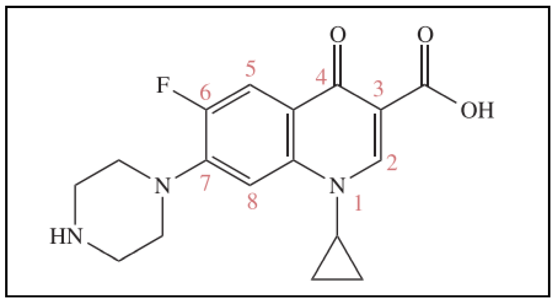
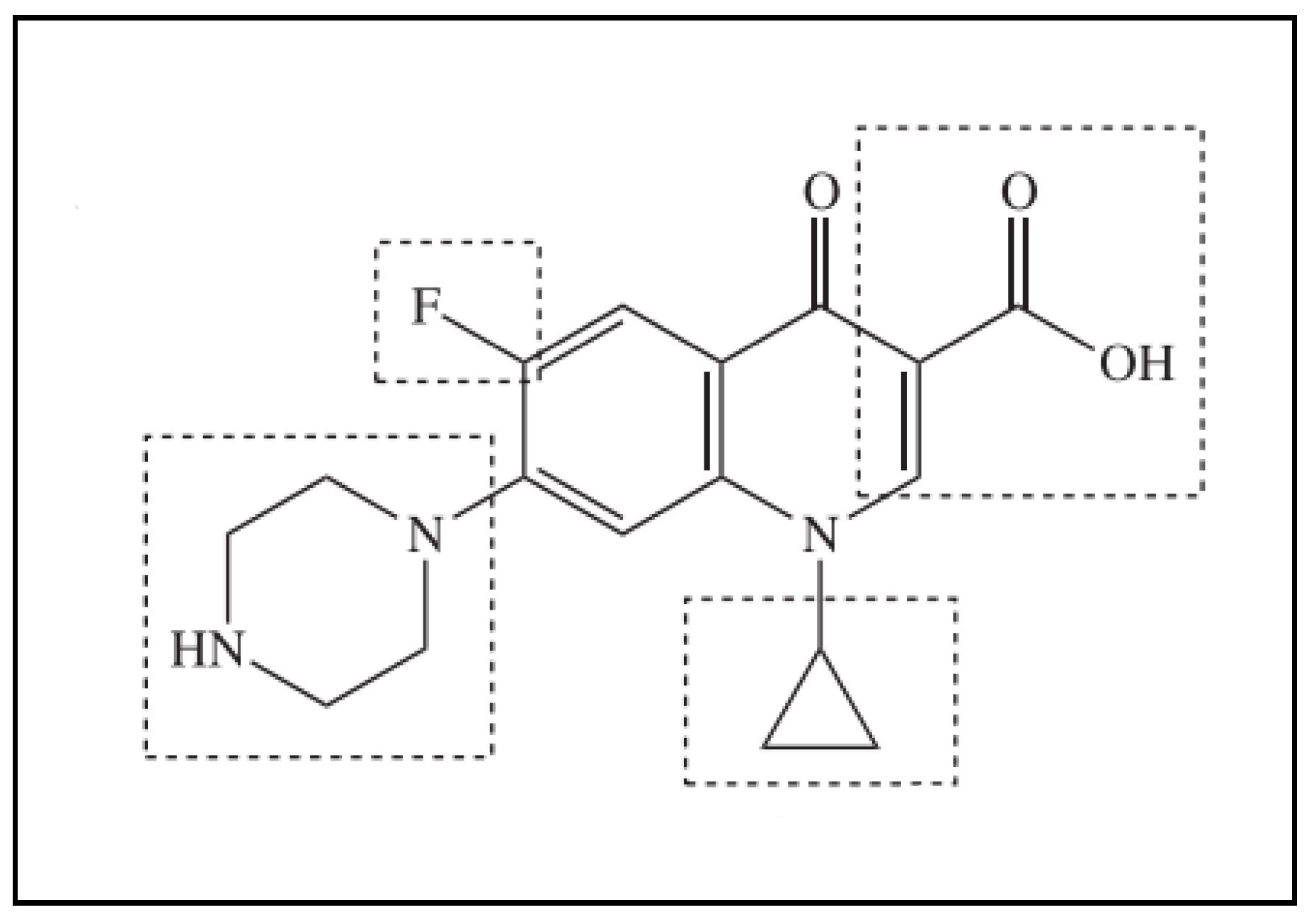
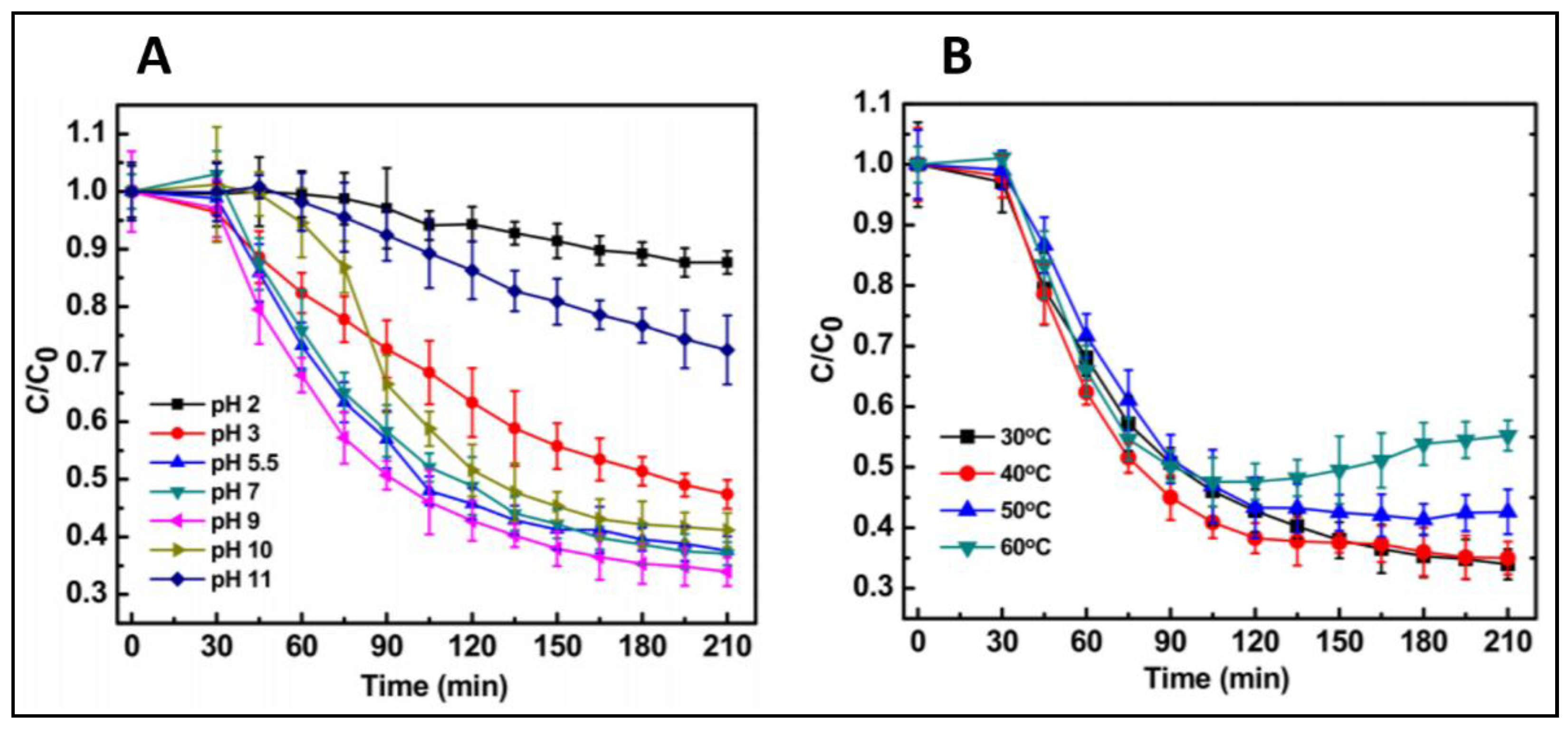
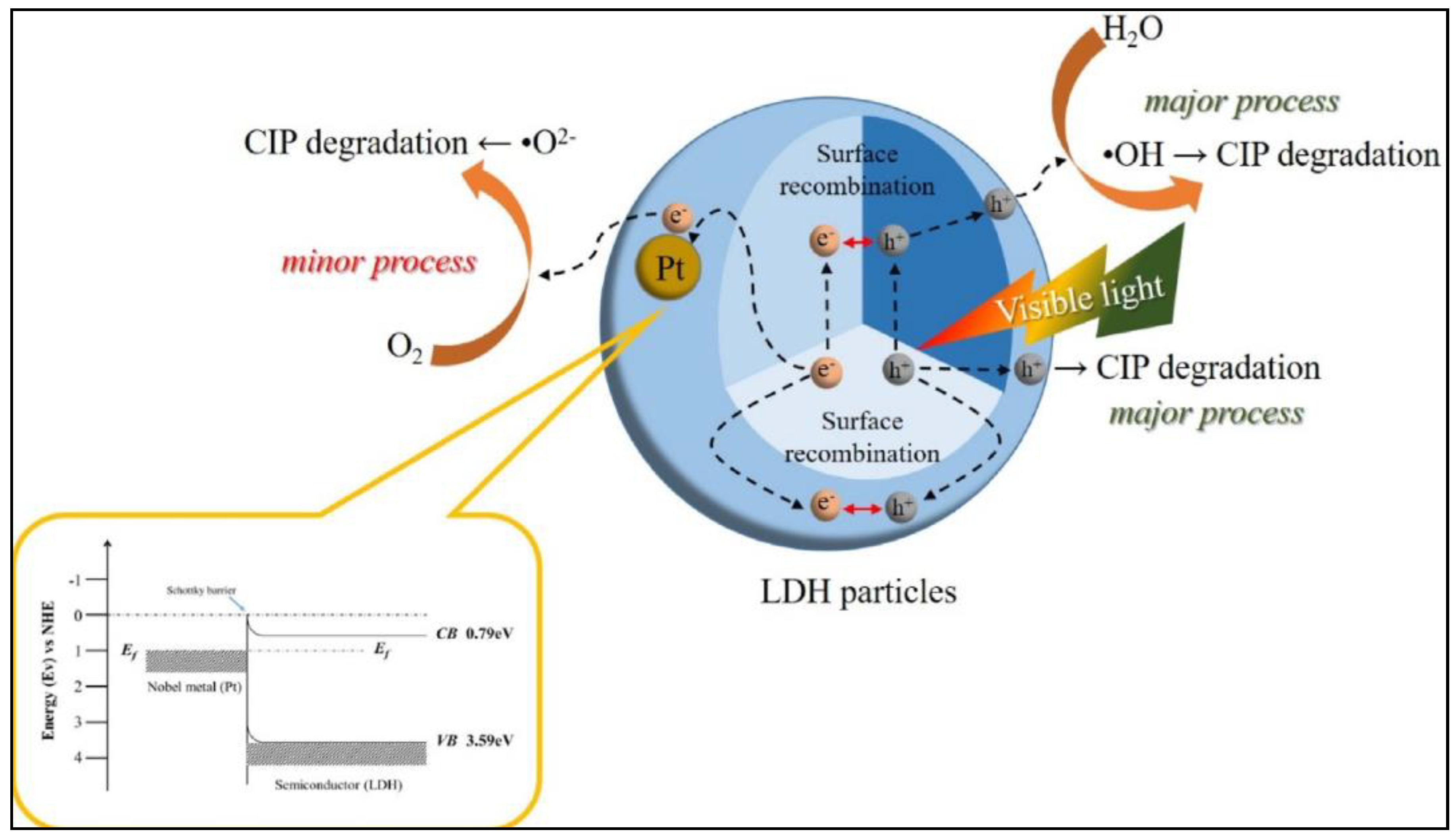
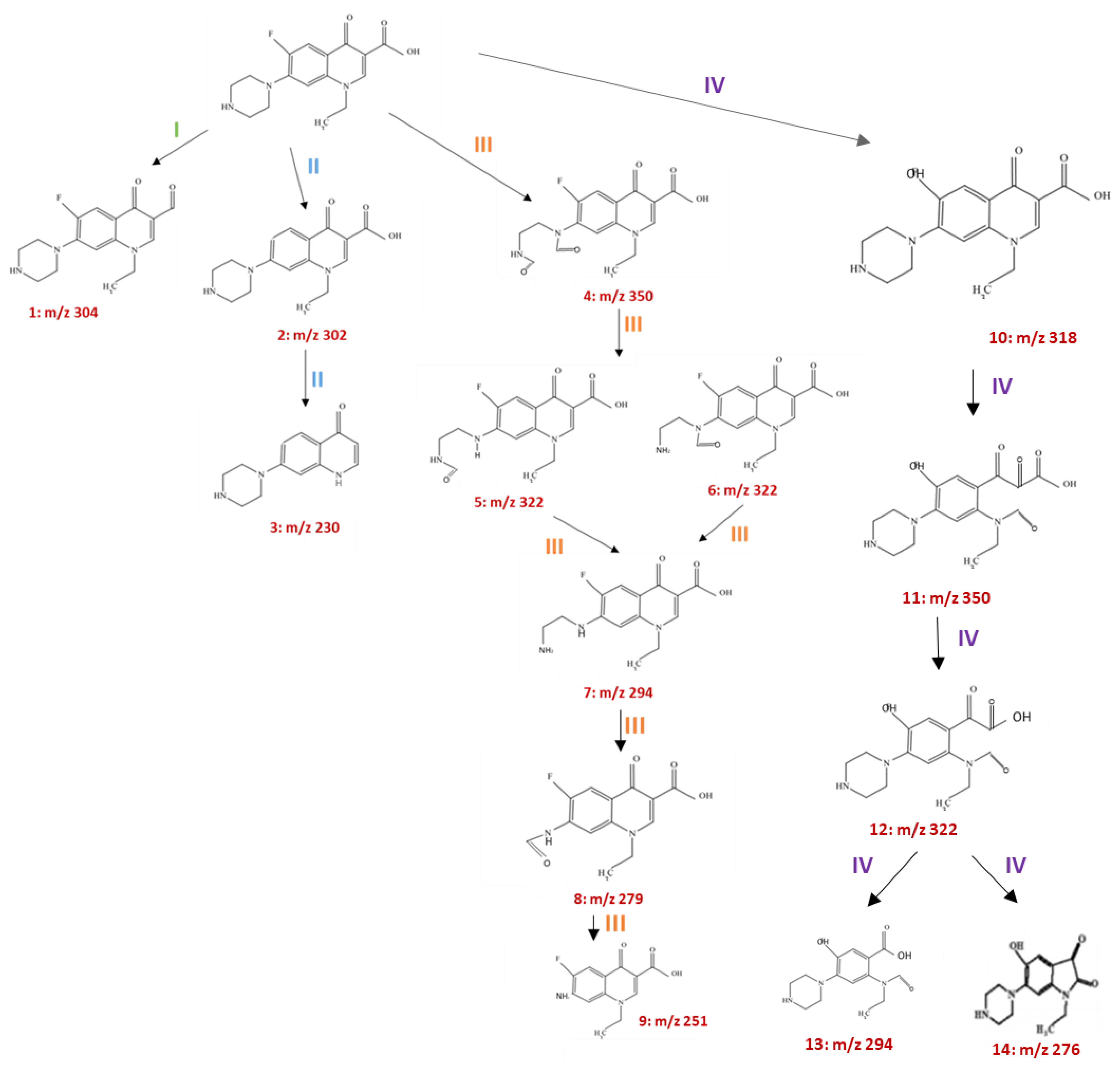
6.1. Photocatalytic Degradation of Ciprofloxacin (CIP)

Ciprofloxacin (CIP) C17H18FN3O3 is a second-generation fluoroquinolone antibiotic that is used to kill and prevent the replication of bacteria. It is used in both humans and animals to treat many medical cases, like skin and urinary tract infections [88]. In a study conducted in 2016, it was reported that CIP represents 73% of the total consumption among fluoroquinolone antibiotics. It was further reported by another study that the detected amount of CIP in hospital wastewater can reach up to 150 μg/L, which is considerably high, and has a potential risk if not eliminated [89]. Considering the frequent detection of CIP in the environment and water, researchers have been working to improve its elimination. CIP removal by the photocatalytic reaction can be achieved by attacking different sites in the CIP structure. Figure 4 shows the chemical structure of CIP and Figure 5 shows different sites that can be attacked by generated free radicals to decompose the CIP structure. For more details of CIP degradation pathways, Huo et.al summarize a variety of approaches and pathways for CIP degradation by photocatalysis [90].
Different studies reported the use of photocatalysts which have been modified to achieve the highest photocatalytic efficiency. Researchers usually start with doping with a transition metal. Transition metals were shown to enhance photocatalysis when added, for example, in a study conducted by Das, Ghosh, Misra, et al., where Fe-doped ZnO was prepared to degrade CIP under visible light irradiation. Here, the authors prepared Fe-doped ZnO following the precipitation route; after that, they induced the photocatalytic reaction by dispersing three different concentrations (100, 150 and 200 mg/L) of the photocatalyst with 10 mg/L of Ciprofloxacin followed by 3.5 h of visible light exposure. The optimum results were obtained when 150 mg/L of Fe-doped ZnO was used with 66% degradation efficiency, which is five times more than the degradation of the negative control. The improved photocatalytic activity was suggested to be due to the delay in recombination that Fe offers, thus the increment in the produced free radicals [91]. Figure 6 represents the degradation of CIP under different conditions. Although Fe, when added, enhances the degradation, its photocatalytic activity is impaired by its thermal instability and fast recombination rate. To overcome such limitations, metalloids such as Boron are used. In one study, Şimşek synthesized boron-doped TiO2 using a solvothermal method to degrade CIP under visible light irradiation. TiO2 was doped with different dopant concentrations (2,4,6,8%) which were then dispersed in 20 mg/L antibiotic solutions. The degradation efficiency was positively correlated with the dopant concentration with the maximum degradation rate of B/TiO2 8% of around 88.32%, which is four times higher than the undoped TiO2. The optimal reaction conditions were achieved when 1.1 g/L of 8% B/TiO2 was used with a solution pH of 7.1 and with the presence of 7.23 mM H2O2. The produced material showed a lower bandgap, and thus better photocatalytic activity, as well as stability, when being reused five times [92]. Non-metallic dopants like graphene oxide (GO) were also used to improve the photocatalytic performance, where sphere-like copper tungstate (CuWO4) deposited on 2D GO was produced by the hydrothermal method. Doping with GO decreased the bandgap of CuWO4 from 2.5 to 2.09 eV, which enhanced the degradation of CIP, as more than 97% of CIP was degraded after one hour under visible light irradiation [93]. Co-doping with non-metals was also reported due to the superior photocatalytic enhancement that it offers. For that, Qu, Xu, and others developed N,S-doped carbon quantum dots embedded with ZNO nanoflowers (N, S-CQDs). A hydrothermal method was used to produce this material, which was tested for the degradation of CIP under visible light. It was found that 92.9% of CIP was degraded after being exposed to simulated sunlight for 20 min, whereas, 85.8% of CIP was degraded when exposed to natural sunlight for 50 min. The photocatalytic degradation efficiency of un-doped CQDs material was tested under simulated sunlight irradiation. The degradation rate was 8.6% after only 20 min of exposure. This is 10 times less efficient than the co-doped material [94]. The utilization of transition metal oxides was also reported, LaNiO3/TiO2 heterojunction was produced by the sol-gel method to degrade organic dyes and the antibiotic CIP under visible light irradiation. The performance of LaNiO3/TiO2 heterojunction was compared to pure LaNiO3 and TiO2. A 70% degradation of 10 mg/L CIP after 3 h of exposure to simulated sunlight was reported, which is significantly more than the pure components. The improvement in photocatalytic activity was attributed to the good separation of electron–hole pairs offered by the heterojunction. Metallic nanoparticles doping, such as Ag, was shown to increase the duration of the photogenerated electron–hole pairs. For that, Alvarez and others developed a AgBr/Ag/Bi2WO6 heterostructure to degrade CIP under visible light irradiation. The authors reported that 57% of CIP was degraded after 5 h of irradiation. The addition of Ag nanoparticles, even at a low weight %, showed a significant change in the material’s photocatalytic behavior. The produced heterostructure showed an improvement in photocatalytic activity compared to the unmodified Bi2WO6. This is mainly due to the presence of Ag as well as the heterojunction formed by the two semiconducting materials AgBr and Bi2WO6 [95]. While the addition of Ag nanoparticles enhanced the photocatalytic activity significantly, it is not cost-effective; other lower-cost materials are suggested for utilization, such as doping with S or B. To overcome the drawbacks, such as material agglomeration, that can happen in some of the modified materials, Zheng et.al. designed graphitized mesoporous carbon TiO2 nanocomposite to overcome material aggregation. It was reported that the large surface area and the mesoporous structure enhance the absorption of the antibiotic in the case of its presence at low concentrations, which enables a convenient interaction with the photocatalyst.
In addition, the graphitized carbon sheets inhibit TiO2 aggregation. The produced material was tested to degrade CIP. The results suggest a quick and complete degradation of CIP in 1.5 h, along with inhibition of Vibrio Fischer bacteria [96]. These findings are important, as there are limited reports on bacterial experimentation. Concerning the enhancement of photocatalysis by morphology modification, a study reported the production of double-shelled ZnSnO3 hollow cubes (ZSO-C), which was prepared by the co-precipitation method and its effect on the degradation of two antibiotics: CIP and sulfamonomethoxine. The ZSO-C was compared to another two materials: one of them was prepared by hydrothermal method with hollow polyhedral shape (ZSO-H), while the other was prepared with the template method (ZSO-T). It was found that ZSO-C has the best photocatalytic efficiency and stability compared to the other two materials, not only in the degradation of the two antibiotics, but also in other dyes such as methylene blue and methyl orange, with a degradation rate of 85.9%, which is 1.5 times better than ZSO-H and two times better than ZSO-T. The authors attributed that to the narrower bandgap and larger surface area of ZSO-C compared to the other two materials [97]. A further study was conducted by Li, Fu, and Zhu; they produced 3D tri pyramid TiO2 by the hydrothermal method to degrade CIP under UV–visible irradiation. They reported that the tri pyramid morphology significantly enhanced the degradation of CIP due to its high specific surface area compared to rod-shaped TiO2. Correspondingly, the tri-pyramid morphology provided more active sites for catalytic interactions [98]. Green, environmentally friendly methods were also utilized. Particularly, the recent photocatalyst “layered double hydroxides” (LDHs), was modified by making an LDHs Pt hybrid, as reported by Z. Li et al. They found that doping LDHs with Pt leads to significant improvement in photocatalytic activity especially in the visible light range [99]. The mechanism by which CIP was degraded by LDHs/Pt hybrid is presented in Figure 7.

6.2. Photocatalytic Degradation of Norfloxacin

Norfloxacin is a broadly administered antibiotic that belongs to the fluoroquinolone antibiotic class. In the past few years, fluoroquinolone antibiotics have caused a major concern due to their extensive use and environmental toxicity. There are few reports on the degradation of norfloxacin by photocatalysis using different materials. The mechanism of degradation have been proposed as well; there are four proposed mechanisms, all sharing the same attack sites (piperazinyl and quinolone moieties). These mechanisms were proposed based on the recognition of the degraded intermediates, as shown in Figure 8. Where Mn:ZnS Quantum Dots were used to degrade norfloxacin Figure 9 represents different parameters redarding the degredation of norfloxacin using Mn:ZnS Quantum Dots [100].
Nevertheless, it is still necessary to summarize what has been done in the literature to know where we stand in terms of different materials’ fabrication and the efficiency of elimination. For this, one study reported the preparation of the In2O3/TiO2 heterostructure by Yu, Chen and others, by the polymeric precursor method to degrade norfloxacin under visible-light irradiation. The antibiotic was completely degraded after 10 min of irradiation. This desirable photocatalytic activity is mainly due to the narrow bandgap that is offered by the heterostructure, which enhanced absorption at the visible range and the large specific surface area that allows for better surface–light interaction [101]. Another study was reported by Jin, Zhou, et al., where they produced spherical-shaped N-doped TiO2 to degrade norfloxacin under visible light irradiation. Within 30 min, 99.53% of the antibiotic was degraded, which cannot be achieved when using TiO2 alone due to the reduction in the bandgap to 2.92 eV offered by the dopant. The authors further assessed the toxicity after norfloxacin degradation against E. coli bacteria; they found that the norfloxacin toxicity reduced to less than 50% after degradation. Moreover, 25% of mineralization was achieved from the photocatalysis [102]. These findings are important, since the antibiotic toxicity against bacteria after the antibiotic degradation was studied; many studies lack this type of experimentation, disregarding its importance. The application of bismuth-based nanocomposites was reported by many studies. One study was done to investigate the effectiveness of Bi2WO6/rGO/Bi25FeO40 heterojunction in the degradation of norfloxacin antibiotics under visible light irradiation. Different ratios of Bi2WO6/rGO/Bi25FeO40 were produced by the hydrothermal method. A total of 8% of Bi2WO6/rGO/Bi25FeO40 showed the highest ability to degrade norfloxacin. This enhancement in photocatalytic properties is due to the efficient charge transfer between Bi2WO6 and Bi25FeO40 and the smaller bandgap in this nanocomposite compared to a single component. Additionally, the high surface area and enhanced light absorption of the produced heterojunction contribute to its superior photocatalytic properties [103]. Besides this, Bi2MoO6/rGO/BiOBr heterostructure was developed by Zhang et al. to degrade norfloxacin under visible light irradiation. The heterostructure showed a superior degradation rate of the antibiotic, with 78.12% degradation. This is approximately four times more than the degradation of Bi2MoO6 and BiOBr alone, and one time better than the degradation of Bi2MoO6/BiOBr heterojunction. The enhancement in the photocatalytic property was attributed to the formation of heterojunction as well as the presence of rGo, which speeds up the electron transfer [104]. A further study was conducted to test the efficiency of the COFe2O4-rGO-BiOBr magnetic nanocomposite for the degradation of norfloxacin antibiotics under visible light irradiation using a mercury lamp. The nanocomposite was prepared by the hydrothermal method. The authors found that COFe2O4-rGO-BiOBr has higher photocatalytic efficiency compared to single or binary systems. This improvement was attributed to the formation of a heterojunction between COFe2O4 and BiOBr. rGO was used to increase the electron transfer rate. The overall photocatalysis rate was intensified due to the faster transfer rate and lower recombination rate. This makes COFe2O4-rGO-BiOBr a candidate material for photocatalysis [105]. There are very few studies regarding the utilization of quantum dots to degrade norfloxacin. One recent study reported the fabrication of novel Mn-doped ZnS quantum dots. The produced material exhibited an excellent degradation property; however, the material was tested only under UV irradiation [100]. A less recent study reported the design of carbon quantum dots loaded with mesoporous g-C3N4. This material showed an enhanced photocatalytic performance for fluoroquinolone antibiotics including norfloxacin compared to the bulk g-C3N4 or the unloaded. It was revealed by scavenging experiments that superoxide anion radical (O2) and photo-hole (h+) are the main active species. The degradation mechanism was proposed based on the produced intermediates [106].

6.3. Photocatalytic Degradation of Tetracyclines

Tetracyclines are a class of broad-spectrum antibiotics that was first utilized in 1940. They are used for the management of bacterial infections caused by Gram-positive and negative bacteria. Tetracyclines are usually consumed by a human for disease management, or animals as a growth-promoter. Tetracyclines cannot be completely metabolized by humans or animals, leading to their entry into the environment, causing many adverse effects on the food chain, and thus affecting human and animal health. Removing such antibiotics by photocatalysis was reported, using different materials and mechanisms of degradation [107,108]. Figure 10 represents the chemical structure of Tetracycline.
There are different proposed mechanisms on the photocatalytic degradation of tetracyclines; some of the reported mechanisms did not show a ring-opening in tetracycline structure. It was reported that degradation without ring opening is not efficient as the antibiotic would still retain its function. It has been reported by some studies that tetracyclines can be degraded by UV or simulated sunlight [109]. Figure 11 represents degradation mechanisms by photolysis and photocatalysis.
Many studies reported a variety of modified materials used to eliminate tetracyclines from wastewater. Various studies reported the modification of graphitic carbon nitrides to achieve the best photocatalytic efficiency. One of these studies reported the fabrication of Ag-doped graphitic carbon nitride (Ag-doped g-C3N4). Tri, Kim et al. tested Ag-doped g-C3N4 against the degradation of the tetracycline under solar light irradiation. The best photocatalytic degradation was when 3 mmol of Ag was used, with a 96.8% degradation rate after 2 h of exposure to solar irradiation. The same experiment was done, but to degrade antibiotics from hospital water; the material showed 89.6% degradation efficiency after 2 h of solar exposure. The produced material showed stability and reserved its degradation ability after six cycles of reuse. The good performance of the doped g-C3N4 is attributed to the efficient charge separation and charge transfer [83]. In another study by Jodeyri et al., they developed an Ag/C3N4-Clinoptilolite nanomaterial to eliminate tetracycline antibiotics under simulated solar light irradiation. They found that 90% of the antibiotics were degraded after 3 h of exposure when Ag/C3N4-Clinoptilolite nano-photocatalyst was used, which showed superior efficiency when compared to Ag/C3N4, C3N4-Clinoptilolite, and C3N4. This enhancement is attributed to the improvement in the specific surface area that Clinoptilolite offers, as well as the SPR, due to the presence of Ag [110]. Further study was done using urea-derived graphitic carbon nitride (g-C3N4) doped with four different metals (K, Na, Mg, and Ca) to find the effect of different metal dopants on morphology, structure, and photocatalytic activity. The produced materials were tested to degrade three different antibiotics (tetracycline, Enrofloxacin ENR, and sulfamethoxazole) under visible light irradiation. All the produced photocatalysts were compared to undoped g-C3N4. Undoped g-C3N4 showed a degradation ability, with a removal rate constant of 0.014/minutes after 2 h of exposure. This rate was significantly increased to 3.3 when Ca was used as a dopant, and 5.4 when K was used as a dopant. The best photocatalytic performance was for g-C3N4 doped with K > Na > Mg > Ca > undoped g-C3N4. The improvement in photocatalytic performance was attributed to the narrowing bandgap, which thus increased charge separation efficiency and extended charge lifetimes [111].
The co-doping of g-C3N4 was also reported. In one study, g-C3N4 was co-doped with Phosphorus and Sulfur by in situ thermal copolymerizations to degrade tetracycline under visible light irradiation. In their experiment, they dispersed 0.5 and 1 g/L of the photocatalyst with 10 mg/L tetracycline, which was then exposed to 100 mW/Cm2 of visible light for 2 h. It is shown that the Co-Doped g-C3N4 is 5.9 times more efficient than the undoped material, with a 70.33% degradation rate. It is suggested that the better performance of the codoped material compared to single-doped or undoped material is due to enhancements in light trapping, surface area and charge separation [112]. An additional study where g-C3N4 was co-doped with Na and Cl was reported. The study showed that the codoped material exhibited enhanced visible light absorption compared to the non-doped. Moreover, the degradation efficiency of TC-HCl was improved due to the reduction in the bandgap region as well as the lowered recombination rate [113]. The plasmonic effect of Au nanomaterials was also utilized, as reported by Xue, Ma, Zhou, et al., and the plasmonic photocatalyst Au/Pt/g-C3N4 showed a 3.4 higher degradation rate than the pure g-C3N4. In this study, the photocatalytic activity of Au/Pt/g-C3N4 was tested for the degradation of the antibiotic tetracycline hydrochloride under visible-light irradiation. The enhanced activity was attributed to the plasmonic effect of gold nanoparticles, which also increased the optical absorption range. Additionally, the electron sink effect of platinum contributed to improvements in the photocatalytic activity [114].
Fewer studies on the degradation of tetracycline using bismuth-based materials were reported. For instance, Wang and others utilized the molten salt method to fabricate Bi4Ti3O12 nanosheets with moderate ratios of top and lateral facets. They found that the best photocatalytic ability was for Bi4Ti3O12, with {001} top facets to {010}/{100} lateral facets. This material showed an improvement in photocatalysis for the degradation of tetracycline hydrochloride (TC-HCl) under Xenon lamp irradiation. The mechanism by which TC-HCl was degraded is mainly by the generation of h+, where O2− plays only a marginal role in photocatalytic degradation [115]. Owing to the superior properties of bismuth ferrite -BiFeO3 (BFO), such as the suitable bandgap (2.2 eV), the low cost and the chemical stability, Zhou, Jiang, Chen, et al. modified BFO to enhance the photocatalytic activity. For that, they produced Er-doped BFO nanoparticles by the sole gel method, which was tested for the degradation of tetracycline hydrochloride under visible-light irradiation with a wavelength of > 420 nm. Different concentrations of Er-doped in BFO were prepared (1%, 3%, and 5% Er) and compared to undoped BFO. The photocatalytic activity was tested by dispersing 200 mg of doped or undoped BFO on 100 mL of a 30 mg/L tetracycline solution, which was then irradiated to assess the photocatalytic degradation efficiency. A linear increment in the surface area of BFO with increasing Er concentration was observed, with the highest surface area for the 5% Er-doped BFO. However, the highest photocatalytic degradation was for the 3% Er-doped BFO with a 75.8% degradation rate which is approximately 3 times higher than the undoped BFO. The enhanced photocatalytic degradation of the doped BFO was attributed to the enhanced absorption wavelength of the doped material especially at the visible range spectra when assessed by UV–vis DRS spectra. Additionally, the doped material revealed a good charge separation and migration, as well as a low recombination rate, compared to the undoped material [116]. Moreover, Wang et al. fabricated a BiVO4/TiO2/RGO nanocomposite for the photocatalytic degradation of four types of tetracyclines antibiotics upon visible light irradiation. They tracked the degradation of antibiotics by detecting the intermediate products that are formed due to antibiotic degradation. They achieved a homogenous distribution of BiOV4 and TiO2 particles when increasing the GO content to 0.3% and 0.5%. The greatest photocatalytic degradation was achieved when 0.5% of GO was used, with 80% antibiotic degradation after 1 h. The photocatalytic efficiency of the produced composite was the best when compared to two produced materials: BiVO4/RGO and TiO2/RGO. The authors claimed that the presence of GO is advantageous, as it separates the photogenerated electron–hole pairs and thus reduces the recombination chances. Hence, the photogenerated electrons will move from the BiVO4 conduction band to TiO2, which improves the degradation rate. Furthermore, the decoration with GO leads to a drop in the bandgap of BiVO4 and TiO2, which also prevents the fast recombination of electron–hole pairs [117].
The photocatalytic activity of Bi2O3/montmorillonite was enhanced by the addition of Ag nanoparticles, as reported by Tun, Wnag, et al., where they produced Ag Bi2O3/montmorillonite nanocomposite by thermal and impregnation method. They tested the produced nanocomposite for the degradation of 20 mg/L tetracycline under visible light irradiation. The results showed significantly enhanced photocatalytic activity when silver is used compared to unmodified Bi2O3/montmorillonite. The dosage of the catalyst was directly related to the degradation rate, as the degradation rate increased from 78.1% to 90% when increasing the dosage from 0.5 to 1 g/L. The enhanced photocatalytic activity was attributed to the presence of more reaction sites, enhanced charge separation, and improved absorption at the visible light spectral range. Additionally, the produced nanocomposite showed good stability and reproducibility when reduced four times, without any significant loss in photocatalytic activity [118].
CdS is another material that was utilized due to its suitable band gap that allows it to absorb light in the visible region (2.4 eV). Nevertheless, its modification is needed to enhance material stability, as, in aqueous solution, CdS is associated with photo-corrosion and the release of toxic Cd+2 ions. For this, the authors designed a CdS/N-doped Carbon composite using the in situ carbonization method to degrade tetracycline antibiotics under visible light irradiation. The produced material achieved 83% degradation of the antibiotic within one hour, which is considered to be effective and comparable to the best performing photocatalysts. A trapping experiment was done to understand the mechanism of degradation. It was found that the degradation of antibiotic occurs in two steps: the first degradation step involves oxidation by three dominant radicals, h+, ·OH and O2, which prompts ring opening followed by a consequent degradation, where complete decomposition is done, with the production of H2O and CO2 as end products. Furthermore, cyclic experiments showed that the material is stable and able to be reused, which makes it a candidate material for future wastewater treatment [119].
An additional study was conducted by Xue, Ma, Zhou et al., where they designed an Ag/ZnO/C nanocomposite. Here, Au and ZnO nanoparticles were deposited on carbonaceous layers by facile calcination and the photo-deposition method. They utilized the produced material to degrade the antibiotic tetracycline hydrochloride under UV and visible-light irradiation. The photocatalytic activity of Ag/ZnO/C was compared to ZnO and ZnO/C, where 100 mg of each photocatalyst was dispersed on 100 mL of 20 mg/L of the antibiotic then exposed to visible light irradiation for 280 min. The best photocatalytic activity (90.6% degradation) was for Ag/ZnO/C, which has enhanced visible light absorption, and due to the presence of Au nanoparticles, which exhibit a plasmonic effect, compared to ZnO alone (15% degradation), due to the large bandgap and low absorption at the visible light spectrum, and ZnO/C (81% degradation rate), which has no SPR effect [120]. Additionally, Semeraro et.al. reported the use of a ZnO/γ-Fe2O3 Paramagnetic Nanocomposite Material, where 88.52% degradation of tetracycline was achieved. Figure 12 represents different experimental parameters in which ZnO/γ-Fe2O3 was tested [68].

6.4. Photocatalytic Degradation of Sulfamethoxazole & Other Antibiotics

Sulfamethoxazole is a widely used antibiotic that belongs to the sulfonamides antibiotic group, which is the oldest antibiotic used in human and veterinary. These antibiotics pose thermal and photostability as well as high solubility in water. This enabled these antibiotics to spread in the environment at a high rate [121]. There are some reports regarding the use of TiO2 to degrade sulfamethoxazole; from older reports, it was already shown that TiO2 can degrade sulfamethoxazole, forming products that are less toxic and biodegradable compared to sulfamethoxazole [122]. Although toxicity with bacterial experimentation was reported, the utilization of visible light instead of UV radiation was not studied. Novel materials, such as TiO2 immobilized on expanded perlite (EP) (EP-TiO2-773), were tested as well by Długosz and colleagues; they reported a noticeable photocatalytic enhancement of EP-TiO2-773 using UV–vis irradiation [121]. The mechanism of degradation was proposed, as shown in Figure 13 A more recent study utilized iron-doped TiO2 (Fe-TiO2) under simulated sunlight irradiation. Different Fe/TiO2 molar ratios were used (0–2%); however, the best photocatalytic degradation was achieved when 1 g/L of 0.04% of Fe/TiO2 molar ratio was tested. A 95% degradation of the antibiotic was achieved after 90 min of exposure to visible light, where hydroxyl radicals were the major contributors to the degradation. It was unexpectedly shown that the presence of bicarbonates at concentrations from 0.125 to 2 g/L have a synergistic effect in the degradation of the antibiotic, along with the photocatalyst. Nevertheless, increasing the complexity of the water matrix was inversely proportional to the degradation rate and reduced the efficiency of the photocatalyst [123]. Figure 14 represents the degradation of sulfamethoxazole under different conditions.
A further example is bismuth-based photocatalyst, which was used in a study done by Ling, Yue, Yuan et al. Dot-shaped TiO2 is produced on microrod Bi2O4 with different molar ratios of Ti to Bi by the two-step hydrothermal method. These photocatalysts were tested for the degradation of Sulfamethoxazole (SMZ) under simulated sunlight as well as natural sunlight. A photochemical reactor was used to experiment with a xenon lamp to simulate the sunlight. A total of 25 mg of the photocatalysts was dispersed in a 60 mL quartz tube, with from 50 of 10 mg/L of the antibiotic. It was found that the best degradation was achieved with the Ti: Bi molar ratio (2.0), where the photocatalytic activity was enhanced by 64% under simulated sunlight and 112% under visible sunlight compared to pure Bi2O4. Moreover, 90% of 10 mg/L of SMZ was degraded under natural sunlight after 2 h of exposure. Scavenging experiments revealed that the main contributors in photodegradation are h+ and O2·−, and the enhanced photocatalytic behavior is due to the effective heterojunction and charge transfer [124]. Moreover, Alvarez and others designed TiO2 doped with boron with different weight % by the sol gel method. The produced material was subjected to surface modification by Au nanoparticles which exhibit SPR effect. The produced material was tested for the degradation of sulfamethoxazole antibiotics. The best mineralization was for 0.5 wt% Au/0.25 wt% BTiO2; this nanocomposite exhibited better mineralization and photocatalytic activity compared to the single components. This is mainly due to the boron doping. which reduced the particle size, thus increasing the surface area and allowing for better light interaction. Additionally, the introduction of Au nanoparticles acted to reduce the recombination rate of the photogenerated electron–hole pairs [125]. Further study with toxicity assessment was performed by Naraginti et al. The authors fabricated Ag3PO4 integrated with N-doped rGO. The degradation efficiency of this composite was assessed against sulfamethoxazole antibiotic, under visible light irradiation. The photocatalytic performance was dependent on variants like pH, the dopant content (N), and catalyst dosage. The best activity was seen when a pH of 5.8, N content of 5.14%, and 0.2 g/L of catalyst were used. These conditions lead to 93.8% degradation of sulfamethoxazole after one hour. It was further reported that the toxicity against bacteria is reduced after the photodegradation [126].
Another antibiotic that belongs to the sulfonamides class is sulfadiazine (SDZ). These drugs have been known to be resistant to degradation by different methods, including biological oxidation. One study was conducted by Dhiman, Dhiman, Kumar, et al., where they used spinal ferrites with the general formula of MFe2O4 due to its comparatively narrow bandgap as well as good electrical and magnetic properties. The authors prepared nano-Zn1−xMgxFe2O4 by the facile sole gel method to find the effect of using Mg-substituted nano-Zinc ferrite in the photocatalytic degradation of pharmaceuticals under visible light irradiation. In their experiment, they dispersed 0.3 g/L of the photocatalyst to 10 mg/L of the antibiotic solution; the reaction was done in a glass photochemical reactor where the samples were exposed either to artificial visible light (400 mW/cm2) or natural sunlight. They found that 99.1% of SDZ was degraded after 90 min of visible light exposure. Scavenging experiments confirmed that hydroxyl radicals are the major ROS that contribute to the degradation of SDZ. The efficient photocatalytic activity was attributed to the introduction of Mg ions, which reduced the bandgap of the photocatalyst, thus offering higher visible light absorption as well as better charge flow [127].
Another great study of the degradation of eight antibiotics (amoxicillin, ampicillin, doxycycline, oxytetracycline, lincomycin, vancomycin, sulfamethazine, and sulfamethoxazole) was done by Do, Nguyen et al., who added Au nanoparticles to enhance the photocatalytic activity of TiO2. They prepared Au nanoparticles NP-decorated TiO2 nanotubes arrays (TNAs) and Au NP-decorated TiO2 nanowire on nanotube arrays (TNWs/TNAs). the degradation was assessed under different light wavelengths (UV and visible light irradiation). The photocatalytic behavior of the produced materials was compared to TiO2-decorated TNAs and TNWs/TNAs without the presence of Au. They found that Au-TiO2-decorated TNWs/TNAs have the highest photocatalytic degradation for the eight antibiotics, with enhanced visible light absorption after 20 min of exposure. The enhanced behavior was attributed to the large surface area of TNWs/TNAs. Additionally, the presence of Au nanoparticles has a plasmonic effect [128]. The degradation of the antibiotic amoxicillin was studied using TiO2 co-doped with Pt and Bi. Salimi, Behbahani, Sobhi, et al. They prepared multiple materials with different concentrations of the dopants. The maximum photocatalytic degradation efficiency was achieved when 1 Pt-5 Bi-TiO2 is used, with a degradation efficiency of 87.67%, which is 21 times more efficient than using undoped TiO2. The authors credited the enhanced efficiency of narrowing the bandgap that the dopants provided (2.7 eV). Additionally, quenching experiments showed that h+ and superoxide radicals are the key players in photocatalytic antibiotic degradation. The reusability and stability for 1 Pt-5 Bi-TiO2 were assessed; there were no changes in photocatalytic ability when recycled for more than four cycles. Moreover, photo corrosion was not detected after being recycled, which signifies the stability of the material [129]. A further study was conducted by Eswar, Ramamurthy, and Madras, where they produced vanadium and nitrogen co-doped TiO2 (V-N TiO2) by the hydrothermal method for the degradation of the antibiotic chloramphenicol, as well as bacterial degradation under visible light irradiation, using a halide lamp with 220 μW/cm2. They used a photochemical reactor to experiment by dispersing 1 g/L of the photocatalyst in 25 ppm aqueous solution of the antibiotic with bacterial suspension (E.Coli). The novelty of this study is in assessing the photocatalytic degradation efficiency of the produced V-N-TiO2 to decompose both the antibiotic and the bacteria when they are in proximity. They found that the best degradation efficiency is achieved when the concentration of V:N is 2 atom%:1 atom%. The authors reported an enhancement of the photocatalytic activity after co-doping, due to the enhancement in the visible region that co-doping offered when enhancing the charge transfer mechanism [128].

7. Conclusions and Perspectives

Semiconductor-assisted photocatalysis is a candidate approach for wastewater treatment from different pharmaceuticals, mainly antibiotics. The degradation of antibiotics is achieved by the formation of free radicals and reactive oxygen species. Different semiconducting materials were studied for antibiotic degradation under visible light irradiation. Semiconducting material modification was achieved, applying different modification methods such as doping, manipulating the morphology and surface area, heterojunction, and SPR. Although different materials showed enhancement in photocatalytic activity after modification, there are only a few studies regarding the safety of some materials using animal models. Furthermore, most, if not all, of the studies are being done at the laboratory scale. The transition from laboratory beaker to a larger scale is definitely complex, but it will provide a more realistic indication of how the behavior will be at the industrial scale. Moreover, most of the studies reported the stability of the photocatalysts under different conditions. However, the elimination of the photocatalysts from the wastewater after the treatment was not fully studied. Another major problem is material stability and reusability at a larger scale. Filling those research gaps will help to guarantee the overall photocatalytic safety and efficiency, and thus improve application.

Author Contributions

S.S., writing and original draft preparation, review and editing; P.T.H., conceptualization; T.M.A., conceptualization, comprehensive revision, approval for submission, supervision. All authors have read and agreed to the published version of the manuscript.

Funding

The publication of this article was funded by the Qatar National Library.

Data Availability Statement

Data available in a publicly accessibility.

Conflicts of Interest

The authors declare no conflict of interest.

References

  1. FAO. Review of World Water Resources by Country: 2. Concepts and Definitions; FAO: Rome, Italy, 2003. [Google Scholar]
  2. Ahmed, S.N.; Haider, W. Heterogeneous photocatalysis and its potential applications in water and wastewater treatment: A review. Nanotechnology 2018, 29, 342001. [Google Scholar] [CrossRef] [PubMed] [Green Version]
  3. Engelman, R.; LeRoy, P. Sustaining water: Population and the future of renewable water supplies. J. Chem. Inf. Model. 1993, 53, 1689–1699. [Google Scholar]
  4. World Water Council. World Water Vision—Making Water Everybody’s Business. J. Chem. Inf. Model. 2000, 53, 1689–1699. [Google Scholar]
  5. Topare, N.S.; Attar, S.J.; Manfe, M.M. Sewage/Wastewater treatment technologies: A review. Sci. Rev. Chem. Commun. 2011, 1, 18–24. [Google Scholar]
  6. Tran, N.H.; Reinhard, M.; Khan, E.; Chen, H.; Nguyen, V.T.; Li, Y.; Goh, S.G.; Nguyen, Q.; Saeidi, N.; Gin, K.Y.-H. Emerging contaminants in wastewater, stormwater runoff, and surface water: Application as chemical markers for diffuse sources. Sci. Total Environ. 2019, 676, 252–267. [Google Scholar] [CrossRef] [PubMed]
  7. Abbassi, B.E.; Saleem, M.A.; Zytner, R.G.; Gharabaghi, B.; Rudra, R. Antibiotics in wastewater: Their degradation and effect on wastewater treatment efficiency. J. Food Agric. Environ. 2016, 14, 95–99. [Google Scholar]
  8. Kurt, A.; Mert, B.K.; Özengin, N.; Sivrioğlu, Ö.; Yonar, T. Treatment of Antibiotics in Wastewater Using Advanced Oxidation Processes (AOPs). In Physico-Chemical Wastewater Treatment and Resource Recovery; IntechOpen: London, UK, 2017. [Google Scholar] [CrossRef] [Green Version]
  9. Chang, Q.; Wang, W.; Regev-Yochay, G.; Lipsitch, M.; Hanage, W.P. Antibiotics in agriculture and the risk to human health: How worried should we be? Evol. Appl. 2015, 8, 240–247. [Google Scholar] [CrossRef] [Green Version]
  10. Wegener, H.C. Antibiotics in animal feed and their role in resistance development. Curr. Opin. Microbiol. 2003, 6, 439–445. [Google Scholar] [CrossRef] [PubMed]
  11. Barancheshme, F.; Munir, M. Development of Antibiotic Resistance in Wastewater Treatment Plants. In Antimicrobial Resistance—A Global Threat; IntechOpen: London, UK, 2019. [Google Scholar] [CrossRef] [Green Version]
  12. Deng, Y.; Zhao, R. Advanced Oxidation Processes (AOPs) in Wastewater Treatment. Curr. Pollut. Rep. 2015, 1, 167–176. [Google Scholar] [CrossRef] [Green Version]
  13. Amor, C.; Marchão, L.; Lucas, M.; Peres, J.A. Application of Advanced Oxidation Processes for the Treatment of Recalcitrant Agro-Industrial Wastewater: A Review. Water 2019, 11, 205. [Google Scholar] [CrossRef] [Green Version]
  14. Mills, A.; Le Hunte, S. An overview of semiconductor photocatalysis. J. Photochem. Photobiol. A Chem. 1997, 108, 1–35. [Google Scholar] [CrossRef]
  15. Dong, H.; Zeng, G.; Tang, L.; Fan, C.; Zhang, C.; He, X.; He, Y. An overview on limitations of TiO2-based particles for photocatalytic degradation of organic pollutants and the corresponding countermeasures. Water Res. 2015, 79, 128–146. [Google Scholar] [CrossRef] [PubMed]
  16. Majumdar, A.; Pal, A. Recent advancements in visible-light-assisted photocatalytic removal of aqueous pharmaceutical pollutants. Clean Technol. Environ. Policy 2020, 22, 11–42. [Google Scholar] [CrossRef]
  17. Li, D.; Shi, W. Recent developments in visible-light photocatalytic degradation of antibiotics. Cuihua Xuebao/Chin. J. Catal. 2016, 37, 792–799. [Google Scholar] [CrossRef]
  18. Ecantas, L.; Shah, S.Q.A.; Cavaco, L.M.; Manaia, C.M.; Walsh, F.; Popowska, M.; Garelick, H.; Bürgmann, H.; Esørum, H. A brief multi-disciplinary review on antimicrobial resistance in medicine and its linkage to the global environmental microbiota. Front. Microbiol. 2013, 4, 96. [Google Scholar] [CrossRef] [Green Version]
  19. Calvete, M.J.; Piccirillo, G.; Vinagreiro, C.S.; Pereira, M.M. Hybrid materials for heterogeneous photocatalytic degradation of antibiotics. Coord. Chem. Rev. 2019, 395, 63–85. [Google Scholar] [CrossRef]
  20. Bad Medicine: How the Pharmaceutical Industry Is Contributing to the Global Rise of Antibiotic-Resistant Superbugs; SumOfUs: New York, NY, USA, 2015. [CrossRef] [Green Version]
  21. Serna-Galvis, E.A.; Silva-Agredo, J.; Giraldo, A.L.; Flórez-Acosta, O.A.; Torres-Palma, R.A. Comparative study of the effect of pharmaceutical additives on the elimination of antibiotic activity during the treatment of oxacillin in water by the photo-Fenton, TiO2-photocatalysis and electrochemical processes. Sci. Total Environ. 2016, 541, 1431–1438. [Google Scholar] [CrossRef] [Green Version]
  22. Van Boeckel, T.P.; Pires, J.; Silvester, R.; Zhao, C.; Song, J.; Criscuolo, N.G.; Gilbert, M.; Bonhoeffer, S.; Laxminarayan, R. Global trends in antimicrobial resistance in animals in low- and middle-income countries. Science 2019, 365, eaaw1944. [Google Scholar] [CrossRef] [Green Version]
  23. Adesokan, H.K.; Akanbi, I.O.; Akanbi, I.M.; Obaweda, R.A. Pattern of antimicrobial usage in livestock animals in south-western Nigeria: The need for alternative plans. Onderstepoort J. Vet. Res. 2015, 82, 1–6. [Google Scholar] [CrossRef]
  24. Bacanlı, M.; Başaran, N. Importance of antibiotic residues in animal food. Food Chem. Toxicol. 2019, 125, 462–466. [Google Scholar] [CrossRef]
  25. Michael, I.; Rizzo, L.; McArdell, C.S.; Manaia, C.M.; Merlin, C.; Schwartz, T.; Dagot, C.; Fatta-Kassinos, D. Urban wastewater treatment plants as hotspots for the release of antibiotics in the environment: A review. Water Res. 2013, 47, 957–995. [Google Scholar] [CrossRef] [Green Version]
  26. Kang, D.H.; Gupta, S.; Rosen, C.J.; Fritz, V.; Singh, A.; Chander, Y.; Murray, H.; Rohwer, C. Antibiotic Uptake by Vegetable Crops from Manure-Applied Soils. J. Agric. Food Chem. 2013, 61, 9992–10001. [Google Scholar] [CrossRef]
  27. Kang, D.H.; Gupta, S.C.; Rosen, C.J.; Fritz, V.; Singh, A.; Chander, Y.; Murray, H. Antibiotic Uptake by Vegetable Crops from Manure-Applied Soils; Technical Report submitted to the North Central Region Sustainable Agricultural Research and Extension (SARE) Program; University of Minnesota: St. Paul, MN, USA, 2012; p. 129. [Google Scholar]
  28. Duong, H.A.; Pham, N.H.; Nguyen, H.T.; Hoang, T.T.; Pham, H.V.; Pham, V.C.; Berg, M.; Giger, W.; Alder, A.C. Occurrence, fate and antibiotic resistance of fluoroquinolone antibacterials in hospital wastewaters in Hanoi, Vietnam. Chemosphere 2008, 72, 968–973. [Google Scholar] [CrossRef]
  29. Sinthuchai, D.; Boontanon, S.K.; Boontanon, N.; Polprasert, C. Evaluation of removal efficiency of human antibiotics in wastewater treatment plants in Bangkok, Thailand. Water Sci. Technol. 2016, 73, 182–191. [Google Scholar] [CrossRef] [PubMed]
  30. Hoyett, Z. Pharmaceuticals and Personal Care Products: Risks, Challenges, and Solutions. In Risk Assessment; IntechOpen: London, UK, 2018. [Google Scholar] [CrossRef] [Green Version]
  31. Ingerslev, F.; Halling-Sørensen, B. Biodegradability of Metronidazole, Olaquindox, and Tylosin and Formation of Tylosin Degradation Products in Aerobic Soil–Manure Slurries. Ecotoxicol. Environ. Saf. 2001, 48, 311–320. [Google Scholar] [CrossRef] [PubMed]
  32. Reis, A.C.; Kolvenbach, B.A.; Nunes, O.C.; Corvini, P.F.-X. Biodegradation of antibiotics: The new resistance determinants—Part I. New Biotechnol. 2020, 54, 34–51. [Google Scholar] [CrossRef] [PubMed]
  33. Directorate, W. Guidelines on Water Purification by Reverse Osmosis (RO); FDA: Silver Spring, MD, USA, 2015.
  34. Kosutic, K.; Dolar, D.; Asperger, D.; Kunst, B. Removal of antibiotics from a model wastewater by RO/NF membranes. Sep. Purif. Technol. 2007, 53, 244–249. [Google Scholar] [CrossRef]
  35. Derakhshan, Z.; Mokhtari, M.; Babaei, F.; Ahmadi, R.M.; Ehrampoush, M.H.; Faramarzian, M. Removal Methods of Antibiotic compounds from Aqueous Environments—A Review. J. Environ. Health Sustain. Dev. 2016, 1, 51–74. [Google Scholar]
  36. Rajendran, S.; Gracia, F.; Stephen, A. Basic Principles, Mechanism, and Challenges of Photocatalysis. In Nanocomposites for Visible Light-Induced Photocatalysis; Springer: Cham, Switzerland, 2017. [Google Scholar]
  37. Pawar, M.; Sendoğdular, S.T.; Gouma, P. A Brief Overview of TiO2 Photocatalyst for Organic Dye Remediation: Case Study of Reaction Mechanisms Involved in Ce-TiO2 Photocatalysts System. J. Nanomater. 2018, 2018, 1–13. [Google Scholar] [CrossRef] [Green Version]
  38. Feng, T.; Feng, G.S.; Yan, L.; Pan, J.H. One-Dimensional Nanostructured TiO2 for Photocatalytic Degradation of Organic Pollutants in Wastewater. Int. J. Photoenergy 2014, 2014, 1–14. [Google Scholar] [CrossRef] [Green Version]
  39. Peighambardoust, N.S.; Asl, S.K.; Maghsoudi, M. The effect of doping concentration of TiO2 nanotubes on energy levels and its direct correlation with photocatalytic activity. Thin Solid Films 2019, 690, 137558. [Google Scholar] [CrossRef]
  40. Xiao, J.; Han, Q.; Xie, Y.; Yang, J.; Su, Q.; Chen, Y.; Cao, H. Is C3N4 Chemically Stable toward Reactive Oxygen Species in Sunlight-Driven Water Treatment? Environ. Sci. Technol. 2017, 51, 13380–13387. [Google Scholar] [CrossRef]
  41. Kumar, S.G.; Rao, K.K. Comparison of modification strategies towards enhanced charge carrier separation and photocatalytic degradation activity of metal oxide semiconductors (TiO2, WO3 and ZnO). Appl. Surf. Sci. 2017, 391, 124–148. [Google Scholar] [CrossRef]
  42. Ge, M.; Cao, C.; Huang, J.; Li, S.; Chen, Z.; Zhang, K.-Q.; Al-Deyab, S.S.; Lai, Y. A review of one-dimensional TiO2nanostructured materials for environmental and energy applications. J. Mater. Chem. A 2016, 4, 6772–6801. [Google Scholar] [CrossRef]
  43. Huang, J.; Yu, H.; Wang, H.; Yu, H.; Cao, Y.; Zou, H.; Liu, Z. Revealing active-site structure of porous nitrogen-defected carbon nitride for highly effective photocatalytic hydrogen evolution. Chem. Eng. J. 2019, 373, 687–699. [Google Scholar] [CrossRef]
  44. Wang, H.; Yong, D.; Chen, S.; Jiang, S.; Zhang, X.; Shao, W.; Zhang, Q.; Yan, W.; Pan, B.; Xie, Y. Oxygen-Vacancy-Mediated Exciton Dissociation in BiOBr for Boosting Charge-Carrier-Involved Molecular Oxygen Activation. J. Am. Chem. Soc. 2018, 140, 1760–1766. [Google Scholar] [CrossRef] [PubMed]
  45. Zhang, W.; Li, Y.; Fan, X.; Zhang, F.; Zhang, G.; Zhu, Y.-A.; Peng, W.; Wang, S.; Duan, X. Synergy of nitrogen doping and structural defects on hierarchically porous carbons toward catalytic oxidation via a non-radical pathway. Carbon 2019, 155, 268–278. [Google Scholar] [CrossRef]
  46. Wang, H.; Zhang, L.; Chen, Z.; Hu, J.; Li, S.; Wang, Z.; Liu, J.; Wang, X. Semiconductor heterojunction photocatalysts: Design, construction, and photocatalytic performances. Chem. Soc. Rev. 2014, 43, 5234–5244. [Google Scholar] [CrossRef]
  47. Djurišić, A.B.; He, Y.; Ng, A.M.C. Visible-light photocatalysts: Prospects and challenges. APL Mater. 2020, 8, 030903. [Google Scholar] [CrossRef] [Green Version]
  48. Wang, S.; Guan, B.Y.; Lou, X.W. (David) Rationally designed hierarchical N-doped carbon@NiCo2O4 double-shelled nanoboxes for enhanced visible light CO2 reduction. Energy Environ. Sci. 2018, 11, 306–310. [Google Scholar] [CrossRef]
  49. Yan, Y.; Han, M.; Konkin, A.; Koppe, T.; Wang, D.; Andreu, T.; Chen, G.; Vetter, U.; Morante, J.R.; Schaaf, P. Slightly hydrogenated TiO2 with enhanced photocatalytic performance. J. Mater. Chem. A 2014, 2, 12708–12716. [Google Scholar] [CrossRef]
  50. Gong, Y.; Li, H.; Jiao, C.; Xu, Q.; Xu, X.; Zhang, X.; Liu, Y.; Dai, Z.; Liu, X.Y.; Chen, W.; et al. Effective hydrogenation of g-C3N4 for enhanced photocatalytic performance revealed by molecular structure dynamics. Appl. Catal. B Environ. 2019, 250, 63–70. [Google Scholar] [CrossRef]
  51. Vaiano, V.; Sacco, O.; Pisano, D.; Sannino, D.; Ciambelli, P. From the design to the development of a continuous fixed bed photoreactor for photocatalytic degradation of organic pollutants in wastewater. Chem. Eng. Sci. 2015, 137, 152–160. [Google Scholar] [CrossRef]
  52. Van Gerven, T.; Mul, G.; Moulijn, J.; Stankiewicz, A. A review of intensification of photocatalytic processes. Chem. Eng. Process. Process. Intensif. 2007, 46, 781–789. [Google Scholar] [CrossRef]
  53. Sacco, O.; Vaiano, V.; Hana, C.; Sannino, D.; Dionysiou, D.D. Photocatalytic removal of atrazine using N-doped TiO2 supported on phosphors. Appl. Catal. B Environ. 2015, 164, 462–474. [Google Scholar] [CrossRef]
  54. Sacco, O.; Vaiano, V.; Sannino, D. Phosphors-Based Photocatalysts for Wastewater Treatment. In Nanophotocatalysis and Environmental Applications; Springer International Publishing: New York, NY, USA, 2019; pp. 119–138. [Google Scholar] [CrossRef]
  55. Alberti, S.; Locardi, F.; Sturini, F.; Speltini, A.; Maraschi, F.; Costa, G.A.; Ferretti, M.; Caratto, V. Photocatalysis in Darkness: Optimization of Sol-Gel Synthesis of NP-TiO2 Supported on a Persistent Luminescence Material and its Application for the Removal of Ofloxacin from Water. J. Nanomed. Nanotechnol. 2018, 9, 1–6. [Google Scholar] [CrossRef]
  56. Khan, M.M.; Adil, S.F.; Al-Mayouf, A.; Adil, S.F. Metal Oxides as Photocatalysts. J. Saudi Chem. Soc. 2015, 19, 462–464. [Google Scholar] [CrossRef] [Green Version]
  57. Kowsari, E. Carbon-Based Nanocomposites for Visible Light-Induced Photocatalysis. In Nanocomposites for Visible Light-Induced Photocatalysis; Springer: Cham, Switzerland, 2017. [Google Scholar]
  58. Ahmad, R.; Ahmad, Z.; Khan, A.U.; Mastoi, N.R.; Aslam, M.; Kim, J. Photocatalytic systems as an advanced environmental remediation: Recent developments, limitations and new avenues for applications. J. Environ. Chem. Eng. 2016, 4, 4143–4164. [Google Scholar] [CrossRef]
  59. Malakootian, M.; Nasiri, A.; Gharaghani, M.A. Photocatalytic degradation of ciprofloxacin antibiotic by TiO2 nanoparticles immobilized on a glass plate. Chem. Eng. Commun. 2020, 207, 56–72. [Google Scholar] [CrossRef]
  60. Bobirică, C.; Bobirică, L.; Râpă, M.; Matei, E.; Predescu, A.M.; Orbeci, C. Photocatalytic Degradation of Ampicillin Using PLA/TiO2 Hybrid Nanofibers Coated on Different Types of Fiberglass. Water 2020, 12, 176. [Google Scholar] [CrossRef] [Green Version]
  61. Duong, T.-N.-B.; Le, M.-V. High efficiency degradation of tetracycline antibiotic with TiO2 - SiO2 photocatalyst under low power of simulated solar light irradiation. AIP Conf. Proc. 2019, 2085. [Google Scholar] [CrossRef]
  62. Qiu, R.; Zhang, D.; Mo, Y.; Song, L.; Brewer, E.; Huang, X.; Xiong, Y. Photocatalytic activity of polymer-modified ZnO under visible light irradiation. J. Hazard. Mater. 2008, 156, 80–85. [Google Scholar] [CrossRef] [PubMed]
  63. Chen, X.; He, Y.; Zhang, Q.; Li, L.; Hu, D.; Yin, T. Fabrication of sandwich-structured ZnO/reduced graphite oxide composite and its photocatalytic properties. J. Mater. Sci. 2010, 45, 953–960. [Google Scholar] [CrossRef]
  64. Chen, T.; Zheng, Y.; Lin, J.-M.; Chen, G. Study on the Photocatalytic Degradation of Methyl Orange in Water Using Ag/ZnO as Catalyst by Liquid Chromatography Electrospray Ionization Ion-Trap Mass Spectrometry. J. Am. Soc. Mass Spectrom. 2008, 19, 997–1003. [Google Scholar] [CrossRef] [PubMed] [Green Version]
  65. Mirzaeifard, Z.; Shariatinia, Z.; Jourshabani, M.; Darvishi, S.M.R. ZnO Photocatalyst Revisited: Effective Photocatalytic Degradation of Emerging Contaminants Using S-Doped ZnO Nanoparticles under Visible Light Radiation. Ind. Eng. Chem. Res. 2020, 59, 15894–15911. [Google Scholar] [CrossRef]
  66. He, J.; Zhang, Y.; Guo, Y.; Rhodes, G.; Yeom, J.; Li, H.; Zhang, W. Photocatalytic degradation of cephalexin by ZnO nanowires under simulated sunlight: Kinetics, influencing factors, and mechanisms. Environ. Int. 2019, 132, 105105. [Google Scholar] [CrossRef]
  67. Chavoshan, S.; Khodadadi, M.; Nasseh, N. Photocatalytic degradation of penicillin G from simulated wastewater using the UV/ZnO process: Isotherm and kinetic study. J. Environ. Health Sci. Eng. 2020, 18, 107–117. [Google Scholar] [CrossRef]
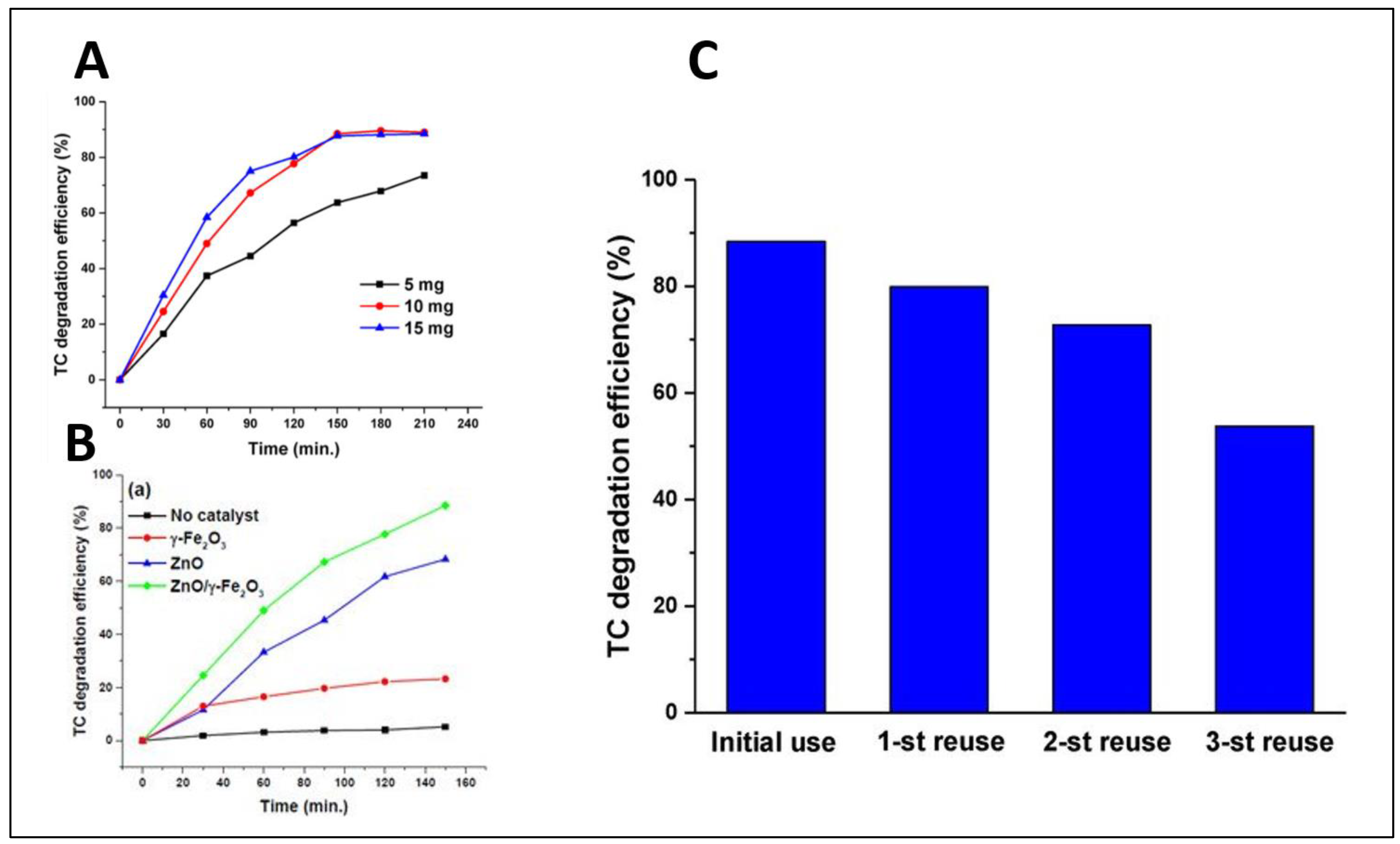
  68. Semeraro, P.; Bettini, S.; Sawalha, S.; Pal, S.; Licciulli, A.; Marzo, F.; Lovergine, N.; Valli, L.; Giancane, G. Photocatalytic Degradation of Tetracycline by ZnO/γ-Fe2O3 Paramagnetic Nanocomposite Material. Nanomaterials 2020, 10, 1458. [Google Scholar] [CrossRef]
  69. Tahir, M.B.; Nabi, G.; Rafique, M.; Khalid, N.R. Nanostructured-based WO3 photocatalysts: Recent development, activity enhancement, perspectives and applications for wastewater treatment. Int. J. Environ. Sci. Technol. 2017, 14, 2519–2542. [Google Scholar] [CrossRef]
  70. Nguyen, T.T.; Nam, S.-N.; Son, J.; Oh, J. Tungsten Trioxide (WO3)-assisted Photocatalytic Degradation of Amoxicillin by Simulated Solar Irradiation. Sci. Rep. 2019, 9, 1–18. [Google Scholar] [CrossRef]
  71. Gholamiabc, P.; Khataeeade, A.; Bhatnagarb, A. Photocatalytic degradation of antibiotic and hydrogen production using diatom-templated 3D WO3−x@mesoporous carbon nanohybrid under visible light irradiation. J. Clean. Prod. 2020, 275, 124157. [Google Scholar] [CrossRef]
  72. Huang, Z.-F.; Song, J.; Pan, L.; Lv, F.; Wang, Q.; Zou, J.-J.; Zhang, X.; Wang, L. Mesoporous W18O49 hollow spheres as highly active photocatalysts. Chem. Commun. 2014, 50, 10959–10962. [Google Scholar] [CrossRef] [PubMed]
  73. Zhang, Z.; Zhang, Z. Bismuth-based photocatalytic semiconductors: Introduction, challenges and possible approaches. J. Mol. Catal. A Chem. 2016, 423, 533–549. [Google Scholar] [CrossRef]
  74. Oudghiri-Hassani, H.; Rakass, S.; Al Wadaani, F.; Al-Ghamdi, K.J.; Omer, A.; Messali, M.; Abboudi, M. Synthesis, characterization and photocatalytic activity of α-Bi2O3 nanoparticles. J. Taibah Univ. Sci. 2015, 9, 508–512. [Google Scholar] [CrossRef] [Green Version]
  75. Bierlein, J.D.; Sleight, A.W. Ferroelasticity in BiVO4. Solid State Commun. 1975, 16, 69–70. [Google Scholar] [CrossRef]
  76. Gotić, M.; Musić, S.; Ivanda, M.; Šoufek, M.; Popović, S. Synthesis and characterisation of bismuth(III) vanadate. J. Mol. Struct. 2005, 744, 535–540. [Google Scholar] [CrossRef]
  77. Ye, L.; Su, Y.; Jin, X.; Xie, H.; Zhang, C. Recent advances in BiOX (X = Cl, Br and I) photocatalysts: Synthesis, modification, facet effects and mechanisms. Environ. Sci. Nano 2014, 1, 90–112. [Google Scholar] [CrossRef]
  78. Zhang, C.; Li, Y.; Shuai, D.; Shen, Y.; Xiong, W.; Wang, L. Graphitic carbon nitride (g-C3N4)-based photocatalysts for water disinfection and microbial control: A review. Chemosphere 2019, 214, 462–479. [Google Scholar] [CrossRef]
  79. Cao, S.; Low, J.; Cheng, B.; Jaroniec, M. Polymeric Photocatalysts Based on Graphitic Carbon Nitride. Adv. Mater. 2015, 27, 2150–2176. [Google Scholar] [CrossRef]
  80. Chen, W.; Jiang, D.; Zhu, M.; Shi, T.; Li, H.; Wang, K. An effective strategy for fabricating highly dispersed nanoparticles on O-C3N4 with enhanced electrocatalytic activity and stability. J. Alloys Compd. 2018, 741, 1203–1211. [Google Scholar] [CrossRef]
  81. Zhang, M.; Dai, Y.; Zhang, S.; Chen, W. Highly efficient photocatalytic activity of boron-doped TiO2 for gas phase degradation of benzene. Rare Met. 2011, 30, 243–248. [Google Scholar] [CrossRef]
  82. Gao, B.; Wang, J.; Dou, M.; Huang, X.; Yu, X. Novel nitrogen-rich g-C3N4 with adjustable energy band by introducing triazole ring for cefotaxime removal. Sep. Purif. Technol. 2020, 241, 116576. [Google Scholar] [CrossRef]
  83. Nguyen-Le, M.-T.; Jitae, K.; Giang, B.L.; Al Tahtamouni, T.; Huong, P.T.; Lee, C.; Nguyen, M.V.; Trung, D.Q. Ag-doped graphitic carbon nitride photocatalyst with remarkably enhanced photocatalytic activity towards antibiotic in hospital wastewater under solar light. J. Ind. Eng. Chem. 2019, 80, 597–605. [Google Scholar] [CrossRef]
  84. Cao, S.-W.; Yuan, Y.; Barber, J.; Loo, J.S.C.; Xue, C. Noble-metal-free g-C3N4/Ni(dmgH)2 composite for efficient photocatalytic hydrogen evolution under visible light irradiation. Appl. Surf. Sci. 2014, 319, 344–349. [Google Scholar] [CrossRef]
  85. Miller, D.R.; Swenson, D.C.; Gillan, E.G. Synthesis and Structure of 2,5,8-Triazido-s-Heptazine: An Energetic and Luminescent Precursor to Nitrogen-Rich Carbon Nitrides. J. Am. Chem. Soc. 2004, 126, 5372–5373. [Google Scholar] [CrossRef]
  86. Fang, J.; Fan, H.; Li, M.; Long, C. Nitrogen self-doped graphitic carbon nitride as efficient visible light photocatalyst for hydrogen evolution. J. Mater. Chem. A 2015, 3, 13819–13826. [Google Scholar] [CrossRef]
  87. Starukh, H.; Praus, P. Doping of Graphitic Carbon Nitride with Non-Metal Elements and Its Applications in Photocatalysis. Catalysts 2020, 10, 1119. [Google Scholar] [CrossRef]
  88. Imam, S.S.; Adnan, R.; Kaus, N.H.M. Photocatalytic degradation of ciprofloxacin in aqueous media: A short review. Toxicol. Environ. Chem. 2018, 100, 518–539. [Google Scholar] [CrossRef]
  89. Ahmadzadeh, S.; Asadipour, A.; Pournamdari, M.; Behnam, B.; Rahimi, H.R.; Dolatabadi, M. Removal of ciprofloxacin from hospital wastewater using electrocoagulation technique by aluminum electrode: Optimization and modelling through response surface methodology. Process. Saf. Environ. Prot. 2017, 109, 538–547. [Google Scholar] [CrossRef]
  90. Huo, P.; Lu, Z.; Liu, X.; Wu, D.; Liu, X.; Pan, J.; Gao, X.; Guo, W.; Li, H.; Yan, Y. Preparation photocatalyst of selected photodegradation antibiotics by molecular imprinting technology onto TiO2/fly-ash cenospheres. Chem. Eng. J. 2012, 189–190, 75–83. [Google Scholar] [CrossRef]
  91. Das, S.; Ghosh, S.; Misra, A.J.; Tamhankar, A.J.; Mishra, A.; Lundborg, C.S.; Tripathy, S.K. Sunlight Assisted Photocatalytic Degradation of Ciprofloxacin in Water Using Fe Doped ZnO Nanoparticles for Potential Public Health Applications. Int. J. Environ. Res. Public Health 2018, 15, 2440. [Google Scholar] [CrossRef] [PubMed] [Green Version]
  92. Şimşek, E.B. Doping of boron in TiO2 catalyst: Enhanced photocatalytic degradation of antibiotic under visible light irradiation. J. Boron 2017, 2, 18–27. [Google Scholar]
  93. Thiruppathi, M.; Kumar, J.V.; Vahini, M.; Ramalingan, C.; Nagarajan, E. A study on divergent functional properties of sphere-like CuWO4 anchored on 2D graphene oxide sheets towards the photocatalysis of ciprofloxacin and electrocatalysis of methanol. J. Mater. Sci. Mater. Electron. 2019, 30, 10172–10182. [Google Scholar] [CrossRef]
  94. Qu, Y.; Xu, X.; Huang, R.; Qi, W.; Su, R.; He, Z. Enhanced photocatalytic degradation of antibiotics in water over functionalized N,S-doped carbon quantum dots embedded ZnO nanoflowers under sunlight irradiation. Chem. Eng. J. 2020, 382, 123016. [Google Scholar] [CrossRef]
  95. Durán-Álvarez, J.C.; Méndez-Galván, M.; Lartundo-Rojas, L.; Rodríguez-Varela, M.; Ramírez-Ortega, D.; Guerrero-Araque, D.; Zanella, R. Synthesis and Characterization of the All Solid Z-Scheme Bi2WO6/Ag/AgBr for the Photocatalytic Degradation of Ciprofloxacin in Water. Top. Catal. 2019, 62, 1011–1025. [Google Scholar] [CrossRef]
  96. Xu, S.; Xu, S.; Wang, Y.; Sun, X.; Gao, Y.; Gao, B. Enhanced degradation of ciprofloxacin by graphitized mesoporous carbon (GMC)-TiO2 nanocomposite: Strong synergy of adsorption-photocatalysis and antibiotics degradation mechanism. J. Colloid Interface Sci. 2018, 527, 202–213. [Google Scholar] [CrossRef]
  97. Dong, S.; Cui, L.; Zhang, W.; Xia, L.; Zhou, S.; Russell, C.K.; Fan, M.; Feng, J.; Sun, J. Double-shelled ZnSnO3 hollow cubes for efficient photocatalytic degradation of antibiotic wastewater. Chem. Eng. J. 2020, 384, 123279. [Google Scholar] [CrossRef]
  98. Li, Y.; Fu, Y.; Zhu, M. Green synthesis of 3D tripyramid TiO2 architectures with assistance of aloe extracts for highly efficient photocatalytic degradation of antibiotic ciprofloxacin. Appl. Catal. B Environ. 2020, 260, 118149. [Google Scholar] [CrossRef]
  99. Li, Z.; Chen, M.; Hu, H.; Zhang, Q.; Tao, D. Mechanochemical synthesis of novel Pt modified ZnAl-LDH for effective ciprofloxacin photodegradation. J. Solid State Chem. 2020, 290, 121594. [Google Scholar] [CrossRef]
  100. Patel, J.; Singh, A.K.; Carabineiro, S. Assessing the Photocatalytic Degradation of Fluoroquinolone Norfloxacin by Mn:ZnS Quantum Dots: Kinetic Study, Degradation Pathway and Influencing Factors. Nanomaterials 2020, 10, 964. [Google Scholar] [CrossRef]
  101. Yu, H.; Chen, F.; Ye, L.; Zhou, H.; Zhao, T. Enhanced photocatalytic degradation of norfloxacin under visible light by immobilized and modified In2O3/TiO2 photocatalyst facilely synthesized by a novel polymeric precursor method. J. Mater. Sci. 2019, 54, 10191–10203. [Google Scholar] [CrossRef]
  102. Jin, X.; Zhou, X.; Sun, P.; Lin, S.; Cao, W.; Li, Z.; Liu, W. Photocatalytic degradation of norfloxacin using N-doped TiO2: Optimization, mechanism, identification of intermediates and toxicity evaluation. Chemosphere 2019, 237, 124433. [Google Scholar] [CrossRef]
  103. Zhang, R.; Zhao, C.; Zhang, T.; Han, Q.; Li, Y.; Liu, Y.; Zeng, K. Ternary Z-Scheme Heterojunction of Bi2WO6 with Reduced Graphene Oxide (rGO) and Bi25FeO40 for Enhanced Visible-Light Photocatalysis. J. Inorg. Organomet. Polym. Mater. 2019, 30, 2152–2162. [Google Scholar] [CrossRef]
  104. Zhang, R.; Han, Q.; Li, Y.; Zhang, T.; Liu, Y.; Zeng, K.; Zhao, C. Fabrication and characterization of high efficient Z-scheme photocatalyst Bi2MoO6/reduced graphene oxide/BiOBr for the degradation of organic dye and antibiotic under visible-light irradiation. J. Mater. Sci. 2019, 54, 14157–14170. [Google Scholar] [CrossRef]
  105. Zhang, R.; Li, Y.; Han, Q.; Zhang, T.; Liu, Y.; Zeng, K.; Zhao, C. Investigation the High Photocatalytic Activity of Magnetically Separable Graphene Oxide Modified BiOBr Nanocomposites for Degradation of Organic Pollutants and Antibiotic. J. Inorg. Organomet. Polym. Mater. 2020, 30, 1703–1715. [Google Scholar] [CrossRef]
  106. Liu, G.; Wang, F.; Feng, Y.; Xie, Z.; Zhang, Q.; Jin, X.; Liu, H.; Liu, Y.; Lv, W.; Liu, G. Facile synthesis of carbon quantum dots loaded with mesoporous g-C3N4 for synergistic absorption and visible light photodegradation of fluoroquinolone antibiotics. Dalton Trans. 2018, 47, 1284–1293. [Google Scholar] [CrossRef]
  107. Li, S.; Hu, J. Photolytic and photocatalytic degradation of tetracycline: Effect of humic acid on degradation kinetics and mechanisms. J. Hazard. Mater. 2016, 318, 134–144. [Google Scholar] [CrossRef]
  108. Wu, S.; Hu, H.; Lin, Y.; Zhang, J.; Hu, Y.H. Visible light photocatalytic degradation of tetracycline over TiO2. Chem. Eng. J. 2020, 382, 122842. [Google Scholar] [CrossRef]
  109. Lv, C.; Lan, X.; Wang, L.; Dai, X.; Zhang, M.; Cui, J.; Yuan, S.; Wang, S.; Shi, J. Rapidly and highly efficient degradation of tetracycline hydrochloride in wastewater by 3D IO-TiO2-CdS nanocomposite under visible light. Environ. Technol. 2019, 1–11. [Google Scholar] [CrossRef] [PubMed]
  110. Jodeyri, M.; Haghighi, M.; Shabani, M. Enhanced-photoreduction deposition of Ag over sono-dispersed C3N4-Clinoptilolite used as nanophotocatalyst for efficient photocatalytic degradation of tetracycline antibiotic under simulated solar-light. J. Mater. Sci. Mater. Electron. 2019, 30, 13877–13894. [Google Scholar] [CrossRef]
  111. Yan, W.; Yan, L.; Jing, C. Impact of doped metals on urea-derived g-C3N4 for photocatalytic degradation of antibiotics: Structure, photoactivity and degradation mechanisms. Appl. Catal. B Environ. 2019, 244, 475–485. [Google Scholar] [CrossRef]
  112. Jiang, L.; Yuan, X.; Zeng, G.; Chen, X.; Wu, Z.; Liang, J.; Zhang, J.; Wang, H.; Wang, H. Phosphorus- and Sulfur-Codoped g-C3N4: Facile Preparation, Mechanism Insight, and Application as Efficient Photocatalyst for Tetracycline and Methyl Orange Degradation under Visible Light Irradiation. ACS Sustain. Chem. Eng. 2017, 5, 5831–5841. [Google Scholar] [CrossRef]
  113. Wang, K.-L.; Li, Y.; Sun, T.; Mao, F.; Wu, J.-K.; Xue, B. Fabrication of Na, Cl co-doped graphitic carbon nitride with enhanced photocatalytic activity for degradation of dyes and antibiotics. J. Mater. Sci. Mater. Electron. 2019, 30, 4446–4454. [Google Scholar] [CrossRef]
  114. Xue, J.; Ma, S.; Zhou, Y.; Zhang, Z.; He, M. Facile Photochemical Synthesis of Au/Pt/g-C3N4 with Plasmon-Enhanced Photocatalytic Activity for Antibiotic Degradation. ACS Appl. Mater. Interfaces 2015, 7, 9630–9637. [Google Scholar] [CrossRef]
  115. Wang, J.; Liu, W.; Zhong, D.; Ma, Y.; Ma, Q.; Wang, Z.; Pan, J. Fabrication of bismuth titanate nanosheets with tunable crystal facets for photocatalytic degradation of antibiotic. J. Mater. Sci. 2019, 54, 13740–13752. [Google Scholar] [CrossRef]
  116. Zhou, J.; Jiang, L.; Chen, D.; Liang, J.; Qin, L.; Bai, L.; Sun, X.; Huang, Y. Facile synthesis of Er-doped BiFeO3 nanoparticles for enhanced visible light photocatalytic degradation of tetracycline hydrochloride. J. Sol-Gel Sci. Technol. 2019, 90, 535–546. [Google Scholar] [CrossRef]
  117. Wang, W.; Han, Q.; Zhu, Z.; Zhang, L.; Zhong, S.; Liu, B. Enhanced photocatalytic degradation performance of organic contaminants by heterojunction photocatalyst BiVO4/TiO2/RGO and its compatibility on four different tetracycline antibiotics. Adv. Powder Technol. 2019, 30, 1882–1896. [Google Scholar] [CrossRef]
  118. Tun, P.P.; Wang, J.; Khaing, T.T.; Wu, X.; Zhang, G. Fabrication of functionalized plasmonic Ag loaded Bi2O3/montmorillonite nanocomposites for efficient photocatalytic removal of antibiotics and organic dyes. J. Alloy. Compd. 2020, 818, 152836. [Google Scholar] [CrossRef]
  119. Cao, H.-L.; Cai, F.-Y.; Yu, K.; Zhang, Y.-Q.; Lü, J.; Cao, R. Photocatalytic Degradation of Tetracycline Antibiotics over CdS/Nitrogen-Doped–Carbon Composites Derived from in Situ Carbonization of Metal–Organic Frameworks. ACS Sustain. Chem. Eng. 2019, 7, 10847–10854. [Google Scholar] [CrossRef]
  120. Xue, J.; Ma, S.; Zhou, Y.; Zhang, Z.; Jiang, P. Synthesis of Ag/ZnO/C plasmonic photocatalyst with enhanced adsorption capacity and photocatalytic activity to antibiotics. RSC Adv. 2015, 5, 18832–18840. [Google Scholar] [CrossRef]
  121. Długosz, M.; Żmudzki, P.; Kwiecień, A.; Szczubiałka, K.; Krzek, J.; Nowakowska, M. Photocatalytic degradation of sulfamethoxazole in aqueous solution using a floating TiO2-expanded perlite photocatalyst. J. Hazard. Mater. 2015, 298, 146–153. [Google Scholar] [CrossRef] [PubMed]
  122. Baran, W.; Sochacka, J.; Wardas, W. Toxicity and biodegradability of sulfonamides and products of their photocatalytic degradation in aqueous solutions. Chemosphere 2006, 65, 1295–1299. [Google Scholar] [CrossRef] [PubMed]
  123. Tsiampalis, A.; Frontistis, Z.; Binas, V.; Kiriakidis, G.; Mantzavinos, D. Degradation of Sulfamethoxazole Using Iron-Doped Titania and Simulated Solar Radiation. Catalysts 2019, 9, 612. [Google Scholar] [CrossRef] [Green Version]
  124. Ling, C.; Yue, C.; Yuan, R.; Qiu, J.; Liu, F.; Zhu, J.-J. Enhanced removal of sulfamethoxazole by a novel composite of TiO2 nanocrystals in situ wrapped-Bi2O4 microrods under simulated solar irradiation. Chem. Eng. J. 2020, 384, 123278. [Google Scholar] [CrossRef]
  125. Durán-Álvarez, J.C.; Santiago, A.L.; Ramírez-Ortega, D.; Acevedo-Peña, P.; Castillón, F.; Ramírez-Zamora, R.M.; Zanella, R. Surface modification of B–TiO2 by deposition of Au nanoparticles to increase its photocatalytic activity under simulated sunlight irradiation. J. Sol-Gel Sci. Technol. 2018, 88, 474–487. [Google Scholar] [CrossRef]
  126. Naraginti, S.; Yu, Y.-Y.; Fang, Z.; Yong, Y.-C. Novel tetrahedral Ag3PO4@N-rGO for photocatalytic detoxification of sulfamethoxazole: Process optimization, transformation pathways and biotoxicity assessment. Chem. Eng. J. 2019, 375, 122035. [Google Scholar] [CrossRef]
  127. Dhiman, P.; Dhiman, N.; Kumar, A.; Sharma, G.; Naushad, M.; Ghfar, A.A. Solar active nano-Zn1−xMgxFe2O4 as a magnetically separable sustainable photocatalyst for degradation of sulfadiazine antibiotic. J. Mol. Liq. 2019, 294, 111574. [Google Scholar] [CrossRef]
  128. Do, T.C.M.V.; Nguyen, D.Q.; Nguyen, K.T.; Le, P.H. TiO2 and Au-TiO2 Nanomaterials for Rapid Photocatalytic Degradation of Antibiotic Residues in Aquaculture Wastewater. Materials 2019, 12, 2434. [Google Scholar] [CrossRef] [Green Version]
  129. Salimi, M.; Behbahanib, M.; Sobhi, H.R.; Gholami, M.; Jafari, A.J.; Kalantari, R.R.; Farzadkia, M.; Esrafili, A. A new nano-photocatalyst based on Pt and Bi co-doped TiO2 for efficient visible-light photo degradation of amoxicillin. New J. Chem. 2019, 43, 1562–1568. [Google Scholar] [CrossRef]
Figure 1. Possible routes of entry of antibiotics into wastewater. Adapted from [30] with modifications.
Catalysts 11 00437 g001
Figure 2. General photocatalytic mechanism. Adapted from [38] with modifications.
Catalysts 11 00437 g002
Figure 3. The down-conversion phosphor way of action. Adapted with permission from [53]. Copyright 2015 Elsevier.
Catalysts 11 00437 g003
Figure 4. Ciprofloxacin (CIP) chemical structure.
Catalysts 11 00437 g004
Figure 5. Different attack sites in CIP structure by free radicals generated by photocatalytic reaction.
Catalysts 11 00437 g005
Figure 6. Degradation of CIP over time under different conditions. (A) the photocatalytic degradation of 10 mg/L CIP using 150 mg/L Fe-ZnO when irradiated with sunlight. The degradation was tested using different pH levels. The best photocatalytic efficiency is near a pH = 9. (B) represents the effect of temperature on CIP photocatalytic degradation at pH = 9. An unexpected trend was obtained as a decreasing in degradation efficiency when increasing the temperature up to 60 °C. C/C0 represents the relative change in CIP with respect to the initial concentration. Adapted from [91].
Catalysts 11 00437 g006
Figure 7. The proposed mechanism for the degradation of CIP using layered double hydroxides (LDHs) Pt hybrid. ESR spin trap experiment was used to determine the dominant free radicals. Scavengers like ascorbic acid, EDTA-2Na and tert-butyl alcohol were used to trap superoxide anion radicals, photogenerated holes, and hydroxyl radicals. Here, metallic Pt acts as springboard that facilitates charge separation and electron movement through Pt/LDH heterojunction interfaces. When Pt is attached to the LDH surface, surface recombination is reduced as the photogenerated electrons transfer to Pt and cannot return due to the presence of Schottky barrier. Simultaneously, the photogenerated holes migrates to LDH external surface and degrades CIP by reacting with H2O to form. OH. Adapted with permission from [99]. Copyright 2020 Elsevier.
Catalysts 11 00437 g007
Figure 8. Proposed mechanism for the degradation of norfloxacin; four main pathways are presented. High-resolution liquid chromatography mass spectrometry was used to determine the produced degradation intermediates. There are 4 suggested pathways; in the first pathway, the norfloxacin is degraded by dehydroxylation reaction. In the second pathway, structure 2 is produced by defluorination reaction, which is transmitted to structure 3 by decarboxylation and deethylation reaction. The third pathway is characterized mainly by the removal of piperazinyl group; in this pathway, ring opening occurs, and 6 intermediates are produced. The fourth pathway is characterized by the opening of the quinolone substituent and the benzene moiety. Adapted from [100].
Catalysts 11 00437 g008
Figure 9. Different parameters were applied to degrade the antibiotic norfloxacin using Mn:ZnS Quantum Dots. (A) showing the effect of pH on norfloxacin, with pH = 10 owining the best photocatalytic efficiency(B) represents a bar chart with the effect of the number of catalysts used. (C) signifies the effect of the initial antibiotic concentration. Adapted from [100].
Catalysts 11 00437 g009
Figure 10. Tetracycline chemical structure.
Catalysts 11 00437 g010
Figure 11. A proposed tetracycline degradation mechanism. The structures of these intermediates were suggested based on their retention time, the fragmentation pattern from the mass spectra and from structures suggested by other studies. There are two pathways of degradation based on the intermediate degradants. In the first pathway, two intermediates are produced, where P5 was produced by the addition of an OH radical to the C11a–C12 double-bond, which then transforms to P4 by H-abstraction at C5a by hydroxyl radical attack. In the second pathway the first intermediate (P3) was produced by the elimination of –CH2, group which either transforms to P1 by dehydration reaction of P2 or by the loss of –CH2 group. Adapted with permission from [107]. Copyright 2016 Elsevier.
Catalysts 11 00437 g011
Figure 12. (A) shows the effect of different ZnO/γ-Fe2O3 concentrations of tetracycline degradation, in which 15 mg showed the highest degradation efficiency (B) represents the TC (%) vs. irradiation time (min) graph with different materials with the best tetracycline degradation efficiency of ZnO/γ-Fe2O3 (C) represents the tetracycline degradation efficiency with tr, using the prepared ZnO/γ-Fe2O3 with slight degradation in the material efficiency after 3 cycles of re-use. Adapted from [68].
Catalysts 11 00437 g012
Figure 13. A proposed mechanism for the degradation of sulfamethoxazole antibiotic using (EP-TiO2-773). The degradation was determined by HPLC analysis, which showed the disappearance of sulfamethoxazole peaks and the appearance of new peaks. Two pathways are mainly involved. The first pathway involves the photogeneration of hydroxyl radicals which resulted from the interaction of holes with water. The second pathway involves the electron transfer from the excited organic molecule to TiO2 conduction band. Adapted with permission from [121]. Copyright 2015 Elsevier.
Catalysts 11 00437 g013
Figure 14. (A) Signifies the effect of iron doping level, where 0.5% Fe/TiO2 showed the most efficient photocatalytic degradation. (B) represents the effect of initial SMZ concertation; as noted, there is a decrement in the rate constant as the concentration of SMZ increases. Adapted with permission from [123]. Copyright 2019 Elsevier.
Catalysts 11 00437 g014
Publisher’s Note: MDPI stays neutral with regard to jurisdictional claims in published maps and institutional affiliations.

