Article

Controlled Synthesis of Au Nanoparticles by Modified Polyol Methods: Determination of Their Size, Shape, and Crystal Structure

1
Institute of Applied Technology, Thu Dau Mot University, Thu Dau Mot City, Vietnam
2
State Key Laboratory of High Performance Ceramics and Superfine Microstructures, Shanghai Institute of Ceramics, Chinese Academy of Sciences, 1295 Dingxi Road, Shanghai 200050, China
3
Faculty of Chemical Engineering, Ho Chi Minh City University of Technology, 268 Ly Thuong Kiet, District 10, Ho Chi Minh City 700000, Vietnam
4
Institute of Research and Development, Duy Tan University, Da Nang 55000, Vietnam
5
Ho Chi Minh City Institute of Physics, 1 Mac Dinh Chi Str., Ben Nghe, District 1, Ho Chi Minh City 700000, Vietnam
6
Faculty of Electronics and Telecommunications, Saigon University, 273 An Duong Vuong, District 5, Ho Chi Minh City 700000, Vietnam
*
Author to whom correspondence should be addressed.
Submission received: 2 September 2021 / Revised: 11 October 2021 / Accepted: 21 October 2021 / Published: 26 October 2021
(This article belongs to the Special Issue Confined Crystals, Quantum Dots, and Nano Crystals)

Abstract

:
We successfully prepared Au nanoparticles using the modified polyol methods and design of experiments. The desirable crystal structure and particle size of Au nanoparticles with various kinds of polyhedral and spherical shapes as well as various kinds of their morphologies or complete and rough spherical crystal surfaces were experimentally predicted in TEM and XRD measurements according to the theoretical calculation and data. The fine crystal formation of Au nanostructures by modified polyol methods with EG, PVP, and NaBH4 is of great importance to their practical applications. Our research shows that the critical nucleation, growth, and formation of sizes, shapes, and morphologies of Au nanoparticles were experimentally discussed in modified polyol methods and design of experiments. In this context, our particle size calculation can hold the greatest attraction for researchers in the field of nanoparticles.

1. Introduction

So far, metal nanoparticles have been widely studied because of their practical applications in electronics and telecommunications, catalysis, energy and environment, health and clinic applications [1,2,3,4,5,6,7]. The unique optical properties of Au, Ag, and Cu single-metal nanoparticles (electron clouds) under suitable light excitation (formation of an electric dipole) are the key foundation of nanoscale electronics and telecommunications [4,5]. In their controlled synthesis, modified polyol methods have been previously discussed in the relationship of Au precursors, reducing agents, protective, and structure-controlling agents in ethylene glycol (EG) or polyEG (PEG) [1,2,3,4,5,6,7]. Their promising applications of functional nanoparticles are used in catalysis and nanobiomedicine [7,8]. The common synthetic methods of metal nanoparticles suggest that Au or gold nanoparticles can be effectively synthesized by modified polyol methods [1,2,3,4,5,6,7]. Recently, Au and Au-based nanoparticles have been potentially used for catalysis, energy, and environment, Au nanoparticles in the diagnosis and treatment of melanoma cancer [9], and fluorescence-based assays in cancer and non-cancerous cells. Therefore, many researchers have proposed methods of controlling the nanosize ranges and the definite shapes of Au nanoparticles by changing the experimental conditions [7,8]. In biomedical applications, Au nanoparticles are used as an antigen carrier and an immunological agent in immunization to prepare antibodies in vivo [9]. However, the highest biocompatibility is silver nanoparticles for plants, animals, and humans [9]. In comparison with the traditional sol-gel method for the synthesis of nanomaterials in alcohol, the polyol method is used to synthesize nanomaterials with typical industrial solvents such alcohols, ethylene glycol (EG), and polyethylene glycol (PEG) [2,3,4,5,6,7,8,9,10]. Both sol-gel and polyol methods can lead to homogeneous nanomaterials and nanoparticles because homogeneous industrial solvents and polymers are under mixing continuously. In the controlled synthesis of nanoparticles, EG and PEG are commonly utilized for the effective synthesis of metal nanomaterials and nanoparticles [11]. Both EG and PEG solvents are effectively used with the addition of a diversity of various reductants to Au precursors, and the addition of a diversity of various protective polymers as a stabilizer for nanoparticles such as poly(vinylpyrrolidone) (i.e., PVP) or hexadecyltrimethylammonium bromide (i.e., CTAB), and tetradodecylammonium bromide (i.e., TCAB) in various solvents such as EG or PEG or various polyols or various kinds of water (i.e., H2O) [12]. The synthetic methods, structures, properties, and applications of Au nanoparticles have been discussed in various works [13,14,15,16,17,18,19,20,21]. Among the promising applications, the very interesting phenomena and optical properties of surface plasmon resonance (SPR) and surface-enhanced Raman scattering (SER) have been utilized in biomedicine [1,2,3,4,5,6]. In the present research, we synthesized Au nanoparticles by modified polyol processes with the addition of NaBH4 in EG and PVP solvents according to suitable time and temperature. We used two measurements (i.e., a combination of XRD and TEM methods), in order to characterize the as-prepared Au nanoparticles. The critical issues of the particle size, the particle shape, and the face-centered cubic (FCC) crystal structure of the as-prepared Au nanoparticles are clearly underlined in detailed discussion.

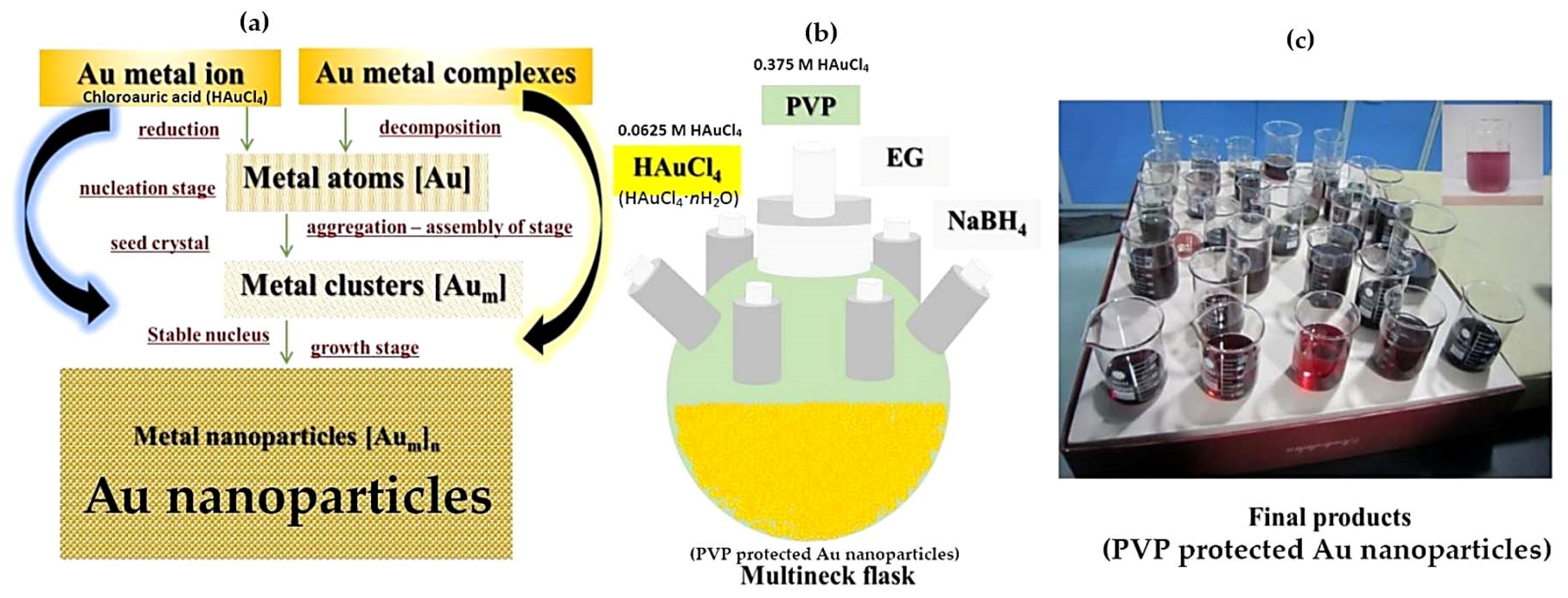
2. Materials and Methods

Chemicals for synthesis were poly(vinylpyrrolidone) (PVP), HAuCl4·3H2O, NaBH4, and ethylene glycol (EG). All chemicals are available from Aldrich or Sigma-Aldrich and meet the synthetic requirements. The addition of NaBH4 was used for the fast crystal formation of Au nanoparticles from HAuCl4. EG was the homogeneous solvent that was used as both a solvent and weak reducing agent. Ethanol, acetone, and hexane were appropriately used for cleaning the very thin layers of PVP covering the surfaces of PVP-protected Au nanoparticles [6,7,8,9,10]. Here, all chemicals were used without any further purification. In one typical process, an amount of EG, an amount of 0.375 M PVP, an amount of 0.0625 M HAuCl4, and NaBH4 were added in a three-neck flask or multineck flask with a reflux system [6]. In the synthetic methodology, the addition of Au, PVP, NaBH4, and other precursors was carried out until all their amounts were completely used with continuous stirring (Scheme 1). The good reduction of HAuCl4 by both EG and NaBH4 occurred according to our design processes, which depended on the technique and experience of the experimenter [10,11,12,13]. In order to remove PVP that existed on the surfaces of as-prepared Au nanoparticles, they were cleaned with an acetone and ethanol/hexane mixture. Finally, clean Au nanoparticles were successfully obtained by the centrifugation procedure. For a successful synthesis of the modified polyol method, the ratios of the volumes and concentrations of the reagents (Au precursors for crystal formation of Au nanoparticles, EG solvent, PVP polymer for the protection of Au nanoparticles, NaBH4, etc.) need to be selected suitably according to the process used. In the study of crystal formation, both XRD and TEM methods were effectively used for the investigation of the crystal structure of as-prepared Au nanoparticles by the polyol process. It is ensured that the final products of Au nanoparticles were studied by TEM and XRD measurements after the removal of PVP. Here, the X-ray diffraction (XRD) method was used for structural information by identifying the crystalline phases of Au nanoparticles within the prepared samples. Here, the Au nanoparticles with the removal of PVP were set on a glass substrate. We used an X-ray diffractometer that was Rigaku D (max 2550 V) operating at 40 kV/200 mA, and Cu-Kα1 radiation at 1.54056 Å. In this study, the FCC crystal structure of Au nanoparticles was proven. In the surface analysis, the JEM-2100F analytical electron microscope was used with standard magnification up to 1,500,000 times, ultra-high image quality and resolution operating at 200 kV, around a 0.1 nm lattice resolution, and around 0.19 nm point resolution. The images of Au nanoparticles by JEM-2100F were used to investigate the interesting properties of the size and the shape of Au nanoparticles.

3. Results and Discussion

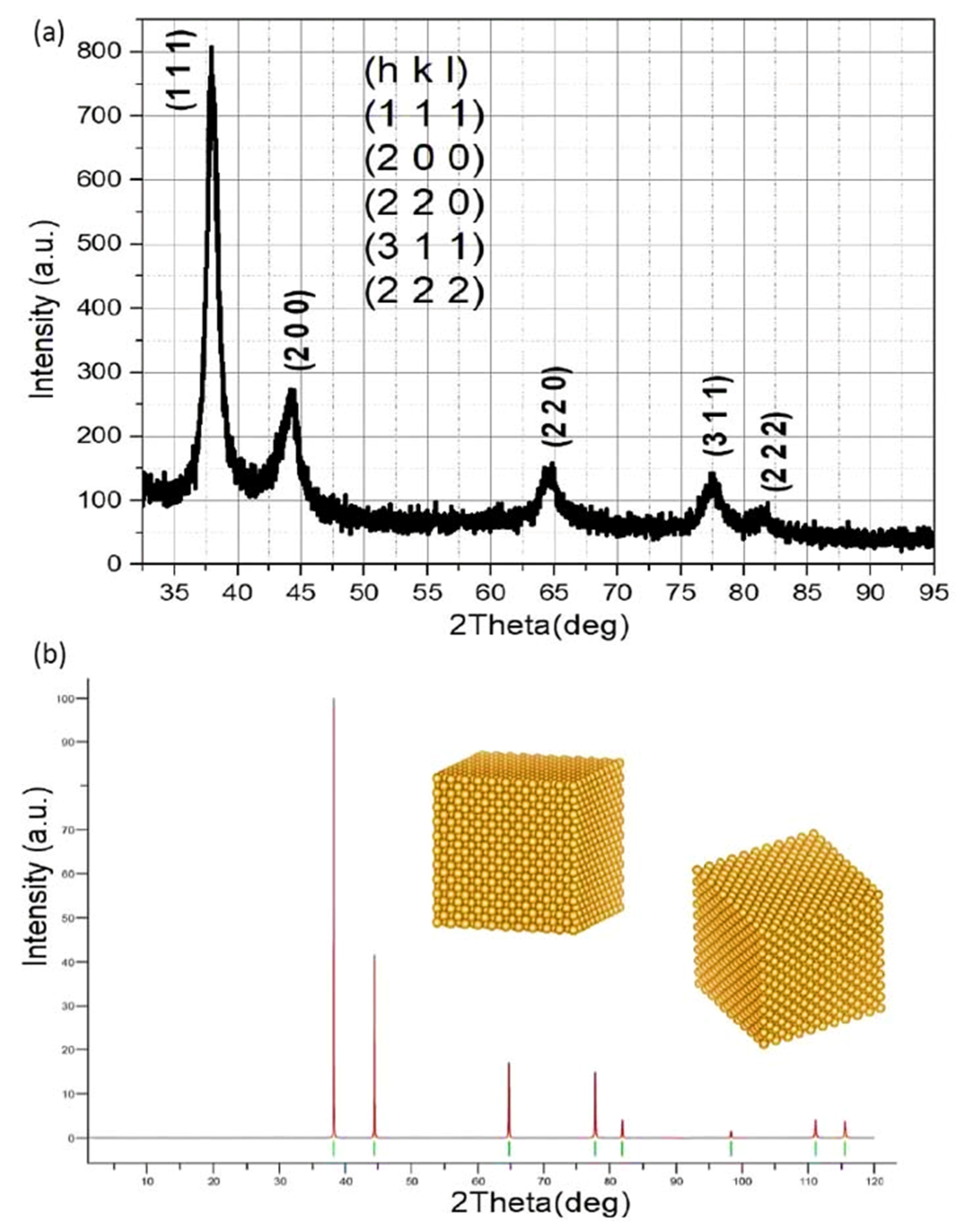
3.1. Determination of Crystal Structure

The exact determination of the crystal structure of Au nanoparticles needs to be undertaken in any experiment. The Au crystal structure of a typical XRD pattern was described by the positions, intensities, and shapes of Bragg reflections from 32.5° to 95° corresponding to the experiment, and from 0° to 120° corresponding to the theory (Figure 1). The crystal structure of the as-prepared Au nanoparticles was very good, which was found in the crystal formation in high crystallization in EG solvent in this polyol method. Therefore, XRD was effectively used to determine the crystal structure of the clean Au nanoparticle PVP formed in EG with the removal of PVP. The results have shown the formation of the crystal structure of the as-prepared Au nanoparticles with high crystallization (Figure 1a). It indicates that all the as-synthesized Au nanoparticles have a crystal structure in high crystallization. In analysis and investigation, we showed the crystal structure of one of the most typical samples prepared by modified polyol methods with the use of a controlling agent (i.e., NaBH4 (reducing agents), AgNO3 (i.e., crystal structure-controlling agents), PVP (i.e., protecting agents) [6,10]). It is known that the crystal formation of Au nanoparticles was confirmed in various homogeneous polyol liquids (i.e., EG, polyEG…). It is certain that Au nanoparticles have a FCC crystal structure from experimental observations and theory [6,13,14,15,16,17,18,19,20,21]. There is a comparison between one sample by XRD where the diffraction peaks at 2θ angles of the prepared Au nanoparticles with the fcc structure were similar to other studies [6,14,15,16,17]. Obviously, Au nanoparticles were determined by the positions of the important diffraction peaks that were shown with the 2θ angles at 38.20°, 44.40°, 64.60°, 77.54°, and 81.72°, corresponding to the fine crystal planes at (111), (200), (220), (311), and (222) or more Miller indices. However, the intensities of their diffraction angles and peaks were very different. In addition, their similar crystal parameters were listed in the PDF-04-0784 standard sample of Au as well as the powder diffraction pattern from the Au crystal model of ten-unit cells. For the determination of crystal structure, the typical values of the distances between atomic planes of Au nanoparticles were about 2.35, 2.03, 1.44, 1.23, and 1.17 Å, corresponding to the (111), (200), (220), (311), and (222) planes, respectively. It is important to note that the intensity of the strongest line, as shown in Figure 1, is the so-called (111) plane. In addition, high crystallization of Au nanoparticles coincided with previous works [15,16,17]. Therefore, the fine crystal phase of as-prepared Au nanoparticles by our polyol process with the addition of NaBH4 was experimentally confirmed because a mixture of EG and PVP was very highly homogeneously mixed in the crystal formation of Au nanoparticles. In Figure 1, the typical crystal parameters such as the values of (h k l), d, θ, 2θ, and I are measured or calculated by nanostructures of Au nanoparticles show very good agreement between the theory and experiment.
Recently, authors have successfully synthesized Au nanoparticles by seeded growth methods [1,2,3,4,5,6]. In general, they have used various kinds of capping agents [12] that are different from the use of PVP as a capping agent [1,2,3,4,5,6,7,8,9,10,11,12]. Recently, NaBH4 has been used in a different way for the controlled synthesis of Au nanoparticles (i.e., fast, moderate, and slow reduction of Au precursor into Au nanoparticles [1,2,3,4,5,6]). In important considerations, researchers have used the NaBH4 reducing agent for the synthesis of Au nanoparticles [2,3,6,12]. TEM and high resolution (HR) TEM results in typical works of Au nanoparticles were compared to our present results. In addition, SPR and SER optical properties of the Au nanoparticles in solvents and solutions are very crucial in promising applications for plasmonic and Raman sensing [4,5,6]. In size and shape analysis, we only used one of the most typical samples for TEM investigation.

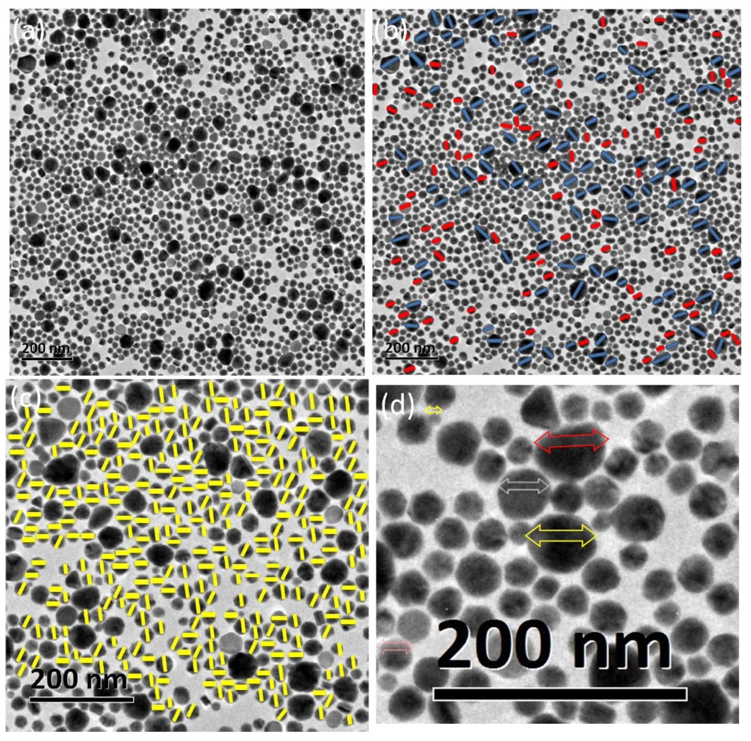
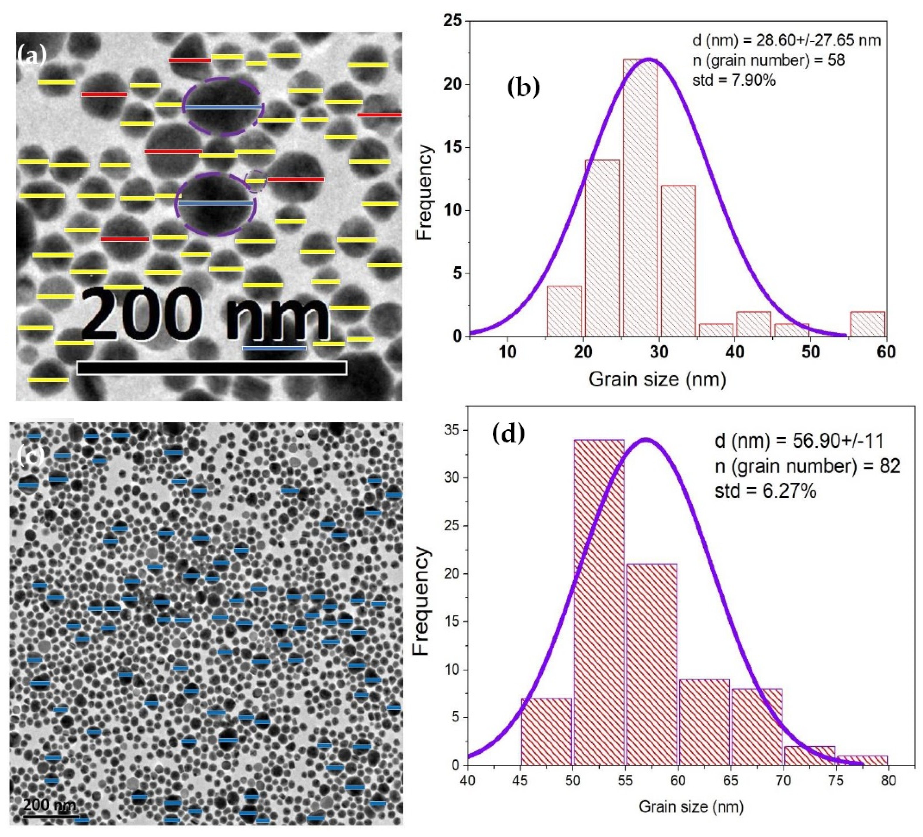
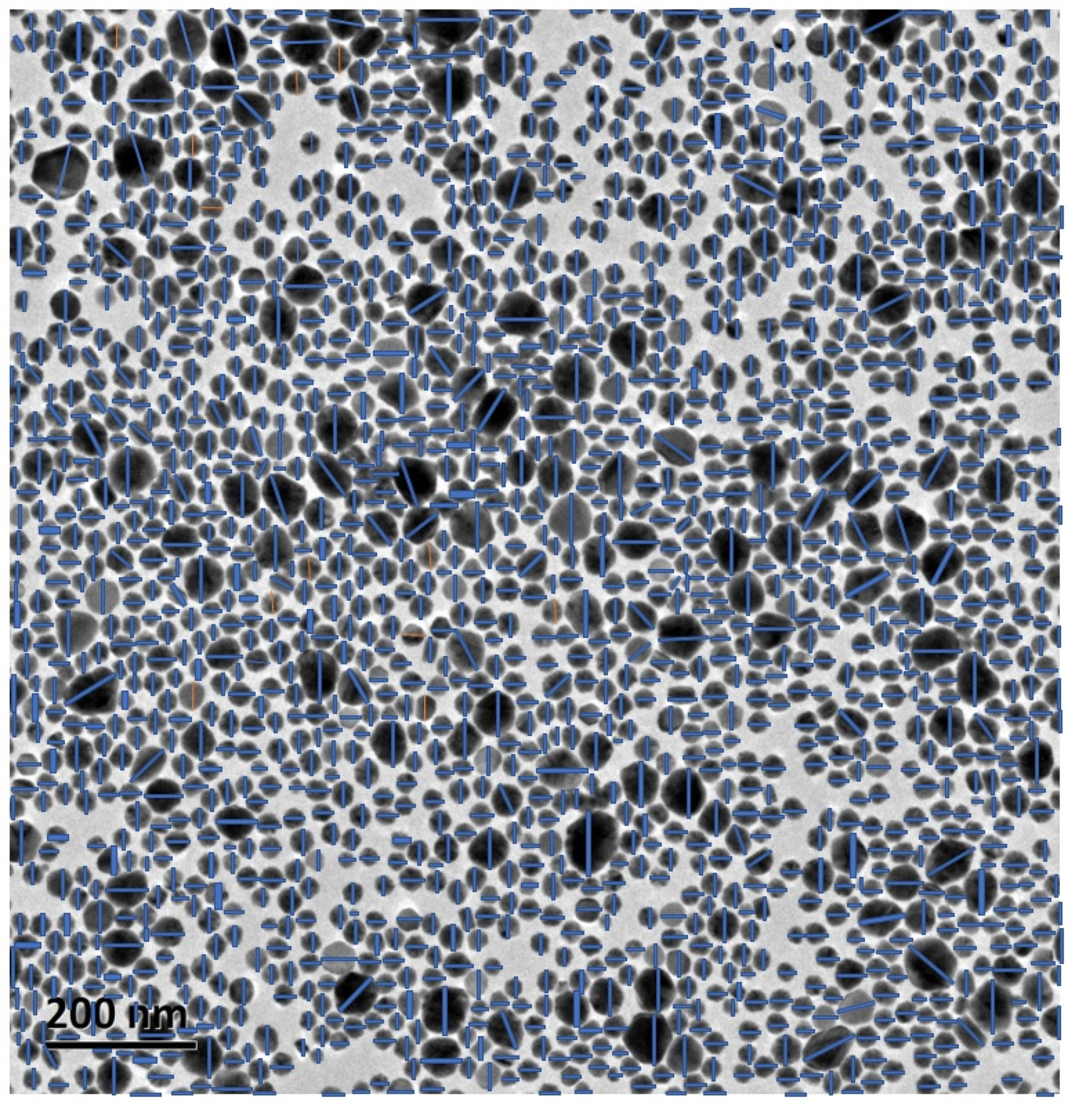
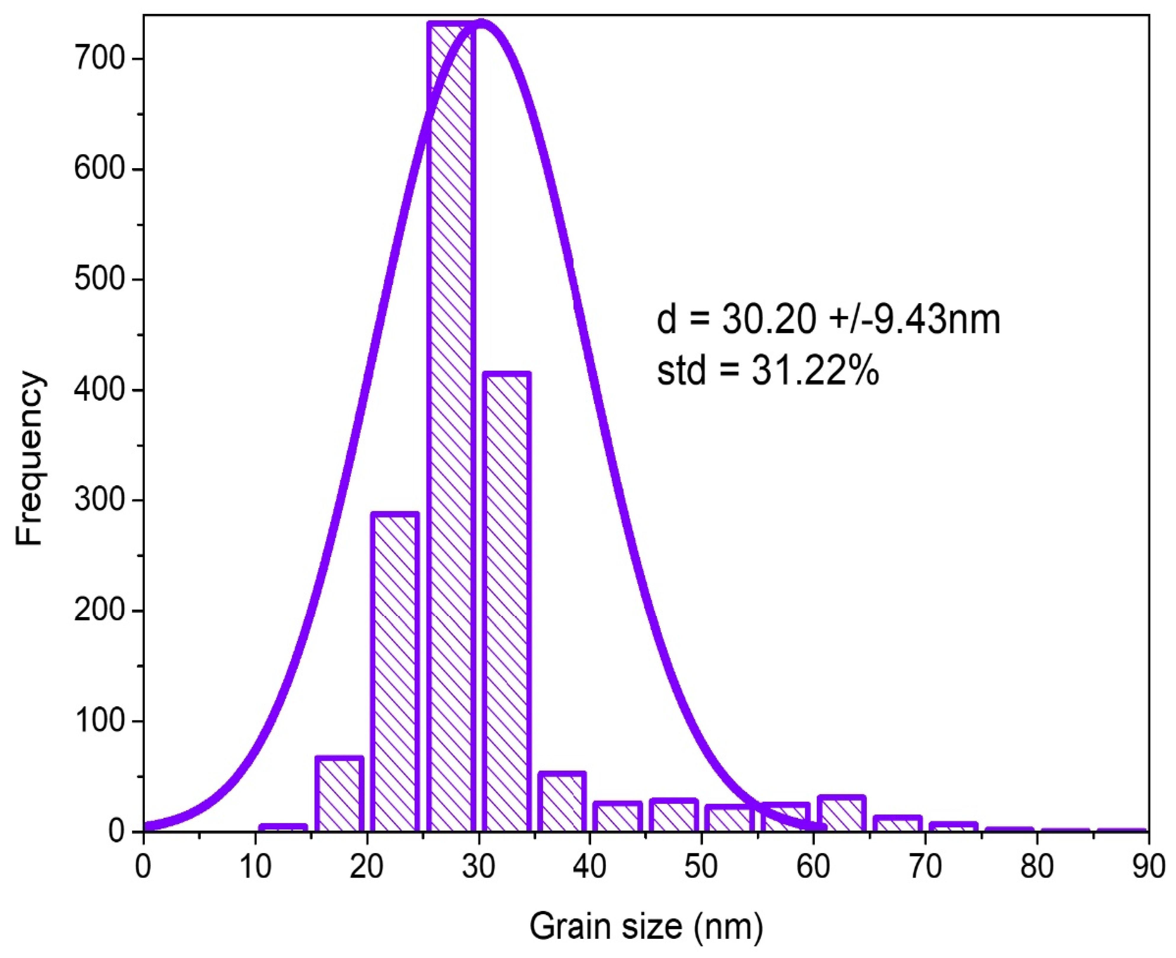
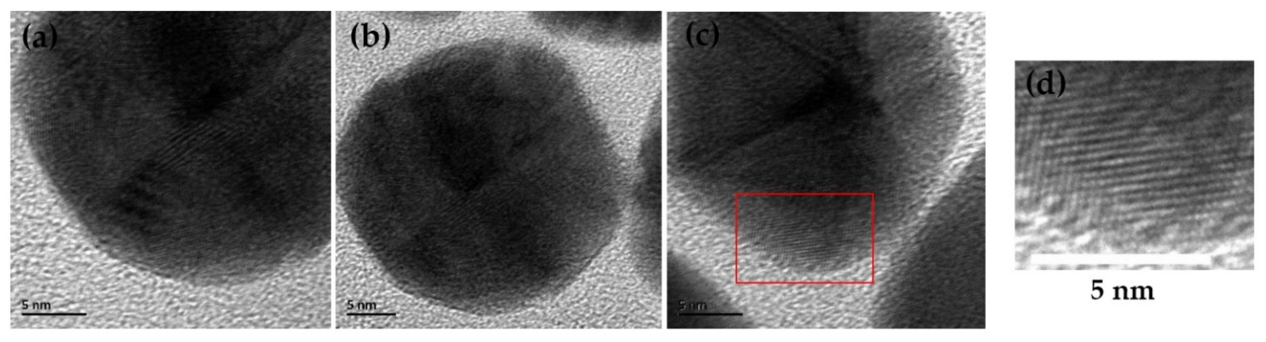
3.2. Determination of Size and Shape of As-Prepared Au Nanoparticles

Figure 2 shows the TEM images of as-prepared Au nanoparticles with the various forms of the fascinating shapes and morphologies such as spheres, polyhedrons, plates, rods, and other undefined shapes in the nanosize range of 60 nm. The various kinds of the sizes of Au nanoparticles were calculated in the ranges of 60 nm, 50 nm, 40 nm, 30 nm, 20 nm, and even 10 nm, of which there was an overlap of particle size ranges according to the smallest and largest particles in a detailed and specific TEM image with a defined bar scale. The largest and smallest particles with the largest and smallest sizes were the limits of the nanoparticle system fabricated in EG. In our process, we prepared a homogeneous nanosystem of Au nanoparticles in a homogeneous mixture of EG and PVP [6,12]. Their common shapes were experimentally found to be the most typical spherical and polyhedral forms. The high roughness or/and flatness surfaces of the as-prepared spherical and polyhedral Au nanoparticles were clearly observed in their morphologies. Here, many Au nanoparticles possessed various large sizes and shapes, but were less than 100 nm. In addition, the self-assembly, combination, and recrystallization of smaller Au nanoparticles less than 10 nm could lead to large nanoparticles, even greater than 100 nm. The largest particles were around 50 nm by the self-assembly of smaller particles [25,26]. Therefore, the large particles result from the combinations of smaller particles, which would be through mechanical motions, collisions, combination, recrystallization, and formations.
In our particle size measurement, a strictly scientific study of particle size and a large number of Au nanoparticles fabricated by the designed polyol processes led to being able to obtain information for any significant improvement of further synthetic processes. To calculate the particle size of the as-prepared Au nanoparticles, it is very necessary to calculate the maximum and minimum sizes of the largest and smallest Au particles, a group of the Au particles seen, and all Au particles measured on that TEM image (Figure 2a). There are many ways to calculate the average particle sizes of large particles, medium size particles, and small particles (Figure 2a–d). Figure 2d shows the rough calculation to arrange, in the order of size, the spherical particles, only a small amount of five Au particles from largest to smallest size, which were about 59.00, 55.52, 42.86, 31.50, and 11.69 nm, respectively, which corresponded roughly to the three nanosized regions of particle sizes of Au nanoparticles based on the evidence of the original TEM image in the ranges of 60, 50, 40, and 20 nm, even the possible smallest nanosized range of 10 nm (1–10 nm) when TEM images of Au nanoparticles were carefully calculated and analyzed in this context (Figure 2a). The average size was calculated to be about 40 nm, corresponding to five Au particles. It is certain that this is the simplest way to determine particle size. In addition, Figure 2d shows mostly spherical shapes of Au nanoparticles with definite sizes. The critical issue indicated that the crystal formation of large crystal particles, medium crystal particles, and very small crystal particles was confirmed. The spherical and cubic particle shapes were the best forms to calculate the particle sizes here. The average grain size (d), standard deviation (std), and grain number (n) were calculated in Figure 3, respectively. In Figure 3a, we obtained the average size of about 28 (±27.65 nm) when 58 particles of various sizes from small to large particles were used to calculate the histogram. We obtained an average size of about 56.90 ± 11 nm after using about 82 particles of only large particles, which indicated that there were nanosize ranges <60 nm. Figure 4 and Figure 5 present a special case of calculation where the average size was calculated to be about 30.20 nm (±9.43 nm), which is relatively similar to the value (d) calculated in Figure 3a.
Figure 6 clearly indicates the typical HRTEM images of three Au nanoparticles showing their lattice fringes of about 0.248 nm, which were assigned to the inter-fringe distance of the crystal planes in the FCC Au nanocrystal. The complexity of the Au atom arrangement on the crystal surface of one such Au nanoparticle is of high interest to scientists and researchers in the further investigation of TEM. In terms of size and surface investigation, it is clear that XRD determined the Au crystal structure of the prepared products, while HRTEM indicated the lattice fringes of Au nanoparticles. The self-assembly phenomenon of Au nanoparticles is popular in various nanosize ranges [25,26]. It is predicted that the large particles were formed by self-assembly via the motions, collisions, and combination of smaller particles. Therefore, the EG-type polyol in this case can be the best environment for Au nanoparticles synthesized in terms of the various sizes, various shapes, and the various morphologies. The experimental crystal formation of Au nanoparticles in the chemical reaction was confirmed by self-attachment, self-aggregation, and self-assembly of the much smaller nanoparticles [25,26,27]. The polyols or alcohols were considered as an excellent environment for the existence of the engineered particles such as the case of the Pd nanoparticles [28]. It is suggested that Au nanoparticles were generated in a homogeneous mixture of EG and PVP during chemical mixing and synthetic reaction. In potential clinical applications, the determination of particle size and crystal structure of Au nanoparticles has become increasingly significant as they are used in virus and COVID-19 pandemic research [29], for example, human ACE2-Au-virustrap-nanostructures for test kits in the capture of SARS-CoV-2 and single-virus SERS detection.

4. Conclusions

In this research, we presented the formation of Au nanoparticles synthesized in EG and PVP with the addition of NaBH4. The Au nanoparticles ranged in size up to 60 nm, with an average size of about 30 nm. In the process of manually calculating each particle, errors can occur despite the careful particle-size calculation of the experimenter. However, there were various kinds of Au nanoparticles shaped in the various ranges of 50 nm, 40 nm, 30 nm, and even 10 nm. Typical shapes for nanosized particles in this range were spherical and polyhedral. It is evident that the modified polyol methods can be promisingly used with the addition of a small content of structure-controlling (i.e., NaBH4) and other chemical reagents in any synthetic process. The spherical and cubic shapes were suggested to be the best forms of particle size calculation in this study. In this research, it is suggested that the TEM method is very important to calculate the parameters of particle size, number of Au nanoparticles, and shape of the Au nanoparticles. Therefore, this research also suggests that the experimenter must choose the experimental conditions of pH, fixed reaction volumes of precursors in solvent (for the effective formation of nanoparticles), the solvent (EG, PEG, or other polyols for media generating nanoparticles), surfactant (PVP, CTAB, other protective agents for nanoparticles), reaction time (fast, moderate, or short time), and stirring speed for synthesis.

Author Contributions

Experimenting, collecting data and results, writing the whole original manuscript, discussion: N.V.L.; Checking and discussing the research results: N.T.N.H., Y.Y., M.N., N.Q.T.N., L.H.P., N.H.T. and H.V.C.; Funding, Y.Y. (China, SICCAS) and N.T.N.H. (Vietnam, TDMU). All authors have read and agreed to the published version of the manuscript.

Funding

This research was funded by Sai Gon University, grant number TĐ2020-19.

Institutional Review Board Statement

Not applicable.

Informed Consent Statement

Not applicable.

Data Availability Statement

Not applicable.

Acknowledgments

Our research was supported by Saigon University through grant number TĐ2020-19. The author (N.V.L.) appreciates the kind research assistance from the Universities and Institutes of Japan (Nagoya Institute of Technology, NITECH; Kyushu University, Kyoto University), China (Shanghai Institute of Ceramics, Chines Academy of Sciences), and Vietnam (Sai Gon University, Thu Dau Mot University, Ho Chi Minh City Institute of Physics, National Foundation for Science & Technology Development) for their financial support during his research of the controlled synthesis of precious nanoparticles by modified polyol methods. Our research was supported by the National Foundation for Science and Technology Development of Vietnam (NAFOSTED). N.V.L. is very grateful to P.M. Tien as the Director of HCMC IOP, and C.H. Thien as the scientific chairman for their very kind support when he carried out his experimental work at HCMC IOP. The authors would like to thank Jeremy Evert at Southwestern Oklahoma State University, USA, for his diligent proofreading of this article.

Conflicts of Interest

The authors declare no conflict of interest.

References

  1. Burda, C.; Chen, X.; Narayanan, R.; El-Sayed, M.A. Chemistry and Properties of Nanocrystals of Different Shapes. Chem. Rev. 2005, 105, 1025–1102. [Google Scholar] [CrossRef] [PubMed]
  2. Fiévet, F.; Ammar-Merah, S.; Brayner, R.; Chau, F.; Giraud, M.; Mammeri, F.; Peron, J.; Piquemal, J.-Y.; Sicard, L.; Viau, G. The polyol process: A unique method for easy access to metal nanoparticles with tailored sizes, shapes and compositions. Chem. Soc. Rev. 2018, 47, 5187–5233. [Google Scholar] [CrossRef]
  3. Ammar, S.; Fiévet, F. Polyol Synthesis: A Versatile Wet-Chemistry Route for the Design and Production of Functional Inorganic Nanoparticles. Nanomaterials 2020, 10, 1217. [Google Scholar] [CrossRef] [PubMed]
  4. Kelly, K.L.; Coronado, E.; Zhao, L.L.; Schatz, G.C. The optical properties of metal nanoparticles: The influence of size, shape, and dielectric environment. J. Phys. Chem. B 2003, 107, 668–677. [Google Scholar] [CrossRef]
  5. Long, N.V.; Ohtaki, M.; Yuasa, M.; Yoshida, S.; Kuragaki, T.; Thi, C.M.; Nogami, M. Synthesis and Self-Assembly of Gold Nanoparticles by Chemically Modified Polyol Methods under Experimental Control. J. Nanomater. 2013, 2013, 793125. [Google Scholar]
  6. Long, N.V.; Yong, Y.; Lin, Z.L.; Cao, Y.; Thi, C.M.; Nogami, M. Controlled Synthesis of Gold Nanoparticles by Modified Polyol Methods for Plasmonic Applications. In New Developments in Gold Nanomaterials Research; Xu, Z., Yong, J.K.C., Eds.; Series: Nanotechnology Science and Technology; Nova Science Publishers, Inc.: Hauppauge, NY, USA, 2016; Chapter 2; pp. 25–37. [Google Scholar]
  7. Long, N.V.; Yong, Y.; Lin, Z.L.; Cao, Y.; Thi, C.M.; Nogami, M. New Developments of Gold Nanoparticles in Plasmonic Sensing. In New Developments in Gold Nanomaterials Research; Xu, Z., Yong, J.K.C., Eds.; Series: Nanotechnology Science and Technology; Nova Science Publishers, Inc.: Hauppauge, NY, USA, 2016; Chapter 7; pp. 115–146. [Google Scholar]
  8. Long, N.V.; Thi, C.M.; Nogami, M. The recent patents and highlights of functionally engineered nanoparticles for potential applications in biology, medicine, and nanomedicine. Curr. Phys. Chem 2014, 4, 173–194. [Google Scholar] [CrossRef]
  9. Long, N.V.; Chien, N.D.; Hayakawa, T.; Hirata, H.; Lakshminarayana, G.; Nogami, M. The Synthesis and Characterization of Platinum Nanoparticles: A Method of Controlling the Size and Morphology. Nanotechnology 2009, 21, 35605. [Google Scholar] [CrossRef] [PubMed]
  10. Huang, C.; Zhao, H.; Peng, Z.; Zheng, B.; Zhang, C.; Wang, J. One-Step Fabrication of Highly Dense Gold Nanoparticles on Polyamide for Surface-Enhanced Raman Scattering. Appl. Surf. Sci. 2021, 561, 149856. [Google Scholar] [CrossRef]
  11. Rodrigues, T.S.; Zhao, M.; Yang, T.H.; Gilroy, K.D.; da Silva, A.G.; Camargo, P.H.; Xia, Y. Synthesis of colloidal metal nanocrystals: A comprehensive review on the reductants. Chem. Eur. J. 2018, 24, 16944–16963. [Google Scholar] [CrossRef] [PubMed]
  12. Yue, H.; Zhao, Y.; Ma, X.; Gong, J. Ethylene glycol: Properties, synthesis, and applications. Chem. Soc. Rev. 2012, 41, 4218–4244. [Google Scholar] [CrossRef]
  13. Wei, M.-Z.; Deng, T.-S.; Zhang, Q.; Cheng, Z.; Li, S. Seed-Mediated Synthesis of Gold Nanorods at Low Concentrations of CTAB. ACS Omega 2021, 6, 9188–9195. [Google Scholar] [CrossRef] [PubMed]
  14. Daniel, M.-C.; Astruc, D. Gold Nanoparticles: Assembly, Supramolecular Chemistry, Quantum-Size-Related Properties, and Applications toward Biology, Catalysis, and Nanotechnology. Chem. Rev. 2004, 104, 293–346. [Google Scholar] [CrossRef] [PubMed]
  15. Suchomel, P.; Kvitek, L.; Prucek, R.; Panacek, A.; Halder, A.; Vajda, S.; Zboril, R. Simple Size-Controlled Synthesis of Au Nanoparticles and Their Size-Dependent Catalytic Activity. Sci. Rep. 2018, 8, 4589. [Google Scholar] [CrossRef] [PubMed] [Green Version]
  16. Lin, S.; Lin, X.; Shang, Y.; Han, S.; Hasi, W.; Wang, L. Self-Assembly of Faceted Gold Nanocrystals for Surface-Enhanced Raman Scattering Application. J. Phys. Chem. C 2019, 123, 24714–24722. [Google Scholar] [CrossRef]
  17. Nwahara, N.; Achadu, O.J.; Nyokong, T. In-Situ Synthesis of Gold Nanoparticles on Graphene Quantum Dots-Phthalocyanine Nanoplatforms: First Description of the Photophysical and Surface Enhanced Raman Scattering Behaviour. J. Photochem. Photobiol. A Chem. 2018, 359, 131–144. [Google Scholar] [CrossRef]
  18. Bai, L.; Ouyang, Y.; Song, J.; Xu, Z.; Liu, W.; Hu, J.; Wang, Y.; Yuan, F. Synthesis of Metallic Nanocrystals: From Noble Metals to Base Metals. Materials 2019, 12, 1497. [Google Scholar] [CrossRef] [Green Version]
  19. González-Rubio, G.; Hilbert, H.; Rosenberg, R.; Ni, B.; Fuhrer, L.; Cölfen, H. Simple Determination of Gold Nanocrystal Dimensions by Analytical Ultracentrifugation via Surface Ligand-Solvent Density Matching. Nanomaterials 2021, 11, 1427. [Google Scholar] [CrossRef]
  20. Hossain, M.K. Nanoassembly of Gold Nanoparticles: An Active Substrate for Size-Dependent Surface-Enhanced Raman Scattering. Spectrochim. Acta Part A Mol. Biomol. Spectrosc. 2020, 242, 118759. [Google Scholar] [CrossRef]
  21. Long, N.V.; Thi, C.M.; Nogami, M.; Ohtaki, M. Novel issues of morphology, size, and structure of Pt nanoparticles in chemical engineering: Surface attachment, aggregation or agglomeration, assembly, and structural changes. New. J. Chem. 2012, 36, 1320–1334. [Google Scholar] [CrossRef]
  22. Long, N.V.; Chien, N.D.; Uchida, M.; Matsubara, T.; Randy, J.; Masayuki, N. Directed and random self-assembly of Pt–Au nanoparticles. Mater. Chem. Phys. 2010, 124, 1193–1197. [Google Scholar] [CrossRef]
  23. Long, N.V.; Ohtaki, M.; Uchida, M.; Jalem, R.; Hirata, H.; Chien, N.D.; Nogami, M. Synthesis and characterization of polyhedral Pt nanoparticles: Their catalytic property, surface attachment, self-aggregation and assembly. J. Colloid Interface Sci. 2011, 359, 339–350. [Google Scholar] [CrossRef] [PubMed]
  24. Favier, I.; Pla, D.; Gómez, M. Palladium nanoparticles in polyols: Synthesis, catalytic couplings, and hydrogenations. Chem. Rev. 2019, 120, 1146–1183. [Google Scholar] [CrossRef] [PubMed]
  25. Yang, Y.; Peng, Y.; Lin, C.; Long, L.; Hu, J.; He, J.; Zeng, H.; Huang, Z.; Li, Z.Y.; Tanemura, M.; et al. Human ACE2-Functionalized Gold “Virus-Trap” Nanostructures for Accurate Capture of SARS-CoV-2 and Single-Virus SERS Detection. Nano-Micro Lett. 2021, 13, 109. [Google Scholar] [CrossRef] [PubMed]
  26. Crystallography Open Database. Available online: http://www.crystallography.net/cod/9008463.html (accessed on 25 September 2021).
  27. VESTA. Available online: https://jp-minerals.org/vesta/en/ (accessed on 25 September 2021).
  28. Momma, K.; Izumi, F. VESTA 3 for three-dimensional visualization of crystal, volumetric and morphology data. J. Appl. Cryst. 2011, 44, 1272–1276. [Google Scholar] [CrossRef]
  29. Gharbi, K.; Mezni, A.; Collière, V.; Philippot, K.; Amiens, C. Controlled Synthesis of Anisotropic Gold Nanoparticles by a Simple Polyol Process and Their Related Optical Properties. J. Tunis. Chem. Soc. 2017, 4, 335–342. [Google Scholar]
Scheme 1. Experimentally synthetic models and controlled synthesis of as-prepared Au nanoparticles by modified polyol methods. (a) The formation of Au nanoparticles. (b) Multineck flask model. (c) The final products of PVP-Au nanoparticles in EG.
Crystals 11 01297 sch001
Figure 1. (a,b) Experimental XRD pattern of as-prepared Au nanoparticles, crystal structure, and crystallographic and unit cells by using Jade 6.0 version (Materials Data, Inc., Livermore, CA, USA), and Vesta software (two models of the fcc ten-unit cells of gold atoms) [22,23,24].
Crystals 11 01297 g001
Figure 2. (a) TEM original image of the as-prepared Au nanoparticles. Scale bar: 200 nm. (b,c) Selected nanoparticles of the original TEM image for the determination of the size and the shape (the use of five particles). (b) Small, medium, and large Au nanoparticles as well as the smallest and largest Au nanoparticles according to the colors of the scale indicators, respectively. (c) Medium Au nanoparticles. (d) Simplification of the determination of particle sizes of the as-prepared Au nanoparticles.
Crystals 11 01297 g002
Figure 3. (a,b) Determination of particle size from the TEM image of the various kinds of as-prepared Au nanoparticles from the smallest to largest particles. The histogram of size particle distribution. Scale bar: 200 nm. (c,d) Determination of particle size from the TEM image of the various kinds of as-prepared Au nanoparticles from the large particles (the use of 58 large particles). The histogram of the size particle distribution (using 82 particles). Scale bar: 200 nm.
Crystals 11 01297 g003
Figure 4. Particle size determination of all the particles that can be observed in the original TEM image (using a large number of Au particles, more than 1700 Au particles). Scale bar: 200 nm.
Crystals 11 01297 g004
Figure 5. The histogram of size particle distribution (using all the particles, more than 1700 Au particles that could be observed in the TEM image in Figure 4) using a large number of Au particles.
Crystals 11 01297 g005
Figure 6. (ac) High resolution TEM images of Au nanoparticles. (d) HRTEM image of a selected area of lattice fringes with a scale bar of 5 nm. Gold atomic arrangement for a crystallographic plane is of very high interest in the Au crystal nanostructure at the atomic level.
Crystals 11 01297 g006
Publisher’s Note: MDPI stays neutral with regard to jurisdictional claims in published maps and institutional affiliations.

