
The Synthesis, Characterization and Applications of Polyhydroxyalkanoates (PHAs) and PHA-Based Nanoparticles

 , , , ,
Abstract
:1. Introduction
1.1. PHA Biosynthesis
1.2. Properties of PHA
1.3. Production of PHA
2. Optimization of PHA Production Using Response Surface Methodology
- Identifying the key variables which majorly influence the process output;
- Conducting the experiments which are designed using statistical tools;
- Choosing a proper mathematical model for the process and estimating its coefficients;
- Simulating the process output using the developed model;
- Verifying the tolerability of the model at various process conditions.
Optimization of PHA Production by Various Parameters
3. Extraction of PHA
3.1. Solvent Extraction
3.2. Digestion Method
3.3. Mechanical Method
3.4. Supercritical (SC) Fluids
4. Characterization of PHA
4.1. Microscopy
4.1.1. Staining and Microscopy
4.1.2. Transmission Electron Microscopy (TEM)
4.1.3. Scanning Electron Microscopy (SEM)
4.2. Spectroscopy
4.2.1. UV-Visible Spectroscopy (UV-Vis)
4.2.2. Fourier Transform-Infrared Spectroscopy (FT-IR)
4.3. Chromatography
4.3.1. Gas Chromatography–Mass Spectrometry (GC–MS)
4.3.2. High Performance Liquid Chromatography (HPLC)
5. Recent Trends in PHA Applications
5.1. PHA as a Coating Agent
5.2. PHAs in Medical Devices
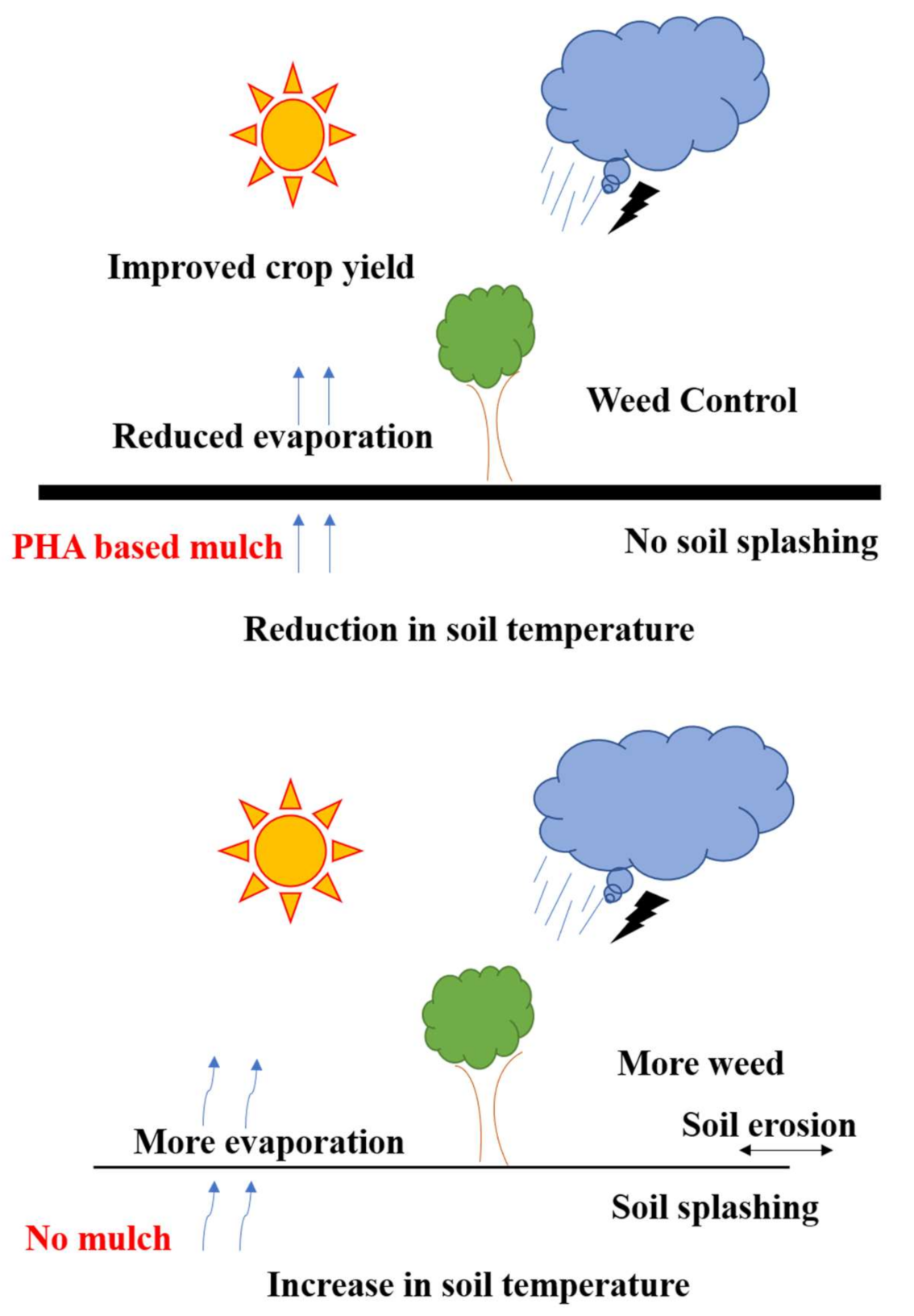
5.3. PHAs in Agriculture
6. PHAs in Nanotechnology
6.1. PHA Nanoparticles
6.1.1. Emulsion Solvent Diffusion
6.1.2. Nanoprecipitation
6.1.3. Dialysis
6.2. Role of PHA in Fabricating Nanoparticles
6.2.1. PHA as an Encapsulating Agent in Nanotechnology
6.2.2. Phasin/PHA Synthase-Based Nanoparticle Synthesis
6.2.3. PHAs in Metal Nanoparticle Synthesis
7. Characterization of Nanoparticles
7.1. Fourier Transform Infrared Spectroscopy
7.2. Scanning Electron Microscope (SEM)
7.3. Atomic Force Microscopy (AFM)
7.4. Zeta Potential and Size Distribution
7.5. X-ray Diffraction Crystallography (X-RD)
8. Applications of PHA Nanoparticles

| S No. | Polymer/Composite | Bioactivities | Drug Loaded with | Against What Type of Organism/Cells | References |
|---|---|---|---|---|---|
| 1. | Poly[R]hydroxyalkanoate (PHA) | Antibacterial activity | Curcumin | B. subtilis | [5] |
| 2. | Poly(3-hydroxybutyrate-co-3-hydroxyvalerate) (P (3HB-co-3HV) | Antibacterial Activity; Biocompatibility Test | Amoxicillin, Levofloxacin | S. aureus, E. coli; human embryonic kidney (HEK) cell line | [215] |
| 3. | Poly(hydroxybutyrate-co-hydroxyhexanoate) (PHBHHx) | Hypoglycemic activity | Insulin | Diabetic rats | [249] |
| 4. | Polyrhydroxyalkanoate (PHA) | Antibacterial activity | - | S. aureus, P. aeruginosa | [250] |
| 5. | PHB, P(HB-12HV) and P(HB-50HV) | In-vitro photocytotoxicity | Porphyrines, Phthalocyanines | Human colon adenocarcinoma HT-29 | [251] |
| 6. | PHA–mPEG deblock copolymer | Biocompatibility Test | Thymoquinone | Prenatal rat neuronal hippocampal cells and NIH/3T3 fibroblast cell line | [252] |
| 7. | Poly(3-hydroxybutyrate-co-3-hydroxyoctanoate) | In vivo anti-cancer activity | Doxorubicin | BALB/c nude mice | [255] |
| 8. | Poly(ε-Caprolactone) and Poly(3-Hydroxybutyrate) | Anti-cancer activity | Chrysin | Human breast cancer cell line MDA-MB-231 | [256] |
| 9. | Polyhydroxybutyrate–Chitosan (PHB-Cs) | Antibacterial activity | Kaempferol | Staphylococcus aureus, Acentibacter baumannii | [257] |
| 10. | Polyhydroxy butyrate/polyethylene glycol (Eu-Pi/PHB-PEG) | Anti-cancer activity | Eugenol, Piperine | Nasopharyngeal cancer (C666-1) cells | [258] |
Author Contributions
Funding
Institutional Review Board Statement
Informed Consent Statement
Conflicts of Interest
References
- Yu, L.; Dean, K.; Li, L. Polymer blends and composites from renewable resources. Prog. Polym. Sci. 2006, 31, 576–602. [Google Scholar] [CrossRef]
- Samrot, A.V.; Avinesh, R.B.; Sukeetha, S.D.; Senthilkumar, P. Accumulation of poly[(R)-3-hydroxyalkanoates] in Enterobacter cloacae SU-1 during growth with two different carbon sources in batch culture. Appl. Biochem. Biotechnol. 2011, 163, 195–203. [Google Scholar] [CrossRef] [PubMed]
- Samrot, A.V.; Bhakyalakshmi, M.; Venkatraman, K.L.; Sahiti, K.; Philip, S.A.; Jahnavi, T.; Senthilkumar, P. Optimization and Characterization of Poly[R]Hydroxyalkanoates of Pseudomonas Aeruginosa. Biosci. Biotechnol. Res. Asia. 2015, 12, 2133–2138. [Google Scholar] [CrossRef] [Green Version]
- Nisha, J.; Mudaliar, N.; Senthilkumar, P.; Kumar, N.; Samrot, A.V. Influence of substrate concentration in accumulation pattern of poly(R) hydroxyalkonoate in Pseudomonas putida SU-8. Afr. J. Microbiol. Res. 2012, 6, 3623–3630. [Google Scholar]
- Senthilkumar, P.; Dawn, S.S.; Samanvitha, K.S.; Kumar, S.S.; Kumar, G.N.; Samrot, A.V. Optimization and characterization of poly[R]hydroxyalkanoate of Pseudomonas aeruginosa SU-1 to utilize in nanoparticle synthesis for curcumin delivery. Biocatal. Agr. Biotechnol. 2017, 12, 292–298. [Google Scholar] [CrossRef]
- Poirier, Y. Green chemistry yields a better plastic. Nat. Biotechnol. 1999, 17, 960–961. [Google Scholar] [CrossRef] [PubMed]
- Anderson, A.J.; Dawes, E.A. Occurrence, metabolism, metabolic role, and industrial uses of bacterial polyhydroxyalkanoates. Microbiol. Rev. 1990, 54, 450–452. [Google Scholar] [CrossRef]
- De Waard, P.; Van der Wal, H.; Huijberts, G.N.; Eggink, G. Heteronuclear NMR analysis of unsaturated fatty acids in poly (3-hydroxyalkanoates). Study of beta-oxidation in Pseudomonas putida. J. Biol. Chem. 1993, 268, 315–319. [Google Scholar] [CrossRef]
- Hazer, B.; Steinbüchel, A. Increased diversification of polyhydroxyalkanoates by modification reactions for industrial and medical applications. Appl. Microbiol. Biotechnol. 2007, 74, 1–12. [Google Scholar] [CrossRef]
- Basnett, P.; Lukasiewicz, B.; Marcello, E.; Gura, H.K.; Knowles, J.C.; Roy, I. Production of a novel medium chain length poly(3-hydroxyalkanoate) using unprocessed biodiesel waste and its evaluation as a tissue engineering scaffold. Microb. Biotechnol. 2017, 10, 1384–1399. [Google Scholar] [CrossRef]
- Sharma, L.; Srivastava, J.K.; Singh, A.K. Biodegradable polyhydroxyalkanoate thermoplastics substituting xenobiotic plastics: A way forward for sustainable environment. In Plant Responses to Xenobiotics; Springer: Singapore, 2016; pp. 317–346. [Google Scholar]
- Loo, C.Y.; Sudesh, K. Polyhydroxyalkanoates: Bio-based microbial plastics and their properties. Malays. Polym. J. 2007, 2, 31–57. [Google Scholar]
- Licciardello, G.; Catara, A.F.; Catara, V. Production of polyhydroxyalkanoates and extracellular products using Pseudomonas Corrugata and P. Mediterranea: A Review. Bioengineering 2019, 146, 105. [Google Scholar] [CrossRef] [Green Version]
- Zinn, M.; Witholt, B.; Egli, T. Occurrence, synthesis, and medical application of bacterial polyhydroxyalkanoate. Adv. Drug Del. Rev. 2001, 53, 5–21. [Google Scholar] [CrossRef]
- Kamm, M.B. Principles of biorefineries. Appl. Microbiol. Biotechnol. 2004, 64, 137–145. [Google Scholar] [CrossRef] [PubMed]
- Devi, A.B.; Nachiyar, C.V.; Kaviyarasi, T.; Samrot, A.V. Characterization of polyhydroxybutyrate synthesized by Bacillus cereus. Int. J. Pharm. Pharm. Sci. 2015, 7, 140–144. [Google Scholar]
- Wang, Y.; Yin, J.; Chen, G.Q. Polyhydroxyalkanoates, challenges and opportunities. Curr. Opin. Biotechnol. 2014, 30, 59–65. [Google Scholar] [CrossRef] [PubMed]
- Lemoigne, M. Produits de dehydration et de polymerisation de l’acide ß-oxobutyrique. Bull. Soc. Chim. Biol. 1926, 8, 770–782. [Google Scholar]
- Xu, M.; Qin, J.; Rao, Z.; You, H.; Zhang, X.; Yang, T.; Wang, X.; Xu, Z. Effect of Polyhydroxybutyrate (PHB) storage on l-arginine production in recombinant Corynebacterium crenatum using coenzyme regulation. Microb. Cell Fact. 2016, 15, 1–12. [Google Scholar] [CrossRef] [Green Version]
- Ebringerova, A.; Heinze, T. Xylan and xylan derivatives-biopolymers with valuable properties. 1 Naturally occurring xylans structures, isolation procedures and properties. Macromol. Rapid Commun. 2000, 556, 542–556. [Google Scholar] [CrossRef]
- Labant, M. Redesigning the Global Food Supply: Although genome editing is gentler than GMO-yielding transgenic manipulation, it may have more impact, enhancing the natural qualities of plants and promoting sustainability. Genet. Eng. Biotechnol. News 2020, 40, 64–67. [Google Scholar] [CrossRef]
- Meereboer, K.W.; Misra, M.; Mohanty, A.K. Review of recent advances in the biodegradability of polyhydroxyalkanoate (PHA) bioplastics and their composites. Green Chem. 2020, 22, 5519–5558. [Google Scholar] [CrossRef]
- Koutinas, A.A.; Wang, R.; Webb, C. Polyhydroxybutyrate production from a novel feedstock derived from a wheat-based biorefinery. Enzym. Microb. Technol. 2007, 40, 1035–1044. [Google Scholar] [CrossRef]
- Hernández-Muñoz, P.; Kanavouras, A.P.K.N.; Gavara, R. Development and characterization of biodegradable films made from wheat gluten protein fractions. J. Agric. Food Chem. 2003, 51, 7647–7654. [Google Scholar] [CrossRef] [PubMed]
- Chen, G.Q.; Jiang, X. Engineering bacteria for enhanced polyhydroxyalkanoates (PHA) biosynthesis. Synth. Syst. Biotechnol. 2017, 2, 192–197. [Google Scholar] [CrossRef]
- Van-Thuoc, D.; Huu-Phong, T.; Thi-Binh, N.; Thi-Tho, N.; Minh-Lam, D.; Quillaguaman, J. Polyester production by halophilic and halotolerant bacterial strains obtained from mangrove soil samples located in Northern Vietnam. Microbiologyopen 2012, 1, 395–406. [Google Scholar] [CrossRef]
- Reusch, N. Poly-p-Hydroxybutyrate/Calcium polyphosphate complexes in eukaryotic membranes. Proc. Soc. Exp. Biol. Med. 1989, 1911, 377–381. [Google Scholar] [CrossRef]
- Potter, M.; Madkour, M.H.; Mayer, F.; Steinbuchel, A. Regulation of phasin expression and polyhydroxyalkanoate (PHA) granule formation in Ralstonia eutropha H16. Microbiology 2002, 148, 2413–2426. [Google Scholar] [CrossRef] [PubMed] [Green Version]
- Potter, M.; Steinbuchel, A. Biogenesis and Structure of Polyhydroxyalkanoate Granules; Springer: Berlin/Heidelberg, Germany, 2006; pp. 109–136. [Google Scholar]
- Tsuge, T.; Yano, K.; Imazu, S.; Numata, K.; Kikkawa, Y.; Abe, H.; Taguchi, K.; Doi, Y. Biosynthesis of polyhydroxyalkanoate (PHA) copolymer from fructose using wild-type and laboratory-evolved PHA synthases. Macromol. Biosci. 2005, 5, 112–117. [Google Scholar] [CrossRef]
- Griebel, R.; Smith, Z.; Merrick, J.M. Metabolism of poly-P-hydroxybutyrate. Purification, composition, and properties of native poly-p-hydroxybutyrate granules from Bacillus megaterium. Biochemistry 1968, 7, 3676–3681. [Google Scholar] [CrossRef]
- Ratledge, C.; Kristiansen, B. Basic Biotechnology, 2nd ed.; Cambridge University Press: Cambridge, UK, 2001. [Google Scholar]
- Tortajada, M.; da Silva, L.F.; Prieto, M.A. Second-Generation functionalized medium-chain-length polyhydroxyalkanoates: The gateway to high-value bioplastic applications. Int. Microbiol. 2013, 16, 1–15. [Google Scholar]
- Wang, Q.; Tappel, R.C.; Zhu, C.; Nomura, C.T. Development of a new strategy for production of medium-chain-length polyhydroxyalkanoates by recombinant Escherichia coli via inexpensive non-fatty acid feedstocks. Appl. Environ. Microbiol. 2012, 78, 519–527. [Google Scholar] [CrossRef] [Green Version]
- Rehm, B.H.; Steinbüchel, A. Biochemical and genetic analysis of PHA synthases and other proteins required for PHA synthesis. Int. J. Biol. Macromol. 1999, 25, 3–19. [Google Scholar] [CrossRef]
- Andler, R.; Pino, V.; Moya, F.; Soto, E.; Valdés, C.; Andreeßen, C. Synthesis of poly-3-hydroxybutyrate (PHB) by Bacillus cereus using grape residues as sole carbon source. Int. J. Biobased Plast. 2021, 3, 98–111. [Google Scholar] [CrossRef]
- Blunt, W.; Sparling, R.; Gapes, D.J.; Levin, D.B.; Cicek, N. The role of dissolved oxygen content as a modulator of microbial polyhydroxyalkanoate synthesis. World J. Microbiol. Biotechnol. 2018, 34, 106. [Google Scholar] [CrossRef] [PubMed]
- Kniewel, R.; Lopez, O.R.; Prieto, M.A. Biogenesis of medium-chain-length polyhydroxyalkanoates. In Handbook of Hydrocarbon and Lipid Microbiology; Geiger, O., Ed.; Springer: Cham, Switzerland, 2019; pp. 457–481. [Google Scholar]
- Jacquel, N.; Lo, C.W.; Wu, H.S.; Wei, Y.H.; Wang, S.S. Solubility of polyhydroxyalkanoates by experiment and thermodynamic correlations. AIChE J. 2007, 53, 2704–2714. [Google Scholar] [CrossRef]
- Cavaliere, C.; Capriotti, A.L.; Cerrato, A.; Lorini, L.; Montone, C.M.; Valentino, F.; Laganà, A.; Majone, M. Identification and Quantification of Polycyclic Aromatic Hydrocarbons in Polyhydroxyalkanoates Produced from Mixed Microbial Cultures and Municipal Organic Wastes at Pilot Scale. Molecules 2021, 26, 539. [Google Scholar] [CrossRef] [PubMed]
- Paduani, M. Microplastics as novel sedimentary particles in coastal wetlands: A review. Mar. Pollut. Bull. 2020, 161, 111739. [Google Scholar] [CrossRef]
- Löbler, M.; Sass, M.; Michel, P.; Hopt, U.; Kunze, C.; Schmitz, K. Differential gene expression after implantation of biomaterials into rat gastrointestine. J. Mater. Sci. Mater. Med. 1999, 10, 797–799. [Google Scholar] [CrossRef] [PubMed]
- Liechty, W.B.; Kryscio, D.R.; Slaughter, B.V.; Peppas, N.A. Polymers for drug delivery systems. Annu. Rev. Chem. Biomol. Eng. 2010, 1, 149–173. [Google Scholar] [CrossRef] [Green Version]
- Woerdeman, D.; Veraverbeke, W.; Verpoest, I.; Delcour, J.; Parnas, R.S. Gluten Biopolymers. U.S. Patent US20929B2, 21 April 2009. [Google Scholar]
- Choi, J.; Lee, S.Y. Process analysis and economic evaluation for poly(3-hydroxybutyrate) production by fermentation. Bioprocess Eng. 1997, 17, 335–342. [Google Scholar] [CrossRef]
- Liao, Q.; Guo, L.; Ran, Y.; Gao, M.; She, Z.; Zhao, Y.; Liu, Y. Optimization of polyhydroxyalkanoates (PHA) synthesis with heat pretreated waste sludge. Waste Manag. 2018, 82, 15–25. [Google Scholar] [CrossRef] [PubMed]
- Koutinas, A.A.; Wang, R.; Webb, C. Restructuring upstream bioprocessing: Technological and economical aspects for production of a generic microbial feedstock from wheat. Biotechnol. Bioeng. 2004, 85, 524–538. [Google Scholar] [CrossRef] [PubMed]
- Beun, J.J.; Paletta, F.; Van Loosdrecht, M.C.M.; Heijnen, J.J. Stoichiometry and kinetics of poly-β-hydroxybutyrate metabolism in aerobic, slow growing, activated sludge cultures. Biotechnol. Bioeng. 2000, 67, 379–389. [Google Scholar] [CrossRef]
- Satoh, Y.; Minamoto, N.; Tajima, K.; Munekata, M. Polyhydroxyalkanoate synthase from Bacillus sp. INT005 is composed of PhaC and PhaR. J. Biosci. Bioeng. 2002, 94, 343–350. [Google Scholar] [CrossRef]
- Lee, I.Y.; Kim, M.K.; Chang, H.M.; Park, Y.K. Effects of propionate on accumulation of poly(β-hydroxy-butyrate-co-β-hydroxyvalerate) and excretion of pyruvate in Alcaligenes eutrophus. Biotechnol. Lett. 1994, 16, 611–616. [Google Scholar] [CrossRef]
- Liebergesell, M.; Sonomoto, K.; Madkour, M.; Mayer, F.; Steinbuchel, A. Purification and characterization of the poly(hydroxyalkanoic acid) synthase from Chromatium vinosum and localization of the enzyme at the surface of poly(hydroxyalkanoic acid) granules. Eur. J. Biochem. 1994, 226, 71–80. [Google Scholar] [CrossRef]
- Jiang, G.; Johnston, B.; Townrow, D.E.; Radecka, I.; Koller, M.; Chaber, P.; Adamus, G.; Kowalczuk, M. Biomass extraction using non-chlorinated solvents for biocompatibility improvement of polyhydroxyalkanoates. Polymers 2018, 10, 731. [Google Scholar] [CrossRef] [PubMed] [Green Version]
- Eggink, G.; de Waard, P.; Huijberts, G.N. Formation of novel poly(hydroxyalkanoates) from long-chain fatty acids. Can. J. Microbiol. 1995, 41, 14–21. [Google Scholar] [CrossRef]
- Poltronieri, P.; Mezzolla, V.; D’Urso, O.F. PHB Production in biofermentors assisted through biosensor applications. Proceedings 2017, 1, 4. [Google Scholar] [CrossRef] [Green Version]
- Lakshmanan, K.; Shamala, T.R. Extraction of polyhydroxyalkanoate from Sinorhizobium meliloti cells using Microbispora sp. culture and its enzymes. Enzym. Microb. Technol. 2006, 39, 1471–1475. [Google Scholar] [CrossRef]
- Labuzek, S.; Radecka, I. Biosynthesis of copolymers of PHB tercopolymer by Bacillus cereus UW85 strain. J. Appl. Microbiol. 2001, 90, 353–357. [Google Scholar]
- Foster, L.J.R.; Sanguanchaipaiwong, V.; Gabelish, C.L.; Hook, J.; Stenzel, M. A natural-synthetic hybrid copolymer of polyhydroxyoctanoate-diethylene glycol: Biosynthesis and properties. Polymer 2005, 46, 6587–6594. [Google Scholar] [CrossRef]
- Qi, Q.; Rehm, B.H.A. Polyhydroxybutyrate biosynthesis in Caulobacter crescentus: Molecular characterization of the polyhydroxybutyrate synthase. Microbiology 2001, 147, 3353–3358. [Google Scholar] [CrossRef] [Green Version]
- Quillaguaman, J.; Hashim, S.; Bento, F.; Mattiasson, B.; Hatti-Kaul, R. Poly(β-hydroxybutyrate) production by a moderate halophile, Halomonas boliviensis LC1 using starch hydrolysate as substrate. J. Appl. Microbiol. 2005, 99, 151–157. [Google Scholar] [CrossRef] [PubMed]
- James, B.W.; Mauchline, W.S.; Dennis, P.J.; Keevil, C.W.; Wait, R. Poly-3-hydroxybutyrate in Legionella pneumophila, an energy source for survival in low-nutrient environments. Appl. Environ. Microbiol. 1999, 65, 822–827. [Google Scholar] [CrossRef] [Green Version]
- Jau, M.H.; Yew, S.P.; Toh, P.S.Y.; Chong, A.S.C.; Chu, W.L.; Phang, S.M.; Najimudin, N.; Sudesh, K. Biosynthesis and mobilization of poly(3-hydroxybutyrate) P(3HB) by Spirulina platensis. Int. J. Biol. Macromol. 2005, 36, 144–151. [Google Scholar] [CrossRef] [PubMed]
- Mukhopadhyay, M.; Patra, A.; Paul, A.K. Production of poly(3-hydroxybutyrate) and poly(3-hydroxybutyrate-co-3-hydroxyvalerate) by Rhodopseudomonas palustris SP5212. World J. Microbiol. Biotechnol. 2005, 21, 765–769. [Google Scholar] [CrossRef]
- Mukhopadhyay, M.; Patra, A.; Paul, A.K. Phototrophic growth and accumulation of poly(3-hydroxybutyrate-co-3-hydroxyvalerate) by purple nonsulfur bacterium Rhodopseudomonas palustris SP5212. J. Polym. 2013, 2013, 523941. [Google Scholar] [CrossRef] [Green Version]
- Lee, S.H.; Oh, D.H.; Ahn, W.S.; Lee, Y.; Choi, J.I.; Lee, S.Y. Production of poly(3-hydroxybutyrate-co-3-hydroxyhexanoate) by high-cell-density cultivation of Aeromonas hydrophila. Biotechnol. Bioeng. 2000, 67, 240–244. [Google Scholar] [CrossRef]
- Senthilkumar, P.; Narendrakumar, G.; Dawn, S.S. Mathematical modelling on accumulation of polyhydroxydecanoates as primary polyhydroxyalkonoates by agrobacterium tumefaciens SU-11 in glucose containing medium. In Proceedings of the International Conference on Mathematical Sciences, Chennai, India, 17–19 July 2014; pp. 668–673. [Google Scholar]
- Shamala, T.R.; Chandrashekar, A.; Vijayendra, S.V.N.; Kshama, L. Identification of polyhydroxyalkanoate (PHA)-producing Bacillus spp. using the polymerase chain reaction (PCR). J. Appl. Microbiol. 2003, 94, 369–374. [Google Scholar] [CrossRef]
- Andreessen, B.; Lange, A.B.; Robenek, H.; Steinbuchel, A. Conversion of Glycerol to Poly(3-Hydroxypropionate) in Recombinant Escherichia coli. Appl. Environ. Microbiol. 2010, 76, 622–626. [Google Scholar] [CrossRef] [Green Version]
- Kritika, S.; Pragya, R.; Nandini, P.; Priti, S. Optimization of PHB (poly-hydroxybutyrate) synthesis by Serratia sp. isolated from soil. Int. J. Curr. Microbiol. App. Sci. 2016, 5, 665–673. [Google Scholar] [CrossRef]
- Senthilkumar, P.; Dawn, S.S.; Samrot, A.V.; Kumar, N.G.; Aashrit, R.D. Production, optimization and characterization of poly[r]hydroxyalkanoate from Enterobacter sp SU16. Indian J. Sci. Technol. 2016, 9, 45. [Google Scholar]
- Yuan, Y.; Zhao, Z.; Wang, L.; Xu, Y.; Chen, H.; Kong, L. Process optimization of CO2 high-pressure and low-temperature explosion puffing drying for apple chips using response surface methodology. Dry. Technol. 2020. [Google Scholar] [CrossRef]
- Chatterjee, S.; Kumar, A.; Basu, S.; Dutta, S. Application of response surface methodology for methylene blue dye removal from aqueous solution using low-cost adsorbent. Chem. Eng. J. 2012, 181–182, 289–299. [Google Scholar] [CrossRef]
- Peasura, P. Application of response surface methodology for modeling of postweld heat treatment process in a pressure vessel steel ASTM A516 grade 70. Sci. World J. 2015, 2015, 318475. [Google Scholar] [CrossRef] [Green Version]
- Bhaumik, R.; Mondal, N.K.; Chattoraj, S.; Datta, J.K. Application of response surface methodology for optimization of fluoride removal mechanism by newly developed. biomaterial. Am. J. Anal. Chem. 2013, 4, 404–419. [Google Scholar] [CrossRef] [Green Version]
- Alkhatib, M.F.; Mamun, A.A.; Akbar, I. Application of response surface methodology (RSM) for optimization of color removal from POME by granular activated carbon. Int. J. Environ. Sci. Technol. 2015, 12, 1295–1302. [Google Scholar] [CrossRef] [Green Version]
- Ratnam, B.V.V.; Rao, S.S.; Rao, M.D.; Rao, M.N.; Ayyanna, C. Optimization of medium constituents and fermentation conditions for the production of ethanol from palmyra jaggery using response surface methodology. World J. Microbiol. Biotechnol. 2005, 21, 399–404. [Google Scholar] [CrossRef]
- Wang, D.S.; Torng, C.C.; Lin, I.P.; Cheng, B.W.; Liu, H.R.; Chou, C.Y. Optimization of nattokinase production conduction using response surface methodology. J. Food Process Eng. 2006, 29, 22–35. [Google Scholar] [CrossRef]
- Shantini, K.; Azami, N.A.; Kai-Hee, H.; Yahya, A.R.M.; Amirul, A.A. Poly(3-hydroxybutyrate-co-3-hydroxyvalerate) copolymer synthesis by using 1-pentanol and oleic acid: Process optimization and polymer characterization. J. Polym. Res. 2021, 28, 247. [Google Scholar] [CrossRef]
- Ghelich, R.; Jahannama, M.R.; Abdizadeh, H.; Torknik, F.S.; Vaezi, M.R. Central Composite Design (CCD)—Response surface methodology (RSM) of effective electrospinning parameters on PVP-B-Hf hybrid nanofibrous cl tar composites for synthesis of HfB2-based composite nanofibers. Compos. B Eng. 2019, 166, 527–541. [Google Scholar] [CrossRef]
- Novak, M.; Koller, M.; Braunegg, G.; Horvat, P. Mathematical modelling as a tool for optimized PHA production. Chem. Biochem. Eng. Q. 2015, 29, 183–220. [Google Scholar] [CrossRef]
- Myers, R.H.; Khuri, A.; Carter, W.H. Response surface methodology: 1966–l988. Technometrics 1989, 31, 137–157. [Google Scholar] [CrossRef]
- Lhamo, P.; Behera, S.K.; Mahanty, B. Process optimization, metabolic engineering interventions and commercialization of microbial polyhydroxyalkanoates production—A state-of-the art review. Biotechnol. J. 2021, 2100136. [Google Scholar] [CrossRef] [PubMed]
- Kumar, M.; Singhal, A.; Verma, P.K.; Thakur, I.S. Production and Characterization of Polyhydroxyalkanoate from Lignin Derivatives by Pandoraea sp. ISTKB. ACS Omega 2017, 2, 9156–9163. [Google Scholar] [CrossRef]
- Sabapathy, P.C.; Devaraj, S.; Parthipan, A.; Kathirvel, P. Polyhydroxyalkanoate production from statistically optimized media using rice mill effluent as sustainable substrate with an analysis on the biopolymer’s degradation potential. Int. J. Biol. Macromol. 2019, 126, 977–986. [Google Scholar] [CrossRef]
- Ojha, N.; Das, N. A Statistical approach to optimize the production of Polyhydroxyalkanoates from Wickerhamomyces anomalus VIT-NN01 using Response Surface Methodology. Int. J. Biol. Macromol. 2018, 107, 2157–2170. [Google Scholar] [CrossRef]
- Mohan, S.V.; Reddy, M.V. Optimization of critical factors to enhance polyhydroxyalkanoates (PHA) synthesis by mixed culture using Taguchi design of experimental methodology. Bioresour. Technol. 2013, 128, 409–416. [Google Scholar] [CrossRef]
- Zihayat, B.; Shakibaie, M.; Sabouri-Shahrbabak, S.; Doostmohammadi, M.; Ameri, A.; Adeli-Sardou, M.; Forootanfar, H. Medium optimization for polyhydroxyalkanoate production by Pseudomonas pseudoalcaligenes strain Te using D-optimal design. Biocatal. Agric. Biotechnol. 2019, 18, 101001. [Google Scholar] [CrossRef]
- Mohanrasu, K.; Rao, R.G.R.; Dinesh, G.H.; Zhang, K.; Prakash, G.S.; Song, D.P.; Muniyasamy, S.; Pugazhendhi, A.; Jeyakanthan, J.; Arun, A. Optimization of media components and culture conditions for polyhydroxyalkanoates production by Bacillus megaterium. Fuel 2020, 271, 117522. [Google Scholar] [CrossRef]
- Elsayed, N.S.; Aboshanab, K.M.; Aboulwafa, M.M.; Yassien, M.A.; Hassouna, N.A. Scaling up, Kinetic modeling, and Economic analysis of poly (3-hydroxybutyrate) production by Bacillus cereus isolate CCASU-P83. Arch. Pharm. Sci. Ain Shams Univ. 2021, 5, 158–170. [Google Scholar]
- Son, H.; Park, G.; Lee, S. Growth-Associated production of poly-β-hydroxybutyrate from glucose or alcoholic distillery wastewater by Actinobacillus sp. EL-9. Biotechnol. Lett. 1996, 18, 1229–1234. [Google Scholar] [CrossRef]
- Ramsay, J.A.; Berger, E.; Ramsay, A.; Chaverie, C. Extraction of poly-3-hydroxybutyrate using chlorinated solvents. Biotechnol. Tech. 1994, 8, 589–594. [Google Scholar] [CrossRef]
- Holmes, P.A. Biologically produced (R)-3-hydroxyalkanoate polymers and copolymers. In Developments in Crystalline Polymers-2; Basseti, D.C., Ed.; Elsevier Applied Science: London, UK; New York, NY, USA, 1988; pp. 1–66. [Google Scholar]
- Vandi, L.J.; Chan, C.M.; Werker, A.; Richardson, D.; Laycock, B.; Pratt, S. Wood-PHA composites: Mapping opportunities. Polymers 2018, 10, 751. [Google Scholar] [CrossRef] [Green Version]
- Fidler, S.; Dennis, D. Polyhydroxyalkanoate production in recombinant Escherichia coli. FEMS Microbiol. Rev. 1992, 9, 231–235. [Google Scholar] [CrossRef]
- Resch, S.; Gruber, K.; Wanner, G.; Slater, S.; Dennis, D.; Lubitz, W. Aqueous release and purification of poly(β-hydroxybutyrate) from Escherichia coli. J. Biotechnol. 1998, 65, 173–182. [Google Scholar] [CrossRef]
- Pagliano, G.; Galletti, P.; Samorì, C.; Zaghini, A.; Torri, C. Recovery of Polyhydroxyalkanoates from single and mixed microbial cultures: A review. Front. Bioeng. Biotechnol. 2021, 9, 624021. [Google Scholar] [CrossRef]
- Heinrich, D.; Madkour, M.H.; Al-Ghamdi, M.A.; Shabbaj, I.I.; Steinbüchel, A. Large scale extraction of poly(3-hydroxybutyrate) from Ralstonia eutropha H16 using sodium hypochlorite. AMB Express. 2012, 2, 59. [Google Scholar] [CrossRef] [Green Version]
- McChalicher, C.W.J.; Srienc, F.; Rouse, D.P. Solubility and degradation of polyhydroxyalkanoate biopolymers in propylene carbonate. AIChE J. 2010, 56, 1616–1625. [Google Scholar] [CrossRef]
- Koller, M.; Bona, R.; Chiellini, E.; Braunegg, G. Extraction of short-chain-length poly [(R)-hydroxyalkanoates] (scl-PHA) by the “anti-solvent” acetone under elevated temperature and pressure. Biotechnol. Lett. 2013, 35, 1023–1028. [Google Scholar] [CrossRef] [PubMed]
- Umesh, M.; Mamatha, C. Comparitive study on efficacy of alkaline and acid extraction for PHA recovery from Bacillus subtilis NCDC0671. Int. J. Recent Sci. Res. 2018, 9, 29467–29471. [Google Scholar]
- Koller, M. Established and advanced approaches for recovery of microbial polyhydroxyalkanoate (PHA) biopolyesters from surrounding microbial biomass. EuroBiotech J. 2020, 4, 113–126. [Google Scholar] [CrossRef]
- Hahn, S.K.; Chang, Y.K.; Kim, B.S.; Lee, K.M.; Chang, H.N. The recovery of poly(3-hydroxybutyrate) by using dispersions of sodium hypochlorite solution and chloroform. Biotechnol. Tech. 1993, 7, 209–212. [Google Scholar] [CrossRef]
- Kurian, N.S.; Das, B. Comparative analysis of various extraction processes based on economy, eco-friendly, purity and recovery of polyhydroxyalkanoate: A review. Int. J. Biol. Macromol. 2021, 183, 1881–1890. [Google Scholar] [CrossRef] [PubMed]
- Mótyán, J.A.; Tóth, F.; Tőzsér, J. Research applications of proteolytic enzymes in molecular biology. Biomolecules 2013, 3, 923–942. [Google Scholar] [CrossRef] [Green Version]
- Yasotha, K.; Aroua, M.K.; Ramachandran, K.B.; Tan, I.K.P. Recovery of medium-chain-length polyhydroxyalkanoates (PHAs) through enzymatic digestion treatments and ultrafiltration. Biochem. Eng. J. 2006, 30, 260–268. [Google Scholar] [CrossRef]
- Israni, N.; Thapa, S.; Shivakumar, S. Biolytic extraction of poly(3-hydroxybutyrate) from Bacillus megaterium Ti3 using the lytic enzyme of Streptomyces albus Tia1. J. Eng. Biotechnol. 2018, 16, 265–271. [Google Scholar] [CrossRef]
- Mannina, G.; Presti, D.; Montiel-Jarillo, G.; Carrera, J.; Suárez-Ojeda, M.E. Recovery of polyhydroxyalkanoates (PHAs) from wastewater: A review. Bioresour. Technol. 2020, 297, 122478. [Google Scholar] [CrossRef]
- Harrison, S.T.L. Bacterial cell disruption: A key unit operation in the recovery of intracellular products. Biotechnol. Adv. 1991, 9, 217–240. [Google Scholar] [CrossRef]
- Harrison, S.T.L.; Chase, H.A.; Dennis, J.S. The lysis of gram negative Alcaligenes Eutrophus by enzymes from Cytophaga. Biotechnol. Tech. 1991, 5, 115–120. [Google Scholar] [CrossRef]
- Tamer, I.M.; Moo-Young, M.; Chisti, Y. Disruption of Alcaligenes latus for recovery of poly(β-hydroxybutyric acid): Comparison of high-pressure homogenization, bead milling, and chemically induced lysis. Ind. Eng. Chem. Res. 1998, 37, 1807–1814. [Google Scholar] [CrossRef]
- Gutt, B.; Kehl, K.; Ren, Q.; Boesel, L.F. Using ANOVA models to compare and optimize extraction protocols of P3HBHV from Cupriavidus necator. Ind. Eng. Chem. Res. 2016, 55, 10355–10365. [Google Scholar] [CrossRef]
- Kunasundari, B.; Sudesh, K. Isolation and recovery of microbial polyhydroxyalkanoates. Express Polym. Lett. 2011, 5, 620–634. [Google Scholar] [CrossRef]
- Diels, A.M.J.; Michiels, C.W. High-Pressure homogenization as a non-thermal technique for the inactivation of microorganisms. Crit. Rev. Microbiol. 2006, 32, 201–216. [Google Scholar] [CrossRef]
- Bury, D.; Jelen, P.; Kalab, M. Disruption of Lactobacillus delbrueckii ssp. bulgaricus 11842 cells for lactose hydrolysis in dairy products: A comparison of sonication, high-pressure homogenization, and bead milling. Innov. Food Sci. Emerg. Technol. 2001, 2, 23–29. [Google Scholar] [CrossRef]
- De Donno, N.L.; Moreno, S.S.; Rene, E.R. Polyhydroxyalkanoate (PHA) production via resource recovery from industrial waste streams: A review of techniques and perspectives. Bioresour. Technol. 2021, 331, 124985. [Google Scholar] [CrossRef] [PubMed]
- Kosseva, M.R.; Rusbandi, E. Trends in the biomanufacture of polyhydroxyalkanoates with focus on downstream processing. Int. J. Biol. Macromol. 2018, 107, 762–778. [Google Scholar] [CrossRef]
- Koller, M. The Handbook of Polyhydroxyalkanoates; CRC Press: Boca Raton, FL, USA, 2021. [Google Scholar]
- Castro, M.D.L.; Jiménez-Carmona, M.M. Where is supercritical fluid extraction going? TrAC Trends Anal. Chem. 2000, 19, 223–228. [Google Scholar] [CrossRef]
- Hejazi, P.; Vasheghani-Farahani, E.; Yamini, Y. Supercritical fluid disruption of Ralstonia eutropha for poly(β-hydroxybutyrate) recovery. Biotechnol. Prog. 2003, 19, 1519–1523. [Google Scholar] [CrossRef] [PubMed]
- Hampson, J.W.; Ashby, R.D. Extraction of lipid-grown bacterial cells by supercritical fluid and organic solvent to obtain pure medium chain-length polyhydroxyalkanoates. J. Am. Oil Chem. Soc. 1999, 76, 1371–1374. [Google Scholar] [CrossRef]
- Godbole, S. Methods for identification, quantification and characterization of polyhydroxyalkanoates. Int. J. Bioassays 2016, 5, 4977. [Google Scholar] [CrossRef]
- Schlegel, H.G.; Lafferty, R.; Krauss, I. The isolation of mutants not accumulating poly-β-hydroxybutyric acid. Archiv für Mikrobiologie 1970, 70, 283–294. [Google Scholar] [CrossRef] [PubMed]
- Ostle, A.G.; Holt, J.G. Nile blue A as a fluorescent stain for poly-beta-hydroxybutyric acid. Appl. Environ. Microbiol. 1982, 44, 238–241. [Google Scholar] [CrossRef] [PubMed] [Green Version]
- Gorenflo, V.; Steinbuchel, A.; Marose, S.; Rosenberg, H.; Scheper, T. Quantification of bacterial polyhydroxyalkanoic acids by Nile red staining. Appl. Microbiol. Biotechnol. 1999, 51, 765–772. [Google Scholar] [CrossRef]
- Spiekermann, P.; Rehm, B.H.; Kalscheuer, R.; Baumeister, D.; Steinbuchel, A. A sensitive, viable-colony staining method using Nile red for direct screening of bacteria that accumulate polyhydroxyalkanoic acids and other lipid storage compounds. Arch. Microbiol. 1999, 171, 73–80. [Google Scholar] [CrossRef]
- Burdon, K.L.J. Bacterial fatty materials in bacterial and fungi revealed by staining dried fixed slide preparations. J. Bacteriol. 1946, 52, 665–678. [Google Scholar] [CrossRef] [Green Version]
- Weibull, C. Characterization of the protoplasmic constituents of Bacillus megaterium. J. Bacteriol. 1953, 66, 696–702. [Google Scholar] [CrossRef] [Green Version]
- Smibert, R.M.; Krieg, N.R. General Characterization. In Manual of Methods for General Bacteriology; Gerdhardt, P., Murray, R.G.E., Costilow, R.N., Nester, E.W., Wood, W.A., Krieg, N.R., Phillips, G.B., Eds.; American Society for Microbiology: Washington, DC, USA, 1981; pp. 409–443. [Google Scholar]
- Mesquita, D.P.; Amaral, A.L.; Leal, C.; Oehmen, A.; Reis, M.A.M.; Ferreira, E.C. Polyhydroxyalkanoate granules quantification in mixed microbial cultures using image analysis: Sudan Black B versus Nile Blue A staining. Analytica Chimica Acta 2015, 865, 8–15. [Google Scholar] [CrossRef] [Green Version]
- Fadipe, T.O.; Jamil, N.; Lawal, A.K. Biosynthesis and characterization of poly-(3)-hydroxyalkanoic acid by Bacillus megaterium SF4 using different carbohydrates. In Microbial Polymers; Vaishnav, A., Choudhary, D.K., Eds.; Springer: Singapore, 2021. [Google Scholar]
- Wei, Y.H.; Chen, W.C.; Huang, C.K.; Wu, H.-S.; Sun, Y.-M.; Lo, C.-W.; Janarthanan, O.-M. Screening and evaluation of polyhydroxybutyrate-producing strains from indigenous isolate Cupriavidus taiwanensis strains. Int. J. Mol. Sci. 2011, 12, 252–265. [Google Scholar] [CrossRef] [PubMed]
- Kitamura, S.; Doi, Y. Staining method of poly(3-hydroxyalkanoic acids) producing bacteria by Nile Blue. Biotechnol. Tech. 1994, 8, 345–350. [Google Scholar] [CrossRef]
- Kabilan, S.; Ayyasamy, M.; Jayavel, S.; Paramasamy, G. Pseudomonas sp. as a source of medium chain length polyhydroxyalkanoates for controlled drug delivery: Perspective. Int. J. Microbiol. 2012, 2012, 317828. [Google Scholar] [CrossRef]
- Tian, J.; Sinskey, A.J.; Stubbe, J. Kinetic studies of polyhydroxybutyrate granule formation in Wautersia eutropha H16 by transmission electron microscopy. J Bacteriol. 2005, 187, 3814–3824. [Google Scholar] [CrossRef] [PubMed] [Green Version]
- Berlanga, M.; Montero, M.T.; Fernández-Borrell, J.; Guerrero, R. Rapid spectrofluorometric screening of poly-hydroxyalkanoate-producing bacteria from microbial mats. Int Microbiol. 2006, 9, 95–102. [Google Scholar]
- Danis, O.; Ogan, A.; Tatlican, P.; Attar, A.; Cakmakci, E.; Mertoglu, B.; Birbir, M. Preparation of poly(3-hydroxybutyrate-co-hydroxyvalerate) films from halophilic archaea and their potential use in drug delivery. Extremophiles 2015, 19, 515–524. [Google Scholar] [CrossRef]
- Gupta, J.; Rathour, R.; Maheshwari, N.; Thakura, I.S. Integrated analysis of whole genome sequencing and life cycle assessment for polyhydroxyalkanoates production by Cupriavidus sp. ISTL7. Bioresour. Technol. 2021, 337, 125418. [Google Scholar] [CrossRef]
- Boonyawanich, S.; Tanikkul, P.; Thenchartanan, P.; Pisutpaisal, N. Productivity of Pseudomonas putida TISTR 1522 in polyhydroxyalkanoates (PHAs) production from saponified palm oil. Appl. Biochem. Biotechnol. 2021, 193, 1086–1098. [Google Scholar] [CrossRef] [PubMed]
- Fialová, D.; Skoupý, R.; Drozdová, E.; Paták, A.; Piňos, J.; Šín, L.; Beňuš, R.; Klíma, B. The application of scanning electron microscopy with energy-dispersive X-ray spectroscopy (SEM-EDX) in ancient dental calculus for the reconstruction of human habits. Microsc. Microanal. 2017, 6, 1207–1213. [Google Scholar] [CrossRef] [Green Version]
- Nwinyi, O.C.; Owolabi, T.A. Scanning electron microscopy and Fourier transmission analysis of polyhydroxyalkanoates isolated from bacteria species from abattoir in Ota, Nigeria. J. King Saud Univ. Sci. 2019, 31, 285–298. [Google Scholar] [CrossRef]
- Govindasamy, S.; Syafiq, I.M.; Amirul, A.; Amin, R.; Bhubalan, K. Dataset on controlled production of polyhydroxyalkanoate-based microbead using double emulsion solvent evaporation technique. Data Brief 2019, 23, 103675. [Google Scholar] [CrossRef]
- Williamson, D.H.; Wilkinson, J.F. The isolation and estimation of the Poly-B-hydroxybutyrate inclusions of Bacillus species. J. Gen. Microbiol. 1958, 19, 198–209. [Google Scholar] [CrossRef] [Green Version]
- Duvigneau, S.; Kettner, A.; Carius, L.; Griehl, C.; Findeisen, R.; Kienle, A. Fast, inexpensive, and reliable HPLC method to determine monomer fractions in poly(3-hydroxybutyrate-co-3-hydroxyvalerate). Appl. Microbiol. Biotechnol. 2021, 105, 4743–4749. [Google Scholar] [CrossRef]
- Law, J.H.; Slepecky, R.A. Assay of poly-β-hydroxybutyric acid. J. Bacteriol. 1971, 82, 33–36. [Google Scholar] [CrossRef] [Green Version]
- Novais, Â.; Peixe, L. Fourier transform infrared spectroscopy (FT-IR) for food and water microbiology. In Application and Integration of Omics-Powered Diagnostics in Clinical and Public Health Microbiology; Moran-Gilad, J., Yagel, Y., Eds.; Springer: Cham, Switzerland, 2021. [Google Scholar] [CrossRef]
- Rakkan, T.; Chana, N.; Sangkharak, K. The Integration of textile wastewater treatment with polyhydroxyalkanoate production using newly isolated Enterobacter strain TS3. Waste Biomass Valor. 2021. [Google Scholar] [CrossRef]
- Bhuwal, A.K.; Singh, G.; Aggarwal, N.K.; Goyal, V.; Yadav, A. Isolation and screening of polyhydroxyalkanoates producing bacteria from pulp, paper, and cardboard industry wastes. Int. J. Biomater. 2013, 2013, 752821. [Google Scholar] [CrossRef] [PubMed] [Green Version]
- Hong, K.; Sun, S.; Tian, W.; Chen, G.Q.; Huang, W. A rapid method for detecting bacterial polyhydroxyalkanoates in intact cells by fourier transform infrared spectroscopy. Appl. Microbiol. Biotechnol. 1999, 51, 523–526. [Google Scholar] [CrossRef]
- Tyagi, P.; Sharma, A. Utilization of crude paper industry effluent for Polyhydroxyalkanoate (PHA). Environ. Technol. Innov. 2021, 23, 101692. [Google Scholar] [CrossRef]
- Kansiz, M.; Domínguez-Vidal, A.; McNaughton, D.; Lendl, B. Fourier-transform infrared (FTIR) spectroscopy for monitoring and determining the degree of crystallisation of polyhydroxyalkanoates (PHAs). Anal. Bioanal. Chem. 2007, 388, 1207–1213. [Google Scholar] [CrossRef] [PubMed]
- Braunegg, G.; Sonnleitner, B.; Lafferty, R.M. A rapid gas chromatographic method for the determination of Poly-B-hydroxybutyric acid in microbial biomass. Eur. J. Appl. Microbiol. Biotechnol. 1978, 6, 29–37. [Google Scholar] [CrossRef]
- Andler, R.; Valdés, C.; Urtuvia, V.; Andreeßen, C.; Barrera, D. Fruit residues as a sustainable feedstock for the production of bacterial polyhydroxyalkanoates. J. Clean. Prod. 2021, 307, 127236. [Google Scholar] [CrossRef]
- Morya, R.; Sharma, A.; Kumar, M.; Tyagi, B.; Singh, S.S.; Thakur, I.S. Polyhydroxyalkanoate synthesis and characterization: A proteogenomic and process optimization study for biovalorization of industrial lignin. Bioresour. Technol. 2021, 320, 124439. [Google Scholar] [CrossRef]
- Satoh, H.; Sakamoto, T.; Kuroki, Y.; Kudo, Y.; Mino, T. Application of the alkaline-digestion-HPLC method to the rapid determination of polyhydroxyalkanoate in activated sludge. J. Water Environ. Technol. 2016, 14, 411–421. [Google Scholar] [CrossRef] [Green Version]
- Kichise, T.; Taguchi, S.; Doi, Y. Enhanced accumulation and changed monomer composition in polyhydroxyalkanoate (PHA) copolyester by in vitro evolution of Aeromonas caviae PHA synthase. Appl. Environ. Microbiol. 2002, 68, 2411–2419. [Google Scholar] [CrossRef] [PubMed] [Green Version]
- Bedade, D.K.; Edson, C.B.; Gross, R.A. Emergent approaches to efficient and sustainable polyhydroxyalkanoate production. Molecules 2021, 26, 3463. [Google Scholar] [CrossRef]
- Li, Z.; Yang, J.; Loh, X.J. Polyhydroxyalkanoates: Opening doors for a sustainable future. NPG Asia Mater. 2016, 8, 265. [Google Scholar] [CrossRef]
- Ansari, S.; Sami, N.; Yasin, D.; Ahmad, N.; Fatma, T. Biomedical applications of environmental friendly poly-hydroxyalkanoates. Int. J. Biol. Macromol. 2021, 183, 549–563. [Google Scholar] [CrossRef] [PubMed]
- Zhang, J.; Shishatskaya, E.I.; Volova, T.G.; da Silva, L.F.; Chen, G.Q. Polyhydroxyalkanoates (PHA) for therapeutic applications. Mater. Sci. Eng. C 2018, 1, 144–150. [Google Scholar] [CrossRef] [Green Version]
- Yang, M.; Zhu, S.; Chen, Y.; Chang, Z.; Chen, G.; Gong, Y.; Zhao, N.; Zhang, X. Studies on bone marrow stromal cells affinity of poly (3-hydroxybutyrate-co-3-hydroxyhexanoate). Biomaterials 2004, 25, 1365–1373. [Google Scholar] [CrossRef] [PubMed]
- Montazer, M.; Harifi, T. New approaches and future aspects of antibacterial food packaging: From nanoparticles coating to nanofibers and nanocomposites, with foresight to address the regulatory uncertainty. In Nanotechnology in the Agri-Food industry; Grumezescu, A.M., Ed.; Elsevier: Amsterdam, The Netherlands, 2017; pp. 533–565. [Google Scholar]
- Basumatary, I.B.; Mukherjee, A.; Katiyar, V.; Kumar, S. Biopolymer-based nanocomposite films and coatings: Recent advances in shelf-life improvement of fruits and vegetables. Crit. Rev. Food Sci. Nutr. 2020, 30, 1–24. [Google Scholar] [CrossRef]
- Guillard, V.; Gaucel, S.; Fornaciari, C.; Angellier-coussy, H. The next generation of sustainable food packaging to preserve our environment in a circular economy context. Front. Nutr. 2018, 5, 121. [Google Scholar] [CrossRef] [Green Version]
- Singh, R.S.; Kaur, N. Microbial biopolymers for edible film and coating applications. In Advances in Industrial Biotechnology; Nawani, N.N., Khetmalas, M., Razdan, P.N., Pandley, A., Eds.; IK International Publishing House: New Delhi, India; Singh: New Kanpur, India, 2013; pp. 187–216. [Google Scholar]
- Rodríguez-Contreras, A.; Guillem-Marti, J.; Lopez, O.; Manero, J.M.; Ruperez, E. Antimicrobial PHAs coatings for solid and porous tantalum implants. Colloids Surf. B Biointerfaces 2019, 182, 110317. [Google Scholar] [CrossRef] [PubMed]
- Castro-Mayorga, L.J.; Fabra, J.M.; Cabedo, L.; Lagaron, M.J. On the use of the electrospinning coating technique to produce antimicrobial polyhydroxyalkanoate materials containing in situ stabilized silver nanoparticles. Nanomaterials 2016, 7, 4. [Google Scholar] [CrossRef] [Green Version]
- Rastogi, V.K.; Samyn, P. Bio-Based Coatings for Paper Applications. Coatings 2015, 5, 887–930. [Google Scholar] [CrossRef] [Green Version]
- Oryan, A.; Alidadi, S.; Moshiri, A.; Maffulli, N. Bone regenerative medicine: Classic options, novel strategies, and future directions. J. Orthop. Surg. Res. 2014, 9, 18. [Google Scholar] [CrossRef] [Green Version]
- Peng, Q.; Sun, X.; Gong, T.; Wu, C.Y.; Zhang, T.; Tan, J.; Zhang, Z.-R. Injectable and biodegradable thermosensitive hydrogels loaded with PHBHHx nanoparticles for the sustained and controlled release of insulin. Acta Biomater. 2013, 9, 5063–5069. [Google Scholar] [CrossRef]
- Poltronieri, P.; Kumar, P. Polyhydroxyalcanoates (PHAs) in industrial applications. In Handbook of Ecomaterials; Martínez, L., Kharissova, O., Kharisov, B., Eds.; Springer: Cham, Switzerland, 2017. [Google Scholar] [CrossRef]
- Lee, Y.F.; Sridewi, N.; Ramanathan, S.; Sudesh, K. The influence of electrospinning parameters and drug loading on polyhydroxyalkanoate (PHA) nanofibers for drug delivery. Int. J. Biotechnol. Wellness Ind. 2015, 4, 103–113. [Google Scholar]
- Sintim, H.Y.; Flury, M. Is biodegradable plastic mulch the solution to agriculture’s plastic problem? Environ. Sci. Technol. 2017, 51, 1068–1069. [Google Scholar] [CrossRef] [PubMed]
- Shamsuddin, I.M.; Jafar, J.A.; Shawai, A.S.A.; Yusuf, S.; Lateefah, M.; Aminu, I. Bioplastics as better alternative to petroplastics and their role in national sustainability: A review. Adv. Biosci. Bioeng. 2017, 5, 63–70. [Google Scholar] [CrossRef] [Green Version]
- Danimer Scientific. Danimer Scientific Film Resins. Compostable Options for a Wide Range of Products. 2018. Available online: https://danimerscientific.com/compostable-solutions/a-family-of-biopolymers/film-resins/ (accessed on 5 June 2021).
- Chen, G.G.Q. Plastics from Bacteria: Natural Functions and Applications; Springer: Berlin/Heidelberg, Germany, 2009; Volume 14, pp. 1–16. [Google Scholar]
- Guerrini, S.; Borreani, G.; Voojis, H. Biodegradable materials in agriculture: Case histories and perspectives. In Soil Degradable Bioplastics for a Sustainable Modern Agriculture; Malinconico, M., Ed.; Springer: Berlin/Heidelberg, Germany, 2017. [Google Scholar] [CrossRef]
- Amelia, T.S.M.; Govindasamy, S.; Tamothran, A.M.; Vigneswari, S.; Bhubalan, K. Applications of PHA in Agriculture. In Biotechnological Applications of Polyhydroxyalkanoates; Springer: Singapore, 2019; pp. 347–361. [Google Scholar] [CrossRef]
- Bala, I.; Hariharan, S.; Kumar, M.N.V.R. PLGA nanoparticles in drug delivery: The state of the art. Crit. Rev. Ther. Drug Carr. Syst. 2004, 21, 387–422. [Google Scholar] [CrossRef]
- Barichello, J.M.; Morishita, M.; Takayama, K.; Nagai, T. Encapsulation of hydrophilic and lipophilic drugs in PLGA nanoparticles by the nanoprecipitation method. Drug Dev. Ind. Pharm. 1999, 25, 471–476. [Google Scholar] [CrossRef]
- Reis, E.C.C.; Borges, A.P.B.; Fonseca, C.C.; Martinez, M.M.M.; Eleotério, R.B.; Morato, G.O.; Oliveira, P.M. Biodegradation of a hydroxyapatite-polyhydroxybutyrate composite. Braz. Arch. Biol. Technol. 2010, 53, 817–826. [Google Scholar] [CrossRef] [Green Version]
- Shrivastav, A.; Kim, H.Y.; Kim, Y.R. Advances in the Applications of Polyhydroxyalkanoate Nanoparticles for Novel Drug Delivery System. Biomed Res. Int. 2013, 2013, 581684. [Google Scholar] [CrossRef]
- Anderson, J.M.; Rodriguez, A.; Chang, D.T. Foreign body reaction to biomaterials. Semin. Immunol. 2008, 20, 86–100. [Google Scholar] [CrossRef] [PubMed] [Green Version]
- Basnett, P.; Ching, K.Y.; Stolz, M.; Knowles, J.C.; Boccaccini, A.R.; Smith, C.L.; Locke, I.C.; Roy, I. Aspirin-loaded P(3HO)/P(3HB) blend films: Potential materials for biodegradable drug-eluting stents. Bioinspired Biomimetric. Nanobiomater. 2013, 2, 141–153. [Google Scholar] [CrossRef]
- Deepak, V.; Pandian, S.R.K.; Kalishwaralal, K.B.; Gurunathan, S. Purification, immobilization, and characterization of nattokinase on PHB nanoparticles. Bioresour. Technol. 2009, 100, 6644–6646. [Google Scholar] [CrossRef]
- Shakeri, F.; Shakeri, S.; Hojjatoleslami, M. Preparation and Characterization of Carvacrol Loaded Polyhydroxybutyrate Nanoparticles by Nanoprecipitation and Dialysis Methods. J. Food Sci. 2014, 79, N697–N705. [Google Scholar] [CrossRef]
- Kumar, R.; Lal, S. Synthesis of organic nanoparticles and their applications in drug delivery and food nanotechnology: A Review. J. Nanomater. Mol. Nanotechnol. 2014. [Google Scholar] [CrossRef]
- Zielińska, A.; Carreiró, F.; Oliveira, A.M.; Neves, A.; Pires, B.; Venkatesh, D.N.; Durazzo, A.; Lucarini, M.; Eder, P.; Silva, A.M.; et al. Polymeric nanoparticles: Production, characterization, toxicology and ecotoxicology. Molecules 2020, 25, 3731. [Google Scholar] [CrossRef]
- Quintanar-Guerrero, D.; Allémann, E.; Doelker, E.; Fessi, H. A mechanistic study of the formation of polymer nanoparticles by the emulsification-diffusion technique. Colloid Polym. Sci. 1997, 275, 640–647. [Google Scholar] [CrossRef] [Green Version]
- Xiong, Y.C.; Yao, Y.; Zhan, X.Y.; Chen, Q. Application of polyhydroxyalkanoates nanoparticles as intracellular sustained drug-release vectors. J. Biomater. Sci. 2010, 21, 127–140. [Google Scholar] [CrossRef]
- Umesh, M.; Priyanka, K.; Thazeem, B.; Preethi, K. Biogenic PHA nanoparticle synthesis and characterization from Bacillus subtilis NCDC0671 using orange peel medium. Int. J. Polym. Mater. Polym. Biomater. 2018, 67, 996–1004. [Google Scholar] [CrossRef]
- Mora-Huertas, C.E.; Fessi, H.; Elaissari, A. Polymer-based nanocapsules for drug delivery. Int. J. Pharm. 2010, 385, 113–142. [Google Scholar] [CrossRef]
- Rawat, M.; Saraf, S. Formulation optimization of double emulsification method for preparation of enzyme loaded Eudragit S100 microspheres. J. Microencapsul. 2009, 26, 306–314. [Google Scholar] [CrossRef]
- Gossmann, R.; Langer, K.; Mulac, D. New Perspective in the formulation and characterization of didodecyldimethylammonium bromide (DMAB) stabilized poly(Lactic-co-Glycolic Acid) (PLGA) nanoparticles. PLoS ONE 2015, 1, e0127532. [Google Scholar] [CrossRef] [Green Version]
- Wu, L.P.; Wang, D.; Parhamifar, L.; Hall, A.; Chen, G.Q.; Moghimi, S.M. Poly(3-hydroxybutyrate-co-R-3-hydroxyhexanoate) nanoparticles with polyethylenimine coat as simple, safe, and versatile vehicles for cell targeting: Population characteristics, cell uptake, and intracellular trafficking. Adv. Healthc. Mater. 2014, 3, 817–824. [Google Scholar] [CrossRef] [PubMed]
- Hernández-Giottonini, K.Y.; Rodríguez-Córdova, R.J.; Gutiérrez-Valenzuela, C.A.; Peñuñuri-Miranda, O.; Zavala-Rivera, P.; Guerrero-Germán, P.; Lucero-Acuña, A. PLGA nanoparticle preparations by emulsification and nanoprecipitation techniques: Effects of formulation parameters. RSC Adv. 2020, 10, 4218–4231. [Google Scholar] [CrossRef] [Green Version]
- Ganachaud, F.; Katz, J.L. Nanoparticles and nanocapsules created using the ouzo effect: Spontaneous emulsification as an alternative to ultrasonic and high-shear devices. ChemPhysChem 2005, 6, 209–216. [Google Scholar] [CrossRef]
- Quintanar-Guerrero, D.; Allémann, E.; Fessi, H.; Doelker, E. Preparation techniques and mechanism of formation of biodegradable nanoparticles from preformed polymers. Drug Dev. Ind. Pharm. 1998, 24, 1113–1128. [Google Scholar] [CrossRef] [PubMed]
- Vauthier, C.; Dubernet, C.; Fattal, E.; Pinto-Alphandary, H.; Couvreur, P. Poly(alkylcyanoacrylates) as biodegradable materials for biomedical applications. Adv. Drug Deliv. Rev. 2003, 55, 519–548. [Google Scholar] [CrossRef]
- Dimitrova, B.; Ivanov, I.B.; Nakache, E. Mass transport effects on the stability of Emulsion: Emulsion Films with acetic acid and acetone diffusing across the interface. J. Dispers. Sci. Technol. 1988, 9, 321–341. [Google Scholar] [CrossRef]
- Singh, R.; Lillard, J.W. Nanoparticle-based targeted drug delivery. Exp. Mol. Pathol. 2009, 86, 215–223. [Google Scholar] [CrossRef] [Green Version]
- Senthilkumar, P.; Dawn, S.S.; Saipriya, C.; Samrot, A.V. Surfactant (span20) influenced synthesis of polyhydroxybutyrate nanoparticles for hydrophobic drug delivery. Rasayan J. Chem. 2018, 11, 1686–1695. [Google Scholar] [CrossRef]
- Chronopoulou, L.; Fratoddi, I.; Palocci, C.; Venditti, I.; Russo, M.V. Osmosis based method drives the self-assembly of polymeric chains into micro-and nanostructures. Langmuir 2009, 25, 11940–11946. [Google Scholar] [CrossRef] [PubMed]
- Oh, I.; Lee, K.; Kwon, H.-Y.; Lee, Y.-B.; Shin, S.-C.; Cho, C.-S.; Kim, C.-K. Release of adriamycin from poly(γ-benzyl-L-glutamate)/poly(ethylene oxide) nanoparticles. Int. J. Pharm. 1999, 181, 107–115. [Google Scholar] [CrossRef]
- York, P. Strategies for particle design using supercritical fluid technologies. Pharm. Sci. Technol. Today 1999, 2, 430–440. [Google Scholar] [CrossRef]
- Samrot, A.V.; Sahithya, C.S.; Sruthi, D.P.; Selvarani, J.A.; Raji, P.; Prakash, P.; Ponnaiah, P.; Petchi, I.; Pattammadath, S.; Purayil, S.L.; et al. Itraconazole coated super paramagnetic iron oxide nanoparticles for antimicrobial studies. Biointerface Res. Appl. Chem. 2020, 10, 6218–6225. [Google Scholar]
- Samrot, A.V.; Bhavya, K.S.; Sruthi, D.P.; Raji, P.; Paulraj, P. Synthesis of SPIONs to deliver drug in-vitro and to use as contrasting agent. Int. J. Adv. Res. Eng. Technol. 2020, 11, 200–208. [Google Scholar]
- Tarrahi, R.; Fathi, Z.; Seydibeyoğlu, M.Ö.; Doustkhah, E.; Khataee, A. Polyhydroxyalkanoates (PHA): From production to nanoarchitecture. Int. J. Biol. Macromol. 2020, 146, 596–619. [Google Scholar] [CrossRef]
- Lee, J.; Jung, S.G.; Park, C.S.; Kim, H.Y.; Batt, C.A.; Kim, Y.R. Tumor-specific hybrid polyhydroxybutyrate nanoparticle: Surface modification of nanoparticle by enzymatically synthesized functional block copolymer. Bioorg. Med. Chem. Lett. 2011, 21, 2941–2944. [Google Scholar] [CrossRef]
- Rey, D.A.; Strickland, A.D.; Kirui, D.; Niamsiri, N.; Batt, C.A. In vitro self-assembly of gold nanoparticle-coated poly(3-hydroxybutyrate) granules exhibiting plasmon-induced thermo-optical enhancements. ACS Appl. Mater. Interfaces 2010, 2, 1804–1810. [Google Scholar] [CrossRef] [PubMed]
- Castro-Mayorga, J.; Martínez-Abad, A.; Fabra, M.; Olivera, C.; Reis, M.; Lagarón, J. Stabilization of antimicrobial silver nanoparticles by a polyhydroxyalkanoate obtained from mixed bacterial culture. Int. J. Biol. Macromol. 2014, 71, 103–110. [Google Scholar] [CrossRef]
- Cárdenas-Triviño, G.; Linares-Bermúdez, N.; Núñez-Decap, M. Synthesis and properties of bionanocomposites of polyhydroxybutyrate-polylactic acid doped with copper and silver nanoparticles. Int. J. Polym. Sci. 2019, 2019, 4520927. [Google Scholar] [CrossRef] [Green Version]
- Jayakumar, A.; Prabhu, K.; Shah, L.; Radha, P. Biologically and environmentally benign approach for PHB-silver nanocomposite synthesis and its characterization. Polym. Test. 2020, 81, 106197. [Google Scholar] [CrossRef]
- Robert, M.S.; Francis, X.W.; David, J.K. Spectrometric Identification of Organic Compounds, 8th ed.; Wiley: Hoboken, NJ, USA, 2014. [Google Scholar]
- Mohan, S.; Oluwafemi, O.S.; Kalarikkal, N.; Thomas, S.; Songca, S.P. Biopolymers—Application in nanoscience and nanotechnology. In Recent Advances in Biopolymers; Perveen, F.K., Ed.; IntechOpen: London, UK, 2016; pp. 47–72. [Google Scholar]
- Senthilkumar, P.; Dawn, S.S.; Samanvitha, S.K.; Saipriya, C.; Samrot, A.V. Surfactant mediated synthesis of polyhydroxybutyrate (PHB) nanoparticles for sustained drug delivery. IET Nanobiotechnol. 2019, 13, 416–427. [Google Scholar]
- Ojha, N.; Das, N. Green formulation of microbial biopolyesteric nanocarriers toward in vitro drug delivery and its characterization. Curr. Microbiol. 2021, 78, 2061–2070. [Google Scholar] [CrossRef] [PubMed]
- Kolev, T.M.; Velcheva, E.A.; Stamboliyska, B.A.; Spiteller, M. DFT and experimental studies of the structure and vibrational spectra of curcumin. Int. J. Quantum. Chem. 2005, 102, 1069–1079. [Google Scholar] [CrossRef]
- Gerber, C.; Binnig, G.; Fechs, H.; Marti, O.; Rohrer, H. Scanning tunneling microscope combined with a scanning electron microscope. In Perspectives in Condensed Matter Physics; A Critical Reprint Series; Neddermeyer, H., Ed.; Springer: Dordrecht, The Netherland, 1986; Volume 6. [Google Scholar]
- Postek, M. The Scanning Electron Microscope; CRC Press: New York, NY, USA, 1997. [Google Scholar]
- Zhou, W.; Wang, Z.L. Scanning Microscopy for Nanotechnology; Springer: New York, NY, USA, 2007. [Google Scholar]
- Babos, G.; Rydz, J.; Kawalec, M.; Klim, M.; Fodor-Kardos, A.; Trif, L.; Feczkó, T. Poly(3-Hydroxybutyrate)-Based nanoparticles for sorafenib and doxorubicin anticancer drug delivery. Int. J. Mol. Sci. 2020, 21, 7312. [Google Scholar] [CrossRef] [PubMed]
- Faisalina, A.F.; Sonvico, F.; Colombo, P.; Amirul, A.A.; Wahab, H.A.; Majid, M.I.A. Docetaxel-Loaded Poly(3HB-co-4HB) biodegradable nanoparticles: Impact of copolymer composition. Nanomaterials 2020, 10, 2123. [Google Scholar] [CrossRef]
- Nachiyar, C.V.; Devi, A.B.; Namasivayam, S.K.R.; Arul, M.R. Levofloxacin loaded polyhydroxybutyrate nano drug conjugate for In-vitro controlled drug release. Res. J. Pharm. Biol. Chem. Sci. 2015, 6, 116–119. [Google Scholar]
- Schwenke, H.; Knoth, J. A highly sensitive energy-dispersive X-ray spectrometer with multiple total reflection of the exciting beam. Nucl. Instrum. Methods 1982, 193, 239–243. [Google Scholar] [CrossRef]
- Scimeca, M.; Bischetti, S.; Lamsira, H.K.; Bonfiglio, R.; Bonanno, E. Energy dispersive X-ray (EDX) microanalysis: A powerful tool in biomedical research and diagnosis. Eur. J. Histochem. 2018, 62, 89–98. [Google Scholar] [CrossRef]
- Salahuddin, N.; Gaber, M.; Mousa, M.; Abdelwahab, M.A. Poly(3-hydroxybutyrate)/poly(amine)-coated nickel oxide nanoparticles for norfloxacin delivery: Antibacterial and cytotoxicity efficiency. RSC Adv. 2020, 10, 34046–34058. [Google Scholar] [CrossRef]
- Mühlen, Z.; Mühlen, E.Z.; Niehus, H.; Mehnert, W. Atomic force microscopy studies of Solid Lipid Nanoparticles. Pharm. Res. 1996, 13, 1411–1416. [Google Scholar] [CrossRef]
- Yadavalli, V.K.; Ehrhardt, C.J. Atomic force microscopy as a biophysical tool for nanoscale forensic investigations. Sci. Justice 2021, 61, 1–12. [Google Scholar] [CrossRef]
- Tseng, A.A. Advancements and challenges in development of atomic force microscopy for nanofabrication. Nano Today 2011, 6, 493–509. [Google Scholar] [CrossRef]
- Jalili, N.; Laxminarayana, K. A review of atomic force microscopy imaging systems: Application to molecular metrology and biological sciences. Mechatronics 2004, 14, 907–945. [Google Scholar] [CrossRef]
- Shi, H.G.; Farber, L.; Michaels, J.N.; Dickey, A.; Thompson, K.C.; Shelukar, S.D.; Hurter, P.N.; Reynolds, S.D.; Kaufman, M.J. Characterization of crystalline drug nanoparticles using atomic force microscopy and complementary techniques. Pharm. Res. 2003, 20, 479–484. [Google Scholar] [CrossRef] [PubMed]
- Polakovič, M.; Görner, T.; Gref, R.; Dellacherie, E. Lidocaine loaded biodegradable nanospheres. II. Modelling of drug release. J. Control. Release 1999, 60, 169–177. [Google Scholar] [CrossRef]
- Kilicay, E.; Erdal, E.; Hazer, B.; Türk, M.; Denkbas, E.B. Antisense oligonucleotide delivery to cancer cell lines for the treatment of different cancer types. Artif. Cells Nanomed. Biotechnol. 2016, 44, 1938–1948. [Google Scholar] [CrossRef] [PubMed]
- Lowry, G.V.; Hill, R.J.; Harper, S.; Rawle, A.F.; Hendren, C.O.; Klaessig, F.; Nobbman, U.; Sayre, P.; Rumble, J. Guidance to improve the scientific value of zeta-potential measurements in nano. Environ. Sci. Nano 2016, 5, 953–965. [Google Scholar] [CrossRef]
- Vogel, R.; Pal, A.K.; Jambhrunkar, S.; Patel, P.; Thakur, S.S.; Reategui, E.; Parekh, H.S.; Saa, P.; Stassinopoluos, A.; Broom, M.F. High-Resolution Single Particle Zeta Potential Characterisation of Biological Nanoparticles using Tunable Resistive Pulse Sensing. Sci Rep. 2017, 7, 17479. [Google Scholar] [CrossRef] [Green Version]
- Xu, R. Progress in nanoparticles characterization: Sizing and zeta potential measurement. Particuology 2008, 6, 112–115. [Google Scholar] [CrossRef]
- Muller, R.H.; Davis, S.S.; Illum, L.; Mark, E. Particle charge and surface hydrophobicity of colloidal drug carriers. In Targeting of Drugs with Synthetic Systems; Gregoriadis, G., Senior, J., Poste, G., Eds.; Plenum: New York, NY, USA, 1986; pp. 239–263. [Google Scholar]
- Maia, J.L.; Santana, M.H.A.; Re, M.I. The effect of some processing conditions on the characteristics of biodegradable microspheres obtained by an emulsion solvent evaporation process. Braz. J. Chem. Eng. 2004, 21, 1–12. [Google Scholar] [CrossRef]
- Francis, L.; Meng, D.; Knowles, J.; Keshavarz, T.; Boccaccin, A.R.; Roy, I. Controlled delivery of gentamicin using poly(3-hydroxybutyrate) microspheres. Int. J. Mol. Sci. 2011, 12, 4294–4314. [Google Scholar] [CrossRef]
- Wang, Y.; Wang, X.; Wie, K.; Zhao, N.; Zhang, S.; Chen, J. Fabrication, characterization and long-term in vitro release of hydrophilic drug using PHBV/HA composite microspheres. Mater. Lett. 2007, 61, 1071–1076. [Google Scholar] [CrossRef]
- Sahoo, S.K.; Panyam, J.; Prabha, S.; Labhasetwar, V. Residual polyvinyl alcohol associated with poly(d,l-lactide-co- glycolide) nanoparticles affects their physical properties and cellular uptake. J. Control. Release 2002, 82, 105–114. [Google Scholar] [CrossRef]
- Lee, S.C.; Oh, J.T.; Jang, M.H.; Chung, S.I. Quantitative analysis of polyvinyl alcohol on the surface of poly(d,l-lactide-co-glycolide) microparticles prepared by solvent evaporation method: Effect of particle size and PVA concentration. J. Control. Release 1999, 59, 123–132. [Google Scholar] [CrossRef]
- Londoño-Restrepo, S.M.; Jeronimo-Cruz, R.; Millán-Malo, B.M.; Rivera-Muñoz, E.M.; Rodriguez-García, M.E. Effect of the nano crystal size on the X-ray diffraction patterns of biogenic hydroxyapatite from human, bovine, and porcine bones. Sci. Rep. 2019, 9, 5915. [Google Scholar] [CrossRef]
- Anbukarasu, P.; Sauvageau, D.; Elias, A. Tuning the properties of polyhydroxybutyrate films using acetic acid via solvent casting. Sci. Rep. 2015, 5, 17884. [Google Scholar] [CrossRef] [PubMed] [Green Version]
- Mahaling, B.; Katti, D.S. Fabrication of micro-structures of poly [(R)-3-hydroxybutyric acid] by electro-spraying/-spinning: Understanding the influence of polymer concentration and solvent type. J. Mater. Sci. 2014, 49, 4246–4260. [Google Scholar] [CrossRef]
- Samrot, A.V.; Burman, U.; Philip, S.A.; Shobana, N.; Chandrasekaran, K. Synthesis of curcumin loaded polymeric nanoparticles from crab shell derived chitosan for drug delivery. Inform. Med. Unlocked 2018, 10, 159–183. [Google Scholar] [CrossRef]
- Gadgil, B.S.; Killi, N.; Rathna, G.V. Polyhydroxyalkanoates as biomaterials. MedChemComm 2017, 8, 1774–1787. [Google Scholar] [CrossRef] [PubMed]
- Cañadas, O.; García-García, A.; Prieto, M.A.; Pérez-Gil, J. Polyhydroxyalkanoate nanoparticles for pulmonary drug delivery: Interaction with lung surfactant. Nanomaterials 2021, 11, 1482. [Google Scholar] [CrossRef]
- Xu, J.; Chen, Y.; Jiang, X.; Gui, Z.; Zhang, L. Development of hydrophilic drug encapsulation and controlled release using a modified nanoprecipitation method. Processes 2019, 7, 331. [Google Scholar] [CrossRef] [Green Version]
- Peng, Q.; Zhang, Z.R.; Gong, T.; Chen, G.Q.; Sun, X. A rapid-acting, long-acting insulin formulation based on a phospholipid complex loaded PH BHHx nanoparticles. Biomaterials 2012, 33, 1583–1588. [Google Scholar] [CrossRef]
- El-Malek, F.A.; Rofeal, M.; Farag, A.; Omar, S.; Khairy, H. Polyhydroxyalkanoate nanoparticles produced by marine bacteria cultivated on cost effective Mediterranean algal hydrolysate media. J. Biotechnol. 2021, 328, 95–105. [Google Scholar] [CrossRef] [PubMed]
- Pramual, S.; Assavanig, A.; Bergkvist, M.; Batt, C.A.; Sunintaboon, P.; Lirdprapamongkol, K.; Svasti, J.; Niamsiri, N. Development and characterization of bio-derived polyhydroxyalkanoate nanoparticles as a delivery system for hydrophobic photodynamic therapy agents. J. Mater. Sci. Mater. Med. 2016, 27, 40. [Google Scholar] [CrossRef]
- Shah, M.; Naseer, M.I.; Choi, M.H.; Kim, M.O.; Yoon, S.C. Amphiphilic PHA-mPEG copolymeric nanocontainers for drug delivery: Preparation, characterization and in vitro evaluation. Int. J. Pharm. 2010, 400, 165–175. [Google Scholar] [CrossRef]
- Deepak, V.; Ilangovan, S.; Sampathkumar, M.V.; Victoria, M.J.; Pasha, S.P.B.S.; Pandian, S.B.R.K.; Gurunathan, S. Medium optimization and immobilization of purified fibrinolytic URAK from Bacillus cereus NK1 on PHB nanoparticles. Enzym. Microb. Technol. 2010, 47, 297–304. [Google Scholar] [CrossRef]
- Wang, Z.; Wu, H.; Chen, J.; Zhang, J.; Yao, Y.; Chen, G.Q. A novel self-cleaving phasin tag for purification of recombinant proteins based on hydrophobic polyhydroxyalkanoate nanoparticles. Lab Chip 2008, 8, 1957–1962. [Google Scholar] [CrossRef] [PubMed]
- Zhang, C.; Zhao, L.; Dong, Y.; Zhang, X.; Lin, J.; Chen, Z. Folate-mediated poly(3-hydroxybutyrate-co-3-hydroxyoctanoate) nanoparticles for targeting drug delivery. Eur. J. Pharm. Biopharm. 2010, 76, 10–16. [Google Scholar] [CrossRef] [PubMed]
- Halevas, E.; Kokotidou, C.; Zaimai, E.; Moschona, A.; Lialiaris, E.; Mitraki, A.; Lialiaris, T.; Pantazaki, A. Evaluation of the hemocompatibility and anticancer potential of poly(ε-Caprolactone) and poly(3-Hydroxybutyrate) microcarriers with encapsulated chrysin. Pharmaceutics 2021, 13, 109. [Google Scholar] [CrossRef] [PubMed]
- Rofeal, M.; El-Malek, F.A.; Qi, X. In vitro assessment of green polyhydroxybutyrate/chitosan blend loaded with kaempferol nanocrystals as a potential dressing for infected wounds. Nanotechnology 2021, 32, 375102. [Google Scholar] [CrossRef] [PubMed]
- Sun, X.; Veeraraghavan, V.P.; Surapaneni, K.M.; Hussain, S.; Mathanmohun, M.; Alharbi, S.A.; Aladresi, A.A.M.; Chinnathambi, A. Eugenol-piperine loaded polyhydroxy butyrate/polyethylene glycol nanocomposite-induced apoptosis and cell death in nasopharyngeal cancer (C666-1) cells through the inhibition of the PI3K/AKT/mTOR signaling pathway. J. Biochem. Mol. Toxicol. 2021, 35, e22700. [Google Scholar] [CrossRef]


| Microorganism | Carbon Source | PHA Yield (%w/v) | Solvent | PHA | Extraction Method | References |
|---|---|---|---|---|---|---|
| Cupriavidus necator, | Vegetable oil | 95 | Cyclohexanone | PHB | Solvent extraction | [52] |
| Pseudomonas aeruginosa | Castor oil and euphorbia oil | 20–30 | Chloroform | PHA | Solvent extraction | [53] |
| E. coli BL21 | Glucose | 20 | Lysozyme and proteinase K | PHB | Enzyme extraction | [54] |
| Sinorhizobium meliloti | Sucrose | 50 | Triton X-100-EDTA | PHA | Surfactant–Chelate | [55] |
| Bacillus cereus | Glucose | 9 | Chloroform | PHB | Solvent extraction | [56] |
| Pseudomonas oleovorans | Octanoic acid | 34 | Chloroform | PH0-DEG | Solvent extraction | [57] |
| Caulobacter crescentus | Caulobacter medium, glucose | 18 | Chloroform | PHB | Solvent extraction | [58] |
| Halomonas boliviensis | Starch hydolysate, maltose, maltotetraose and maltohexaose | 56 | Chloroform | PHB | Solvent extraction | [59] |
| Legionella pneumophila | Nutrient broth | Chloroform | PHB | Solvent extraction | [60] | |
| Spirulina platensis | Carbon dioxide | 10 | Methanol | PHB | Solvent extraction | [61] |
| Rhodopseudomonas palustris SP5212 | Acetate, malate, fumarate, succinate, propionate, malonate, gluconate, butyrate, glycerol, citrate | 7.7 | Chloroform | PHB, PHBV | Solvent extraction | [62] |
| Rhodopseudomonas palustris SP5212 | Hydroxybutyrate, valerat | 49.06, 30 | Chloroform | PHB, PHBV | Solvent extraction | [63] |
| Aeromonas hydrophila | Lauric acid, oleic acid | 28 | Hexane | PHA | Solvent extraction | [64] |
| Agrobacterium tumefaciens SU-11 | Glucose | 43 | Chloroform and sodium hypochlorite solution | PHA | Solvent extraction | [65] |
| Bacillus sp. | Sucrose | 11–41 | PHB | Solvent extraction | [66] | |
| Escherichia coli | Glycerol | 5.2 | Chloroform | PHA | Solvent extraction | [67] |
| Serratia sp. | Xylose | 37.50 | Methanol | PHB | Solvent extraction | [68] |
| Enterobacter sp. SU16 | Glucose | 40 | PHA | Non solvent Precipitation | [69] |
Publisher’s Note: MDPI stays neutral with regard to jurisdictional claims in published maps and institutional affiliations. |
© 2021 by the authors. Licensee MDPI, Basel, Switzerland. This article is an open access article distributed under the terms and conditions of the Creative Commons Attribution (CC BY) license (https://creativecommons.org/licenses/by/4.0/).
Share and Cite
Samrot, A.V.; Samanvitha, S.K.; Shobana, N.; Renitta, E.R.; Senthilkumar, P.; Kumar, S.S.; Abirami, S.; Dhiva, S.; Bavanilatha, M.; Prakash, P.; et al. The Synthesis, Characterization and Applications of Polyhydroxyalkanoates (PHAs) and PHA-Based Nanoparticles. Polymers 2021, 13, 3302. https://0-doi-org.brum.beds.ac.uk/10.3390/polym13193302
Samrot AV, Samanvitha SK, Shobana N, Renitta ER, Senthilkumar P, Kumar SS, Abirami S, Dhiva S, Bavanilatha M, Prakash P, et al. The Synthesis, Characterization and Applications of Polyhydroxyalkanoates (PHAs) and PHA-Based Nanoparticles. Polymers. 2021; 13(19):3302. https://0-doi-org.brum.beds.ac.uk/10.3390/polym13193302
Chicago/Turabian StyleSamrot, Antony V., Sree K. Samanvitha, N. Shobana, Emilin R. Renitta, P. Senthilkumar, Suresh S. Kumar, S. Abirami, S. Dhiva, M. Bavanilatha, P. Prakash, and et al. 2021. "The Synthesis, Characterization and Applications of Polyhydroxyalkanoates (PHAs) and PHA-Based Nanoparticles" Polymers 13, no. 19: 3302. https://0-doi-org.brum.beds.ac.uk/10.3390/polym13193302