
An Electrospun Porous CuBi2O4 Nanofiber Photocathode for Efficient Solar Water Splitting
by

Xiuhua Yuan
1, 
Yeling Liu
2,
Hui Yuan
2,
Bingxin Liu
2,
Tianyu Guo
3,
Huawei Zhou
2 and
Xia Li
2,* 1
School of Mechanical and Automotive Engineering, Liaocheng University, Liaocheng 252000, China
2
Department of Chemistry, Liaocheng University, Liaocheng 252000, China
3
Department of Art and Science, University of Vermont, Burlington, VT 05405, USA
*
Author to whom correspondence should be addressed.
Polymers 2021, 13(19), 3341; https://0-doi-org.brum.beds.ac.uk/10.3390/polym13193341
Submission received: 28 July 2021
/
Revised: 11 August 2021
/
Accepted: 12 August 2021
/
Published: 29 September 2021
(This article belongs to the Special Issue Advanced Electrospinning Technology)
Abstract
:While the CuBi2O4-based photocathode has emerged as an ideal candidate for photoelectrochemical water splitting, it is still far from its theoretical values due to poor charge carrier transport, poor electron–hole separation, and instability caused by self-photoelectric-corrosion with electrolytes. Establishing synthesis methods to produce a CuBi2O4 photocathode with sufficient cocatalyst sites would be highly beneficial for water splitting. Here, the platinum-enriched porous CuBi2O4 nanofiber (CuBi2O4/Pt) with uniform coverage and high surface area was prepared as a photocathode through an electrospinning and electrodeposition process for water splitting. The prepared photocathode material was composed of a CuBi2O4 nanofiber array, which has a freestanding porous structure, and the Pt nanoparticle is firmly embedded on the rough surface. The highly porous nanofiber structures allow the cocatalyst (Pt) better alignment on the surface of CuBi2O4, which can effectively suppress the electron–hole recombination at the electrolyte interface. The as-fabricated CuBi2O4 nanofiber has a tetragonal crystal structure, and its band gap was determined to be 1.8 eV. The self-supporting porous structure and electrocatalytic activity of Pt can effectively promote the separation of electron–hole pairs, thus obtaining high photocurrent density (0.21 mA/cm2 at 0.6 V vs. RHE) and incident photon-to-current conversion efficiency (IPCE, 4% at 380 nm). This work shows a new view for integrating an amount of Pt nanoparticles with CuBi2O4 nanofibers and demonstrates the synergistic effect of cocatalysts for future solar water splitting.
1. Introduction
It is imperative to find sustainable alternative energy to cope with humankind’s energy source crisis [1,2]. Photoelectrochemical water splitting for hydrogen under solar irradiation is seen as the ultimate way to solve the energy crisis [3,4,5,6]. A critical challenge for photoelectrochemical water splitting is the low conversion efficiency suffering from poor charge carrier transport and poor electron–hole separation. In order to improve the efficiency of water splitting, it is necessary to explore a new type of photoelectric material with the best band gap and photocurrent starting potential [7,8,9,10]. In this regard, copper-based oxides-based photocathodes with natural p-type conductivity are a very good choice for their high photocurrents [10,11,12]. Nevertheless, the Cu 3d character in the conduction band of Cu2O will lead to photoelectron-induced self-reduction and poor operational stability [10,11]. Therefore, it is urgent to develop a new type of copper-based metal oxide photocathode, so that the Cu2O conduction band is sheared, and the photogenerated electrons are directed to the redox stable metal orbitals.
Substantial studies have revealed CuBi2O4, a multinary p-type metal oxide semiconductor that alloys Cu2O with Bi oxide, and its ternary alloy structure allows the photogenerated electrons to be directed toward redox stable metal orbitals [12]. Such CuBi2O4 possesses a sufficiently narrow direct bandgap and exceptionally positive photocurrent onset potential (>1.0 V vs. RHE), thus improving solar energy utilization [13,14,15,16]. For example, a powder-type CuBi2O4 photocatalyst can be realized by hypoxic calcination [17], hydrothermal synthesis [18], and the sol−gel method [19]. Although the photocathode prepared by powder-type CuBi2O4 has various desirable properties, it has not achieved a high photoelectric conversion efficiency. Photocathode corrosion often occurs during oxygen reduction due to the poor transport property of the carrier (~1.2 × 10⁻³ cm2/Vs) [14]. Therefore, substantial improvements in activity and stability are greatly needed.
Recently, the coupling of film-type CuBi2O4 with different noble metal decorative materials has attracted widespread attention due to its synergistic effect, which can increase photoelectrochemical activity [20]. Such CuBi2O4 film can be realized by hydrothermal synthesis, chemical bath deposition [21], and a template-directed method [22]. For example, Xu et al. demonstrated Au coating film-type CuBi2O4 photocathodes with high photoelectrochemical activity through coupling p-type doping with Au and gradient Cu-vacancy doping [23,24]. Cao et al. fabricated CuBi2O4 film decorated with Pt nanoparticles using atomic layer deposition and indicated an attractive p-type material in water splitting without concern for the corrosion problem in aqueous electrolytes [16]. Park et al. reported a CuO|CuBi2O4 film coated with Pt layers for water splitting and showed more than double the photoactivity compared to the corresponding monolayer photocathode [25]. The photoelectrochemical properties of such polycrystalline thin films can vary significantly, which mainly depend on their morphological details (such as uniform coverage, surface area, and the size and number of cocatalysts) [26,27,28,29]. Although the thin-film CuBi2O4 photocathode system has a high application efficiency, its application scope is limited, especially in dense thin films, which often suffer from the recombination of electron–hole pairs [20]. Therefore, designing a simple and effective synthesis method to obtain a stable extensible film-type CuBi2O4 photoelectrode with sufficient active sites will be very conducive to improving water splitting.
Electrospinning provides a simple and scalable synthesis method to fabricate one-dimensional nanomaterial [30] and has been proven particularly useful in the field of photocatalysis [31,32,33]. While electrospinning has been used to fabricate CuO nanofibers [34] and BiVO4 nanotubes [35] for solar water splitting, no studies have been reported on fabricating CuBi2O4 nanofibers. Here, the novel platinum-enriched porous CuBi2O4 nanofiber (CuBi2O4/Pt) with uniform coverage and high surface area was prepared as a photocathode through an electrospinning and electrodeposition process for efficient water splitting. The prepared photocathode material was composed of a CuBi2O4 nanofiber array, which has a freestanding porous structure, and the Pt nanoparticle was firmly embedded on the rough surface. The porous nanofiber structure makes the cocatalyst (Pt) better arranged on the CuBi2O4 surface, which effectively prevents the electron–hole pair recombination at the electrolyte interface. The as-fabricated CuBi2O4 nanofiber has a tetragonal crystal structure, and its band gap was determined to be 1.8 eV. The self-supporting porous structure and cocatalytic activity of Pt can effectively promote the separation of electron–hole pairs, resulting in high photocurrent density (0.21 mA/cm2 at 0.6 V vs. RHE) and IPCE (4% at 380 nm). This study provides a new idea for the integration of Pt nanoparticles and CuBi2O4 nanofibers and provides a synergistic catalyst for future solar water splitting.
2. Materials and Methods
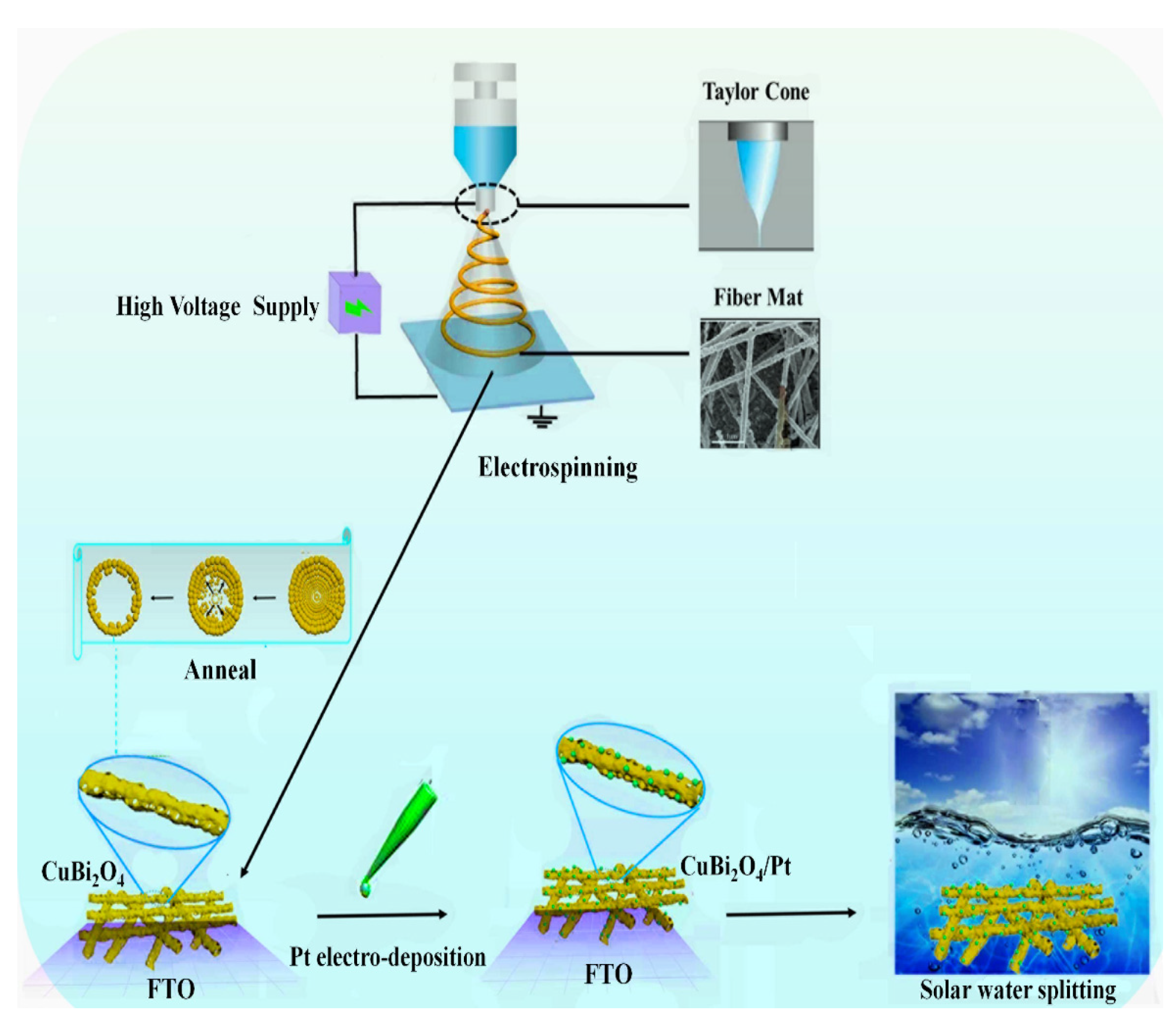
The CuBi2O4/Pt nanofiber film was synthesized via a three-step process: electrospinning, annealing, and deposition, which is shown in Figure 1.
2.1. Materials
The polyvinylpyrrolidone (PVP, K90, Mw = 1,300,000) and chloroplatinic acid (H2PtCl6·6H2O) were from Aladdin, Shanghai, China. The bismuth nitrate pentahydrate (Bi(NO3)3·5H2O), cupric nitrate (Cu(NO3)2·3H2O), N,N-Dimethylformamide (DMF), and acetic acid (CH3COOH) were obtained from J&K Chemical Ltd., B, Beijing, China. All materials were of analytical grade without further purification.
2.2. Preparation of Porous CuBi2O4/Pt Nanofiber Film
The synthesis of the precursor solution followed two steps: first of all, the Bi(NO3)3·5H2O and Cu(NO3)2·3H2O were added to a mixture of acetic acid and DMF and stirred 1 h to ensure dissolution; then the PVP was added to the above mixture and stirred 10 h to form the homogeneous precursor (Figure S1a, Supplementary Materials). The electrospinning was carried out with a self-made apparatus [35], which was composed of a plastic syringe, a high voltage supply, and a plate collector. The homogeneous precursor was injected into the syringe with a stainless steel needle (diameter = 0.5 mm). The FTO glass (OPV-FTO-22-07, 2.5 × 3 cm2) was pasted on the counter plate to collect nanofibers. The electrospinning was performed at a distance of 20 cm between the tip of the steel needle and the plate collector, at a high voltage of 20 kV, at an injection rate of 0.1 mL/h, and with an air humidity of 40%. After electrospinning for 25 min, the films collected on the FTO glass (Figure S1b, Supplementary Materials) were dried at 100 °C for 5 h. Based on the thermogravimetry curve (Figure S2, Supplementary Materials), the nanofiber film on FTO was annealed at 520 °C for 1 h and naturally cooled down to ambient temperature. Finally, the photocathode of CuBi2O4 nanofibers (Figure S1c, Supplementary Materials) was successfully obtained.
The Pt nanoparticles were loaded onto the CuBi2O4 nanofibers by electrodeposition, as the PtCl6 2+ can be reduced to Pt nanoparticles at low potential. A three-electrode system was employed with an as-prepared nanofiber on FTO glass (working electrode), an Ag/AgCl reference electrode, and a platinum counter electrode. The electrolyte was 0.1 mM H2PtCl6•6H2O in 0.1 M potassium borate buffer (pH = 7.0). The electrodeposition was carried out using an electrochemical workstation (Zahner Zennium) at −0.20 V versus Ag/AgCl for 1 min.
2.3. Physical Characterization
In order to investigate the nanofiber, its morphology was measured by a scanning electron microscope (SEM, Zeiss Merlin), an energy-dispersive X-ray spectroscope (EDS), and a transmission electron microscope (TEM, JEM-2100). Its chemical element and crystallinity were characterized by X-ray photoelectron spectroscopy (XPS, ESCALAB Xi +) and X-ray diffraction (XRD, Bruker Smart-1000CCD diffractometer), respectively. The surface area of the CuBi2O4 nanofiber was measured by the Brunauer–Emmett–Teller (BET, Micromeritics ASAP2460, Norcross, GA, USA). The UV-visible diffuse reflectance spectrum was characterized by a UV-vis spectrophotometer (PE lambda 750) with an integrated sphere attachment.
The photocatalytic H2 was carried out using the CEL-PAEM-D8 photocatalytic activity evaluation system, which consisted of a gas chromatograph (AgilentTechnologies GC-7890B) and a 300 W Xe lamp (MicroSolar 300, Perfect Light). The circulation water of 25 °C was applied to maintain the reaction temperature of the solution. The photoelectrochemical experiments on the CuBi2O4/Pt nanofiber photocathode were performed on the Zahner electrochemical workstation in a three-electrode cell (the CuBi2O4/Pt nanofibers on FTO, Ag/AgCl reference electrode, and a platinum counter electrode, respectively). For photocurrent measurements, the electrolyte was 0.2 M PBS (pH 7.0). The Xe lamp (CEL-HXF300-T3, P = 100 mW/cm2, AM1.5) was used as the illumination source. The light intensity was adjusted by a calibrated photodetector. For incident photon-to-current efficiency (IPCE) measurements, the CIMPS TLS03 model (Zahner tunable light source system) was employed for monochromatic light excitation. The chopped photocurrent–voltammetry measurement was carried out with a scan speed of 10 mV/s and a chopped light time of 8 s.
3. Results
3.1. Morphology and Structure of Nanofibers
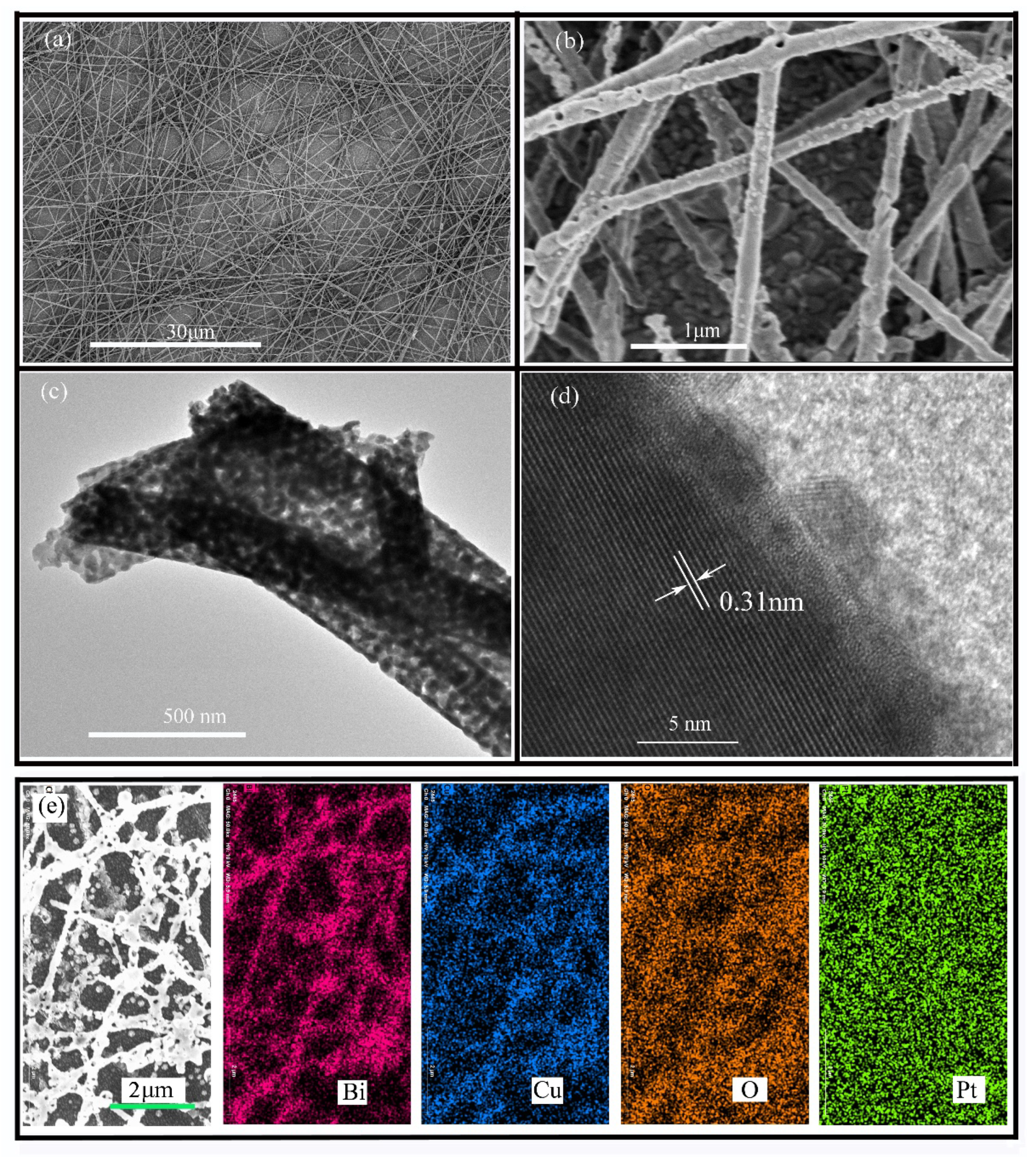
As indicated in Figure S1b (Supplementary Materials), a typical digital photo of the as-spun film before annealing shows white. After high-temperature annealing, the photocathode shown in Figure S1c (Supplementary Materials) changes to a transparent yellow, which is similar to the spray CuBi2O4 photocathode. The morphology of the CuBi2O4 nanofibers was evaluated by TEM and SEM measurements. As shown in Figure 2a, the randomly oriented nanofibers inherited the one-dimensional structure, and the nonwoven film, which was composed of nanofibers, showed typically interconnected flyover-like network form. The high-magnification image of Figure 2b shows these nanofibers possess a porous fiber structure, which has sufficient surface active sites for photocatalytic reaction. As shown in Figure S3 (Supplementary Materials), the average diameter of the porous nanofiber was 225 nanometers, and its length was up to several dozen micrometers. Under low magnification, Figure 2c reveals one-dimensional morphology, and numerous connective nanoparticles make up the products, similar to the SEM results. The distance between adjacent lattice planes was measured to be 0.31 nm, which belongs to the crystal plane (211) of the CuBi2O4 tetragonal phase (JCPDS 01-080-0996). Figure 2e shows the elemental mapping of the CuBi2O4 nanofiber after depositing Pt. It can be seen that the Bi, Cu, and O elements were uniformly distributed inside the nanofiber. In addition, the Pt element was uniformly distributed on the surface of the nanofiber.
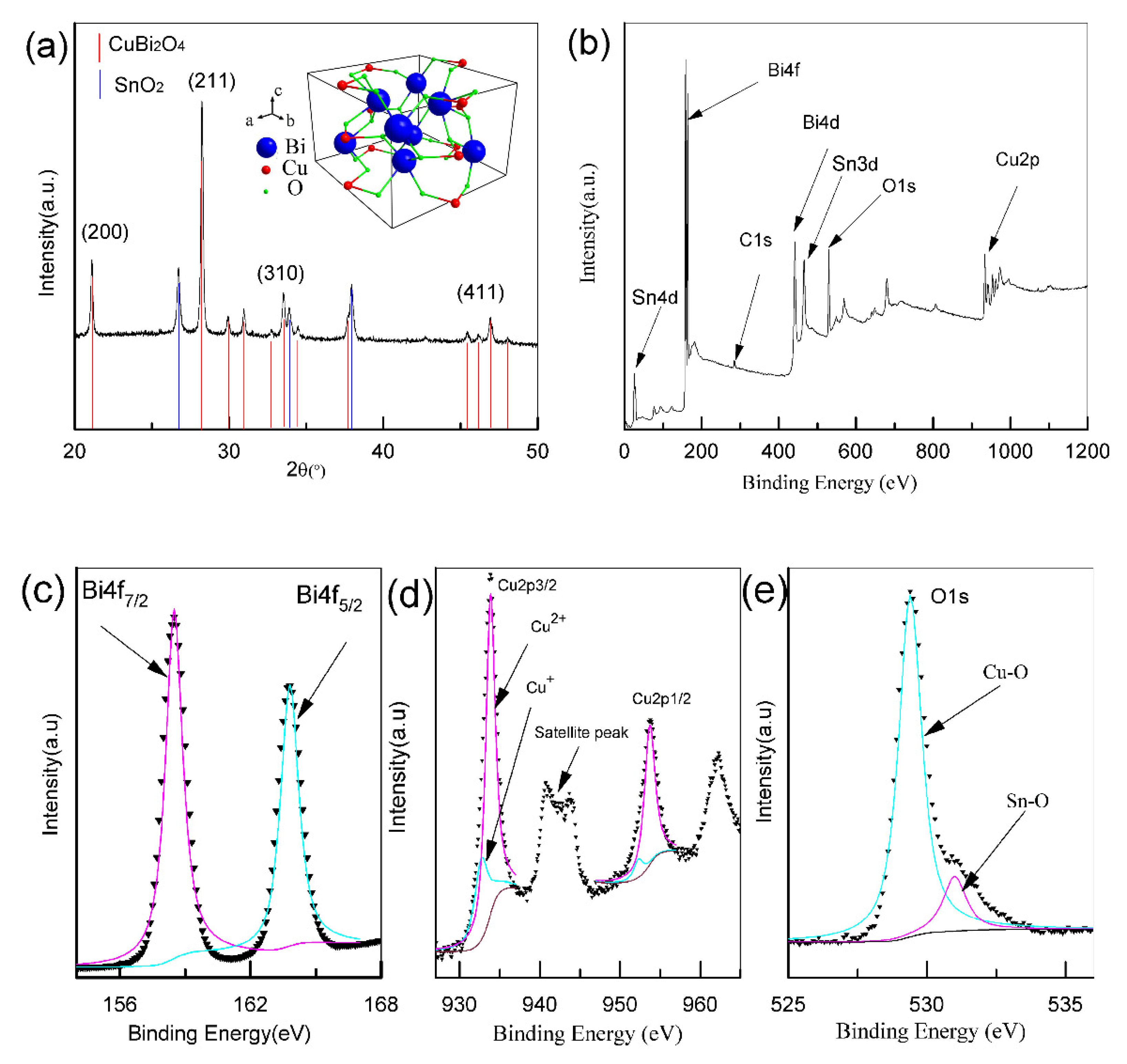
As shown in Figure 3a, the diffraction peaks in 20.9°, 28.1°, 33.5°, and 46.7° corresponded to the crystal planes (200), (211), (310), and (411) of the tetragonal CuBi2O4, respectively (JCPDS 01-080-0996). The SnO2 phase of FTO glass was also marked on the same figure with an orange line. No other diffraction peak was found in the XRD pattern, which confirmed the high crystalline and phase purity of CuBi2O4 after annealing. Detailed information about the surface element composition as well as the chemical state can be obtained by XPS. The survey scan spectrum (Figure 3b) revealed that these nanofibers were composed of Cu, Bi, and O elements. As shown in the high-resolution XPS spectra (Figure 3c–e), two main asymmetric peaks at 159.0 eV and 164.7 eV were attributed to Bi4f7/2 and Bi4f5/2, corresponding to the oxidation state of Bi3+. Then, the peaks at 954.0 and 934.4 eV were attributed to Cu 2p1/2 and Cu 2p3/2. Together with a satellite peak at 942.0 eV, the copper mainly existed in the form of Cu2+. Besides, the asymmetrical O1s peak (Figure 3e) ranging from 527 eV to 535 eV was fitted into two peaks at 529.4 and 531.0 eV, which were attributed to Cu–O and Sn–O (SnO2 phase of FTO glass) bonds, respectively. Together, both the XRD and XPS results confirmed that these nanofibers were highly crystallized.
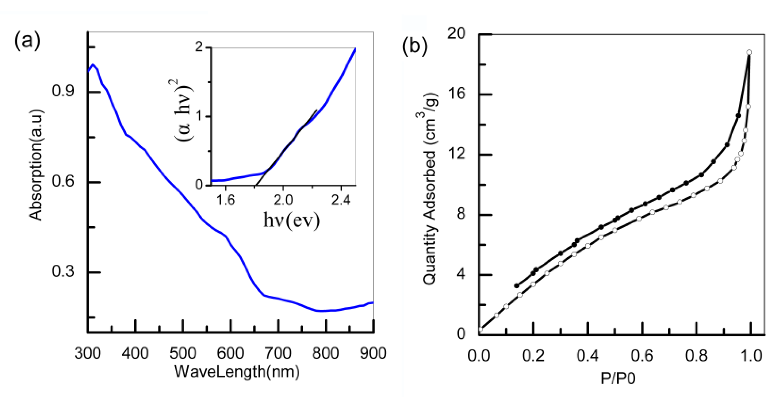
As indicated in Figure 4a, the CuBi2O4 nanofibers exhibited strong UV-vis absorbance in both the ultraviolet and visible light regions, and their absorption cutoff wavelength was about 650 nm. By employing the linear part of (αhv)2 vs. hv, the band gap was calculated to be 1.8 eV, which is similar to that reported in other studies (1.74 eV) [17]. From the nitrogen adsorption and desorption isotherms of CuBi2O4 nanofibers (Figure 4b), the specific surface area of the CuBi2O4 nanofibers was calculated to be 20.5 m2g−1 using the Brunauer–Emmet–Teller model, which was larger than the value (14 m2g−1) of nanoparticles reported in other research [36].
3.2. Photoelectrochemical Performance
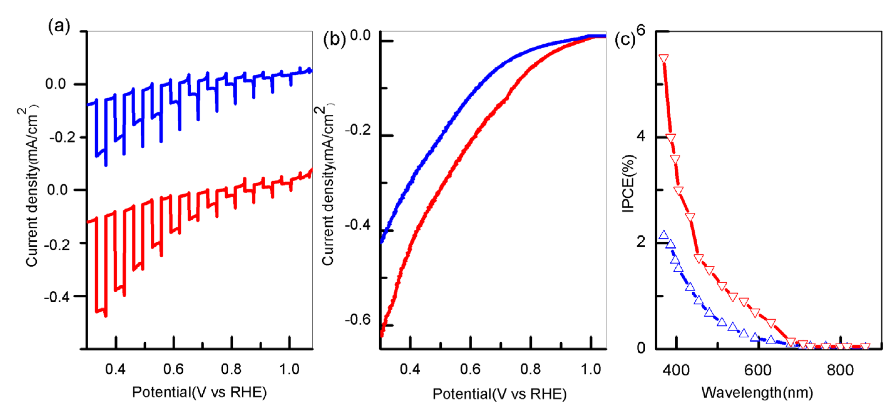
To investigate the photoelectrochemical activity of the CuBi2O4 photocathode, the photocurrent–voltammetry measurement under AM 1.5 was performed. As shown in Figure 5a, the photocurrent started to appear at the initial potential of 1.0 V vs. RHE, increased rapidly when the lamp was turned on, and decreased when the lamp was turned off, indicating that the photocurrent was generated under light irradiation. Interestingly, the instantaneous photocurrent overshoot could also be observed when the lamp was switched on/off, which indicates that electrons accumulate in the space charge layer and reverse recombination occurs between electrons and holes. Moreover, the chopped photocurrent for the CuBi2O4/Pt nanofibers showed little cathodic transient spikes, which presumably were caused by surface recombination. Figure 5b indicates the photocurrent–voltage (J–V) curves under AM 1.5 irradiation. The photocurrent of CuBi2O4 nanofibers rose slowly when decreasing the potential, and yielded −0.12 mA/cm2 at 0.6 V vs. RHE. On the contrary, after depositing the Pt nanoparticles, their photocurrent rose quickly when decreasing the potential and yielded −0.21 mA/cm2 at 0.6 V vs. RHE. Remarkably, the photocurrent of CuBi2O4/Pt nanofibers was about 75% higher than that of the pristine nanofibers. Then, the IPCE measurement by the tunable light source TLS03 model at 0.6 V vs. RHE was performed. As indicated in Figure 5c, with the increase in illuminant wavelength, the IPCE values gradually decreased to zero at 650 nm (1.8 eV), which was consistent with its band gap energy. Significantly, compared to those of pristine nanofibers (1.8% at 380 nm), the as-prepared CuBi2O4/Pt nanofibers showed a higher IPCE reaching up to 4% at 380 nm. As shown in Table S1, the nanofibers decorated with Pt exhibit higher photoelectrochemical performance (the value of photocurrent and IPCE) than that of CuBi2O4 nanofilm (0.15 mA/cm2 at 0.6 V vs. RHE) [21]. Nevertheless, the gradient self-doping nanofilm (0.50 mA/cm2 at 0.6 V vs. RHE) showed higher photoelectrochemical performance than that of the as-prepared CuBi2O4/Pt nanofiber due to its internal electric field promoting charge separation. [19].
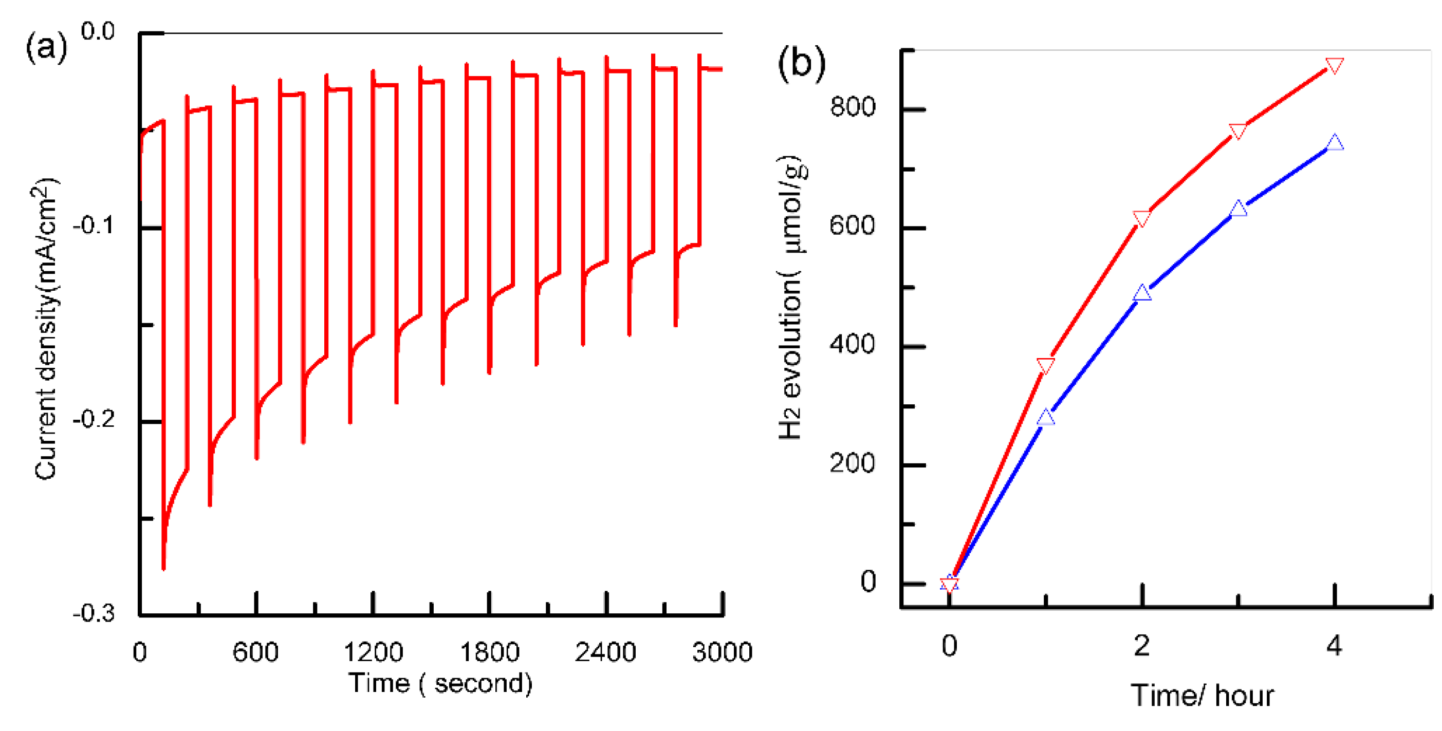
As indicated in Figure 6a, the photocurrent of the CuBi2O4/Pt nanofibers photocathode decreased obviously with the increase in time at 0.6 V versus RHE and decreased by nearly 40% after 25 min illumination. Nevertheless, the photostability of the CuBi2O4/Pt nanofibers photocathode was better than that of the pure Cu2O [37] and CuO (52% reduction of photocurrent after 25 min illumination [38]) photocathode. The photocurrent decay was mainly caused by photocorrosion, and a similar phenomenon also appeared at the CuBi2O4 photocathode consisting of open windows and struts [14]. The photocatalytic H2 production of the fabricated nanofibers was measured by the gas chromatography-mass spectrometer (GC-7890B). As shown in Figure 6b, CuBi2O4/Pt nanofibers exhibited higher photocatalytic performance (380 μmol/(g·h)) compared to the CuBi2O4 nanofibers (290 μmol/(g·h)). Nonetheless, its photocatalytic performance was much lower than that of Pt/TiO2 nanosheet with exposed (001) facet (8500 μmol/(g·h), [39]). The main reason was that although the one-dimensional nanostructure could reduce the recombination of photogenerated electron–hole pairs, some electron–hole pairs still recombined due to the poor transport property of the carrier (~1.2 × 10⁻³ cm2/Vs, [14]). In addition, the photocatalytic activity of these nanofibers slightly decreased with time due to photocorrosion. The instability of CuBi2O4 nanofibers presents a major challenge for solar water splitting, and protection layers using atomic layer deposition were essential in order to use them as a practical photocathode.
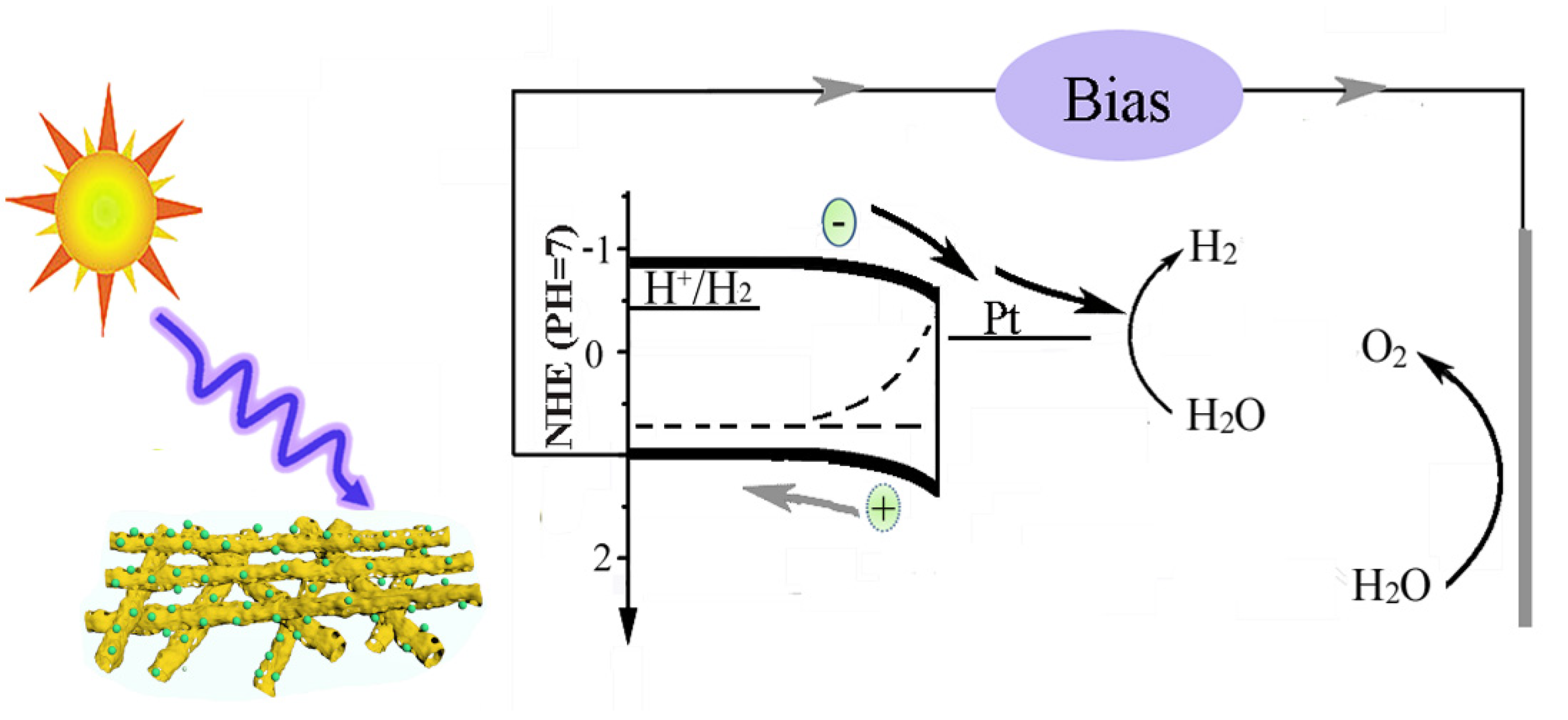
Based on the above analysis of experimental data, the transfer process of the electron–hole pair in the photocathode was shown in Figure 7. Under light illumination, the CuBi2O4 nanofibers could absorb photons and excite the valence electron to the conduction band. Then the photogenerated electron moved to the interface between the photocathode and the electrolyte due to the downward band bending and injected into the electrolyte to take part in the reduction reaction of hydrogen, which was similar to the charge transfer of BiVO4 anode [35]. As shown in Figure 5a, there are amounts of recombination of electron–hole pairs during the water splitting reaction. The photoelectrochemical test demonstrates that the CuBi2O4/Pt nanofibers show better photocatalytic activity. The main reasons are as follows: Firstly, nanofibers have a large specific surface area and porous structure, which maintain good contact with electrolytes and enrich the active sites. Moreover, the nanofibers, possessing a one-dimensional structure, can also shorten the length of hole diffusing to the FTO substrate and decrease the recombination of the electron–hole pair. In addition, the Pt cocatalyst can also efficiently extract a photogenerated electron from the space charge layer. Therefore, more electrons can be transferred into electrolytes to take part in the reduction reaction of hydrogen, and the electron–hole recombination is significantly hindered. In summary, the main reasons for the enhanced photoelectrochemical performance of CuBi2O4/Pt nanofiber photocathode are porous nanofibers and cocatalysts.
4. Conclusions
In summary, the platinum-enriched porous CuBi2O4 nanofibers (CuBi2O4/Pt) with uniform coverage and high surface area were prepared as a photocathode through an electrospinning and electrodeposition process for improving the photoelectrochemical water splitting. The CuBi2O4 nanofibers showed an average diameter of 225 nanometers, and lengths up to several dozens of micrometers. The porous nanofiber structures allow the cocatalyst (Pt) to have better alignment on the surface of CuBi2O4, which can effectively hinder the electron–hole recombination at the electrolyte interface. These nanofibers have a tetragonal crystal structure, and their band gap was determined to be 1.8 eV. After depositing Pt nanoparticles, their photocurrent density was 0.21 mA/cm2 at 0.6 V vs. RHE under AM 1.5 illumination, and the IPCE was 4% at 380 nm. The enhanced photoelectrochemical ability was mainly attributed to the porous nanofibers, large specific surface area, and the cocatalytic activity of Pt nanoparticles. This work shows a new view for integrating an amount of Pt nanoparticles with CuBi2O4 nanofibers, indicating the synergistic effect of cocatalysts for efficient storage of solar energy into hydrogen.
Supplementary Materials
The following are available online at https://0-www-mdpi-com.brum.beds.ac.uk/article/10.3390/polym13193341/s1, Figure S1: Digital micrograph of (a) solution precursor, (b) PVP-CuBi2O4 nanofiber mat before annealing and (c) CuBi2O4 nanofiber mat after annealing; Figure S2: TG-DSC curve of the crystallization of CuBi2O4 nanofiber; Figure S3: CuBi2O4 nanofiber (a) SEM, (b) corresponding diameter distribution; Table S1: Comparison of photocurrent data reported in the literature with the photocurrent value obtained in the present study.
Author Contributions
Conceptualization, X.Y.; methodology, Y.L.; formal analysis, H.Y.; investigation, T.G.; resources; H.Z., data curation, B.L.; writing—original draft preparation, X.Y.; writing—review and editing; X.L., funding acquisition. All authors have read and agreed to the published version of the manuscript.
Funding
This work was supported by the Natural Science Foundation of Shandong Province (No. ZR2019MB068), and A Project of Shandong Province Higher Educational Science and Technology Program (No. KJ2018BZC043).
Institutional Review Board Statement
Not applicable.
Informed Consent Statement
Not applicable.
Data Availability Statement
The data presented in this study are available upon request from the corresponding author.
Conflicts of Interest
The authors declare no conflict of interest.
References
- Cook, T.R.; Dogutan, D.K.; Reece, S.Y.; Surendranath, Y.; Teets, T.; Nocera, D. Solar energy supply and storage for the legacy and non legacy worlds. Chem. Rev. 2010, 110, 6474–6502. [Google Scholar] [CrossRef]
- Chu, S.; Majumdar, A. Opportunities and challenges for a sustainable energy future. Nature 2012, 488, 294–303. [Google Scholar] [CrossRef]
- Roger, I.; Shipman, M.A.; Symes, M.D. Earth-abundant catalysts for electrochemical and photoelectrochemical water splitting. Nat. Rev. Chem. 2017, 1, 1–13. [Google Scholar] [CrossRef]
- Yao, T.; An, X.; Han, H.; Chen, J.Q.; Li, C. Photoelectrocatalytic materials for solar water splitting. Adv. Energy Mater. 2018, 8, 1800210–1800237. [Google Scholar] [CrossRef]
- Xu, P.; Mccool, N.S.; Mallouk, T.E. Water splitting dye-sensitized solar cells. nanotoday 2017, 14, 42–58. [Google Scholar] [CrossRef]
- Jiang, C.; Moniz, S.J.A.; Wang, A.; Zhang, T.; Tang, J. Photoelectrochemical devices for solar water splitting—Materials and challenges. Chem. Soc. Rev. 2017, 46, 4645–4660. [Google Scholar] [CrossRef] [Green Version]
- Kim, J.H.; Lee, J.S. Solar water splitting: Elaborately modified BiVO4 photoanodes for solar water splitting. Adv. Mater. 2019, 31, 1806938–1806973. [Google Scholar] [CrossRef]
- Wang, D.; Chang, G.; Zhang, Y.; Jie, C.; Yang, J.; Shao, S.; Wang, L.; Fan, C.; Wang, L. Hierarchical three-dimensional branched hematite nanorod arrays with enhanced mid-visible light absorption for high-efficiency photoelectrochemical water splitting. Nanoscale 2016, 8, 12697–12701. [Google Scholar] [CrossRef]
- Pang, H.; Zhao, G.; Liu, G.; Zhang, H.; Hai, X.; Wang, S.; Song, H.; Ye, J. Interfacing photosynthetic membrane protein with mesoporous WO3 photoelectrode for solar water oxidation. Small 2018, 14, 1800104–1800114. [Google Scholar] [CrossRef] [PubMed]
- Bagal, I.V.; Chodankar, N.R.; Hassan, M.A.; Waseem, A.; Johar, M.A.; Kim, D.H.; Ryu, S.W. Cu2O as an emerging photocathode for solar water splitting—A status review. Int. J. Hydrogen Energy 2019, 44, 21351–21378. [Google Scholar] [CrossRef]
- Guo, X.; Diao, P.; Xu, D.; Huang, S.; Yang, Y.; Jin, T.; Wu, Q.; Xiang, M.; Zhang, M. CuO/Pd composite photocathodes for photoelectrochemical hydrogen evolution reaction. Int. J. Hydrogen Energy 2014, 39, 7686–7696. [Google Scholar] [CrossRef]
- Li, C.; He, J.; Xiao, Y.; Delaunay, J.J. Earth-abundant Cu-based metal oxide photocathodes for photoelectrochemical water splitting. Energy Environ. Sci. 2020, 13, 3269–3306. [Google Scholar] [CrossRef]
- Sharma, G.; Zhao, Z.; Sarker, P.; Nail, B.A.; Osterloh, F. Electronic structure, photovoltage, and photocatalytic hydrogen evolution with p-CuBi2O4 nanocrystals. J. Mater. Chem. A 2016, 4, 2936–2942. [Google Scholar] [CrossRef]
- Berglund, S.P.; Abdi, F.F.; Bogdanoff, P.; Chemseddine, A.; Friedrich, D.; Roel, V.D.K. Comprehensive evaluation of CuBi2O4 as a photocathode material for photoelectrochemical water splitting. Chem. Mater. 2016, 28, 4231–4242. [Google Scholar] [CrossRef]
- Hahn, N.T.; Holmberg, V.C.; Korgel, B.A.; Mullins, C.B. Electrochemical synthesis and characterization of p-CuBi2O4 thin film photocathodes. J. Phys. Chem. C 2012, 116, 6459–6466. [Google Scholar] [CrossRef]
- Cao, D.; Nasori, N.; Wang, Z.; Yan, M.; Yong, L. P-type CuBi2O4: An easily accessible photocathodic material for high-efficient water splitting. J. Mater. Chem. A 2016, 4, 8995–9001. [Google Scholar] [CrossRef]
- Yang, J.; Du, C.; Wen, Y.; Zhang, Z.; Cho, K.; Chen, R.; Shan, B. Enhanced photoelectrochemical hydrogen evolution at p-type CuBi2O4 photocathode through hypoxic calcination. Int. J. Hydrogen Energy 2018, 43, 9549–9557. [Google Scholar] [CrossRef]
- Abdulkarem, A.M.; Li, J.; Aref, A.A.; Lu, R.; Elssfah, E.M.; Hui, W.; Ge, Y.; Ying, Y. CuBi2O4 single crystal nanorods prepared by hydrothermal method: Growth mechanism and optical properties. Mater. Res. Bull. 2011, 46, 1443–1450. [Google Scholar] [CrossRef]
- Wang, F.; Septina, W.; Chemseddine, A.; Abdi, F.F.; Friedrich, D.; Bogdanoff, P.; Krol, R.V.D.; Tilley, D.; Berglund, S.P. Gradient self-doped CuBi2O4 with highly improved charge separation efficiency. J. Am. Chem. Soc. 2017, 139, 15094–15103. [Google Scholar] [CrossRef] [Green Version]
- Kang, D.; Hill, J.C.; Park, Y.; Choi, K.S. Photoelectrochemical properties and photostabilities of high surface area CuBi2O4 and Ag-doped CuBi2O4 photocathodes. J. High Energy Phys. 2016, 28, 4331–4340. [Google Scholar] [CrossRef]
- Oh, W.D.; Lua, S.K.; Dong, Z.; Lim, T.T. A novel three-dimensional spherical CuBi2O4 consisting of nanocolumn arrays with persulfate and peroxymonosulfate activation functionalities for 1H-benzotriazole removal. Nanoscale 2015, 7, 8149–8158. [Google Scholar] [CrossRef]
- Li, J.; Griep, M.; Choi, Y.S.; Chu, D. Photoelectrochemical overall water splitting with textured CuBi2O4 as a photocathode. Chem. Commun. 2018, 54, 3331–3334. [Google Scholar] [CrossRef]
- Xu, N.; Li, F.; Gao, L.; Hu, H.; Hu, Y.; Long, X.; Ma, J.; Jin, J.N. Cu-codoped carbon nanosheet/Au/CuBi2O4 photocathodes for efficient photoelectrochemical water splitting. ACS Sustain. Chem. Eng. 2018, 6, 7257–7264. [Google Scholar] [CrossRef]
- Wang, F.; Chemseddine, A.; Abdi, F.F.; Krol, R.; Berglund, S.P. Spray pyrolysis of CuBi2O4 photocathodes: Improved solution chemistry for highly homogeneous thin films. J. Mater. Chem. A 2017, 5, 12838–12847. [Google Scholar] [CrossRef]
- Park, H.S.; Lee, C.; Reisner, E. Photoelectrochemical reduction of aqueous protons with a CuO/CuBi2O4 heterojunction under visible light irradiation. Phys. Chem. Chem. Phys. 2014, 16, 22462–22465. [Google Scholar] [CrossRef] [Green Version]
- Xu, Y.; Jian, J.; Li, F.; Liu, W.; Jia, L.; Wang, H. Porous CuBi2O4 photocathodes with rationally engineered morphology and composition towards high-efficiency photoelectrochemical performance. J. Mater. Chem. 2019, 7, 21997–22004. [Google Scholar] [CrossRef]
- Patil, R.; Kelkar, S.; Naphade, R.; Ogale, S. Low temperature grown CuBi2O4 with flower morphology and its composite with CuO nanosheets for photoelectrochemical water splitting. J. Mater. Chem. A 2014, 2, 3661–3668. [Google Scholar] [CrossRef]
- Yao, B.; Zhang, J.; Fan, X.; He, J.; Li, Y. surface engineering of nanomaterials for photo-electrochemical water splitting. Small 2019, 15, 1803746–1803766. [Google Scholar] [CrossRef]
- Reddy, C.V.; Reddy, K.R.; Shetti, N.P.; Shim, J.; Aminabhavi, T.M. Dionysiou D.D. Hetero-nanostructured metal oxide-based hybrid photocatalysts for enhanced photoelectrochemical water splitting—A review. Int. J. Hydrogen Energy 2020, 45, 18331–18347. [Google Scholar] [CrossRef]
- Kumar, P.S.; Sundaramurthy, J.; Subramanian, S.; Babu, V.J.; Singh, G.; Allakhverdiev, S.I.; Ramarkrishna, S. Hierarchical electrospun nanofibers for energy harvesting, production and environmental remediation. Energy Environ. Sci. 2014, 7, 3192–3222. [Google Scholar] [CrossRef]
- Joly, D.; Jung, J.W.; Kim, I.D. Demadrille, Electrospun materials for solar energy conversion: Innovations and trends. J. Mater. Chem. C 2016, 4, 10173–10197. [Google Scholar] [CrossRef]
- Hildebrandt, N.C.; Soldat, J.; Marschall, R. Layered perovskite nanofibers via electrospinning for overall water splitting. Small 2015, 11, 2051–2057. [Google Scholar] [CrossRef]
- Jo, H.S.; Kim, M.W.; Joshi, B.; Samuel, E.; Yoon, H.; Swihart, M.T.; Yoon, S. Ni-core CuO-shell fibers produced by electrospinning and electroplating as efficient photocathode materials for solar water splitting. Nanoscale 2018, 10, 9720–9728. [Google Scholar] [CrossRef]
- Hu, G.; Hu, C.; Zhu, Z.; Lei, Z.; Qiang, W.; Zhang, H.L. Construction of Au/CuO/Co3O4 tri-component heterojunction nanotubes for enhanced photocatalytic oxygen evolution under visible light irradiation. ACS Sustain. Chem. Eng. 2018, 6, 8801–8808. [Google Scholar]
- Yuan, X.; Sun, X.; Zhou, H.; Zeng, S.; Liu, D. Free-standing electrospun W-doped BiVO4 porous nanotubes for the efficient photoelectrochemical water oxidation. Front. Chem. 2020, 8, 1–10. [Google Scholar] [CrossRef]
- Zhang, Y.C.; Yang, H.; Wang, W.P.; Zhang, H.M.; Li, R.S.; Wang, X.X.; Yu, R.C. A promising supercapacitor electrode material of CuBi2O4 hierarchical microspheres synthesized via a coprecipitation route. J. Alloys Compd. 2016, 684, 707–713. [Google Scholar] [CrossRef]
- Kim, H.; Bae, S.; Jeon, D.; Ryu, J. Fully solution-processable Cu2O–BiVO4 photoelectrochemical cells for bias-free solar water splitting. Green Chem. 2018, 20, 3732–3742. [Google Scholar] [CrossRef]
- Pulipaka, S.; Boni, N.; Ummethala, G.; Meduri, P. CuO/CuBi2O4 heterojunction photocathode: High stability and current densities for solar water splitting. J. Catal. 2020, 387, 17–27. [Google Scholar] [CrossRef]
- Yu, J.; Qi, L.; Jaroniec, M. Hydrogen Production by Photocatalytic water splitting over Pt_TiO2 nanosheets with exposed (001) facets. J. Phys. Chem. C 2010, 114, 13118–13125. [Google Scholar] [CrossRef]
Figure 1.
Schematic illustration of the CuBi2O4/Pt nanofiber fabrication process.

Figure 2.
The CuBi2O4 nanofibers’ (a,b) SEM images and (c,d) TEM images; (e) the elemental mapping after depositing Pt.
Figure 2.
The CuBi2O4 nanofibers’ (a,b) SEM images and (c,d) TEM images; (e) the elemental mapping after depositing Pt.

Figure 3.
XRD pattern (a); XPS spectrum (b); high-resolution XPS spectra for (c) Bi4f; (d) Cu2p; and (e) O1s.
Figure 3.
XRD pattern (a); XPS spectrum (b); high-resolution XPS spectra for (c) Bi4f; (d) Cu2p; and (e) O1s.

Figure 4.
UV-vis absorption spectra (a); N2 adsorption–desorption isotherms (b) of CuBi2O4 nanofibers.
Figure 4.
UV-vis absorption spectra (a); N2 adsorption–desorption isotherms (b) of CuBi2O4 nanofibers.

Figure 5.
(a) Chopped photocurrent vs. voltage curves; (b) photocurrent vs. voltage (J–V) curves; (c) IPCE spectrum(blue and red lines represent before and after depositing Pt).
Figure 5.
(a) Chopped photocurrent vs. voltage curves; (b) photocurrent vs. voltage (J–V) curves; (c) IPCE spectrum(blue and red lines represent before and after depositing Pt).

Figure 6.
(a) Chopped photocurrent vs. time curve at 0.6 V vs. RHE; (b) the time courses of H2 evolution under AM 1.5 with methanol as a sacrificial reagent (blue and red lines represent before and after depositing Pt).
Figure 6.
(a) Chopped photocurrent vs. time curve at 0.6 V vs. RHE; (b) the time courses of H2 evolution under AM 1.5 with methanol as a sacrificial reagent (blue and red lines represent before and after depositing Pt).

Figure 7.
The transport process of electron–hole pairs.

Publisher’s Note: MDPI stays neutral with regard to jurisdictional claims in published maps and institutional affiliations. |
© 2021 by the authors. Licensee MDPI, Basel, Switzerland. This article is an open access article distributed under the terms and conditions of the Creative Commons Attribution (CC BY) license (https://creativecommons.org/licenses/by/4.0/).
Share and Cite
MDPI and ACS Style
Yuan, X.; Liu, Y.; Yuan, H.; Liu, B.; Guo, T.; Zhou, H.; Li, X. An Electrospun Porous CuBi2O4 Nanofiber Photocathode for Efficient Solar Water Splitting. Polymers 2021, 13, 3341. https://0-doi-org.brum.beds.ac.uk/10.3390/polym13193341
AMA Style
Yuan X, Liu Y, Yuan H, Liu B, Guo T, Zhou H, Li X. An Electrospun Porous CuBi2O4 Nanofiber Photocathode for Efficient Solar Water Splitting. Polymers. 2021; 13(19):3341. https://0-doi-org.brum.beds.ac.uk/10.3390/polym13193341
Chicago/Turabian StyleYuan, Xiuhua, Yeling Liu, Hui Yuan, Bingxin Liu, Tianyu Guo, Huawei Zhou, and Xia Li. 2021. "An Electrospun Porous CuBi2O4 Nanofiber Photocathode for Efficient Solar Water Splitting" Polymers 13, no. 19: 3341. https://0-doi-org.brum.beds.ac.uk/10.3390/polym13193341
Note that from the first issue of 2016, this journal uses article numbers instead of page numbers. See further details here.