
Chemical-Physical Behaviour of Microgels Made of Interpenetrating Polymer Networks of PNIPAM and Poly(acrylic Acid)

 , , ,
, , ,  , and
, and
Abstract
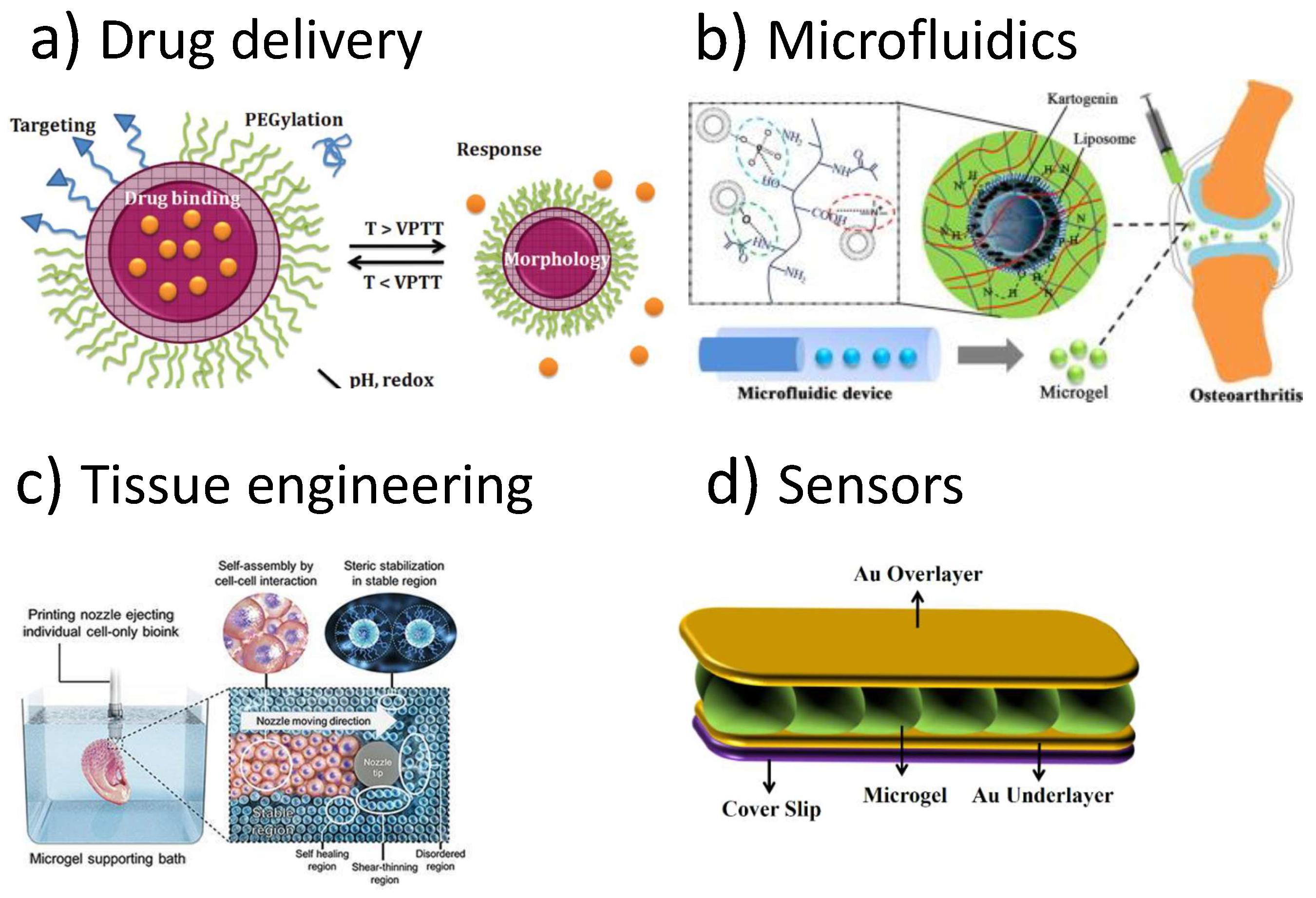
:1. Introduction
2. Experimental Methods
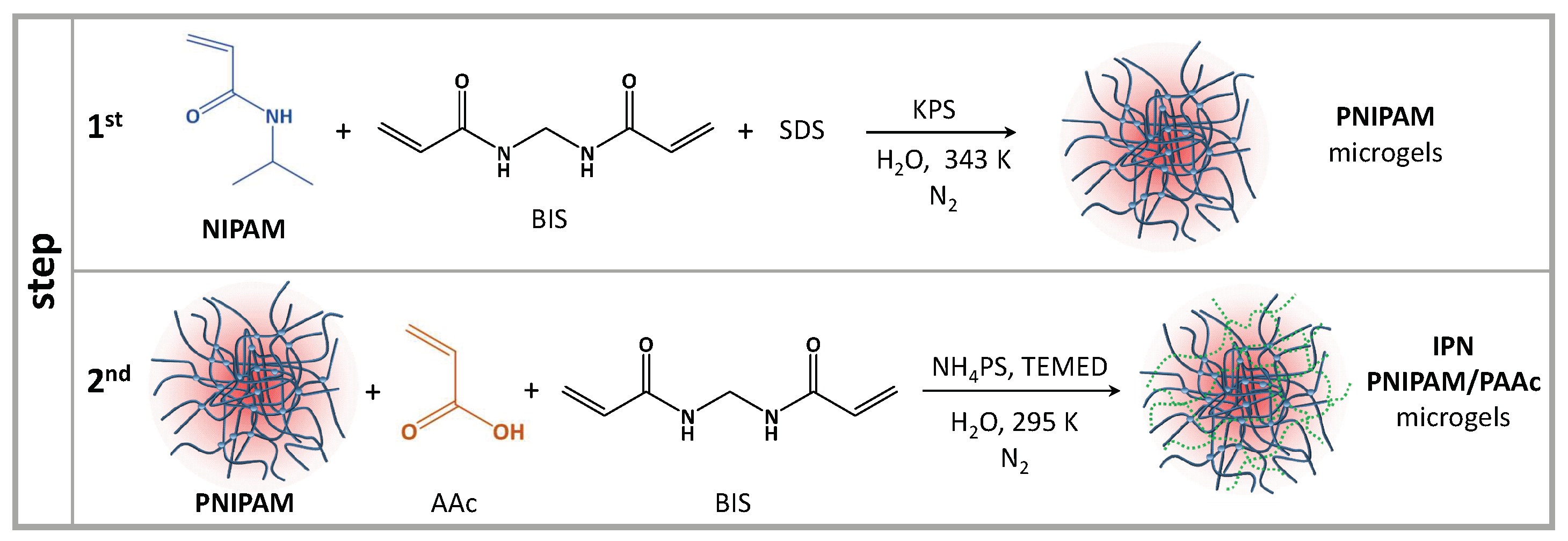
2.1. Sample Preparation
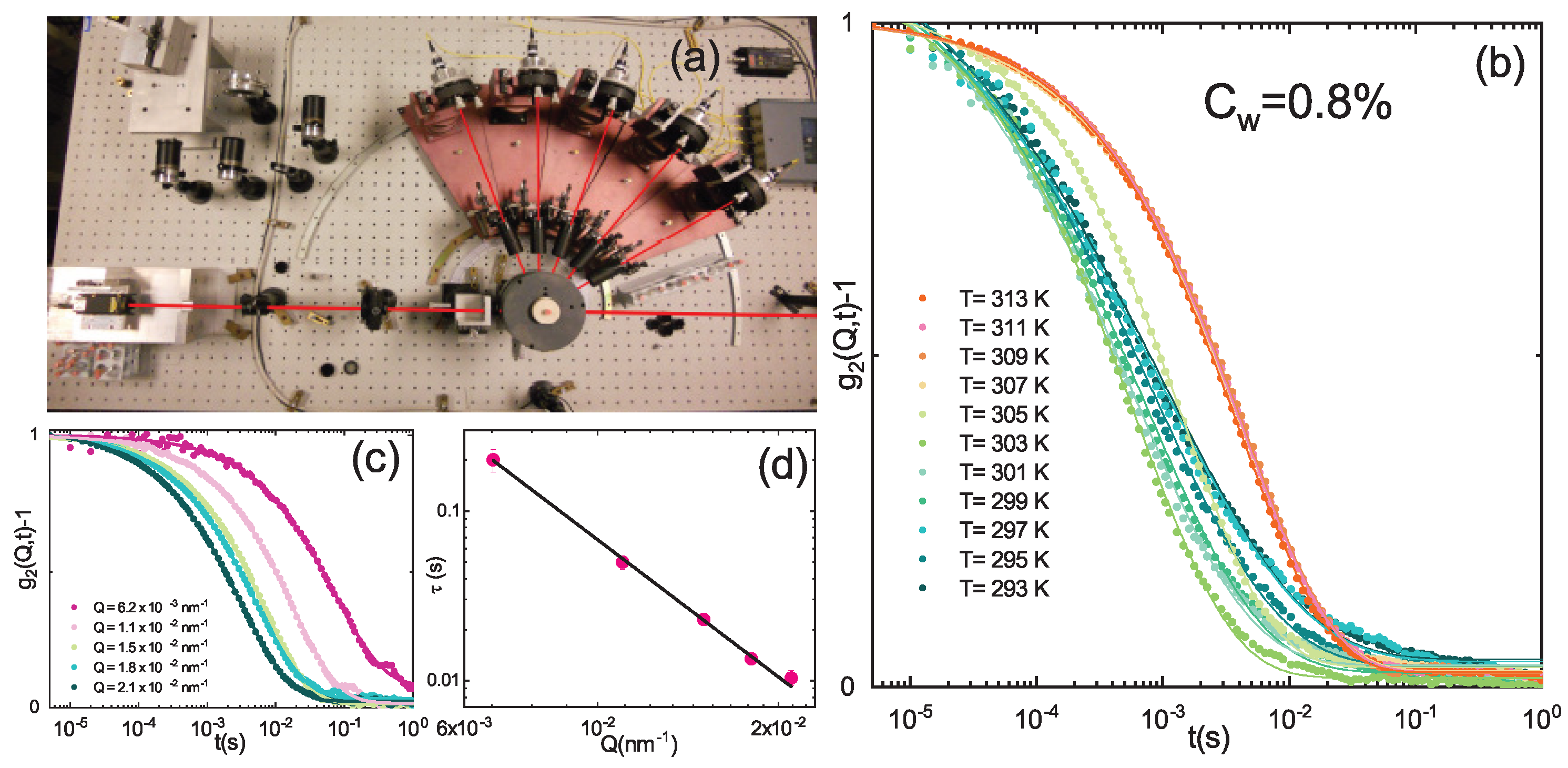
2.2. Dynamic Light Scattering
2.3. Small-Angle Neutron Scattering
2.4. Raman Spectroscopy
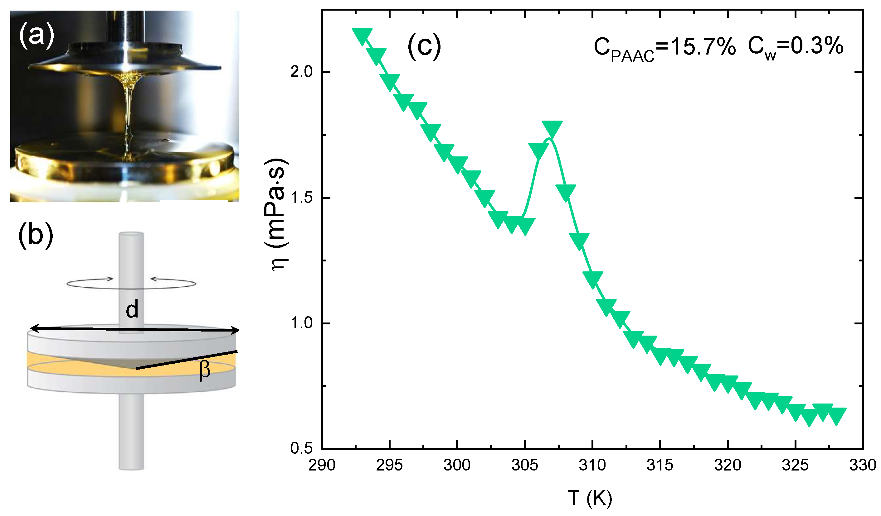
2.5. Rheological Measurements
2.6. Electrophoretic Measurements
3. Results
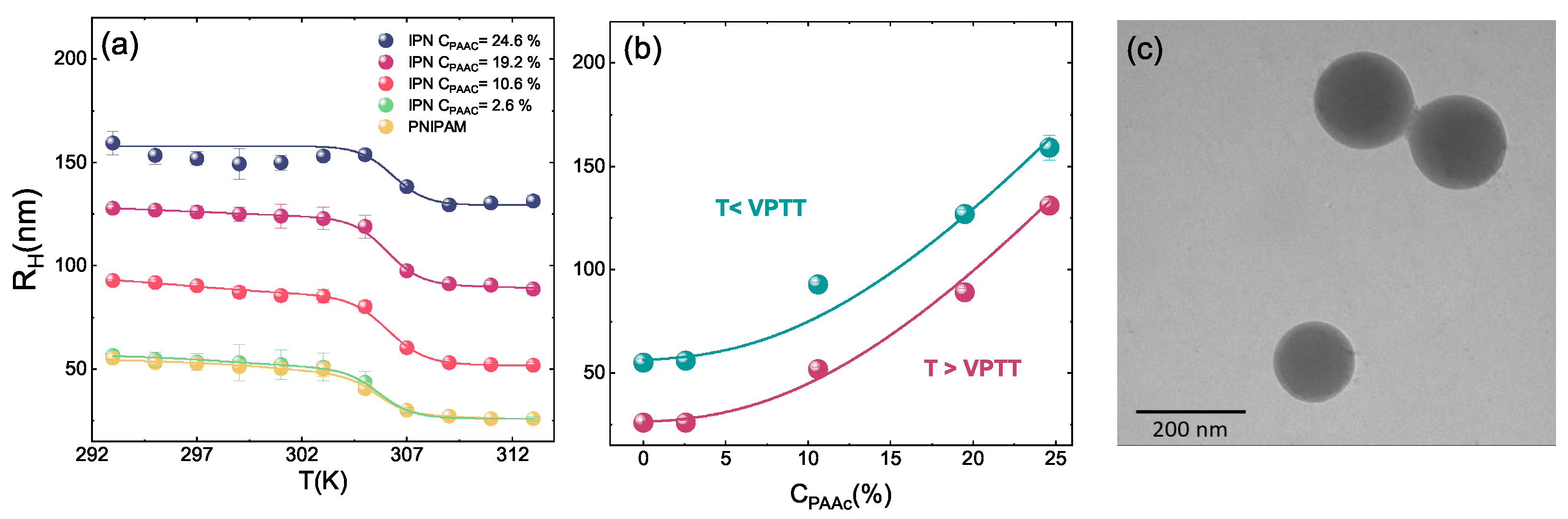
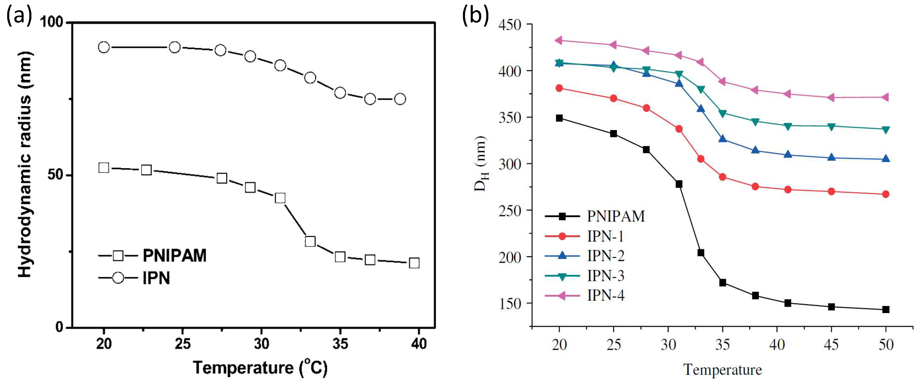
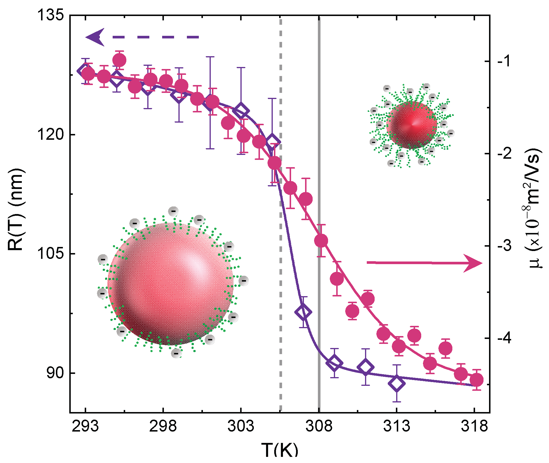
3.1. Particle Size and Volume Phase Transition
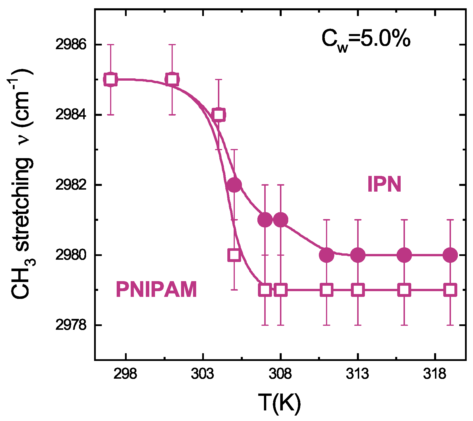
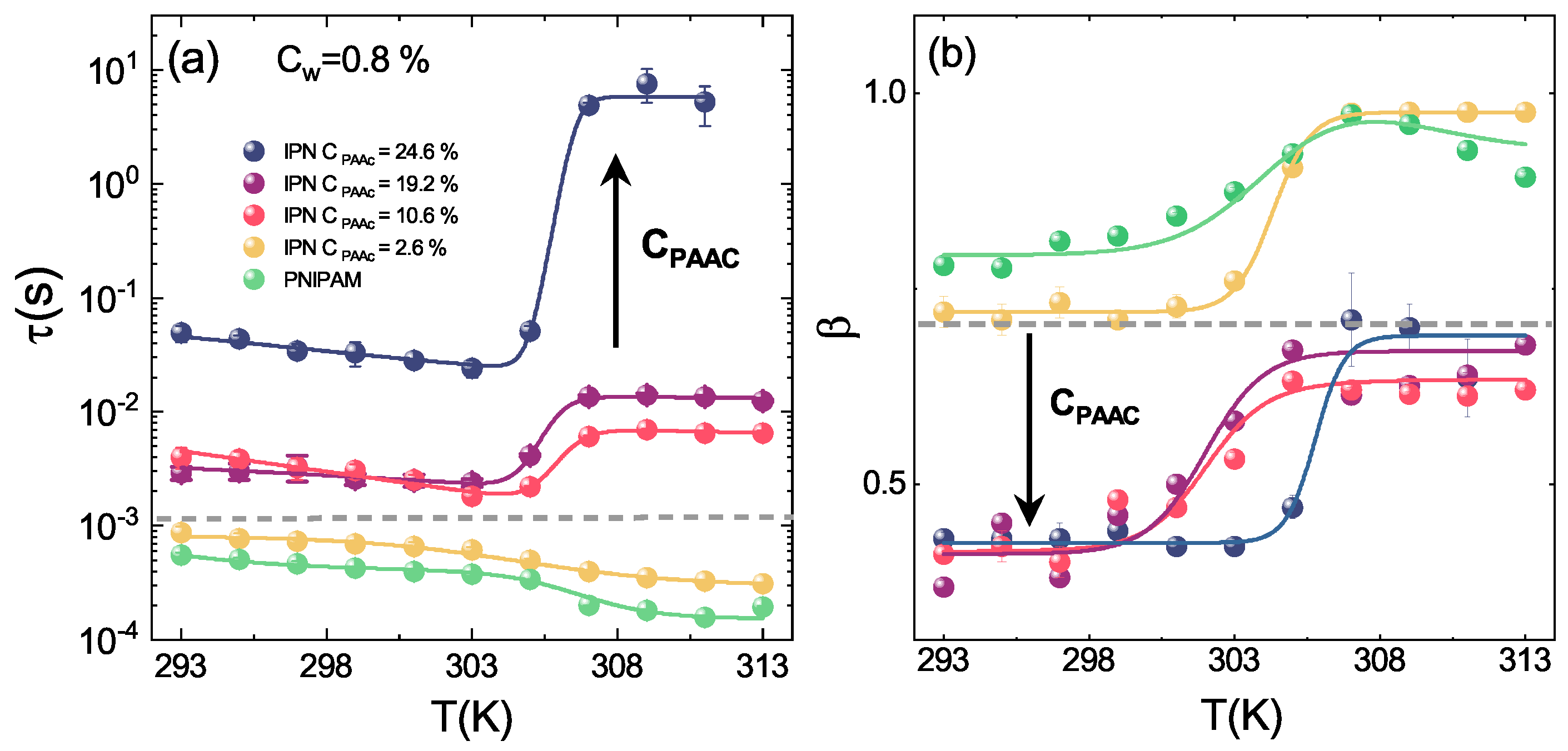
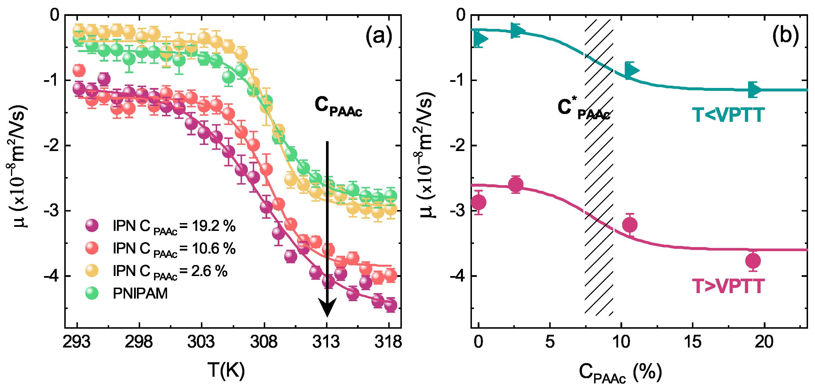
3.2. Molecular Mechanism Driving the VPT
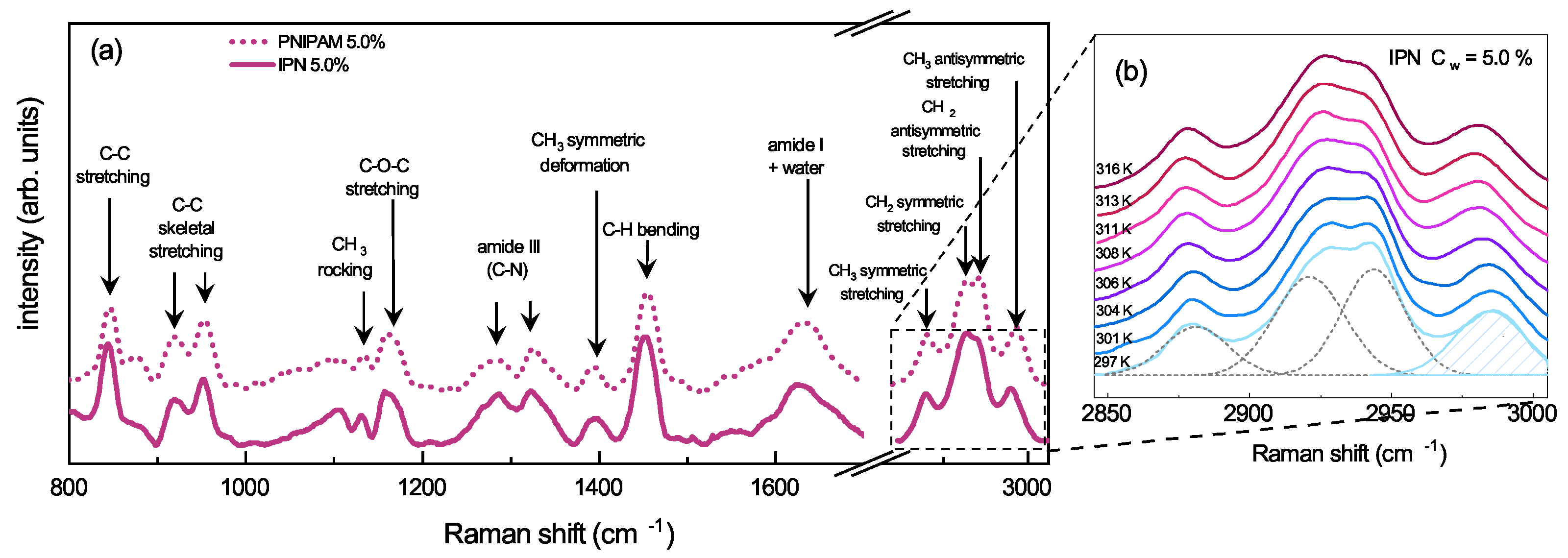
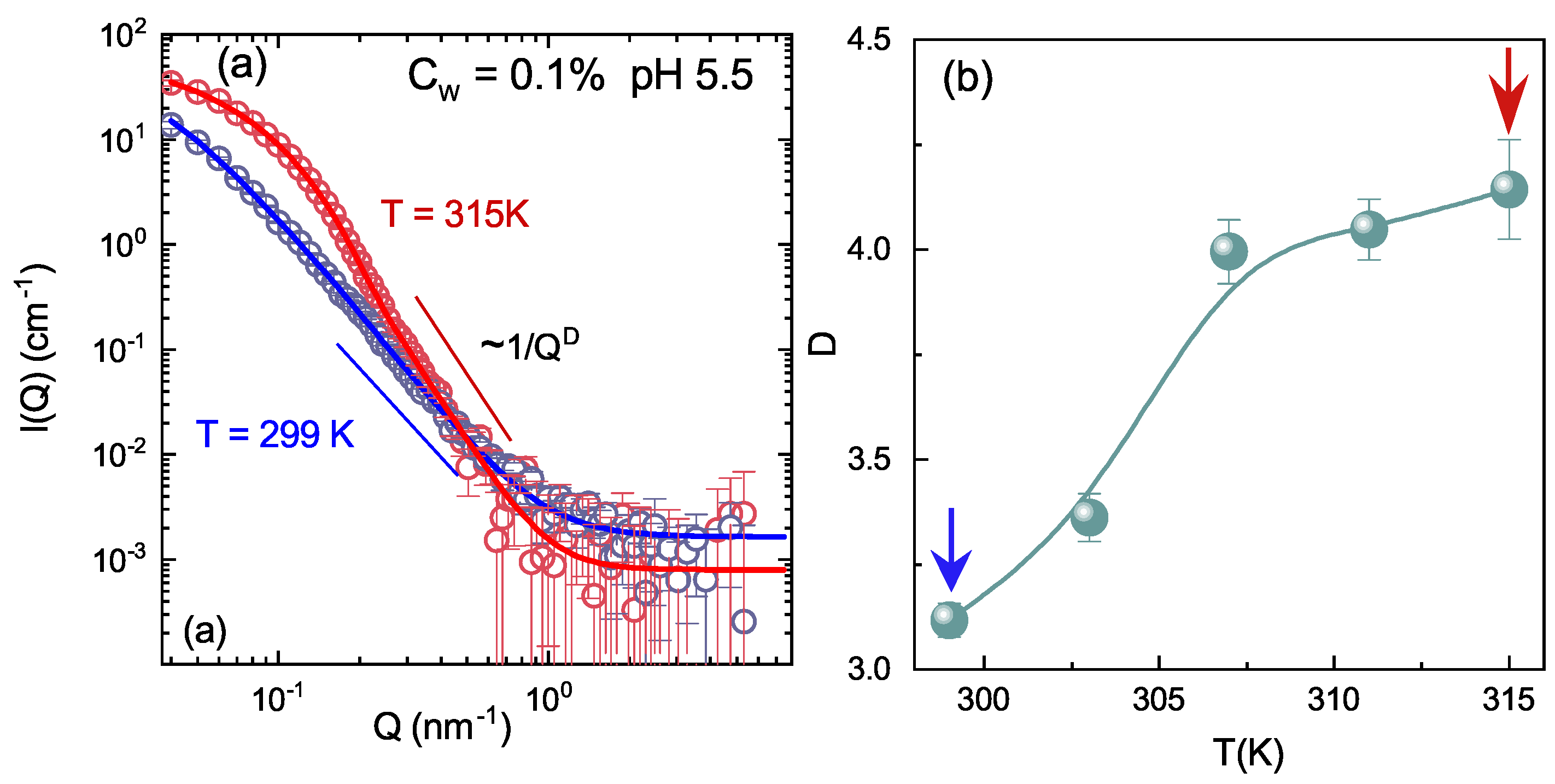
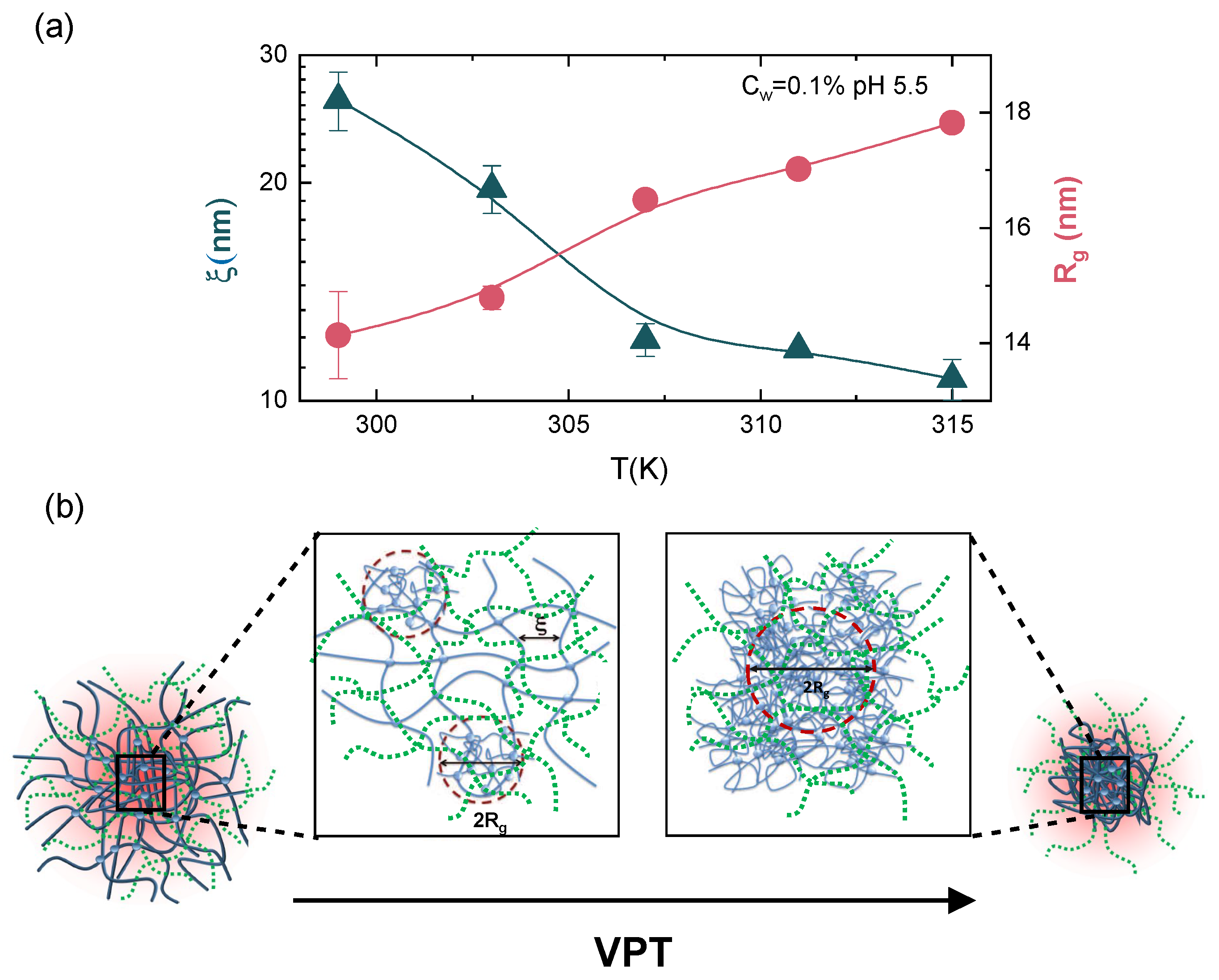
3.3. Local Structure across the VPT
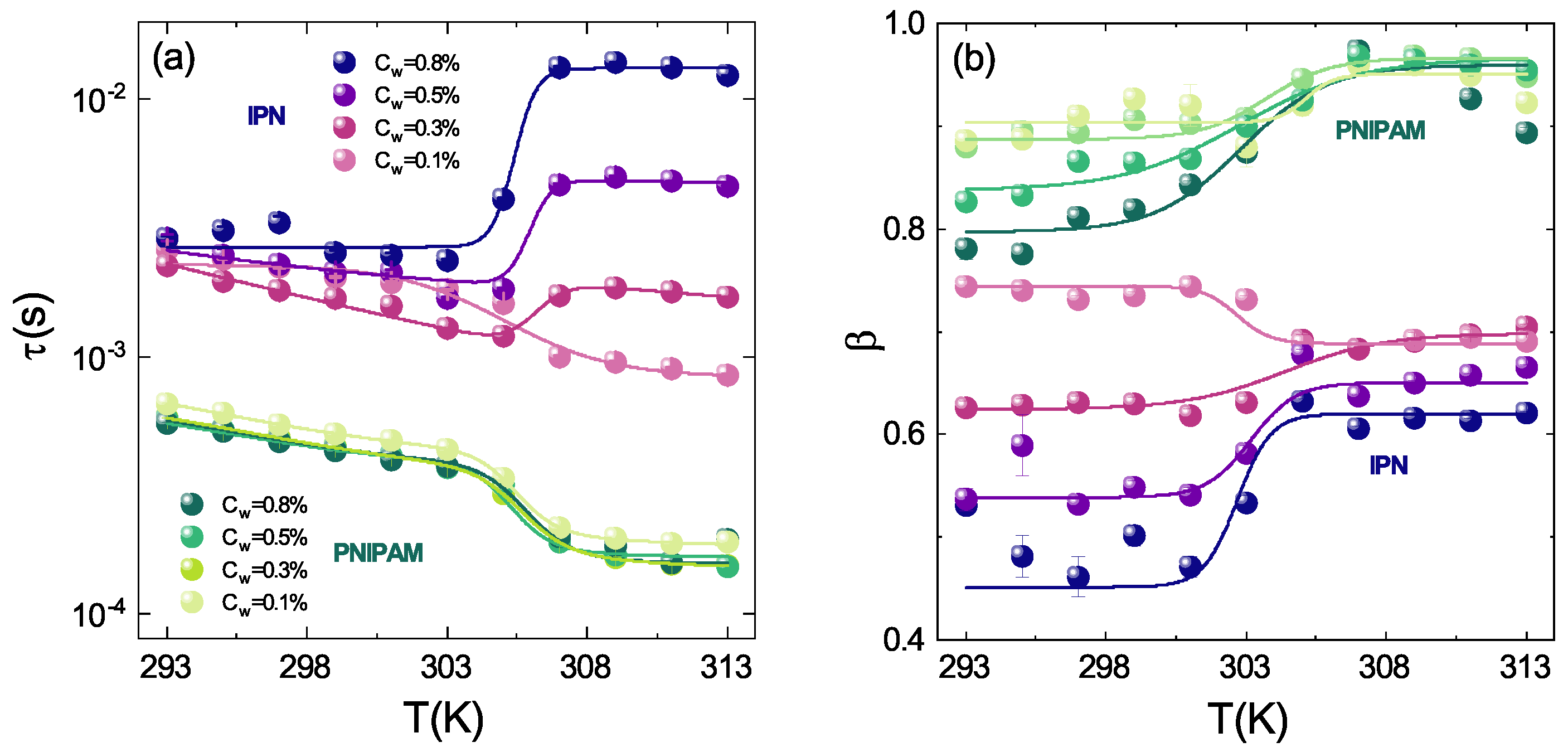
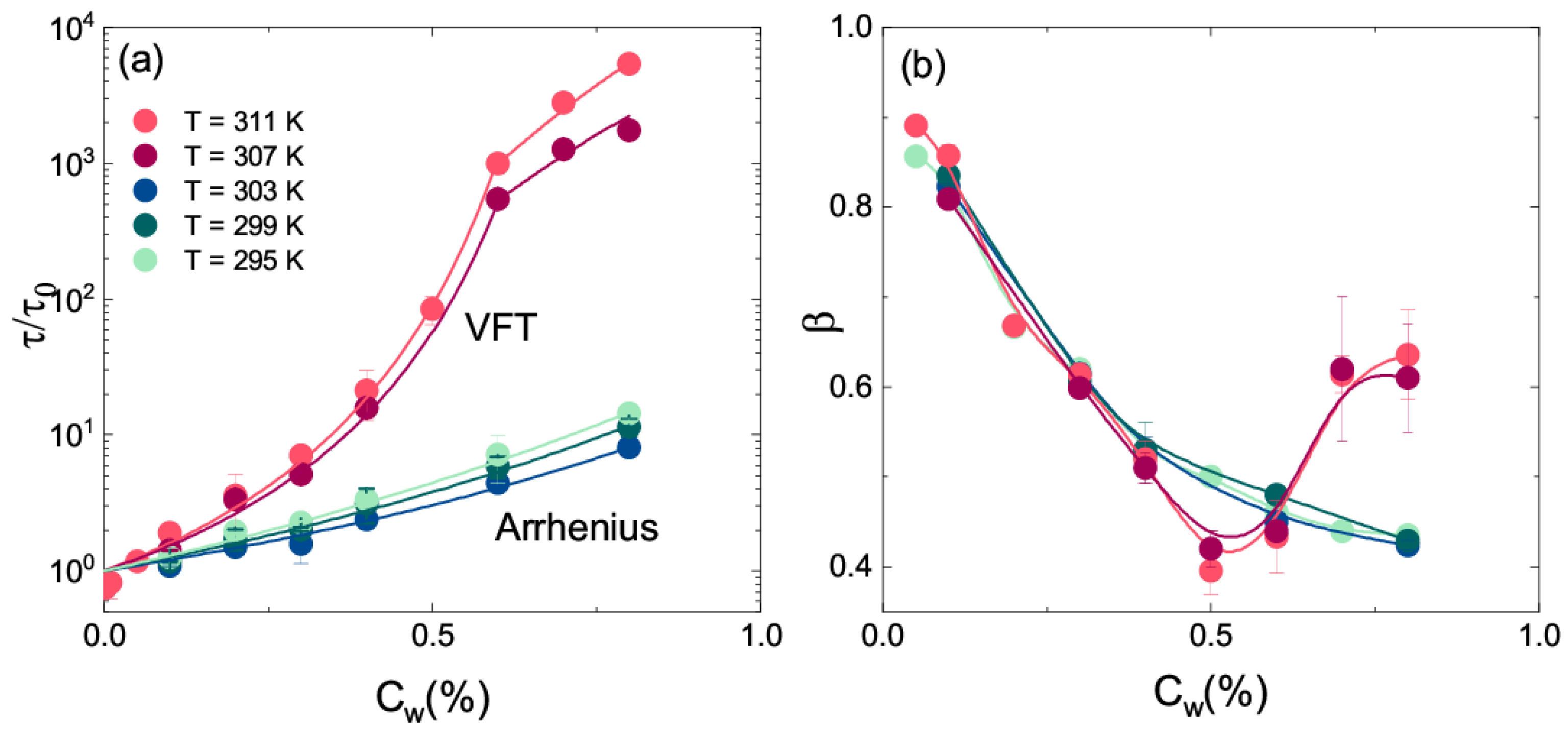
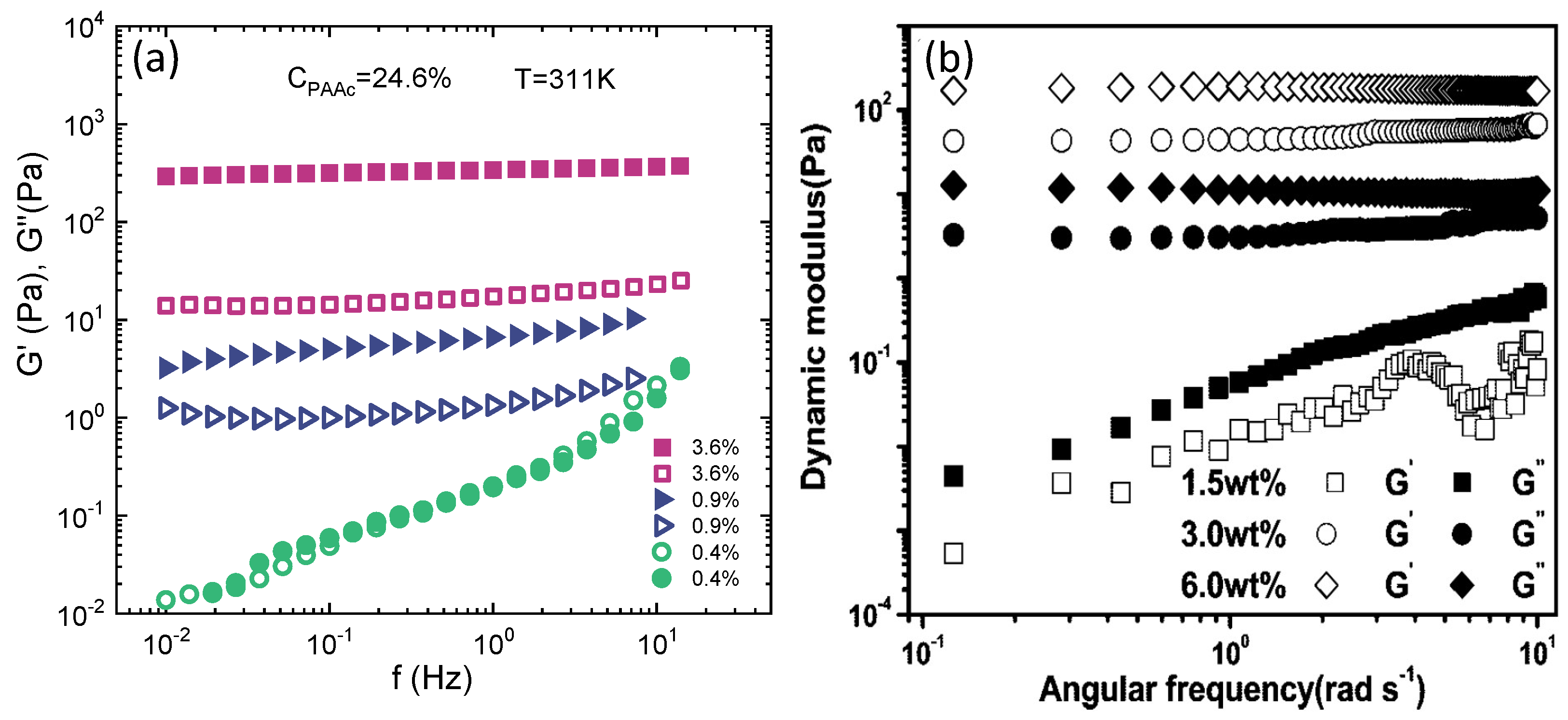
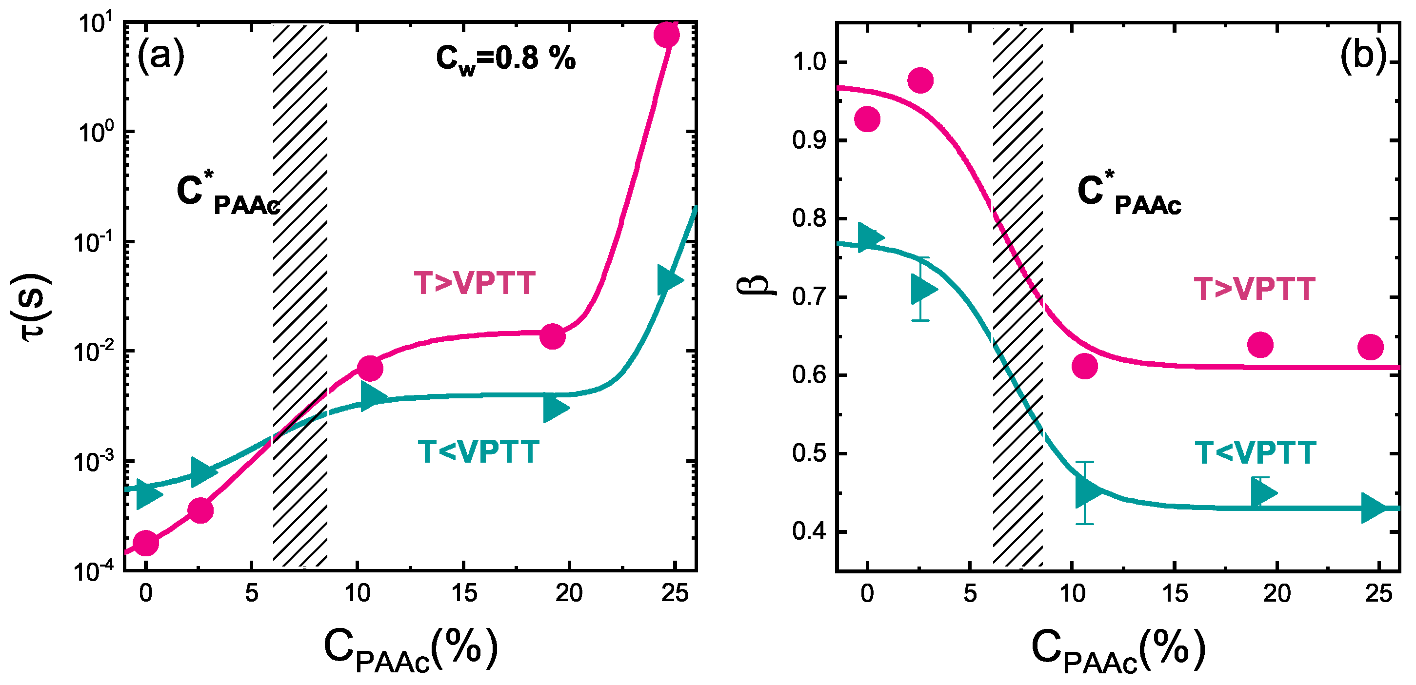
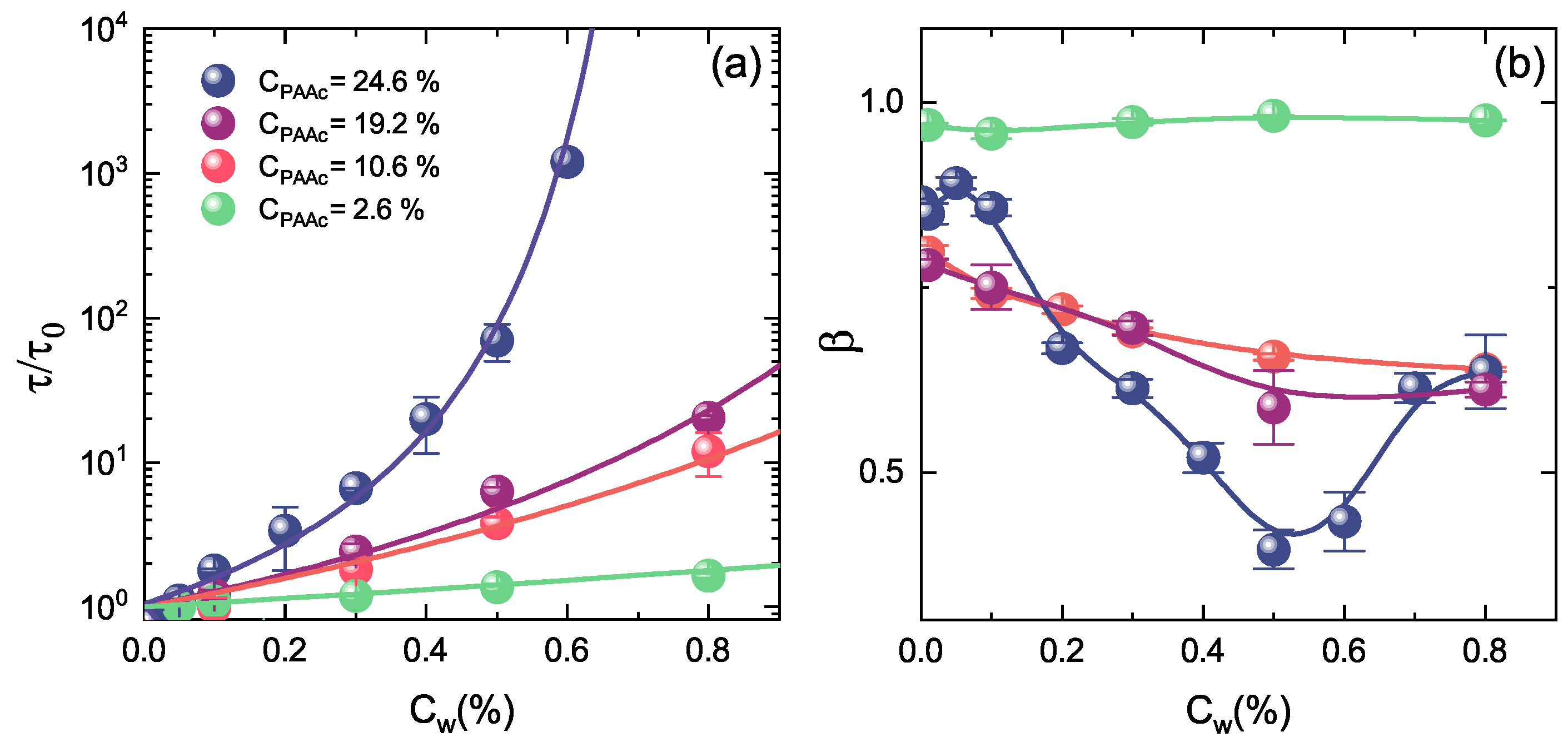
3.4. Concentration Dependence
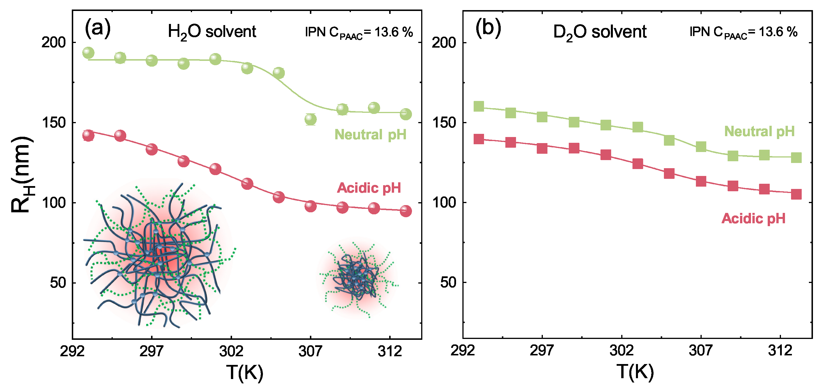
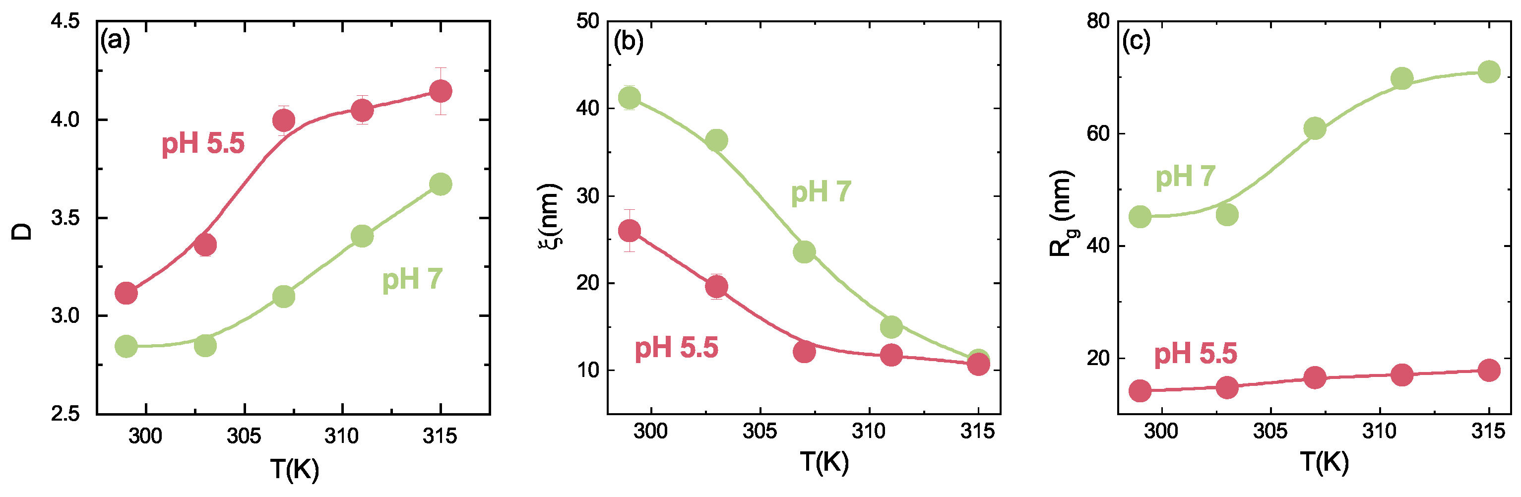
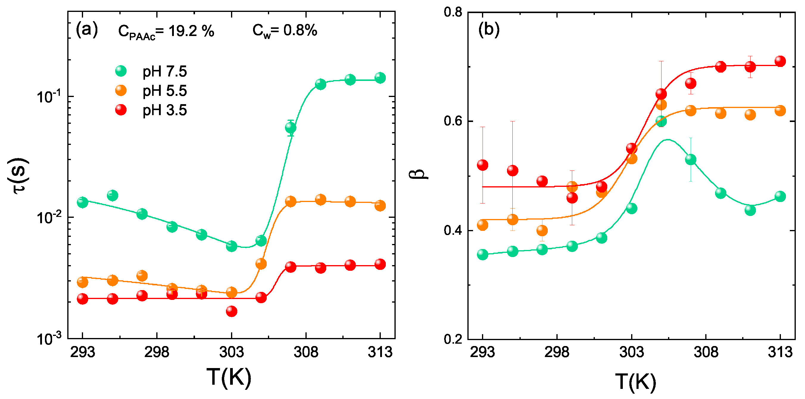
3.5. pH Dependence
4. Conclusions
Author Contributions
Funding
Informed Consent Statement
Data Availability Statement
Acknowledgments
Conflicts of Interest
References
- Yu, R.; Zheng, S. Poly(acrylic acid)-grafted Poly(N-isopropyl acrylamide) Networks: Preparation, Characterization and Hydrogel Behavior. J. Biomater. Sci. 2011, 22, 2305–2324. [Google Scholar] [CrossRef] [PubMed]
- Janovak, L.; Varga, J.; Kemeny, L.; Dekany, I. Investigation of the structure and swelling of poly(N-isopropyl-acrylamideacrylamide) and poly(N-isopropyl-acrylamide-acrylic acid) based copolymer and composite hydrogels. Colloid Polym. Sci. 2008, 286, 1575–1585. [Google Scholar] [CrossRef]
- Zhang, J.; Chu, L.Y.; Li, Y.K.; Lee, Y. Dual thermo and pH-sensitive poly(N-isopropylacrylamide-co-acrylic acid) hydrogels with rapid response behaviors. Polymer 2007, 48, 1718–1728. [Google Scholar] [CrossRef]
- Champ, S.; Xue, W. Concentrating aqueous solutions of water soluble polymers by thermoreversible swelling of poly [(N–isopropylacrylamide)–co–(acrylic acid)] hydrogels. Macromol. Chem. Phys. 2000, 201, 931–940. [Google Scholar] [CrossRef]
- Tian, Q.; Zhao, X.; Tang, X.; Zhang, Y. Hydrophobic association and temperature and pH sensitivity of hydrophobicallymodified poly(N–isopropylacrylamide/acrylic acid) gels. J. Appl. Polym. Sci. 2003, 87, 2406–2413. [Google Scholar] [CrossRef]
- Adem, E.; Burillo, G.; Bucio, E.; Magaña, C.; Avalos-Borja, M. Characterization of interpenetrating networks of acrylic acid (AAc) and N-isopropylacrylamide (NIPAAm) synthesized by ionizing radiation. Radiat. Phys. Chem. 2009, 78, 549–552. [Google Scholar] [CrossRef]
- Burillo, G.; Briones, M.; Adem, E. IPN’s of acrylic acid and Nisopropylacrylamide by gamma and electron beam irradiation. Nucl. Instrum. Methods Phys. Res. 2007, 265, 104–108. [Google Scholar] [CrossRef]
- Xia, X.; Hu, Z.; Marquez, M. Physically bonded nanoparticle networks: A novel drug delivery system. J. Control. Release 2005, 103, 21–30. [Google Scholar] [CrossRef]
- Cong, H.; Zheng, S. Poly(N-isopropylacrylamide)-block-poly(acrylic acid) hydrogels: Synthesis and rapid thermoresponsive properties. Colloid Polym. Sci. 2014, 292, 2633–2645. [Google Scholar] [CrossRef]
- Wang, H.; Wu, X.; Zhu, Z.; Liu, C.S.; Zhang, Z. Revisit to phase diagram of poly(N-isopropylacrylamide) microgel suspensions by mechanical spectroscopy. J. Chem. Phys. 2014, 140, 024908. [Google Scholar] [CrossRef] [PubMed]
- Mohanty, P.S.; Paloli, D.; Crassous, J.J.; Zaccarelli, E.; Schurtenberger, P. Effective interactions between soft-repulsive colloids: Experiments, theory and simulations. J. Chem. Phys. 2014, 140, 094901. [Google Scholar] [CrossRef] [PubMed]
- Hellweg, T.; Dewhurst, C.; Brückner, E.; Kratz, K.; Eimer, W. Colloidal crystals made of poly(N-isopropylacrylamide) microgel particles. Colloid Polym. Sci. 2000, 278, 972–978. [Google Scholar] [CrossRef]
- Paloli, D.; Mohanty, P.S.; Crassous, J.J.; Zaccarelli, E.; Schurtenberger, P. Fluid–solid transitions in soft-repulsive colloids. Soft Matter 2013, 9, 3000–3004. [Google Scholar] [CrossRef]
- Lyon, L.A.; Fernandez-Nieves, A. The Polymer/Colloid Duality of Microgel Suspensions. Annu. Rev. Phys. Chem. 2012, 63, 25–43. [Google Scholar] [CrossRef]
- Wu, J.; Zhou, B.; Hu, Z. Phase behavior of thermally responsive microgel colloids. Phys. Rev. Lett. 2003, 90, 048304. [Google Scholar] [CrossRef] [Green Version]
- Smeets, N.M.B.; Hoare, T. Designing responsive microgels for drug delivery applications. J. Polym. Sci. Part A Polym. Chem. 2013, 51, 3027–3043. [Google Scholar] [CrossRef]
- Yang, J.; Zhu, Y.; Wang, F.; Deng, L.; Xu, X.; Cui, W. Microfluidic liposomes-anchored microgels as extended delivery platform for treatment of osteoarthritis. Chem. Eng. J. 2020, 400, 126004. [Google Scholar] [CrossRef]
- Yoshida, R.; Okano, T. Stimuli-Responsive Hydrogels and Their Application to Functional Materials; Biomedical Applications of Hydrogels Handbook; Springer: New York, NY, USA, 2010. [Google Scholar] [CrossRef]
- Islam, M.R.; Ahiabu, A.; Li, X.; Serpe, M.J. Poly (N-isopropylacrylamide) Microgel-Based Optical Devices for Sensing and Biosensing. Sensors 2014, 14, 8984–8995. [Google Scholar] [CrossRef] [Green Version]
- Vinogradov, S.V. Colloidal microgels in drug delivery applications. Curr. Pharm. Des. 2006, 12, 4703–4712. [Google Scholar] [CrossRef] [Green Version]
- Hamidi, M.; Azadi, A.; Rafie, P. Hydrogel nanoparticles in drug delivery. Adv. Drug Deliv. Rev. 2008, 60, 1638–1649. [Google Scholar] [CrossRef] [PubMed]
- Kabanov, A.; Vinogradov, S. Nanogels as Pharmaceutical Carriers: Finite Networks of Infinite Capabilities. Angew. Chem. Int. Ed. 2009, 48, 5418–5429. [Google Scholar] [CrossRef] [PubMed] [Green Version]
- Saunders, B.R.; Laajam, N.; Daly, E.; Teow, S.; Hu, X.; Stepto, R. Microgels: From responsive polymer colloids to biomaterials. Adv. Colloid Interface Sci. 2009, 147–148, 251–262. [Google Scholar] [CrossRef]
- Park, J.S.; Yang, H.N.; Woo, D.G.; Jeon, S.Y.; Park, K.H. Poly(N-isopropylacrylamide-co-acrylic acid) nanogels for tracing and delivering genes to human mesenchymal stem cells. Biomaterials 2013, 34, 8819–8834. [Google Scholar] [CrossRef] [PubMed]
- Maya, S.; Sarmento, B.; Nair, A.; Rejinold, N.S.; Nair, S.V.; Jayakumar, R. Smart Stimuli Sensitive Nanogels in Cancer Drug Delivery and Imaging: A Review. Curr. Pharm. Des. 2013, 19, 7203–7218. [Google Scholar] [CrossRef]
- Meena, L.K.; Rather, H.; Kedaria, D.; Vasita, R. Polymeric microgels for bone tissue engineering applications—A review. Int. J. Polym. Mater. Polym. Biomater. 2020, 69, 381–397. [Google Scholar] [CrossRef]
- Nasimova, I.R.; Vyshivannaya, O.V.; Gallyamov, M.O.; Kozhunova, E.Y. Thermo- and pH-Sensitive Microgels Based on Interpenetrating Networks as Components for Creating Polymeric Materials. Polym. Sci. Ser. A 2019, 61, 773–779. [Google Scholar] [CrossRef]
- Nasimova, I.R.; Rudyak, V.Y.; Doroganov, A.P.; Kharitonova, E.P.; Kozhunova, E.Y. Microstructured Macromaterials Based on IPN Microgels. Polymers 2021, 13, 1078. [Google Scholar] [CrossRef]
- Sanzari, I.; Buratti, E.; Huang, R.; Tusan, C.G.; Dinelli, F.; Evans, N.D.; Prodromakis, T.; Bertoldo, M. Poly(N-isopropylacrylamide) based thin microgel films for use in cell culture applications. Sci. Rep. 2020, 10, 6126. [Google Scholar] [CrossRef]
- Buratti, E.; Sanzari, I.; Dinelli, F.; Prodromakis, T.; Bertoldo, M. Formation and Stability of Smooth Thin Films with Soft Microgels Made of Poly(N-Isopropylacrylamide) and Poly(Acrylic Acid). Polymers 2020, 12, 2638. [Google Scholar] [CrossRef] [PubMed]
- Kim, J.; Serpe, M.J.; Lyon, L.A. Photoswitchable Microlens Arrays. Angew. Chem. Int. Ed. 2005, 44, 1333–1336. [Google Scholar] [CrossRef] [PubMed]
- Lyon, L.A.; Hendrickson, G.R.; Meng, Z.; John Iyer, A.N.S. Exploiting the Optical Properties of Microgels and Hydrogels as Microlenses and Photonic Crystals in Sensing Applications. In Microgel Suspensions; John Wiley & Sons, Ltd.: Hoboken, NJ, USA, 2011; Chapter 14; pp. 355–374. [Google Scholar] [CrossRef]
- Debord, J.; Eustis, S.; Byul Debord, S.; Lofye, M.; Lyon, L. Color-Tunable Colloidal Crystals from Soft Hydrogel Nanoparticles. Adv. Mater. 2002, 14, 658–662. [Google Scholar] [CrossRef]
- Islam, M.R.; Xie, S.; Huang, D.; Smyth, K.; Serpe, M.J. Poly (N-Isopropylacrylamide) microgel-based optical devices for humidity sensing. Anal. Chim. Acta 2015, 898, 101–108. [Google Scholar] [CrossRef] [PubMed]
- Di Napoli, B.; Franco, S.; Severini, L.; Tumiati, M.; Buratti, E.; Titubante, M.; Nigro, V.; Gnan, N.; Micheli, L.; Ruzicka, B.; et al. Gellan Gum Microgels as Effective Agents for a Rapid Cleaning of Paper. ACS Appl. Polym. Mater. 2020, 2, 2791–2801. [Google Scholar] [CrossRef]
- Bonelli, N.; Montis, C.; Mirabile, A.; Berti, D.; Baglioni, P. Restoration of paper artworks with microemulsions confined in hydrogels for safe and efficient removal of adhesive tapes. Proc. Natl. Acad. Sci. USA 2018, 115, 5932–5937. [Google Scholar] [CrossRef] [PubMed] [Green Version]
- Pelton, R.H.; Chibante, P. Preparation of aqueous lattices with N-isopropylacrylamide. Colloids Surf. 1986, 20, 247–256. [Google Scholar] [CrossRef]
- Saunders, B.R.; Vincent, B. Microgels particles as model colloids: Theory, properties and applications. Adv. Colloid Interface Sci. 1999, 80, 1–25. [Google Scholar] [CrossRef]
- Pelton, R.H. Temperature-sensitive aqueous microgels. Adv. Colloid Interface Sci. 2000, 85, 1–33. [Google Scholar] [CrossRef]
- Das, M.; Zhang, H.; Kumacheva, E. MICROGELS: Old Materials with New Applications. Annu. Rev. Mater. Res. 2006, 36, 117–142. [Google Scholar] [CrossRef]
- Karg, M.; Hellweg, T. New “smart” poly(NIPAM) microgels and nanoparticle microgel hybrids: Properties and advances in characterisation. Curr. Opin. Colloid Interface Sci. 2009, 14, 438–450. [Google Scholar] [CrossRef]
- Lu, Y.; Ballauff, M. Thermosensitive core-shell microgels: From colloidal model systems to nanoreactors. Prog. Polym. Sci. 2011, 36, 767–792. [Google Scholar] [CrossRef]
- Rovigatti, L.; Gnan, N.; Tavagnacco, L.; Moreno, A.; Zaccarelli, E. Numerical modelling of non-ionic microgels: An overview. Soft Matter 2019, 15, 1108–1119. [Google Scholar] [CrossRef] [PubMed]
- Gnan, N.; Zaccarelli, E. The microscopic role of deformation in the dynamics of soft colloids. Nat. Phys. 2019, 15, 683–688. [Google Scholar] [CrossRef]
- Wu, J.; Huang, G.; Hu, Z. Interparticle Potential and the Phase Behavior of Temperature-Sensitive Microgel Dispersions. Macromolecules 2003, 36, 440–448. [Google Scholar] [CrossRef]
- Tan, B.H.; Pelton, R.H.; Tam, K.C. Microstructure and rheological properties of thermo-responsive poly(N-isopropylacrilamide) microgels. Polymers 2010, 51, 3238–3243. [Google Scholar] [CrossRef]
- Zhu, P.W.; Napper, D.H. Light scattering studies of poly(N-isopropylacrylamide) microgel particle in mixed water-acetic acid solvents. Macromol. Chem. Phys. 1999, 200, 1950–1955. [Google Scholar] [CrossRef]
- Kratz, K.; Eimer, W. Swelling Properties of Colloidal Poly(N-Isopropylacrylamide) Microgels in Solution. Ber. Bunsenges. Phys. Chem. 1998, 102, 848–854. [Google Scholar] [CrossRef]
- Kratz, K.; Hellweg, T.; Eimer, W. Structural changes in PNIPAM microgel particles as seen by SANS, DLS and EM techniques. Polymer 2001, 42, 6631–6639. [Google Scholar] [CrossRef]
- Bao, L.; Zhaj, L. Preparation of Poly(N-isopropylacrylamide) Microgels using Different Initiators Under Various pH Values. Macromol. Sci. 2006, 43, 1765–1771. [Google Scholar] [CrossRef]
- Hellweg, T.; Dewhurst, C.D.; Eimer, W.; Kratz, K. PNIPAM-co-polystyrene core-shell microgels: Structure, swelling behavior, and crystallization. Langmuir 2004, 20, 4333–4335. [Google Scholar] [CrossRef]
- Xiong, W.; Gao, X.; Zao, Y.; Xu, H.; Yang, X. The dual temperature/pH-sensitive multiphase behavior of poly (Nisopropylacrylamide-co-acrylic acid) microgels for potential application in in situ gelling system. Colloids Surf. B Biointerfaces 2011, 84, 103–110. [Google Scholar] [CrossRef] [PubMed]
- Meng, Z.; Cho, J.K.; Debord, S.; Breedveld, V.; Lyon, L.A. Crystallization Behavior of Soft, Attractive Microgels. J. Phys. Chem. B 2007, 111, 6992–6997. [Google Scholar] [CrossRef] [PubMed]
- Lyon, L.A.; Debord, J.D.; Debord, S.B.; Jones, C.D.; McGrath, J.G.; Serpe, M.J. Microgel Colloidal Crystals. J. Phys. Chem. B 2004, 108, 19099–19108. [Google Scholar] [CrossRef]
- Holmqvist, P.; Mohanty, P.S.; Nägele, G.; Schurtenberger, P.; Heinen, M. Structure and Dynamics of Loosely Cross-Linked Ionic Microgel Dispersions in the Fluid Regime. Phys. Rev. Lett. 2012, 109, 048302. [Google Scholar] [CrossRef] [Green Version]
- Debord, S.B.; Lyon, L.A. Influence of Particle Volume Fraction on Packing in Responsive Hydrogel Colloidal Crystals. J. Phys. Chem. B 2003, 107, 2927–2932. [Google Scholar] [CrossRef]
- Hu, Z.; Xia, X. Hydrogel nanoparticle dispersions with inverse thermoreversible gelation. Adv. Mater. 2004, 16, 305–309. [Google Scholar] [CrossRef]
- Xia, X.; Hu, Z. Synthesis and Light Scattering Study of Microgels with Interpenetrating Polymer Networks. Langmuir 2004, 20, 2094–2098. [Google Scholar] [CrossRef]
- Zhou, J.; Wang, G.; Zou, L.; Tang, L.; Marquez, M.; Hu, Z. Viscoelastic Behavior and In Vivo Release Study of Microgel Dispersions with Inverse Thermoreversible Gelation. Biomacromolecules 2008, 9, 142–148. [Google Scholar] [CrossRef] [Green Version]
- Xing, Z.; Wang, C.; Yan, J.; Zhang, L.; Li, L.; Zha, L. pH/temperature dual stimuli-responsive microcapsules with interpenetrating polymer network structure. Colloid Polym. Sci. 2010, 288, 1723–1729. [Google Scholar] [CrossRef]
- Liu, X.; Guo, H.; Zha, L. Study of pH/temperature dual stimuli-responsive nanogels with interpenetrating polymer network structure. Polym. Int. 2012, 61, 1144–1150. [Google Scholar] [CrossRef]
- Nigro, V.; Angelini, R.; Bertoldo, M.; Castelvetro, V.; Ruocco, G.; Ruzicka, B. Dynamic light scattering study of temperature and pH sensitive colloidal microgels. J. Non Cryst. Solids 2015, 407, 361–366. [Google Scholar] [CrossRef]
- Nigro, V.; Angelini, R.; Bertoldo, M.; Bruni, F.; Ricci, M.; Ruzicka, B. Local structure of temperature and pH-sensitive colloidal microgels. J. Chem. Phys. 2015, 143, 114904. [Google Scholar] [CrossRef]
- Nigro, V.; Angelini, R.; Bertoldo, M.; Ruzicka, B. Swelling of responsive-microgels: Experiments versus models. Colloids Surf. A 2017, 532, 389–396. [Google Scholar] [CrossRef] [Green Version]
- Nigro, V.; Angelini, R.; Bertoldo, M.; Bruni, F.; Ricci, M.; Ruzicka, B. Dynamical behavior of microgels of Interpenetrated Polymer Networks. Soft Matter 2017, 13, 5185–5193. [Google Scholar] [CrossRef] [PubMed]
- Romeo, G.; Ciamarra, M.P. Elasticity of compressed microgel suspensions. Soft Matter 2013, 9, 5401–5406. [Google Scholar] [CrossRef] [Green Version]
- Romeo, G.; Imperiali, L.; Kim, J.; Fernández-Nieves, A.; Weitz, D.A. Origin of de-swelling and dynamics of dense ionic microgel suspensions. J. Chem. Phys. 2012, 136, 124905. [Google Scholar] [CrossRef]
- Kratz, K.; Hellweg, T.; Eimer, W. Influence of charge density on the swelling of colloidal poly(N-isopropylacrylamide-co-acrylic acid) microgels. Colloids Surf. A 2000, 170, 137–149. [Google Scholar] [CrossRef]
- Jones, C.D.; Lyon, L.A. Synthesis and Characterization of Multiresponsive Core-Shell Microgels. Macromolecules 2000, 33, 8301–8303. [Google Scholar] [CrossRef]
- Mattsson, J.; Wyss, H.M.; Fernandez-Nieves, A.; Miyazaki, K.; Hu, Z.; Reichman, D.; Weitz, D.A. Soft colloids make strong glasses. Nature 2009, 462, 83–86. [Google Scholar] [CrossRef]
- Ma, J.; Fan, B.; Liang, B.; Xu, J. Synthesis and characterization of Poly(N-isopropylacrylamide)/Poly(acrylic acid) semi-IPN nanocomposite microgels. J. Colloid Interface Sci. 2010, 341, 88–93. [Google Scholar] [CrossRef]
- Nigro, V.; Angelini, R.; King, S.; Franco, S.; Buratti, E.; Bomboi, F.; Mahmoudi, N.; Corvasce, F.; Scaccia, R.; Church, A.; et al. Apparatus for simultaneous dynamic light scattering–small angle neutron scattering investigations of dynamics and structure in soft matter. Rev. Sci. Instrum. 2021, 92, 023907. [Google Scholar] [CrossRef]
- Nigro, V.; Angelini, R.; Rosi, B.; Bertoldo, M.; Buratti, E.; Casciardi, S.; Sennato, S.; Ruzicka, B. Study of network composition in interpenetrating polymer networks of poly(N isopropylacrylamide) microgels: The role of poly (acrylic acid). J. Colloid Interface Sci. 2019, 545, 210–219. [Google Scholar] [CrossRef] [Green Version]
- Nigro, V.; Ruzicka, B.; Ruta, B.; Zontone, F.; Bertoldo, M.; Buratti, E.; Angelini, R. Relaxation Dynamics, Softness, and Fragility of Microgels with Interpenetrated Polymer Networks. Macromolecules 2020, 53, 1596–1603. [Google Scholar] [CrossRef]
- Franco, S.; Buratti, E.; Ruzicka, B.; Nigro, V.; Zoratto, N.; Matricardi, P.; Zaccarelli, E.; Angelini, R. Volume fraction determination of microgel composed of interpenetrating polymer networks of PNIPAM and Polyacrylic acid. J. Phys. Condens. Matter 2021. Epub ahead of print. [Google Scholar]
- Franco, S.; Buratti, E.; Nigro, V.; Zaccarelli, E.; Ruzicka, B.; Angelini, R. Glass and Jamming Rheology in Soft Particles made of PNIPAM and polyacrylic acid. Int. J. Mol. Sci. 2021, 22, 4032. [Google Scholar] [CrossRef]
- Micali, N.; Bertoldo, M.; Buratti, E.; Nigro, V.; Angelini, R.; Villari, V. Interpenetrating polymer network microgels in water: Effect of composition on the structural properties and electrosteric interactions. ChemPhysChem 2018, 19, 2894–2901. [Google Scholar] [CrossRef]
- Kohlrausch, R. Theorie des elektrischen rckstandes in der leidener flasche. Ann. Phys. 1854, 2, 179–214. [Google Scholar] [CrossRef] [Green Version]
- Williams, G.; Watts, D.C. Non-Symmetrical Dielectric Relaxation Behavior Arising from a Simple Empirical Decay Function. J. Chem. Soc. Faraday Trans. 1970, 66, 80–85. [Google Scholar] [CrossRef]
- Sierra-Martin, B.; Retama, J.R.; Laurenti, M.; Barbero, A.F.; Cabarcos, E.L. Structure and polymer dynamics within PNIPAM-based microgel particles. Adv. Colloid Interface Sci. 2014, 205, 113–123. [Google Scholar] [CrossRef] [PubMed]
- Shibayama, M.; Tanaka, T.; Han, C.C. Small angle neutron scattering study of poly(N-isopropyl acrylamide) gels near their volume-phase transition temperature. J. Chem. Phys 1992, 97, 6829–6841. [Google Scholar] [CrossRef]
- Shibayama, M. Small angle neutron scattering on polymer gels: Phase behavior, inhomogeneities and deformation mechanisms. Polym. J. 2011, 43, 18–34. [Google Scholar] [CrossRef] [Green Version]
- Shibayama, M. Spatial inhomogeneity and dynamic fluctuations of polymer gels. Macromol. Chem. Phys. 1998, 199, 1–30. [Google Scholar] [CrossRef]
- Glebov, A.; Mokhun, O.; Rapaport, A.; Vergnole, S.; Smirnov, V.; Glebov, L.B. Volume Bragg gratings as ultra-narrow and multiband optical filters. In Proceedings of the SPIE, Brussels, Belgium, 16–19 April 2012; Volume 8428, p. 84280C. [Google Scholar]
- Tscharnuter, W. Mobility measurements by phase analysis. Appl. Opt. 2001, 40, 3995–4003. [Google Scholar] [CrossRef] [PubMed]
- Minor, M.; van der Linde, A.; van Leeuwen, H.; Lyklema, J. Dynamic Aspects of Electrophoresis and Electroosmosis: A New Fast Method for Measuring Particle Mobilities. J. Colloid Interface Sci. 1997, 189, 370–375. [Google Scholar] [CrossRef]
- Connah, M.; Kaszuba, M.; Morfesis, A. High Resolution of Zeta Potential Measurements: Analysis of Multi-component Mixtures. J. Dispers. Sci. Technol. 2002, 23, 663–669. [Google Scholar] [CrossRef]
- Cors, M.; Wiehemeier, L.; Oberdisse, J.; Hellweg, T. Deuteration-Induced Volume Phase Transition Temperature Shift of PNIPMAM Microgels. Polymers 2019, 11, 620. [Google Scholar] [CrossRef] [Green Version]
- Angelini, R.; Ruocco, G. Viscosity measurements in a solution undergoing inverse melting. Philos. Mag. 2007, 87, 553–558. [Google Scholar] [CrossRef] [Green Version]
- Angelini, R.; Ruocco, G.; De Panfilis, S. Phase diagram of a solution undergoing inverse melting. Phys. Rev. E 2008, 78, 020502. [Google Scholar] [CrossRef] [PubMed] [Green Version]
- Howe, A.; Desrousseaux, S.; Lunel, L.; Tavacoli, J.; Yow, H.N.; Routh, A.F. Anomalous viscosity jump during the volume phase transition of poly(N-isopropylacrylamide) particles. Adv. Colloid Interface Sci. 2009, 147–148, 124–131. [Google Scholar] [CrossRef]
- Ahmed, Z.; Gooding, E.; Pimenov, K.; Wang, L.; Asher, S. UV Resonance Raman Determination of Molecular Mechanism of Poly(N-isopropylacrylamide) Volume Phase Transition. J. Chem. Phys. B 2009, 113, 4248–4256. [Google Scholar] [CrossRef] [Green Version]
- Nigro, V.; Ripanti, F.; Angelini, R.; Sarra, A.; Bertoldo, M.; Buratti, E.; Postorino, P.; Ruzicka, B. Molecular mechanisms driving the microgels behaviour: A Raman spectroscopy and dynamic light scattering study. J. Mol. Liq. 2019, 284, 718–724. [Google Scholar] [CrossRef] [Green Version]
- Pastorczak, M.; Kozanecki, M.; Ulanski, J. Water-Polymer interactions in PVME hydrogels—Raman spectroscopy studies. Polymer 2009, 50, 4535–4542. [Google Scholar] [CrossRef]
- Maeda, Y. Infrared and Raman Spectroscopy of Temperature-Responsive Polymers: Chemistry, Properties and Applications; Wiley: Hoboken, NJ, USA, 2018; pp. 197–223. [Google Scholar] [CrossRef]
- Tsuboi, Y.; Nishino, M.; Kitamura, N. Laser-Induced Reversible Volume Phase Transition of a Poly(N-isopropylacrylamide) Gel Explored by Raman Microspectroscopy. Polym. J. 2008, 40, 367–374. [Google Scholar] [CrossRef] [Green Version]
- Dybal, J.; Trchova, M.; Schmidt, P. The role of water in structural changes of poly(N-isopropylacrylamide) and poly(N-isopropylmethacrylamide) studied by FTIR, Raman spectroscopy and quantum chemical calculations. Vib. Spectrosc. 2009, 51, 44–51. [Google Scholar] [CrossRef]
- Schmidt, P.; Dybal, J.; Trchova, M. Investigations of the hydrophobic and hydrophillic interactions in polymer-water systems by ATR FTIR and Raman spectroscopy. Vib. Spectrosc. 2006, 42, 278–283. [Google Scholar] [CrossRef]
- Pelton, R.H.; Pelton, H.M.; Morphesis, A.; Rowells, R.L. Particle Sizes and Electrophoretic Mobilities of Poly(N-isopropylacrylamide) Latex. Langmuir 1989, 5, 816–818. [Google Scholar] [CrossRef]
- Daly, E.; Saunders, B. Temperature-dependent electrophoretic mobility and hydrodynamic radius measurements of poly(N-isopropylacrylamide) microgel particles: Structural insights. Phys. Chem. Chem. Phys. 2000, 2, 3187–3193. [Google Scholar] [CrossRef]
- Hoare, T.; Pelton, R. Electrophoresis of functionalized microgels: Morphological insights. Polymer 2005, 46, 1139–1150. [Google Scholar] [CrossRef]
- Gröhn, F.; Antonietti, M. Intermolecular Structure of Spherical Polyelectrolyte Microgels in Salt-Free Solution. 1. Quantification of the Attraction between Equally Charged Polyelectrolytes. Macromolecules 2000, 33, 5938–5949. [Google Scholar] [CrossRef]


| PNIPAM | PAAc | BIS | |
|---|---|---|---|
| IPN C = 2.6% | 94.4 | 2.6 | 3.0 |
| IPN C = 10.6% | 85.7 | 10.6 | 3.7 |
| IPN C = 15.7% | 76.7 | 15.7 | 7.6 |
| IPN C = 19.2% | 73.6 | 19.2 | 7.2 |
| IPN C = 24.6% | 67.7 | 24.6 | 7.7 |
Publisher’s Note: MDPI stays neutral with regard to jurisdictional claims in published maps and institutional affiliations. |
© 2021 by the authors. Licensee MDPI, Basel, Switzerland. This article is an open access article distributed under the terms and conditions of the Creative Commons Attribution (CC BY) license (https://creativecommons.org/licenses/by/4.0/).
Share and Cite
Nigro, V.; Angelini, R.; Bertoldo, M.; Buratti, E.; Franco, S.; Ruzicka, B. Chemical-Physical Behaviour of Microgels Made of Interpenetrating Polymer Networks of PNIPAM and Poly(acrylic Acid). Polymers 2021, 13, 1353. https://0-doi-org.brum.beds.ac.uk/10.3390/polym13091353
Nigro V, Angelini R, Bertoldo M, Buratti E, Franco S, Ruzicka B. Chemical-Physical Behaviour of Microgels Made of Interpenetrating Polymer Networks of PNIPAM and Poly(acrylic Acid). Polymers. 2021; 13(9):1353. https://0-doi-org.brum.beds.ac.uk/10.3390/polym13091353
Chicago/Turabian StyleNigro, Valentina, Roberta Angelini, Monica Bertoldo, Elena Buratti, Silvia Franco, and Barbara Ruzicka. 2021. "Chemical-Physical Behaviour of Microgels Made of Interpenetrating Polymer Networks of PNIPAM and Poly(acrylic Acid)" Polymers 13, no. 9: 1353. https://0-doi-org.brum.beds.ac.uk/10.3390/polym13091353